General
- Biology SME Y3 SB File Uploaded 1/11/21, 16:56
UNIT 3: REGULATION OF GLUCOSE LEVEL AND TEMPERATURE
Key unit competence
Explain the mechanism of the regulation of blood glucose levels and
regulation of temperature in living organismsIntroductory activity 3
The human body maintains constant different substances in the blood, a
process called homeostasis. The figures below show different organs
involved in the regulation of blood glucose level in the body.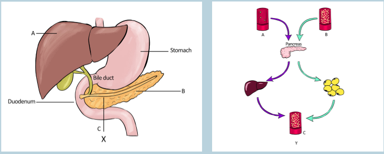
Observe the illustrations X and Y above and answer to the questions that
follow:
a) What are the parts represented by the letters A, B and C on the
illustration X?
b) All the organs shown in the illustration X are involved in the digestion of
food. What are the functions of A and B in the digestion?
c) What are the organs involved in the regulation of blood glucose level
on the illustration X? In which way does each organ state help in this
regulation?d) The illustration Y shows the regulation of blood glucose level. What
does the letters A, B and C show in this regulation?
e) Alpha and beta cells are responsible for producing the hormones
that are involved in the regulation of blood glucose level. Which
organ on the illustration Y produces these hormones?
f) Compare the mechanism of working of the organs A and B in the
regulation of blood glucose level.3.1 Structure and functions of the liver and the pancreas
Activity 3.1
Each organ of our body is made of different tissues which are also composed
of cells. These cells carry out different functions that help in the functioning
of the organ. Refer to the image below to answer the questions that follow: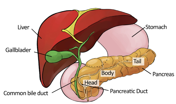
a) Observe the liver and the pancreas and make short notes on their
structures.
b) What are the functions of the liver and the pancreas?
c) Which hormones are produced by the pancreas and what are their
functions?
d) Compare the modes of action of insulin and glucagon.
e) Examine what happens when the blood glucose regulation fails?3.1.1 Importance of glucose
Glucose is one of the most important carbohydrates molecules in our body.
Body requires glucose to carry out some of its most important functions. Glucose
is synthesized in green plants, from carbon dioxide, CO2 and water, H2O with
the help of energy from sunlight. This process is known as photosynthesis.
The reverse of the photosynthesis reaction i.e., breakdown of glucose in the
presence of oxygen to form carbon dioxide and water releasing the energy, is
the main source of power for all the living organisms. The excess glucose in
plants is stored in the form of starch which serves as foods for various animals.Glucose as an energy source
Almost 80 per cent of carbohydrates in our food are converted to glucose during
digestion in the alimentary canal. Fructose and galactose is the other main
product of carbohydrates digestion. After absorption from the alimentary tract,
fructose and galactose are converted into glucose in the liver. And therefore,
glucose constitutes more than 95 per cent of all the carbohydrates circulating
in the blood.Body cells require glucose continuously for its various metabolic activities. These
cells directly absorbed glucose from the blood. Once inside the cells, glucose
combines with a phosphate moiety to form Glucose-6-phosphate with the
help of enzyme glucokinase in liver and hexokinase in most other cells. This
phosphorylation reaction is irreversible and helps to retain the glucose inside the
cells. However, in liver cells, renal tubular epithelial cells and intestinal epithelial
cells, an enzyme glucose phosphatase converts the glucose-6-phosphate
back to glucose.The complete oxidation of one molecule of glucose into carbon dioxide and
water inside the cells produces as many as 38 molecules of ATP (2 from
glycolysis, 2 from the Krebs cycle and 34 from the oxidative phosphorylation).3.1.2 Role of liver and pancreas in glucose regulation
Our body maintains a narrow range of glucose concentration in the blood
between 80 mg/dL to 120 mg/dL which may increase up to 180 mg/dL after a
meal containing high amount of carbohydrates. The hormones responsible for
the regulation of blood sugar level— insulin and glucagon are secreted by the
pancreas. The excess glucose in our blood is converted into glycogen in the
liver. Therefore, pancreas and liver play a vital role in the regulation of blood
sugar concentration.Role of liver in glucose regulation
The liver is the largest internal solid organ in the body second to the skin as the
largest organ overall. It performs various functions in our body, including synthesis
and storage of proteins and fats, carbohydrates metabolism, formation and
secretion of bile, detoxification and excretion of potentially harmful compounds.
Liver contains two main cell types: Kupffer cells and Hepatocytes.1) Kupffer cells are a type of macrophage that capture and break down
old, worn out red blood cells passing through liver sinusoids.
2) Hepatocytes are cuboidal epithelial cells that line the sinusoids and
make up the majority of cells in the liver. Hepatocytes perform most of the
liver’s functions—metabolism, storage, digestion, and bile production.Hepatocytes cells contain various enzymes which help in the regulation of blood
glucose.
These are:1) Glycogen synthase; responsible for glycogen biosynthesis (Glycogenesis).
When the concentration of glucose in the blood increases beyond the normal
value, the excess glucose is converted to glycogen in liver with the help of
enzyme glycogen synthase.2) Glycogen phosphorylase; responsible for breaking down of glycogen
(Glycogenolysis). When the blood glucose level drops, the enzyme
glycogen phosphorylase convert glycogen to glucose-6-phosphate.
Other two enzymes, glucan transferase and glucosidase also help in
glycogenolysis.3) Glucose phosphatase; responsible for conversion of glucose-6-
phosphate to glucose in the liver. Glucose is then released into the blood
stream, thereby increasing the blood glucose level.Role of the pancreas in glucose regulation
Pancreas is the most important endocrine organ for the regulation of blood
glucose. It secretes insulin and glucagon, the two main hormones responsible
for the regulation of blood glucose.
1) Insulin: When the blood glucose concentration increases rapidly, for
example after a meal with high carbohydrates content, pancreas secretes
insulin hormone into the blood stream. Insulin binds to its receptors and
increases the rate of glucose uptake, storage and utilization by almost all
tissues of the body resulting in lowering of blood glucose level. Besides,
insulin also stimulates glycogenesis, lipid and proteins biosynthesis
which helps in decreasing blood glucose concentration.2) Glucagon: In response to decrease in blood glucose concentration,
pancreas secretes glucagon which activates the enzyme glycogen
phosphorylase responsible for degradation of glycogen to glucose-6-
phosphate. Glucose-6-phosphate is then dephosphorylated to form
glucose and finally released into the blood stream thereby increasing
the blood glucose level. Glucagon also stimulates gluconeogenesis i.e.,
biosynthesis of glucose from non-carbohydrate compounds like pyruvate
and amino acids.3.1.3 Detailed structure of liver lobule and islet of
langerhansLiver and liver lobules
The liver is a roughly triangular in shape and extends across the entire abdominal
cavity under the diaphragm. Most of the liver’s mass is located on the right
hypochondrium (i.e., upper part of the abdomen) as well as part of the abdomen
(Figure 3.3). The liver is made of very soft, pinkish-brown tissues encapsulated
by a connective tissue capsule. This capsule is further covered and reinforced
by the peritoneum of the abdominal cavity, which protects and holds the liver.The liver consists of 4 distinct lobes: the left, right, caudate, and quadrate lobes.
The falciform ligament divides the liver into two main lobes, right and left. The
larger right lobe is again sub-divided into three lobes, the right lobe proper, the
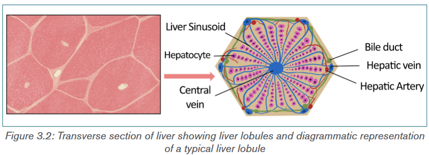
caudate lobe and the quadrate lobe (Figure 3.1). Each liver lobe is made up of
about 100,000 small hexagonal functional units known as lobules. A typical liver
lobule comprises rows of liver cells, hepatocytes, radiating out from a central
vein. The six angles of the hexagon are occupied by a portal triad comprising a
hepatic portal vein, a hepatic artery and a bile duct. The portal veins and arteries
are connected to the central vein through a network of capillary-like tubes called
sinusoids (Figure 3.2). Blood flows out of the sinusoids into the central vein and
is transported out of the liver.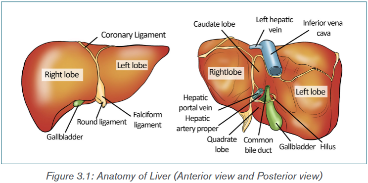
Pancreas
The pancreas is an elongated, tapered organ, located in the abdominal region,
behind the stomach and next to the duodenum—the first part of the small
intestine (Figure 3.3). The right side of the organ, called the head, is the widest
part of the organ and lies in the curve of the duodenum. The tapered left side
which extends slightly upward is the body of the pancreas.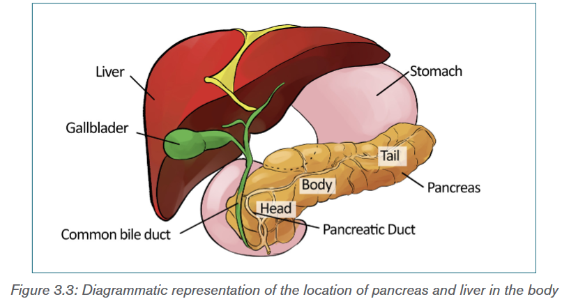
Structure and function of pancreas
Pancreas has two main functional components:1) Exocrine cells, the acini—Cells that release digestive enzymes
into the gut via the pancreatic duct. These enzymes include trypsin
and chymotrypsin to digest proteins; amylase for the digestion of
carbohydrates; and lipase to break down fats. The pancreatic duct joins
the common bile duct to form the ampulla of Vater in the duodenum. The
pancreatic juices and bile (from gallbladder) released into the duodenum
help the body to digest fats, carbohydrates as well as proteins.2) Endocrine pancreas: Highly vascularized groups of cells known as
the Islets of Langerhans within the exocrine tissue constitute the
endocrine pancreas (Figure 3.4). The human pancreas has 1–2 million
islets of Langerhans. It contains four different types of cells which
are distinguished from one another by their morphology and staining
characteristics:i) Alpha cells: Which secrete glucagon, constitute about 25 per
cent of all the cells of islet of Langerhans.ii) Beta cells: The most abundant of the islet cells constitute about
60% of the cells. They release insulin, a hormone involved in
decreasing the blood glucose level.iii) Delta cells: Constitute about 10 per cent of total cells and secrete
somatostatin which regulates both the alpha and beta cells.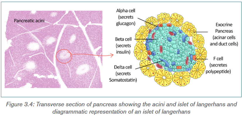
Application activity 3.1
1) The homeostatic level of blood glucose is around 90 mg per 100 ml
of blood. Three person have taken their blood glucose levels using a
glucometer and their results are:
Peter: 85 mg per 100 cm3 of blood
Mary: 130 mg per 100 cm3 of blood
John: 65 mg per 100 cm3 of blood
Interpret these results obtained by using a glucometer?3.2 Control mechanisms by hormones
Activity 3.2
Different hormones are involved in the regulation of blood glucose level. List
and explain those hormones and their functions.3.2.1 Homeostatic control of blood glucose concentration
by insulin and glucagon
Insulin and glucagon are the major hormones responsible for the regulation of
blood glucose. Both insulin and glucagon are secreted by the pancreas, and are
referred to as pancreatic endocrine hormones.Insulin
Insulin was first discovered in 1922 by Banting and Best. Although there is
always a low level of insulin secreted by beta cells of pancreas, the amount
secreted into the blood increases as the blood glucose level rises. In the blood,
it circulates entirely in an unbound form with plasma half-life of about 6 minutes.
Only a small portion of the insulin binds with the insulin receptors of the target
cells while the rest is degraded by the enzyme insulinase, mainly in liver and to
a lesser extends in kidney and muscles.Functions of insulin
Binding of insulin to the receptors stimulates the rate of glucose uptake, storage
and utilization by almost all tissues of the body mainly in muscles, adipose tissue
and liver. Other important functions of insulin include:
i) Insulin promotes glycogenesis by activating enzyme glycogen synthase.
ii) Insulin inactivates liver phosphorylase, the key enzyme of glycogenolysis.
iii) Insulin promotes lipid synthesis by increasing the conversion of excess
glucose into fatty acids in the liver. These fatty acids are transported
as triglycerides to the adipose tissue where it is deposited as fat.
iv) Insulin inhibits the enzymes responsible for gluconeogenesis in liver.
v) Insulin promotes protein synthesis by increasing the rate of transcription
and translation. It also stimulates transport of many amino acids into the
cells.
vi) Insulin inhibits breakdown of lipids and proteins.Regulation of insulin secretion
The secretion of insulin by beta cells of islet of Langerhans depends on the
following factors:
i) Blood glucose level: Increased in the blood glucose level stimulates
the insulin secretion while decreased in the blood glucose concentration
inhibits the secretion.
ii) Blood fatty acids and amino acids concentration: Insulin secretion
is also stimulated by increased in the concentration of blood’s fatty acids
and amino acids concentration and inhibited when its concentration
decreased.
iii) Gastrointestinal hormones: Insulin secretion increases moderately
in response to several gastrointestinal hormones—gastrin, secretin,
cholecystokinin and gastric inhibitory peptide.
iv) These hormones are released after the person takes meal and the
increased in insulin secretion can be regarded as preparation for the
glucose and amino acids uptake by cells.
v) Other hormones: Other hormones that are associated with the
increase in the insulin secretion are glucagon, growth hormone, cortisol,
progesterone and estrogen.Glucagon
Glucagon is secreted by the alpha cells of the pancreatic islets in response
to low blood glucose levels and to events whereby the body needs additional
glucose, such as in response to vigorous exercise.Functions of glucagon
The effect of glucagon in regulating blood glucose level is exactly opposite to
insulin:
i) The most important function of glucagon is activation of glycogen
phosphorylase enzyme responsible for degradation of glycogen to glucose-
6-phosphates. The glucose-6-phosphate is then dephosphorylated to
glucose and finally released into the blood stream resulting in increase in
blood glucose concentration.
ii) Glucagon also stimulates the increase in rate of amino acid uptake and its
conversion into glucose, i.e., gluconeogenesis.
iii) Glucagon activates adipose cell lipase enzyme which stimulates lipids
metabolism.
iv) Glucagon also inhibits the storage of triglycerides in the liver by preventing
the liver from removing fatty acids from the blood.v) Glucagon also enhances the strength of the heart; increases blood flow in
some tissues, especially the kidneys; enhances bile secretion; and inhibits
gastric acid secretion.Regulation of glucagon secretion
Glucagon secretion increases with the decrease in the concentration of
blood glucose level while the increasing concentration of glucose inhibits its
secretion. Other factors which stimulate glucagon secretion are, increase in the
concentration of amino acids in blood and vigorous physical exercise.Negative-positive feedback mechanism
A positive feedback mechanism is the exact opposite of a negative feedback
mechanism. With negative feedback, the output reduces the original effect
of the stimulus. In a positive feedback system, the output enhances the
original stimulus. Negative feedback is an important regulatory mechanism for
physiological function in all living cells. It occurs when a reaction is inhibited by
increase concentration of the product. Regulation of blood glucose level is an
excellent example of homeostatic control through negative feedback mechanism
(Figure 3.5).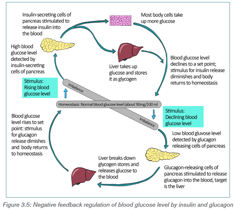
Response to an increase in blood glucose level
When there is increase in blood glucose level, the beta cells of the pancreatic
islets of Langerhans increase the release of insulin into the blood. Insulin
binds to receptors on the cell membrane and stimulates the cells to increase
glucose uptake. This led to decrease in blood glucose level. Besides, insulin
also stimulates glycogenesis and glycolysis while inhibiting glycogenolysis,
gluconeogenesis, lipolysis etc. which all contributes in reducing blood glucose
levels.Response to a decrease in blood glucose level
Decreased in blood glucose level stimulates the alpha cells of pancreas islets
to increase the secretion of glucagon. Glucagon activates enzyme glycogen
phosphorylase in the liver and muscle cells which start glycogenolysis. It also
promotes gluconeogenesis, lipid metabolism etc. The overall effect of glucagon
is increase in the concentration of blood glucose.3.2.2 Other hormones involved in glucose regulation
Other than insulin and glucagon, there are many hormones which contribute to
the regulation of blood glucose level (Figure 3.6). They are:
1) Somatostatin: It is secreted by delta cells of pancreatic islet of
Langerhans in response to many factors related to ingestion of food like
increased concentration of glucose, amino acids, fatty acids and several
gastrointestinal hormones released from the upper gastrointestinal tract.
Somatostatin acts locally within the islets of Langerhans and inhibits the
secretion of both insulin and glucagon. It also reduces the motility of
the stomach, duodenum, and gallbladder and decreases the secretion
and absorption in the gastrointestinal tract. Hence, lowers overall blood
glucose level.2) Epinephrine: Commonly known as Adrenaline, it is secreted by the
medulla of the adrenal glands in response to strong emotions such as
fear or anger. It causes increases in the heart rate, muscle strength, blood
pressure and sugar metabolism. In response, it enhances the process of
glycogenolysis, increasing the overall blood glucose concentration.3) Cortisol: It is also known as stress hormone and is secreted by the
adrenal cortex of the adrenal gland in response to stress. Cortisol
enhances gluconeogenesis and increases the concentration of glucose
in the blood.4) Adrenocorticotropic Hormone (ACTH): In response to various
stresses, hypothalamus secretes corticotropin-releasing hormone which
stimulates anterior pituitary to secrete ACTH. It stimulates adrenal cortex
to release the cortisol hormones.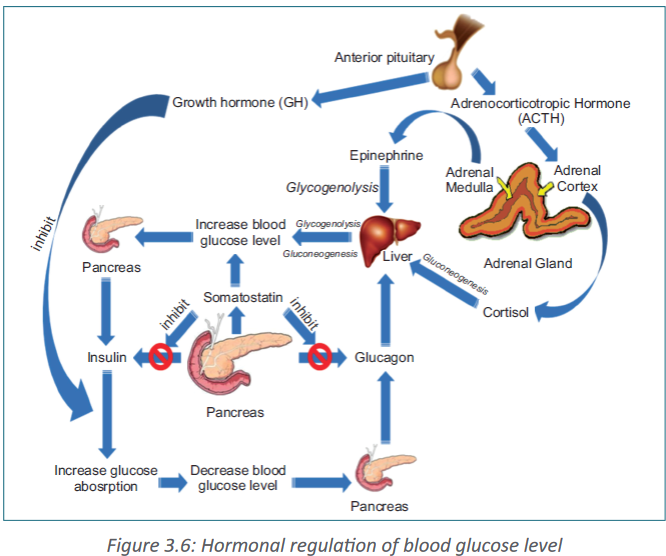
5) Growth hormone (GH): It is another anterior pituitary hormone which
antagonizes the action of insulin by inhibiting the glucose uptake by cells
and increasing the blood glucose level.6) Gastrointestinal hormones: The hormones released by gastrointestinal
tract such as gastrin, secretin, cholecystokinin and gastric inhibitory
peptide etc. increase the digestion and absorption of nutrients in the
gastrointestinal tracts. These hormones stimulate the pancreas to secrete
insulin in anticipation of the increase in blood glucose level.3.2.3 Mechanism of hormonal regulation
Our body maintains certain variables like temperature, pH etc. within a safe range
so that it does not cause any harm to the body and the internal environment
remains stable and relatively constant. This is known as homeostasis.
Hormones are chemical messenger that are directly released into the blood
stream. They play very important role in maintaining the homeostasis.Steps of hormonal signaling
Hormonal signal transduction is a complex process which involves the following
steps:
i) Hormones are first synthesis in particular cells of an organ and stored for
secretion in response to certain stimulus.
ii) When the organ receives the stimulus; hormones are secreted directly
into the blood stream.iii) Blood carries the hormone to the target cell(s).
iv) The hormone is recognized by the specific receptor in the cell membrane
or by the intracellular receptor protein.
v) The hormonal signal is relayed and amplified through a series of signal
transduction process in the target cells which lead to cellular response.3.2.4 Cause of blood sugar imbalances in the body
Our body obtains glucose from various food sources or synthesis in the liver and
muscles from other compounds like pyruvate, lactate, glycerol, and glucogenic
amino acids. The blood carries glucose to all the cells in the body where it is
metabolized to produce energy.Blood sugar levels keep on fluctuating throughout the day increasing after
meals and decreasing in between the meals. When the blood glucose level
rises beyond the normal value, the condition is known as hyperglycaemia. On
the other hand, hypoglycaemia or low blood sugar is the condition in which the
blood glucose level is below normal (~80 mg/dL).Hyperglycaemia
High blood glucose level can be caused due to various reasons like:
i) Carbohydrates: Eating food containing too much of carbohydrates. The
body of a person cannot process high levels of carbohydrates fast enough
to convert it into energy.
ii) Insulin control: The pancreas of the individual are unable to produce
enough insulin.
iii) Stress: Stress stimulates the secretion of certain hormones like cortisol
and epinephrine etc., which increases the blood glucose level.
iv) Low levels of exercise: Daily exercise is a critical contributor to regulating
blood sugar levels.
v) Infection, illness, or surgery: With illness, blood sugar levels tend to
rise quickly over several hours.
vi) Other medications: Certain drugs, especially steroids, can affect blood
sugar levels.A high blood sugar level can be a symptom of diabetes. If hyperglycaemia
persists for several hours, it can leads to dehydration. Other symptoms of
hyperglycaemia include dry mouth, thirst, frequent urination, blurry vision, dry,
itchy skin, fatigue or drowsiness, weight loss, increased appetite, difficulty
breathing, dizziness upon standing, rapid weight loss, increased drowsiness
and confusion, unconsciousness or coma.Hypoglycaemia
Hypoglycaemia is generally defined as a serum glucose level below 80 mg/dL.
Symptoms typically appear when the blood glucose levels reach below 70 mg/
dL and levels below 60 mg/dL can be fatal.Common causes of low blood sugar include the following:
i. Overmedication with insulin or antidiabetic pills
ii. Use of alcohol
iii. Skipped meals
iv. Severe infection
v. Adrenal insufficiency
vi. Kidney failure
vii. Liver failure, etc.
Common symptoms of hypoglycaemia include trembling, clammy skin,
palpitations (pounding or fast heart beats), anxiety, sweating, hunger, and
irritability. If the brain remains deprived of glucose for longer period, a later set of
symptoms can follows like difficulty in thinking, confusion, headache, seizures,
and coma. And ultimately, after significant coma or loss of consciousness, death
can occur.3.2.5 Diabetes mellitus
Diabetes mellitus (commonly referred to as diabetes) is a chronic condition
associated with abnormally high levels of sugar in the blood due to impaired
carbohydrate, fat, and protein metabolism. It can be due to absence or insufficient
production of insulin by the pancreas, or inability of the body to properly use
insulin. Hence, there are two types of diabetes mellitus – Type I causes by lack
of insulin secretion and Type II, caused by reduced sensitivity of target cells to
insulin.Type I diabetes
It is known as insulin dependent diabetes mellitus (IDDM) and it is due to
insufficient insulin production by the beta cells of pancreatic islet of Langerhans
or due to absence of the beta cells itself. Since the pancreas makes very little or
no insulin at all, glucose cannot get into the body’s cells and remain in the blood
leading to hyperglycemia. The concentration of blood glucose level can be as
high as 300 – 1,200 mg/dL. The symptoms of Type I diabetes include:i) Loss of glucose in urine; due to increase in blood glucose, concentration
goes beyond 180 mg/dL.ii) Dehydration; due to osmotic loss of water from cells and inability to
reabsorb water in kidney.
iii) Tissue injury; due to damages blood vessels in many tissues.
iv) Metabolic acidosis; due to increased fat metabolism.
v) Depletion of body’s protein; due to increase protein metabolism.Treatment of Type I Diabetes
Persons with Type I diabetes require treatment to keep blood sugar levels within
a target range which includes:
i) Taking insulin from external source everyday either through injections or
using an insulin pump.
ii) Monitoring blood sugar levels several times a day.
iii) Eating a healthy diet that spreads carbohydrate throughout the day.
iv) Regular physical activity or exercise. Exercise helps the body to use
glucose more efficiently.
v) It may also lower your risk for heart and blood vessel disease.
vi) Not smoking.
vii) Not drinking alcohol if you are at risk for periods of low blood sugar.Type II diabetes
Also known as non-insulin dependent diabetes mellitus (NIDDM), it is
due to the inability of cells to take up glucose from the blood. It can be either
due to defective insulin receptors over cell surfaces or abnormality of blood
plasma protein, amylin. Due to decrease sensitivity of cells to insulin, a condition
known as insulin resistance, the beta cells secrete large amount of insulin into
the blood stream resulting in increased concentration of insulin in blood. This
condition is known as hyperinsulinaemia. Type II diabetes are more common
and account for almost 80–90 per cent of the total diabetes mellitus cases.The symptoms of type II diabetes include:
i) Obesity, especially accumulation of abdominal fat;
ii) Fasting hyperglycaemia;
iii) Lipid abnormalities such as increased blood triglycerides and decreased
blood high density lipoprotein-cholesterol; and
iv) Hypertension.Treatment of Type II Diabetes
There’s no cure for diabetes, so the treatment aims to keep the blood glucose
levels as normal as possible and to control the symptoms and prevent health
problems developing later in life. In type II diabetes, the pancreas is still working
but our body develops insulin resistance and is unable to effectively convert
glucose into energy leaving too much glucose in the blood. Therefore, Type II
diabetes can be managed through lifestyle modification including:
i) Healthy diet as eating well helps manage our blood glucose levels and
body weight.
ii) Regular exercise helps the insulin work more effectively, lowers your blood
pressure and reduces the risk of heart disease.
iii) Regular monitoring of blood glucose levels to test whether the treatment
being followed is adequately controlling blood glucose levels or we need
to adjust the treatment.Importance of controlled diet in diabetes
Controlled diet is very important for diabetic patients because blood sugar is
mostly affected by the food one eats. The glycaemic index of a food measures
how the food affects the blood glucose level. The higher the glycaemic index
of the food, the greater the potential of increasing blood glucose. Therefore, in
order to control glucose levels in the blood, it is important that diabetic primarily
chooses low glycaemic index carbohydrates like dried beans and legumes
such as lentils and pintos, non-starchy vegetables, fruits, whole grain bread
and cereals, sweet potatoes etc. Foods like white bread, white rice, cornflakes,
white potatoes, popcorn, pineapple, and melons are high glycaemic index foods
and should be eaten moderately.Because people with diabetes are at risk of high blood pressure, it makes sense
to also choose foods that are heart healthy (i.e., lean, low-fat) and the ones that
are low in salt. Increasing the amount of fibre in diet and reducing fat intake,
particularly saturated fat, can help prevent diabetes or manage the diabetic
condition from developing any complications. Therefore, one should:
i) Increase the consumption of high-fibre foods, such as wholegrain bread
and cereals, beans and lentils, and fruits and vegetables.
ii) Choose foods that are low in fat for example, replace butter, ghee and
coconut oil with low-fat spreads and vegetable oil.
iii) Choose skimmed and semi-skimmed milk, and low-fat yoghurts.
iv) Eat fish and lean meat rather than fatty or processed meat, such as
sausages and burgers.
v) Grill, bake, poach or steam food instead of frying or roasting it.vi) Avoid high-fat foods, such as mayonnaise, chips, crisps, pasties,
poppadums and samosas.
vii) Eat fruit, unsalted nuts and low-fat yoghurts as snacks instead of cakes,
biscuits, bombay mix or crisps etc.Coping with situation of diabetics and hypertension
Blood pressure is the measure of the force of blood pushing against blood
vessel walls. The heart pumps blood into the arteries, which carry the blood
throughout the body. The normal blood pressure is less than 120 (systolic) over
80 (diastolic). High blood pressure, also called hypertension, is dangerous
because it makes the heart work harder to pump blood out to the body and
contributes to hardening of the arteries, or atherosclerosis, to stroke, kidney
disease, and to the development of heart failure. Diabetics are more likely to
develop high blood pressure and other heart and circulation related problems,
because diabetes damages arteries and makes them targets for hardening
(atherosclerosis). Obesity is another main factor which is responsible for
hypertension.When it comes to preventing diabetes complications, normal blood pressure
is as important as good control of blood glucose levels. Therefore, to treat
and help prevent high blood pressure, one should control their blood glucose,
stop smoking, eat healthy, maintain a healthy body weight, limit alcohol and salt
consumption and exercise regularly.3.2.6 Monitoring of blood glucose levels
Blood glucose monitoring is a way of testing the concentration of glucose
in the blood (glycaemia). As mentioned earlier, the concentration of blood
glucose is fluctuating throughout the day. Under certain physiological disorders,
especially when the person is suffering from diabetes mellitus, the blood glucose
concentration can increase well above the normal concentration. Most people
with type II diabetes need to monitor their blood sugar levels at home. A blood
glucose test is generally performed by piercing the skin (typically, on the finger)
to draw blood, then applying the blood to a chemically active disposable ‘test-
strip’ or to a biosensors.1. Dipstick test
A dipstick or the reagent strips is a narrow strip of plastic with small pads
attached to it. Each pad contains specific reagents for a different reaction,
thus allowing for the simultaneous determination of several compounds. The
blood glucose test use enzymes glucose oxidase and hexokinase which
are specific to glucose, embedded on a test strip or a dipstick. When the
blood sample is applied onto the strip, the enzymes catalyzed glucose specific
reaction which changes the colour. The chemical reaction involved in the
glucose oxidase test is as follows: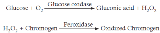
Numbers of chromogen like potassium iodide, tetramethylbenzine,
O-tolidinehydrochloride, 4-aminoantipyrine etc. are used in the dipstick. The
colour reaction of the dipsticks is kinetic and will continue to react after the
prescribed time. Therefore, reading taken after the prescribed time can give
false result.2. Biosensors
A biosensor is a device which is composed of two elements; a bio-receptor
that is an immobilized sensitive biological element (e.g. enzyme, DNA probe,
antibody) recognizing the analyte (e.g. enzyme substrate, complementary DNA,
antigen) and a transducer, used to convert biochemical signal resulting from
the interaction of the analyte with the bioreceptor into an electronic signal. The
intensity of generated signal is directly or inversely proportional to the analyte
concentration. For example, the glucose biosensor is based on the fact that
the immobilized Glucose oxidase enzyme which catalyzes the oxidation of β-D-
glucose by molecular oxygen producing gluconic acid and hydrogen peroxide.
An electrochemical transducer converts this reaction into electronic signal
which appears on the screen of the glucose meter.3. Continuous glucose monitoring
Continuous glucose monitoring systems (CGMS) use a glucose sensor
inserted under the skinin the form of a small needle. The signal from the sensor
is transmitted wirelessly and the result is recorded in a small recording device.
The monitor of the device updates and displays the blood sugar level every few
minutes. The glucose sensor needs to be removed and replaced at least once
per week.Advantages of continuous glucose monitoring:
i) The monitor displays blood sugar level every few minutes, allowing one to
see whether the level is increasing, decreasing, or is stable.
ii) The receiver can also be set to alarm if the blood sugar level is above or
below a pre-set level.
iii) The blood sugar results from the continuous monitor can be downloaded
to a computer, allowing you to check blood sugar trends over time.The only disadvantage of continuous monitor other than the cost is its inaccuracy
compared to more traditional accurate dipstick method. Therefore, most experts
recommend continuous glucose monitoring along with several finger sticks
daily to calibrate the CGMS device and to verify that the sensor readings are
accurate.Roles of adrenaline in the control of blood sugar level
Adrenaline, a natural stimulant created in the kidney’s adrenal gland, travels
through the bloodstream and controls functions of the autonomous nervous
system, including the secretion of saliva and sweat, heart rate and pupil dilation.
The substance also plays a key role in the human flight-or-flight response.The “fight or flight” hormone that gives us a quick boost of extra energy to
cope with danger — including the danger of low blood glucose. When blood
glucose levels drop too low, the adrenal glands secrete epinephrine (also called
adrenaline), causing the liver to convert stored glycogen to glucose and release
it, raising blood glucose levels. Epinephrine also causes many of the symptoms
associated with low blood glucose, including rapid heart rate, sweating, and
shakiness. The epinephrine response spurs the liver to correct low blood glucose
or at least raise blood glucose levels long enough for a person to consume
carbohydrate.3.2.7. Detection of glucose in urine
Urine analysis can be used to test pH, protein, glucose, ketones, occult blood,
bilirubin, urobilinogen, nitrite, leukocyte esterase etc. in the urine sample. Simple
test for glucose in urine can be used to diagnose diabetes mellitus. Generally,
healthy person do not loss glucose in their urine whereas a person with diabetes
mellitus loses small to large quantities of glucose in their urine.Detection of glucose in urine
The presence of glucose in the urine is called glycosuria (or glucosuria).
The urine analysis of glucose is based on enzyme glucose oxidase which is
impregnated in a dipstick (reaction described in previous section).Detection of protein in urine
The glomerular filtrate of a normal kidney contains little amount of low–molecular
weight protein. Most of these proteins get reabsorbed in the tubules with less
than 150 mg being excreted through urine per day. Therefore, the abnormal
increase in the amounts of protein in the urine, Proteinurea, can be an important
indicator of renal diseases. There are certain physiologic conditions such as
exercise and fever that can lead to increased protein excretion in the urine in the
absence of renal disease.Proteinuria is a symptom of chronic kidney disease (CKD), which can be due
to diabetes, high blood pressure, and diseases that cause inflammation
in the kidneys. Therefore, urine analysis for protein is part of a routine medical
assessment for everyone. If CKD is not checked in time, it can lead to end-
stage renal disease (ESRD), when the kidneys completely stop functioning.
A person with ESRD requires a kidney transplant or regular blood-cleansing
treatments called dialysis to further sustain.The tests for proteinuria are based either on the “protein error of indicators”
principle (ability of protein to alter the colour of some acid-base indicators without
altering the pH) or on the ability of protein to be precipitated by acid or heat.
According to “protein error of indicators” principle, a protein-free solution of
tetrabromphenol blue at pH 3 is yellow in colour and its colour changes from
yellow to blue (or green) when the pH increases from pH 3 to pH 4. However,
in the presence of protein (albumin), the colour changes occur between pH 2
and 3 i.e., an “error” occurs in the behaviour of the indicator. The method is more
sensitive to albumin than to other proteins, whereas the heat and acid tests are
sensitive to all proteins.The test result may show false-positive results in a highly buffered alkaline urine,
which may result from alkaline medication or stale urine. Also, if the dipstick
is left in the urine for too long, the buffer could be washed out of the reagent
resulting in increased pH and the strip may turn blue or green even if protein is
not present. On the other hand, false-negative results can occur in dilute urines
or when the urine contains proteins other than albumin in higher concentrations.Detection of ketones in urine
As discussed earlier, ketones, or ketone bodies are formed during lipid
metabolism. One of the intermediate products of fatty acid breakdown is acetyl
CoA. If the lipid metabolism and carbohydrate metabolism are in balanced,
Acetyl-CoA enters the citric acid cycle (Krebs cycle) where it reacts with
oxaloacetate to form citrate. When carbohydrate is not available in the cells,
all available oxaloacetate get converted to glucose and so none is available for
condensation with Acetyl- CoA. As such, Acetyl-CoA cannot enter the Krebs
cycle and is diverted to form ketone bodies.Application activity 3.2
An experiment was carried out with two groups of people. Group X has type
I diabetes mellitus while group Y did not (control group). Every 15 minutes’
blood samples were taken from all members of both groups and the mean of
levels of insulin, glucagon, and glucose were calculated. After an hour, every
person was given a glucose drink. The results are shown in the figure below: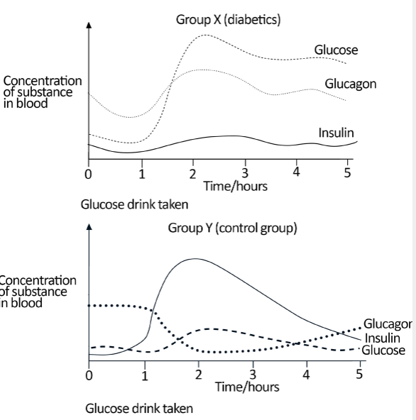
a) Name a hormone other than insulin and glucagon that is involved in
regulating blood glucose levels.
b) State two differences between groups X and Y in the way insulin
secretion responds to the drinking of glucose.
c) Suggest a reason why the glucose level falls in both groups during the
first hour.
d) Using information from the graphs, explain the changes in the blood
glucose level in group Y after the glucose drink.
e) Explain the difference in blood glucose level in group X compared to
group Y.
f) Suggest what might happen to the blood glucose level of group X if
they had no food intake over the next 24 hours.3.3 Adaptations of animals to temperature changes in the
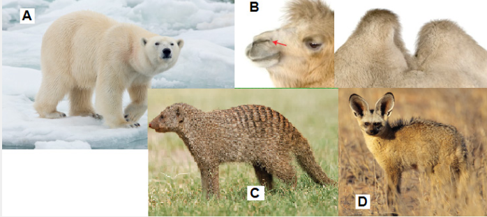
environmentActivity 3.3
Observe the photo below and answer the questions that follow:
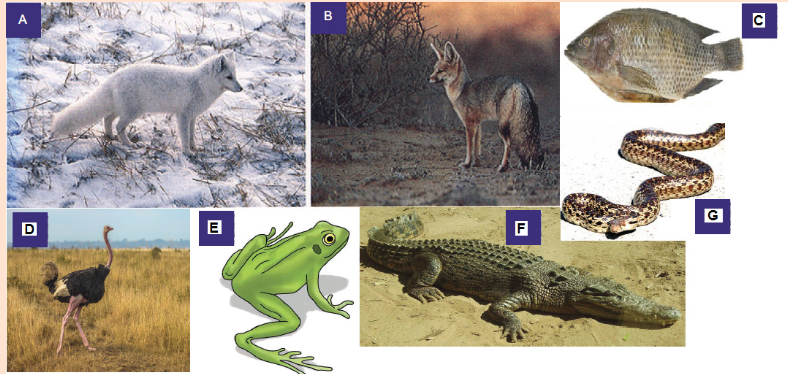
a) Show 2 main differences between individual A and individual E.
b) How is individual C different from individual D?
c) The individual A is adapted to live in cold environments. Analyze it
carefully to identify any two characteristics that this animal has.
d) Which among the animals on the photo is adapted to live in hot climates?
Justify your answer.Thermoregulation is the ability of an organism to keep its body temperature
within certain boundaries, even when the surrounding temperature is very
different. This process is one aspect of homeostasis: a dynamic state of stability
between an animal’s internal environment and its external environment.One of the most important examples of homeostasis is the regulation of body
temperature. Not all animals can do this physiologically. Animals that maintain a
fairly constant body temperature (birds and mammals) are called endotherms,
while those that have a variable body temperature (all others) are called
ectotherms. Endotherms normally maintain their body temperatures at around
35 - 40°C, so are sometimes called warm-blooded animals, but in fact
ectothermic animals can also have very warm blood during the day by basking in
the sun, or by extended muscle activity. The difference between the two groups
is thus that endothermic animals use internal corrective mechanisms, whilst
ectotherms use behavioral mechanisms (e.g. lying in the sun when cold, movinginto shade when hot). Such mechanisms can be very effective, particularly when
coupled with internal mechanisms to ensure that the temperature of the blood
going to vital organs (brain, heart) is kept constant.3.3.1 Importance of temperature regulation
Besides water, our body consists of many inorganic and organic compounds
including proteins, lipids, carbohydrates etc. Among these, proteins are the most
important compounds and are regarded as “workhorse” molecules of life, taking
part in essentially every structure and activity of life. Proteins make up about 75
per cent of the dry weight of our bodies and serve four important functions:
i) They are nutrients.
ii) They also form the structural components of our body including skin, hair
etc. They are building materials for living cells, appearing in the structures
inside the cell and within the cell membrane.
iii) As haemoglobin, Hb they carry oxygen to all the body organs and
iv) They function as biological catalysts as enzymes facilitating and
controlling various chemical reactions of our body.Protein molecules are often very large and are made up of hundreds to thousands
of amino acid units. They are of varying shape and size. For examples, keratins, a
protein in hair and collagen in tendons and ligaments linear chains of amino acids.
Other proteins called globular proteins, fold up into specific shapes and often
more than one globular unit are bound together. Enzymes are globular proteins.
Though large, enzymes typically have a small working region, known as active
site which acts as the binding site of ligands. The shape of globular proteins is
held together by many forces, including highly resistant strong covalent bonds.
However, there are also many weak forces, like hydrogen bonds, which are
susceptible to pH, osmolality and temperature changes.Since the function of enzymes is attributed to its shape, small changes in the
shape can greatly reduce its function. Every enzyme has an optimal temperature
at which it works best and this temperature is approximately the normal body
temperature of the body. Therefore, in order to ensure the optimal function of
the enzymes within, the core body temperature need to be maintained more or
less constant. If the body temperature falls below the normal value, the enzymes
catalyzed reactions of the animal will be slowed. Similarly, too much rise in body
temperature might result in enzyme denaturation and hence reduced catalytic
activities. Rise in body temperature also reduces the oxygen carrying capacity
of haemoglobin. Increasing temperature weakens and denatures the bond
between oxygen and haemoglobin which in turn decreases the concentration
of the oxyhaemoglobin. This can lead to hypoxia – a condition in which tissues
receive insufficient oxygen supply from the blood.3.3.2 Adaptations of animals to temperature changes in
the environment
From deepest corner of the sea to high mountains, living organisms have colonized
almost everywhere. However, they are not distributed evenly with different
species found in different areas. Many abiotic factors including temperature,
humidity, soil chemistry, pH, salinity, oxygen levels etc., influence the availability
of species in certain area. Each species has certain set of environmental
conditions within which it can best survive and reproduce to which they are best
adapted. This is known as limits of tolerance (i.e., the upper and lower limits
to the range of particular environmental factors within which an organism can
survive). No organism can survive if the environmental factor is below its lower
limits of tolerance or above the higher limits. Therefore, organisms having a
wide range of tolerance are usually distributed widely, while those with a narrow
range have a more restricted distribution. For examples, euryhaline fishes
(like salmon) can survive wide range of salt concentration and therefore
are found both in freshwater and salt water environment while stenohaline
fishes are found only in saltwater or freshwater.Temperature is one of the most important factors which directly or indirectly
influence the distribution of organisms to a large extend. For example, polar
bears can survive very well in low temperatures ranges, but would die from
overheating in the tropics. On the other hand, a giraffe does very well in the
heat of the African savanna, but would quickly freeze to death in the Arctic.
Compared to ectotherms or cold blooded animals, endotherms due to their
ability to generate their own body heat, are generally more widely distributed.
Besides, all the organisms have varying degree of morphological, physiological
or behavioral adaptations that helps them to survive the extreme temperature
conditions of their habitat.Effect of temperature
As discussed above, all the living organisms have a particular range of
temperature within which they can best survive and reproduce. Temperature
below or above this temperature ranges are harmful to the organism in various
ways. Some of the well-known effects of temperature on living organisms are
given below.
1. Effect of temperature on cells: If the temperature is too cold, the cell
proteins could be destroyed due to the formation of ice, or as the water is
lost, the cytoplasm can become highly concentrated. Conversely, extreme
heat can coagulate cell proteins.
2. Effect on metabolism: Most of metabolic activities of microbes, plants
and animals are regulated by enzymes and the functions of enzymes aregreatly affected by temperature. Therefore, increase or decrease in the body
temperature will greatly affect the various metabolic activities. For example,
the activity of liver arginase enzyme upon arginine increases gradually
with increase in the temperature from 17°C to 48°C. With the increase in
temperature beyond 48°C, the enzymatic activity decreased sharply.3. Effect on reproduction: Changes in temperature affect both the maturation
of gonads i.e., gametogenesis and fecundity of animals. For example, some
animal species can breed throughout the year, some only in summer or in
winter, while some species have two breeding periods, spring and autumn.
Therefore, temperature determines the breeding seasons of most organisms.
Also, it was observed that female Chrotogonus trachyplerus an acridid insect
lays highest number of eggs per female at the temperature of 30°C and
decreases with increase in temperature from 30°C to 35°C.4. Effect on sex ratio: In certain animals like copepod Maerocyclops
albidu, rises in temperature significantly increase the number of male
offspring. Similarly, in plague flea, Xenopsyll acheopis, males’ population
outnumbered females when the mean temperature is between 21°C to 25°C.
However, further decreases in temperature reverse the conditions with the
considerable increases in female population.5. Effect on growth and development: In general growth and development
of eggs and larvae is more rapid in warm temperatures. For example, Trout
eggs develop four times faster at 15°C than at 5°C. On the other hand,
seeds of many plants will not germinate and the eggs and pupae of some
insects will not hatch until chilled.6. Effect on colouration: Animals generally have a darker pigmentation in
warm and humid climates than those found in cool and dry climates. This
phenomenon is known as Gioger rule. In the frog Hylaand the horned toad
Phrynosoma, low temperatures have been known to induce darkening. Some
prawn turn light coloured with increasing temperature.7. Effect on morphology: Temperatures have profound effects on the size of
animals and various body parts. Endotherms generally attain a larger body
size (reduced surface-mass ratio) in colder temperatures than in warmer
temperatures. As such the colder regions harbour larger species. Conversely,
the poikilotherms (ectotherms) tend to be smaller in colder regions. We will
discuss the various morphological modifications due to extreme climates in
the later sections.8. Effect on animal behaviour: Temperature certainly has profound effect
on the behavioural pattern of animals. The advantage gained by certain cold
blooded animals through thermotaxis or orientation towards a source of
heat are quite interesting. Ticks locate their warm blood hosts by a turningreaction to the heat of their bodies. Certain snakes such as rattle snakes,
copper heads, and pit vipers are able to detect mammals and birds by their
body heat which remains slightly warmer than the surroundings.9. Effect on animal distribution: Since the optimum temperature for many
organisms varies, temperature imposes a restriction on the distribution of
species. The diversity of animals and plants gradually decrease as we move
from equator towards the pole.Morphological Adaptations
1. Body size and shape: Ectotherms or cold-blooded animals whose body
temperature depends on the temperature of external environments are usually
smaller in size compared to endotherms or warm blooded animals. For instance,
compare the size of elephant, blue whales and crocodiles or snakes. Within
the same species, individuals living in the colder climates tend to be larger
than those living in warmer climates. This is known as Bergmann’s rule. For
example, whitetail deer in the southern part of the United States have a smaller
body size than white tail deer in the northern states the far northern states.2. Body Extremities: According to Allen’s rule, animals living in the colder
climates have more rounded and compact form. This is achieved by reducing
the size of the body extremities i.e., ears, limbs, tails etc. On the other hand,
animals living in the warmer climates have longer body extremities. For instance,
compare the size of the ear of Arctic fox with that of the Desert fox (Figure 13.2).
Longer body extremities increase the surface to volume ratio of the desert fox
which enable them to lose heat more easily.Most cold-blooded organisms have either an elongated or a flat body shape.
For example, fishes, snakes, lizards, and worms have long and slender body
form which ensures rapid heat up and cool down processes.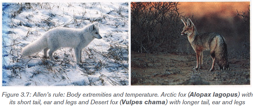
Both Bergmann’s rule and Allen’s rule depend on simple principle that “the ratio
of surface area to volume of an object is inversely proportional to the volume of the
object”. In other words, the smaller an animal is, the higher the surface area-to-
volume ratio. Higher surface area-to-volume ratio ensures these animals to lose
heat relatively quickly and cool down faster, so they are more likely to be found
in warmer climates. Larger animals, on the other hand, have lower surface area-
to-volume ratios and lose heat more slowly, so and they are more likely to be
found in colder climates.3. Insulation: All the marine mammals have a thick insulating layer of fat
known as Blubber, just beneath the skin. It covers the entire body of animals
such as seals, whales, and walruses (except for their fins, flippers, and flukes)
and serves to stores energy, insulates heat, and increases buoyancy. Thickness
of blubber can range from a couple of inches in dolphins and smaller whales,
to 4.3 inches in polar bears to more than 12 inches in some bigger whales. To
insulate the body, blood vessels in blubber constrict in cold water. Constriction
of the blood vessels reduces the flow of blood to the skin and minimizes the
heat loss. In such animals, skin surface temperature is nearly identical to the
surrounding water, though at a depth of around 50 mm beneath the skin, the
temperature is the same as their core temperature.Some marine mammals, such as polar bears and sea otters, have a thick fur
coat, as well as blubber, to insulate them. The blubber insulates in water
while fur insulates in air or terrestrial environment. The feathers of the birds also
function in insulating the body from cold temperature.Physiological Adaptations
1. Evaporation: In a cold region, i.e., when the surrounding environment of
the animal is cold than the body temperature, conduction and radiation are
the main ways an animal will dissipate heat. However, in warmer region, the air
temperature is often higher than the animal’s body temperatures, so the only
physiological thermoregulatory mechanism available is evaporation. Animals
use three evaporative cooling techniques that include sweating, panting, and
saliva spreading.(a) Sweating: It is the loss of water through sweat glands found in the skin of
mammals. The number of sweat glands can vary from none in whales, few in dogs
to numerous in humans. Most small mammals do not sweat because they would
lose too much body mass if they did. For example, in a hot desert the amount
of water a mouse would lose through sweating to maintain a constant body
temperature would be more than 20% of its body weight per hour, which could
be lethal for the animal. Therefore, smaller mammals use other techniques to cool
down their body. On the other hand, sweating is an important thermoregulatory
mechanism for primates including humans. An adult human can loss as much as
10–12 litres of water per day through sweating.(b) Panting: It is rapid, shallow respiration that cools an animal by increased
evaporation from the respiratory surfaces. It is a common thermoregulatory
technique used by small animals like dogs and rodents to loss heat.(c) Saliva spreading: It is a means of thermoregulation used by marsupials.
Under extreme heat, saliva will drip from animal’s mouth and is then wiped on its
fore and hind legs. This technique induces the cooling effect of evaporation by
wetting the fur. However, since the animal cannot spread saliva while moving,
they need to adapt other evaporative techniques during such situation.2. Counter current mechanism: As mentioned above, in addition to its role in
the transport of oxygen and food, circulatory system of our body is responsible for
distribution of heat throughout the body. This is true in case of both endotherms
and ectotherms. In endotherms, most of the body heat is generated in brain,
liver, heart and skeletal muscles. This heat is transported to other parts of the
body through blood. On the other hand, in ectotherms, the circulatory system
help in transporting heat from skin to others body parts. The counter current
heat exchanger is generally located in body extremities like limbs, neck, gills,
which are directly in contact to the external environment.In cold region, when the warm blood flows through the arteries, the blood gives
up some of its heat to the colder blood returning from the extremities in the
veins running parallel to the arteries. Such veins are located in the deeper side
of the body and carry the warm blood to the heart and most of the body heat is
retained. Such mechanism can operate with remarkable efficiency. For instance,
a seagull can maintain a normal temperature in its torso while standing with its
unprotected feet in freezing water (Figure 3.8).When the external temperature is higher than the body temperature and heat
loss is not a problem, most of the venous blood from the extremities returns
through veins located near the surface. If the core body temperature becomes
too high, the blood supply to the surface and extremities of the body is increased
enabling heat to be released to the surroundings.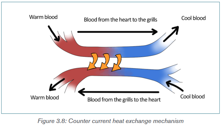
3. Hyperthermia: Hyperthermia is a condition of having the body temperature
greatly above the normal. Although all the endotherms can maintain a constant
body temperature, some are able to raise their body temperature as a way
to decrease the amount of water and energy used for thermoregulation. For
example, camels and gazelles can increase their body temperature by 5–7°C
during the day when the animal is dehydrated. Hyperthermia helps in saving
water by letting their body temperature increase instead of using evaporative
cooling to keep it at a constant temperature.4. Water retention: Human body obtains about 60 per cent of the water they
need from ingested liquid, 30 per cent from ingested food, and 10 per cent from
metabolism. While rodent adapted to arid conditions obtains approximately 90
per cent from metabolism and 10 per cent from ingested food. The predaceous
marsupial Mulgara species can go its whole life without ingesting water but by
obtaining water from the food they eat and from metabolism. The fawn hopping
mouse eats seed, small insects, and green leaves for moisture, and Kowaris eat
insects and small mammals to obtain water. These animals have specialized
kidneys with extra microscopic tubules to extract most of the water from their
urine and return it to the blood stream. And much of the moisture that would be
exhaled in breathing is recaptured in the nasal cavities by specialized organs.Many desert dwelling insects tap plant fluids such as nectar or sap from stems,
while others extract water from the plant parts they eat, such as leaves and
fruit. The abundance of insects permits insectivorous birds, bats and lizards
to thrive in the desert. Elf owls survive on katydids and scorpions. Pronghorns
can survive on the water in cholla fruits. Kit foxes can satisfy their water needs
with the water in their diet of kangaroo rats, mice, and rabbits, along with small
amounts of vegetable material.5. Excretion: As mentioned above, desert dwelling mammals and birds have
specialized kidneys with long loops of Henle compared to animals that live in
aquatic environments and less arid regions. A longer tubules help in reabsorbing
most of the water from their urine and return it to blood stream. As a result, the
urine becomes highly concentrated. In these animals, most of the water in the
faeces gets reabsorbed in the alimentary canals and colon. Camels produce
dryer faeces than other ruminants. For example, sheep produce faeces with 45
per cent water after 5 days of water deprivation, while camels produce faeces
with 38 per cent water even after 10 days of water deprivation. The ability to
excrete concentrate urine and dry faeces is an important adaptation to arid
conditions. Desert rodents can have urine five times as concentrated as that of
humans.Behavioural adaptations
Behavioural adaptations are used to reduce the amount of heat gained or lost by
animals, and, thereby, reducing the amount of energy and water to maintain the
body temperature. Ectoderms or cold blooded animals rely on their behaviour to
maintain a favourable body temperature.1. Nocturnality: It is the simplest form of behavioural adaptation characterized
by activity during the night and sleeping during the day. As such, nocturnal
animals avoid direct exposure to heat of the day, thereby preventing loss of
water needed for evaporative cooling. The night temperatures are generally
15–20°C colder than the daytime, so require much less energy and water to
regulate body temperature. Most of the desert animals like quoll, bilby, and the
spinifex hopping mouse, are nocturnal. Other large animals like lions prefer to
hunt at night are to conserve water.Crepuscular animals are those animals that are mainly active during twilight
(i.e., the period before dawn and that after dusk). Examples include hamsters,
rabbits, jaguars, ocelots, red pandas, bears, deer, moose, spotted hyenas etc.
Many moths, beetles, flies, and other insects are also crepuscular in habit.
These crepuscular animals take advantage of the slightly cooler mornings and
evenings to escape the daytime heat, and to evaporate less water.2. Microhabitat: Among the diurnal animals (animals which are mainly active
during the day and rest during night), the use of microhabitat like burrows, shade
is another type of behavioural adaptation to avoid the daytime heat. Fossorial
animals (digging animals), such as mulgaras, spent much of their time below
ground eating stored food. Lizards and snakes seek a sunny spot in the morning
to warm up their body temperatures more quickly and remain in shade when the
temperature rises.3. Migration: It is the physical movement of animals over a long distance
from one area to another. It is found in all major animal groups, including birds,mammals, fish, reptiles, amphibians, insects, and crustaceans. Many factors
like climate, food, the season of the year or mating could lead to migration. It
helps the animals in avoiding the extreme environmental conditions by moving
to more favourable places. For example, many migratory birds like arctic tern
(Sterna paradisaea) migrate north-south, with species feeding and breeding in
high northern latitudes in the summer, and moving some hundreds of miles
south during the winter to escape the extreme cold of north. Monarch butterflies
spend the summer in Canada and the Northern America and migrate as far
south as Mexico for the winter.4. Hibernation and Aestivation: Warm blooded animals which do not
migrate generally survive the extreme cold condition of winter by sleeping.
Hibernation is the state of dormancy during the cold conditions, i.e., winter.
During hibernation, body temperature drops, breathing and heart rate slows,
and most of the body’s metabolic functions are put on hold in a state of quasi-
suspended animation. This allows them to conserve energy, and survive the
winter with little or no food.Many insects spend the winter in different stages of their lives in a dormant
state. Such phenomenon is known as diapause. During diapauses, insect’s
heartbeat, breathing and temperature drop. Some insects spend the winter as
worm-like larvae, while others spend as pupae. Some adult insects die after
laying their eggs in the fall and eggs hatch into new insects in the spring when
the food supply and temperature become favorable.Aestivation or summer dormancy on the other hand, is a state of animal dormancy,
characterized by inactivity and a lowered metabolic rate, in response to high
temperatures and arid conditions. It allows an animal to survive the scarcity of
water or food as aestivating animal can live longer off its energy reserves due
to the lowered metabolism, and reduced water loss though lowered breathing
rates. Lung fishes, toad, salamander, desert tortoise, swamp turtles are some of
the other non-mammalian animals which undergo aestivation.5. Social behavior: Among all the adaptations, living together is one of the
most important adaptations of the animal kingdom. Animals can derive a lot of
benefit from spending time with other members of the same species like finding
food, defense against predators and care for their young. For example, emperor
penguins can survive the harsh Antarctica winter huddling together in groups
that may comprise several thousand penguins. Huddling greatly reduces the
surface area of the group compared to individuals and a great deal of warmth
and body fat is conserved. Many social mammals, including many rodents, pigs
and primates survive extreme cold by huddling together in groups.6. Locomotion: Different types of locomotion require varying amount of energy.
Many mammals like kangaroo, hares hop, which is an energy efficient type oflocomotion. When animals go from walking to running, there is an increasing
energy cost; however, once kangaroos start moving, there is no additional
energy cost. This is because when a kangaroo lands, energy is stored in the
tendons of its hind legs which is used to power the next hop.Application activity 3.3
1) The figure below shows different animals living in different climates
a) Which animal(s) on the photo appears to be adapted to live in cold
climates? Why?
b) Which animal(s) on the photo appears to be adapted to live in hot
climates? Why?
c) What are the adaptations of the animal A that help it to survive in its
environment?
d) What is the functions of the humps on the animal B?
e) Some animals such as the animal A hibernate during the winter. Explain
the importance of hibernation to these animals.3.4 Response to cold and hot conditions by endothermic
and ectothermic animalsActivity 3.4
1) The figure below shows different animals living in different climates
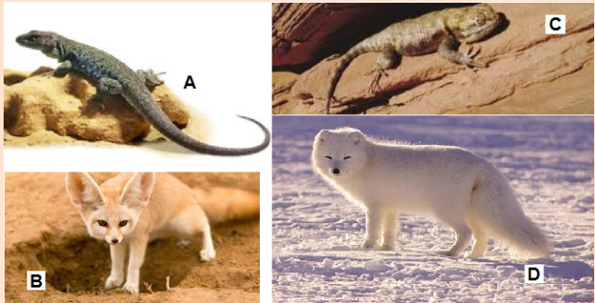
a) The animals A and B are reptiles under different environmental
conditions. Compare their behaviors in regards to how they regulate
their temperature.
b) The animals’ C and D are mammals under different environmental
conditions. Compare their behaviors in regards to how they regulate
their temperature.
c) What are the adaptations of the animal D that help it to survive in its
environment?
d) How is the animal A different to animal D according to how they regulate
their body temperature.3.4.1 Endotherms’ response to temperature changes
Endothermic organism can maintain relatively high body temperatures within a
narrow range. Since most of the body heat is produced as a result of various
metabolic activities, thermoregulation in endotherms depends on food and
water availability. For example, bear undergoes hibernation during the winter
because there is no sufficient food during the cold season. On the other hand,
in arid environment like deserts, many deserts animals are nocturnal to avoid the
extreme daytime heat to avoid loss of water through evaporation.Response to hot temperature
When the body temperature increases in response to the external temperature,
the body’s temperature control system uses three important mechanisms to
reduce the body heat. These are:1. Vasodilation of blood vessels in the skin: The blood vessels in skin become
intensely dilated due to the inhibition of the sympathetic centres in the posterior
hypothalamus that cause vasoconstriction. Vasodilation increases the rate blood
flow to the skin and as a result, the amount of heat transfer from the core of the
body increases tremendously.2. Sweating: As discussed in the previous section, sweating is an important
adaptation to lose body heat through evaporative cooling. An increase in 1°C in
body temperature causes enough sweating to remove ten times the basal rate
of body heat production.3. Decrease in heat production: As mentioned above, metabolic activities of
the body are the main source of body heat. The mechanisms that cause excess
heat production, such as shivering and chemical thermogenesis, are strongly
inhibited when exposed to hot temperature.Response to cold temperature
In response to cold temperature, the temperatures control system performs
exactly opposite mechanism to that performs in hot temperature. These are:1. Vasoconstriction of blood vessels in the skin: The blood vessels in the skin
constrict under the influence of posterior hypothalamic sympathetic centres
which reduce the blood flow to the skin.2. Piloerection: Piloerection means hairs “standing on end”. Sympathetic
stimulation causes the arrector pili muscles attached to the hair follicles to
contract, which brings the hairs to an upright stance. The upright projection of
the hairs allows them to entrap a thick layer of air next to the skin which acts as
insulator, so that transfer of heat to the surroundings is greatly depressed.3. Increase in heat production (thermogenesis): Endothermic metabolic
rates are several times higher than those of ectotherms. The metabolic heat
production of endotherms is regulated in response to fluctuations in the
environment temperature. This phenomenon is known as adaptive thermogenesis
or facultative thermogenesis. It can be defined as “Heat production by metabolic
processes in response to environmental temperature with the purpose of
protecting the organism from cold exposure and buffering body temperature
from environmental temperature fluctuations”. Under cold temperature stress,
heat production by the metabolic activities increased tremendously by promoting
shivering, sympathetic excitation of heat production, and thyroxine secretion.These mechanisms will be discussed later. Extreme shivering can increase the
temperature four to five times the normal production.3.4.2 Ectotherms’ response to temperature changes
Ectotherms cannot maintain stable body temperature and their body temperature
relies on the external temperature. They depend more on energy assimilation
rather than utilizing it for temperature regulation. Therefore, ectotherms regulate
their body temperature behaviourally and by cardiovascular modulation of
heating and cooling rates. At the same time, metabolism and other essential
rate functions are regulated so that reaction rates remain relatively constant
even when body temperatures vary. This process is known as acclimatization or
temperature compensation. For example, many fish adjust metabolic capacities
to compensate for seasonal variation in water temperature with the result that
metabolic performance remains relatively stable throughout the year. Reptiles
often regulate their body temperature to different levels in different seasons
to minimize the behavioural cost of thermoregulation. At the same time, tissue
metabolic capacities are adjusted to counteract thermodynamically-induced
changes in rate functions.Response to hot temperature
When the external temperature increases, ectotherms protect their bodies from
overheating using various mechanisms. These are:1. Use of microhabitat: Under extreme heat conditions, many ectotherms like
lizards and snakes prefer to stay in shade, either beneath the rocks, crevices or
underground burrows.Amphibians and fishes enter cold water when their body temperature increases.
2. Acclimatization: If a salamander living at 10°C is exposed to 20°C, its
metabolic rate increases rapidly. But if the exposure to the higher temperature
lasts for several days, the animal experiences a compensating decrease in the
metabolic rate. This decrease in the metabolic rate is due to acclimatization.
The higher metabolic rate is due to the increase in the enzymes activity with
temperature. However, with prolonged exposure to the condition, the metabolic
rates decrease to prevent excessive energy loss. Ectotherms also exhibit
acclimatization of temperature tolerance range with animal acclimated to high
temperature are able to tolerate higher temperature than those exposed only to
low temperature. Similarly, cold acclimated animals have better tolerance to low
temperature than high temperature acclimated animal.Response to cold temperature
Ectotherms response to cold temperature is exactly opposite to the response
shown when exposed to hot temperature. That is:1. Basking to sun: When the body temperature of the ectotherms becomes
colder than the normal, the animals either bask to sunlight to warm up the body
or move to a warmer place. Under extreme cold conditions, all the metabolic
activities may cease and the animals enter the state of torpor (reduced metabolic
activities).2. Cold Acclimatization: Decrease in the temperature result in reduced
metabolic rate. Therefore, as a compensatory measure to meet the require body
metabolism, the cold acclimatization of ectotherms is characterized by increase
in concentration of various metabolic enzymes. There is also significant increase
in the mitochondria and capillaries concentration in the skeletal muscle. This
increase the ATP production through aerobic respiration in these tissues.
Therefore, in those animals which have prolonged exposure to cold temperature,
there may be increase in the locomotion, though the basal rates of metabolism
remain below the warm acclimatized animals.Application activity 3.4
1. a) Describe the importance of hibernation to animals.
b) The camel is one of the animals adapted to live in deserts. Explain
three of its adaptations that help it to survive in arid conditions.
c) State three adaptations of animals to living in cold climates.3.5 Role of the brain
Activity 3.5
Find information about the role of hypothalamus and different thermoreceptors
in temperature regulation. Make short notes and present them in front of the
class.So far we have discussed that on the basis of types of thermoregulation, all the
living organisms can be classified into two groups – ectotherms and endotherms.
Endotherms can regulate their body temperature within a narrow range through
various physiological mechanisms while ectotherms being depended on external
temperature mostly rely on their behaviour to maintain body temperature. But
how do these animals sense and counter the changing temperature of their
body will be discussed in the section.Thermoreceptors
A thermoreceptor is a sensory receptor which is basically the receptive
portion of a sensory neuron that converts the absolute and relative changes
in temperature, primarily within the innocuous range to nerves impulses.
Thermoreception is the sense by which an organism perceives the
temperature of the external and internal environment from the information supply
by thermoreceptors. In vertebrates, most of the thermoreceptors are found in
skins which are actually free nerve endings. Deep body thermoreceptors are
also found mainly in the spinal cord, in the abdominal viscera, and in or around
the great veins in the upper abdomen and thorax region.Mammals have at least two types of thermoreceptors: the warm receptors,
those that detect heat or temperatures above normal body temperature and cold
receptors, those that detect cold or temperatures below body temperature. The
warm receptors are generally unmyelinated nerves fibres, while cold receptors
have thinly myelinated axons and hence faster conduction velocity. Increasing
body temperature results in an increase in the action potential discharge rate
of warm receptors while cooling results in decrease. On the other hand, cold
receptors’ firing rate increases during cooling and decreases during warming.
Another types of receptor called nociceptors, detect pain due to extreme cold
or heat which is beyond certain threshold limits.A specialized form of thermoception known as distance thermoreception is found
in some snakes like pit viper and boa, use a specialized type of thermoreceptor
which can sense the infrared radiation emitted by hot objects. The snake’s
face has a pair of holes, or pits, lined with temperature sensors. These sensors
indirectly detect infrared radiation by its heating effect on the skin inside the pit
which helps them to locate their warm blooded prey. The common vampire bat
may also have specialized infrared sensors on its nose.Hypothalamus
The hypothalamus is a very small, but extremely important part of the brain
that acts as the link between the endocrine and nervous systems of the
body. The hypothalamus plays a significant role in the endocrine system and is
responsible for maintaining the body’s homeostasis by stimulating or inhibiting
many key processes, including body temperature, fluid and electrolyte
balance, appetite and body weight, glandular secretions of the stomach
and intestines, production of substances that influence the pituitary gland to
release hormones and sleep cycles.Role of Hypothalamus in thermoregulation
Thermoregulation is carried out almost entirely by nervous feedback mechanisms,
and almost all these operate through temperature-regulating centres located inthe hypothalamus (Figure 3.7). The hypothalamus contains large numbers of
heat-sensitive as well as cold sensitive neurons which acts as thermoreceptor,
sensing the temperature of the brain. The posterior hypothalamus region
contains the thermoregulatory centre which integrate the signals from of all
the thermoreceptors found in skin, deep organs and skeletal muscles, as well
as from the anterior hypothalamus and control the heat-producing and heat-
conserving reactions of the body.Cooling Mechanism
When the body temperature increases beyond the set-point, the anterior
hypothalamus is heated. The posterior hypothalamus senses the heat and
inhibits the adrenergic activity of the sympathetic nervous system, which control
vasoconstriction and metabolic rate. This causes cutaneous vasodilation and
increase heat loss through skin. It also reduces the body metabolic rate resulting
in decreasing heat production through metabolic reactions. Under intense
heating, the cholinergic sympathetic fibres innervating the sweat glands release
acetylcholine, stimulating the secretion of sweat. Many behavioural responses
to heat, such as lethargy, resting in shade, lying down with limbs spread out,
etc., decreases heat production and increases heat loss.Heating Mechanism
When the body temperature falls below the set-point, the body regulating
mechanism tries to reduce heat loss and increase heat production. The
immediate response to cold is vasoconstriction throughout the skin. The
result is vasoconstriction of the skin blood vessels, reducing the blood flow
and subsequent heat loss through skin. Sympathetic stimulation also causes
piloerection and reduces the heat loss from the body by trapping heat within
the body hair.The primary motor centre for shivering is excited by the cold signals from skin
and spinal cord which cause shivering of the skeletal muscles. Intense shivering
can increase the body heat production four to five times normal. Cooling
the anterior hypothalamic due to decrease in body temperature stimulates
hypothalamus to increases the production of the neurosecretory hormone
thyrotropin-releasing hormone. This hormone in turn stimulates the anterior
pituitary gland, to secrete thyroid-stimulating hormone. Thyroid-stimulating
hormone then stimulates thyroid glands to increased output of thyroxine. The
increased thyroxine level in the blood increases the rate of cellular metabolism
throughout the body and hence increases heat production.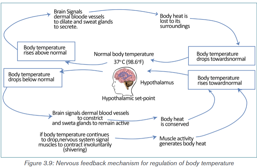
Application activity 3.5
1) The diagram shows the way in which temperature is regulated in body
of a mammal.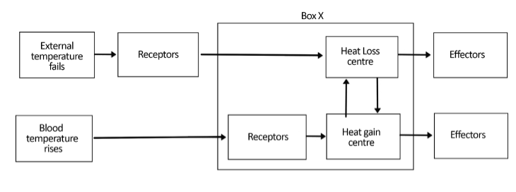
a) Which part of the brain is represented by box X?
b) i) How does the heat loss center control the effectors which lower the
body temperature?
ii) Explain how blood vessels can act as effectors and lower the body
temperature?3.6 Temperature controls in plants
Activity 3.6
Observe carefully the photos below and answer to the questions that follow:
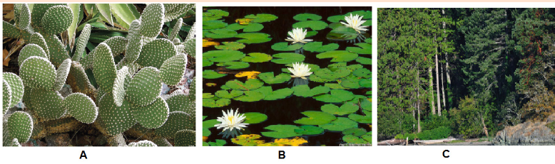
a) In which habitat do these plants live?
b) What are the adaptations of plant A that help it to survive in its
environment?
c) Make a comparison between plant A and plant B.Like all the other living organisms, plants depend on enzymes catalyzed chemical
reactions for their growth and development. For example, plants synthesize their
own food from water and carbon dioxide using sunlight through photosynthesis.
The process of photosynthesis involves a series of complex enzyme system
and other proteins. Therefore, along with carbon dioxide, water, light, nutrients
and humidity, temperature is also one of the limiting factors for growth and
development of plants.Unlike animals, plants remain fixed in a particular site and absorb heat from the
sunlight. The excess heat from the body is released to the surrounding through
radiation and evaporation. The process of evaporation of water from the leaves
and stem of plants to the surrounding environment is known as transpiration. It
occurs through stomata, small opening located on the underside of the leaves.
The stomata are specialized cells in the leaves which can open or close, limiting
the amount of water vapour that can evaporate. Higher temperature causes the
opening of stomata whereas colder temperature causes the opening to close.
The opening of the stomata and hence the transpiration rate of plants depends
on environmental conditions such as light, temperature, the level of atmospheric
CO2 and relative humidity. Higher relative humidity leads to more opening,
while higher CO2 levels lead to closing of stomata. Under high environmental
temperature, the plant body gets heat up. In order to cool down, the plant
increases its transpiration rate. The evaporative loss of water from the plant’s
body lowers the temperature.Besides transpiration, many plants have different adaptations that help them
survive in extreme temperature conditions ranging from hot and arid deserts
to cold and snow covered mountains. These adaptations make it difficult for
the plant to survive in a different place other than the one they are adapted to.
This explains why certain plants are found in one area, but not in another. For
example, cactus plants, adapted to desert conditions can’t survive in the Arctic.These adaptations will be discussed later in this unit.
3.6.1 Effect of temperature changes on plants
The most obvious effect of temperature on plants is changes in the rate of
photosynthesis and respiration. Both processes increase with rise in the
temperature up to a certain limit. However, increase in temperature beyond the
limits, the rate of respiration exceeds the rate of photosynthesis and the plants
productivity decreases.Another important effect of temperature is during the process of germination
of seeds. Like most other processes it also depends on various factors
including air, water, light, and, of course, temperature. In many plant species,
germination is triggered by either a high or low temperature period that destroys
germination inhibitors. This allows the plant to measure the end of winter season
for spring germination or end of summer for fall germination. For example, winter
adapted plant seeds remain dormant until they experience cooler temperatures.
Temperature of 4°C is cool enough to end dormancy for most cool dormant
seeds, but some groups, especially within the family Ranunculaceae and others,
need conditions cooler than –5°C. On the other hand, some plants like Fire
poppy (Papaver californicum) seeds will only germinate after hot temperatures
during a forest fire which cracks their seed coats. The fire does not cause direct
germination, rather weakens the seed coat to allow hydration of the embryo.Pollination is another phenological stage of plants sensitive to temperature
extremes across all species. Since pollination is carried out by pollinators like
honey bees, butterflies etc., any factors including temperature that affect these
pollinators will certainly affect the process.Heat adapted plants
In extremely hot and dry desert region with annual rainfall averages less than
10 inches per year, and there is a lot of direct sunlight shining on the plants,
the main strategy for the survival of the plants is to avoid extensive water loss
through transpiration. Therefore, in such region many plants called succulents,
like cactus can store water in their stems or leaves. Some plants are leafless
or have small seasonal leaves that only grow after rains. These leafless plants
conduct photosynthesis in their green stems. Leaves are often modified into
spines to discourage animals from eating plants for water. Also waxy coatingon stems and leaves help reduce water loss. Other plants have very long root
systems that spread out wide or go deep into the ground to absorb water.On the other hand, in hot and humid tropical rainforest, the abundance of water
can cause problems such as promoting the growth of bacteria and fungi which
could be harmful to plants. Heavy rainfall also increases the risk of flooding, soil
erosion, and rapid leaching of nutrients from the soil. Plants grow rapidly and
quickly use up any organic material left from decomposing plants and animals.
The tropical rainforest is very thick, and not much sunlight is able to penetrate
to the forest floor. However, the plants at the top of the rainforest in the canopy
must be able to survive the intense sunlight. Therefore, the plants in the tropical
rainforest usually have large leaves with drip tips and waxy surfaces allow water
to run off easily. Some plants grow on other plants to reach the sunlight.Similarly, in aquatic plants adapted for life in water, the leaves are very large,
fleshy and waxy coated. Increase surface area allows plants to lose excess
water while the shiny wax coating discourages the growth of microbes. The
roots and stems are highly reduced since water, nutrients, and dissolved gases
are absorbed from the water directly through the leaves.Cold adapted plants
In extremely cold region like tundra which is characterized by a permanently
frozen sub-layer of soil called permafrost, the drainage is poor and evaporation
slow. With the region receiving very little precipitation, about 4 to 10 inches
per year usually in the form of snow or ice, plant life is dominated by small,
low growing mosses, grasses, and sedges. Plants are darker in colour, some
even red which helps them absorb solar heat. Some plants are covered with
hair which helps keep them warm while others grow in clumps to protect one
another from the wind and cold.In a slightly warmer temperate forest, with temperature varies from hot in the
summer to below freezing point in the winter, many trees are deciduous that is
they drop their leaves in the autumn to avoid cold winter, and grow new ones in
spring. These trees have thin, broad, light-weight leaves that can capture a lot
of sunlight to make a lot of food during the warm weather and when the weather
gets cooler, the broad leaves cause too much water loss and can be weighed
down by too much snow, so the tree drops its leaves. They usually have thick
bark to protect against cold winters.Application activity 3.6
1) The diagram below shows a transverse section of a leaf Ammophila
arenaria, which is a xerophyte. The photomicrograph shows the details
of the area indicated by the box in the diagram.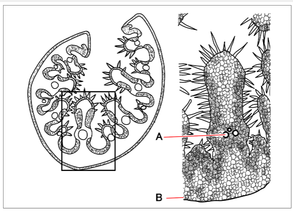
a) Name the parts labelled A and B.
b) Describe two xeromorphic features shown in this leaf and, in each case,
indicate how the feature helps to reduce transpiration.Skills Lab 3

Procedure:
1) Wash your hands with soap and water and dry them properly.
2) Prepare the blood glucose meter with the test strip according to the
manufacturer’s instructions.
3) Use the lancet device to prick the side of your fingertip with a lancet.
4) Place a drop of blood onto the correct part of the test strip.
5) The strip will draw up the blood into the meter and show a digital
reading of the blood glucose level within seconds.
6) Note the reading.
7) Use a clean cotton ball to apply pressure to the fingertip for a few
moments until the bleeding stops.
8) Similarly, measure the blood glucose level of your friends.
9) Compare your blood glucose level with that of your friends.Discussion:
In general, a fasting blood glucose reading (taken before a meal) should be
between 72 mg/dL to 126 mg/dL. And a blood glucose reading 2 hours after
a meal should be between 90 mg/dL to 180 mg/dL.Precautions:
1) Make sure the lancelet is properly sterilized.
2) Insert the test strip properly.End unit assessment 3
I. Multiple Choice Questions
1) Which of the following monosaccharides is not a product of
carbohydrate metabolism in our body?
(a) Glucose (b) Fructose (c) Ribose (d) Galactose2) Which of the following is not a part of portal triad?
(a) Central vein (b) Hepatic artery
(c) Hepatic portal vein (d) Bile duct.3) Somatostatin is secreted by
(a) Alpha cells (b) Beta cells
(c) Delta cells (d) F cellsThe process of formation of glucose from non-carbohydrates source in the
body is known as
(a) Glycogenesis (b) Gluconeogenesis
(c) Glycolysis (d) Glycogenolysis5) Which of the following hormone is responsible for decreasing blood
glucose level?
(a) Glucagon (b) Insulin (c) Somatostastin (d) Adrenaline6) The enzyme used in the dipstick for testing concentration of glucose is
(a) Glucose oxidase (b) Glycogen phosphorylase
(c) Glucose phosphatase (d) GlucosidaseII. State whether the following statements are True (T) or False (F)
1) Excess glucose in the body is stored in the form of glycogen.
2) Trypsin is an enzyme used for carbohydrate digestion.
3) Bile salt is secreted by exocrine liver.
4) Glucagon is secreted by pancreas in response to high blood glucose
concentration.
5) Insulin administration is recommended for person with type II diabetes
mellitus.6) Type I diabetes mellitus is cause due to insufficient secretion of insulin
by beta cells.
7) Ketone bodies are formed when our body have excessive fat metabolism.
8) Hyperinsulinaemia is associated with type II diabetes mellitus.
9) All the living organisms have a particular range of temperature within
which they can best survive and reproduce.
10) Nocturnality is the simplest form of behavioral adaptation characterized
by activity during the day and sleeping during the night.
11) Crepuscular animals take advantage of the slightly cooler mornings
and evenings to escape the daytime heat, and to evaporate less water.
12) Body temperature of Ectotherms rely on the external temperature.
13) Thermoregulation in endotherms depends on food and water availability.
14) Glycogenolysis is the breakdown of glucose to form pyruvate.III Long Answer Type Questions
1) List few adaptive features shown by plants inhabiting extreme cold and
hot environments.
2) Explain the role of the brain and thermoreceptors in temperature
regulation.
3) In your own words, explain the importance of maintaining fairly constant
temperatures for efficient metabolism.
4) Describe the functions of liver and pancreas in regulating blood
glucose level.
5) Discuss in brief the importance of urine analysis in diagnosis diabetes
mellitus.
6) The control of blood glucose concentration involves a negative
feedback mechanism.
a) What are the stimuli, receptors and effectors in this control mechanism?
b) Explain how negative feedback is involved in this homeostatic
mechanism.7) An investigation was carried out to determine the response of
pancreatic cells to an increase in the glucose concentration of the
blood. A person who had been told not to eat or drink anything other
than water for 12 hours then took a drink of a glucose solution. Blood
samples were taken from the person at one hour intervals for five hours,
and the concentration of glucose, insulin and glucagon in the blood
and the concentration of glucose, insulin and glucagon in the blood
were determined. The results are shown in the graph below: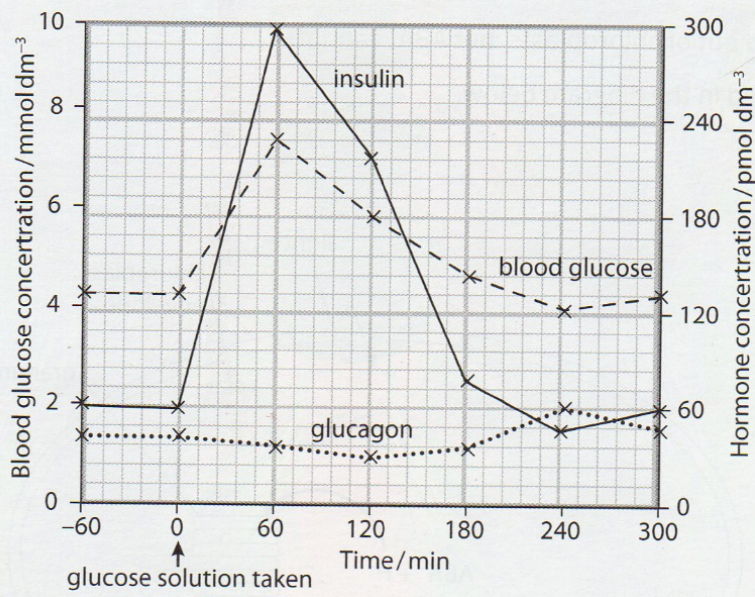
a) Explain why the person was told not to eat or drink anything other than
water for 12 hours before having the glucose drink.
b) Use the information in the figure to describe the response of the
pancreatic cells to an increase in the glucose concentration.
c) Outline the role of insulin when the glucose concentration in the blood
increases.
d) Suggest how the results will change if the investigation continued longer
than five hours without the person taking any food.
e) Outline the sequence of events that follows the binding of glucagon to
its membrane receptor on a liver cell.