Topic outline
General
- Biology SME Y3 SB File Uploaded 1/11/21, 16:56
UNIT 1: POPULATION AND NATURAL RESOURCES
Key unit competence
Describe the factors affecting population size and the importance of natural
resourcesIntroductory activity 1
Living organisms in their natural habitat are different in number where by some are
still represented by a significant number (figure A) While others can disappear when
they are not protected (Figure B). The change in number of organisms does not
happen abruptly without any reasons behind. Refer to the figures and do activity
below :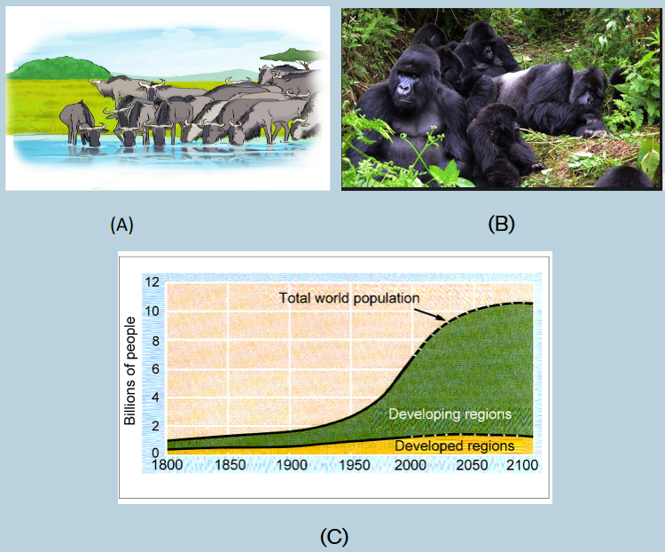
a) Referring to figure B above, identify the reasons that were behind
their decrease?
b) Referring to figure A, why does others species still represented by a
significant number?
c) Observe the graph C and identify what it indicates in terms of population
growth, especially in developing countries. What do you think would be
the effect on the nature and what measures would be taken to maintain
the nature?1.1 Population characteristics
Activity 1.1
The human population size in some areas increases yet their habitat does
not increase. The pyramid of age structure in that area shows there are
more young people than adults.The growth pattern below shows that there is an increase in population
of these areas which is a result of high birth rate compared to death rate.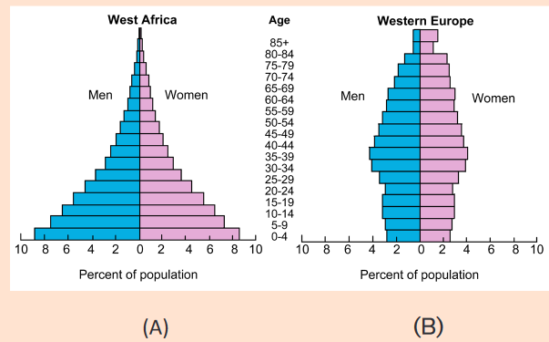
d) Among bolded terms (human population size, pyramid of age
structure, growth pattern, birth rate and death rate), one of them
is better applied to the above figures. Find out the corresponding term
based on the parameters presented on both figure A and B.
e) Based on the shapes of figure A and B. Find out the figure that
corresponds to the description done in above text. Explain how you
have arrived to your choice.
f) Using the school library and additional information from the internet,
Explain bolded terms found in the text at the start of this activity.Populations are dynamic, constantly changing components of ecosystem. They
are commonly described using the following characteristics:1.1.1 Population density
Population density is defined as the numbers of individuals per unit area or per
unit volume of environment. Larger organisms as trees may be expressed as
100 trees per square kilometer. For example, the number of Acacia tree species
per square kilometer in the Akagera National park, whereas smaller ones like
phytoplanktons (as algae) as 1 million cells per cubic meter of water. In terms of
weight it may be 50 kilograms of fish per hectare of water surface.1.1.2 Population age structure
One important demographic variable in present and future growth trends is a
country’s age structure, the relative number of individuals of each age in the
population. The age structure of a population is the distribution of people
of various ages. Age structure is commonly graphed as “pyramids” like those
in figure below.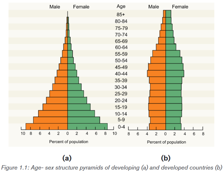
The shapes of the age-sex structure pyramids shown above show the age
sex-structure of a developing and developed country. The main characteristics
of developing countries including some of the African countries in terms of
population growth include high death rate; high birth rate and low life expectancy,
while the main characteristics of developed countries such as most Europeancountries in terms of population growth have low death rate, low birth rate and
longer life expectancy. The age structure of a population affects a nation’s key
socioeconomic issues. For example, countries with young populations (high
percentage under age 15) need to invest more in schools while countries with
older populations (high percentage ages 65 and over) need to invest more in
the health sector.1.1.3 Population explosion
The human population increased relatively slowly until about 1950, at which
time approximately 500 million people inhabited Earth. Our population doubled
to 1 billion within the next two centuries, doubled again to 2 billion between
1850 and 1930, and doubled still again by 1975 to more than 4 billion. The
global population is now more than 6.6 billion people and is increasing by about
75 million each year. Population ecologists predict a population of 7.8-10.8
billion people on Earth by the year 2050.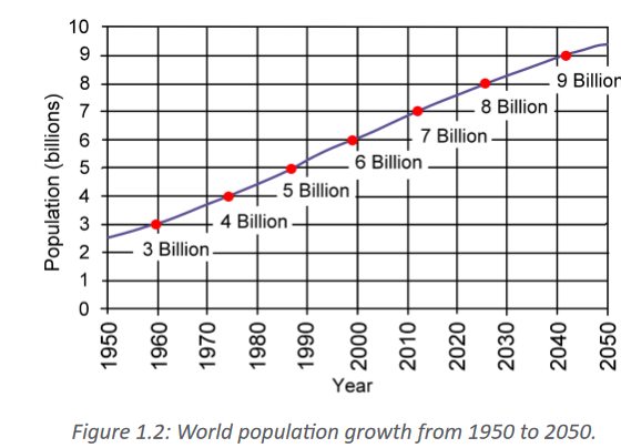
Such human population increase impacts negatively the environment. For
instance, human population explosion contributes to pollution leading to;
ozone depletion, eutrophication, acid rain, global deforestation, soil
erosion and desertification. A population explosion is a sudden increase in
the number of individuals in a particular species. Human population explosions
is sometimes cited as a cause of resource scarcity and a lack of opportunity for
individuals.One of the best way of regulating human population increase in different
countries including Rwanda is practicing the family planning. Family planning
is the practice of controlling the number of children in a family and the intervals
between their births. If married couples are sexually active, they have to adopt
at least one family planning technique such as contraception and timing of
reproduction. Other techniques commonly used include; sexuality education,prevention and management of sexually transmitted infections, pre-conception
counselling and management, and infertility management.1.1.4 Birth and death rates
Birth rate is the ratio of live births in a specified area to the adults in population
of that area. It is usually expressed per one thousand individuals per year. It is
estimated from this calculation:
Death rate is the ratio of deaths to the adults in population of a particular area
during a particular period of time. It is usually calculated as the number of deaths
per one thousand individuals per year and it is estimated from this calculation: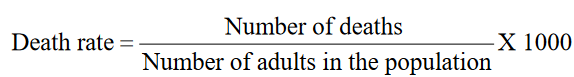
1.1.5 Population growth patterns
Population growth patterns are graphs (population growth curves) in which
increases in size are plotted per unit time. When a population size increases,
the growth rate also increases. The factors that contribute to the population
growth are immigration of new species as well as the birth rate. Population
growth is also influenced negatively by emigration and death rate.Application activity 1.1
1) In a habitat, there are 200 adult lions. Each year, 20 lions are produced
while 5 lions die.
a) Calculate the birth rate of this population.
b) Calculate the death rate of that population
2) A population of 820 insects occupies a surface area of 1.2 km2.
These insects gather nectar from a population of 560 flowering plants
which occupy a surface area of 0.2km2. Which population has greater
density?1.2 Factors affecting population density
Activity 1.2
Observe the figures below and respond to the following questions:
a) Observe the figures above and identify what is taking place in each
figure.
b) Based on what is happening as a result of interaction between organisms
or not, make two Groups from the above figures and find the names that
correspond to those two groups.
c) By use of books or search engine describe how identified factors in (a)
affect the population density.Populations are differently distributed. The distribution and the density are
controlled by environmental factors, which can either increase or decrease the
population size by affecting birthrate, death rate, immigration and emigration.
These factors are grouped into two major categories: Density -dependent
factors and Density- independent factors.1.2.1 Density-dependent factors
Without some type of negative feedback between population density and the
vital rates of birth and death, a population would never stop growing. Density
dependent factors are factors whose effects on the size or growth of the
population vary with the population density. The density dependent factors
include the following: availability of food or resources, predation, disease and
migration.a) Competition for resources
In a crowded population, increasing population density intensifies competition
for declining nutrients and other resources, resulting in a lower birth rate.
Crowding can reduce reproduction by plants and many animal populations also
experience internal competition for food and other resources.
b) Diseases
Population density can also influence the health and thus the survival of
organisms. If the transmission rate of a particular disease depends on a certain
level of crowding in a population, the disease would impact more the population
with high density. Among plants, the severity of infection by fungal pathogens
is often greater in locations where the density of the host plant population is
higher. Animals, too, can experience an increased rate of infection by pathogens
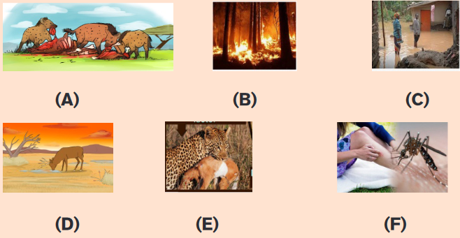
at high population densities.c) Predation
Predation is also an important cause of density-dependent mortality if a predator
encounters and captures more food as the population density of the prey
increases. As a prey population builds up, predators may feed preferentially
on that species, consuming a higher percentage of them which affects directly
population density.1.2.2 Density-independent factors
Density independent factors can affect the population regardless of their density.Most density independent factors are abiotic factors, such as volcanic eruptions,
temperature, storms, floods, draught, chemical pesticides and major habitat
disruption. Even if all population can be affected by these factors, the most
vulnerable appear to be on small organisms with large population such as
insects.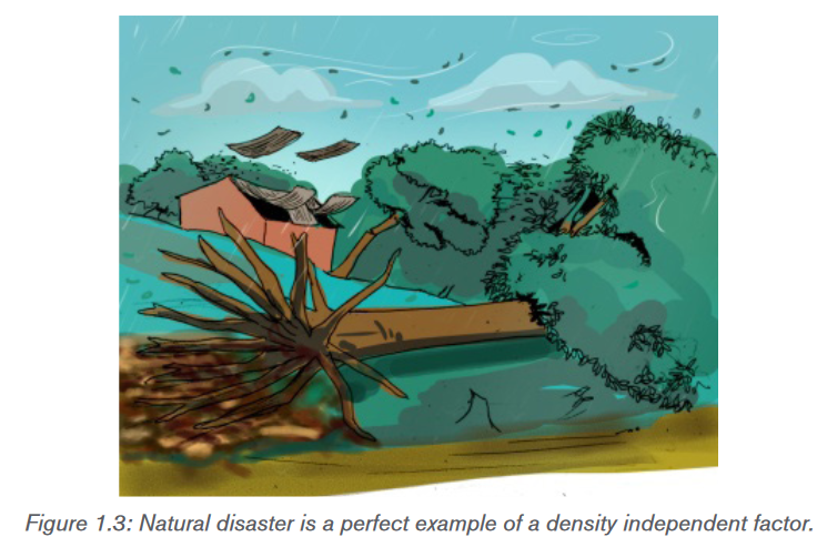
Application activity 1.2
1) A population of field mice increases after a farmer leaves his field
unharvest for a season. Which of the following categories does this
factor fall into? Explain your choice.
a) Density Independent Factors,
b) Density Dependent Factors,
c) Increased death rate
2) Compare the density -dependent and density independent factors. In
your comparison highlight examples of those factors.1.3 Methods or techniques of measuring and estimating
population densityActivity 1.3
Using strings/ropes, a decameter and quadrats in your school garden, carry
out the following field work:
a) Move in the school garden and make a line transect of 15 meters by the
use of a decameter and rope or a string.
b) Count all plants species found at each five meters across transect.
c) On the ground, apply five different quadrats of one square meter
separated by 2.5 meters and count different plants species within each
quadrat. The sketch that show the disposition of quadrats upon 15
meters is as follow: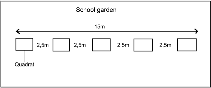
Record your samples in the following table with respect to each quadrat:
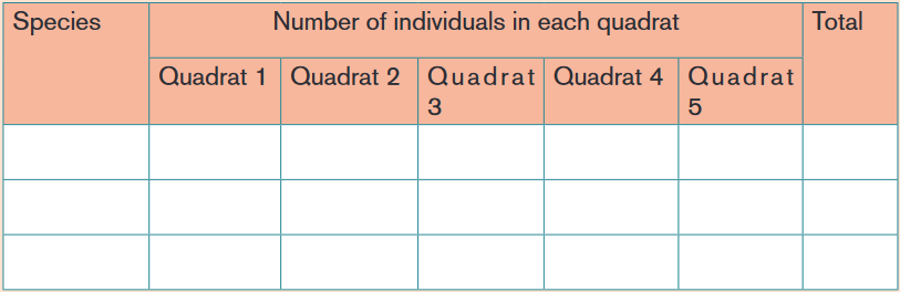
a) Calculate the population density and species frequency for each studied
quadrat.
b) Compare the results of different quadrats.1.3.1 Quadrat method
A quadrat is a square frame that marks off an area of ground, or water, where
you can identify different species present and/or take a measurement of their
abundance. Before any experiment, the decision on a suitable size for the
quadrat and the number of samples to use is taken. Samples must be selected
randomly to avoid any bias, such as choosing to take all of samples from the
place with fewest species simply because it is the easiest to do. This would
not represent the whole area you are surveying. A quadrat method enables the
calculations of 3 aspects of species distribution including; species frequency,
species density and species percentage cover.1.3.2 Species frequency
Species frequency is a measure of the chance (probability) of a particular species
being found within any one of the quadrat, and it is found simply by recording
whether the species was present in each analyzed quadrat. For example, if a
quadrat is placed 40 times, and a given plant was identified in 20 samples, then
the species frequency for this plants equals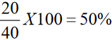
1.3.3 Species density
Species density is a quantity of how many individuals there are per unit area, it
can also be the number of species in a sampled area for example, per square
meter. To achieve this, one takes the total number of counted individuals and
then divide it by the number of quadrats done. An example is:
Total number of individuals= 250
Total area of quadrats =500m2
Species density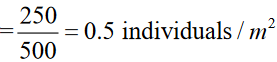
1.3.4 Species cover
Species cover is a measure of the proportion of ground occupied by the species
and gives an estimate of the area covered by the species as the percentage of
the total area. For example, if there are 100 small squares in one quadrat, then
the squares in which the plant species are present are counted. If plants are
found in 25 squares within that quadrat, the conclusion is that the plant covers
25% of the area1.3.5 Line transect method
Line transect is a tape or string laid along the ground in a straight line between
two poles as a guide to a sampling method used to measure the distribution
of organisms. For example, the investigation on change at the edge of a field
where it becomes very marshy is done by randomly selecting a starting point
in the field and lay out a measuring tape in a straight line to the marshy area,
and then sample the organisms that are present along the line, which is called
a transect. The simplest way to do this is to record the identity of the organisms
that touch the line at set distances for example, every two meters.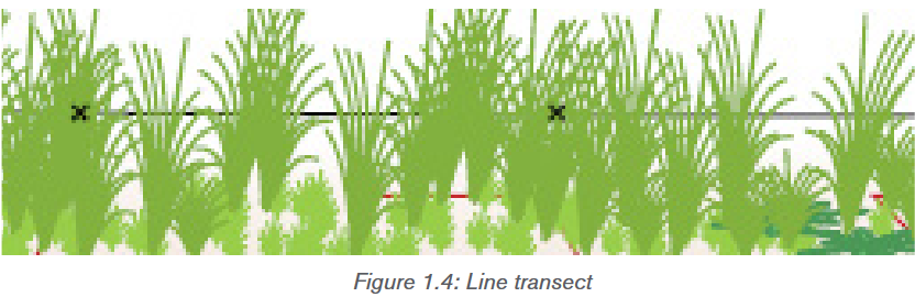
1.3.6 Capture-recapture method
Capture-recapture method involves capturing the organism, marking it without
any harm, and release it in the same area so that it can resume a normal role in
the population. For example, fish can be netted and their opercula is netted with
aluminium discs, birds can be netted and rings can be attached to their legs,
small animals may be tagged by dyes, or by clipping the fur in distinctive pattern,
while arthropods can be marked with paint. In all cases, some form of coding
may be adopted so that individual organisms are identified. Having trapped,
counted and marked are representative sample of the population.At a later stage, the population is trapped again and counted, and the population
size is estimated using the Lincoln index as follows:Estimated total population
=

Where:
N1: the number of organisms in initial sample,
N2: the number of organism in a second sample,
N: the number of marked organisms recaptured.Application activity 1.3
1) Conduct a survey using a quadrat of 0.5m2 and found the following
statistics for a couch grass by quadrat:
a) Calculate the species frequency, and the species density of couch
grass from the results of this survey.
b) Given that the total surface area of the school ground is 200 m2 and
couch grasses were found on 50 m2. Calculate the percentage cover
occupied by couch grasses.A fish farmer wanted to know the total population in her fish pond. She netted
240 fish and tagged (marked) their opercula with aluminium discs. She
released those fish into the pond. After one week, she netted again 250 fish
among which 15 had the aluminium discs. Calculate the estimated population
from marked individuals.1.4 Population growth patterns and Environmental
resistanceActivity 1.4The following graphs are of insect’s growth in separate conditions, study it
and answer the following questions: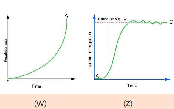 a) What does these graphs represent based on parameters presented
on horizontal and vertical axis?
b) Based on the shape of the graphW from 0 to A and the shape of Z from
A up to C findout their similarities and their differences.
c) Explain how does food supply brings fluctuation which is the result of
the shape B to C on graph Z.1.4.1 Population growth patterns
Population growth patterns are graphs also called population growth curves in
which the increases in size are plotted per unit time. Two types of population
growth patterns may occur depending on specific environmental conditions:a) Exponential growth pattern / J-shaped curve
Exponential growth is a pattern of population growth in which a population
starts out growing slowly but grows faster as population size increases. An
exponential growth pattern also called J shapes curve occurs in an ideal,
and unlimited environmental resources. In such an environment there will be
no competition. Initially population growth is slow as there is a shortage of
reproducing individuals that may be widely dispersed. As population numbers
increase, the rate of growth similarly increases, resulting in an exponential
J-shaped curve. Exponential population growth can be seen in populations that
are very small or in regions that are newly colonized by a species.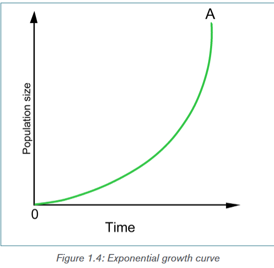 b. Logistic growth pattern / sigmoid growth curve
Logistic growth is a pattern of population growth in which growth slows and
population size levels off as the population approaches the carrying capacity. A
logistic growth pattern also called S-shaped curve occurs when environmental
factors slow the rate of growth.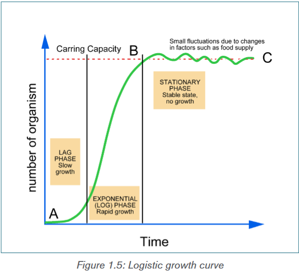 The sigmoid or S- shaped curve represented by the figure 1.8 shows three main
stages in population growth: The lag phase where there is a slow growth, the
log phase or exponential growth phase, also called logarithmic phase,
in which the number of individuals increases at a faster rate and the plateau
phase or stationary phase, in which the number of individuals are stabilized.Causes of the exponential phase are various and include the plentiful of resources
such as; food, space or light, little or no competition from other organisms,
and favourable abiotic factors such as; temperature or oxygen and reduced of
lack of predation or diseases. The stationary phase, however is caused by a
balanced number of; births plus the number of immigrations and the number of
deaths plus the number of emigration. Other causes may include; the increase of
mortality caused by predators and diseases, excess of wastes and competition
for available resources such as food, space, shelter and minerals. Some of these
causes may include the carrying capacity explained as is the maximum number
of individuals that a particular habitat can support.1.4.2 Environmental resistance
Environmental resistance is the total sum of limiting factors, both biotic and
abiotic, which act together to prevent the maximum reproductive potential also
called biotic potential from being realized. It includes external factors such as
predation, food supply, heat, light and space, and internal regulatory mechanisms
such as intraspecific competition and behavioral adaptations.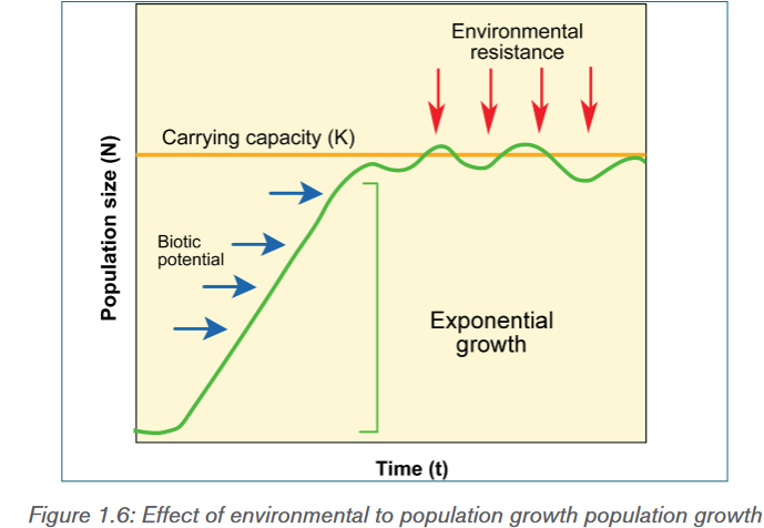 1.4.3 Environmental balance
A balance of nature is the stable state in which natural communities of animals and
plants exist, and are maintained by competition, adaptation and other interactions
between members of the communities and their non-living environment. Every
biotic factor, affects or causes a change in the natural environment. For
example, when a living organism interacts with the environment, this causes a
change in the environment. The following are some of the examples of biotic
factors are include animals, plants, fungi, bacteria, and protists and their effects
on balance of nature can be seen through the following phenomena:
– Respiration: when animals are respiring, they take in oxygen and give
out carbon dioxide (CO2) from respiration. The CO2 can be taken in
by plant leaves and be used in the process of photosynthesis to make
food and give out oxygen.
– Predation: when animals, for example, predate on other animals, this
reduces the numbers of prey, which in turn affects the ecosystem.
– Parasitism: cause diseases that may slow down the growth rate of a
population and/or reduces the number of organisms.
– Competition: when organisms compete over nutritional resources,
this could reduce the growth of a populationApplication activity 1.4A small group of mice invaded a new habitat with unlimited resources and
their population grew rapidly. A flood then swept through the habitat and three
quarters of the mice died. Two months later, the population was increasing
again.
a) What role did the flood play for the mouse population?
b) Draw a graph depicting the population history of this mouse.1.5 Renewable natural resourcesActivity 1.5Observe the figures below carefully and respond to the following
questions: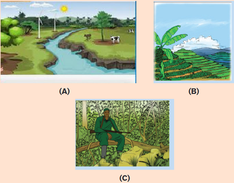 Natural resources refer to materials or substances occurring in environment
and which can be exploited for economic gain. They are also resources that
exist without actions of humankind Natural resources such as; solar energy,wind, air, water, soil and biomass (plants and animals) are renewable natural
resources. Below are the examples of renewable natural resource: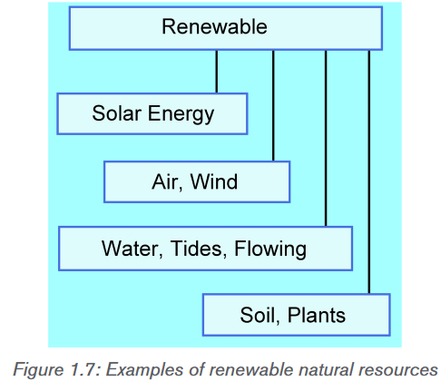 1.5.1 Importance of renewable natural resources in
economic growth of Rwanda
• Water is used for irrigation, domestic activities, industrial use, and mining.
• Lakes and rivers are source of food (fish) for humans and contribute for
recreation (tourism).
• Land serves as the storehouse of water, minerals, livestock, and home for
wild animals which generate an income in different ways.
• Soil contributes to agricultural crop production, and supports forest and
food crops.
• Trees are the major sources of timber, construction materials and firewood
and contribute to fight against erosion, water and air purification and wind
protection.
• Some plants are source of food and money for humans and other animals.
• National Parks contribute to economic development of the country through
tourism.1.5.2 Methods of conserving renewable natural resources
They are various methods used for conservation of renewable natural resources
and they include:• Planting trees to prevent soil erosion. The vegetation prevents soil erosion
but also is a home for most insects, birds and some symbiotic plants. This
creates a habitat for wildlife hence conserving wild organisms• Practicing of judicious ways to conserve water in our homes: This
entails simple practices like ensuring that taps are closed when they are not
in use. Using less water during domestic activities aids to conserve lots of
water in our homes.• Growing vegetation in catchment areas: Catchment areas act as a
source of water that flows in; streams, rivers and oceans. Vegetation in the
catchment areas allows sufficient infiltration of water into deeper soil layers
thus leading to formation of ground water• Prior treatment of human sewage and Industrial wastes: Water flowing
from industries comes with many toxic wastes that must be treated before
getting to the natural water bodies. This reduces harm inform of pollutants
e.g. chemical and thermal forms.• Practice of in-situ and ex-situ conservation of wild plants which
involves conservation of flora in their natural habitats and outside the natural
habitats respectively. This requires setting up measures that protect areas
such as national parks and game reserves. The ex-situ conservation of plants
uses the areas such as; pollen banks, DNA banks, seed banks, botanical
gardens, tissue culture banks among others.• Ensure the recycling of wastes: These wastes include; plastics, paper bags
that have resulted to tones of garbage. Recycling entails re-manufacturing of
already used materials. This reduces the amount of waste available reducing
soil and water pollution.• Practice crop rotation: Planting the same crops for a long period of time
reduces soil fertility. The practice of crop rotation will restore and maintain
soil fertility thus conserving the soil.• Construction of terraces in sloping land: This will prevent soil erosion as
water tends to run downhill.Application activity 1.51. You live in place which is dominated by sloping lands and bare soil then
your parents complain about their soil that is washed away by the rainfall.
What can you do to help your parent to prevent that sloping land?2. The water bill at your home is always high and you are given a responsibility
to reduce it as some who attended secondary school. Implement the measures
that will reduce that water bill at your home.1.6 Non-renewable natural resourcesActivity 1.6Observe the figures and respond to the following questions: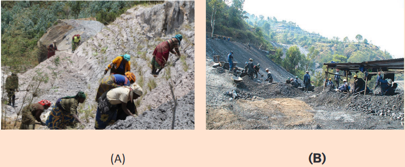 a) Based on the on figures above identify the activities that are taking place
on both A and B.
b) Identify the effects of activity taking place on figure B.
c) Find out the purpose of activity taking place on figure A.Non-renewable natural resource are resources of economic value that cannot
be readily replaced by natural means on a level equal to its consumption.
They include fossil fuels, oil, coal natural gas cited among many others as it is
indicated below: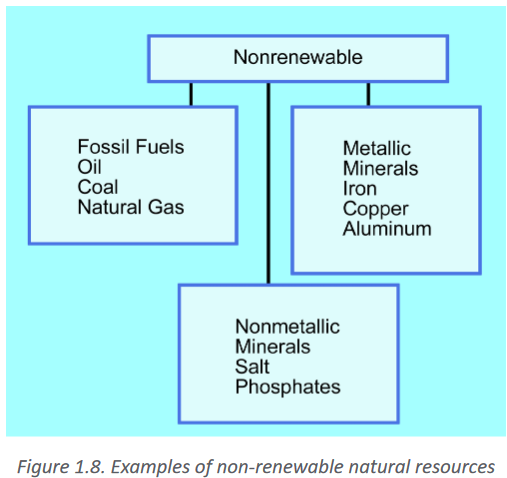 1.6.1 Importance of nonrenewable natural resources in
economic growth of Rwanda• Minerals including gravel, metals, sand, and stones are used for construction
and for income generation for the country.
• Imported fossil fuels derivatives such as gas oil and asphalts are used
as source of energy and construction of asphaltic roads to easy the
transportation.
• Natural gas e.g. gas methane from Kivu is used as source energy.
• Some animals including; mountain gorillas in Volcanoes National Park,
lions in Akagera National Park and many other wild animals contribute to
economic development of the country through tourism.1.6.2 Methods of conserving nonrenewable natural
resourcesThere are various a methods used for conservation of nonrenewable natural
resources and they include:
• The use of alternative sources of energy such as solar and wind energy because
they do not produce harmful gases that damage the ozone layer compared to
the burning of fossils fuels such as coal and charcoal.
• Use pipelines to transport oil: During oil transportation on ships, spills can
happen which will negatively affect both plant and animal life. Therefore, use of
pipelines is more recommended
• Putting in place of policies and regulations to prevent poaching
because poachers continue to kill many animals such as; elephants and rhinos,
for their tusks and skins which are sold off in the black market. Poachers are a
major threat to our biodiversity as they are slowly making some species extinct.
• Use of bio-fuels and biogas: For more than a century, fossil fuels have
been a major source of energy. However, they are depleting rapidly, this calls
for alternative sources of fuel such as bio-fuels and biogas which mainly reduce
the occurrence of air pollution.
• Establish special schemes to preserve endangered plant and animal
species: This includes; botanical gardens, sanctuaries that may be established
to protect the endangered species so that they can be available for future
generationApplication activity 1.61) Different industries are making cars and motorcycles that use electricity
instead of fuel. What is the contribution of that method compared to
the one that uses fuel?2) Why does mining companies that extract minerals legally, are obliged
to restore the mining site after the completion of extraction of minerals
at that mining company.Skills lab 1Biogas is a type of biofuel that is naturally produced from the decomposition
of organic waste. When organic matter, such as food scraps and animal
waste, break down in an anaerobic environment (an environment absent of
oxygen) they release a blend of gases, primarily methane and carbon dioxide.
People are encouraged to use biogas in their home as alternative source
of energy. This can reduce the rate of deforestation which can result in
maintenance of plant and animal species as well as soil protection against
erosion. Sensitization can be a tool to help people to have these alternative
sources of energy in large number.Procedure
– Select 10 families at your village.
– Record the families that have biogas.
– Select other ten families which use woods in cooking.
– Record the money spent by each family while cooking either using
biogas or woods
– Compare the money spent by each family
– Prepare action of sensitizing people on using biogas based on
recorded data.Evaluation sheet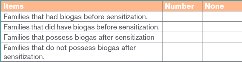 End unit assessment 1I. Choose the letter of the answer that best complete each statement
1. During population growth
a) Birth rate increases
b) Death rate increases
c) Birth rate decreases.
d) Birth rate and death rate decreases.2. Population that reaches the carrying capacity of its environment is
said to have reached
a) logistic growth
b) exponential growth
c) density dependence
d) a steady state3. On a logistic growth curve, the portion of the curve in which the
population grows rapidly is called
a) logistic growth
b) a steady state
c) exponential growth
d) carrying capacity4. Which of the following is a characteristic of developing countries?
a) A fast population growth due to a high death rate but higher birth rate.
b) A fast population growth due to a high birth rate but falling death rate.
c) A slow population growth due to a low birth rate and falling death
rate.
d) A slow population growth due to a low birth rate and low death rate.5. Which of the following would be an example of population density?
a) 100 caterpillars
b) 100 caterpillars per mango tree
c) 100 caterpillars clumped into 5 specific areasII. Open questions
1. How can a density dependent factor, such as a food supply affect the
carrying capacity of a habitat?
2. Describe how density dependent and density dependent factors regulate
population growth.
3. Suggest the reasons why the luck of available clean water could be a
limiting factor for a country’s population.
4. a) Distinguish between carrying capacity and biotic potential.
b) Explain how environmental resistance affects the population growth.
5. Students made a survey of blackjack (Bidens pilosa) growing their school
environment. Ten quadrats of 1.0 m2 were placed randomly in the garden
and the number of blackjack plants in each quadrat was counted.
The results are summarized in the following table: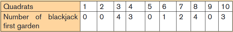 Calculate: a. the species frequency of blackjack in this gardens.
b. The species density of blackjack plants in that area.
c. Explain why it is important to use randomly placed quadrats.6. Describe how has the growth of Earth’s human population has changed
in 2 recent centuries? Give your answer in terms of growth rate and the
number of people added each year?7. Construct a bar graph showing the age structure of a given country
using the following data: Pre-reproductive years (0-14) are 42 percent;
reproductive years (15-44) are 39 percent; post-reproductive years (45-
85+) are 19 percent. Interpret obtained graph.8. Explain the relationship between a growing population and the environment9. Observe the pictures below and respond to the following questions.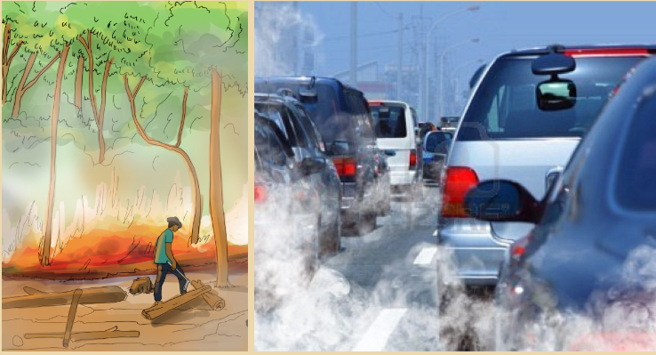 a) Identify the human activities shown above that harm the natural
resources.
b) Describe all effects of the identified activities on the environment.
c) Suggest the possible measures to solve the above problems.UNIT 2: ENERGY AND CELLULAR RESPIRATION
Key unit competence
Describe the structure and importance of ATP, and outline the roles of the
coenzymes NAD, FAD and coenzyme A during cellular respiration and the
process of cellular respiration.Introductory activity 2.1
Living organisms perform different tasks like running, moving and pumping
substances across cell membranes as shown on the figures below: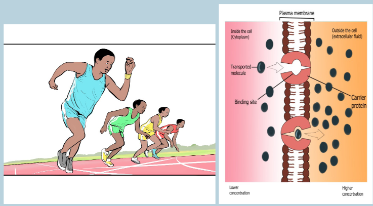
a) What is the requirement to perform such activities and others that seem
like these?
b) By which mechanism do you think is taking place in organism cells to
obtain such requirement? In which form this requirement would appear?2.1 Energy of living organisms
Activity 2.1
Observe the figures below and answer to the following questions

a) The figures A represents the activity that requires energy, based on
figure A above identify other more activities that requires energy.
b) What could be the name of figure B, its main chemical parts and its
roles for living organisms?2.1.1 Need for energy by organisms
Without some input of energy, natural processes tend to break down in
randomness and disorder. Living organisms have high ordered systems that
require a constant input of energy to prevent them becoming disordered which
would lead to their death. This energy comes from the breakdown of organic
molecules to make adenosine triphosphate (ATP) which is a source of energy
needed to carry out processes that are essential to life.More precisely energy is needed for:
• Metabolism which involves specifically the anabolism process in which
simple substances are build up into complex ones e.g. monosaccharides
are built up into polysaccharides and amino acids are built up into
polypeptides• Active transport of ions and different molecules against a concentration
gradient across cell membranes. The transport of sodium (Na+), potassium
(K+) magnesium (Mg+), calcium (Ca+) and chloride (Cl-) across the plasma
membrane cannot be possible without the use of energy. The transport
proteins that move solutes against their concentration gradients are all
carrier proteins rather than channel proteins. Active transport enables a
cell to maintain internal concentrations of small solutes that differ from
concentrations in its environment. Some transport proteins act as pumps,moving substances across a membrane against their concentration
or electrochemical gradients. Energy is usually supplied by adenosine
triphosphate (ATP) hydrolysis.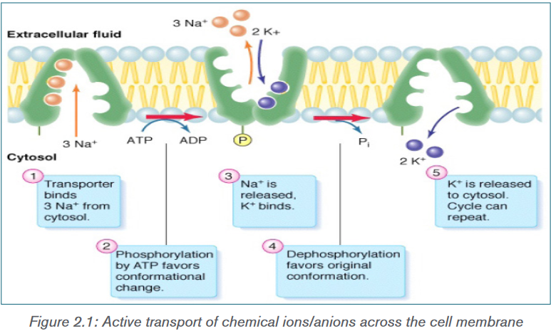
• Movement within an organism when substances move in the body e.g.
circulation of blood and of the orgasm it’s self during locomotion due to
muscular contraction or movement of cilia and flagella.
• Maintenance, repair and division of cell and organelles within
them.
• Maintenance of body temperature in endothermic organisms e.g.
birds and mammals that need energy to replace that lost as heat to the
surrounding environment.
• Production of substances used within organism e.g. enzymes and
hormones.2.1.2 Structure of adenosine triphosphate (ATP)
The special carrier of energy is the molecule of adenosine triphosphate (ATP).
The ATP molecule is a phosphorylated nucleotide and it has three parts:
• Adenine: is a nitrogen containing organic base belongs to the group
called purines
• Ribose: is a pentose sugar molecule means it has 5-carbon ring structure
that act as the backbone where the other parts are attached.
• Phosphates: that are chain of three phosphate groups.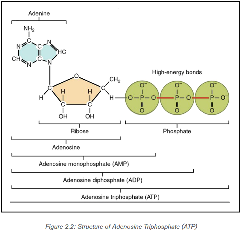
ATP has the following biological functions in the cell:
a) Active transport
ATP plays a critical role in the transport of macromolecules such as proteins
and lipids into and out of the cell membrane. It provides the required energy for
active transport mechanisms to carry such molecules against a concentration
gradient.b) Cell signaling
ATP has key functions of both intracellular and extracellular signaling. In nervous
system, adenosine triphosphate modulates the neural development, the control
of immune systems, and of neuron signaling.c) Structural maintenance
ATP plays a very important role in preserving the structure of the cell by helping
the assembly of the cytoskeletal elements. It also supplies energy to the flagella
and chromosomes to maintain their appropriate functioning.d) Muscle contraction
ATP is critical for the contraction of muscles. It binds to myosin to provide
energy and facilitate its binding to actin to form a cross-bridge. Adenosine
diphosphate (ADP) and phosphate group (Pi) are then released and a new ATP
molecule binds to myosin. This breaks the cross-bridge between myosin and
actin filaments, thereby releasing myosin for the next contraction.e) Synthesis of DNA and RNA
The adenosine from ATP is a building block of RNA and is directly added to
RNA molecules during RNA synthesis by RNA polymerases. The removal
of pyrophosphate provides the energy required for this reaction. It is also a
component of DNA.Application activity 2.1
1) Energy is contained within ATP, draw and label its structure. On
diagram show the names that result from the combination of different
parts of ATP.
2) The person faints on playground as a result of doing vigorous physical
exercise for long time. What can you do to save the life of that person?2.2 Adenosine triphosphate (ATP) and coenzyme in
respirationActivity 2.2
Based on the structure of ATP molecule, explain how the synthesis and
breakdown of ATP is done.2.2.1 Synthesis and breakdown of ATP
a) Breakdown of ATP
Adenosine triphosphate (ATP) is the energy currency for cellular processes. It
provides the energy for both energy-consuming endergonic reactions and
energy-releasing exergonic reactions. The three phosphate groups in ATP
structure are the main key to how ATP stores energy. Each phosphate group
is very negatively charged so they repel one another which makes the covalent
bonds that link to be unstable. These unstable covalent bonds are broken easily
because they have low activation energy. When the first two phosphates are
removed 30.5Kjmol-1 are released for each phosphate group and 14.2 KJ mol-1
are released for the removal of the final phosphate group. In living cells, usually
only the terminal phosphate group is removed as follow: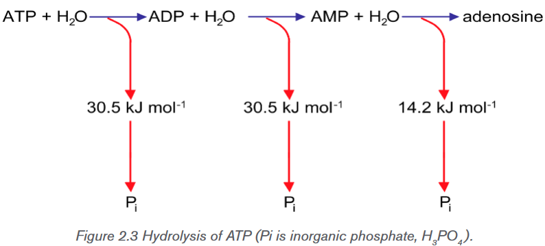
These reactions are all reversible. It is the interconversion of ATP and ADP that
is all-important in providing energy for the cell:
The calculated ∆G for the hydrolysis of one mole of ATP into ADP and Pi is
estimated at −7.3 kcal/mole equivalent to −30.5 kJ/mol. However, this is only
true under standard conditions, and the ∆G for the hydrolysis of one mole of
ATP in a living cell is almost double the value at standard conditions and equals
-14 kcal/mol or −57 kJ/mol. ATP is a highly unstable molecule. Unless quickly
used to perform work, ATP spontaneously dissociates into ADP + Pi, and the
free energy released during this process is lost as heat. To harness the energy
within the bounds of ATP, cells use a strategy called energy coupling.The hydrolysis of ATP to ADP and Pi is a reversible reaction, where the reverse
reaction combines ADP + Pi to regenerate ATP from ADP as it is shown in the
equation above.b) Synthesis of ATP
Energy for ATP synthesis can become available in two ways. In respiration, energy
released by reorganizing chemical bonds (chemical potential energy) during
making some ATP. However, most ATP in cells is generated using electrical
potential energy. This energy is from the transfer of electrons by electron carriers
in mitochondria and chloroplasts. It is stored as a difference in proton (hydrogen
ion) concentration across some phospholipid membranes in mitochondria and
chloroplasts, which are essentially impermeable to protons. Protons are then
allowed to flow down their concentration gradient (by facilitated diffusion)
through a protein that spans the phospholipid bilayer. Part of this protein acts
as an enzyme that synthesizes ATP and is called ATP synthase. The transfer
of three protons allows the production of one ATP molecule, provided that ADP
and an inorganic phosphate group (Pi) are available inside the organelle. This
process occurs in both mitochondria and chloroplasts and it was first proposed
by Peter Mitchell in 1961 and is called chemiosmosis.Since the hydrolysis of ATP releases energy, ATP synthesis must require an
input of free energy. Recall that free energy is the portion of system’s energy
that can perform work when temperature and pressure are uniform throughout
the system. The synthesis of ATP from ADP involves the addition of a phosphate
molecule, which is called phosphorylation reaction. This Phosphorylation is
catalyzed by the enzyme ATP synthase (sometimes called ATP synthetase or
ATPase).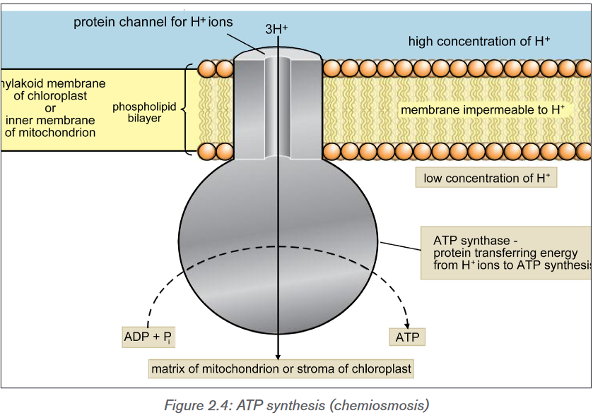
2.2.2 Roles of coenzymes in respiration
The transformation of succinate to fumarate, the sub-products of the breakdown
of glucose during glycolysis process, two hydrogens are transferred to flavin
adenine dinucleotide (FAD), forming FADH2. The reduced coenzymes NADH
and FADH2 transfer higher energy electrons to the electron transport chain.
Finally, another coenzyme called coenzyme A sometimes abbreviated by CoA,
a sulfur-containing compound is attached via its sulfur atom to the two-carbon
intermediate, forming acetyl CoA. The Acetyl CoA has a high potential energy,
which is used to transfer the acetyl group to a molecule in the citric acid cycle
(Krebs cycle), a reaction that is therefore highly exergonic producing great
number of energy in the form of ATP.Application activity 2.2
Application activity 2.2
1) Using the chemical equations explain the synthesis and the hydrolysis
of ATP in a living cell.
2) The hydrolysis and synthesis of ATP are reversible reactions. Estimate
the amount of energy for each process.
3) Calculate the amount of energy produced by 5 moles of ATP
a) Under standard conditions
b) In a living cell2.3 Respiratory substrates and their relative energy values
Activity 2.3
Activity 2.3: Simple combustion experiments to determine the relative energy
values of different food substances.
– Cut up a range of dried foods into small pieces around 1 cm square
or 0.5 cm cubed.
– Use the measuring cylinder to measure 20 cm3 of water into the
boiling tube.
– Clamp the boiling tube to the clamp stand.
– Measure the temperature of the water with the thermometer. Record
the temperature in a suitable table.
– Impale the piece of food carefully on a mounted needle.– Light the Bunsen burner and hold the food in the flame until it
catches a light.
– As soon as the food is alight, put it under the boiling tube of water as
shown on figure and keep the flame under the tube.
– Hold the food in place until the food has burnt completely.
– As soon as the food has burned away completely and the flame
has gone out, stir the water carefully with the thermometer and
measure the temperature of the water again. Note down the highest
temperature reached.
– Repeat the procedure for other foods.
– Calculate the rise in temperature each time and Calculate the energy
released from each food by using this formula.
Where 4.2 represents the value of the specific heat capacity of water, in
joules per gram per degree Celsius. If the number is more than 1000 J/g,
express it as kilojoules (kJ):
1 kilojoule = 1000 joules
Compare obtained results.
Follow the set up below:
A respiratory substrate refers to the substance required for cellular respiration
to derive energy through oxidation. They include carbohydrates, lipids and
proteins.Carbohydrates include any of the group of organic compounds consisting
of carbon, hydrogen and oxygen, usually in the ratio 1:2:1. The examples of
carbohydrates include sugars, starch and cellulose. Carbohydrates are the
most abundant of all classes of biomolecules, and glucose whose chemicalformula is C6H12O6 is the most known and the most abundant. Its breakdown
produces energy in the following way: C6H12O6 +6 O2→6 CO2 +6 H2O+Energy
(ATP + heat).This breakdown is exergonic metabolic reaction, having a free-energy change of
-686 kcal (-2,870 kJ) per mole of glucose decomposed.Lipids include diverse group of compounds which are insoluble in water but
dissolved readily in other lipids and in organic solvents such as ethanol (alcohol).
Lipids mainly fats and oils contain carbon, hydrogen and oxygen, though the
proportion of oxygen is lower than in carbohydrates. Fats and oils have a higher
proportion of hydrogen than either carbohydrates or proteins. This property
makes them a more concentrated source of energy, where each gram of fat or
oil yields about 38kJ (38 kJ/g) more than twice the energy yield of a gram of
carbohydrate.Proteins are other respiratory substrate. They are large and complex biological
molecules which play many and diverse roles during respiration. They mainly
work as enzymes. Enzyme is a biological catalyst that controls biochemical
reactions in living organisms.
Back to glucose when it is broken down during the process called glycolysis,
the dehydrogenases enzymes transfer electrons from substrates, here glucose,
to NAD+ which in turn forms NADH. At this stage the electron transport chain
accepts electrons from NADH and passes these electrons from one molecule
to another in electron chain transfer leading to a controlled release of energy
for the synthesis of ATP. At the end of the chain, the electrons are combined
with molecular oxygen and hydrogen ions (H+) to form one molecule of water.
When NAD is oxidized, its oxidized form NAD+ is converted into its reduced
from NADH, and two molecules of ATP are produced.Application activity 2.2
1) Calculate the amount of energy produced by 5moles of glucose in kcal
and kJ if one mole of glucose produce -686 kcal and 2,870 kJ per mole
of glucose.
2) Specify the number of ATP produced by glycolysis during respiration
process.2.4 Measurement of respiration and respiratory quotients
Activity 2.3
– Set up the boiling tube so it is vertical and supported in a water bath
such as a beaker.
– Use pea seeds that have been soaked for 24 hours and rinsed in 1%
formaldehyde for 5 minutes.
– Kill an equal quantity of soaked seeds by boiling them for 5 minutes.
– Cool the boiled seeds in cold tap water; rinse them in bleach or
formaldehyde for 5 minutes as before.
– Start with a water bath at about 20 °C and allow the seeds to adapt
to that temperature for a few minutes before taking any readings.
– Record the initial and final positions of the water drop with a
permanent marker with small label onto the glass.
– Measure the distance travelled by colored dye (or drop of water) with
a ruler.
– Repeat the procedure (introducing a new bubble each time) at a
range of different temperatures, remembering to allow time for the
seeds to adapt to the new conditions before taking further readings.
– Interpret your observation. Follow the set up below: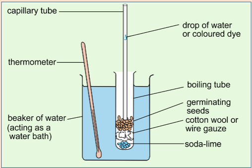
The rate of respiration is measured by the use of respirometer device, typically
by measuring oxygen consumed and the carbon dioxide given out. It can also
be used to measure the depth and frequency of breathing, and allows the
investigation on how factors such as; age, or chemicals can affect the rate of
respiration. Currently, the computer technology is also used to automatically
measure the volume of gases exchanged and drawing off small samples to
analyze the proportions of oxygen and carbon dioxide in the gases.The respiratory quotient (RQ) is the ratio of the volume of carbon dioxide
produced to the volume of oxygen used in respiration during the same period
of time. The RQ is often assumed to equal the ratio of carbon dioxide expired:
oxygen inspired during a given time as it is summarized in the following formula:
The RQ is important as it can indicate whether the respiration is aerobic or
anaerobic.
As each molecule of gas occupies the same volume, this would give RQ =
1.0, and this is common for all carbohydrates. Further studies indicated the
respiratory quotient to be 0.9 for proteins and 0.7 for fats, and concluded that
an, RQ greater than 1.0 indicates anaerobic respiration, while RQ equals or less
than 1.0 indicates aerobic respiration.Note that respiration during germination, especially in early stages was also
studied. Results indicated that it is difficult for oxygen to penetrate the seed
coat, so that at this stage, the RQ is about 3 to 4. Later when the seed coat is
shed, it becomes easier for oxygen to reach respiration tissues and the levels of
RQ falls. Results indicated that eventually seeds with large carbohydrate stores
have an RQ around 1.0 and those with large lipid stores have RQs of 0.7 to 0.8.a. Measuring and obtaining the RQ values in invertebrate (e.g. woodlice)
In this particular respirometer, woodlice have been placed in a boiling tube
which is connected to a U-tube. The U-tube acts as a manometer (a device for
measuring pressure changes). The other end of the U-tube is connected to a
control tube which is treated in exactly the same way as the first tube, except
that it has no woodlice but instead glass beads which take up the same volume
as the woodlice. The two boiling tubes (but not the manometer) are kept in
water bath at constant temperature. The U-tube contains a colored liquid which
moves according to the pressure exerted on it by the gases in the two boiling
tubes. Both tubes contain potassium hydroxide solution which absorbs any
carbon dioxide produced. The setup is summarized below: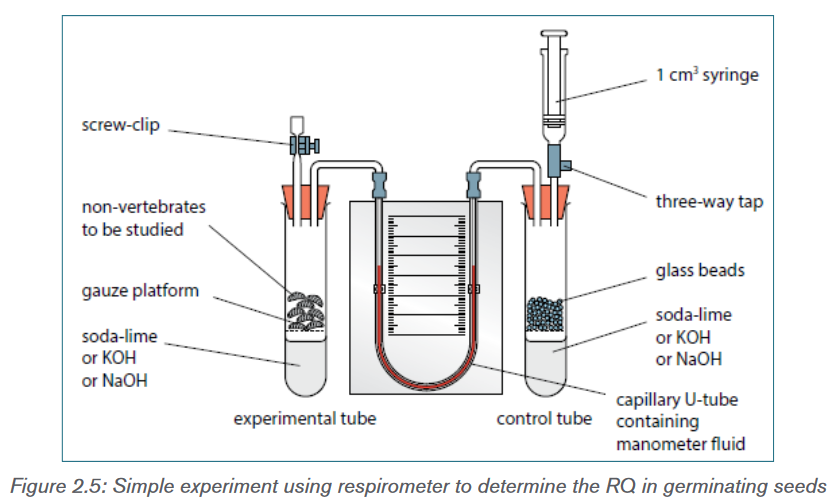
When the woodlice respire aerobically, they consume oxygen, which causes
the liquid to move in the U- tube in the direction of arrows. The rate of oxygen
consumption can be estimated by timing how long it takes for the liquid to
rise through a certain height. The experiment can be repeated by replacing the
potassium hydroxide solution with water. Comparing the changes in manometer
liquid level with and without potassium hydroxide solution gives an estimate of
carbon dioxide production can be used to measure the respiratory quotient.If the internal radius of the manometer tube is known, the volumes of gases can
be calculated using the equation:Volume of gases = π r2 h,
Where π is equal to 3.14, r is the internal radius of the tube and h is the
distance moved by the liquid.b. Measuring and obtaining the RQ values during seed germination
processDuring seed germination, CO2 is released. To test its presence, chemicals
including Sodium hydroxide or Potassium hydroxide are used due to their ability
to absorb CO2. As the germinating seeds use oxygen, pressure reduces in tube
A so the manometer level nearest to the seeds rises (figure 2.8). The syringe is
used to return the manometer fluid levels to normal. The volume of oxygen used
is calculated by measuring the volume of gas needed from the syringe to return
the levels to the original values. If water replaces the sodium hydroxide, then the
carbon dioxide evolved can be measured. The setup is summarized below: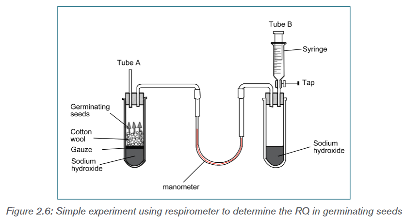
This graph suggests that the seed begins with carbohydrate as a metabolite,
changes to fat/oil then returns to mainly using carbohydrate.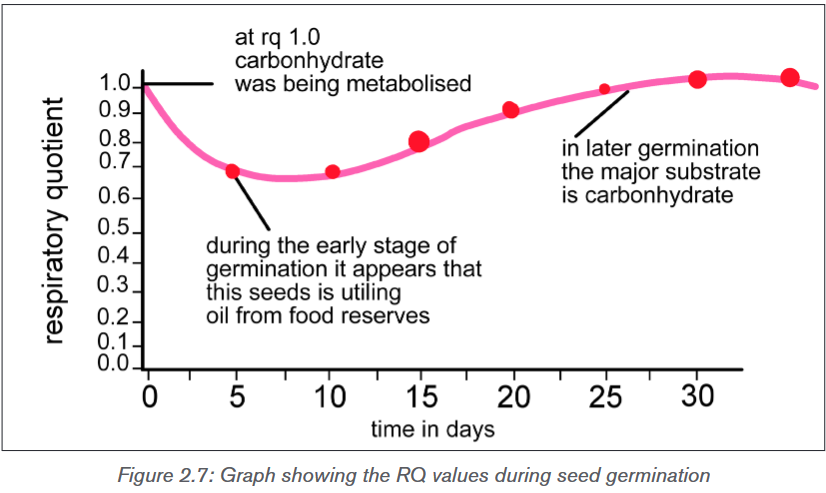
Application activity 2.4
1) Using the following equation of oleic acid (a fatty acid found in olive
oil):
a) Calculate the RQ for the complete aerobic respiration.
b) Based on your findings, state which substrate is being respired
2) Measurements of oxygen uptake and carbon dioxide production by
germinating seeds in a respirometer showed that 25 cm3 of oxygen
was used and 17.5 cm3 of carbon dioxide was produced over the
same time period.
i) Calculate the RQ for these seeds.
ii) Identify the respiratory substrate used by the seeds.2.5 Aerobic respiration and Glycolysis
Activity 2.5
Glycolysis process
Observe the figure below and do the following activities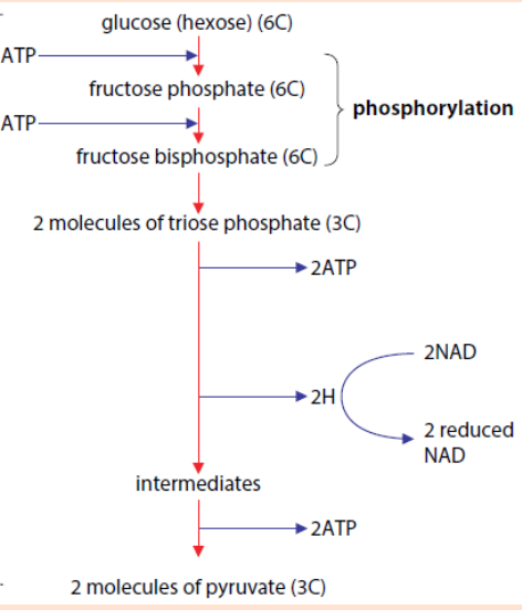
a) If this representation on figure above (→ATP) shows energy used and
this (ATP→) represent energy produced during this process. Identify the
energy used and energy produced then calculate net energy produced
during this process.
b) According to your observation, what are the end products of this
process above?Cellular respiration is the complex process in which cells make adenosine
triphosphate (ATP) by breaking down organic molecules. The energy stored
in ATP can then be used to drive processes requiring energy, including
biosynthesis, locomotion or transportation of molecules across cell membranes.
The main fuel for most cells is carbohydrate, usually glucose which is used by
most of the cells as respiratory substrate. Some other cells are able to break
down fatty acids, glycerol and amino acids.Glucose breakdown can be divided into four stages: glycolysis, the link
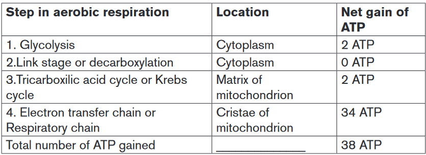
reaction, the Krebs cycle and oxidative phosphorylation.Glycolysis is the splitting or lysis of a glucose molecule. It is a multi-step
process in which a glucose molecule with six carbon atoms is eventually split
into two molecules of pyruvate, each with three carbon atoms. Energy from ATP
is needed in the first steps, and it is released in the later steps to synthesize ATP.
There is a net gain of two ATP molecules per molecule of glucose broken down.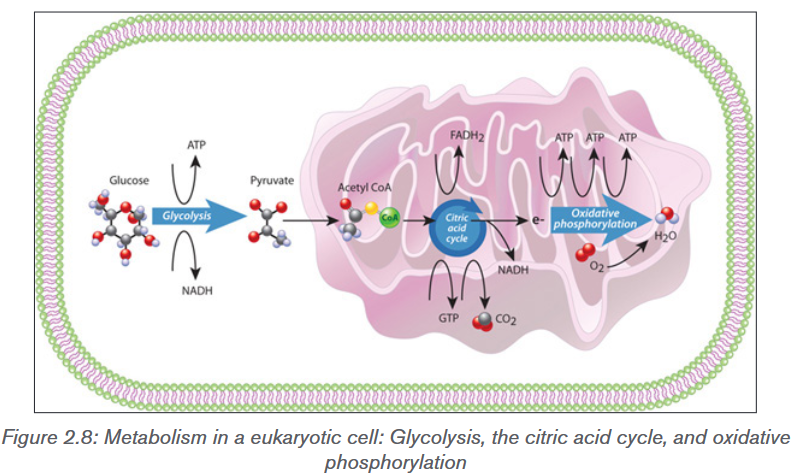
Glycolysis takes place in the cytoplasm of a cell. Within the mitochondrion, the
citric acid cycle occurs in the mitochondrial matrix, and oxidative metabolism
occurs at the internal folded mitochondrial membranes (cristae). Glucose entersthe cell and is phosphorylated by the enzyme called hexokinase, which transfers
a phosphate group from ATP to the sugar. The ATP used in this process has
2 advantages: the charge of the phosphate group traps the sugar in the cell
because the plasma membrane is impermeable to large ions. Phosphorylation
also makes glucose more chemically reactive. Even though glycolysis consumes
two ATP molecules, it produces a gross of four ATP molecules (4 ATP), and
a net gain of two ATP (2 ATP) molecules for each glucose molecule that is
oxidized. Glycolysis results in a net gain of two ATP (2ATP), two NADH and two
pyruvate molecules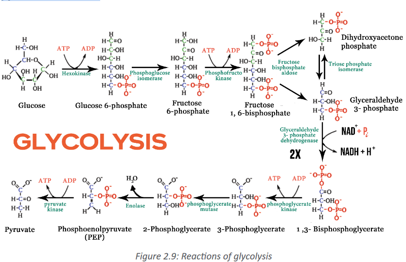
Application activity 2.5
1) Why is ATP needed for glycolysis?
2) How many gross ATP molecules are produced during glycolysis from
one glucose molecule?
3) How many NADH are made during glycolysis?
4) The following flowchart summarizes the reactions that take place in
glycolysis
Glucose → 2 × glyceraldehydes 3-phoshate → 2 × pyruvate
a) How many carbon atoms are there in glucose, glyceraldehydes
3-phoshate and pyruvate?
b) What is the net gain of ATP in glycolysis?2.6 Link reaction and Krebs cycle (TCA cycle)
Activity 2.6
Use the figure below and do the following activities:
a) The above figure summarizes two stages that take place during
respiration, observe it and identify the number of CO2, ATP, reduced
FAD and reduced NAD.
b) Knowing that the above stages involve two molecule of pyruvates
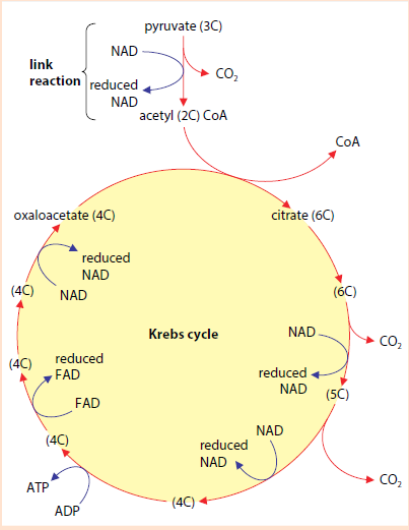
calculate the total number of CO2, ATP, reduced FAD and reduced NAD.2.6.1 Link reaction
Pyruvate, the end product of glycolysis is oxidized to Acetyl-CoA by enzymes
located in the mitochondrion of eukaryotic cells as well as in the cytoplasm
of prokaryotes. In the conversion of pyruvate to Acetyl-CoA, one molecule of
NADH and one molecule of CO2 are formed (Figure 2.10). This step is also
known as the link reaction or transition step, as it links glycolysis to the Krebs
cycle.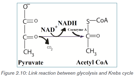
Krebs cycle (Citric acid cycle)
The coenzyme has a sulphur atom, which attaches the acetyl fragment by an
unstable bond. This activates the acetyl group for the first reaction of the Krebs
cycle also called citric acid cycle or Tricarboxylic Acid Cycle (TCA). It is also
known as the citric acid cycle, because the first molecule formed when an acetyl
group joins the cycle. When oxygen is present, the mitochondria will undergo
aerobic respiration which leads to the Krebs cycle.In the presence of oxygen, when acetyl-CoA is produced, the molecule then
enters the citric acid cycle inside the mitochondrial matrix, and gets oxidized
to CO2 while at the same time reducing NAD+ to NADH. NADH can then be
used by the electron transport chain to create more ATP as part of oxidative
phosphorylation. For the complete oxidation of one glucose molecule, two
Acetyl-CoA must be metabolized by the Krebs cycle. Two waste products
namely H2O and CO2, are released during this cycle.The citric acid cycle is an 8-step process involving different enzymes and co-
enzymes. Throughout the entire cycle, Acetyl-CoA (2 carbons) combines with
oxaloacetate (4 carbons) to produce citrate. Citrate (6 carbons) is rearranged
to a more reactive form called isocitrate (6 carbons). Isocitrate (6 carbons) is
modified to α-Ketoglutarate (5 carbons), Succinyl-CoA, Succinate, Fumarate,
Malate, and finally to Oxaloacetate. The net energy gain from one cycle is 3 NADH,
1 FADH2, and 1 Guanosine Triphosphate (GTP). The GTP may subsequently
be used to produce ATP. Thus, the total energy yield from one whole glucose
molecule (2 pyruvate molecules) is 6 NADH, 2 FADH2, and 2 ATP. 2 molecules
of carbon dioxide are also produced in one cycle (for a total of 4 molecules of
carbon dioxide from one glucose molecule).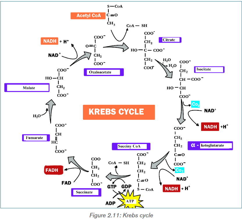
Application activity 2.6
1) Use the chemical equation to show the conversion of pyruvate into
acetyl-coA.
2) Identify and note the main products of the Krebs cycle from one
glucose molecule2.7 Oxidative phosphorylation
Activity 2.7
Observe the figure below and do the following activities
a) This figure summarizes last stage that take place during cellular
respiration, observe it and identify the role of reduced NAD, reduced
FAD and oxygen in this stage.
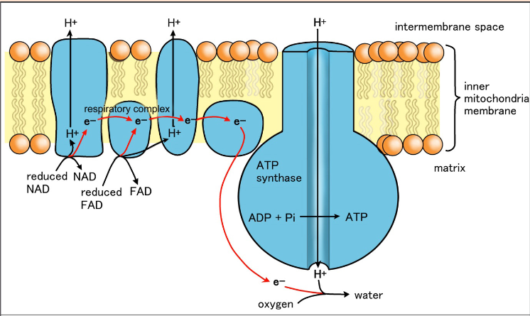
b) Give the explanation of the above figure.In the final stage of aerobic respiration known as the oxidative phosphorylation,
the energy for the phosphorylation of ADP to ATP comes from the activity of the
electron transport chain. Oxidative Phosphorylation is the production of ATP
using energy derived from the redox reactions of an electron transport chain.In eukaryotes, oxidative phosphorylation occurs in the mitochondrial cristae. It
comprises the electron transport chain that establishes a proton gradient across
the inner membrane by oxidizing the NADH produced from the Krebs cycle. ATP
is synthesized by the ATP synthase enzyme when the chemiosmosis gradient
is used to drive the phosphorylation of ADP. Chemiosmosis is the production
of ATP from ADP using the energy of hydrogen ion gradients. The electrons
are finally transferred to oxygen and, with the addition of two protons, water is
formed. The average ATP yield per NADH is probably 3 and for FADH2 of this
electron carrier is worth a maximum of only two molecules of ATP.
Role of oxygen in chemiosmosis
ATP can be synthesized by chemiosmosis only if electrons continue to move
from molecule to molecule in the electron transport chain. Oxygen serves as
the final acceptor of electrons. By accepting electrons from the last molecule in
the electron transport chain, and allows additional electrons to pass along the
chain. As a result, ATP can continue to be synthesized. Oxygen also accepts
the protons that were once part of the hydrogen atoms supplied by NADH and
FADH2. By combining with both electrons and protons, oxygen forms water as
shown in the following equation: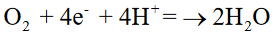
Overview of aerobic respiration
A considerable number of ATP is produced during oxidative phosphorylation and
it is estimated between 32 and 34 ATPs. These are added to 2 ATP produced
during glycolysis and 2 ATP produced during citric cycle. The total number
of ATP produced during a complete respiration process for one molecule of
glucose is then estimated between 36 and 38 ATPs.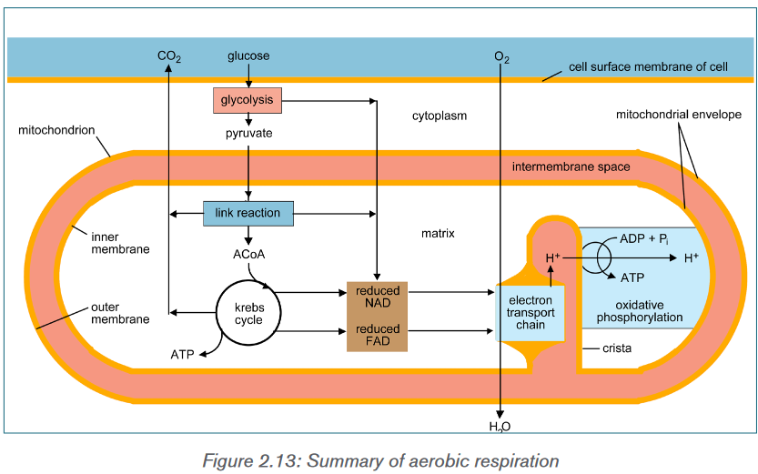
Note that the amount of ATP produced from glucose is usually less than 38
ATP for the following reasons: some ATP is used to transport pyruvate from the
cytoplasm into the mitochondria and some energy is used to transport NADH
produced in glycolysis from the cytoplasm into the cristae of mitochondria.Overall net gain of energy from glucose
Application activity 2.7
1) a) How many ATP are formed from 1 NADH?
b) How many ATP are formed from 1 FADH?
2) How many ATP are formed after a complete oxidation of one glucose
molecule.2.8 Efficiency of aerobic respiration
Activity 2.8
During the complete oxidation of a molecule of glucose it is estimated to
produce 686Kcal. Knowing that inside the cell each ATP produced is
equivalent to 7.3 Kcal,Considering all the amount of ATP produced, find out the percentage of
energy that is equivalent to amount of total ATP produced during aerobic
respiration. Use below formula for your calculations: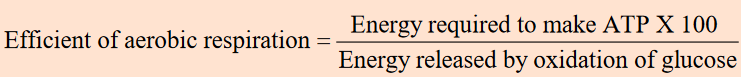
The complete oxidation of glucose produces the energy estimated at 686 Kcal.
Under the condition that exists inside most of the cells, the production of a
standard amount of ATP from ADP absorbs about 7.3 Kcal. Glucose molecule
can generate up to 38 ATP molecules in aerobic respiration. The efficiency of
aerobic.
This result indicates that the efficiency of aerobic respiration equals 40%. The
remained energy (around 60%) is lost from the cell as heat.Application activity 2.8
1) 1. Under which conditions can aerobic respiration take place in animal
cells?
2) 2. Calculate the efficiency aerobic respiration, when a complete
oxidation of glucose produce the energy estimated at 500Kcal under a
production of a standard amount of ATP from ADP absorbed is about
7.3 Kcal.2.9 Efficiency of anaerobic respiration
Activity 2.9
Anaerobic respiration in yeast
a) Boil some water to expel all the dissolved oxygen.
b) When cool, use the boiled water to make up a 5% solution of glucose
and a 10% suspension of dried yeast.
c) Place 5 Cm3 of the glucose solution and 1 Cm3 of the yeast suspension
in a test-tube and cover the mixture with a thin layer of liquid paraffin to
exclude atmospheric oxygen
d) Fit a delivery tube as shown in figure below and allow it to dip into clear
limewater.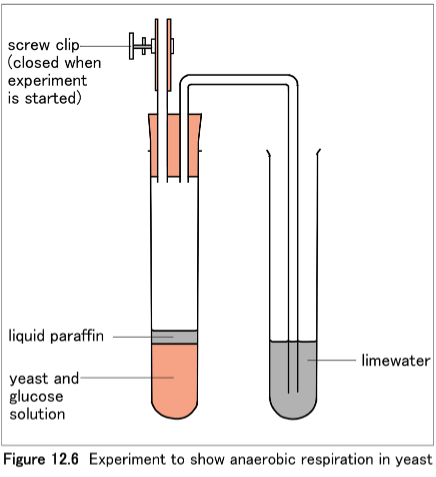
Observe the change that takes place in test tube containing, then explain the
bases of such change.Without oxygen, pyruvate (pyruvic acid) is not metabolized by cellular respiration
but undergoes a process of fermentation. The pyruvate is not transported into
the mitochondrion, but it remains in the cytoplasm, where it is converted to
waste products like alcohol or lactic acid or other compounds depending on
the kind of cells that are active which may be removed from the cell. This serves
the purpose of oxidizing the electron carriers so that they can perform glycolysis
again and removing the excess pyruvate. Fermentation oxidizes NADH to NAD+
so it can be re-used in glycolysis.In the absence of oxygen, fermentation prevents the build-up of NADH in
the cytoplasm and provides NAD+ for glycolysis. This waste product varies
depending on the organism. In skeletal muscles, the waste product is lactic acid.
This type of fermentation is called lactic acid fermentation. In yeast and plants,
the waste products are ethanol and carbon dioxide. This type of fermentation is
known as alcoholic or ethanol fermentation. The ATP generated in this process
is made by substrate-level phosphorylation, which does not require oxygen.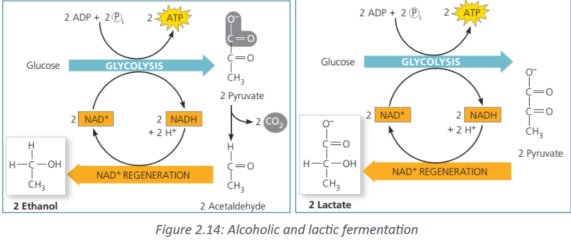
Fermentation is less efficient at using the energy from glucose since only 2 ATP
are produced per glucose, compared to the 38 ATP per glucose produced by
aerobic respiration. This is because the waste products of fermentation still
contain plenty of energy. Glycolytic ATP, however, is created more quickly.Due to the fact that anaerobic respiration produces only 2 ATP, the efficiency
of anaerobic respiration is less than that of aerobic respiration. It is calculated
as follows:Efficiency of aerobic respiration = Energy required to make ATP x 100 Energy
released by oxidation of glucose 2 ATP x 7.3 Kcal x 100 687 Kcal =2%.The production of a small yield of ATP from anaerobic respiration in yeast and
mammalian muscle tissue, including the concept of oxygen debt.Standing still, the person absorbs oxygen at the resting rate of 0.2 dm3 min−1.
(This is a measure of the person’s metabolic rate.) When exercise begins,
more oxygen is needed to support aerobic respiration in the person’s muscles,
increasing the overall demand to 2.5 dm3 min−1. However, it takes four minutes for
the heart and lungs to meet this demand, and during this time lactic fermentation
occurs in the muscles. Thus the person builds up an oxygen deficit. For the next
three minutes, enough oxygen is supplied. When exercise stops, the person
continues to breathe deeply and absorb oxygen at a higher rate than when
at rest. This post-exercise uptake of extra oxygen, which is ‘paying back’ the
oxygen deficit, is called the oxygen debt.The oxygen is needed for:
• Conversion of lactate to glycogen in the liver
• Re-oxygenation of haemoglobin in the blood
• A high metabolic rate, as many organs are operating at above resting
levels.The presence of the lactic acid is sometimes described as an “oxygen debt”.
This is because significant quantities of lactic acid can only be removed
reasonably quickly by combining with oxygen. However, the lactic acid was
only formed due to lack of sufficient oxygen to release the required energy to
the muscle tissue via aerobic respiration. Lactic acid can accumulate in muscle
tissue that continues to be over-worked. Eventually, so much lactic acid can
build-up that the muscle ceases working until the oxygen supply that it needs
has been replenished, this is called muscle crampsTo repay such an oxygen debt, the body must take in more oxygen in order to
get rid of the additional unwanted waste product lactic acid. Mineral depletion,
inadequate blood supply and Nerve compression can be the causes of muscle
cramps.Application activity 2.9
1) Under which conditions can anaerobic respiration take place in animal
cells?
2) Calculate the efficiency of anaerobic, when a complete oxidation of
glucose produce the energy estimated at 200 Kcal under a production
of a standard amount of ATP from ADP absorbed is about 7.3 Kcal2.10 Factors which affect the rate of respiration
Activity 2.10
– Fill a small vacuum flask with beans grains or pea seeds that have
been soaked for 24 hours and rinsed in 1% formaldehyde for 5
minutes.
– Kill an equal quantity of soaked seeds by boiling them for 5 minutes.
– Cool the boiled seeds in cold tap water, rinse them formaldehyde for
5 minutes as before and then put them in a vacuum flask of the same
size as the first one.
– Place a thermometer in each flask so that its bulb is in the middle of
the seeds.
– Plug the mouth of each flask with cotton wool and leave both flasks
for 2 days, noting the thermometer readings whenever possible. Set
it as follow: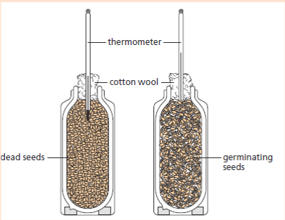
a) What is the purpose of soaking seeds for 24 hours and in formaldehyde
for 5 minutes.
b) Why do you need flask containing dead seeds?
c) Compare the temperature change in those two flasks and explain those
changes.Cellular respiration is the process of conversion of chemical energy stored in
the food to ATP or higher energy compounds. The factors that affect the cellular
respiration are:a. Amount of nutrients
If the amount of nutrients is high, then the energy is high in the cellular respiration.
The nutrients which can go through cellular respiration and transform intoenergy are fat, proteins and carbohydrates. The amount of nutrients available to
transform into energy depend upon the diet of the person.b. Temperature
The rate of the cellular respiration increases if the body temperature is warmer.
The lower the temperature, the slower the rate of cellular respiration. The reason
for this is enzymes which are present in cellular respiration process. Enzyme
reactions require optimum temperatures.c. State of the cell
Metabolically active cells such as neurons, root of human hair have higher
respiration rate than the dormant cells such as skin cells and bone cells. This is
because metabolically active cells can store energy in the body because of the
many metabolic reactions that take place in them.d. Water
It is the medium where the reaction happens. When a cell is dehydrated the
respiration and other metabolism decreases.e. Cellular activity
Some cells need more energy than others. For example, growing cells or very
active cells such as neurons need a lot of energy.f. O2 /CO2 content
When there is high mount of O2 and lower amount of CO2 there is increase of the
rate of respiration. This is because oxygen is needed during aerobic respiration.g. ATP/ADP range
When there is more ATP than ADP, respiration rate slows down to avoid excess
of ATP.Application activity 2.10
1) Explain how proteins and lipids are metabolized for energy during
respiration
2) Explain why the body does not use fats to produce energy as
carbohydrates given that they produce much energy than carbohydrates.2.11 Use of other substrates in respiration.
Activity 2.11
When someone has eaten carbohydrates such as cassava and sweet pota-
toes you do not feel hungry in the same time as another one who has con-
sumed milk or cheese.
1) Can you suggest the reason for this?
2) Which one can take a short time for digestion and why?Carbohydrates are the first nutrients that most organisms can catabolize for
energy. In some cases, living things must be able to metabolize other energy-
rich nutrients to obtain energy in times of starvation. Most organisms possess
metabolic pathways that, when necessary, metabolize proteins, lipids. In each
case, the larger molecules are first digested into their component parts, which
the cell may reassemble into macromolecules for its own use. Otherwise, they
may be metabolized for energy by feeding into various parts of glycolysis or the
Krebs cycle
Carbohydrates, fats and proteins can all be used for cellular respiration.
Monomers of these foods enter glycolysis or the Krebs cycle at various points.
Glycolysis and the Krebs cycle are catabolic pathways through which all kinds
of food molecules are channeled to oxygen as their final acceptor of electrons.Application activity 2.11
1) Explain how proteins and lipids are metabolized for energy during
respiration
2) Explain why the body does not use fats to produce energy as
carbohydrates given that they produce much energy than carbohydrates.Skill lab 2
Fried breads are slices of bread that have been fried in oil or butter.
1) On a sheet of paper write down the ingredients used to make fried
bread.
2) Write down all requirement to make fried bread.
3) Investigate the procedures and make your own fried bread according
to that procedures investigated.
4) Compare your fried bread with the one sold in shops.
5) Present some samples to your teacher.End unit assessment 2
1. Explain the reasons why chemical energy is the most important type of
energy for living organisms.
2. Why do all organisms need energy and where does this energy come
from?
3. Give the structure of ATP and specify its importance to living organisms?
4. The equation C57H104O6 + 80O2→ 57CO2 + 52H2O + Energy represents
oxidation of lipids. Calculate RQ for this equation.
5. Calculate the total amount of energy produced for:
a) 3 moles of hydrolysed ATP
b) moles of synthesized ATP
c) 5 moles of decomposed glucose
6. Active mitochondria can be isolated from liver cells. If these mitochondria
are then incubated in a buffer solution containing a substrate, such as
succinate, dissolved oxygen will be used by mitochondria. The concentration
of dissolved oxygen in the buffer solution can be measured using an electrode.
When this experiment was done, the concentration of dissolved oxygen was
measured every minute for five minutes. Sodium azide (NaN3) which combines
with cytochromes and prevents electron transport was added thereafter. The
results are shown in the graph below.
a) Suggest what effect the addition of sodium azide will have on the
production of ATP and give an explanation for your answer.
b) Explain why the concentration of oxygen decreased during the first
five minutes.c) Suggest what effect the addition of sodium azide will have on the
production of ATP and give an explanation for your answer7. During an experiment, the mouse was inside the bell jar. The air pipe from
the bell jar was connected to the first beaker containing lime water and filter
pump. The glass wool containing soda lime covered by a piece of paper was
connected to the second beaker by air pipe. Another air pipe was connected
from the second beaker containing lime water to the belly jar in the first step.
The set of the experiment looked like the following: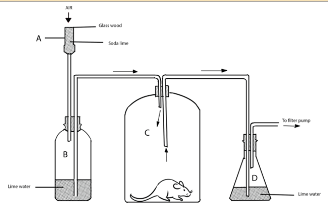
8. What are the major differences between cellular respiration and
photosynthesis?
9. Compare aerobic respiration with anaerobic respiration or fermentation.
10. A student set up an experiment using germinating seeds and boiled seeds
as shown in the diagram below: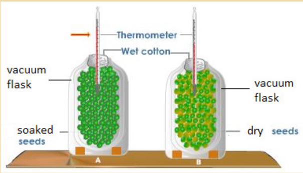
a) State the objective of this experiment and the observation made after
24 hours?
b) Account for the observation made in (a) above?
c) Suggest why vacuum flasks were used in the experiment?
d) What was the purpose of the set-up B?
11) The diagram summarizes how glucose can be used to produce ATP,
without the use of oxygen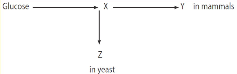
Which compounds are represented by the letters X, Y and Z?
12) Complete the table below:
UNIT 3: REGULATION OF GLUCOSE LEVEL AND TEMPERATURE
Key unit competence
Explain the mechanism of the regulation of blood glucose levels and
regulation of temperature in living organismsIntroductory activity 3
The human body maintains constant different substances in the blood, a
process called homeostasis. The figures below show different organs
involved in the regulation of blood glucose level in the body.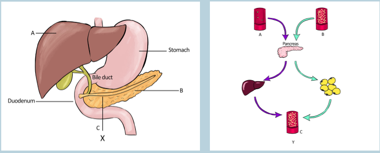
Observe the illustrations X and Y above and answer to the questions that
follow:
a) What are the parts represented by the letters A, B and C on the
illustration X?
b) All the organs shown in the illustration X are involved in the digestion of
food. What are the functions of A and B in the digestion?
c) What are the organs involved in the regulation of blood glucose level
on the illustration X? In which way does each organ state help in this
regulation?d) The illustration Y shows the regulation of blood glucose level. What
does the letters A, B and C show in this regulation?
e) Alpha and beta cells are responsible for producing the hormones
that are involved in the regulation of blood glucose level. Which
organ on the illustration Y produces these hormones?
f) Compare the mechanism of working of the organs A and B in the
regulation of blood glucose level.3.1 Structure and functions of the liver and the pancreas
Activity 3.1
Each organ of our body is made of different tissues which are also composed
of cells. These cells carry out different functions that help in the functioning
of the organ. Refer to the image below to answer the questions that follow: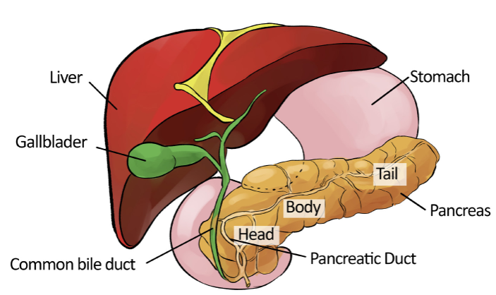
a) Observe the liver and the pancreas and make short notes on their
structures.
b) What are the functions of the liver and the pancreas?
c) Which hormones are produced by the pancreas and what are their
functions?
d) Compare the modes of action of insulin and glucagon.
e) Examine what happens when the blood glucose regulation fails?3.1.1 Importance of glucose
Glucose is one of the most important carbohydrates molecules in our body.
Body requires glucose to carry out some of its most important functions. Glucose
is synthesized in green plants, from carbon dioxide, CO2 and water, H2O with
the help of energy from sunlight. This process is known as photosynthesis.
The reverse of the photosynthesis reaction i.e., breakdown of glucose in the
presence of oxygen to form carbon dioxide and water releasing the energy, is
the main source of power for all the living organisms. The excess glucose in
plants is stored in the form of starch which serves as foods for various animals.Glucose as an energy source
Almost 80 per cent of carbohydrates in our food are converted to glucose during
digestion in the alimentary canal. Fructose and galactose is the other main
product of carbohydrates digestion. After absorption from the alimentary tract,
fructose and galactose are converted into glucose in the liver. And therefore,
glucose constitutes more than 95 per cent of all the carbohydrates circulating
in the blood.Body cells require glucose continuously for its various metabolic activities. These
cells directly absorbed glucose from the blood. Once inside the cells, glucose
combines with a phosphate moiety to form Glucose-6-phosphate with the
help of enzyme glucokinase in liver and hexokinase in most other cells. This
phosphorylation reaction is irreversible and helps to retain the glucose inside the
cells. However, in liver cells, renal tubular epithelial cells and intestinal epithelial
cells, an enzyme glucose phosphatase converts the glucose-6-phosphate
back to glucose.The complete oxidation of one molecule of glucose into carbon dioxide and
water inside the cells produces as many as 38 molecules of ATP (2 from
glycolysis, 2 from the Krebs cycle and 34 from the oxidative phosphorylation).3.1.2 Role of liver and pancreas in glucose regulation
Our body maintains a narrow range of glucose concentration in the blood
between 80 mg/dL to 120 mg/dL which may increase up to 180 mg/dL after a
meal containing high amount of carbohydrates. The hormones responsible for
the regulation of blood sugar level— insulin and glucagon are secreted by the
pancreas. The excess glucose in our blood is converted into glycogen in the
liver. Therefore, pancreas and liver play a vital role in the regulation of blood
sugar concentration.Role of liver in glucose regulation
The liver is the largest internal solid organ in the body second to the skin as the
largest organ overall. It performs various functions in our body, including synthesis
and storage of proteins and fats, carbohydrates metabolism, formation and
secretion of bile, detoxification and excretion of potentially harmful compounds.
Liver contains two main cell types: Kupffer cells and Hepatocytes.1) Kupffer cells are a type of macrophage that capture and break down
old, worn out red blood cells passing through liver sinusoids.
2) Hepatocytes are cuboidal epithelial cells that line the sinusoids and
make up the majority of cells in the liver. Hepatocytes perform most of the
liver’s functions—metabolism, storage, digestion, and bile production.Hepatocytes cells contain various enzymes which help in the regulation of blood
glucose.
These are:1) Glycogen synthase; responsible for glycogen biosynthesis (Glycogenesis).
When the concentration of glucose in the blood increases beyond the normal
value, the excess glucose is converted to glycogen in liver with the help of
enzyme glycogen synthase.2) Glycogen phosphorylase; responsible for breaking down of glycogen
(Glycogenolysis). When the blood glucose level drops, the enzyme
glycogen phosphorylase convert glycogen to glucose-6-phosphate.
Other two enzymes, glucan transferase and glucosidase also help in
glycogenolysis.3) Glucose phosphatase; responsible for conversion of glucose-6-
phosphate to glucose in the liver. Glucose is then released into the blood
stream, thereby increasing the blood glucose level.Role of the pancreas in glucose regulation
Pancreas is the most important endocrine organ for the regulation of blood
glucose. It secretes insulin and glucagon, the two main hormones responsible
for the regulation of blood glucose.
1) Insulin: When the blood glucose concentration increases rapidly, for
example after a meal with high carbohydrates content, pancreas secretes
insulin hormone into the blood stream. Insulin binds to its receptors and
increases the rate of glucose uptake, storage and utilization by almost all
tissues of the body resulting in lowering of blood glucose level. Besides,
insulin also stimulates glycogenesis, lipid and proteins biosynthesis
which helps in decreasing blood glucose concentration.2) Glucagon: In response to decrease in blood glucose concentration,
pancreas secretes glucagon which activates the enzyme glycogen
phosphorylase responsible for degradation of glycogen to glucose-6-
phosphate. Glucose-6-phosphate is then dephosphorylated to form
glucose and finally released into the blood stream thereby increasing
the blood glucose level. Glucagon also stimulates gluconeogenesis i.e.,
biosynthesis of glucose from non-carbohydrate compounds like pyruvate
and amino acids.3.1.3 Detailed structure of liver lobule and islet of
langerhansLiver and liver lobules
The liver is a roughly triangular in shape and extends across the entire abdominal
cavity under the diaphragm. Most of the liver’s mass is located on the right
hypochondrium (i.e., upper part of the abdomen) as well as part of the abdomen
(Figure 3.3). The liver is made of very soft, pinkish-brown tissues encapsulated
by a connective tissue capsule. This capsule is further covered and reinforced
by the peritoneum of the abdominal cavity, which protects and holds the liver.The liver consists of 4 distinct lobes: the left, right, caudate, and quadrate lobes.
The falciform ligament divides the liver into two main lobes, right and left. The
larger right lobe is again sub-divided into three lobes, the right lobe proper, the
caudate lobe and the quadrate lobe (Figure 3.1). Each liver lobe is made up of
about 100,000 small hexagonal functional units known as lobules. A typical liver
lobule comprises rows of liver cells, hepatocytes, radiating out from a central
vein. The six angles of the hexagon are occupied by a portal triad comprising a
hepatic portal vein, a hepatic artery and a bile duct. The portal veins and arteries
are connected to the central vein through a network of capillary-like tubes called
sinusoids (Figure 3.2). Blood flows out of the sinusoids into the central vein and
is transported out of the liver.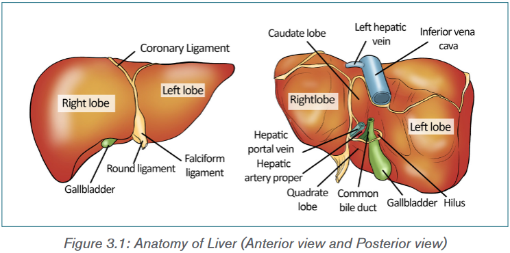
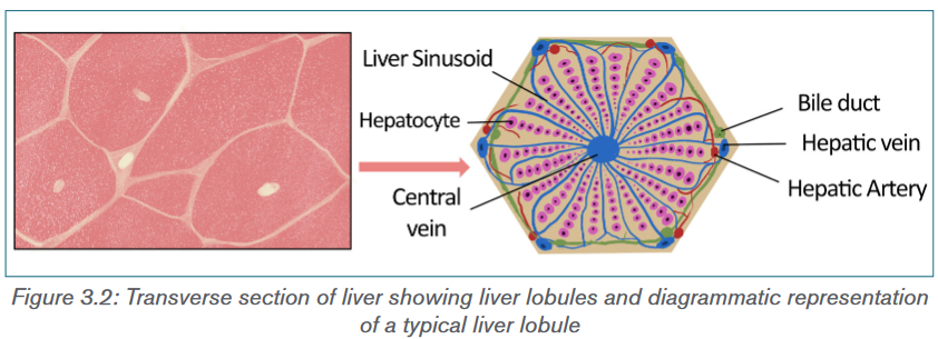
Pancreas
The pancreas is an elongated, tapered organ, located in the abdominal region,
behind the stomach and next to the duodenum—the first part of the small
intestine (Figure 3.3). The right side of the organ, called the head, is the widest
part of the organ and lies in the curve of the duodenum. The tapered left side
which extends slightly upward is the body of the pancreas.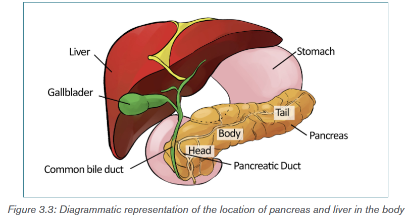
Structure and function of pancreas
Pancreas has two main functional components:1) Exocrine cells, the acini—Cells that release digestive enzymes
into the gut via the pancreatic duct. These enzymes include trypsin
and chymotrypsin to digest proteins; amylase for the digestion of
carbohydrates; and lipase to break down fats. The pancreatic duct joins
the common bile duct to form the ampulla of Vater in the duodenum. The
pancreatic juices and bile (from gallbladder) released into the duodenum
help the body to digest fats, carbohydrates as well as proteins.2) Endocrine pancreas: Highly vascularized groups of cells known as
the Islets of Langerhans within the exocrine tissue constitute the
endocrine pancreas (Figure 3.4). The human pancreas has 1–2 million
islets of Langerhans. It contains four different types of cells which
are distinguished from one another by their morphology and staining
characteristics:i) Alpha cells: Which secrete glucagon, constitute about 25 per
cent of all the cells of islet of Langerhans.ii) Beta cells: The most abundant of the islet cells constitute about
60% of the cells. They release insulin, a hormone involved in
decreasing the blood glucose level.iii) Delta cells: Constitute about 10 per cent of total cells and secrete
somatostatin which regulates both the alpha and beta cells.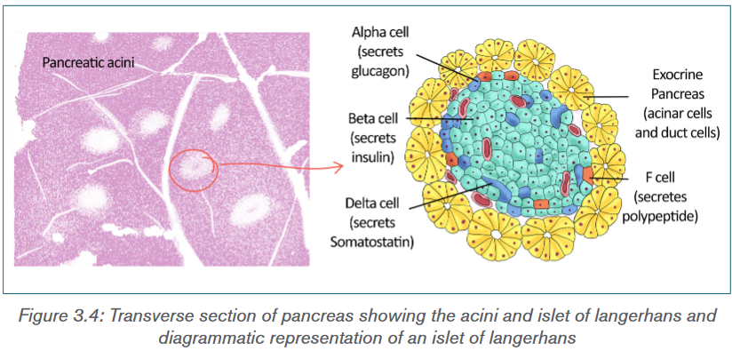
Application activity 3.1
1) The homeostatic level of blood glucose is around 90 mg per 100 ml
of blood. Three person have taken their blood glucose levels using a
glucometer and their results are:
Peter: 85 mg per 100 cm3 of blood
Mary: 130 mg per 100 cm3 of blood
John: 65 mg per 100 cm3 of blood
Interpret these results obtained by using a glucometer?3.2 Control mechanisms by hormones
Activity 3.2
Different hormones are involved in the regulation of blood glucose level. List
and explain those hormones and their functions.3.2.1 Homeostatic control of blood glucose concentration
by insulin and glucagon
Insulin and glucagon are the major hormones responsible for the regulation of
blood glucose. Both insulin and glucagon are secreted by the pancreas, and are
referred to as pancreatic endocrine hormones.Insulin
Insulin was first discovered in 1922 by Banting and Best. Although there is
always a low level of insulin secreted by beta cells of pancreas, the amount
secreted into the blood increases as the blood glucose level rises. In the blood,
it circulates entirely in an unbound form with plasma half-life of about 6 minutes.
Only a small portion of the insulin binds with the insulin receptors of the target
cells while the rest is degraded by the enzyme insulinase, mainly in liver and to
a lesser extends in kidney and muscles.Functions of insulin
Binding of insulin to the receptors stimulates the rate of glucose uptake, storage
and utilization by almost all tissues of the body mainly in muscles, adipose tissue
and liver. Other important functions of insulin include:
i) Insulin promotes glycogenesis by activating enzyme glycogen synthase.
ii) Insulin inactivates liver phosphorylase, the key enzyme of glycogenolysis.
iii) Insulin promotes lipid synthesis by increasing the conversion of excess
glucose into fatty acids in the liver. These fatty acids are transported
as triglycerides to the adipose tissue where it is deposited as fat.
iv) Insulin inhibits the enzymes responsible for gluconeogenesis in liver.
v) Insulin promotes protein synthesis by increasing the rate of transcription
and translation. It also stimulates transport of many amino acids into the
cells.
vi) Insulin inhibits breakdown of lipids and proteins.Regulation of insulin secretion
The secretion of insulin by beta cells of islet of Langerhans depends on the
following factors:
i) Blood glucose level: Increased in the blood glucose level stimulates
the insulin secretion while decreased in the blood glucose concentration
inhibits the secretion.
ii) Blood fatty acids and amino acids concentration: Insulin secretion
is also stimulated by increased in the concentration of blood’s fatty acids
and amino acids concentration and inhibited when its concentration
decreased.
iii) Gastrointestinal hormones: Insulin secretion increases moderately
in response to several gastrointestinal hormones—gastrin, secretin,
cholecystokinin and gastric inhibitory peptide.
iv) These hormones are released after the person takes meal and the
increased in insulin secretion can be regarded as preparation for the
glucose and amino acids uptake by cells.
v) Other hormones: Other hormones that are associated with the
increase in the insulin secretion are glucagon, growth hormone, cortisol,
progesterone and estrogen.Glucagon
Glucagon is secreted by the alpha cells of the pancreatic islets in response
to low blood glucose levels and to events whereby the body needs additional
glucose, such as in response to vigorous exercise.Functions of glucagon
The effect of glucagon in regulating blood glucose level is exactly opposite to
insulin:
i) The most important function of glucagon is activation of glycogen
phosphorylase enzyme responsible for degradation of glycogen to glucose-
6-phosphates. The glucose-6-phosphate is then dephosphorylated to
glucose and finally released into the blood stream resulting in increase in
blood glucose concentration.
ii) Glucagon also stimulates the increase in rate of amino acid uptake and its
conversion into glucose, i.e., gluconeogenesis.
iii) Glucagon activates adipose cell lipase enzyme which stimulates lipids
metabolism.
iv) Glucagon also inhibits the storage of triglycerides in the liver by preventing
the liver from removing fatty acids from the blood.v) Glucagon also enhances the strength of the heart; increases blood flow in
some tissues, especially the kidneys; enhances bile secretion; and inhibits
gastric acid secretion.Regulation of glucagon secretion
Glucagon secretion increases with the decrease in the concentration of
blood glucose level while the increasing concentration of glucose inhibits its
secretion. Other factors which stimulate glucagon secretion are, increase in the
concentration of amino acids in blood and vigorous physical exercise.Negative-positive feedback mechanism
A positive feedback mechanism is the exact opposite of a negative feedback
mechanism. With negative feedback, the output reduces the original effect
of the stimulus. In a positive feedback system, the output enhances the
original stimulus. Negative feedback is an important regulatory mechanism for
physiological function in all living cells. It occurs when a reaction is inhibited by
increase concentration of the product. Regulation of blood glucose level is an
excellent example of homeostatic control through negative feedback mechanism
(Figure 3.5).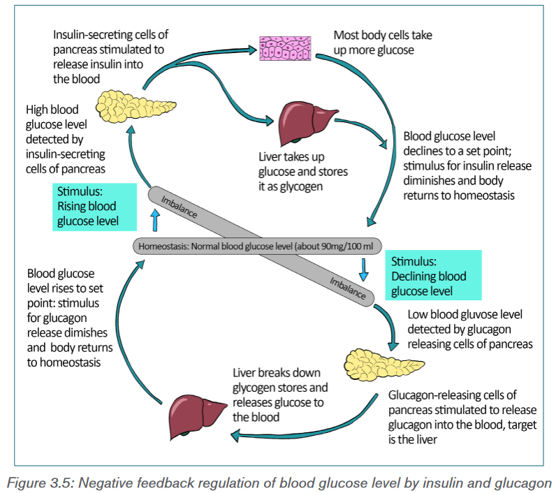
Response to an increase in blood glucose level
When there is increase in blood glucose level, the beta cells of the pancreatic
islets of Langerhans increase the release of insulin into the blood. Insulin
binds to receptors on the cell membrane and stimulates the cells to increase
glucose uptake. This led to decrease in blood glucose level. Besides, insulin
also stimulates glycogenesis and glycolysis while inhibiting glycogenolysis,
gluconeogenesis, lipolysis etc. which all contributes in reducing blood glucose
levels.Response to a decrease in blood glucose level
Decreased in blood glucose level stimulates the alpha cells of pancreas islets
to increase the secretion of glucagon. Glucagon activates enzyme glycogen
phosphorylase in the liver and muscle cells which start glycogenolysis. It also
promotes gluconeogenesis, lipid metabolism etc. The overall effect of glucagon
is increase in the concentration of blood glucose.3.2.2 Other hormones involved in glucose regulation
Other than insulin and glucagon, there are many hormones which contribute to
the regulation of blood glucose level (Figure 3.6). They are:
1) Somatostatin: It is secreted by delta cells of pancreatic islet of
Langerhans in response to many factors related to ingestion of food like
increased concentration of glucose, amino acids, fatty acids and several
gastrointestinal hormones released from the upper gastrointestinal tract.
Somatostatin acts locally within the islets of Langerhans and inhibits the
secretion of both insulin and glucagon. It also reduces the motility of
the stomach, duodenum, and gallbladder and decreases the secretion
and absorption in the gastrointestinal tract. Hence, lowers overall blood
glucose level.2) Epinephrine: Commonly known as Adrenaline, it is secreted by the
medulla of the adrenal glands in response to strong emotions such as
fear or anger. It causes increases in the heart rate, muscle strength, blood
pressure and sugar metabolism. In response, it enhances the process of
glycogenolysis, increasing the overall blood glucose concentration.3) Cortisol: It is also known as stress hormone and is secreted by the
adrenal cortex of the adrenal gland in response to stress. Cortisol
enhances gluconeogenesis and increases the concentration of glucose
in the blood.4) Adrenocorticotropic Hormone (ACTH): In response to various
stresses, hypothalamus secretes corticotropin-releasing hormone which
stimulates anterior pituitary to secrete ACTH. It stimulates adrenal cortex
to release the cortisol hormones.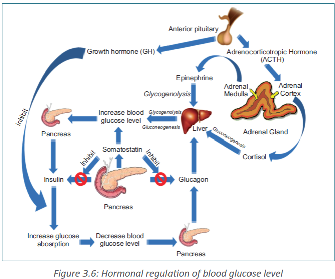
5) Growth hormone (GH): It is another anterior pituitary hormone which
antagonizes the action of insulin by inhibiting the glucose uptake by cells
and increasing the blood glucose level.6) Gastrointestinal hormones: The hormones released by gastrointestinal
tract such as gastrin, secretin, cholecystokinin and gastric inhibitory
peptide etc. increase the digestion and absorption of nutrients in the
gastrointestinal tracts. These hormones stimulate the pancreas to secrete
insulin in anticipation of the increase in blood glucose level.3.2.3 Mechanism of hormonal regulation
Our body maintains certain variables like temperature, pH etc. within a safe range
so that it does not cause any harm to the body and the internal environment
remains stable and relatively constant. This is known as homeostasis.
Hormones are chemical messenger that are directly released into the blood
stream. They play very important role in maintaining the homeostasis.Steps of hormonal signaling
Hormonal signal transduction is a complex process which involves the following
steps:
i) Hormones are first synthesis in particular cells of an organ and stored for
secretion in response to certain stimulus.
ii) When the organ receives the stimulus; hormones are secreted directly
into the blood stream.iii) Blood carries the hormone to the target cell(s).
iv) The hormone is recognized by the specific receptor in the cell membrane
or by the intracellular receptor protein.
v) The hormonal signal is relayed and amplified through a series of signal
transduction process in the target cells which lead to cellular response.3.2.4 Cause of blood sugar imbalances in the body
Our body obtains glucose from various food sources or synthesis in the liver and
muscles from other compounds like pyruvate, lactate, glycerol, and glucogenic
amino acids. The blood carries glucose to all the cells in the body where it is
metabolized to produce energy.Blood sugar levels keep on fluctuating throughout the day increasing after
meals and decreasing in between the meals. When the blood glucose level
rises beyond the normal value, the condition is known as hyperglycaemia. On
the other hand, hypoglycaemia or low blood sugar is the condition in which the
blood glucose level is below normal (~80 mg/dL).Hyperglycaemia
High blood glucose level can be caused due to various reasons like:
i) Carbohydrates: Eating food containing too much of carbohydrates. The
body of a person cannot process high levels of carbohydrates fast enough
to convert it into energy.
ii) Insulin control: The pancreas of the individual are unable to produce
enough insulin.
iii) Stress: Stress stimulates the secretion of certain hormones like cortisol
and epinephrine etc., which increases the blood glucose level.
iv) Low levels of exercise: Daily exercise is a critical contributor to regulating
blood sugar levels.
v) Infection, illness, or surgery: With illness, blood sugar levels tend to
rise quickly over several hours.
vi) Other medications: Certain drugs, especially steroids, can affect blood
sugar levels.A high blood sugar level can be a symptom of diabetes. If hyperglycaemia
persists for several hours, it can leads to dehydration. Other symptoms of
hyperglycaemia include dry mouth, thirst, frequent urination, blurry vision, dry,
itchy skin, fatigue or drowsiness, weight loss, increased appetite, difficulty
breathing, dizziness upon standing, rapid weight loss, increased drowsiness
and confusion, unconsciousness or coma.Hypoglycaemia
Hypoglycaemia is generally defined as a serum glucose level below 80 mg/dL.
Symptoms typically appear when the blood glucose levels reach below 70 mg/
dL and levels below 60 mg/dL can be fatal.Common causes of low blood sugar include the following:
i. Overmedication with insulin or antidiabetic pills
ii. Use of alcohol
iii. Skipped meals
iv. Severe infection
v. Adrenal insufficiency
vi. Kidney failure
vii. Liver failure, etc.
Common symptoms of hypoglycaemia include trembling, clammy skin,
palpitations (pounding or fast heart beats), anxiety, sweating, hunger, and
irritability. If the brain remains deprived of glucose for longer period, a later set of
symptoms can follows like difficulty in thinking, confusion, headache, seizures,
and coma. And ultimately, after significant coma or loss of consciousness, death
can occur.3.2.5 Diabetes mellitus
Diabetes mellitus (commonly referred to as diabetes) is a chronic condition
associated with abnormally high levels of sugar in the blood due to impaired
carbohydrate, fat, and protein metabolism. It can be due to absence or insufficient
production of insulin by the pancreas, or inability of the body to properly use
insulin. Hence, there are two types of diabetes mellitus – Type I causes by lack
of insulin secretion and Type II, caused by reduced sensitivity of target cells to
insulin.Type I diabetes
It is known as insulin dependent diabetes mellitus (IDDM) and it is due to
insufficient insulin production by the beta cells of pancreatic islet of Langerhans
or due to absence of the beta cells itself. Since the pancreas makes very little or
no insulin at all, glucose cannot get into the body’s cells and remain in the blood
leading to hyperglycemia. The concentration of blood glucose level can be as
high as 300 – 1,200 mg/dL. The symptoms of Type I diabetes include:i) Loss of glucose in urine; due to increase in blood glucose, concentration
goes beyond 180 mg/dL.ii) Dehydration; due to osmotic loss of water from cells and inability to
reabsorb water in kidney.
iii) Tissue injury; due to damages blood vessels in many tissues.
iv) Metabolic acidosis; due to increased fat metabolism.
v) Depletion of body’s protein; due to increase protein metabolism.Treatment of Type I Diabetes
Persons with Type I diabetes require treatment to keep blood sugar levels within
a target range which includes:
i) Taking insulin from external source everyday either through injections or
using an insulin pump.
ii) Monitoring blood sugar levels several times a day.
iii) Eating a healthy diet that spreads carbohydrate throughout the day.
iv) Regular physical activity or exercise. Exercise helps the body to use
glucose more efficiently.
v) It may also lower your risk for heart and blood vessel disease.
vi) Not smoking.
vii) Not drinking alcohol if you are at risk for periods of low blood sugar.Type II diabetes
Also known as non-insulin dependent diabetes mellitus (NIDDM), it is
due to the inability of cells to take up glucose from the blood. It can be either
due to defective insulin receptors over cell surfaces or abnormality of blood
plasma protein, amylin. Due to decrease sensitivity of cells to insulin, a condition
known as insulin resistance, the beta cells secrete large amount of insulin into
the blood stream resulting in increased concentration of insulin in blood. This
condition is known as hyperinsulinaemia. Type II diabetes are more common
and account for almost 80–90 per cent of the total diabetes mellitus cases.The symptoms of type II diabetes include:
i) Obesity, especially accumulation of abdominal fat;
ii) Fasting hyperglycaemia;
iii) Lipid abnormalities such as increased blood triglycerides and decreased
blood high density lipoprotein-cholesterol; and
iv) Hypertension.Treatment of Type II Diabetes
There’s no cure for diabetes, so the treatment aims to keep the blood glucose
levels as normal as possible and to control the symptoms and prevent health
problems developing later in life. In type II diabetes, the pancreas is still working
but our body develops insulin resistance and is unable to effectively convert
glucose into energy leaving too much glucose in the blood. Therefore, Type II
diabetes can be managed through lifestyle modification including:
i) Healthy diet as eating well helps manage our blood glucose levels and
body weight.
ii) Regular exercise helps the insulin work more effectively, lowers your blood
pressure and reduces the risk of heart disease.
iii) Regular monitoring of blood glucose levels to test whether the treatment
being followed is adequately controlling blood glucose levels or we need
to adjust the treatment.Importance of controlled diet in diabetes
Controlled diet is very important for diabetic patients because blood sugar is
mostly affected by the food one eats. The glycaemic index of a food measures
how the food affects the blood glucose level. The higher the glycaemic index
of the food, the greater the potential of increasing blood glucose. Therefore, in
order to control glucose levels in the blood, it is important that diabetic primarily
chooses low glycaemic index carbohydrates like dried beans and legumes
such as lentils and pintos, non-starchy vegetables, fruits, whole grain bread
and cereals, sweet potatoes etc. Foods like white bread, white rice, cornflakes,
white potatoes, popcorn, pineapple, and melons are high glycaemic index foods
and should be eaten moderately.Because people with diabetes are at risk of high blood pressure, it makes sense
to also choose foods that are heart healthy (i.e., lean, low-fat) and the ones that
are low in salt. Increasing the amount of fibre in diet and reducing fat intake,
particularly saturated fat, can help prevent diabetes or manage the diabetic
condition from developing any complications. Therefore, one should:
i) Increase the consumption of high-fibre foods, such as wholegrain bread
and cereals, beans and lentils, and fruits and vegetables.
ii) Choose foods that are low in fat for example, replace butter, ghee and
coconut oil with low-fat spreads and vegetable oil.
iii) Choose skimmed and semi-skimmed milk, and low-fat yoghurts.
iv) Eat fish and lean meat rather than fatty or processed meat, such as
sausages and burgers.
v) Grill, bake, poach or steam food instead of frying or roasting it.vi) Avoid high-fat foods, such as mayonnaise, chips, crisps, pasties,
poppadums and samosas.
vii) Eat fruit, unsalted nuts and low-fat yoghurts as snacks instead of cakes,
biscuits, bombay mix or crisps etc.Coping with situation of diabetics and hypertension
Blood pressure is the measure of the force of blood pushing against blood
vessel walls. The heart pumps blood into the arteries, which carry the blood
throughout the body. The normal blood pressure is less than 120 (systolic) over
80 (diastolic). High blood pressure, also called hypertension, is dangerous
because it makes the heart work harder to pump blood out to the body and
contributes to hardening of the arteries, or atherosclerosis, to stroke, kidney
disease, and to the development of heart failure. Diabetics are more likely to
develop high blood pressure and other heart and circulation related problems,
because diabetes damages arteries and makes them targets for hardening
(atherosclerosis). Obesity is another main factor which is responsible for
hypertension.When it comes to preventing diabetes complications, normal blood pressure
is as important as good control of blood glucose levels. Therefore, to treat
and help prevent high blood pressure, one should control their blood glucose,
stop smoking, eat healthy, maintain a healthy body weight, limit alcohol and salt
consumption and exercise regularly.3.2.6 Monitoring of blood glucose levels
Blood glucose monitoring is a way of testing the concentration of glucose
in the blood (glycaemia). As mentioned earlier, the concentration of blood
glucose is fluctuating throughout the day. Under certain physiological disorders,
especially when the person is suffering from diabetes mellitus, the blood glucose
concentration can increase well above the normal concentration. Most people
with type II diabetes need to monitor their blood sugar levels at home. A blood
glucose test is generally performed by piercing the skin (typically, on the finger)
to draw blood, then applying the blood to a chemically active disposable ‘test-
strip’ or to a biosensors.1. Dipstick test
A dipstick or the reagent strips is a narrow strip of plastic with small pads
attached to it. Each pad contains specific reagents for a different reaction,
thus allowing for the simultaneous determination of several compounds. The
blood glucose test use enzymes glucose oxidase and hexokinase which
are specific to glucose, embedded on a test strip or a dipstick. When the
blood sample is applied onto the strip, the enzymes catalyzed glucose specific
reaction which changes the colour. The chemical reaction involved in the
glucose oxidase test is as follows: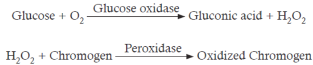
Numbers of chromogen like potassium iodide, tetramethylbenzine,
O-tolidinehydrochloride, 4-aminoantipyrine etc. are used in the dipstick. The
colour reaction of the dipsticks is kinetic and will continue to react after the
prescribed time. Therefore, reading taken after the prescribed time can give
false result.2. Biosensors
A biosensor is a device which is composed of two elements; a bio-receptor
that is an immobilized sensitive biological element (e.g. enzyme, DNA probe,
antibody) recognizing the analyte (e.g. enzyme substrate, complementary DNA,
antigen) and a transducer, used to convert biochemical signal resulting from
the interaction of the analyte with the bioreceptor into an electronic signal. The
intensity of generated signal is directly or inversely proportional to the analyte
concentration. For example, the glucose biosensor is based on the fact that
the immobilized Glucose oxidase enzyme which catalyzes the oxidation of β-D-
glucose by molecular oxygen producing gluconic acid and hydrogen peroxide.
An electrochemical transducer converts this reaction into electronic signal
which appears on the screen of the glucose meter.3. Continuous glucose monitoring
Continuous glucose monitoring systems (CGMS) use a glucose sensor
inserted under the skinin the form of a small needle. The signal from the sensor
is transmitted wirelessly and the result is recorded in a small recording device.
The monitor of the device updates and displays the blood sugar level every few
minutes. The glucose sensor needs to be removed and replaced at least once
per week.Advantages of continuous glucose monitoring:
i) The monitor displays blood sugar level every few minutes, allowing one to
see whether the level is increasing, decreasing, or is stable.
ii) The receiver can also be set to alarm if the blood sugar level is above or
below a pre-set level.
iii) The blood sugar results from the continuous monitor can be downloaded
to a computer, allowing you to check blood sugar trends over time.The only disadvantage of continuous monitor other than the cost is its inaccuracy
compared to more traditional accurate dipstick method. Therefore, most experts
recommend continuous glucose monitoring along with several finger sticks
daily to calibrate the CGMS device and to verify that the sensor readings are
accurate.Roles of adrenaline in the control of blood sugar level
Adrenaline, a natural stimulant created in the kidney’s adrenal gland, travels
through the bloodstream and controls functions of the autonomous nervous
system, including the secretion of saliva and sweat, heart rate and pupil dilation.
The substance also plays a key role in the human flight-or-flight response.The “fight or flight” hormone that gives us a quick boost of extra energy to
cope with danger — including the danger of low blood glucose. When blood
glucose levels drop too low, the adrenal glands secrete epinephrine (also called
adrenaline), causing the liver to convert stored glycogen to glucose and release
it, raising blood glucose levels. Epinephrine also causes many of the symptoms
associated with low blood glucose, including rapid heart rate, sweating, and
shakiness. The epinephrine response spurs the liver to correct low blood glucose
or at least raise blood glucose levels long enough for a person to consume
carbohydrate.3.2.7. Detection of glucose in urine
Urine analysis can be used to test pH, protein, glucose, ketones, occult blood,
bilirubin, urobilinogen, nitrite, leukocyte esterase etc. in the urine sample. Simple
test for glucose in urine can be used to diagnose diabetes mellitus. Generally,
healthy person do not loss glucose in their urine whereas a person with diabetes
mellitus loses small to large quantities of glucose in their urine.Detection of glucose in urine
The presence of glucose in the urine is called glycosuria (or glucosuria).
The urine analysis of glucose is based on enzyme glucose oxidase which is
impregnated in a dipstick (reaction described in previous section).Detection of protein in urine
The glomerular filtrate of a normal kidney contains little amount of low–molecular
weight protein. Most of these proteins get reabsorbed in the tubules with less
than 150 mg being excreted through urine per day. Therefore, the abnormal
increase in the amounts of protein in the urine, Proteinurea, can be an important
indicator of renal diseases. There are certain physiologic conditions such as
exercise and fever that can lead to increased protein excretion in the urine in the
absence of renal disease.Proteinuria is a symptom of chronic kidney disease (CKD), which can be due
to diabetes, high blood pressure, and diseases that cause inflammation
in the kidneys. Therefore, urine analysis for protein is part of a routine medical
assessment for everyone. If CKD is not checked in time, it can lead to end-
stage renal disease (ESRD), when the kidneys completely stop functioning.
A person with ESRD requires a kidney transplant or regular blood-cleansing
treatments called dialysis to further sustain.The tests for proteinuria are based either on the “protein error of indicators”
principle (ability of protein to alter the colour of some acid-base indicators without
altering the pH) or on the ability of protein to be precipitated by acid or heat.
According to “protein error of indicators” principle, a protein-free solution of
tetrabromphenol blue at pH 3 is yellow in colour and its colour changes from
yellow to blue (or green) when the pH increases from pH 3 to pH 4. However,
in the presence of protein (albumin), the colour changes occur between pH 2
and 3 i.e., an “error” occurs in the behaviour of the indicator. The method is more
sensitive to albumin than to other proteins, whereas the heat and acid tests are
sensitive to all proteins.The test result may show false-positive results in a highly buffered alkaline urine,
which may result from alkaline medication or stale urine. Also, if the dipstick
is left in the urine for too long, the buffer could be washed out of the reagent
resulting in increased pH and the strip may turn blue or green even if protein is
not present. On the other hand, false-negative results can occur in dilute urines
or when the urine contains proteins other than albumin in higher concentrations.Detection of ketones in urine
As discussed earlier, ketones, or ketone bodies are formed during lipid
metabolism. One of the intermediate products of fatty acid breakdown is acetyl
CoA. If the lipid metabolism and carbohydrate metabolism are in balanced,
Acetyl-CoA enters the citric acid cycle (Krebs cycle) where it reacts with
oxaloacetate to form citrate. When carbohydrate is not available in the cells,
all available oxaloacetate get converted to glucose and so none is available for
condensation with Acetyl- CoA. As such, Acetyl-CoA cannot enter the Krebs
cycle and is diverted to form ketone bodies.Application activity 3.2
An experiment was carried out with two groups of people. Group X has type
I diabetes mellitus while group Y did not (control group). Every 15 minutes’
blood samples were taken from all members of both groups and the mean of
levels of insulin, glucagon, and glucose were calculated. After an hour, every
person was given a glucose drink. The results are shown in the figure below: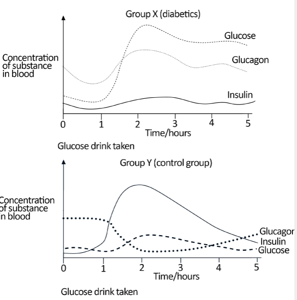
a) Name a hormone other than insulin and glucagon that is involved in
regulating blood glucose levels.
b) State two differences between groups X and Y in the way insulin
secretion responds to the drinking of glucose.
c) Suggest a reason why the glucose level falls in both groups during the
first hour.
d) Using information from the graphs, explain the changes in the blood
glucose level in group Y after the glucose drink.
e) Explain the difference in blood glucose level in group X compared to
group Y.
f) Suggest what might happen to the blood glucose level of group X if
they had no food intake over the next 24 hours.3.3 Adaptations of animals to temperature changes in the
environmentActivity 3.3
Observe the photo below and answer the questions that follow:
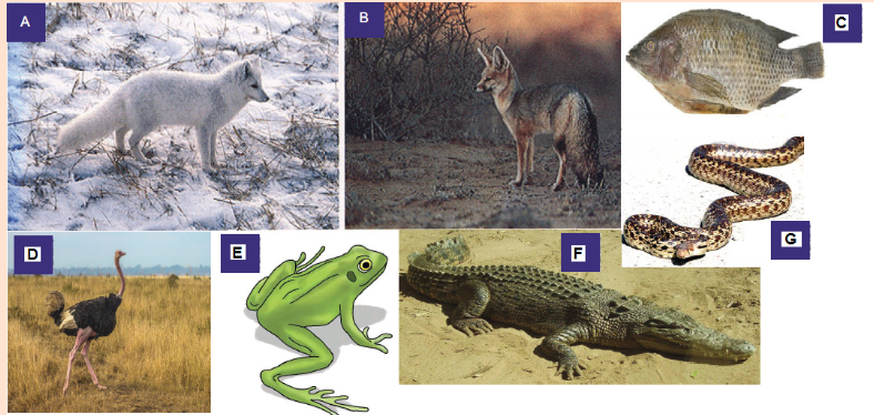
a) Show 2 main differences between individual A and individual E.
b) How is individual C different from individual D?
c) The individual A is adapted to live in cold environments. Analyze it
carefully to identify any two characteristics that this animal has.
d) Which among the animals on the photo is adapted to live in hot climates?
Justify your answer.Thermoregulation is the ability of an organism to keep its body temperature
within certain boundaries, even when the surrounding temperature is very
different. This process is one aspect of homeostasis: a dynamic state of stability
between an animal’s internal environment and its external environment.One of the most important examples of homeostasis is the regulation of body
temperature. Not all animals can do this physiologically. Animals that maintain a
fairly constant body temperature (birds and mammals) are called endotherms,
while those that have a variable body temperature (all others) are called
ectotherms. Endotherms normally maintain their body temperatures at around
35 - 40°C, so are sometimes called warm-blooded animals, but in fact
ectothermic animals can also have very warm blood during the day by basking in
the sun, or by extended muscle activity. The difference between the two groups
is thus that endothermic animals use internal corrective mechanisms, whilst
ectotherms use behavioral mechanisms (e.g. lying in the sun when cold, movinginto shade when hot). Such mechanisms can be very effective, particularly when
coupled with internal mechanisms to ensure that the temperature of the blood
going to vital organs (brain, heart) is kept constant.3.3.1 Importance of temperature regulation
Besides water, our body consists of many inorganic and organic compounds
including proteins, lipids, carbohydrates etc. Among these, proteins are the most
important compounds and are regarded as “workhorse” molecules of life, taking
part in essentially every structure and activity of life. Proteins make up about 75
per cent of the dry weight of our bodies and serve four important functions:
i) They are nutrients.
ii) They also form the structural components of our body including skin, hair
etc. They are building materials for living cells, appearing in the structures
inside the cell and within the cell membrane.
iii) As haemoglobin, Hb they carry oxygen to all the body organs and
iv) They function as biological catalysts as enzymes facilitating and
controlling various chemical reactions of our body.Protein molecules are often very large and are made up of hundreds to thousands
of amino acid units. They are of varying shape and size. For examples, keratins, a
protein in hair and collagen in tendons and ligaments linear chains of amino acids.
Other proteins called globular proteins, fold up into specific shapes and often
more than one globular unit are bound together. Enzymes are globular proteins.
Though large, enzymes typically have a small working region, known as active
site which acts as the binding site of ligands. The shape of globular proteins is
held together by many forces, including highly resistant strong covalent bonds.
However, there are also many weak forces, like hydrogen bonds, which are
susceptible to pH, osmolality and temperature changes.Since the function of enzymes is attributed to its shape, small changes in the
shape can greatly reduce its function. Every enzyme has an optimal temperature
at which it works best and this temperature is approximately the normal body
temperature of the body. Therefore, in order to ensure the optimal function of
the enzymes within, the core body temperature need to be maintained more or
less constant. If the body temperature falls below the normal value, the enzymes
catalyzed reactions of the animal will be slowed. Similarly, too much rise in body
temperature might result in enzyme denaturation and hence reduced catalytic
activities. Rise in body temperature also reduces the oxygen carrying capacity
of haemoglobin. Increasing temperature weakens and denatures the bond
between oxygen and haemoglobin which in turn decreases the concentration
of the oxyhaemoglobin. This can lead to hypoxia – a condition in which tissues
receive insufficient oxygen supply from the blood.3.3.2 Adaptations of animals to temperature changes in
the environment
From deepest corner of the sea to high mountains, living organisms have colonized
almost everywhere. However, they are not distributed evenly with different
species found in different areas. Many abiotic factors including temperature,
humidity, soil chemistry, pH, salinity, oxygen levels etc., influence the availability
of species in certain area. Each species has certain set of environmental
conditions within which it can best survive and reproduce to which they are best
adapted. This is known as limits of tolerance (i.e., the upper and lower limits
to the range of particular environmental factors within which an organism can
survive). No organism can survive if the environmental factor is below its lower
limits of tolerance or above the higher limits. Therefore, organisms having a
wide range of tolerance are usually distributed widely, while those with a narrow
range have a more restricted distribution. For examples, euryhaline fishes
(like salmon) can survive wide range of salt concentration and therefore
are found both in freshwater and salt water environment while stenohaline
fishes are found only in saltwater or freshwater.Temperature is one of the most important factors which directly or indirectly
influence the distribution of organisms to a large extend. For example, polar
bears can survive very well in low temperatures ranges, but would die from
overheating in the tropics. On the other hand, a giraffe does very well in the
heat of the African savanna, but would quickly freeze to death in the Arctic.
Compared to ectotherms or cold blooded animals, endotherms due to their
ability to generate their own body heat, are generally more widely distributed.
Besides, all the organisms have varying degree of morphological, physiological
or behavioral adaptations that helps them to survive the extreme temperature
conditions of their habitat.Effect of temperature
As discussed above, all the living organisms have a particular range of
temperature within which they can best survive and reproduce. Temperature
below or above this temperature ranges are harmful to the organism in various
ways. Some of the well-known effects of temperature on living organisms are
given below.
1. Effect of temperature on cells: If the temperature is too cold, the cell
proteins could be destroyed due to the formation of ice, or as the water is
lost, the cytoplasm can become highly concentrated. Conversely, extreme
heat can coagulate cell proteins.
2. Effect on metabolism: Most of metabolic activities of microbes, plants
and animals are regulated by enzymes and the functions of enzymes aregreatly affected by temperature. Therefore, increase or decrease in the body
temperature will greatly affect the various metabolic activities. For example,
the activity of liver arginase enzyme upon arginine increases gradually
with increase in the temperature from 17°C to 48°C. With the increase in
temperature beyond 48°C, the enzymatic activity decreased sharply.3. Effect on reproduction: Changes in temperature affect both the maturation
of gonads i.e., gametogenesis and fecundity of animals. For example, some
animal species can breed throughout the year, some only in summer or in
winter, while some species have two breeding periods, spring and autumn.
Therefore, temperature determines the breeding seasons of most organisms.
Also, it was observed that female Chrotogonus trachyplerus an acridid insect
lays highest number of eggs per female at the temperature of 30°C and
decreases with increase in temperature from 30°C to 35°C.4. Effect on sex ratio: In certain animals like copepod Maerocyclops
albidu, rises in temperature significantly increase the number of male
offspring. Similarly, in plague flea, Xenopsyll acheopis, males’ population
outnumbered females when the mean temperature is between 21°C to 25°C.
However, further decreases in temperature reverse the conditions with the
considerable increases in female population.5. Effect on growth and development: In general growth and development
of eggs and larvae is more rapid in warm temperatures. For example, Trout
eggs develop four times faster at 15°C than at 5°C. On the other hand,
seeds of many plants will not germinate and the eggs and pupae of some
insects will not hatch until chilled.6. Effect on colouration: Animals generally have a darker pigmentation in
warm and humid climates than those found in cool and dry climates. This
phenomenon is known as Gioger rule. In the frog Hylaand the horned toad
Phrynosoma, low temperatures have been known to induce darkening. Some
prawn turn light coloured with increasing temperature.7. Effect on morphology: Temperatures have profound effects on the size of
animals and various body parts. Endotherms generally attain a larger body
size (reduced surface-mass ratio) in colder temperatures than in warmer
temperatures. As such the colder regions harbour larger species. Conversely,
the poikilotherms (ectotherms) tend to be smaller in colder regions. We will
discuss the various morphological modifications due to extreme climates in
the later sections.8. Effect on animal behaviour: Temperature certainly has profound effect
on the behavioural pattern of animals. The advantage gained by certain cold
blooded animals through thermotaxis or orientation towards a source of
heat are quite interesting. Ticks locate their warm blood hosts by a turningreaction to the heat of their bodies. Certain snakes such as rattle snakes,
copper heads, and pit vipers are able to detect mammals and birds by their
body heat which remains slightly warmer than the surroundings.9. Effect on animal distribution: Since the optimum temperature for many
organisms varies, temperature imposes a restriction on the distribution of
species. The diversity of animals and plants gradually decrease as we move
from equator towards the pole.Morphological Adaptations
1. Body size and shape: Ectotherms or cold-blooded animals whose body
temperature depends on the temperature of external environments are usually
smaller in size compared to endotherms or warm blooded animals. For instance,
compare the size of elephant, blue whales and crocodiles or snakes. Within
the same species, individuals living in the colder climates tend to be larger
than those living in warmer climates. This is known as Bergmann’s rule. For
example, whitetail deer in the southern part of the United States have a smaller
body size than white tail deer in the northern states the far northern states.2. Body Extremities: According to Allen’s rule, animals living in the colder
climates have more rounded and compact form. This is achieved by reducing
the size of the body extremities i.e., ears, limbs, tails etc. On the other hand,
animals living in the warmer climates have longer body extremities. For instance,
compare the size of the ear of Arctic fox with that of the Desert fox (Figure 13.2).
Longer body extremities increase the surface to volume ratio of the desert fox
which enable them to lose heat more easily.Most cold-blooded organisms have either an elongated or a flat body shape.
For example, fishes, snakes, lizards, and worms have long and slender body
form which ensures rapid heat up and cool down processes.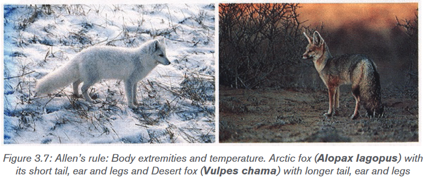
Both Bergmann’s rule and Allen’s rule depend on simple principle that “the ratio
of surface area to volume of an object is inversely proportional to the volume of the
object”. In other words, the smaller an animal is, the higher the surface area-to-
volume ratio. Higher surface area-to-volume ratio ensures these animals to lose
heat relatively quickly and cool down faster, so they are more likely to be found
in warmer climates. Larger animals, on the other hand, have lower surface area-
to-volume ratios and lose heat more slowly, so and they are more likely to be
found in colder climates.3. Insulation: All the marine mammals have a thick insulating layer of fat
known as Blubber, just beneath the skin. It covers the entire body of animals
such as seals, whales, and walruses (except for their fins, flippers, and flukes)
and serves to stores energy, insulates heat, and increases buoyancy. Thickness
of blubber can range from a couple of inches in dolphins and smaller whales,
to 4.3 inches in polar bears to more than 12 inches in some bigger whales. To
insulate the body, blood vessels in blubber constrict in cold water. Constriction
of the blood vessels reduces the flow of blood to the skin and minimizes the
heat loss. In such animals, skin surface temperature is nearly identical to the
surrounding water, though at a depth of around 50 mm beneath the skin, the
temperature is the same as their core temperature.Some marine mammals, such as polar bears and sea otters, have a thick fur
coat, as well as blubber, to insulate them. The blubber insulates in water
while fur insulates in air or terrestrial environment. The feathers of the birds also
function in insulating the body from cold temperature.Physiological Adaptations
1. Evaporation: In a cold region, i.e., when the surrounding environment of
the animal is cold than the body temperature, conduction and radiation are
the main ways an animal will dissipate heat. However, in warmer region, the air
temperature is often higher than the animal’s body temperatures, so the only
physiological thermoregulatory mechanism available is evaporation. Animals
use three evaporative cooling techniques that include sweating, panting, and
saliva spreading.(a) Sweating: It is the loss of water through sweat glands found in the skin of
mammals. The number of sweat glands can vary from none in whales, few in dogs
to numerous in humans. Most small mammals do not sweat because they would
lose too much body mass if they did. For example, in a hot desert the amount
of water a mouse would lose through sweating to maintain a constant body
temperature would be more than 20% of its body weight per hour, which could
be lethal for the animal. Therefore, smaller mammals use other techniques to cool
down their body. On the other hand, sweating is an important thermoregulatory
mechanism for primates including humans. An adult human can loss as much as
10–12 litres of water per day through sweating.(b) Panting: It is rapid, shallow respiration that cools an animal by increased
evaporation from the respiratory surfaces. It is a common thermoregulatory
technique used by small animals like dogs and rodents to loss heat.(c) Saliva spreading: It is a means of thermoregulation used by marsupials.
Under extreme heat, saliva will drip from animal’s mouth and is then wiped on its
fore and hind legs. This technique induces the cooling effect of evaporation by
wetting the fur. However, since the animal cannot spread saliva while moving,
they need to adapt other evaporative techniques during such situation.2. Counter current mechanism: As mentioned above, in addition to its role in
the transport of oxygen and food, circulatory system of our body is responsible for
distribution of heat throughout the body. This is true in case of both endotherms
and ectotherms. In endotherms, most of the body heat is generated in brain,
liver, heart and skeletal muscles. This heat is transported to other parts of the
body through blood. On the other hand, in ectotherms, the circulatory system
help in transporting heat from skin to others body parts. The counter current
heat exchanger is generally located in body extremities like limbs, neck, gills,
which are directly in contact to the external environment.In cold region, when the warm blood flows through the arteries, the blood gives
up some of its heat to the colder blood returning from the extremities in the
veins running parallel to the arteries. Such veins are located in the deeper side
of the body and carry the warm blood to the heart and most of the body heat is
retained. Such mechanism can operate with remarkable efficiency. For instance,
a seagull can maintain a normal temperature in its torso while standing with its
unprotected feet in freezing water (Figure 3.8).When the external temperature is higher than the body temperature and heat
loss is not a problem, most of the venous blood from the extremities returns
through veins located near the surface. If the core body temperature becomes
too high, the blood supply to the surface and extremities of the body is increased
enabling heat to be released to the surroundings.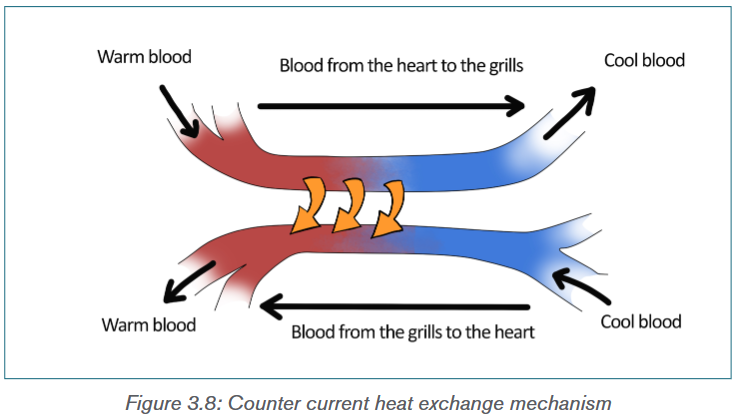
3. Hyperthermia: Hyperthermia is a condition of having the body temperature
greatly above the normal. Although all the endotherms can maintain a constant
body temperature, some are able to raise their body temperature as a way
to decrease the amount of water and energy used for thermoregulation. For
example, camels and gazelles can increase their body temperature by 5–7°C
during the day when the animal is dehydrated. Hyperthermia helps in saving
water by letting their body temperature increase instead of using evaporative
cooling to keep it at a constant temperature.4. Water retention: Human body obtains about 60 per cent of the water they
need from ingested liquid, 30 per cent from ingested food, and 10 per cent from
metabolism. While rodent adapted to arid conditions obtains approximately 90
per cent from metabolism and 10 per cent from ingested food. The predaceous
marsupial Mulgara species can go its whole life without ingesting water but by
obtaining water from the food they eat and from metabolism. The fawn hopping
mouse eats seed, small insects, and green leaves for moisture, and Kowaris eat
insects and small mammals to obtain water. These animals have specialized
kidneys with extra microscopic tubules to extract most of the water from their
urine and return it to the blood stream. And much of the moisture that would be
exhaled in breathing is recaptured in the nasal cavities by specialized organs.Many desert dwelling insects tap plant fluids such as nectar or sap from stems,
while others extract water from the plant parts they eat, such as leaves and
fruit. The abundance of insects permits insectivorous birds, bats and lizards
to thrive in the desert. Elf owls survive on katydids and scorpions. Pronghorns
can survive on the water in cholla fruits. Kit foxes can satisfy their water needs
with the water in their diet of kangaroo rats, mice, and rabbits, along with small
amounts of vegetable material.5. Excretion: As mentioned above, desert dwelling mammals and birds have
specialized kidneys with long loops of Henle compared to animals that live in
aquatic environments and less arid regions. A longer tubules help in reabsorbing
most of the water from their urine and return it to blood stream. As a result, the
urine becomes highly concentrated. In these animals, most of the water in the
faeces gets reabsorbed in the alimentary canals and colon. Camels produce
dryer faeces than other ruminants. For example, sheep produce faeces with 45
per cent water after 5 days of water deprivation, while camels produce faeces
with 38 per cent water even after 10 days of water deprivation. The ability to
excrete concentrate urine and dry faeces is an important adaptation to arid
conditions. Desert rodents can have urine five times as concentrated as that of
humans.Behavioural adaptations
Behavioural adaptations are used to reduce the amount of heat gained or lost by
animals, and, thereby, reducing the amount of energy and water to maintain the
body temperature. Ectoderms or cold blooded animals rely on their behaviour to
maintain a favourable body temperature.1. Nocturnality: It is the simplest form of behavioural adaptation characterized
by activity during the night and sleeping during the day. As such, nocturnal
animals avoid direct exposure to heat of the day, thereby preventing loss of
water needed for evaporative cooling. The night temperatures are generally
15–20°C colder than the daytime, so require much less energy and water to
regulate body temperature. Most of the desert animals like quoll, bilby, and the
spinifex hopping mouse, are nocturnal. Other large animals like lions prefer to
hunt at night are to conserve water.Crepuscular animals are those animals that are mainly active during twilight
(i.e., the period before dawn and that after dusk). Examples include hamsters,
rabbits, jaguars, ocelots, red pandas, bears, deer, moose, spotted hyenas etc.
Many moths, beetles, flies, and other insects are also crepuscular in habit.
These crepuscular animals take advantage of the slightly cooler mornings and
evenings to escape the daytime heat, and to evaporate less water.2. Microhabitat: Among the diurnal animals (animals which are mainly active
during the day and rest during night), the use of microhabitat like burrows, shade
is another type of behavioural adaptation to avoid the daytime heat. Fossorial
animals (digging animals), such as mulgaras, spent much of their time below
ground eating stored food. Lizards and snakes seek a sunny spot in the morning
to warm up their body temperatures more quickly and remain in shade when the
temperature rises.3. Migration: It is the physical movement of animals over a long distance
from one area to another. It is found in all major animal groups, including birds,mammals, fish, reptiles, amphibians, insects, and crustaceans. Many factors
like climate, food, the season of the year or mating could lead to migration. It
helps the animals in avoiding the extreme environmental conditions by moving
to more favourable places. For example, many migratory birds like arctic tern
(Sterna paradisaea) migrate north-south, with species feeding and breeding in
high northern latitudes in the summer, and moving some hundreds of miles
south during the winter to escape the extreme cold of north. Monarch butterflies
spend the summer in Canada and the Northern America and migrate as far
south as Mexico for the winter.4. Hibernation and Aestivation: Warm blooded animals which do not
migrate generally survive the extreme cold condition of winter by sleeping.
Hibernation is the state of dormancy during the cold conditions, i.e., winter.
During hibernation, body temperature drops, breathing and heart rate slows,
and most of the body’s metabolic functions are put on hold in a state of quasi-
suspended animation. This allows them to conserve energy, and survive the
winter with little or no food.Many insects spend the winter in different stages of their lives in a dormant
state. Such phenomenon is known as diapause. During diapauses, insect’s
heartbeat, breathing and temperature drop. Some insects spend the winter as
worm-like larvae, while others spend as pupae. Some adult insects die after
laying their eggs in the fall and eggs hatch into new insects in the spring when
the food supply and temperature become favorable.Aestivation or summer dormancy on the other hand, is a state of animal dormancy,
characterized by inactivity and a lowered metabolic rate, in response to high
temperatures and arid conditions. It allows an animal to survive the scarcity of
water or food as aestivating animal can live longer off its energy reserves due
to the lowered metabolism, and reduced water loss though lowered breathing
rates. Lung fishes, toad, salamander, desert tortoise, swamp turtles are some of
the other non-mammalian animals which undergo aestivation.5. Social behavior: Among all the adaptations, living together is one of the
most important adaptations of the animal kingdom. Animals can derive a lot of
benefit from spending time with other members of the same species like finding
food, defense against predators and care for their young. For example, emperor
penguins can survive the harsh Antarctica winter huddling together in groups
that may comprise several thousand penguins. Huddling greatly reduces the
surface area of the group compared to individuals and a great deal of warmth
and body fat is conserved. Many social mammals, including many rodents, pigs
and primates survive extreme cold by huddling together in groups.6. Locomotion: Different types of locomotion require varying amount of energy.
Many mammals like kangaroo, hares hop, which is an energy efficient type oflocomotion. When animals go from walking to running, there is an increasing
energy cost; however, once kangaroos start moving, there is no additional
energy cost. This is because when a kangaroo lands, energy is stored in the
tendons of its hind legs which is used to power the next hop.Application activity 3.3
1) The figure below shows different animals living in different climates
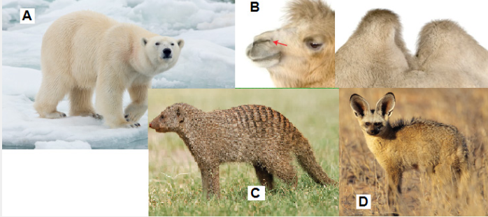
a) Which animal(s) on the photo appears to be adapted to live in cold
climates? Why?
b) Which animal(s) on the photo appears to be adapted to live in hot
climates? Why?
c) What are the adaptations of the animal A that help it to survive in its
environment?
d) What is the functions of the humps on the animal B?
e) Some animals such as the animal A hibernate during the winter. Explain
the importance of hibernation to these animals.3.4 Response to cold and hot conditions by endothermic
and ectothermic animalsActivity 3.4
1) The figure below shows different animals living in different climates
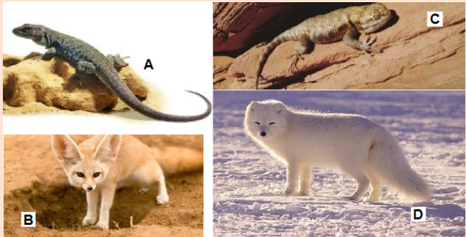
a) The animals A and B are reptiles under different environmental
conditions. Compare their behaviors in regards to how they regulate
their temperature.
b) The animals’ C and D are mammals under different environmental
conditions. Compare their behaviors in regards to how they regulate
their temperature.
c) What are the adaptations of the animal D that help it to survive in its
environment?
d) How is the animal A different to animal D according to how they regulate
their body temperature.3.4.1 Endotherms’ response to temperature changes
Endothermic organism can maintain relatively high body temperatures within a
narrow range. Since most of the body heat is produced as a result of various
metabolic activities, thermoregulation in endotherms depends on food and
water availability. For example, bear undergoes hibernation during the winter
because there is no sufficient food during the cold season. On the other hand,
in arid environment like deserts, many deserts animals are nocturnal to avoid the
extreme daytime heat to avoid loss of water through evaporation.Response to hot temperature
When the body temperature increases in response to the external temperature,
the body’s temperature control system uses three important mechanisms to
reduce the body heat. These are:1. Vasodilation of blood vessels in the skin: The blood vessels in skin become
intensely dilated due to the inhibition of the sympathetic centres in the posterior
hypothalamus that cause vasoconstriction. Vasodilation increases the rate blood
flow to the skin and as a result, the amount of heat transfer from the core of the
body increases tremendously.2. Sweating: As discussed in the previous section, sweating is an important
adaptation to lose body heat through evaporative cooling. An increase in 1°C in
body temperature causes enough sweating to remove ten times the basal rate
of body heat production.3. Decrease in heat production: As mentioned above, metabolic activities of
the body are the main source of body heat. The mechanisms that cause excess
heat production, such as shivering and chemical thermogenesis, are strongly
inhibited when exposed to hot temperature.Response to cold temperature
In response to cold temperature, the temperatures control system performs
exactly opposite mechanism to that performs in hot temperature. These are:1. Vasoconstriction of blood vessels in the skin: The blood vessels in the skin
constrict under the influence of posterior hypothalamic sympathetic centres
which reduce the blood flow to the skin.2. Piloerection: Piloerection means hairs “standing on end”. Sympathetic
stimulation causes the arrector pili muscles attached to the hair follicles to
contract, which brings the hairs to an upright stance. The upright projection of
the hairs allows them to entrap a thick layer of air next to the skin which acts as
insulator, so that transfer of heat to the surroundings is greatly depressed.3. Increase in heat production (thermogenesis): Endothermic metabolic
rates are several times higher than those of ectotherms. The metabolic heat
production of endotherms is regulated in response to fluctuations in the
environment temperature. This phenomenon is known as adaptive thermogenesis
or facultative thermogenesis. It can be defined as “Heat production by metabolic
processes in response to environmental temperature with the purpose of
protecting the organism from cold exposure and buffering body temperature
from environmental temperature fluctuations”. Under cold temperature stress,
heat production by the metabolic activities increased tremendously by promoting
shivering, sympathetic excitation of heat production, and thyroxine secretion.These mechanisms will be discussed later. Extreme shivering can increase the
temperature four to five times the normal production.3.4.2 Ectotherms’ response to temperature changes
Ectotherms cannot maintain stable body temperature and their body temperature
relies on the external temperature. They depend more on energy assimilation
rather than utilizing it for temperature regulation. Therefore, ectotherms regulate
their body temperature behaviourally and by cardiovascular modulation of
heating and cooling rates. At the same time, metabolism and other essential
rate functions are regulated so that reaction rates remain relatively constant
even when body temperatures vary. This process is known as acclimatization or
temperature compensation. For example, many fish adjust metabolic capacities
to compensate for seasonal variation in water temperature with the result that
metabolic performance remains relatively stable throughout the year. Reptiles
often regulate their body temperature to different levels in different seasons
to minimize the behavioural cost of thermoregulation. At the same time, tissue
metabolic capacities are adjusted to counteract thermodynamically-induced
changes in rate functions.Response to hot temperature
When the external temperature increases, ectotherms protect their bodies from
overheating using various mechanisms. These are:1. Use of microhabitat: Under extreme heat conditions, many ectotherms like
lizards and snakes prefer to stay in shade, either beneath the rocks, crevices or
underground burrows.Amphibians and fishes enter cold water when their body temperature increases.
2. Acclimatization: If a salamander living at 10°C is exposed to 20°C, its
metabolic rate increases rapidly. But if the exposure to the higher temperature
lasts for several days, the animal experiences a compensating decrease in the
metabolic rate. This decrease in the metabolic rate is due to acclimatization.
The higher metabolic rate is due to the increase in the enzymes activity with
temperature. However, with prolonged exposure to the condition, the metabolic
rates decrease to prevent excessive energy loss. Ectotherms also exhibit
acclimatization of temperature tolerance range with animal acclimated to high
temperature are able to tolerate higher temperature than those exposed only to
low temperature. Similarly, cold acclimated animals have better tolerance to low
temperature than high temperature acclimated animal.Response to cold temperature
Ectotherms response to cold temperature is exactly opposite to the response
shown when exposed to hot temperature. That is:1. Basking to sun: When the body temperature of the ectotherms becomes
colder than the normal, the animals either bask to sunlight to warm up the body
or move to a warmer place. Under extreme cold conditions, all the metabolic
activities may cease and the animals enter the state of torpor (reduced metabolic
activities).2. Cold Acclimatization: Decrease in the temperature result in reduced
metabolic rate. Therefore, as a compensatory measure to meet the require body
metabolism, the cold acclimatization of ectotherms is characterized by increase
in concentration of various metabolic enzymes. There is also significant increase
in the mitochondria and capillaries concentration in the skeletal muscle. This
increase the ATP production through aerobic respiration in these tissues.
Therefore, in those animals which have prolonged exposure to cold temperature,
there may be increase in the locomotion, though the basal rates of metabolism
remain below the warm acclimatized animals.Application activity 3.4
1. a) Describe the importance of hibernation to animals.
b) The camel is one of the animals adapted to live in deserts. Explain
three of its adaptations that help it to survive in arid conditions.
c) State three adaptations of animals to living in cold climates.3.5 Role of the brain
Activity 3.5
Find information about the role of hypothalamus and different thermoreceptors
in temperature regulation. Make short notes and present them in front of the
class.So far we have discussed that on the basis of types of thermoregulation, all the
living organisms can be classified into two groups – ectotherms and endotherms.
Endotherms can regulate their body temperature within a narrow range through
various physiological mechanisms while ectotherms being depended on external
temperature mostly rely on their behaviour to maintain body temperature. But
how do these animals sense and counter the changing temperature of their
body will be discussed in the section.Thermoreceptors
A thermoreceptor is a sensory receptor which is basically the receptive
portion of a sensory neuron that converts the absolute and relative changes
in temperature, primarily within the innocuous range to nerves impulses.
Thermoreception is the sense by which an organism perceives the
temperature of the external and internal environment from the information supply
by thermoreceptors. In vertebrates, most of the thermoreceptors are found in
skins which are actually free nerve endings. Deep body thermoreceptors are
also found mainly in the spinal cord, in the abdominal viscera, and in or around
the great veins in the upper abdomen and thorax region.Mammals have at least two types of thermoreceptors: the warm receptors,
those that detect heat or temperatures above normal body temperature and cold
receptors, those that detect cold or temperatures below body temperature. The
warm receptors are generally unmyelinated nerves fibres, while cold receptors
have thinly myelinated axons and hence faster conduction velocity. Increasing
body temperature results in an increase in the action potential discharge rate
of warm receptors while cooling results in decrease. On the other hand, cold
receptors’ firing rate increases during cooling and decreases during warming.
Another types of receptor called nociceptors, detect pain due to extreme cold
or heat which is beyond certain threshold limits.A specialized form of thermoception known as distance thermoreception is found
in some snakes like pit viper and boa, use a specialized type of thermoreceptor
which can sense the infrared radiation emitted by hot objects. The snake’s
face has a pair of holes, or pits, lined with temperature sensors. These sensors
indirectly detect infrared radiation by its heating effect on the skin inside the pit
which helps them to locate their warm blooded prey. The common vampire bat
may also have specialized infrared sensors on its nose.Hypothalamus
The hypothalamus is a very small, but extremely important part of the brain
that acts as the link between the endocrine and nervous systems of the
body. The hypothalamus plays a significant role in the endocrine system and is
responsible for maintaining the body’s homeostasis by stimulating or inhibiting
many key processes, including body temperature, fluid and electrolyte
balance, appetite and body weight, glandular secretions of the stomach
and intestines, production of substances that influence the pituitary gland to
release hormones and sleep cycles.Role of Hypothalamus in thermoregulation
Thermoregulation is carried out almost entirely by nervous feedback mechanisms,
and almost all these operate through temperature-regulating centres located inthe hypothalamus (Figure 3.7). The hypothalamus contains large numbers of
heat-sensitive as well as cold sensitive neurons which acts as thermoreceptor,
sensing the temperature of the brain. The posterior hypothalamus region
contains the thermoregulatory centre which integrate the signals from of all
the thermoreceptors found in skin, deep organs and skeletal muscles, as well
as from the anterior hypothalamus and control the heat-producing and heat-
conserving reactions of the body.Cooling Mechanism
When the body temperature increases beyond the set-point, the anterior
hypothalamus is heated. The posterior hypothalamus senses the heat and
inhibits the adrenergic activity of the sympathetic nervous system, which control
vasoconstriction and metabolic rate. This causes cutaneous vasodilation and
increase heat loss through skin. It also reduces the body metabolic rate resulting
in decreasing heat production through metabolic reactions. Under intense
heating, the cholinergic sympathetic fibres innervating the sweat glands release
acetylcholine, stimulating the secretion of sweat. Many behavioural responses
to heat, such as lethargy, resting in shade, lying down with limbs spread out,
etc., decreases heat production and increases heat loss.Heating Mechanism
When the body temperature falls below the set-point, the body regulating
mechanism tries to reduce heat loss and increase heat production. The
immediate response to cold is vasoconstriction throughout the skin. The
result is vasoconstriction of the skin blood vessels, reducing the blood flow
and subsequent heat loss through skin. Sympathetic stimulation also causes
piloerection and reduces the heat loss from the body by trapping heat within
the body hair.The primary motor centre for shivering is excited by the cold signals from skin
and spinal cord which cause shivering of the skeletal muscles. Intense shivering
can increase the body heat production four to five times normal. Cooling
the anterior hypothalamic due to decrease in body temperature stimulates
hypothalamus to increases the production of the neurosecretory hormone
thyrotropin-releasing hormone. This hormone in turn stimulates the anterior
pituitary gland, to secrete thyroid-stimulating hormone. Thyroid-stimulating
hormone then stimulates thyroid glands to increased output of thyroxine. The
increased thyroxine level in the blood increases the rate of cellular metabolism
throughout the body and hence increases heat production.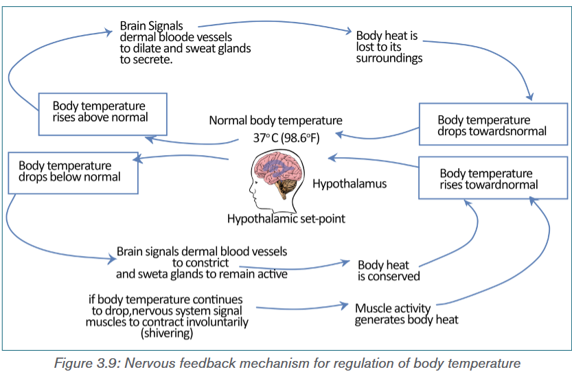
Application activity 3.5
1) The diagram shows the way in which temperature is regulated in body
of a mammal.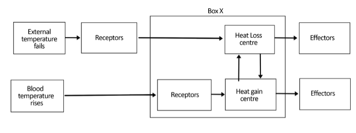
a) Which part of the brain is represented by box X?
b) i) How does the heat loss center control the effectors which lower the
body temperature?
ii) Explain how blood vessels can act as effectors and lower the body
temperature?3.6 Temperature controls in plants
Activity 3.6
Observe carefully the photos below and answer to the questions that follow:
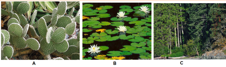
a) In which habitat do these plants live?
b) What are the adaptations of plant A that help it to survive in its
environment?
c) Make a comparison between plant A and plant B.Like all the other living organisms, plants depend on enzymes catalyzed chemical
reactions for their growth and development. For example, plants synthesize their
own food from water and carbon dioxide using sunlight through photosynthesis.
The process of photosynthesis involves a series of complex enzyme system
and other proteins. Therefore, along with carbon dioxide, water, light, nutrients
and humidity, temperature is also one of the limiting factors for growth and
development of plants.Unlike animals, plants remain fixed in a particular site and absorb heat from the
sunlight. The excess heat from the body is released to the surrounding through
radiation and evaporation. The process of evaporation of water from the leaves
and stem of plants to the surrounding environment is known as transpiration. It
occurs through stomata, small opening located on the underside of the leaves.
The stomata are specialized cells in the leaves which can open or close, limiting
the amount of water vapour that can evaporate. Higher temperature causes the
opening of stomata whereas colder temperature causes the opening to close.
The opening of the stomata and hence the transpiration rate of plants depends
on environmental conditions such as light, temperature, the level of atmospheric
CO2 and relative humidity. Higher relative humidity leads to more opening,
while higher CO2 levels lead to closing of stomata. Under high environmental
temperature, the plant body gets heat up. In order to cool down, the plant
increases its transpiration rate. The evaporative loss of water from the plant’s
body lowers the temperature.Besides transpiration, many plants have different adaptations that help them
survive in extreme temperature conditions ranging from hot and arid deserts
to cold and snow covered mountains. These adaptations make it difficult for
the plant to survive in a different place other than the one they are adapted to.
This explains why certain plants are found in one area, but not in another. For
example, cactus plants, adapted to desert conditions can’t survive in the Arctic.These adaptations will be discussed later in this unit.
3.6.1 Effect of temperature changes on plants
The most obvious effect of temperature on plants is changes in the rate of
photosynthesis and respiration. Both processes increase with rise in the
temperature up to a certain limit. However, increase in temperature beyond the
limits, the rate of respiration exceeds the rate of photosynthesis and the plants
productivity decreases.Another important effect of temperature is during the process of germination
of seeds. Like most other processes it also depends on various factors
including air, water, light, and, of course, temperature. In many plant species,
germination is triggered by either a high or low temperature period that destroys
germination inhibitors. This allows the plant to measure the end of winter season
for spring germination or end of summer for fall germination. For example, winter
adapted plant seeds remain dormant until they experience cooler temperatures.
Temperature of 4°C is cool enough to end dormancy for most cool dormant
seeds, but some groups, especially within the family Ranunculaceae and others,
need conditions cooler than –5°C. On the other hand, some plants like Fire
poppy (Papaver californicum) seeds will only germinate after hot temperatures
during a forest fire which cracks their seed coats. The fire does not cause direct
germination, rather weakens the seed coat to allow hydration of the embryo.Pollination is another phenological stage of plants sensitive to temperature
extremes across all species. Since pollination is carried out by pollinators like
honey bees, butterflies etc., any factors including temperature that affect these
pollinators will certainly affect the process.Heat adapted plants
In extremely hot and dry desert region with annual rainfall averages less than
10 inches per year, and there is a lot of direct sunlight shining on the plants,
the main strategy for the survival of the plants is to avoid extensive water loss
through transpiration. Therefore, in such region many plants called succulents,
like cactus can store water in their stems or leaves. Some plants are leafless
or have small seasonal leaves that only grow after rains. These leafless plants
conduct photosynthesis in their green stems. Leaves are often modified into
spines to discourage animals from eating plants for water. Also waxy coatingon stems and leaves help reduce water loss. Other plants have very long root
systems that spread out wide or go deep into the ground to absorb water.On the other hand, in hot and humid tropical rainforest, the abundance of water
can cause problems such as promoting the growth of bacteria and fungi which
could be harmful to plants. Heavy rainfall also increases the risk of flooding, soil
erosion, and rapid leaching of nutrients from the soil. Plants grow rapidly and
quickly use up any organic material left from decomposing plants and animals.
The tropical rainforest is very thick, and not much sunlight is able to penetrate
to the forest floor. However, the plants at the top of the rainforest in the canopy
must be able to survive the intense sunlight. Therefore, the plants in the tropical
rainforest usually have large leaves with drip tips and waxy surfaces allow water
to run off easily. Some plants grow on other plants to reach the sunlight.Similarly, in aquatic plants adapted for life in water, the leaves are very large,
fleshy and waxy coated. Increase surface area allows plants to lose excess
water while the shiny wax coating discourages the growth of microbes. The
roots and stems are highly reduced since water, nutrients, and dissolved gases
are absorbed from the water directly through the leaves.Cold adapted plants
In extremely cold region like tundra which is characterized by a permanently
frozen sub-layer of soil called permafrost, the drainage is poor and evaporation
slow. With the region receiving very little precipitation, about 4 to 10 inches
per year usually in the form of snow or ice, plant life is dominated by small,
low growing mosses, grasses, and sedges. Plants are darker in colour, some
even red which helps them absorb solar heat. Some plants are covered with
hair which helps keep them warm while others grow in clumps to protect one
another from the wind and cold.In a slightly warmer temperate forest, with temperature varies from hot in the
summer to below freezing point in the winter, many trees are deciduous that is
they drop their leaves in the autumn to avoid cold winter, and grow new ones in
spring. These trees have thin, broad, light-weight leaves that can capture a lot
of sunlight to make a lot of food during the warm weather and when the weather
gets cooler, the broad leaves cause too much water loss and can be weighed
down by too much snow, so the tree drops its leaves. They usually have thick
bark to protect against cold winters.Application activity 3.6
1) The diagram below shows a transverse section of a leaf Ammophila
arenaria, which is a xerophyte. The photomicrograph shows the details
of the area indicated by the box in the diagram.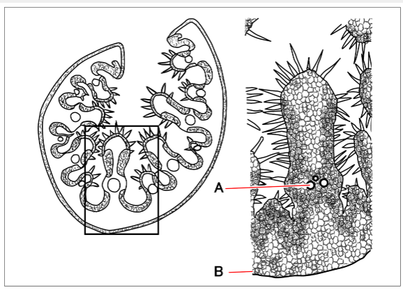
a) Name the parts labelled A and B.
b) Describe two xeromorphic features shown in this leaf and, in each case,
indicate how the feature helps to reduce transpiration.Skills Lab 3

Procedure:
1) Wash your hands with soap and water and dry them properly.
2) Prepare the blood glucose meter with the test strip according to the
manufacturer’s instructions.
3) Use the lancet device to prick the side of your fingertip with a lancet.
4) Place a drop of blood onto the correct part of the test strip.
5) The strip will draw up the blood into the meter and show a digital
reading of the blood glucose level within seconds.
6) Note the reading.
7) Use a clean cotton ball to apply pressure to the fingertip for a few
moments until the bleeding stops.
8) Similarly, measure the blood glucose level of your friends.
9) Compare your blood glucose level with that of your friends.Discussion:
In general, a fasting blood glucose reading (taken before a meal) should be
between 72 mg/dL to 126 mg/dL. And a blood glucose reading 2 hours after
a meal should be between 90 mg/dL to 180 mg/dL.Precautions:
1) Make sure the lancelet is properly sterilized.
2) Insert the test strip properly.End unit assessment 3
I. Multiple Choice Questions
1) Which of the following monosaccharides is not a product of
carbohydrate metabolism in our body?
(a) Glucose (b) Fructose (c) Ribose (d) Galactose2) Which of the following is not a part of portal triad?
(a) Central vein (b) Hepatic artery
(c) Hepatic portal vein (d) Bile duct.3) Somatostatin is secreted by
(a) Alpha cells (b) Beta cells
(c) Delta cells (d) F cellsThe process of formation of glucose from non-carbohydrates source in the
body is known as
(a) Glycogenesis (b) Gluconeogenesis
(c) Glycolysis (d) Glycogenolysis5) Which of the following hormone is responsible for decreasing blood
glucose level?
(a) Glucagon (b) Insulin (c) Somatostastin (d) Adrenaline6) The enzyme used in the dipstick for testing concentration of glucose is
(a) Glucose oxidase (b) Glycogen phosphorylase
(c) Glucose phosphatase (d) GlucosidaseII. State whether the following statements are True (T) or False (F)
1) Excess glucose in the body is stored in the form of glycogen.
2) Trypsin is an enzyme used for carbohydrate digestion.
3) Bile salt is secreted by exocrine liver.
4) Glucagon is secreted by pancreas in response to high blood glucose
concentration.
5) Insulin administration is recommended for person with type II diabetes
mellitus.6) Type I diabetes mellitus is cause due to insufficient secretion of insulin
by beta cells.
7) Ketone bodies are formed when our body have excessive fat metabolism.
8) Hyperinsulinaemia is associated with type II diabetes mellitus.
9) All the living organisms have a particular range of temperature within
which they can best survive and reproduce.
10) Nocturnality is the simplest form of behavioral adaptation characterized
by activity during the day and sleeping during the night.
11) Crepuscular animals take advantage of the slightly cooler mornings
and evenings to escape the daytime heat, and to evaporate less water.
12) Body temperature of Ectotherms rely on the external temperature.
13) Thermoregulation in endotherms depends on food and water availability.
14) Glycogenolysis is the breakdown of glucose to form pyruvate.III Long Answer Type Questions
1) List few adaptive features shown by plants inhabiting extreme cold and
hot environments.
2) Explain the role of the brain and thermoreceptors in temperature
regulation.
3) In your own words, explain the importance of maintaining fairly constant
temperatures for efficient metabolism.
4) Describe the functions of liver and pancreas in regulating blood
glucose level.
5) Discuss in brief the importance of urine analysis in diagnosis diabetes
mellitus.
6) The control of blood glucose concentration involves a negative
feedback mechanism.
a) What are the stimuli, receptors and effectors in this control mechanism?
b) Explain how negative feedback is involved in this homeostatic
mechanism.7) An investigation was carried out to determine the response of
pancreatic cells to an increase in the glucose concentration of the
blood. A person who had been told not to eat or drink anything other
than water for 12 hours then took a drink of a glucose solution. Blood
samples were taken from the person at one hour intervals for five hours,
and the concentration of glucose, insulin and glucagon in the blood
and the concentration of glucose, insulin and glucagon in the blood
were determined. The results are shown in the graph below: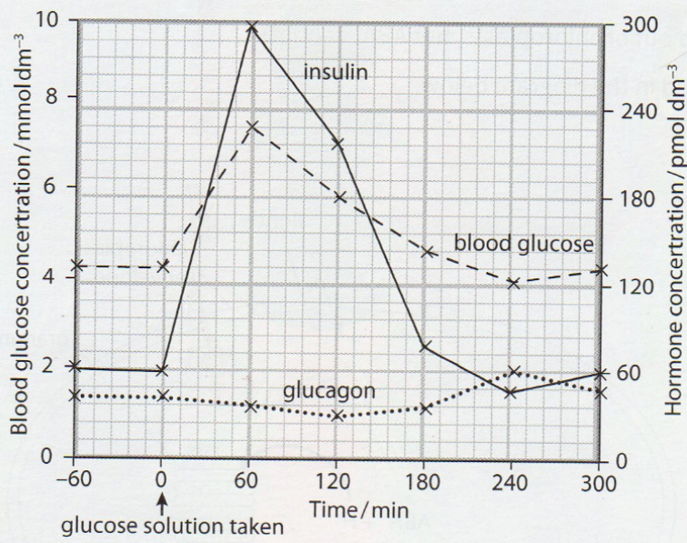
a) Explain why the person was told not to eat or drink anything other than
water for 12 hours before having the glucose drink.
b) Use the information in the figure to describe the response of the
pancreatic cells to an increase in the glucose concentration.
c) Outline the role of insulin when the glucose concentration in the blood
increases.
d) Suggest how the results will change if the investigation continued longer
than five hours without the person taking any food.
e) Outline the sequence of events that follows the binding of glucagon to
its membrane receptor on a liver cell.UNIT 4: PRINCIPLES OF GENE TECHNOLOGY AND ITS APPLICATIONS
Key unit competence
Explain the principles of gene technology and evaluate how gene technology
is applied in areas of medicine, forensic science and agricultureIntroductory activity 4
You hear about them all the time. They are often depicted in cartoons, comic
books, movies, and science fiction as mad scientists. These are the scientists
who take a gene from one organism and place it into an unrelated organism.
These are the scientists who make hormones that farmers inject into the cows
that produce the milk we drink.These are the scientists who modify the crops we eat, creating what some
people call “Franken foods” or genetically modified organisms. The figure
below shows a GMO tomato and a GMO rice also called golden rice.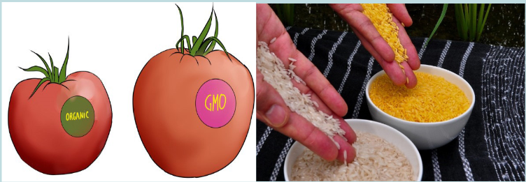
You may have wondered if it might soon be possible to replace a beloved
family member or pet, or bring back extinct species through cloning, or even
clone yourself. You might worry about a future where parents unwilling to fix
their children’s “genetic defects” face discrimination.Use the image above and your own knowledge to answer the questions
that follow:
a) Who are these scientists who make such manipulations?
b) What do they do?
c) What are the basic tools that these scientists use?
d) What are the possible products that they do?
e) Is anyone trying to determine if it is unhealthy to eat these modified
foods, whether genetically modified plants will cause environmental
problems, or if genetically modified animals are less healthy than their
counterparts?4.1 Recombinant DNA technology
Activity 4.1
arpenters require tools such as hammers, screwdrivers, and saws; surgeons
require scalpels, forceps, and stitching needles; and mechanics require hoists,
wrenches, and pumps. These individuals use their implements to modify,
deconstruct, or build a system that they are working with. Just like any other
technicians, molecular biologists use tools to complete a project. The tools
in their laboratories may aid them in investigating genetic disorders, altering
the genetic makeup of organisms so that they produce useful products such
as insulin, or analysing DNA evidence in a criminal investigation. Find out the
possible tools used in recombinant DNA technology and their functions.Genetic engineering, also known as recombinant DNA (rDNA) technology,
means altering the genes in a living organism to produce a Genetically Modified
Organism (GMO) with a new genotype. Various kinds of genetic modification
are possible: inserting a foreign gene from one species into another, forming a
transgenic organism; altering an existing gene so that its product is changed; or
changing gene expression so that it is translated more often or not at all.4.1.1 Techniques of genetic engineering
Genetic engineering is a very young discipline, and is only possible due to
the development of techniques from the 1960s onwards. These techniques
have been made possible from our greater understanding of DNA and how it
functions following the discovery of its structure by Watson and Crick in 1953.
Although the final goal of genetic engineering is usually the expression of a genein a host, in fact most of the techniques and time in genetic engineering are
spent isolating a gene and then cloning it.An overview of gene transfer
There are many different ways in which a GMO may be produced, but these
steps are essential.
• The gene that is required is identified. It may be cut from a chromosome,
made from mRNA by reverse transcription or synthesized from nucleotides.
• Multiple copies of the gene are made using the technique known as the
polymerase chain reaction (PCR).
• The gene is inserted into a vector which delivers the gene to the cells of
the organism. Examples of vectors are plasmids, viruses and liposomes.
• The vector takes the gene into the cells.
• The cells that have the new gene are identified and cloned.To perform these steps, the genetic engineer needs a ‘tool kit’ consisting of:
• Enzymes, such as restriction endonucleases, DNA ligase and
reverse transcriptase
• Vectors, including plasmids and viruses
• Host cell, a living system (microbial, plant, animal) in which the vector can
be propagated.
• Genes coding for easily identifiable substances that can be used as
markers.
4.1.2 Plasmids
A plasmid is a genetic structure, in some cells, that can replicate independently
of the chromosomes; it is typically a small circular DNA strand in the cytoplasm
of a bacterium or protozoan. Plasmids are much used in the laboratory during
manipulation of genes as vectors.The properties of plasmids are:
– It is big enough to hold the desired gene.
– It is circular (or more accurately a closed loop), so that it is less likely
to be broken down.
– It contains control sequences, such as a transcription promoter, so that
the gene will be replicated or expressed.
– It contains marker genes, so that cells containing the vector can be
identified.Plasmids are not the only type of vector that can be used. Viruses can also
be used as vectors. Another group of vectors are liposomes, which are tiny
spheres of lipid containing the DNA.The production of genetically modified organisms (GMO), also called transgenic
organisms, is a multistage process which can be generally summarized as
follows:
– Identification of the gene of interest.
– Isolation of the gene of interest.
– Cutting of gene of interest and opening of plasmid with restriction
enzymes in order to have sticky ends.
– Associating the gene with an appropriate promoter and poly -A
sequence and insertion into plasmids.
– Multiplying the plasmid in bacteria and recovering the cloned construct
for injection.
– Transference of the construct into the recipient tissue, usually fertilized
eggs.
– Integration of gene into recipient genome.
– Expression of gene in recipient genome.
– Inheritance of gene through further generations.Application activity 4.1
1) Explain briefly the terms below:
a) Recombinant DNA
b) Transgenic organism
c) Enzyme
2) State the main tools of a genetic engineer and their functions.4.2 Roles of enzymes in genetic engineering
Activity 4.2
Enzymes are biological molecules which speed up the rates of chemical
reactions in our body. There are different enzymes with different functions
used in genetic engineering. Find the information about these enzymes and
their possible functions.The enzymes involved in gene manipulation include; restriction endonucleases
(restriction enzymes), methylases, ligase and reverse transcriptase.4.2.1 Restriction enzymes
These are enzymes that cut DNA at specific sites. They are properly called
restriction endonucleases because they cut the bonds in the middle of the
polynucleotide chain. Each type of restriction enzyme recognizes a characteristic
sequence of nucleotides that is known as its recognition site. A recognition
site is a specific sequence within double-stranded DNA, usually palindromic
and consisting of four to eight nucleotides, that a restriction endonuclease
recognizes and cleaves. Molecular biologists can use these enzymes to cut
DNA in a predictable and precise manner.Most restriction enzymes make a staggered cut in the two strands, forming
sticky ends.The cut ends are “sticky” because they have short stretches of single-stranded
DNA. These sticky ends will stick (or anneal) to another piece of DNA by
complementary base pairing, but only if they have both been cut with the same
restriction enzyme. Restriction enzymes are highly specific, and will only cut
DNA at specific base sequences, 4-8 base pairs long.Restriction enzymes are produced naturally by bacteria as a defense against
viruses (they “restrict” viral growth), but they are enormously useful in genetic
engineering for cutting DNA at precise places (“molecular scissors”).
Short lengths of DNA cut out by restriction enzymes are called restriction
fragments. There are thousands of different restriction enzymes known, with
over a hundred different recognition sequences. Restriction enzymes are named
after the bacteria species they came from, so Eco R1 is from E. coli strain R.Table 4.1: List of some restriction enzymes and their respective recognition sites
The ends of DNA fragments produced from a cut by different restriction
endonucleases differ, depending on where the phosphodiester bonds are
broken in the recognition site. In the example in Table 4.1, EcoRI produces
sticky ends; that is, both fragments have DNA nucleotides that are now
lacking their respective complementary bases. These overhangs are produced
because EcoRI cleaves between the guanine and the adenine nucleotide on
each strand. Since A and G are at opposite ends of the recognition site on each
of the complementary strands, the result is the overhang. Another restriction
endonuclease, SmaI, produces blunt ends, which means that the ends of the
DNA molecule fragments are fully base paired (Table 4.1).Sticky ends are fragment end of a DNA molecule with short single stranded
overhangs, resulting from cleavage by a restriction enzyme. Blunt ends are
fragment ends of a DNA molecule that are fully base paired, resulting from
cleavage by a restriction enzyme.Restriction enzymes are named according to the bacteria from which they
originate.For example, the restriction enzyme BamHI is named as follows:
– B represents the genus Bacillus
– am represents the species amyloliquefaciens
– H represents the strain
– I mean that it was the first endonuclease isolated from this strain
– Following the same pattern, the rationale for the name of the restriction
enzyme Hind II is the following:
– H represents the genus Haemophilus
– in represents the species influenzae
– d represents the strain Rd
– II means that it was the second endonuclease isolated from this strain.Generally speaking, the first letter is the initial of the genus name of the organism
from which the enzyme is isolated. The second and third letters are usually the
initial letters of the species name. The fourth letter indicates the strain, while
the numerals indicate the order of discovery of that particular enzyme from that
strain of bacteria.4.2.2 Methylases
Restriction endonucleases must be able to distinguish between foreign DNA
and the genetic material of their own cells; otherwise a bacterium’s DNA would
be in danger of being cleaved by its own immune system. Methylases are
enzymes that add a methyl group to one of the nucleotides found in a restriction
endonuclease recognition site, altering its chemical composition. In prokaryotes,
they modify the recognition site ofa respective restriction endonuclease by placing
a methyl group on one of the bases, preventing the restriction endonuclease
from cutting the DNA into fragments. When foreign DNA is introduced into the
bacterium, it is not methylated, rendering it defenceless against the bacterium’s
restriction enzymes. Methylases are important tools for a molecular biologist
when working with prokaryotic organisms. They allow the molecular biologist to
protect a gene fragment from being cleaved in an undesired location.
4.2.3 DNA ligase
This enzyme repairs broken DNA by joining two nucleotides in a DNA strand.
It is commonly used in genetic engineering to do the reverse of a restriction
enzyme that is to join together complementary restriction fragments. The sticky
ends allow two complementary restriction fragments to harden, but only by
weak hydrogen bonds, which can quite easily be broken by gentle heating.
The backbone is still incomplete. DNA ligase completes the DNA backbone by
forming covalent bonds. T4 DNA ligase is an enzyme that originated from the
T4 bacteriophage and which is used to join together DNA blunt or sticky ends.
So, DNA ligase is able to join complementary sticky ends produced by the same
restriction enzyme via a condensation reaction:iv) Complementary sticky ends produced by Hind III
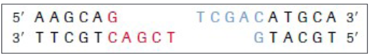
v) Hydrogen bonds form between complementary bases. DNA ligase
reconstitutes the phosphodiester bond in DNA backbones.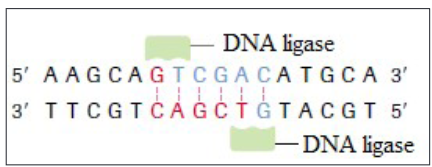
vi) If fragments are not complementary, then hydrogen bonds will not form.
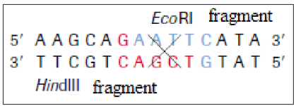
4.2.4 Reverse transcriptase
Reverse transcription is a process whereby a mRNA is converted into cDNA
(complementary DNA, also called copy of DNA). It requires the enzymes called
reverse transcriptase. It is shown by this reaction:Application activity 4.2
1) Which of the following tools of recombinant DNA technology is
incorrectly paired with its use?
a) Restriction enzyme: cut DNA into smaller segments of various sizes.
b) DNA ligase: enzyme that cuts DNA, creating the sticky ends of
restriction fragments
c) DNA polymerase: used to make many copies of DNA
d) Reverse transcriptase: production of cDNA from mRNA2) The diagram below shows the stages in the insertion of the gene for
insulin into a bacterium.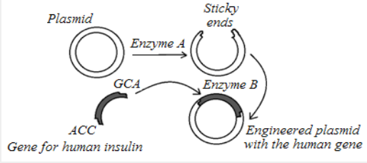
a) Name the substance that makes up the plasmid.
b) Identify the enzyme labelled A. what is its role?
c) Identify enzyme B on the diagram. What is its role?
d) What term is given to a length of DNA formed from different sources?4.3 Polymerase chain reaction (PCR)
Activity 4.3
The polymerase chain reaction is a process which can be carried out in a
laboratory to make large quantities of identical DNA from very small samples.
The process is summarized in the flowchart.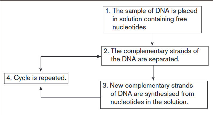
a) At the end of one cycle, two molecules of DNA have been produced
from each original molecule. How many DNA molecules will have been
produced from one molecule of DNA after 5 complete cycles?
b) Suggest one practical use to which this technique might be applied.
c) Give two ways in which the polymerase chain reaction differs from the
process of transcription.
d) The polymerase chain reaction involves semi-conservative replication.
Explain what is meant by semi-conservative replication.The Polymerase Chain reaction (PCR) is a method widely used in molecular
biology to make several copies of a specific DNA segment. Using PCR, copies
of DNA sequences are exponentially amplified to generate thousands to millions
of more copies of that particular DNA segment. DNA can clone (or amplify) DNA
samples as small as a single DNA molecule. It is a newer technique, havingbeen developed in 1983 by KARY Mullis, for which discovery he won the
Nobel Prize in 1993. The polymerase chain reaction is simply DNA replication in
a test tube. If a length of DNA is mixed with the four nucleotides (A, T, C and G)
and the enzyme DNA polymerase in a test tube, then the DNA will be replicated
many times.The polymerase chain reaction (PCR) is an automated process, making it both
rapid and efficient. It requires the following:
• The DNA fragment to be copied.
• Taq polymerase – DNA polymerase obtained from the bacterium Thermus
aquaticus, after which it is named. The bacterium lives in hot springs, and
so the remarkable feature of Taq polymerase is that it is very tolerant to
heat (it is thermostable) and does not denature at the high temperatures
of the polymerase chain reaction so that take place. DNA polymerase is
an enzyme is an enzyme capable of joining together tens of thousands of
nucleotides in a matter of minutes.
• Primers: short sequences of nucleotides that have a set of bases
complementary to those at one end of each of the two DNA fragments.
• Nucleotides: which contain each of the four bases found in DNA. They
are nucleotide triphosphate (dNTPs) as energy is required for the synthesis
of the phosphodiester bonds.
• Thermocycler: a computer-controlled machine that varies temperatures
precisely over a period of time.The polymerase chain reaction is illustrated in the figure 4.2 and is carried out
in three stages:
• Separation of the DNA double helix: the mixture containing DNA
fragments, primers, dNTPs and Taq polymerase is placed in a vessel in
the thermocycler. The temperature is increases to 95ºC causing the two
strands of the DNA fragments to separate as hydrogen bonds are broken.
• Annealing of the primers: the mixture is cooled to 55ºC causing the
primers to join (anneal) to their complementary bases at the end of
the DNA fragment. The primers provide the starting sequences for Taq
polymerase to begin DNA copying because Taq polymerase can only attach
nucleotides to the end of an existing chain. Primers also prevent the two
separate strands from simply rejoining.
• Synthesis of DNA: the temperature is increased to 72ºC. This is the
optimum temperature for the Taq polymerase to add complementary
nucleotides along each of the separated DNA strands. It begins at the
primer on both strands and adds the nucleotides in sequence until it
reaches the end of the chain.Each original DNA molecule has now been replicated to form two molecules.
The cycle is repeated from step 2 and each time the number of DNA molecules
doubles. This is why it is called a chain reaction, since the number of molecules
increases exponentially, like an explosive chain reaction. Typically, PCR is run
for 20-30 cycles. This is known as DNA amplification. The complete cycle
takes around two minutes. After only 25 cycles over a million copies of the DNA
can be made and 100 billion copies can be manufactured in just a few hours.
The PCR has revolutionized many aspects of science and medicine. Even the
minutest sample of DNA from a single hair or a speck of blood can now be
multiplied to allow forensic examination and accurate cross-matching.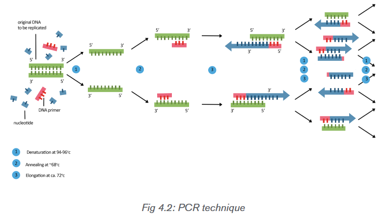
Applications of the PCR technique
PCR is useful in forensic criminal investigations, medical diagnosis, paternity
testing and genetic research, and only requires a small amount of DNA to
work. In criminal investigations, forensic scientists can find enough DNA in a
hair follicle or one cell to use as a starting point for PCR. Therefore, only a
small amount of DNA evidence is needed because it can be copied over and
over again. PCR can also improve medical diagnoses, such as confirming the
presence of the AIDS-causing virus. HIV cannot be detected immediately by
looking for antibodies, because it takes time for the body to build antibodies
against it. Traditional testing relies on the detection of these antibodies. With
PCR, primers can be designed to complement short regions of the DNA of
HIV. The DNA can be amplified and then examined for the presence of the HIV
genome. Another application of PCR is that researchers can use it to determine,
from fossil remains, whether or not two species are closely related.Application activity 4.3
1) Which of the following are required in a polymerase chain reaction?
a) DNA polymerase, template strand and primers.
b) RNA polymerase, template strand and primers
c) RNA polymerase, template strand and ligase
d) RNA polymerase, ligase and primers.
2) Each cycle of a polymerase chain reaction (PCR) takes 5 minutes. If
there are 1000 DNA molecules at the start of the reaction, how long will it
take for the number of fragments produced by the reaction to be greater than
1 million?
a) 15 minutes
b) 35 minutes
c) 50 minutes
d) 55 minutes4.4 Gel electrophoresis
Activity 4.4
Explain the process of gel electrophoresis, the process by which
electrophoresis takes place and the possible importance of this technique.Gel electrophoresis is a laboratory technique used to separate mixtures of
DNA, RNA or proteins according to molecular size. In gel electrophoresis, the
molecules to be separated are pushed by an electrical field through a gel that
contains small pores.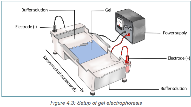
In a common gel electrophoresis setup, a nucleic acid such as DNA is loaded
into wells at one end of the gel and then migrates toward the positive electrode
at the opposite end. The rate of migration of fragments varies with size. The
steps of gel electrophoresis are shown below.
– The DNA samples are cut with a restriction enzyme into smaller
segments of various sizes. The DNA is then placed in wells made on a
thick gel.
– An electric current runs through the gel for a given period of time.
Negatively charged DNA fragments migrate toward the positively
charged end of the porous gel. Smaller DNA fragments migrate faster
and farther than longer fragments, and this separates the fragments by
size. The gel floats in a buffer solution within a chamber between two
electrodes.
– The DNA is transferred to a nylon membrane and radioactive probes
are added. The probes bind to complementary DNA.
– The X-ray film is exposed to the radiolabeled membrane. The resulting
pattern of bands is called a DNA fingerprint.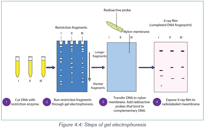
During electrophoresis, DNA fragments migrate through the gel at a rate that
is inversely proportional to the logarithm of their size. The shorter the fragment
is, the faster it will travel because of its ability to navigate through the pores in
the gel more easily than a large fragment can. Larger fragments are hampered
by their size. Hence, the longer a nucleotide chain, the longer it takes for the
migration.Gel electrophoresis takes advantage of DNA’s negative charge. A solution
containing different-size fragments to be separated is placed in a well. A
well is a depression at one end of the gel. The gel itself is usually a square or
rectangular slab and consists of a buffer containing electrolytes and agarose,
or possibly polyacrylamide. Agarose is a gel-forming polysaccharide found in
some types of seaweed that is used to form a gel meshwork for electrophoresis.
Polyacrylamide is an artificial polymer used to form a gel meshwork for
electrophoresis.The gel is loaded while it is submerged in a tray containing an electrolytic solution
called the buffer. Using direct current, a negative charge is placed at one end
of the gel where the wells are, and a positive charge is placed at the opposite
end of the gel. The electrolyte solution conveys the current through the gel. The
negatively charged DNA will migrate toward the positively charged electrode,
with the shorter fragments migrating faster than the longer fragments, achieving
separation. Small molecules found within the loading dye migrate ahead of all
the DNA fragments. Since the small molecules can be visualized, the electrical
current can be turned off before they reach the end of the gel.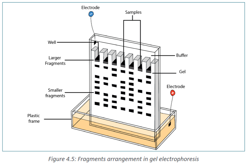
Once gel electrophoresis is complete, the DNA fragments are made visible by
staining the gel. The set of fragments generated with a particular restriction
enzyme produces a banding pattern characteristic for that DNA. The most
commonly used stain is ethidium bromide. Ethidium bromide is a flat molecule
that fluoresces under ultraviolet (UV) light and is able to insert itself among the
rungs of the ladder of DNA. When the gel is subjected to UV light, the bands
of DNA are visualized because the ethidium bromide is inserted among the
nucleotides. The size of the fragments is then determined using a molecular
marker as a standard. The molecular marker, which contains fragments of known
size, is run under the same conditions (in the same gel) as the digested DNA.Gel electrophoresis is not limited to the separation of nucleic acids but is also
commonly applied to proteins. Proteins are usually run on polyacrylamide
gels, which have smaller pores, because proteins are generally smaller in size
than nucleic acids. Proteins, however, are not negatively charged; thus, when
researchers want to separate proteins using gel electrophoresis, they must
first mix the proteins with a detergent called sodium dodecyl sulfate. This
treatment makes the proteins unfold into a linear shape and coats them with a
negative charge, which allows them to migrate toward the positive end of the gel
and be separated. Finally, after the DNA, RNA, or protein molecules have been
separated using gel electrophoresis, bands representing molecules of different
sizes can be detected. The gel electrophoresis is used for different purposes
such as DNA analysis, protein and antibody interactions, testing antibiotics and
testing vaccines.Application activity 4.4
The gel shown in the figure below was run after bacterial DNA was digested
using restriction enzymes where A, B, C and D are the comb lane of the gel.
In your notebook, indicate on the gel
a) Where the positive electrode was located;
b) Where the negative electrode was located;
c) The location of the largest band;
d) The location of the smallest band;
e) The number of cuts that were made on the linear fragment of DNA to
produce this number of bands.
4.5 Production of human proteins by recombinant DNA
technologyActivity 4.4
The figure below shows the process by which bacteria can be bioengineered
to produce human insulin. Follow each of the steps used to produce GMO
bacteria. Use the figure to make a list of steps followed when producing
a genetically modified organism bacterium.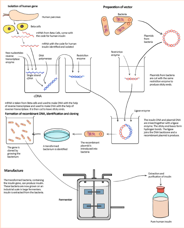
Production of insulin
One form of diabetes mellitus is caused by the inability of the pancreas to
produce insulin. Before insulin from GM bacteria became available, people with
this form of diabetes were treated with insulin extracted from the pancreases
of pigs or cattle. In the 1970s, biotechnology companies began to work on the
idea of inserting the gene for human insulin into a bacterium and then using
this bacterium to make insulin. They tried several different approaches, finally
succeeding in the early1980s. This form of human insulin became available
in1983.The procedure involved in the production of insulin is shown in the figure 4.6.
There were problems in locating and isolating the gene coding for human
insulin from all of the rest of the DNA in a human cell. Instead of cutting out
the gene from the DNA in the relevant chromosome, researchers extracted
mRNA for insulin from pancreatic β cells, which are the only cells to express
the insulin gene. These cells contain large quantities of mRNA for insulin as
they are its only source in the body. The mRNA was then incubated with the
enzyme reverse transcriptase which comes from the group of viruses called
retroviruses. As its name suggests, this enzyme reverses transcription, using
mRNA as a template to make single-stranded DNA. These single-stranded DNA
molecules were then converted to double-stranded DNA molecules using DNA
polymerase to assemble nucleotides to make the complementary strand. The
genetic engineers now had insulin genes that they could insert into plasmids to
transform the bacterium Escherichia coli.The main advantage of this form of insulin is that there is now a reliable supply
available to meet the increasing demand. Supplies are not dependent on factors
such as availability through the meat trade.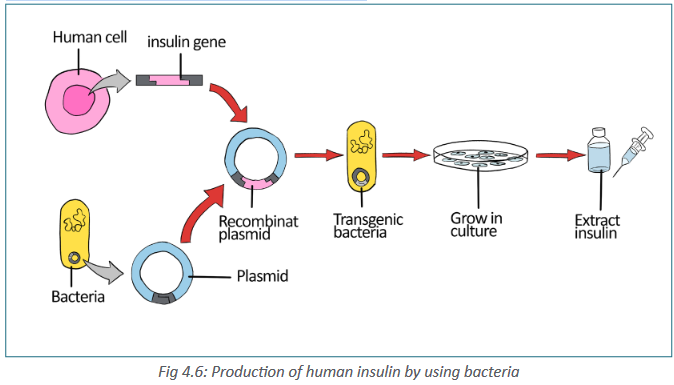
There were problems in locating and isolating the gene coding for human insulin
from all of the rest of the DNA in a human cell. Instead of cutting out the gene
from the DNA in the relevant chromosome, these are steps involved in human
insulin production:
– Researchers extracted mRNA for insulin from pancreatic β cells, which
are the only cells to express the insulin gene. These cells contain large
quantities of mRNA for insulin as they are its only source in the body.
– The mRNA was then incubated with the enzyme reverse transcriptase
which comes from the group of viruses called retroviruses. As its name
suggests, this enzyme reverses transcription, using mRNA as a template
to make single stranded DNA.
– These single-stranded DNA molecules were then converted to
double stranded DNA molecules using DNA polymerase to assemble
nucleotides to make the complementary strand.
– The genetic engineers now had insulin genes that they could insert into
plasmids to transform the bacterium Escherichia coli.
– When the bacterial cells copy their own DNA, they also copy the
plasmids and the donor genes that plasmids carry. After the cells have
grown into colonies, on an industrial scale in large fermenters insulin is
extracted from the bacteria.Production of bovine growth hormone
Recombinant (r) bovine growth hormone is a protein that has been made by
manipulating the DNA sequence (gene) that carries the instructions for, or
encodes, the growth hormone protein so it can be produced in the laboratory.
Hormones are substances secreted from specialized glands. Hormones travel
through the bloodstream to affect their target organs. Growth hormone acts on
many different organs to increase the overall size of the body. Before the advent
of genetic technologies, growth hormone was procured from the pituitary glands
of slaughtered cows and then injected into live cows.The same technique has been used to obtain human growth hormone from
the pituitary glands of human cadavers. When the human growth hormone is
injected into humans who have a condition called pituitary dwarfism, their size
increases. However, harvesting the growth hormone from the pituitary glands
of cows and humans is laborious, and many cadavers are necessary to obtain
small amounts of the protein.Producing rBGH
The first step in the production of the rBGH protein is to transfer the BGH gene
from the nucleus of a cow cell into a bacterial cell. Bacteria with the BGH gene
will then serve as factories to produce millions of copies of this gene and its
protein product; making many copies of a gene is called cloning the gene.Cloning a gene using bacterial cells
The following steps are involved in moving a BGH gene into a bacterial cell:
a) BGH gene is cut from the cow chromosome using restriction enzymes
that leave “sticky ends” with specific base sequences.
b) A plasmid from a bacterium is cut with the same restriction enzymes,
creating the same “sticky ends” as the cow gene.
c) The cleaved gene and plasmid are placed together in a test tube.
Complementary “sticky ends” fit together, resulting in a recombinant
plasmid.
d) The recombinant plasmid is reinserted into a bacterial cell.
e) The plasmids and the bacterial cells replicate, making millions of copies
of the rBGH gene.
f) The rBGH genes produce large quantities of rBGH proteins that are
harvested, purified, and injected into cows to increase milk production.Application activity 4.4
1) Rearrange the statements below to produce a flow diagram showing
the steps involved in producing bacteria capable of synthesizing a
human protein such as human growth hormone (hGH).
1. Insert the plasmid into a host bacterium.
2. Isolate mRNA for hGH.
3. Insert the DNA into a plasmid and use ligase to seal the ‘nicks’ in
the sugar–phosphate chains.
4. Use DNA polymerase to clone the DNA.
5. Clone the modified bacteria and harvest hGH.
6. Use reverse transcriptase to produce cDNA.
7. Use a restriction enzyme to cut a plasmid vector.4.6 Use of microarrays in the analysis of genomes and in
detecting mRNAActivity 4.6.
Indicate and explain the use and applications of the microarray DNA
technologyA DNA microarray consists of tiny amounts of a large number of single-stranded
DNA fragments representing different genes fixed to a glass slide in a tightlyspaced array, or grid. (The microarray is also called a DNA chip by analogy to a
computer chip.) Ideally, these fragments represent all the genes of an organism.The mRNA from the organism or the cell to be tested is labelled with a fluorescent
dye and added to the chip. When the mRNAs bind to the microarray, a fluorescent
pattern results that is recorded by a computer. Now the investigator knows what
DNA is active in that cell or organism. A researcher can use this method to
determine the difference in gene expression between two different cell types,
such as between liver cells and muscle cells.A mutation microarray, the most common type, can be used to generate a
person’s genetic profile. The microarray contains hundreds to thousands
of known disease-associated mutant gene alleles. Genomic DNA from the
individual to be tested is labelled with a fluorescent dye, and then added to
the microarray. The spots on the microarray fluoresce if the individual’s DNA
binds to the mutant genes on the chip, indicating that the individual may have
a particular disorder or is at risk for developing it later in life. This technique
can generate a genetic profile much more quickly and inexpensively than older
methods involving DNA sequencing.Microarrays have proved a valuable tool to identify the genes present in an
organism’s genome and to find out which genes are expressed within cells.
They have allowed researchers to study very large numbers of genes in a short
period of time, increasing the information available. A microarray is based on a
small piece of glass or plastic usually 2 cm2 (Figure 4.7). Short lengths of single-
stranded DNA are attached to this support in a regular two-dimensional pattern,
with 10 000 or more different positions per cm2. Each individual position has
multiple copies of the same DNA probe. It is possible to search databases to
find DNA probes for a huge range of genes. Having selected the gene probes
required, an automated process applies those probes to the positions on the
microarray.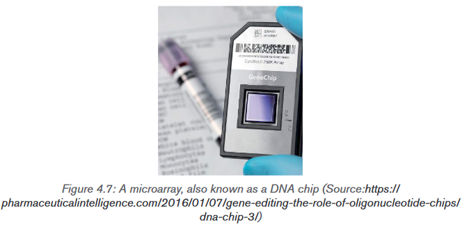
When microarrays are used to analyze genomic DNA, the probes are from
known locations across the chromosomes of the organism involved and are
500 or more base pairs in length. A single microarray can even hold probes from
the entire human genome.Microarrays can be used to compare the genes present in two different species.
DNA is collected from each species and cut up into fragments and denatured
to give lengths of single-stranded DNA. The DNA is labelled with fluorescent
tags so that – for example – DNA from one species may be labelled with green
tags and DNA from the other species labelled with red tags. The labelled DNA
samples are mixed together and allowed to hybridize with the probes on the
microarray. Any DNA that does not bind to probes on the microarray is washed
off. The microarray is then inspected using ultraviolet light, which causes the
tags to fluoresce. Where this happens, we know that hybridization has taken
place because the DNA fragments are complementary to the probes. Green
and red fluorescent spots indicate where DNA from one species only has
hybridized with the probes. Where DNA from both species hybridize with a
probe, a yellow colour is seen. Yellow spots indicate that the two species have
DNA with exactly the same base sequence. This suggests that they have the
same genes (Figure 4.8). The microarray is then scanned so that the data can
be read by a computer. Data stored by the computer indicate which genes are
present in both species, which genes are only found in one of the species and
which genes are not present in either species.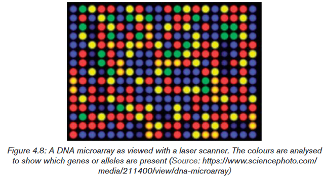
Using microarray analysis, researchers can quickly compare gene expression in
different samples, such as those obtained from normal and cancerous tissues.
The knowledge gained from such gene expression studies is making a significant
contribution to the study of cancer and other diseases.Application activity 4.6.
Using your knowledge of the microarray DNA technology, explain three uses
of this technique.4.7 Gene therapy and genetic screening
Activity 4.7
Gene technology can be involved in the detection and treatment of genetic
disorders. Discuss on different cases of genetic disorders that are treated by
using gene therapy.4.7.1 Genetic screening
Genetic screening is the analysis of a person’s DNA to check for the presence
of a particular allele. This can be done in adults, in a foetus or embryo in the
uterus, or in a newly formed embryo produced by in vitro fertilisation. An adult
woman with a family history of breast cancer may choose to be screened for the
faulty alleles of the genes Brca-1 and Brca-2, which considerably increase an
individual’s chance of developing breast cancer. Should the results be positive,
the woman may elect to have her breasts removed (elective mastectomy) before
such cancer appears.In 1989, the first ‘designer baby’ was created. Officially known as pre-implantation
genetic diagnosis (PGD), the technique involved mixing the father’s sperm with
the mother’s eggs (oocytes) in a dish – that is, a ‘normal’ IVF procedure. It was
the next step that was new. At the eight-cell stage, one of the cells from the tiny
embryo was removed. The DNA in the cell was analysed and used to predict
whether or not the embryo would have a genetic disease for which both parents
were carriers. An embryo that was not carrying the allele that would cause the
disease was chosen for implantation, and embryos that did have this allele were
discarded.Since then, many babies have been born using this technique. It has been used
to avoid pregnancies in which the baby would have had Duchenne muscular
dystrophy, thalassaemia, haemophilia, Huntington’s disease and others. In
2004, it was first used in the UK to produce a baby that was a tissue match with
an elder sibling, with a view to using cells from the umbilical cord as a transplant
into the sick child.For some time, genetic testing of embryos has been leaving prospective parents
with very difficult choices to make if the embryo is found to have a genetic
condition such as Down’s syndrome or cystic fibrosis. The decision about
whether or not to have a termination is very difficult to make. Now, though,
advances in medical technology have provided us with even more ethical issues
to consider.4.7.2 Gene therapy
Gene therapy is the alteration of a genetic sequence in an organism to prevent
or treat a genetic disorder.Gene technology and our rapidly increasing knowledge of the positions of
particular genes on our chromosomes have given us the opportunity to identify
many genes that are responsible for genetic disorders such as sickle cell anaemia
and cystic fibrosis. When genetic engineering really began to get going in the
1990s, it was envisaged that it would not be long before gene technology could
cure these disorders by inserting ‘normal’ alleles of these genes into the cells.Gene therapy has proved to be far more difficult than was originally thought. The
problems lie in getting normal alleles of the genes into a person’s cells and then
making them work properly when they get there. In theory, a normal allele of the
defective gene could be inserted into the somatic cells of the tissue affected by
the disorder. For gene therapy of somatic cells to be permanent, the cells that
receive the normal allele must be ones that multiply throughout the patient’s life.
Bone marrow cells, which include the stem cells that give rise to all the cells
of the blood and immune system, are prime candidates. One type of severe
combined immunodeficiency (SCID) is caused by a single defective gene. If the
treatment is successful, the patient’s bone marrow cells will begin producing
the missing protein, and the patient will be cured.The most common vectors that are used to carry the normal alleles into host cells
are viruses (often retroviruses or lentiviruses) or small spheres of phospholipid
called liposomes. Occasionally ‘naked’ DNA is used. The first successful
gene therapy was performed in 1990 on a four-year-old girl from Cleveland,
Ohio. She suffered from the rare genetic disorder known as severe combined
immunodeficiency (SCID). In this disorder, the immune system is crippled and
sufferers die in infancy from common infections. Children showing the condition
are often isolated inside plastic ‘bubbles’ to protect them from infections.Application activity 4.7
DNA technology is increasingly being used in the diagnosis of genetic and
other diseases and offers potential for better treatment of genetic disorders
or even permanent cures. Suggest the advantages of genetic screening and
therapy over the normal methods of treating diseases.4.8 Genetically modified organisms in agriculture
Activity 4.8
Discuss on the different GMO used in agriculture and the possible advantages
of these plants over the natural plants.4.8.1 Gene technology and agriculture
Many new products have been developed using this technology. Crops have
been genetically engineered to increase yield, hardiness, uniformity, insect and
virus resistance, and herbicide tolerance. The vast bulk of genetically modified
plants grown around the world are crop plants modified to be resistant to
herbicides or crops that are resistant to insect pests. These modifications
increase crop yield. A few crops, such as vitamin A, enhanced rice, provide
improved nutrition.4.8.2 Use of Agrobacterium tumefaciens to transfer genes
in plantsAgrobacterium is a bacterium that uses a horizontal gene transfer (HGT).
HGT is the transfer of DNA between different genomes. HGT can occur in
bacteria through transformation, conjugation and transduction. However, it is
also possible for HGT to occur between eukaryotes and bacteria. Bacteria have
three ways of transferring bacteria DNA between cells:1) Transformation: The uptake and incorporation of external DNA into the
cell thereby resulting in the alteration of the genome.2) Conjugation: The exchange of genetic material through cell-to-cell
contact of two bacterial cells. A strand of plasmid DNA is transferred to
the recipient cell and the donor cell then synthesis DNA to replace the
strand that was transferred to the recipient cell.3) Transduction: A segment of bacterial DNA is carried from one bacterial
cell to another by a bacteriophage. The bacteriophage infects a bacterial
cell and takes up bacterial DNA. When this phage infects another cell,
it transfers the bacterial DNA to the new cell. The bacteria can then
become a part of the new host cell.Agrobacterium has the ability to transfer DNA between itself and plants
and is therefore commonly used in genetic engineering. The process of using
Agrobacterium for genetic engineering is illustrated in the diagram below.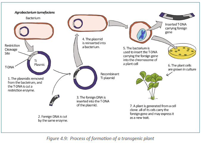
Summary of formation of a transgenic plant:
– The Agrobacterium cell contains a bacterial chromosome and a Tumor
inducing plasmid (Ti Plasmid).
– The Ti plasmid is removed from the agrobacterium cell and a restriction
enzyme cleaves the T-DNA restriction site.
– The foreign gene of interest is inserted into the middle of the TDNA.
– Recombinant plasmids can be introduced into cultured plant cells by
electroporation. Or plasmids can be returned to Agrobacterium, which
is then applied as a liquid suspension to the leaves of susceptible
plants, infecting them. Once a plasmid is taken into a plant cell, its
TDNA integrates into the cell’s chromosomal DNA.a. Golden rice
Golden rice is a staple food in many parts of the world, where people are
poor and rice forms the major part of their diet. Deficiency of vitamin A is a
common and serious problem; its deficiency can cause blindness. In the 1990s,
a project was undertaken to produce a variety of rice that contained carotene in
its endosperm. Genes for the production of carotene were extracted from maize
and the bacterium Pantonoea ananatis. These genes, together with promoters,
were inserted into plasmids. The plasmids were inserted into bacteria called
Agrobacterium tumefaciens. These bacteria naturally infect plants and so could
introduce the genetically modified plasmid into rice cells. The rice embryos, now
containing the carotene genes, were grown into adult plants.This genetically modified rice is called golden rice, because it contains a lot of
yellow pigment carotene. The genetically modified rice is being bred into other
varieties of rice to produce varieties that grow well in the conditions in different
parts of the world, with the same yield, pest resistance and eating qualities as
the original varieties.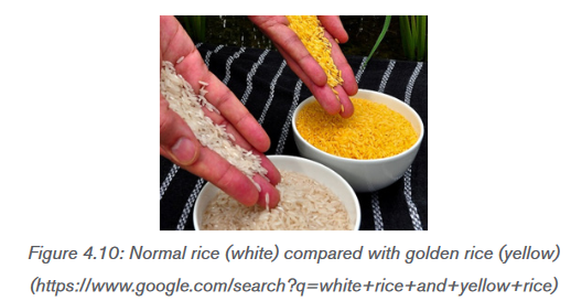
b. Herbicide-resistant crops
Herbicide-resistant crops called oil seed rape or Brassica napus, is grown in
many parts of the world as a source of vegetable oil which is used as biodiesel
fuel, as a lubricant and in human and animal foods. Natural rape seed oil contains
substances that are undesirable in oil that is to be used in human or animal
food. A hybrid, was made to produce low concentrations of these undesirable
substances, called canola (Canadian oilseed low acid), and this name is
now often used to mean any variety of oil seed rape. Gene technology has been
used to produce herbicide-resistant strains. Growing an herbicide-resistant
crop allows fields to be sprayed with herbicide after the crop has germinated,
killing any weeds that would otherwise compete with the crop for space, light,
water or ions. This increases the yield of the crop.c. Insect pests-resistant plants
Another important agricultural development is that of genetically modified plants
protected against attack by insect pests. Bt maize is genetically engineered
(GE) plant that produces crystal (Cry) proteins or toxins derived from the soil
bacterium, Bacillus thuringiensis (Bt), hence the common name “Bt maize”. Bt
maize plant has revolutionized pest control in a number of countries, but there
still are questions about its use and impact.Application activity 4.8
Explain why are Ti plasmids are used to insert genes into plant cells?
4.9 Significance of genetic engineering in improving the
quality and yield of crop plants and livestockActivity 4.9
The figure below shows an Atlantic salmon and a GMO salmon. A GM salmon
and non-GM salmon are of the same age.
Suggest any advantage of growing GM salmon over non-GM salmon and
discuss how the GM salmons are produced.4.9.1 Why are animals genetically modified?
Genetically modified animals are animals that have been genetically
modified for a variety of purposes including producing drugs, enhancing
yields, increase resistance to disease, etc. The vast majority of genetically
modified animals are at the research stage with the number close to entering
the market remains small. The process of genetically engineering mammals is a
slow, tedious, and expensive process. Researchers have genetically engineered
a number of mammals, from laboratory animals to farm animals, as well as birds,
fish and insects.The most widely used genetically modified animals are laboratory animals, such
as the fruitfly (Drosophila) and mice. Genetically engineered animals enable
scientists to gain an insight into basic biological processes and the relationships
between mutations and disease. However, farm animals, such as sheep, goats
and cows, can also be genetically modified to enhance specific characteristics.
These can include milk production and disease resistance, as well as improving
the nutritional value of the products they are farmed for. For example, cows,
goats and sheep have been genetically engineered to express specific proteins
in their milk.The majority of work on genetically modified farm animals is still in the research
phase and is yet to be used commercially. The advantages and disadvantages
associated with genetically modifying animals for agriculture, divided up into
four key areas:
4.9.2 Why are crop plants genetically modified?
Crop plants are genetically modified to increase their shelf life, yield and nutritive
value.• To increase the shelf life: to increase the time of ripening and the time of
storage.
• Improving the yield of crop plants has been the driving force behind the
vast majority of genetic engineering. Yield can be increased when plants
are engineered to be resistant to pesticides and herbicides, drought, and
freezing. For example, a gene from an Arctic fish has been transferred into
a strawberry to help prevent frost damage.Many people believe that improving farmers’ yields may help decrease world
hunger problems. Others argue that, since there is already enough food being
produced to feed the entire population, it might make more sense to use less
technological approaches to feeding the hungry. Significant numbers of people
around the world are malnourished, hungry, or starving, not due to a shortage of
food but because access to food is tied to access to money or land.• Genetic engineers may also be able to increase the nutritive value of crops.
Some genetic engineers have increased the amount of β -carotene in
rice, a staple food for many of the world’s people. Scientists hope the
engineered rice will help decrease the number of people who become
blind in underdeveloped nations because cells require β-carotene in order
to synthesize vitamin A, a vitamin required for vision. Therefore, eating this
genetically modified rice, called Golden Rice, increases a person’s ability
to synthesize vitamin A.Application activity 4.9
a) What is the importance of using golden rice in developing countries?
b) Suggest any three importance of using GM organism?4.10 Ethical and social implications of using genetically
modified organisms (GMOs).Activity 4.10
From your daily life experience, discuss the ethical and social implications of
using genetically modified crops in food production.Ethics includes moral principles that control or influence a person’s behaviour.
It includes a set of standards by which a community regulates its behaviour
and decides as to which activity is legitimate and which is not. Bioethics maybe viewed as a set of standards that may be used to regulate our activities in
relation to the biological world. Biotechnology, particularly recombinant DNA
technology, is used for exploitation of the biological world by various ways.Some genetically modified plants are grown in strict containment of glasshouses,
but a totally different set of problems emerges when genetically engineered
organisms such as crop plants and organisms for the biological control of pests
are intended for use in the general environment. Few countries would object
to the growth of genetically modified crops that produce vaccines for human
or animal use, yet there are people who object to the growth of pro-vitamin A
enhanced rice. The major bioethical concerns pertaining to biotechnology are
summarized below:
– When animals are used for production of certain pharmaceutical
proteins, they are treated as factory machines.
– Introduction of a transgene from one species into another species
violates the integrity of species.
– The transfer of human genes into animals or vice-versa is great ethic
threat to humanity.
– Biotechnology is disrespectful to living beings, and only exploits them
for the benefit of humans.
– Genetic modification of organism can have unpredictable/ undesirable
effects when such organisms are introduced into the ecosystem.Moreover, most objections are raised against the growth of herbicide-resistant
or insect-resistant crops as follow:
– The modified crop plants may become agricultural weeds or invade
natural habitats.
– The introduced gene may be transferred by pollen to wild relatives
whose hybrid offspring may become more invasive.
– The introduced gene may be transferred by pollen to unmodified plants
growing on a farm with organic certification.
– The modified plants may be a direct hazard to humans, domestic animals
or other beneficial animals, by being toxic or producing allergies.
– The herbicide that can now be used on the crop will leave toxic residues
in the crop.
– Genetically modified seeds are expensive, as is herbicide, and their
cost may remove any advantage of growing a resistant crop.
– Growers mostly need to buy seed each season, keeping costs high,
unlike for traditional varieties, where the grower kept seed from one
crop to sow for the next– In parts of the world where a lot of genetically modified crops are grown,
there is a danger of losing traditional varieties with their desirable
background genes for particular localities This requires a program of
growing and harvesting traditional varieties and setting up a seed bank
to preserve them.
Application activity 4.10
Discuss ethical and social implications raised against insect-resistant crops.
4.11 Bioinformatics
Activity 4.11
Dicuss on the importance of Bioinformatics and its importance. Thereafter;
Explain how the bioinformatics has contributed to the progress in DNA
sequence analysis.Bioinformatics is the collection, processing and analysis of biological
information and data using computer software. In other words, it is the branch
of biology that is concerned with the acquisition, storage, and analysis of the
information found in nucleic acid and protein sequence data. Bioinformatics
combines biological data with computer technology and statistics. It builds up
databases and allows links to be made between them. The databases hold
gene sequences of complete genomes, amino acid sequences of proteins and
protein structures.4.11.1 Scientists use bioinformatics to analyze genomes
and their functions
Different government agencies carried out their mandate to establish databases
and provide software with which scientists could analyse the sequence data. For
example, in the United States, a joint endeavour between the National Library of
Medicine and the National Institutes of Health (NIH) created the National
Center for Biotechnology Information (NCBI), which maintains a website
(www.ncbi.nlm.nih.gov) with extensive bioinformatics resources. On this site are
links to databases, software, and a wealth of information about genomics and
related topics. Similar websites have also been established by the European
Molecular Biology Laboratory (EMBL) and the DNA Data Bank of Japan,
two genome centers with which the NCBI collaborates.These large, comprehensive websites are complemented by others maintained
by individual or small groups of laboratories. Smaller websites often provide
databases and software designed for a narrower purpose, such as studying
genetic and genomic changes in one particular type of cancer.The NCBI database of sequences is called Genbank. As of August 2007, it
included the sequences of 76 million fragments of genomic DNA, totaling 80
billion base pairs! Genbank is constantly updated, and the amount of data it
contains is estimated to double approximately every 18 months. Any sequence
in the database can be retrieved and analyzed using software from the NCBI
website or elsewhere.UniProt (universal protein resource) holds information on the primary sequences
of proteins and the functions of many proteins, such as enzymes. The search tool
BLAST (basic local alignment search tool) is an algorithm for comparing primary
biological sequence information, such as the primary sequences of different
proteins or the nucleotide sequences of genes. Researchers use BLAST to find
similarities between sequences that they are studying and those already saved
in databases. When a genome has been sequenced, comparisons can be made
with other known genomes. For example, the human genome can be compared
to the genomes of the fruit fly, Drosophila, the nematode worm, or the malarial
parasite, Plasmodium. All the information about the genome of Plasmodium is
now available in databases. This information is being used to find new methods
to control the parasite. For example, being able to read gene sequences is
providing valuable information in the development of vaccines for malaria.4.11.2 Applications of bioinformatics
Bioinformatics has various applications in human genetics. For example,
researchers found the function of the protein that causes cystic fibrosis by
using the computer to search for genes in model organisms that have thesame sequence. Because they knew the function of this same gene in model
organisms, they could deduce the function in humans. This was a necessary
step toward possibly developing specific treatments for cystic fibrosis. The
human genome has 3 billion known base pairs, and without the computer it
would be almost impossible to make sense of these data. For example, it is now
known that an individual’s genome often contains multiple copies of a gene. But
individuals may differ as to the number of copies called copy number variations.
Now it seems that the number of copies in a genome can be associated with
specific diseases. The computer can help make correlations between genomic
differences among large numbers of people and disease. It is safe to say
that without bioinformatics, our progress in determining the function of DNA
sequences; in comparing our genome to model organisms; in knowing how
genes and proteins interact in cells; and so forth, would be extremely slow.
Instead, with the help of bioinformatics, progress should proceed rapidly in
these and other areas.Application activity 4.11
a) Describe 2 applications of bioinformatics
b) Explain the role of bioinformatics following the sequencing of genome of
Plasmodium in the control and prevention of malaria.Skills lab 4
Sensitize people about the use of DNA in crime investigation
In violent crimes, body fluids or small pieces of tissue may be left at the scene
or on the clothes or other possessions of the victim or assailant. If enough
blood, semen, or tissue is available, forensic laboratories can determine the
blood type or tissue type by using antibodies to detect specific cell-surface
proteins. However, such tests require fairly fresh samples in relatively large
amounts. Also, because many people have the same blood or tissue type,
this approach can only exclude a suspect; it cannot provide strong evidence
of guilt.DNA testing, on the other hand, can identify the guilty individual with a
high degree of certainty, because the DNA sequence of every person is
unique (except for identical twins).Genetic markers that vary in the population can be analyzed for a given person
to determine that individual’s unique set of genetic markers, or genetic profile.(This term is preferred over “DNA fingerprint” by forensic scientists, who want
to emphasize the heritable aspect of these markers rather than simply the
fact that they produce a pattern on a gel that, like a fingerprint, is visually
recognizable). The Rwanda Forensic Laboratory can now use DNA test to
convict criminals and to help to solve different problems such as paternity
testing.Student-teachers will have to sensitize people about the behavior that they
need to take for example if there is someone who has been murdered. They
will have to avoid to touching him because they can be taken as guilty or
they can make the police unable to find the murderer due to the fact that the
murdered person has been touched by a lot number of people.End unit assessment 4
I. Choose the letter corresponding to the best answer.
1) Different enzymes are used in the various steps involved in the
production of bacteria capable of synthesizing a human protein. Which
step is catalyzed by a restriction enzyme?
a) Cloning DNA
b) Cutting open a plasmid vector
c) Producing cDNA from mRNA
d) Reforming the DNA double helix2) What describes a promoter?
a) A length of DNA that controls the expression of a gene.
b) A piece of RNA that binds to DNA to switch off a gene.
c) A polypeptide that binds to DNA to switch on a gene
d) A triplet code of three DNA nucleotides that codes for ‘stop’3) Which statement correctly describes the electrophoresis of DNA
fragments?
a) Larger fragments of DNA move more rapidly to the anode than
smaller fragments.
b) Positively charged fragments of DNA move to the anode.
c) Small negatively charged fragments of DNA move rapidly to the
cathode.d) Smaller fragments of DNA move more rapidly than larger fragments.
II. OPEN ENDED QUESTIONS
1) The table shows enzymes that are used in gene technology. Copy and
complete the table to show the role of each enzyme.
a) Explain what is meant by:
i) Gene therapy
ii) Genetic screening.
b) Explain why it is easier to devise a gene therapy for condition caused by
a recessive allele than for one caused by a dominant allele.
2) Refer to what you have studied on DNA and PCR,
a) How many molecules of DNA are produced from one double-stranded
starting molecule, after eight cycles of PCR?
b) Explain why it is not possible to use PCR to increase the number of
RNA molecules in the same way as it is used to increase the number of
DNA molecules.
3) The latest estimate of the number of genes in the human genome is 21
000. Before the invention of microarrays, it was very time consuming to
find out which genes were expressed in any particular cell.
a) Explain how it is possible to find out which genes are active in a cell at
a particular time in its development.
b) Why is it not possible to use the same technique to find out which
genes are active in red blood cells?III. Long Answer Type Questions
1) Bacteria are used in genetic engineering. The diagram outlines the
process of inserting human insulin genes into bacteria using genetic
engineering.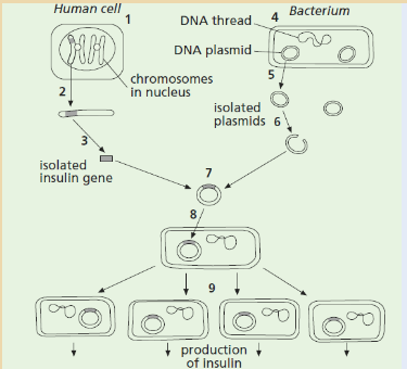
Complete the table below by identifying one of the stages shown in the
diagram that matches each description.
The diagram below shows a map of pBr322, a small piece of double-stranded,
circular DNA found in a bacterium in addition to the bacterial chromosome.
The genes for ampicillin resistance (Ampres) and tetracycline resistance (Tetres)
are indicated.
Pst 1 is a restriction endonuclease (enzyme) that has its effect at the site
shown. Pst 1 recognizes the base sequence and acts on
the DNA between guanine and adenine bases.
a) State the name given to such a piece of circular DNA.
b) Explain the use of such DNA in genetic engineering
c) Using the information given:
i) Explain what is meant by the term restriction endonuclease.
ii) Explain what is meant by the term sticky ends.UNIT 5: VARIATION AND ARTIFICIAL AND NATURAL SELECTION
Key unit competence
Explain variation and mutation as a source of biodiversity, the role of artificial
and natural selection in the production of varieties of animals and plants with
increased economic importanceIntroductory activity 5
Human population is classified as a single species ”homo sapiens” .Small
group of individuals with different skin colour do not look the same ,here is
a representation of human population observe it carefully then answer to the
questions below..
a) Some individuals in the figure above look like they are an intermediate
of other skin colour, from your observation is there any cause of this?
b) All of us we are human being but we do not look the same, why?5.1 Variation
All living organisms on the earth are unique, individuals of different species are
easy to differentiate and even those of the same species present differences
(morphological, physiological, cytological and behaviouristic). Such differences
among individuals of the same species are referred to as “Variation”. These
differences between cells, individual organisms, or groups of organisms of any
species are caused either by genetic differences (genotypic variation) or by
the effect of environmental factors on the expression of the genetic potentials
(phenotypic variation). So, organisms that have helpful variations tend to survive
better, and reproduce more. As they reproduce, their genes (including the
helpful genes) become more common in the gene pool, and these variations
spread out more and more.5.1.1 Genetic variation
Activity 5.1.1
A mutation in one gene causes the shell of the Japanese land snail (Euhadra)
to spiral in the opposite direction from others. Snails with opposite spirals
cannot mate, resulting in reproductive isolation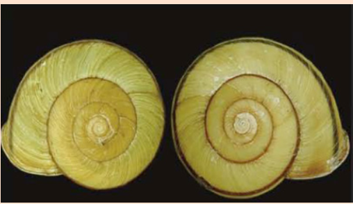
Using the knowledge acquired in genetics, what types of variation is indicated
by the Japanese Land snail?Genetic variation result from the differences in DNA sequences of individuals
(gene make up), those variations can be inherited by the transfer of genes.There are three primary sources of genetic variation:
• Variation from mutations are changes in the DNA. A single mutation
can have a large effect, but in many cases, evolutionary change is based
on the accumulation of many mutations. Ex, base substitution (Glu →Val),
deletion and insertion
• Variation from gene flow/gene migration/allele flow is any
movement of genes from one population to another and is an important
source of genetic variation. Ex, a bee carrying pollen from one flower
population to another
• Variation from recombination/ reproduction can introduce new
gene combinations into a population. This genetic shuffling/ genetic
recombination (meiosis & crossing over) is another important source of
genetic variation. At meiosis, the process that generates a haploid product
of meiosis whose genotype is different from either of the two haploid
genotypes that constituted the meiotic diploid. The creation of genetic
variation by recombination can be a much faster process than its creation
by mutation. For example, when just two chromosomes with “normal”
survival, taken from a natural population of Drosophila, are allowed to
recombine for a single generation, they produce an array of chromosomes
with 25 to 75 percent as much genetic variation in survival as was present
in the entire natural population from which the parent chromosomes were
sampled. This outcome is simply a consequence of the very large number
of different recombinant chromosomes that can be produced even if we
take into account only single crossovers.• Why is genetic variation important for evolution?
Variation is one of the main things that drive evolution. First, there are limited
resources available, and there is just not enough; food, water, shelter, etc.
available for all organisms. Second, to make matters worse, most species have
many offspring that can possibly survive. Just think of how many insect eggs are
laid compared to the number that make it to adulthood. This leads to competition
for the limited resources.Not all individuals in a species are the same. There are variations in; size, speed,
coloration, etc. These small variations can help or hinder individuals in their
survival. These variations are caused by small differences in genes. Organisms
that have helpful variations are more likely to survive. On average, they
get more food, get better shelter, etc. Coloration can help a predator get closer
to prey and eat better. Or, for the prey species, coloration can make it harder for
predators to find and eat it. So, organisms that have helpful variations tend to
survive better, and reproduce more. As they reproduce, their genes (including
the helpful genes) become more common in the gene pool, and these variations
spread out more and more.Variation can be influenced by numerous factors including:
i. Independent assortment of chromosomes.
Due to the law of independent assortment, traits are transmitted from parents to
offspring independently of one another.
This occurs at the time of gamete formation. At the time of gamete formation
during meiosis, the parental chromosomes separate randomly hence forming
different gametes with different chromosomes. This independent assortment
gives a wide variety of different gametes and hence individuals.ii. Crossing over
Crossing over allows the alleles on DNA molecules to change positions from
one homologous chromosome segment to another in other word is the transfer
or exchanges of genetic material from one homologous chromosome to another
during gamete formation known as meiosis. The new formed chromosomes are
known as recombinant chromosomes; Genetic recombination is responsible for
genetic diversity in species or population.iii. Random mating
Random mating involves individuals pairing by chance, not according to their
genotypes or phenotypes. Random mating is a source of variation in a population.
For example, a population in which mating only occur between organisms of
similar phenotypes as red beetles mating with red beetles and yellow beetles
mating with yellow beetles, will tend to show less variation than a population
where crosses are random.iv. Random fertilization of gametes
Random fertilization means that the collection of genes within one gametes,
each gametes contain a unique set of gene combination, and the ova is fertilized
randomly by the male gamete as a result each zygote is unique hence the
variation among individuals.v. Mutations
Mutation is a random change in the sequence of DNA, either due to errors
during DNA replication or by the influence of environmental factors. Mutations in
gametes cell can be inherited while somatic mutations are not transmitted from
generation to generation (not inherited).vi. Environmental factors
These variations caused by environmental factors are not inherited, environmental
variation are not prominent in animals as in plants, and this is due to the
environmental effect on the meristems of various parts. Some environmental
factors that can induce variation include, availability of food, light intensity,
Temperature, water, minerals etc…Application activity 5.1.1
1) Which of the following give rise to genetic variation in a population?
a) Crossing over and independent assortment in meiosis
b) Different environmental conditions
c) Random mating and fertilization.
d) Mutation.
i) a, b, c and d
ii) a, b and c only
iii) a, c and d only
iv) b, c and d only
2) Variation caused by environmental factors are not inherited. Why?
3) What is random mating?5.1.2 Phenotypic variation
Activity 5.1.2
1) Observe the following figures of students and make analysis on
their size (weight and height). These students live together in the
same school which means that the type of food they consume is
the same.Figure of students
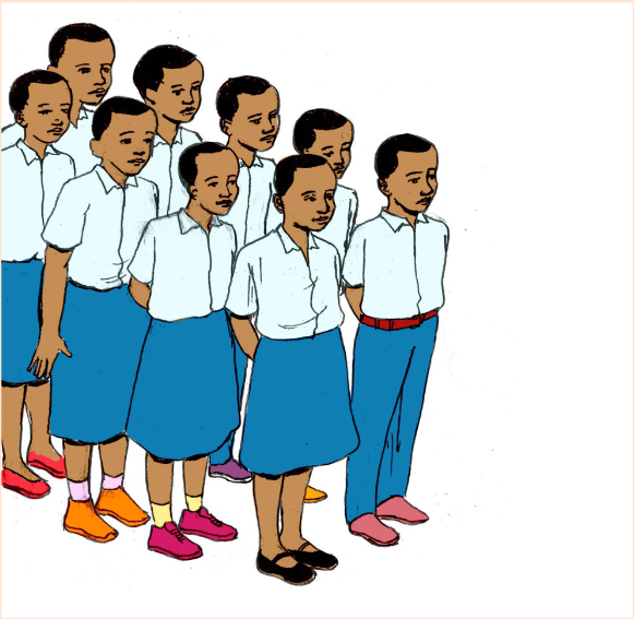
a) Write down your observation
b) Try to form 3 groups according to their height
c) By looking on their size can you try to make 3 groups according to
their weight?
d) By considering weight and height, why are not the same to those
soldiersPhenotypic variations can be brought about by genes or environmental
factors or a combination of both genes and environment they are not inherited.
So, there are characteristics that are not inherited but influenced by the
environmental factors, a child that get insufficient food will not grow to the size
expected, a cat with a skin disease may have bald patches in its coat. Those
conditions are not inherited. Such Phenotypic variations can be divided into two
types such as continuous variations or quantitative and Discontinuous variations
or Qualitative variations.a) Continuous variation
Continuous variation is variation which does not show clear cut differences i.e. it
shows a gradual change from one extreme to another. Characteristics such as;
human height and weight show continuous variation, and are usually determined
by a large number of genes (i.e. polygenic) and/ or considerable environmental
influence. Some examples of continuous variation are: Height, weight, heart
rate, finger length, and leaf length. They are also called fluctuating variationsIn continuous variations/quantitative:
• Different alleles at a single gene locus have a small effect on the phenotype
• Different genes have the same, often additive, effect on the phenotype
• A large number of genes may have a combined effect on a particular
phenotypic trait, these genes are known as polygenesA typical example of continuous variation is height. There are no distinct
categories of height; people are not either tall or short. There are all possible
intermediates between very short and very tall (Figure 5.1).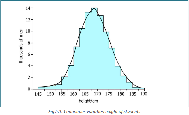
Continuously variable characteristic is greatly influenced by environment. A
person may inherit tallness trait and yet not get enough food to grow tall. A plant
may have a gene for large fruits but not get enough water, minerals and sunlight
to grow large fruits.b) Discontinuous variation.
Discontinuous variation is indicated as a variation where there is a clear difference
among individuals there is no intermediates, in human you are male or female
apart from abnormalities, Sex are inherited in a discontinuous way, some people
are able to roll they tongue in a tube other can’t do it.There are many characteristics that are difficult to classify as continuous or
non-discontinuous such as human eye colour people can be classified roughly
as having blue eyes or brown eyes, but there are also categories described as
grey, Hazel or green.A typical example of discontinuous variation is human blood group; discontinuous
variations is controlled by a single pair of alleles or small number of genes.A person is one of four blood group: A, B, AB and O there is no blood group
between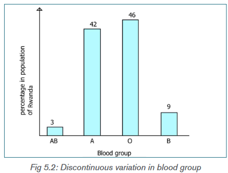
The major distinctions between continuous and discontinuous variations in
inheritance are as follows:Continuous variations have the following characteristics:
– The variations fluctuate around an average or mean of species.
– Direction of continuous variations is predictable.
– They are already present in the population.– Continuous variations are formed due to chance segregation of
chromosomes during gamete formation, crossing over and chance
pairing during fertilization.
– They can increase adaptability of the race but cannot form new species.
– Continuous variations are connected with the mean or average of the
species by intermediate stages.
– The continuous variations are also called fluctuations.
– When represented graphically, continuous variations give a smooth bell
shaped curve - They are very common
– Continuous variations do not disturb the genetic system.Discontinuous variations have the following characteristics:
– A mean or average is absent in discontinuous variations.
– The direction of discontinuous variations is unpredictable.
– Discontinuous variations are new variations though similar variations
might have occurred previously.
– Discontinuous variations are produced by changes in genome or genes.
– Discontinuous variations are the fountain head of continuous variations
as well as evolution
– These variations are not connected with the parental type by intermediate
stages.
– Discontinuous variations are also known as mutations or sports.
– A curve is not produced when discontinuous variations are represented
graphically.
– These variations appear occasionally.
– They disturb the genetic system of the organism
Table 5.1. Comparison between Discontinuous and continuous variation
Application activity 5.1.2
The histogram shows the height of wheat plants in an experiment plot.
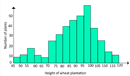
Based on the figure
a) Which type of variation is shown by the height of each of the strains of
wheat plants
b)Give other example of discontinuous variation5.2 Natural selection
Natural selection is a process that results in the adaptation of an organism to
its environments by means of selectively reproducing changes in its genotype
or genetic constitution. In 1858, Charles Darwin and Alfred Russel Wallace
published a theory of evolution by natural selection.
Individuals with certain variants of the trait may survive and are capable to
reproduce more than less successful individuals with unfavorable characters;
therefore, the population evolves. Over time, this process can result in
populations that specialize for particular ecological niches (microevolution) and
may eventually result in speciation (the emergence of new species also known
as macroevolution). In other words, natural selection is a key process to change
organisms and make them suitable to different environment.The allele responsible for the variation that help individuals to survive better is
inherited by the offspring, they will survive and transmit the trait to its offspring,
in time this particular variety outnumber and finally replace the original variety.This is known as “the survival of the fittest” but this doesn’t indicate good health
to an organism but the one which is well fitted to the conditions of environment.Factors of natural selection
Activity.5.2.1
The puffball can produce billions of offspring, if all offspring produced survived
to maturity they would carpet the surrounding land surface.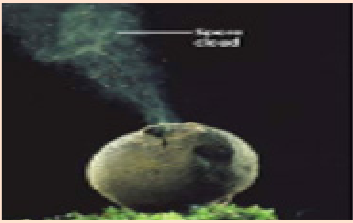
For your observation, how can the factor of producing a high number of
offspring for an organism have an impact on surviving in the environment?Natural selection which is one of the evolution means is due to several factors in
a population including over production and environmental factors.Role of over production and variation in natural selection
Over production is the production of more offspring that can be supported
by the available resources (food, light, space…). Individuals possessing genes
that help them to survive in an environment and this trait is transmitted from
generation to generations.Darwin appreciated that all species have the potential to increase their numbers
exponentially, he realized that, in nature, population rarely, if ever, increased
in size at such rate. The reason why reproductive rate is high is because an
individual cannot control the climate, availability of food, rate of predation etc.
Therefore, the production of sufficient offspring ensures a sufficient large
population surviving during hash conditions.Application 5.2.1
1) In the natural selection species that is able to produce a high number
of offspring ensure the possibility of great number offspring to survive.
How do we call such process?
2) How does over production lead to competition?Environmental factors
Activity 5.2.2
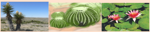
The figure above represents plants into different environments, observe them
carefully and
a) Write down your observations according their environments and their
structures.
b) Mention your differential observation of the plant (a), (b), (C) according
to their environment.Environmental factors as forces of natural selection, Environment is a responsible
agent of natural selection. Thus, it selects and determines individuals in different
ways according to different types of natural selections.Selection pressure
Selection pressure are environmental factors that limit the population of species
it is also known as environmental resistance.
It includes:• Availability of resources:
– Competition for food
– Competition for a space in which to live, breed and rear young
– Competition for mating etc.• Environmental conditions:
– Need for light, water, oxygen,
– Climate changes temperature, whether conditions or geographical
access• Biological factors: Predators and diseases(pathogens)
Selection pressure can be density dependent or independent density factor,
and it extent varies from time to time and place to place.
The selection is of three main types:
• Stabilizing selection
• Direction selection
• Disruptive selectiona) Stabilizing selection
Stabilizing selection is a type of natural selection in which a population mean
stabilizes on a particular non-extreme trait value as result of genetic diversity
decreases as illustrated in the figure below.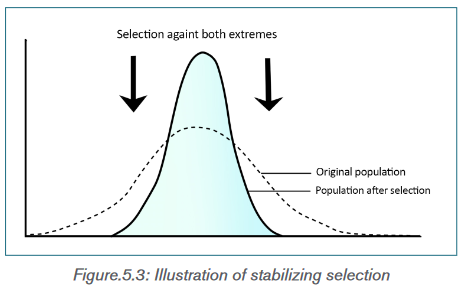
As illustrated in the above figure (Fig. 5.3), in stabilizing selection, natural selection
favors the individuals in the population with the intermediate phenotypes. These
individuals have greater survival and reproductive success. Individuals with
extreme phenotypes are less adaptive and are therefore eliminated.b) Directional selection
Directional selection is a mode of natural selection in which a single or new
fit phenotype is favored when exposed to environmental changes, causing a
population genetic variance or allele frequency to continuously shift in one
direction or one end of the spectrum of existing variation.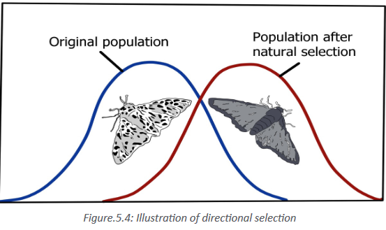
c) Disruptive or diversifying selection
In disruptive selection, both the extreme phenotypes in the population are
selected and become more prevalent. The individuals with extreme phenotypes
or end- phenotypic spectrum have greater survival and reproductive success.
The disruptive selection pressure increases the chances of the advantageous
alleles to be passed on to the next generation. By disruptive selection, the
intermediate phenotype is selected against and gradually decreases in number
from generation to generation, and may become extinct.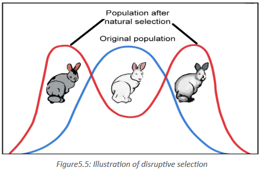
From the above figure, disruptive selection many generations may cause the
formation of two separate gene pools and the formation of new species.
From the factors of natural selection some examples that indicate natural
selection today are known and include:
• Antibiotic resistance,
• industrial melanism
• pesticides resistance in insect and mammalsApplication activity 5.2.2
1) Classify the following figures according to the types of natural selection
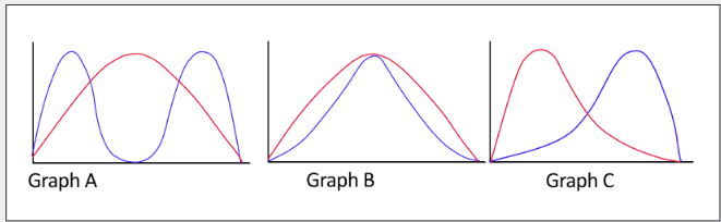
Key: Blue line indicates a given population after natural selection while red
line indicates a given population before natural selection.
2) Give 2 examples of natural selection due to environment?A. Antibiotic resistance in bacteria
Activity 5.2.2.A
Those Petri dishes contain gel that are nutrient for the growth of bacteria, the
red gel in each of these petri dishes has been inoculated with bacteria. The
small blue circles are discs impregnated with antibiotics but some bacteria
can grow around those antibiotics that are supposed to kill them.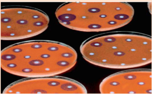
From the observation above what can you say on those bacteria that are able
to grow in the presence of Antibiotics?Antibiotic resistance occurs when an antibiotic has lost its ability to effectively
control or kill bacterial growth; in other words, the bacteria are “resistant” and
continue to multiply in the presence of therapeutic levels of an antibiotic.When the antibiotic is used the bacteria can develop the ability to defeat the
drugs designed to kill them. These bacteria survive to this antibiotic continue to
live and produce offspring that are resistant to this antibiotic.Antibiotic resistance is a natural process even though a number of bacteria drug
resistance is attributed to human being by overuse and misuse of antibiotics. In
some countries and over the Internet, antibiotics can be purchased without a
doctor’s prescription. Patients sometimes take antibiotics unnecessarily, to treat
viral illnesses like the common cold.How do bacteria become resistant?
Some bacteria are naturally resistant to certain types of antibiotics. However,
bacteria may also become resistant in two ways: by a genetic mutation or by
acquiring resistance from another bacterium.How does antibiotic resistance spread?
Genetically, antibiotic resistance spreads through bacteria populations both
“vertically,” when new generations inherit antibiotic resistance genes, and
“horizontally,” when bacteria share or exchange sections of genetic material
with other bacteria. Horizontal gene transfer can even occur between different
bacterial species. Environmentally, antibiotic resistance spreads as bacteria
themselves move from place to place; bacteria can travel via air, water and wind.People can pass the resistant bacteria to others; for example, by coughing
or contact with unwashed hands.Can bacteria lose their antibiotic resistance?
Yes, antibiotic resistance traits can be lost, but this reverse process occurs more
slowly. If the selective pressure that is applied by the presence of an antibiotic
is removed, the bacterial population can potentially revert to a population of
bacteria that responds to antibiotics.Application activity 5.2.2.A
1) Compare the two diagram below and differentiate them in the two
categories of resistance transmission in bacteria.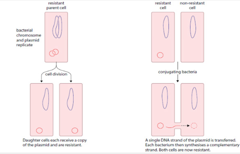
B. Pesticides resistance in insect and mammals
Activity 5.2.2.B
Discuss and explain the pesticides resistance in insect and mammals and
present your findings.Pesticide resistance means a decreased ability of pesticides to kill pest, Pest
species evolve pesticide resistance via natural selection and it can be passed
from one generation to the other trough reproduction.a) Pesticide resistance in insect
The intensive use of insecticide and genetics are the fundamental factors of
insecticide resistance. By natural selection insect with the genes that confer
the resistance to the insect survive and transmit the trait to the next generation,
most of pest species including insect produce large broods which increase the
probability of mutations and ensures large resistant populations.Resistance can be for a single insecticide, but it is more common that insect
resistance can be developed to the pesticide with the same mode of action.
This is known as cross resistance while multiple resistance is when insects
resist to two or more pesticides. In addition to resistance there is Tolerance, it
is not a result of selection pressure but a natural tendency, for example mature
caterpillar are tolerant to many insecticides than their young one.b) Pesticide resistance in mammals
Resistant weed species have now been reported and about 10 species of
small mammals and plants attacking nematodes are known to be resistant.
Resistance in mammals has affected the control of rat populations. A chemical
called warfarin has been used to kill rats since the 1950s.It is given to rats in the
form of food baited with the chemical. Once ingested, warfarin prevents blood
clotting, causing hemorrhaging and death of the rats. A warfarin –resistant
allele arose in the rat population that allow them to survive when warfarin was
ingested.Application 5.2.2.B
Answer by true or false
1) Due to the development of exoskeleton in some insect like caterpillar
they become more susceptible to pesticides than their young ones?
2) In our house there is a lot of mice, we use to kill them using ‘sumu ya
Panya”but it can’t kill them nowadays, they are resistant to it.
3) To fight against malaria we can use a mosquito net which kills
Anopheles, Anopheles do not resist on it.C. Industrial melanism
Activity 5.2.2.C
1) Observe the following diagram of moth and answer to the question
below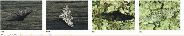
a) Write down your observation about the colour of moth and its back
ground.
b) Why those moth do not have the same colour?Industrial melanism is an effect of evolution prominent in several arthropods
where melanism has evolved in an environment where the air is greatly affected
by Sulphur dioxide and dark soot deposits. Darker pigmented individuals are
better fitted into those polluted environments which favors their camouflage.
Other explanation links this industrial melanism with immunization (strengthening
of immunity), absorption of heat at high rate in reduced sunlight and ability to
excrete trace element into melanic scales and feathers.Industrial melanism can be observed in more than 70 species of Lepidoptera
(butterflies and moths).Application 5.2.2.C
In the light forest, after the end of the simulation 76% light moths and 24%
dark moths were observed. Relate this to the industrial melanism and find an
explanation to those number.5.3 Artificial selection
Activity 5.3
1. The original Rwandan cattle (a) from which individuals were first
domesticated use to have long horns, but now days many cattle (b) do
not look the same as the original one.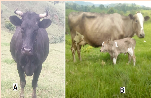
With the daily life experience and the observation on the figures
above, why does the number of traditional cows (a) are decreasing in
Rwandan societies.Artificial selection or selective breeding involves the selection of trait of interest
done by human being not environment and use them as the parent of the
next generation Artificial selection has been practiced by humans for several
centuries. It has played an important role in the evolution of modern crop plants,
farm animals and domestic pets from the wild ancestors. This led to a population
with desired traits. Artificial selection is used by humans to produce varieties ofanimals and plants that have an increased economic importance. It is considered
as a safe way of developing new strains of organisms, compared with genetic
engineering, and is a much faster process than natural selection. However,
artificial selection removes variation from a population, leaving it susceptible
to disease and unable to cope with changes in environmental conditions.
Potentially, therefore, artificial selection puts a species at risk of extinction.Comparison between natural selection and artificial selection
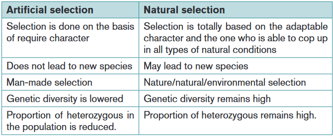
Two methods of carrying out selective breeding are known as in breeding and
out breedingApplication 5.3
1) At break time one of the student who come from Mamba sector in
Gisagara district told us about the agriculture activity, she said that
before sowing ground nuts their parents select seeds that look good.
I was asking myself why they can’t saw any seed.
a) From your knowledge is this selection, artificial or natural selection
b) Give other examples of artificial selection that you knowMethods of Artificial selection
Activity 5.3.1
1. Carefully observe the following diagram and write down what are your
observation of the activity that take place.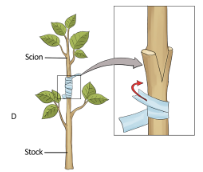
a) Inbreeding
In breeding methods, is a method of artificial selection in which there is a
breeding of closely relative’s individuals with a desired trait, in this case the
chances to obtain offspring showing the desired characteristics are greater.
This characteristic of interest is retained as far as possible and the origin of the
desirable trait is spontaneous mutation. The inbreeding presents some negative
consequences like the loss of vigour, with the population being weakened by
lack of diversity, the increase of expression of recessive allele that is why there is
a need of introducing new genes from outside to make the population healthier
and stronger.b) Outbreeding
This method of artificial selection involves crossing unrelated individuals showing
two different characteristics in order to obtain an individual which combine both
characteristics for example by crossing a crop plant that gives an excellent yield
with the one which resist to disease in the expectation of a plant with a high
yield and disease resistance. It frequently produces tougher individuals with a
better chance of survival. This is called hybrid vigourSelective breeding in cattle: Nowadays milk dairy industry are interested in
modern-day cattle for milk production and farmers involved in it use selective
breeding by artificial insemination.In the selection farmers follows some factors including:
• Volume of milk produced each day
• Length of milking(lactation) period
• Protein and fat content of milk• Disease resistance,
The selective breeding process
• Selecting a suitable cow and bull by consulting the pedigree records of
each and trough progeny testing.
• Collection of sperm from the selected bull and storing them by freezing
• Detection of when the cow is in oestrus by observing changes in her
behavior, e.g. Increase restlessness, feeding less.
• Artificially inseminating the defrosted semen into the cow.
• Checking that fertilization has occurred and the calf in cow.
Both artificial insemination and embryo transplantation can be used.Application 5.3.1
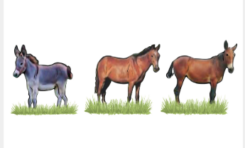
Before leading the following passage observe carefully the fig of animals
above.The hybrid offspring of donkey (I) and horse (J) is a mule (k), which is robust
but sterile, animals can be crossed according to the characteristic needed in
artificial selection.
a) Which type of artificial selection indicated above?
b) What can you do, to prevent the sterility of your hybrid(mule)Skills lab
This exercise illustrates the effect of natural selection on populations of
predators and prey. Students-teacher, in groups of four, will represent
predators, each with a different adaptation for capturing their prey. The prey
will consist of different species represented by different colored beans.Procedure.
1) Each team of 4 students will count out exactly 100 dried beans of each
color.
2) Thoroughly mix the beans and spread them evenly over your’habitat.’your
habitat depends on the weather.
i) If the weather is poor, it is dark outside, or your instructor would
rather, your habitat will be a tray of sediment in the classroom.
ii) If the weather is lovely,or your instructor is adventurous,you will do
this about lab outside.Each team will mark off 1m*1m ‘’habitat’’ in
the grass using yarn, a meter stick,and wood stakes.
iii) All ‘prey’ are confined to the habitat,wherever it is!
3) Each student (predotor) will have a different feeding apparatus:A
fork,spoon,Knife or forceps.
4) When everyone is ready, predators will spend 60 seconds capturing
prey with their devices and depositing them into a cup while obeying
the following rules:
i) Predators must only use their capture device to capture prey
ii) Predators may not scoop prey up with their cup.
iii) If predators ‘eat’ too much of the environment, they will become
constipated and die.5) Each predator determines the number of prey captured and records
results in data
Sheet: Generation1.4) Calculate and fill the statistics on the data sheet (see example
below).
Data sheet: Generation1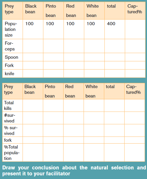
End unit assessment 5
1) Suggest how each of the following might decrease the chances of
an antibiotic resistant strain of bacteria developing
a) Limiting the use of antibiotics to cases where there is a real need
b) Regularly changing the type of antibiotic that is prescribed for a
particular disease
c) Using two or more antibiotics together to treat a bacterial infection.
2) Differentiate between natural selection from artificial selection
3) Some individuals of the swallowtail butterfly scientifically known as
Papilio machaon of the family papilionidae pupate on brown stems
or leaves; others pupate on green stems or leaves. Two distinct
colour forms of the pupae are found, namely brown and green, with
very few intermediates.
a) What type of natural selection does this example show?
b) Explain why the intermediate colour formed would be at selective
disadvantage.
4) Copy and complete the table to compare artificial selection with
natural selection
Distinguish inbreeding and outbreeding
6) Explain why artificial breeding is beneficial to man?UNIT 6: EVOLUTION AND SPECIATION
Key unit competence
Analyze the relevance of theories of evolution and explain the process of
speciation.Introductory activity 1
During Kwita Izina Ceremony (naming a newborn Gorilla) in Rwanda. On
Rwanda television I saw an image of mountain gorilla, it was closely related
to human being. Later while I was reading biology book I found this image
which shows human being and their ancestors
a) Observe carefully the image above and record the similarities among
A and D, and D and F.
b) Write a short note of your observation about the image.
6.1 Theories of evolutionActivity 6.1
1) Observe the diagram below of plant (vegetables), do you see any
relationship among those types of vegetables
According to the most biologists the principal questions in biology is “where do
all living things come from?” but we know that life comes from the pre-existing
life means that every species descends from other species, it is what we call
“evolution”Evolution is a changeover successive generation of inheritable trait of a
population or it is the process by which new species are formed from pre-
existing ones over a period of time. As there is emergence of new species
others are disappearing, the species that disappear are said to become extinct.
An enormous fossil, such as those of early birds, provides evidence of evolution.
Genetics studies of populations of bacteria, protists, plants, insects, and even
humans provide further evidence of the history of the change among organisms
that live or have lived on earth.Theory of evolution is a short term for theory of evolution by natural selection
which was proposed by Charles Darwin and Alfred Russel Wallace in the
nineteenth century.Four main theories of evolution are known:
• Lamarckism or theory of inheritance of acquired characters
• Darwinism or theory of natural selection.
• Neo-Darwinism or modern concept or Synthetic theory of evolution and
• Creation Theory.a) Lamarckism
Lamarckism or theory of inheritance of acquired characters developed by Jean
Baptist Lamarck (1744-1829) French Biologist. His theory is based on the
inheritance of acquired characteristic (variations) in the body of organism in the
response to the environment conditionsi) Assumptions of Lamarck’s theory
• Organisms tend to increase in size as they become more complex to a
predetermined limit.
• When influenced by the environment, body changes can be induced in
organisms.
• Organisms acquire new features because of need.
• Development of an organ and its effectiveness is promoted by its use
whereas its disuse brings about decline.
• Acquired features are inherited by future generations.ii) Merits/Advantages
• Lamarck was able to show that the environment influences the course of
evolution.
• He observed that features are passed down from parents to their offspring.
• He was able to recognize that as organism increase in size, they become
more complex to a predetermined limit. (Predetermine: to determine or
decide in advance)iii) Demerits /disadvantages
• Acquired changes are not heritable as they are influenced by genes.
• Somatic changes are not heritable as they are not passed through
reproduction.
• The process of gametogenesis is not related to occupation or their activity.
• Use or disuse of somatic cells does not affect gamete formation.b) Darwinism
The evolution is not a modern concept, since the ancient time, philosophers,
Aristotle, Socrates, Confucius and others have suggested that complex species
evolved from simple pre-existing ones by a process of continuous and gradual
change. In nineteenth century Charles Darwin an A. Wallace published the paper
describing their theory of evolution by natural selection later on 24 November
1859 Darwin published the book “The origin of species by means of Natural
selection or the preservation of favoured races in the struggle for life’’ containing
many evidence to support the theory.According to Darwin’s theory:
– Each species living today arose from a pre-existing species.
– All species have evolved from one ancestral type.
– Natural selection provides the mechanism for one species to change
into another.The main evidence for his first suggestion, which has been called descent with
modification, comes from fossils.Darwin use different observations in his research including the following:
a) Reproductive powers of living organism/over production/biotic
potential: Over production is the production of more offspring that can
be supported by the available resources this ensures the surviving of a
high number of offspring and the geometric or exponential growth of the
population.
b) Scarcity of resources: Darwinism states that, the increase of the
population geometrically is not directly proportional to the increase of
resources (food, space...) which increase in arithmetic way.
c) Struggle for existence: Darwin deduced on the basis of 1 and 2 that
members of the species were constantly competing with each other in an
effort to survive. In this struggle for existence only a few would live long
enough to breed
d) Survival of the fittest by natural selection: Among the offspring there
will be some better able to withstand the prevailing conditions. That is,
some will be better adapted (fitter) to survive in the struggle for existence.
These types are more likely to survive long enough to breed. Darwin’s idea
of evolution by natural selection is relatively simple but misunderstood. To
find out how it works, imagine a population of beetles:i) Variation in the beetles’ population some are green and some brown
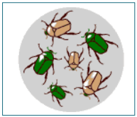
ii) Green beetles tend to reproduce less as they are eaten by predators than
brown one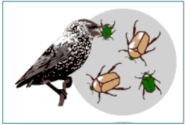
iii) Surviving beetles pass they brown genes to their offspring
iv) The brown coloration the important trait which allows the beetles to have
more offspring and to survive, will dominate the population and eventually
all beetles will be brown.
e) Inheritance of useful trait/like produce like: The selected individuals
produce offspring with the useful trait so that they can fit into the
environment.Darwin’s theory was based on three main observations:
i) Within a population are organisms with varying characteristics, and these
variations are inherited (at least in part) by their offspring.
ii) Organisms produce more offspring than are required to replace their
parents
iii) On average, population numbers remain relatively constant and no
population gets bigger indefinitely.After his observations Darwin concluded that within a population many individuals
do not survive and fail to reproduce.Assumptions of Darwinism
– Most organisms have the potential to produce large number of
offspring or progeny than the environment can support. This leads to
still competition as the numbers of organisms are fairly stable.
– All organisms, even of the same species vary in a few characteristics,
– Only those organisms of a given species with variations that adapt them
to the environment, survive the competition and live. There is survival for
the fittest by natural selection.
– The features favored/selected by nature survive and are inherited.
Therefore, new species may develop by natural selection, which is one
of the forces of evolution. Merits of Darwin’s theory of natural selection
– Species always change as the environment changes.
– Species are compared with their ancestors due to presence of
similarities in characteristics.
– Enough data are / can be collected for explaining variation in a population
that may result into formation of a new species. Demerits of Darwin’s theory of natural selection
– Not all variations inherited, except for only genetic variations.
– It provides inadequate explanation of existence of many vestigial
structures in organisms.
– Explanation on deleterious mutations that are retained in a population
is not adequate.c) Neo-Darwinism
Neo-Darwinism is the modern theory of evolution that incorporates scientific
evidence particularly from genetics and molecular biology, the Neo-Darwinism
combine the work of Mendel genetics and Darwin, for example, we now know that
the variations that are so important in natural selection come about by random
and spontaneous changes in genes, particularly from mutations in reproductive
cells. According to Neo-Darwinism, nature selects those individuals with
beneficial mutations and allows them to be passed to their offspring through
reproduction from generation to generation. The mutations are transmitted
within the population and if selected by nature, they may form a new species.d) Special creation
It is believed that a special being, God created the universe and all living
organisms (bible Genesis 1:1-2; Psalm 139:13-14). In this theory, heavens and
earth were first created. Light, day and night were created next and subsequently,
all living things with human beings the last in the creation. It shows that there
was direct creation of organism with no precursor to life.6.1. 1 Evidence for evolution
a) Fossils
The evidence for evolution are provided mainly by the study of fossil
(paleontology). Fossils come into different forms, such as imprints, the burrow
of worm, or mineralized bone preserved by natural process in rocks, ice etc. The
study of fossils show how the organisms have changed over time.Relative and radiometric dating are the method used by scientist to determine
the age of fossils and rocks.b) Anatomy and Embryology
Anatomy or comparative anatomic structures is the study of biological different
organisms. Structures in different species that have similar internal frame, work,
position and embryonic development are said to be homologous. For example,
bones in the appendages of a human, dog, bird, and whale all share the same
overall construction resulting from their origin in the appendages of a common
ancestor. Overtime, evolution led to changes in the shapes and sizes of these
bones in different species, but they have maintained the same overall layout.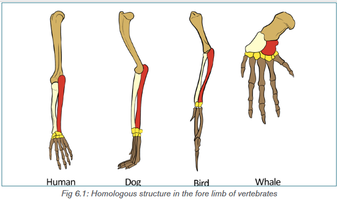
Paleontologists have found fossils showing how the bones of lizard-like ancestor
evolved into the ear bones of modern mammals.The fact that two different organisms look alike does not always suggest a
close evolutionary relationship. Structures of unrelated species can evolve
to look alike because structures are adapted of similar functions. These are
called analogous structures. Another evidence is vestigial structures
body structure with no function or which do not serve their original purpose but
probably useful in the ancestors.c) Comparative embryology
The study for embryo it is called embryology, an embryo is an unborn or
unhatched animal or human young in its earliest phases. Therefore, species that
show a similar embryonic development are assumed to be closely related, even
if the adult stages are very different. For example, echinoderms (the phylum
containing starfish and sea urchins) are believed to be related to chordates (the
phylum including vertebrates) because of similarities in their early embryonic
development.d) Comparative biochemistry and cell biology
The most persuasive evidence that organisms have evolved from a common
ancestor comes from studies comparing the cell biology and biochemistry of
different organisms, which reveal that:
– The genetic code contained within nucleic acids is almost universal
– Physiological processes vital to all organisms, such as respiration,
follow very similar metabolic pathways.– ATP is the universal energy currency
The cellular and biochemical details of organisms are quite similar, but any
differences can give an idea of how closely different species are related.
Species that are closely related would be expected to differ only slightly from
each other. Detailed comparisons of DNA, metabolic pathways, key proteins,
and organelles such as ribosomes have been used to work out the evolutionary
relationships of organisms. For example, ribosomes inside mitochondria and
chloroplast are similar to those in bacteria, suggesting that these organelles
may have evolved from bacteria. Mammalian blood proteins can be tested to see
how similar they are to human blood proteins: blood serum from the mammal in
question is added to rabbit serum containing anti-human antibodies.Application Activities 6.1
1) The diagram below indicates the part of front limb of different animals,
if the labeled diagram is an arm of human being label other diagram by
corresponding them, according to how they have evolve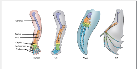
2) The skull of chimpanzee and that of human being are shown her below
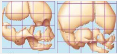
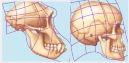
The above diagram corresponds to the adult skull, relate them with their fetus
3) Correct this statement
Mitochondrial DNA differences are inconsistent with the existence of a recent
human common ancestor for all ethnic groups.6.2 Cause of evolution
Activity 6.2
Find out the cause of evolution and discuss it among your classmates.
It is difficult to meet Hardy-Weinberg equilibrium in real populations. The
Hardy-Weinberg Theorem describes populations in which allele frequencies
are not changing means that it does not evolve.
The force behind evolution are mainly summarized in four factors:
• Competition changes in the environment.
• Sexual reproduction.
• Mutations.
• Gene recombination.
• Industrialization.
• Effect of drugs or chemical resistance.
• Artificial selection.a) Competition changes in the environment
Imagine that we are plunged into a new ice age. The climate becomes much
colder, so that snow covers the ground for almost all of the year. Assuming
that rabbits can cope with these conditions, white rabbits now have a selective
advantage during seasons when snow lies on the ground, as they are better
camouflaged (like the hare in figure 6.3.). Rabbits with white fur are more likely
to survive and reproduce, passing on their alleles for white fur to their offspring.
The frequency of the allele for white coat increases at the expense of the allele
for agouti. Over many generations, almost all rabbits will come to have white
coats rather than agouti.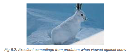
b) Sexual reproduction
Sexual reproduction is a reproduction using gametes (male gametes and female
gametes) each gamete contain a unique set of gene combination, and the ova is
fertilized randomly by the male gamete as a result each zygote is unique hence
the variation among individuals.c) Mutation
Mutation creates new genetic variation in a gene pool. It is how all new alleles first
arise. In sexually reproducing species, the mutations that matter for evolution are
those that occur in gametes. Only these mutations can be passed to offspring.
For any given gene, the chance of a mutation occurring in a given gamete is
very low. Thus mutations alone do not have much effect on allele frequencies.
However, mutations provide the genetic variation needed for other forces of
evolution to actd) Gene recombination
Natural selection is usually the most powerful mechanism or process causing
evolution to occur, however, it only selects among the existing variation already
in a population. It does not create new genetic varieties or new combinations or
varieties. One of the sources of those new combinations of genes is recombination
during meiosis. It is responsible for producing genetic combinations not found
in earlier generations.e) Industrialization
Many species of organisms, especially insect species, have two or more
adult body forms that are genetically distinct from one another, but which are
contained within the same interbreeding population. This condition is known as
polymorphism (another type of natural selection). The peppered moth (Biston
betularia), for example, has two main forms with different wing colours. One form
has pale wings with dark markings; the other form is called melanic because the
wings contain large amounts of melanin (a black pigment), so they are almost
black.f) Effect of drugs or chemical resistance
Drug resistance is a reduction in effectiveness of medication such as
antimicrobial in treating a disease or condition. Antibiotic resistance is a severe
problem throughout the world. For example, some strains of the common
bacterium Staphylococcus aureus are resistant to antibiotics such as penicillin
and methicillin. Penicillin resistance has probably evolved in the following way:
– By chance, a mutation produces an individual bacterium with an allele
that allows it to produce an enzyme, penicillinase, which deactivates
penicillin.
– This bacterium is immediately resistant to penicillin. (As bacteria have
only one strand of DNA and one copy of each gene, the mutant allele is
expressed immediately and is not masked by a dominant allele.)
– If the population to which the mutant belongs is exposed to penicillin,
the mutant will survive and reproduce whereas those without the mutant
will be killed.g) Artificial selection
Over the years, humans have used artificial selection to create dog breeds over
the past 150 years or so, humans have been specifically mating dogs that look
a certain way to create the animals we now keep as pests via a process known
as breeding. This is artificial selection, where one species (humans) directs the
traits that get passed down to future generations of another species (dogs).Application activity 6.2
1) Numerous factors can induce the evolution of species, observe the
diagram below then suggest the cause of the loss of hair.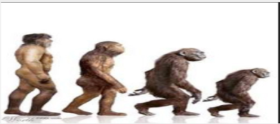
2) After understanding the evolution, give the factors that are inducing
today’s evolution.6.3 Speciation
Activity 6.3
1. Observe the diagram below, and write a short notes for your observation
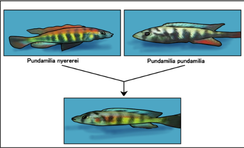
Speciation is the evolution of new species from the existing ones. A species is a
group of organisms with similar features which can interbreed to produce fertile
offspring, and which are reproductively isolated from other species. Organisms
which do not interbreed under normal circumstances to produce fertile offspring
are regarded as reproductively isolated. Mechanisms that prevent the formationof hybrids are called prezygotic isolating mechanisms, Prezygotic (before a
zygote is formed) isolating. Mechanisms include:
– Individuals not recognizing one another as potential mates or not
responding to mating behavior
– Animals being physically unable to mate
– Incompatibility of pollen and stigma in plants
– Inability of a male gamete to fuse with a female gamete.The mechanisms that affect the ability of hybrids to produce fertile offspring
are called postzygotic isolating mechanisms. Postzygotic isolating mechanisms
include:
– Failure of cell division in the zygote
– Non-viable offspring (offspring that soon die)
– Viable, but sterile offspring.The most important isolating mechanism is thought to be geographical isolation,
in which two populations originally of the same species are separated from each
other by a physical barrier such as a mountain, river, or ocean.a) Allopatric speciation
Allopatric means ‘different countries’ and describes the form of speciation where
two populations become geographically isolated. Geographical isolation
may be the result of any physical barrier between two populations which
prevents them interbreeding. These barriers include oceans, rivers, mountains
ranges and deserts. Which proves a barrier to one species may be no problem
to another. The isolated populations then undergo phenotypic divergence as:
• They independently undergo genetic drift
• Different mutations arise in two populations
• They become subjected to dissimilar selective pressureb) Sympatric speciation
Sympatric literally means. (‘Same country’) Sympatric speciation occurs when
organisms inhabiting the same area become reproductively isolated into two
groups for reasons other than geographical barriers. Such reasons might
include:
1) The genitalia of two groups may be incompatible (mechanical isolation):
It may be physically impossible for the penis of a male mammal to enter
the female’s vagina
2) The gametes may be prevented from meeting: In animals, the sperm may
not survive in the female’s reproductive tract or, in plants; the pollen tube
may fail to grow.3) Fusion of the gametes may not take place: Despite the sperm reaching
the ovum, or the pollen tube entering the micropyle, the gametes may be
incompatible and so will not fuse.
4) Development of the embryo may not occur (hybrid inevitability): Despite
fertilization taking place, further development may not occur, or fatal
abnormalities may arise during early growth
5) Polyploidy (hybrid sterility): When individuals of different species breed,
the sets of chromosomes from each parent are obviously different. These
sets are unable to pair up during meiosis and so the offspring cannot
produce gametes.
6) Behavioral isolation: Before copulation can take place, many animals
undergo elaborate courtship behavior. This behavior is often stimulated
by the colour and markings on the members of the opposite sex, the call
of a mate or particular actions of a partner.Application activity 6.3
1) Which type of speciation is indicated by the diagram below
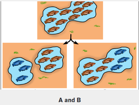
2) Which of the following is a correct definition of speciation?
a) When one species has a genetic mutation, allowing it to breed with
another species
b) When a species has a genetic defect, making it a brand new species
c) The process by which a species goes extinct, allowing a new species
a chance to live in anew habitat
d) An evolutionary process that leads to the formation of a new species.3) Which of the following is not true in the formation of a new species?
a) If an isolated population has a new environmental conditions new
traits can be favored eventually leading to the inability to reproduce
with the original population.
b) A mutation causes a population to breed with a different species.
c) Reproductive isolation can occur by the formation of a mountain
range.
d) A population needs to become reproductively isolated.6.4 Mechanisms of speciation
Activity 6.4
The following image are for two different animals.
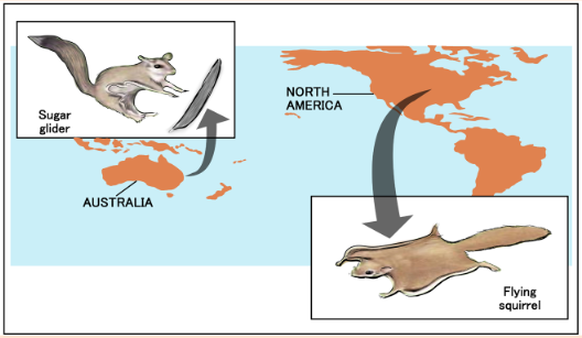
a) Write down the similarities and differences in these animals on the
above image.
b) Can you consider them as a single species?a) Continental drift
The continents which now exist have not always appeared as they do today.
At one time, the earth had a single large land mass called Pangaea. This is
thought to have broken up into two parts, a northern Laurasia and a southern
Gondwanaland. Over millions of years, the two great land masses split up and
moved by a process called continental drift to form our present continents. Thetheory that these land masses were once joined is supported by the discovery
in Australia, South Africa, South America, and Antarctica of fossils belonging to
the same extinct species. Fossils in North and South America show differences
between the species, suggesting that these two continents have only joined
together relatively recently. Before this, their fauna (animals) and flora (plants)
were geographically isolated and evolved independently.Australia shows many excellent examples of species that evolved independently
following its geographical isolation. It is thought that Australia became isolated
about 120 million years ago, when marsupials (mammals without a placenta but
with a pouch in which the young develop) and eutherian mammals (mammals
with a true placenta) diverged from a common ancestorb) Migration
Migration also called gene flow is any movement of individuals, and/or the
genetic material they carry, from one population to another. Gene flow includes
lots of different kinds of events, such as pollen being blown to a new destination
or people moving to new cities or countries. If gene versions are carried to a
population where those gene versions previously did not exist, gene flow can
be a very important source of genetic variation. In the graphic below, the gene
version for brown coloration moves from one population to another.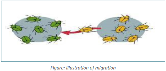
c) Divergent evolution
A single species evolves into several new species that live in different ways. The
five of Darwin’s finches are a good example. There are separate species
of finch in the group, all of which probably evolved from individuals belonging to
ne mainland species.The islands have few other bird species. In the absence of competition, the
finches became adapted to fill all the available niches. In particular, they evolved
a wide range of beak sizes and shapes so that they could take advantage of the
food sources on the different islands. The evolution of an ancestral species into
different species to fill different niches is called adaptive radiationd) Convergent evolution
Unrelated species independently evolve similarities when adapting to similar
environments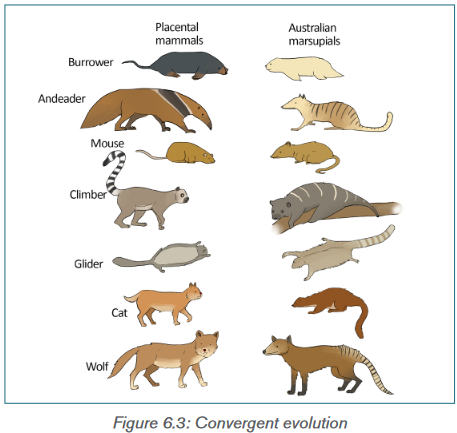
6.1: Table isolating mechanisms
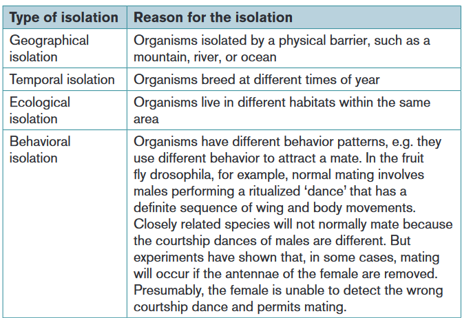
Application activity 6.4
1) Observe the following birds
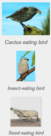
Observe those figures, what is the type of speciation?
2) Which effect of natural selection is likely to lead to speciation?
a) Differences between populations are increased.
b) The range of genetic variation is reduced.
c) The range of phenotypic variation is reduced.
d) Favorable alleles are maintained in the population.Skills lab
Formulate models
Camouflage provides an adaptive advantage Camouflage is a structural
adaptation that allows organisms to blend with their surroundings. In this
activity, you’ll discover how natural selection can result in camouflage
adaptations in organisms.Procedure
Working with a partner, punch 100 dots from a sheet of white paper with
a paper hole punch. Repeat with a sheet of black paper. These dots will
represent black paper.
1) Scatter both white and black dots on a sheet of black paper.
2) Decide whether you or your partner will role-play a bird.
3) The ‘’bird ‘’looks away from the paper, then turns back and immediately
picks up the first dot he or she sees.
4) Repeat step 4 for one minuteAnalysis
1) Observe what color dots were most often collected?
2) Infer how does color affect the survival rate of insects?
Hypothesize what might happen over many generations to a similar
population in nature?End unit assessment 6
1) Name two examples of adaptive radiation.
2) What effect did industrial pollution have on:
a) The frequency of the C (melanic) allele within a population of peppered
moths.
b) The rate of mutation of the c allele to the C allele
3) Explain what is meant by heterozygous advantage, using the sickle-cell
allele as an example.
4) Answer the following questions:
a) Distinguish between homologous structures and analogous structures
with specific examples.
b) Name the type of evolution exhibited by comparing:
i) Flipper of whale and forelimb of desert rat.
ii) Wing of a bat and wing of butterfly
iii) Wing of a flamingo and wing of an insectEssay questions
1) Explain the various evidences of organic evolution.
2) Explain Darwin’s theory of natural selection. The environment or
nature selects the individual with variations that are favored by the
environment. These compete with the others and able to reach sexual
maturity, reproduce and pass over the favorable characteristics to their
offspring.
3) What do you understand by Lamarckism? How does it differ from
Darwinism?
4) How can you convince that evolution progress?
5) A Darwin and Lamarck contribution to science is unparalleled. Discuss.REFERENCES
1) Campbell, N.A., Reece, J.B., Urry, A.L., Cain, L.M, Wasserman, A.S.,
Minorksy, V.P and Jackson, B.R. (2008). Biology. 8th edition. Pearson
international, San Francisco, USA.
2) Mackean, D.G., Hayward D. (2014). Biology. 3rd edition Cambridge
International Examinations, UK
3) Mader, S. (2004). Biology. 8th Edition. Mc Graw Hill. New York, USA.
4) Mary, J., Richard, F., Jennifer, G., Dennis, T. (2014). Biology. 4th edition,
Cambridge International AS and A Level, UK,
5) Miller, L. (2006). Prentice Hall Biology. Pearson Prentice Hall.
6) Michael Kent, 2000, Advanced BIOLOGY, Oxford. New York
7) Glenn and Susan Tole, 2015, Biology in context, Oxford University Press,
UK
8) Michael Kent, 2000, Advanced biology, Oxford University Press, UK
9) Taylor D. J., (1999), Biological Sciences, 3rd edition, Cambridge University
Press, UK.
10) Jones, M., Fosbery, R., Gregory, J., and Taylor, D. (2014). Cambridge
International AS and A Level Biology course book fourth Edition. United
Kingdom: Cambridge University Press.
11) Neil A. Campbell, Reece J.B., Urry L.A., Cain M.L., Wasserman S.A.,
Minorsky P.V., and Jackson R.B. (2008). Biology. Pearson Benjamin
Cummings. 8th Ed. San Francisco, US.
12) https://www.sciencephoto.com/media/211400/view/dna-microarray
13) https://pharmaceuticalintelligence.com/2016/01/07/gene-editing-the-
role-of-oligonucleotide-chips/dna-chip-3/
14) https://en.wikipedia.org/wiki/Dolly_(sheep) http://sphweb.bumc.bu.edu/
otlt/MPH-