Topic outline
General
- Physics SME Y3 TG File Uploaded 1/11/21, 16:40
UNIT 1: SOUND WAVES
Key Unit Competence:Analyse the effects of sound waves in elastic medium
Introductory activity
What are the properties which explain mostly the behavior of sound?
1. a) Most people like to listen to music, but hardly anyone likes to listen
to noise. In your own view, how is musical sound different from noise?
b) A guitarist as shown in the figure above plays guitar. The sound is
made by the vibration of the guitar string and propagates as a wavethrough the air and reaches your ear.
i) Assuming you are near by the guitarist and your friend is behind you,
who can hear more sound? Explain your reasoning
ii) If another person playing flute comes in and plays it. Can you distinguish
sound from the flute from that of a guitar? How are the two soundsdifferent?
2. a) Now, while they are playing their instruments you keep moving away
and coming towards a point where they are playing the instruments.
Explain the variations of sound heard by you.
b) Do you think there would be any change in the sound if you(the
observer) and the players (the source) remained in the same position?
3. With scientific explanations explain why you may not be able tocommunicate well in a room where music is being played at a high tone.
1.1 PRODUCTION OF STATIONARY SOUND WAVES
Activity 1.1
Look at the Fig.1.2 and then answer the following questions.
1. The two students in the figure above are producing sound. In each case,
describe the method of production of sound.
2. Imagine that the student replaces the flute with a longer one, would the
sound produced remain the same?Explain you answer.
3. Do you think a guitar with longer string produces the same sound as
the one with a shorter string? Defend your answer using scientificexplanations.
1.1.1 Sound in pipes
The source of any sound is vibrating object. Almost any object can vibrate and hence
be a source of sound. For musical instruments, the source is set into vibration by
striking, plucking, bowing, or blowing. Standing waves (also known as stationary
waves are superposition of two waves moving in opposite directions, each having
the same amplitude and frequency) are produced and the source vibrates at its
natural resonant frequencies. The most widely used instruments that produce
sound waves make use of vibrating strings, such as the violin, guitar, and piano or
make use of vibrating columns of air, such as the flute, trumpet, and pipe organ.
They are called wind instruments.
We can create a standing wave:
• In a tube, which is open on both ends. The open end of a tube is approximately
a node in the pressure (or an antinode in the longitudinal displacement).
• In a tube, which is open on one end and closed on the other end. The closed
end of a tube is an antinode in the pressure (or a node in the longitudinal
displacement).
In both cases a pressure node is always a displacement antinode and vice versa.
A node is a point half way between the crest and the trough. The line that connects
the nodes is the nodal line. The nodal line shows the original position of the matter
carrying the wave.
Displacement node means that a very thin slice of the medium at the node does
not move (zero displacement). If you have a standing wave in a half-open tube,
there will be a displacement node (and a pressure antinode) at the closed end.
This is due to the fact that the molecules cannot move back and forth at the closed
end.In the open end you will, on the other hand, have a pressure node (and thus a
displacement antinode). This is due to the fact that the pressure at the end of thetube is equal to that of the surrounding air.
Pressure node does not mean that the pressure is low; it simply means that the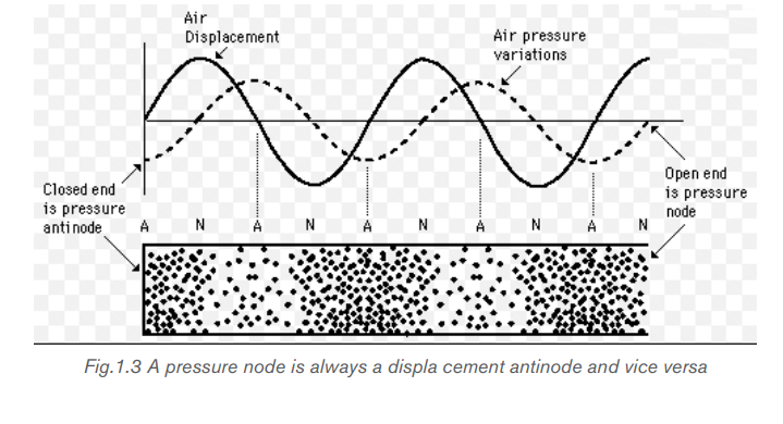
pressure is constant. Similarly, the pressure at the antinode is not “high”; it simplyhas the largest oscillations from low pressure to high pressure.
a. Tube of length L with two open ends
An open pipe is one which is open at both ends. The length of the pipe is thedistance between consecutive antinodes. But the distance between consecutive
The longest standing wave in a tube of length L with two open ends has displacement
antinodes (pressure nodes) at both ends. It is called the fundamental.
Notes with higher frequencies than fundamental can be obtained from the pipe by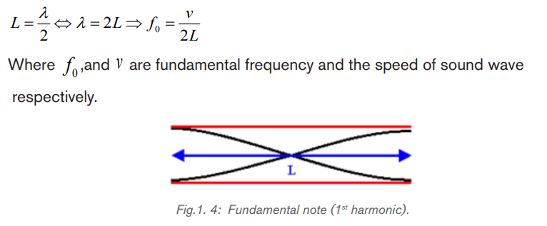
blowing harder. The stationary wave in the open pipe has always an antinode at
each end.
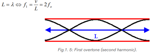
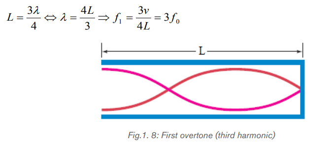
The next longest standing wave in a tube of length L with two open ends is the
second harmonic (first overtone). It also has displacement antinodes at eachend.
The second overtone is obtained from Fig. 1.6 and is the third harmonic.

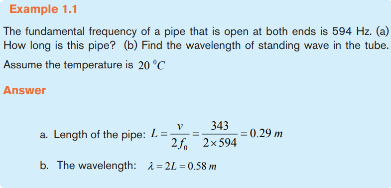
Example 1.1
b. Tube of length L with one open end and one closed end.
The longest wavelength of standing wave in a tube of length L with one open
end and one closed end has a displacement antinode at the open end and adisplacement node at the closed end. This is the fundamental.

The next longest standing wave in a tube of length in a tube of length L with one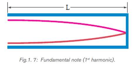
open end and one closed end is the third harmonic (second overtone). It alsohas a displacement antinode at one end and a node at the other.
An odd-integer number of quarter wavelength has to fit into the tube of length L.

frequencies. Only odd harmonics of the fundamental are natural frequencies.
Example 1.2
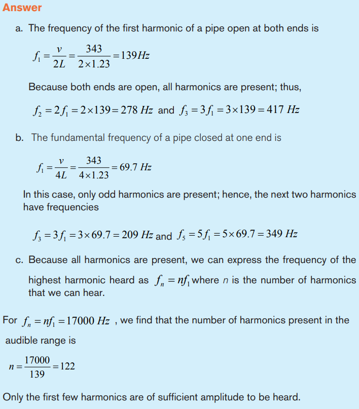
A section of drainage culvert 1.23 m in length and makes a howling noise when
the wind blows.
a. Determine the frequencies of the first three harmonics of the culvert if it is
open at both ends. Take v = 343 m/s as the speed of sound in air.
b. What are the three lowest natural frequencies of the culvert if it is blocked
at one end?
c. For the culvert open at both ends, how many of the harmonics present fallwithin the normal human hearing range (20 Hz to 17 000 Hz)?
1.1.2 Vibrating strings
The string is a tightly stretched wire or length of gut. When it is struck, bowed or
plucked, progressive transverse waves travel to both ends, which are fixed, where
they are reflected to meet the incident waves. A stationary wave pattern is formed
for waves whose wavelengths fit into the length of the string, i.e. resonance occurs.
If you shake one end of a cord (slinky) and the other end is kept fixed, a continuous
wave will travel down to the fixed end and be reflected back, inverted. The
frequencies at which standing waves are produced are the natural frequencies
or resonant frequencies of the cord. A progressive sound wave (i.e. a longitudinal
wave) is produced in the surrounding air with frequency equal to that of the
stationary transverse wave on the string.
Now let consider a cord stretched between two supports that is plucked like a
guitar or violin string. Waves of a great variety of frequencies will travel in both
directions along the string, will be reflected at the ends, and will be travel back in
the opposite direction. The ends of the string, since they are fixed, will be nodes.
Consider a string of length L fixed at both ends, as shown in Fig.1.10. Standing
waves are set up in the string by a continuous superposition of wave incident on
and reflected from the ends.
Note that there is a boundary condition for the waves on the string. The ends of the
string, because they are fixed, must necessarily have zero displacement and are,therefore, nodes by definition.
Fig.1. 10 (a) A string of length L fixed at both ends. The normal modes of vibration form a harmonic
series: (b) the fundamental note; (c) First overtone; (d) the second overtone (Halliday, Resneck, &Walker, 2007).
Example 1.3
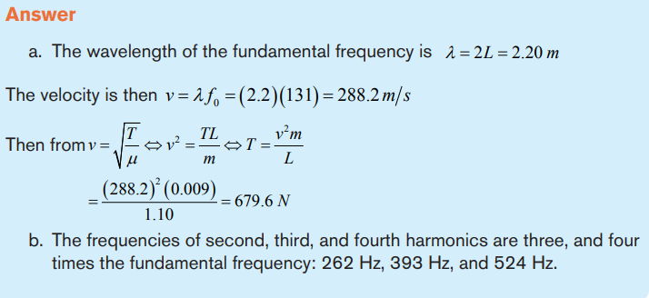
A piano string is 1.10 m long and has mass of 9 g.
a. How much tension must the string be under, if it is to vibrate at a fundamental
frequency of 131 Hz?b. What are the frequencies of the first four harmonics?
Resonance of sound
We have seen that a system such as a taut string is capable of oscillating in one or
more normal modes of oscillation. If a periodic force is applied to such a system,
the amplitude of the resulting motion is greater than normal when the frequency
of the applied force is equal to or nearly equal to one of the natural frequencies
of the system. This phenomenon is known as resonance. Although a block–
spring system or a simple pendulum has only one natural frequency, standing-wave
systems can have a whole set of natural frequencies.
Because oscillating systems exhibit large amplitude when driven at any of its natural
frequencies, these frequencies are often referred to as resonance frequencies.
Fig.1.11shows the response of an oscillating system to various driving frequencies,
where one of the resonance frequencies of the system is denoted by
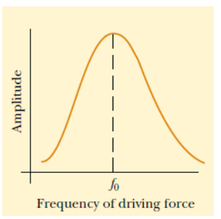
Fig.1. 11: Graph of the amplitude versus driving frequency for oscillating system. The amplitude is
a maximum at the resonance frequency. Note that the curve is not symmetric (Halliday, Resneck, &Walker, 2007)
A more spectacular example is a singer breaking a wine glass with her amplified
voice. A good-quality wine glass has normal-mode frequencies that you can hear
by tapping it.
If the singer emits a loud note with a frequency corresponding exactly to one of
these normal-mode frequencies, large-amplitude oscillations can build up andbreak the glass (Fig. 1.12)

Fig.1. 12 : Some singers can shatter a wine glass by maintaining a certain frequency of their voice
for seconds, (a) Standing-wave pattern in a vibrating wine glass. (b) A wine glass shattered by theamplified sound of a human voice
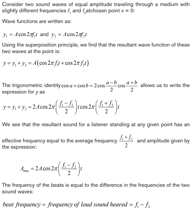
Beats and its phenomena
Beats occur when two sounds-say, two tuning forks- have nearly, but not exactly,
the same frequencies interfere with each other. A crest may meet a trough at
one instant in time resulting in destructive interference. However, at later time the
crest may meet a crest at the same point resulting in constructive interference. To
see how beats arise, consider two sound waves of equalamplitudes and slightlydifferent frequencies as shown on the figure below.
Fig.1. 13: Beats occur as a result of the superposition of two sound waves of slightly different
frequencies (Cutnell & Johnson, 2006).
In 1.00 s, the first source makes 50 vibrations whereas the second makes 60. We
now examine the waves at one point in space equidistant from the two sources.
The waveforms for each wave as a function of time, at a fixed position, are shown
on the top graph of Fig. 1.13; the red line represents the 50 Hz wave, and the blue
line represents the 60 Hz wave. The lower graph in Fig. 1.13 shows the sum of
the two waves as a function of time. At the time the two waves are in phase they
interfere constructively and at other time the two waves are completely out of phase
and interfere destructively. Thus, the resultant amplitude is large every 0.10 s and
drops periodically in between. This rising and falling of the intensity is what is heard
as beats. In this case the beats are 0.10 s apart. The beat frequency is equal tothe difference in frequencies of the two interfering waves.
The interference pattern varies in such a way that a listener hears an alternation
between loudness and softness. The variation from soft to loud and back to soft
is called a Beat. The phenomena of beats can be used to measure the unknownfrequency of a note.
Application activity 1.1
1. Is the wavelength of the fundamental standing wave in a tube open
at both ends greater than, equal to, or less than the wavelength of
the fundamental standing wave in a tube with one open end and one
closed end? Explain your answer.
2. You blow across the opening of a bottle to produce a sound. What
must be the approximate height of the bottle for the fundamental note
to be a middle C (with the wavelength of 1.29 m).
3. Two loudspeakers are separated by 2.5 m. A person stands at 3.0 m
from one and at 3.5 m from the other one. Assume a sound velocity of
343 m/s.
a. What is the minimum frequency to present destructive interference at
this point?
b. Calculate the other two frequencies that also produce destructive
interference.
4. How would you create a longitudinal wave in a stretched spring? Would
it be possible to create a transverse wave in that spring?
5. In mechanics, massless strings are often assumed. Why is this not a
good assumption when discussing waves on strings?
6. Draw the second harmonic (The second lowest tone it can make.) of
a one end fixed, one end open pipe. Calculate the frequency of this
mode if thepipe is 53.2 cm long, and the speed of sound in the pipe is
317 m/s.
7. Calculate the wavelengths below. The length given is the length of thewaveform in the picture bellow:
8. A guitar string is 64 cm long and has a fundamental Mi frequency of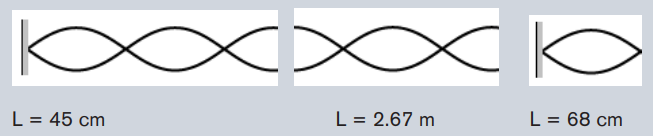
330 Hz. When pressing in the first fret (nearest to the tuning keys) see
figure bellow the string is shortened in such a way that it plays a Fa
note having a frequency of 350 Hz. Calculate the distance betweenthis first fret and the nut necessary to get this effect.
9. Why is a pulse on a string considered to be transverse?
10. A guitar string has a total length of 90 cm and a mass of 3.6 g. From
the bridge to the nut there is a distance of 60 cm and the string has
a tension of 520 N. Calculate the fundamental frequency and the firsttwo overtones
1.2 CHARACTERISTICS AND PROPERTIES OF SOUND WAVES
Activity 1.2
Read the scenario below and answer the questions that follow.
On an interview for Physics placement in a certain school in Rwanda, Claudette
a S.6 leaver who had applied for the job was asked about sound waves
during the interview.
She was asked to state the properties of sound waves. Confidently, she
responded that the properties are reflection, refraction, diffraction and
interference. This was enough to make Claudette pass the first level of the
interview.
However, in the second step, she was required to discuss different media
in which sound waves can propagate. Claudette started discussing these
different media. What surprised the interviewer was Claudette’s ability to relate
sound waves to other kinds of waves stating that these waves behave thesame way when they pass from one medium to another.
Looking at Claudette’s face, the interviewer asked her to discuss the laws
governing reflection and refraction of sound waves. With a smile, she
started by saying that since sound waves have the same properties as for light;
these laws therefore do not change.
As she was attempting to state them, the interviewer stopped her and
congratulated her upon her confidence and bravery she showed in the room.
She was directly told that she was successful and she was given the job.
Claudette is now working as assistant S2 Physics Tutor and doubles as a
Physics laboratory attendant.
Questions
a. Explain the meaning of underlined terms used in the text above?
b. Do you think, it was 100% correct for Claudette to relate sound waves
to light waves? Explain?
c. There is where she was asked to discuss the different media in which
sound waves can propagate. Discuss these different media and talk
about speed of sound waves in the stated media.
d. In one of the paragraphs, Claudette said that the laws governing reflection
and refraction of sound waves were similar to those of light. Can you
explain these laws (Use diagrams where possible)
e. Assuming that you were an interviewer and the interview was out of 80.What mark would you award to Claudette? Why?
1.2.1 Properties of sound waves
Most of us start our lives by producing sound waves! We spend much of our life
surrounded by objects which produce sound waves. Most machines in use vibrate
and produce sound so the only sure way to silence them would be to put them in
vacuum where there would be no surrounding medium for the vibrating surfaces
of the machine to push against, hence no sound waves. Some physiologists
are concerned with how speech is produced, how speech impairment might be
corrected, how hearing loss can be alleviated.
Sound is associated with our sense of hearing and, therefore, with the physiology
of our ears that intercept the sound and the psychology of our brain which interprets
the sensations that reach our ears. Sound waves are longitudinal mechanical waves
that can travel through solids, liquids, or gases.
As the sound wave propagates, many interactions can occur, including reflection,
refraction, diffraction and interference. When a sound wave hits a surface, a part ofthe energy gets scattered while a part of it is absorbed.
a. Reflection of sound wave
Fixed end
First consider an elastic rope stretched from end to end. One end will be securely
attached to a pole on a lab bench while the other end will be held in the hand in
order to introduce pulses (single disturbance, on vibration) into the medium as
shown in Fig.1.14. Because the right end of the rope is attached to a pole (which is
attached to a lab bench), the last particle of the rope will be unable to move whena disturbance reaches it. This end of the rope is referred to as a fixed end.
Fig.1. 14 An elastic securely tied to a pole can be used to study the behavior
of waves at a fixed end
If a pulse is introduced at the left end of the rope, it will travel through the rope
towards the right end of the medium. This pulse is called the incident pulse since
it is incident towards (i.e., approaching) the boundary with the pole.
When the incident pulse reaches the boundary, two things occur:
• A portion of the energy carried by the pulse is reflected and returns towards
the left end of the rope. The disturbance that returns to the left after bouncing
off the pole is known as the reflected pulse.
• A portion of the energy carried by the pulse is transmitted to the pole, causing
the pole to vibrate.
When one observes the reflected pulse off the fixed end, there are several notable
observations. First the reflected pulse is inverted. That is, if an upward displaced
pulse is incident towards a fixed end boundary, it will reflect and return as adownward displaced pulse.
Similarly, if a downward displaced pulse is incident towards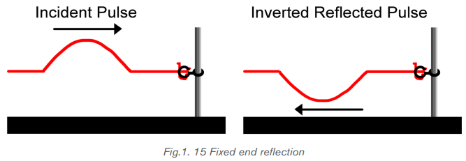
a fixed end boundary,
it will reflect and return as an upward displaced pulse.
The inversion of the reflected pulse can be explained by returning to our conceptions
of the nature of a mechanical wave. When a crest reaches the end of a medium
(“medium A”), the last particle of the medium A receives an upward displacement.
This particle is attached to the first particle of the other medium (“medium B”) on
the other side of the boundary. As the last particle of medium A pulls upwards on
the first particle of medium B, the first particle of medium B pulls downwards on the
last particle of medium A.
In general, Reflection leaves wavelength, speed, amplitude and frequencyunchanged.
Free End Reflection
Suppose a rope is attached to a ring that is loosely fit around the pole as in Fig.1.16.
Because the right end of the rope is no longer secured to the pole, the last particle
of the rope will be able to move when a disturbance reaches it. This end of the ropeis referred to as a free end.
Fig.1. 16 If the end of elastic rope not fastened to the pole then it will befree
to move up and down. This provides for the study of wave behavior at free end
When an upward displaced pulse is incident upon a free end, it returns as an
upward displaced pulse after reflection. And when a downward displaced pulse is
incident upon a free end, it returns as a downward displaced pulse after reflectionas in Fig.1.17. Inversion is not observed in free end reflection.
The reflection of sound waves can end up with any of the two phenomena either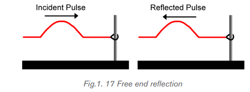
an echo or reverberation:
• Echo occurs when a reflected sound wave reaches the ear 0.1 s after we
hear the original sound. If the time elapsed between the arrivals of the two
sound waves is more than 0.1 s, then the sensation of the first sound will get
died out. An echo sounder or fathometer is a device used on a ship for thepurpose of measuring the depth of the sea.
In a small room the sound is also heard more than once, but the time differencesare so small that the sound just seems to loom. This is known as reverberation.
b. Refraction and Snell’s law and waves
Refraction of waves is the change in direction of waves as they pass from one
medium to another. The bending of waves is accompanied by the change in speed
and wavelength of the wave. So, if there is any change in media, the wave speed
changes. Sound waves travel with less velocity in cool air than they do in the warmer
air.
When a wave travels from deep water to shallow water in such a way that it meets
the boundary between the two depths straight on, no change in direction occurs.
On the other hand, if a wave meets the boundary at an angle, the direction of traveldoes change. This phenomenon is called refraction (Fig.1.18)
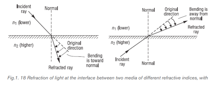
Snell’s law (also known as Snell–Descartes law or the law of refraction) is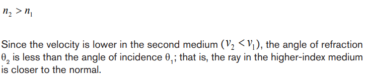
a formula used to describe the relationship between the angles of incidence
and refraction, when referring to light or other waves passing through a boundary
between two different isotropic media, such as water, glass, or air.
Snell’s law states that the ratio of the sines of the angles of incidence and refraction
is equivalent to the ratio of phase velocities in the two media, or equivalent to thereciprocal of the ratio of the indices of refraction:

Where
Comparisons between the characteristics of the transmitted pulse and the reflected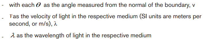
pulse lead to the following observations.
• The transmitted pulse (in the less dense medium) is traveling faster than the
reflected pulse (in the denser medium).
• The transmitted pulse (in the less dense medium) has a larger wavelength
than the reflected pulse (in the denser medium).
• The speed and the wavelength of the reflected pulse are the same as thespeed and the wavelength of the incident pulse.
Because this is less than the incident angle of 30°, the refracted ray is bent
toward the normal, as expected. Its change in direction is called the angleof deviation and is given by

c. Diffraction
Diffraction is the name given to the phenomenon in which a wave spreads out as
it passes through a small aperture or around an obstacle. Diffraction patterns are
formed when the diffracted waves interfere with one another to produce light and
dark bands on a screen or piece of film. Diffraction patterns are most intense when
the size of the aperture or obstacle is comparable to the size of the wavelength of
the wave. Similar effects are observed when light waves travel through a medium
with a varying refractive index. Diffraction is due to the wave nature of light
When light passes through an opening it is observed to spread out. This is knownas diffraction and becomes more pronounced with narrower openings.
Diffraction occurs with all waves, including sound waves, water waves, and
electromagnetic waves such as visible light, x-rays and radio waves. Since diffraction
occurs for waves, but not for particles, it can serve as one means for distinguishingthe nature of light.
d. Interference and principle of Superposition
Interference occurs when two or more waves traveling through the same medium
overlap and combine together. Interference of incident and reflected waves isessential to the production of resonant standing waves.
We can have constructive and destructive interference:
• If a person stands equidistant from two speakers which are playing the same
sound in phase, i.e. which are moving in and out together, then the two waves
arrive in phase after traveling the same distance. Crest meets crest and
trough meets trough at the location of the person. The amplitudes of the two
waves add and the sound is loudest here.
• If the two speakers play the same sound but are out of phase, i.e. one is
moving out while the other is moving in, and then the sound has a low volume
at the location of the person equidistant from the two speakers. This can
easily be demonstrated by switching the wires on one of the speakers. (This
is why you need to pay attention to the color of the wires when setting up your
stereo). Dead spots in an auditorium are sometimes produced by destructive
interference.
In general, the term “interference” refers to what happens when two or
more waves pass through the same region at the same time.
The principle of superposition
Combining the displacements of the separate pulses at each point to obtain the
actual displacement is an example of the principle of superposition: “When
two waves overlap, the actual displacement of any point on the string at any time
is obtained by adding the displacement the point would have if only the first wave
were present and the displacement it would have if only the second wave werepresent”.
In other words, the wave function y(t, x) that describes the resulting motion in thissituation is obtained by adding the two wave functions for the two separate waves:
As we saw with transverse waves, when two waves meet, they create a third wave
that is a combination of the other two waves. This third wave is actually the sum of
the two waves at the points where they meet. The two original waves are still there
and will continue along their paths after passing through each other. After passingthe third wave no longer exists.
1.2.2 Characteristics of sound waves
Usually, the characteristics used to describe waves are period, frequency,
wavelength, and amplitude.
a. Frequency ranges
Any periodic motion has a frequency, which is the number of complete cycles in
a second and a period which is the time used to complete one cycle. While the
frequency is measured in Hertz (Hz), the period is measured in seconds (s). For a
wave, the frequency is the number of wave cycles that pass a point in a second. A
wave’s frequency equals the frequency of the vibrating source producing the wave.
Sound waves are classified into three categories that cover different frequency
ranges:
• Audible sound
Audible sound lies within the range of sensitivity of the human ear. They can
be generated in a variety of ways, such as musical instruments, human voices, or
loudspeakers. It is almost impossible to hear sounds outside the range of 20 Hz to
20 kHz. These are the limits of audibility for human beings but the range decreaseswith age.
Hearing is the perception of sound. The hearing mechanism involves some
interesting physics. The sound wave that impinges upon our ear is a pressure wave.
The ear is a transducer that converts sound waves into electrical nerve impulsesin a manner much more sophisticated than, but analogous to, a microphone.
• Infrasonic waves
Infrasonic waves have frequencies below the audible range. They are sound
waves with frequencies that are below 20 Hz limit.
Some animals such as elephants can use infrasonic waves to communicate
effectively with each other, even when they are separated by many kilometers. Their
large ears enable them to detect these low frequency sound waves which haverelatively long wavelengths.
Young bat-eared fox and Rhinoceros (Fig.1.21) also use infrasonic as low as 5 Hzto call one another. They have ears adapted for the detection of very weak sounds.
A number of animals are sensitive to infrasonic frequencies. It is believed by many
zoologists that this sensitivity in animals such as elephants may be helpful in
providing them with early warning of earthquakes and weather disturbances. It has
been suggested that the sensitivity of birds to infrasound aids their navigation and
even affects their migration.
• Ultrasonic waves
Ultrasonic waves have frequencies above the audible range. They are sound
waves whose frequencies are higher than 20 KHz. You may have used a “silent”
whistle to retrieve your dog. The ultrasonic sound emitted by that device is easily
heard by dogs, although humans cannot detect it at all. Ultrasonic waves are alsoused in medical imaging.
Some marine mammals, such as dolphin, whales, and porpoises use sound waves
to locate distant objects. In this process, called echolocation, a dolphin produces
a rapid train of short sound pulses that travel through the water, bounce off distant
objects, and reflect back to the dolphin. From these echoes, dolphins can determine
the size, shape, speed, and distance of their potential prey. Experiments have
shown that at distance of 114 m, a blindfolded dolphin can locate a stainless-steel
sphere with a diameter of 7.5 cm and can distinguish between a sheet of aluminum
and a sheet of copper. The Ultrasonic waves emitted by a dolphin enable it to seethrough bodies of other animals and people (Fig.1.22).
Skin muscles and fat are almost transparent to dolphins, so they see only a thin
outline of the body but the bones, teeth and gas-filled cavities are clearly apparent.
Physical evidence of cancers, tumors, heat attacks, and even emotional shake can
all be seen by dolphin. What is more interesting, the dolphin can reproduce the
sonic signals that paint the mental image of its surroundings, and thus the dolphin
probably communicates its experience to other dolphins. It needs no words or
symbol for fish, for example, but communicates an image of the real thing.
Dogs, cats and mice can hear ultrasound frequencies up to 450 000 Hz. Some
animals not only hear ultrasound but also use ultrasonic to see in dark. Bats also
use echo to navigate through air. Bats use ultrasonic with frequencies up to 100000 Hz to move around and hunt (Fig.1.23).
The waves reflect off objects and return the bat’s ears. The time it takes for the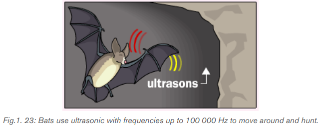
sound waves to return tells the bat how far it is from obstacles or prey. The bat uses
the reflected sound waves to build up a picture of what lies ahead.
The process of imaging using Sonar (Sound Navigation and Ranging) is the same
as the echo-locating sonar of a submarine or a bat. The observer sends out a brief
pulse of ultrasound and waits for an echo. The pulse travels out, reflects off the
target and returns. The ultrasound machine uses pulses because the same device
acts as both transmitter and receiver.
Ultrasound has been used in a variety of clinical settings, including obstetrics and
gynecology, cardiology and cancer detection. The main advantage of ultrasound
is that certain structures can be observed without using radiation. Ultrasound can
also be done much faster than X-rays or other radiographic techniques.
Ultrasonic waves can be used to produce images of objects inside the body thus
Physicians use ultrasonic to observe fetuses. Ultrasound has frequencies too high
for you to hear. Echoes from ultrasound waves can show what is inside the body.Echo is a reflection of sound off the surface of an object.
In medicine, ultrasonic is used as a diagnostic tool, to destroy diseased tissue,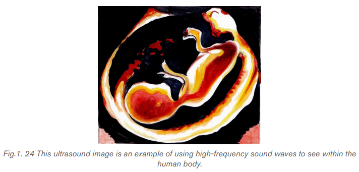
and to repair damaged tissue. Ultrasound examination of the heart is known as
echocardiography.
Many animals hear a much wider range of frequencies than human beings do. For
example, dog whistles vibrate at a higher frequency than the human ear can detect,
while evidence suggests that dolphins and whales communicate at frequencies
beyond human hearing (ultrasound) see Fig.1.25 below.(Cutnell & Johnson, 2006).
b. Wavelength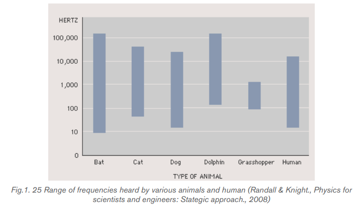
Wavelength is the distance covered by a wave in a period. It is represented by
the separation between a point on one wave and a similar point on the next cycle
of the wave. For a transverse wave, wavelength is measured between adjacent
crests or between adjacent troughs. For a longitudinal wave such as sound wave,
wavelength is the distance between adjacent compressions or rarefaction.
c. Speed of sound
For a periodic wave, the shape of the string at any instant is a repeating pattern. The
length of one complete wave pattern is the distance from one crest to the next or
from one trough to the next or from any point to the corresponding point on the next
repetition of the wave shape. We call this distance the wavelength of the wave,
denoted by the Greek letter lambda (λ).
The wave pattern travels with constant speed and advances a distance of onewavelength in a time interval of one period T. So, the wave speed is given by
where f is the frequency of the wave.
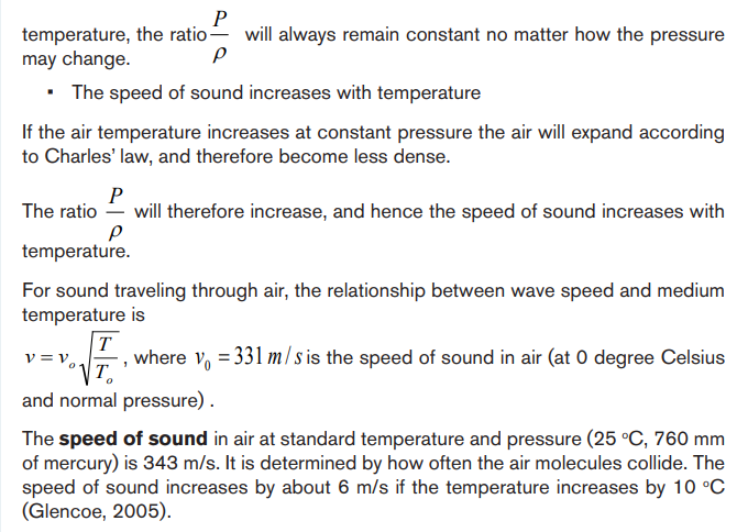
Sound travels faster in liquids and solids than in gases, since the particles in liquids
and solids are closer together and can respond more quickly to the motion of
their neighbors. As examples, the speed of sound is 331 m/s in air, 1500 m/s in
water and 5000 m/s in iron (though these mediums the seed of sound can changedepending on temperature and pressure). Sound does not travel in vacuum.


d. Amplitude
The amplitude of a wave is the maximum displacement of the medium from its rest
position. The amplitude of a transverse wave is the distance from the rest positionto a crest or a trough. The more energy a wave has, the greater is its amplitude.
Application activity 1.2
1. The correct statement about sound waves is that:
A. They are transverse waves
B. They can be polarized
C. They require material medium to propagate
2. Sound travels in
A. Air C. Water
B. Iron D. All of these
3. Two men talk on the moon. Assuming that the thin layer of gases on the
moon is negligible, which of the following is the right answer:
A. They hear each other with lower frequency
B. They hear each other with higher frequency
C. They can hear each other at such frequency
D. They cannot hear each other at all
4. Do you expect an echo to return to you more quickly on a hot day or a
cold day? Explain your answer.
A. Hot day.
B. Cold day.
C. Same on both days.
5. A sound wave is different than a light wave in that a sound wave is:
A. Produced by an oscillating object and a light wave is not.
B. Not capable of traveling through a vacuum.
C. Not capable of diffracting and a light wave is.
D. Capable of existing with a variety of frequencies and a light wave
has a single frequency.
6. A spider of mass 0.30 g waits in its web of negligible mass see Fig.
below. A slight movement causes the web to vibrate with a frequencyof about 15 Hz.
a. Estimate the value of the spring stiffness constant k for the web assuming
simple harmonic motion.
b. At what frequency would you expect the web to vibrate if an insect of
mass 0.10 g were trapped in addition to the spider?
7. Dolphins use sound waves to locate food. Experiments have shown
that a dolphin can detect a 7.5 cm target 110 m away, even in murky
water. For a bit of “dinner” at that distance, how much time passes
between the moment the dolphin emits a sound pulse and the moment
the dolphin hears its reflection and thereby detects the distant target?
8. By what factor would you have to multiply the tension in a stretched
string in order to double the wave speed? Explain your answer.
9. (a) The range of audible frequencies is from about 20 Hz to 20 000 Hz.
What is the range of the wavelengths of audible sound in air?
(b) The range of visible light extends from 400 nm to 700 nm. What is
the range of visible frequencies of light?
(c) Surgeons can remove brain tumors by using a cavitron ultrasonic
surgical aspirator, which produces sound waves of frequency 23
kHz. What is the wavelength of these waves in air?
(d) Sound having frequencies above the range of human hearing (about
20 000 Hz) is called ultrasound. Waves above this frequency can
be used to penetrate the body and to produce images by reflecting
from surfaces. In a typical ultrasound scan, the waves travel through
body tissue with a speed of 1500 m/s. For a good, detailed image,
the wavelength should be no more than 1.0 mm. What frequencysound is required for a good scan?
1.3 CHARACTERISTICS OF MUSICAL NOTES
Activity 1.3
The physical characteristics of a sound wave are directly related to the
perception of that sound by a listener.
1. What is the difference between the sound of whistle and that of drum?
2. Mutoni is playing the same notes on different musical instruments, can
you predict which musical instruments is played without seeing them?Explain your answers.
A musical note is produced by vibrations that are regular and repeating, i.e. by
periodic motion. Non-periodic motion results in noise which is not pleasant to the
ear. Many behaviors of musical note can be explained using a few characteristics:
intensity and loudness, frequency and pitch, and quality or timber
1.3.1. Pitch and frequency
The sound of a whistle is different from the sound of a drum. The whistle makes a
high sound. The drum makes a low sound. The highness or lowness of a sound is
called its pitch. The higher the frequency, the higher is the pitch. The frequency of
an audible sound wave determines how high or low we perceive the sound to be,
which is known as pitch.
Frequency refers to how often something happens or in our case, the number of
periodic, compression-rarefaction cycles that occur each second as a sound wave
moves through a medium and is measured in Hertz (Hz) or cycles/second. The term
pitch is used to describe our perception of frequencies within the range of human
hearing.
If a note of frequency 300 Hz and note of 600 Hz, are sounded by a siren, the
pitch of the higher note is recognized to be an upper octave of the lower note.
The musical interval between two notes is an upper octave if the ratio of their
frequencies is 2:1. It can be shown that the musical interval between two notes
depends on the ratio of their frequencies, and not on the actual frequencies.
Whether a sound is high-pitched or low-pitched depends on how fast something
vibrates. Fast vibrations make high-pitched sounds. Slow vibrations make low
pitched sounds.
Do not confuse the term pitch with frequency. Frequency is the physical
measurement of the number of oscillations per second. Pitch is a psychological
reaction to sound that enables a person to place the sound on a scale from high
to low, or from treble to bass. Thus, frequency is the stimulus and pitch is the response.
Although pitch is related mostly to frequency, they are not the same. A
phrase such as “the pitch of the sound” is incorrect because pitch is not a physicalproperty of the sound. The octave is a measure of musical frequency.
1.3.2 Intensity, amplitude and ear response
A police siren makes a loud sound. Whispering makes a soft sound. Whether a
sound is loud or soft depends on the force or power of the sound wave. Powerful
sound waves travel farther than weak sound waves. To talk to a friend across the
street you have to shout and send out powerful sound waves. Your friend would
never hear you if you whispered.
A unit called the decibel measures the power of sound waves. The sound waves
of a whisper are about 10 decibels. Loud music can have a level of 120 decibels
or more. Sounds above 140 decibels can actually make your ears hurt. The energy
carried by a sound wave is proportional to the square of its amplitude. The energy
passing in a unit area per unit time is called the intensity of the wave.

Sound intensity level
To the human ear the change in loudness when the power of a sound increases
from 0.1 W to 1.0 W is the same as when 1W to 10 W. The ear responds to the
ratio of the power and not to their difference.
We measure sound level intensity in terms of “decibels”. The unit bel is named afterthe inventor of the telephone, Alexander Graham Bell (1847–1922).
The decibel is a “relative unit” which is actually dimensionless, comparing a givensound to a standard intensity which represents the smallest audible sound:
The intensity of 0dB represents the softest audible sound (threshold of human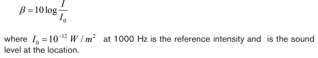
hearing), while 80 dB (i.e., moderately loud music) represents an intensity which isone hundred million times greater.
The physical characteristics of a sound wave are directly related to the perception
of that sound by a listener. For a given frequency the greater the pressure amplitude
of a sinusoidal sound wave, the greater the perceived loudness.
The loudness or softness of sound depends on the intensity of the sound wave
reaching the person concerned. Loudness is a subjective quantity unlike intensity.
Sound that is not wanted or unpleasant to the ear is called noise. High intensity
can damage hearing. The higher the intensity, the louder is the sound. Our ears,
however, do not respond linearly to the intensity. A wave that carries twice theenergy does not sound twice as loud.
Anatomy of human ear
The human ear is a remarkably sensitive detector of sound. Mechanical detectors
of sound can barely match the ear in detecting low intensity sounds. The ear has a
function of transforming the vibrational energy of waves into electrical signals that
are carried to the brain by ways of nerves as does a microphone.
The ear consists of three main parts: the outer ear, the middle ear and the inner ear.
In the outer ear, sounds waves from the outside travel down the ear canal to the
eardrum which vibrates in response to the colliding waves.The inner ear consists
of three small bones known as the hammer, anvil and stirrup which transfer the
vibrations of the eardrum to the inner ear at the oval window.
The function of the inner ear is to transduce vibration into nervous impulses. While
doing so, it also produces a frequency (or pitch) and intensity (or loudness) analysis
of the sound. Nerve fibres can fire at a rate of just under 200 times per second.
Sound level information is conveyed to the brain by the rate of nerve firing, for
example, by a group of nerves each firing at a rate at less than 200 pulses per
second. They can also fire in locked phase with acoustic signals up to about 5
kHz. At frequencies below 5 kHz, groups of nerve fibres firing in lock phase with
an acoustic signal convey information about frequency to the brain. Above about
5 kHz frequency information conveyed to the brain is based upon the
place of stimulation on the basilar membrane. As an aside, music translated
up into the frequency range above 5 kHz does not sound musical. (Hallowell, Davis;Richard,S., 1970)
This delicate system of levers, coupled with the relatively large area of the eardrum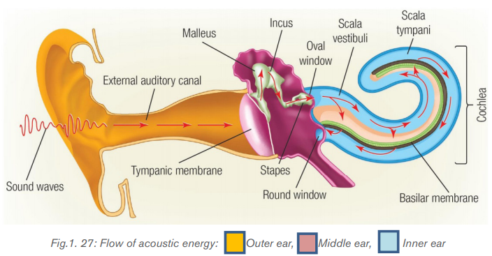
compared to the area of the oval window, results in pressure being amplified by
a factor of about 40. The inner ear consists of the semicircular canals, which are
important for controlling balance, and the liquid filled cochlea where the vibrationalenergy of sound waves is transformed into electrical energy and sent to the brain.
Logarithmic response of the ear versus intensity
The ear is not equally sensitive to all frequencies. To hear the same loudness for
sounds of different frequencies requires different intensities. Studies done overlarge numbers of people have produced the curves shown on Fig.1.28.
On this graph, each curve represents sounds that seemed to be equally loud. The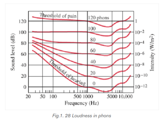
number labelling each curve represents the loudness level which is numerically
equal to the sound level in dB at 1000 Hz. The units are called phons.
Example: The curve labelled 40 represents sounds that are heard by an average
person to have the same loudness as 1000 Hz sound with a sound level of 40 dB.
From this 40 phon curve, we see that a 100 Hz tone must be at a level of about
62 dB to be perceived as loud as a 1000 Hz tone of only 40 dB.
Two aspects of any sound are immediately evident to human listener: loudness
and the pitch. Each refers to a sensation in the consciousness of the listener. But
to each of these subjective sensations there corresponds a physically measurable
quantity.
Loudness refers to the intensity in the sound wave. Intensity is related to the
energy transported by a wave per unit time across a unit area perpendicular to the
energy flow. Intensity is proportional to the square of the wave amplitude.
A unit called a phon is used to express loudness numerically. Phons differ from
decibels because the phon is a unit of loudness perception, whereas the decibel is
a unit of physical intensity. Fig.1.28 shows the relationship of loudness to intensity
(or intensity level) and frequency for persons with normal hearing. The curved lines
are equal-loudness curves. Each curve is labelled with its loudness in phons. Any
sound along a given curve is perceived as equally loud by the average person. The
curves were determined by having large numbers of people compare the loudness
of sounds at different frequencies and sound intensity levels. At a frequency of
1000 Hz, phons are taken to be numerically equal to decibels.
Because of this relationship between the subjective sensation of loudness and the
physically measurable quantity intensity, sound intensity levels are usually specified
on a logarithmic scale. The unit of this scale is a bel, after the inventor Alexander
Graham Bell.
1.3.3 Quality or timbre
If the same note is sounded on the violin and then on the piano, an untrained listener
can tell which instrument is being used, without seeing it. We would never mistake
a piano for flute. We say that the quality or timbre of note is different in each case.
The manner in which an instrument is played strongly influences the sound quality.
Two tones produced by different instruments might have the same fundamental
frequency (and thus the same pitch) but sound different because of different
harmonic content. The difference in sound is called tone color, quality, or timbre.A violin has a different timbre than a piano.
Application activity 1.3
1. Complete each of the following sentences by choosing the correct
term from the following words: loudness, pitch, sound quality, echoes,
intensity and noise
a. The ------------ of a sound wave depends on its amplitude
b. Reflected sound waves are called ---------------------------
c. Two different instruments playing the same note sound different because
of ------------------
2. Plane sound wave of frequency 100 Hz fall normally on a smooth wall.
At what distances from the wall will the air particles have:
a. Maximum amplitude of vibration
b. Minimum amplitude of vibration?
Give reasons for your answer. The speed of sound in air may be taken
as 340 m/s
3. A boy whistles a sound with the power of 4 0.5 10 W− × . What will be his
sound intensity at a distance of 5 m?
4. Calculate the intensity level equivalent to an intensity 1nW/m2
5. If the statement is true, write true. If it is false, change the underlined
word or words to make the statement true.
a. Intensity is mass per unit volume.
b. Loudness is how the ear perceives frequency
c. Music is a set of notes that are pleasing
6. The sound level of sound whose intensity is 10 2 I Wm 1.0 10 / − = × whatwill be the sound intensity level?
1.4 THE DOPPLER EFFECT AND ITS APPLICATIONS
Activity 1.4
1. People use sound for other things other than talking and making music.
In your own words, give more examples and explanations to support this
statement.
2. Imagine you are standing beside a road and a police car with its siren
turned on, drives by you. What do you notice about the heard sound?
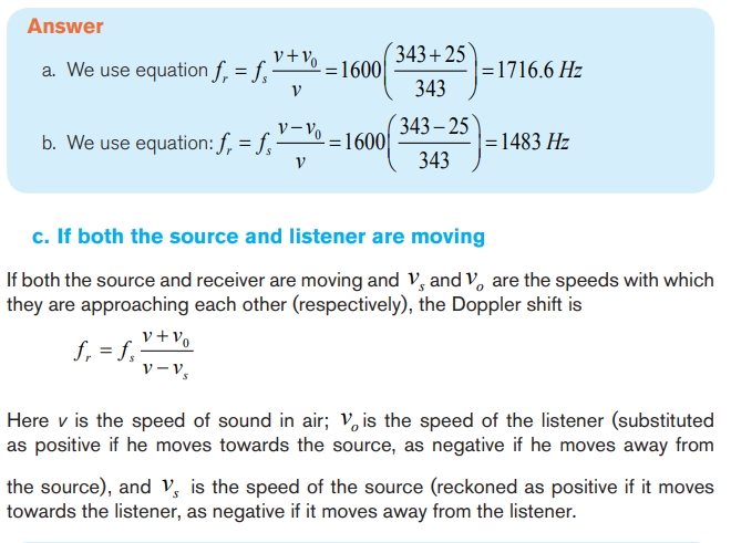
3. In the second case, the same police car turned and comes towards you.
Comment on the heard sound4. Compare and contrast the sounds heard in case 2 and 3.
1.4.1 Doppler Effect
Doppler’s effect is the apparent variation in frequency of a wave due to the relative
motion of the source of the wave and the observer.
The effect takes its name from the Austrian Mathematician Christian Johann Doppler
(1803-1853), who first stated the physical principle in 1842. Doppler’s principle
explains why, if a source of sound of a constant pitch is moving toward an observer,
the sound seems higher in pitch, whereas if the source is moving away it seems
lower. This change in pitch can be heard by an observer listening to the whistle ofan express train from a station platform or another train.

Hence the frequency you hear is higher than the frequency emitted by the
approaching source.Example 1.10
If a source emits a sound of frequency 400 Hz when is at rest, then when the
source moves toward a fixed observer with a speed of 30 m/s, what frequency
does the observer hears knowing that the speed of a sound in air at roomtemperature is 343m/s?


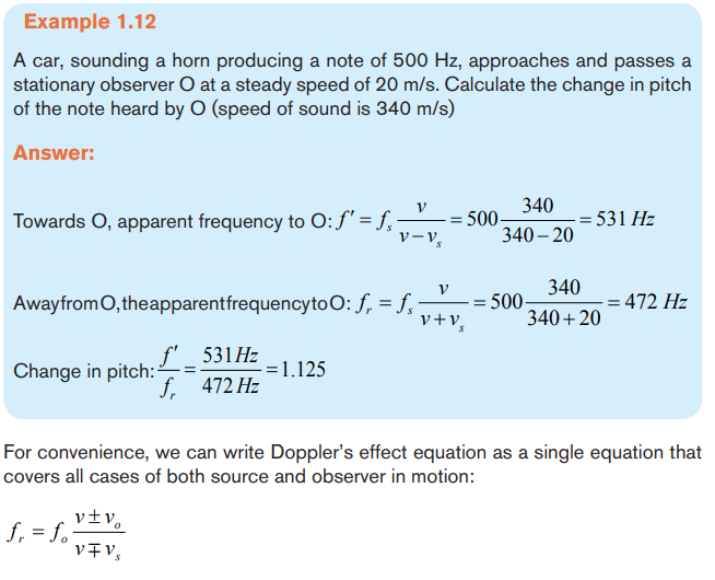
The upper signs apply if source and/or observer move toward each other. The
lower signs apply if they are moving apart. The word toward is associated with
an increase in observed frequency. The words away from are associated with a
decrease in observed frequency.
Although the Doppler’s effect is most typically experienced with sound waves, it
is a phenomenon that is common to all waves. For example, the relative motion
of source and observer produces a frequency shift in light waves. The Doppler’s
effect is used in police radar systems to measure the speeds of motor vehicles.
Likewise, astronomers use the effect to determine the speeds of stars, galaxies,and other celestial objects relative to the Earth.
Example1.13
As an ambulance travels east down a highway at a speed of 33.5 m/s, its siren
emits sound at a frequency of 400 Hz. What frequency is heard by a person in a
car traveling west at 24.6 m/s
a. as the car approaches the ambulance and
b. as the car moves away from the ambulance?
c. Suppose the car is parked on the side of the highway as the ambulance
speeds by. What frequency does the person in the car hear as theambulance (i) approaches and (ii) recedes?

1.4.2 Uses of Doppler Effect
Astronomy
Doppler Effect is used to measure the speed at which stars and galaxies are
approaching or receding from us, in a mechanism named red shift or blue shift.
Redshift happens when light seen coming from an object that is moving away is
proportionally increased in wavelength, or shifted to the red end of the spectrum.
Vice versa occurs with blue shift. Since blue light has a higher frequency than red
light, the spectral lines of an approaching astronomical light source exhibit a blue
shift and those of a receding astronomical light source exhibits a redshift.
Medical imaging
In medicine, the Doppler Effect can be used to measure the direction and speed
of blood flow in arteries and veins. This is used in echocardiograms and medical
ultrasonography and is an effective tool in diagnosis of vascular problems.
Radar
The Doppler Effect is used to measure the velocity detected objects where a radar
beam is fired at a moving target. For example, the police use radar to detect a
speeding vehicle. Radio waves are fired using a radar gun at the moving vehicle.
The velocity is calculated using the difference between the emitted frequency and
the reflected frequency. In a similar way, Doppler radar is used by weather stationsto calculate factors like wind speed and intensity
Application activity 1.4
1. Choose the best answer: Bats can fly in the dark without hitting
anything because
A. They are flying mammals C. They are guided by ultrasonic waves
produced by them
B. Their night vision is going D. Of no scientific reason
2. Discuss application of sound waves in medicine and navigation
3. Explain how sonar is used to measure the depth of a sea
4. a. What is meant by Doppler Effect?
b. A police car sound a siren of 1000 Hz as it approaches a stationary
observer at a speed of 33.5 m/s. What is the apparent frequency of
the siren as heard by the observer if the speed of sound in air is 340
m/s.c. Discuss applications of the Doppler Effect.
Skills Lab 1
In this activity, you will design any musical instrument of your choice.
Procedures:
• Think of the instrument you wish to design. You may have two alternatives.
• Check whether the materials can be locally available in your area
• When you have all the required materials, start making it. You can find a
model instrument for reference.
• After you have designed your instrument, try to experiment (play it) to
check whether it is functioning. In case it is not functioning, try to design
it until it works
• When you are done, try to present it to the whole class in presence of
your tutor.
Note: You can ask a place at your school where you can keep your instrumentfor future use by either other students or tutors.
End of unit 1 assessment
For question 1 to 6, choose the letter of the best answer
1. Which of the following affects the frequency of wave?
A. Reflection C. Diffraction
B. Doppler Effect D. All of the above
2. Consider the following statements:
I) Recording of sound on tapes was first invented by Valdemar Poulsen.
II) Audio tapes have magnetic property.
III) The tapes may also be made of PVC (Polyvinyl-chloride)
2. Considering the above statements in question 2 choose the letter of the
best answer:
A. I, II, and III all are correct. C. I and II are correct, III is
wrong
B. I, II, and III all are wrong D. I and II are wrong, III is
correct
3. Nodes are
A. Positions of maximum displacement
B. Positions of no displacement
C. A position between no displacement and maximum displacement
D. None of these
4. Sound waves are:
A. Transverse waves characterized by the displacement of air molecules.
B. Longitudinal waves characterized by the displacement of air molecules.
C. Longitudinal waves characterized by pressure differences.
D. Both (B) and (C).
E. (A), (B), and (C).
5. In which of the following is the wavelength of the lowest vibration mode
the same as the length of the string or tube?
A. A string. D. An open tube.
B. A tube closed at one end. E. None of the above.
C. All of the above.
6. When a sound wave passes from air into water, what properties of the
wave will change?
A. Frequency. D. Wavelength.
B. Wave speed. E. Both frequency and wavelength.
C. Both wave speed and wavelength.
7. Does the phenomenon of wave interference apply only to sinusoidal
waves? Explain.
8. As oppositely moving pulses of the same shape (one upward, one
downward) on a string pass through each other, there is one instant at
which the string shows no displacement from the equilibrium position
at any point. Has the energy carried by the pulses disappeared at this
instant of time? If not, where is it?
9. Can two pulses traveling in opposite directions on the same string
reflect from each other? Explain.
10. When two waves interfere, can the amplitude of the resultant wave
be greater than the amplitude of any of the two original waves? Under
which conditions?
11. When two waves interfere constructively or destructively, is there any
gain or loss in energy? Explain.
12. Explain why your voice seems to sound better than usual when you
sing in the shower.
13. An airplane mechanic notices that the sound from a twin-engine aircraft
rapidly varies in loudness when both engines are running. What could
be causing this variation from loud to soft?
14. Explain how a musical instrument such as a piano may be tuned by
using the phenomenon of beats.
15. Fill in the gap
A. As a sound wave or water ripple travels out from its source, its ---------
----- decreases.
B. The vibrating air in a/an ----------------------------- has displacement
antinodes at both ends.
C. For a /an ……………., the fundamental corresponds to a wavelength
four times the length of the tube.
D. The ……………….. refers to the change in pitch of a sound due to the
motion either of the source or of the observer. If source and observerare
approaching each other, the perceived pitch is …….. If they are moving
apart, the perceived pitch is …………….
16. A bat, moving at 5.00 m/s, is chasing a flying insect. If the bat emits a
40.0 kHz chirp and receives back an echo at 40.4 kHz, at what speed
is the insect moving toward or away from the bat? (Take the speed of
sound in air to be v = 340 m/s.)
17. If you hear the horn of the car whose frequency is 216 Hz at a frequency
of 225 Hz, what is their velocity? Is it away from you or toward you? The
speed of sound is 343 m/s
18. You run at 12.5 m/s toward a stationary speaker that is emitting a
frequency of 518 Hz. What frequency do you hear? The speed of
sound is 343 m/s
19. If you are moving and you hear the frequency of the speaker at 557
Hz, what is your velocity? Is it away from or toward the speaker? Thespeed of sound is 343 m/s
20. Read the following text and answer the question
Researchers have known for decades that whales sing complicated songs.
Their songs can last for 30 min and a whale may repeat the song for two or
more hours. Songs can be heard at a distances of hundreds of kilometers.
There is evidence that whales use variations in the songs to tell other whales
about the location of food and predators. Only the male whales sing, which
has led some researchers to think that songs are also used to attract a male.
The whale songs may be threatened by noise pollution. In the past 50 years,
ocean noise has increased due to human activities. Goods are transported
across the ocean in larger ships than ever before. Large ships use bigger
engines. They produce low-frequency noise by stirring up air bubbles with their
propellers. Unfortunately, whales also use low-frequency sound in their songs,
perhaps because these sounds carry further than high-frequency sounds in
the ocean. Propeller noise from large ships is loud enough to interfere with
whale songs at a distance of 20 km.
Question: Are regulations needed to protect whales from noise?
In your own words, describe the major issue that needs to be resolved about
ocean noise pollution. List three arguments for those who think regulations
should require large ships to reduce noise pollution. List three arguments forthose who think regulations are not necessary.
File: 1UNIT 2: CLIMATE CHANGE AND GREENHOUSE EFFECT
Key Unit Competence:
Evaluate the environmental survey conducted on climate change
and greenhouse effect.
Introductory activity

Most of the solar radiation that is incident onto the Earth is absorbed and the
rest is reflected into the atmosphere. Our Earth acts almost as a black body,
thereby radiating back to the space part of the energy it has absorbed from the
sun. The earth and its atmosphere are a part of the solar system. Life on theEarth cannot exist without the energy from the sun.
a. Basing on Physics concepts, how do humans and plants get energy from
the sun? Account for the use of this energy.
b. Do humans and plants maintain the energy absorbed forever? Explain
your reasoning using physics concepts.
c. State and explain the scientific term used to describe a body that can
absorb or emit radiations that fall on them.
d. Basing on your ideas in question (c) above what could be the effect on
that body if:
i) It maintains the energy for a long time
ii) Reflects energy after a given time.
iii) Do you think that these reflected and absorbed radiations have effect
on the Climate? Why? Discuss your answers with your friends and even
present it to your Physics tutor
e. Some radiations may be prevented from leaving the atmosphere and
remain concentrated in the atmosphere. What do you think are effects of
these gases as they remain in the atmosphere?
f. In your own view what changes have been brought by this concentrationof radiations and how has man tried to control some of the changes.
2.1 CLIMATE CHANGE
Activity 2.1
The sun is the major source of energy (in form of radiation) on the earth. Some
of these radiations are absorbed by different aerosols in the space and part of
it reaches the earth. Our earth has special features that leads to absorption of
these radiations and these processes are continuous.
a. What do you think are the effects of these radiations on the earth and its
atmosphere after absorption and reflection?
b. From your own understanding, what would happen if there is imbalance
between the absorbed and radiated energy in the earth’s atmosphere.
c. Can that incidence be controlled? How?
d. With practical examples, discuss how these changes in climate have
been noticed in our country Rwanda
e. In your own words, what are the scientific measures that can be done toavoid that kind of situation?
2.1.1 Climate change and related facts
Climate is usually defined as the “average weather,” or more rigorously, as the
statistical description in terms of the mean and variability of relevant
quantities over a period of time ranging from months to thousands of years. The
classical period is 3 decades, as defined by the World Meteorological Organization
(WMO). These quantities are most often surface variables such as temperature,
precipitation, and wind. Weather is measured in terms of the following parameters:
wind, temperature, humidity, atmospheric pressure, cloudiness, and precipitation.
In most places, weather can change from hour-to-hour, day-to-day, and season to-season.
Climate can be described as the sum of weather. While the weather is
quite variable, the trend over a longer period, the climate, is more stable.
However, climate change can be observed over a longer period of time. Climate
change refers to any significant change in the climate parameters such as
temperature, precipitation, or wind patterns, among others, that occur over several
decades or longer Natural and human systems have adapted to the prevailing
amount of sunshine, wind, and rain. While these systems can adapt to small
changes in climate, adaptation is more difficult or even impossible if the change in
climate is too rapid or too large. This is the driving concern over anthropogenic, or
human induced, climate change. If climate changes are too rapid, then many natural
systems will not be able to adapt and will be damaged and societies will need to
incur the costs of adapting to a changed climate (REMA)
There has been variation in the atmospheric conditions in a given time. This has
affected the seasons leading to a less output of our produce especially from
agriculture, fishing and other activities.
These changes are sometimes for a short time but also may take a long time. Some of
these changes result from our practices like farming, industrialization, urbanization,
mining and other infrastructure developments. Care should be taken so as these
changes in the atmospheric conditions can be avoided.
Some of important terms we need to know
• Climate feedback: This refers to a process that acts to amplify or reduce
direct warming or cooling effects.
• Climate lag: This is the delay that can occur in a change of some aspect
of climate due to the influence of a factor that is slow acting.
• Climate model: This is a quantitative way of representing the interactions
of the atmosphere, oceans, land surface, and ice. Models can range from
relatively simple to quite comprehensive
This explains a delay that occurs in climate change as a result of some factors that
changes only very slowly. For example, the effects of releasing more carbon dioxide
into the atmosphere occur gradually over time because the ocean takes a long timeto warm up in response to these emissions.
2.1.2 Causes of climate change.
Physics behind climate change and causes
The climate of the earth is controlled by its absorption and the subsequent emission
of that energy.
The Earth’s surface temperature is determined by the balance between the
absorption and emission of Sun’s radiation.
The major cause of climate change is the concentration of greenhouse gases,
especially water vapor and carbon dioxide. These gases trap thermal radiation from
the earth’s surface and this effect keeps the surface warmer than it would be.
a. Human causes
Human activities are major factors that lead to natural greenhouse effect. Most of
activities done by human lead to high concentration of greenhouse gases. From
research it has been found that the concentration of carbon dioxide has risen
by about 30% in this period as compared to pre-industrial period. This gives a
projection that the concentration of these greenhouse gases is still increasing day
and night as people continue using fossil fuels.
Such activities include bush burning, burning of fossil fuels, deforestation and
agriculture. All these activities have a strong impact on the climate of a given area
as they may lead to either warming or cooling the land.
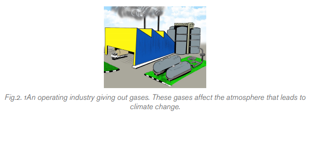
On another hand, industries have also led to the change in climatic conditions as
they emit carbon gases into the atmosphere. From what is happening currently,
the climatic conditions are worsening if the world becomes more industrialized.
Peoples are trying to limit this by making machines that emit less carbon gases(REMA).
b. Natural causes
• Volcanicity: When volcanoes erupt, they throw out large volumes of
Sulphur dioxide (SO2), water vapor, dust, and ash into the atmosphere.
Although the volcanic activity may last only a few days, the large volumes
of gases and ash can influence climatic patterns for years. Millions of
tons of Sulphur dioxide gas can reach the upper levels of the atmosphere
(called the stratosphere) from a major eruption.
• Ocean currents: Oceans plays a major role in the change of climate.
Oceans cover about 71% of the Earth and absorb about twice as much of
the sun’s radiation as the atmosphere or the land surface. Ocean currents
move vast amounts of heat across the planet - roughly the same amount
as the atmosphere does. But the oceans are surrounded by land masses,so heat transport through the water is through channels.
Application activity 2.1
1. Explain the meaning of the following terms.
a) Weather d) Humidity
b) Climate e) Temperature
c) Climatic change
2. What do you think are the factors that lead climatic change in your
area? Make a general conclusion using a case study of Rwanda.
3. Using Physics Concepts, discuss why different areas found in thesame region may have different climatic conditions.
2.2 SOLAR AND BLACK BODY RADIATIONS.
Activity 2.2
The earth receives almost all its energy from the Sun’s radiation. The heat or
energy from the sun prevents the earth from becoming cold and lifeless planet.
Our earth is made in a way that it absorbs some of radiations from the sun and
reflects some.
a. From your scientific understanding, what is the mode of transfer of energy
from the sun to the earth?
b. Do you think all radiations that is emitted from the sun reaches the earth?
Explain your reasoning.
c. Explain factors you think affect the intensity of radiations from the sun
received by the earth.
d. From the introductory statement, it’s clear that the earth absorbs some
of the radiations from the sun. Discuss the factors you think makes the
earth absorb radiations.
e. From knowledge you acquired in unit 2 in year two, what is the scientific
name of the body that reflects absorbs radiations that falls on it.
2.2.1 Intensity of the sun’s radiation and albedo
Sun produces heat of very high intensity that is spread and then received by all
surrounding objects. These objects include all the planets and other objects aroundit.
• The shape of the earth: The earth has a spherical shape and therefore the
sunlight is more spread out near the poles because it is hitting the earth at
an angle, as opposed to hitting the earth straight-on at the equator. There
are also fewer atmospheres at the equator, allowing more sunlight to reach
the earth. Therefore, the intensity varies depending on the geographical
latitude of the earth’s location.
• The earth’s rotation: all areas are not consistently exposed to sunlight.
Areas that are experiencing ‘night time’ are not receiving a lot of the sun’s
power; therefore, the time of the day or night will affect the solar constant.
• The angle of the surface to the horizontal at that particular location: When
the Sun is directly overhead, its rays strike Earth perpendicular to the
ground and so deliver the maximum amount of energy. When the Sun
is lower in the sky, a sunbeam strikes the ground at an angle and so itsenergy is “spread out” over a larger area
The solar constant represents the mean amount of incoming solar electromagnetic
radiation per unit area on the earth’s surface. This constant takes into account
all types of solar radiation, including UV and infrared. The accuracy of the solar
constant is questionable due to the following generalizations: This radiation is
assumed to be incident on a plane perpendicular to the earth’s surface. It is
assumed that the earth is at its mean distance from the sun.
• Our seasons also determine how much Sun’s radiation strikes a square
meter of ground in a given place on the planet’s surface at a given time of
the year. The sun’s radiation is maximum in the summer and it is minimum
in winter.
Scientists use a quantity called “albedo” to describe the degree to which a surface
reflects light that strikes it. It can be calculated by the ratio of reflected radiationfrom the surface to the incident radiation upon it.
The albedo has no units since it is a ratio of the similar quantities.
Being a dimensionless fraction, it can also be expressed as a percentage and is
measured on a scale from 0 (0%) for no reflective power to 1 (100%) for perfect
reflectors. The earth’s albedo is about 0.3, meaning, on average, 30% of the
radiation incident on the earth is directly reflected or scattered back into space. An
object that has no reflective power and completely absorbs radiation is also known
as a black body
The table below gives you some values of estimated albedo for various surfacesexpressed as percentages:


2.2.2 Factors affecting earth’s albedo
Among other factors, the following are some of the factors that affect the albedo
of the earth:
• Clouds. The atmosphere is usually covered with clouds that usually
pass over the earth’s surface. This leads to reduction or increase in
the temperature of the earth’s surface. This is because these clouds
may absorb or reflects back sun’s light to the free space. However, this
depends on the distance from which the clouds are from earth’s surface.
When sun’s radiation is reflected, the earth’s surface is cooled and when
it is absorbed the earth is warmed.
• Oceans While observing from the space, you will find out that water bodies
appear differently from land surfaces. They appear darker and therefore
absorb more sun’s radiations than land. However, some of the radiations
heating the water surface (ocean) may be carried away by the currents
while others may form water vapor.
• Thick vegetation covers or forested areas. Places covered with vegetation
absorb a lot of sun’s radiation. This is because the vegetation cover
provides a dark surface which absorbs more radiations than the bare land.
• Surface albedo. Different surfaces appear differently. Light coloured
surfaces absorb different amounts of radiations than dark coloured
surfaces. Snow covered areas are highly reflective. They thus absorb
less amounts of energy (Sun’s radiation). The snow cover reduces the
heating effect of the earth’s surface. However, if temperatures reduce, the
snow cover reduces leading to the absorption of radiation by the exposedground surface.
2.2.3 Black body radiation
An object that absorbs all radiation falling on it and therefore emitting radiation in
whole spectrum of wavelengths is called a blackbody. At equilibrium temperature,
a blackbody has a characteristic frequency spectrum that depends only on itstemperature.
A perfect blackbody is one that absorbs all incoming light and does not reflect
any. At room temperature, such object would appear to be perfectly black (hence
the term blackbody). However, if heated to a high temperature, a blackbody will
begin to glow with thermal radiation.
Blackbody radiation is radiant energy emitted by an ideal black surface (blackbody)
whose spectral power distribution is only governed by its own temperature.
Blackbody radiation is radiant energy emitted by an ideal black surface (blackbody)
whose spectral power distribution is only governed by its own temperature Black
body radiation is the radiant energy emitted by a black body surface whose spectral
power distribution is governed by its own temperature.LAWS OF BLACK BODY RADIATION
a. Stefan-Boltzmann law
The law states that, “the power per unit area radiated by a surface of a black bodyis directly proportional to the forth power of its temperature”.
Using this formula, we can calculate the amount of power radiated by an object. A
black body which emit in whole spectrum of wavelength would have an emissivityof 1.
Since the earth is not a perfect black body, it has a certain emissivity value.

The emissivity is defined as the power radiated by a surface divided by the power
radiated from a perfect black body of the same surface area and temperature.
In simpler terms, it is the relative ability of a surface to emit energy by radiation. A
true blackbody would have an emissivity of 1 while highly polished silver could have
an emissivity of around 0.02. The emissivity is a dimensionless quantity.
b. Wien’s displacement law
It states that “the maximum wavelength of the emitted energy from a
blackbody is inversely proportional to its absolute temperature”.
This law was formulated by the German physicist Wilhelm Wien in 1893 who
related the temperature of a black body and its wavelength of maximum emissionfollowing the equation.
Remember: It is not good to put on black clothes on a sunny day. This is because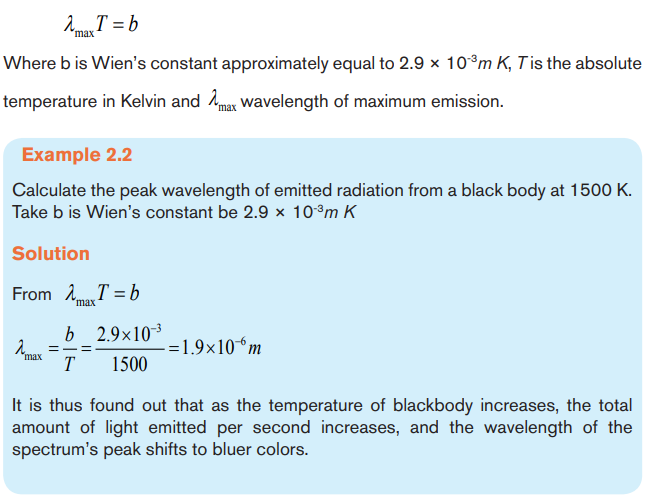
these dark clothes will absorb more radiations from the sun which may be harmfulto our health.
BLACK BODY RADIATION CURVES
This Fig.2.2 shows how the black body radiation curves change at various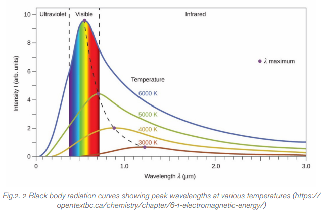
temperatures. The graph indicates that as the temperature increases, the peak
wavelength emitted by the black body decreases. It therefore begins to move from
the infra-red towards the visible part of the spectrum. Again, none of the graphs
touch the x-axis so they emit at every wavelength. This means that some visible
radiation is emitted even at these lower temperatures and at any temperature above
absolute zero, a black body will emit some visible light.
Features/Characteristics of the Graph
• As temperature increases, the total energy emitted increases, because
the total area under the curve increases.
• It also shows that the relationship is not linear as the area does not increase
in even steps. The rate of increase of area and therefore energy increasesas temperature increases.
Application activity 2.2
1. a. With clear explanations, explain the approximate spectral
composition of
the Sun’s radiation before it interacts with Earth’s atmosphere?
b. Is the amount of solar energy that reaches the top of Earth’s
atmosphere constant? Explain giving valid examples and evidence.
c. Are all wavelengths of Sun’s radiation transmitted equally through
Earth’s atmosphere? Explain your reasoning.
2. a. What effect does absorption have on the amount of solar radiation
that reaches Earth’s surface?
b. List down other processes (besides absorption) that affect radiation
reaching the earth’s surface?
c. What can be percentage of incoming solar radiation that is affected
by absorption and scattering (or reflection)?
3. a. Jane Says that clouds have a high albedo while Pierre says land
vegetation has a low albedo? Using Scientific explanations, discuss
what they base on to make their deductions.
b. What do you think are major factors that influence the insolation at a
particular location on a particular day? How do they affect it?
c. What latitudinal regions experience least variation in day-to-day solar
radiation? Which one experiences the greatest? Why?
4. a) Explain the meaning of a blackbody.b) Interpret Stefan-Boltzmann law and Wein’s displacement law.

2.3 GREENHOUSE EFFECTS AND ITS IMPACT ON CLIMATE
CHANGE.
Activity 2.3
a) What do you know about greenhouse?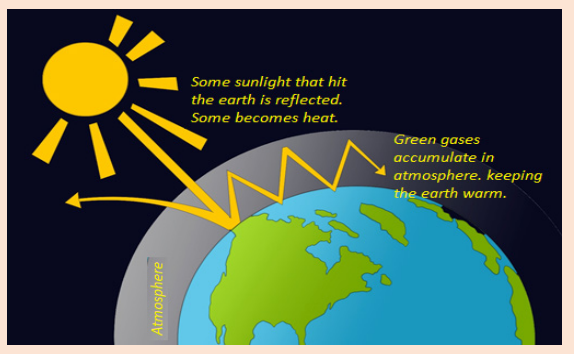
b) Different activities like industrialization and others give out gases after
burning fossil fuels.
i) What special name do you think is given to these gases?
ii) If these gases accumulate in the atmosphere, what effect do they
have on to the temperature of the earth and its atmosphere?
c) Suggest measures that can be done to limit high accumulation of thesegases in the atmosphere.
Greenhouse effect is the process by which thermal radiation from the sun is
prevented from leaving the atmosphere and then re-radiated in different directions.
The relationships between the atmospheric concentration of greenhouse gases
and their radiative effects are well quantified. The greenhouse effect has the
root from greenhouses that becomes warmer when heated by sun’s radiation. The
mode of operation of a greenhouse is that a part of the sunlight radiations incident
on the ground surface of the greenhouse are absorbed and warms the surface
inside the greenhouse. Both the reflected radiations and the heat emitted by the
ground surface in the greenhouse are trapped and re-absorbed inside the structure.
Thus, the temperature rises inside the greenhouse compared to its surrounding
environment.
Therefore, the greenhouse effect heats up the earth’s surface because the green
gases that are in the atmosphere prevent radiations from leaving the atmosphere.
The absorption of these radiations contributes to the increase of the atmosphere’s
temperature.
A greenhouse is constructed by using any material that allows sunlight to pass
through usually plastic or glass. This prevents reflected radiations from leaving
the structure thereby leading to the increase in the temperature within. If a small
puncture is made on to the greenhouse, the temperature within reduces.
Since some of these re-radiated radiations come back to the earth’s surface, they
lead to the increase in the temperature of the earth’s surface leading to globalwarming.

2.3.1 Greenhouse gases
Some gases in the earth’s atmosphere act a bit like the glass in a greenhouse,
trapping the sun’s heat and stopping it from leaking back into space.
Many of these gases occur naturally, but human activity is increasing theconcentrations of some of them in the atmosphere, in particular:

2.3.2 Impact of greenhouse effect on climate change.
With the greenhouse effect, the earth is unable to emit the excess heat to space and
this leads to increase in atmosphere’s temperature and global warming. Scientists
have recorded about 0.75°C increase in the planet’s overall temperature during
the course of the last 100 years. The increased greenhouse effect leads to other
effects on our climate and has already caused: (REMA).
• Greater strength of extreme weather events like: heat waves, tropical
cyclones, floods, and other major storms.
• Increasing number and size of forest fires.
• Rising sea levels (predicted to be as high as about 5.8 cm at the end of
the next century).
• Melting of glaciers and polar ice.
• Increasing acidity in the ocean, resulting in bleaching of coral reefs and
damage to oceanic wildlife.
Solutions to reduce the impact of greenhouse gases
• High efficiency during power production
• Replacing coal and oil with natural gas
• Combined heating and power systems (CHP)
• Renewable energy sources and nuclear power
• Carbon dioxide capture and storage• Use of hybrid vehicles.
2.3.3 Global warming
Global warming is the persistent increase in temperature of the earth’s surface
(both land and water) as well as its atmosphere. Scientists have found out that the
average temperature in the world has risen by about 0.750C in the last 100 years
and about 75% of this rise is from 1975.
Previously the changes were due to natural factors but currently the changes are due
to both natural and human activities. From research, Natural greenhouse maintains
the temperature of the earth making it a better place for human kind and animal life.
However ever since the evolution of industries, there has been significant change
in the temperature. The causes are both natural and human activities and they are
the ones that cause climate change.
Note: If these greenhouse gases were completely not there, the Earth would be
too cold for humans, plants and other creatures to live.
Can you now see the importance of these greenhouse gases! Though they cause
greenhouse effect, they are responsible for regulating the temperature of the earth.
Global warming is damaging the earth’s climate as well as the physical environment.
One of the most visible effects of global warming can be seen in the Arctic region
where glaciers, permafrost and sea ice are melting rapidly. Global warming is
harming the environment in several ways. Global warming has led to: Desertification,Increased melting of snow and ice, Sea level rise, stronger hurricanes and cyclones.
Application activity 2.3
1. Differentiate the term “greenhouse effect from global warming.
2. With clear explanations, explain why it is called the “greenhouse”
effect?
3. From your own reasoning and understanding, why do you think
Environmental experts have become worried about the greenhouse
effect?
4. Below is a bar graph showing emitted Greenhouse gases worldwidefrom 1990 to 2005. Use it to answer the questions that follow
a) From your analysis (using the graph) which kind of gas was emitted in
excess for the specified period?
b) What could be the factors that led that gas to be emitted in large
quantities?
c) What do you think one can do to limit such emissions?
d) From your own point of view, do you think it’s a good idea to stop
completely emission of these gases? Explain your reasoning by givingvalid examples
2.4 CLIMATE CHANGE MITIGATION
Activity 2.4
Read the text below and answer the questions that follow.
The government of Rwanda is trying to sensitize people not to cut down trees
for charcoal, drying wetlands for farming activities and regulating people from
approaching wetlands, forests (Both Natural and artificial) and Fighting all
activities that may lead to climate change.
a. As a good citizen of Rwanda, do you support these plans of the
government? Support your stand with clear justifications
b. If yes what have you done to implement some of these policies?
c. What are some of the New Technologies that the government isadvocating for to stop these negative climate changes?
2.4.1 Climate change mitigation
Climate change mitigation refers to efforts to reduce or prevent emission of
greenhouse gases. Mitigation can mean using new technologies and renewable
energies, making older equipment more energy efficient, or changing management
practices or consumer behavior. (IPCC, 1996)
Climate change is one of the most complex issues we are facing today. It involves
many dimensions science, economics, society, and moral and ethical questions
and is a global problem, felt on local scales that will be around for decades and
centuries to come. Carbon dioxide, the heat-trapping greenhouse gas that has
driven recent global warming, lingers in the atmosphere for centuries, and the earth
(especially the oceans) takes a while to respond to warming.
So even if we stopped emitting all greenhouse gases today, global warming and
climate change will continue to affect future generations. In this way, humanity is
“committed” to some level of climate change.
Because we are already committed to some level of climate change, responding to
climate change involves a two-pronged approach:
1. Reducing emissions and stabilizing the levels of heat-trapping greenhouse
gases in the atmosphere (“mitigation”);2. Adapting to the climate change already in the pipeline (“adaptation”).
2.4.2 Mitigation and adaptation
Because of these changes in climatic conditions, man has devised all possible
measures to see how he can live in harmony on this planet. This has made man to
think harder so that these greenhouse gases can be minimized.
The process of preventing all these greenhouse gases is what is known as
mitigation. This is very important as it is aimed at controlling the rise in temperatures
of the earth while regulating earth’s temperature.
The main goal of mitigation is to reduce human interference to nature thereby
stabilizing the greenhouse gas levels in a given time to allow ecosystem to adapt
naturally to the climate changes. Care should be taken while these adjustments are
made not to affect food production and other economic developments.
Among other strategies, mitigation strategies include:
a) Retrofitting buildings: Retrofitting is the process of modifying something
after it has been manufactured.
Retrofitting a building involves changing its systems or structure after its initial
construction and occupation. This work can improve amenities for the building’s
occupants and improve the performance of the building. As technology develops,
building retrofits can significantly reduce energy and water usage hence conserving
energy sources.
Retrofitting has come to prominence in recent years as part of the drive to
make buildings more thermal efficient and sustainable. This can help cut carbon
emissions, make it cheaper and easier to run buildings, and can contribute to
overcoming poor ventilation and damp problems.
b) Adopting renewable energy sources like solar, wind and small hydroelectric plants:
Human activities are overloading our atmosphere with carbon dioxide and
other global warming emissions. These gases act like a blanket, trapping heat. The
result is a web of significant and harmful impacts, from stronger, more frequent
storms, to drought, sea level rise, and extinction.
In contrast, most renewable energy sources produce little to no global warming
emissions. Even when including “life cycle” emissions of clean energy (ie, the
emissions from each stage of a technology’s lifemanufacturing, installation,
operation, decommissioning), the global warming emissions associated with
renewable energy are minimal.
c) Helping cities develop more sustainable transport such as bus rapid
transit, electric vehicles. This helps in reducing carbon emissions.
d) Promoting more sustainable uses of land and forests and making people
aware of the impacts of mis-using these natural gifts.e) Creating carbon sinks like in big oceans in case there are no alternatives.
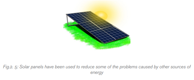
Application activity 2.4
1. Plan and write an essay about climatic change mitigation in Rwanda.
2. Make a research about climate change mitigation in your
neighborhood (either for the school or for your home) and answer
the following questions.
a) What are some of the conditions you have experienced that used in
order to prevent the greenhouse gases?
b) How have you adapted to the changes in conditions you have
mentioned in a) above.c) What are you doing to stop these climatic changes?
Skills Lab 2
Aim: Constructing a greenhouse. (Can be done over a long period
of time)
In this activity you may need
• Polyethene paper (should be relatively white in color)
• Wood
• Nails
• Any fiber that can be used while tying
• Laboratory thermometer.• Bean seeds
Procedures
a) Collect materials listed above.
b) Chose a place where to construct the greenhouse. It may either be near
your school or near your home.
c) Fix and connect the materials together until you make a structure similarto the one indicated in the figure below.
d) Each day measure temperatures and keep noting down records.
e) What do you think are the causes of temperature variations in your
records?
f) Sow seeds of beans in your greenhouse.
g) After seeds have germinated, keep observing changes in the development
of the bean plant.
h) Make a comprehensive report about your Greenhouse. (Include
temperature and vapor changes within the greenhouse).i) Share it with your friends or your physics tutor.
End of unit 2 assessment
Re-write the questions (1) to (8) below in your notebook and circle the best
alternative
1. The emissivity (ε) can be defined as the ratio of
A. emissive power of real body to the emissive power of black body
B. emissive power of black body to the emissive power of real body
C. reflectivity of real body to emissive power of black body
D. Reflectivity of black body to emissive power of real body.
2. Imagine two planets. Planet A is completely covered by an ocean
and has and overall average albedo of 20%. Planet B is blanketed
by clouds and has an overall average albedo of 70%. Which planet
reflects more sunlight back into space?
A. Planet A
B. Planet B
C. the two planets reflect the same amount of light
D. more information is needed to answer this question
3. _______________ is a term used to a process that acts to amplify or
reduce direct warming or cooling effects
A. Climate change C. Climate feedback
B. Weather D. Climate model
4. The filament of an electric bulb has length of 0.5 m and a diameter
of 6x10⁻⁵ m. The power rating of the lamp is 60 W Assuming the
radiation from the filament is equivalent to 80% that of a perfect
black body radiator at the same temperature. The temperature of the
filament is (Stefan Constant is 5.7x10⁻⁸ Wm-2K-4):
A. 1933 K C. 64433333.3 K
B. 796178.3 K D. 60 K
5. The long-term storage of carbon dioxide at the surface of the earth
is termed as
A. Black body radiation C. Solar radiation management
B. Thermal expansion D. Sequestration
6. The balance between the amount of energy entering and exiting the
Earth system is known as
A. Radiative balance C. Paleoclimatology
B. Black body Radiation D. Solar radiation
7. The following are examples of greenhouse gases except
A. Carbon dioxide C. Methane
B. Nitrous oxide D. Oxygen
8. The government of Rwanda is advising the people to conserve the
nature. This is intended to limit the incidence of rise in the temperature.
This conservation of nature
A. Reduces the amount of water vapor that leads to increase in
temperature
B. Reduces the amount of Carbon dioxide in the space
C. Increases the amount of plant species that is required to boost our
tourism industry
D. Provides a green environment for human settlement
9. a) State Wien’s displacement law and its practical implicationsb) Use the graph indicated below to answer the questions that follow
i) What does the graph explain?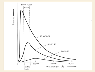
ii) Explain the spectrum that is found between temperatures of 4000
and 7000 K
iii) Why do you think the three curves have different shapes
10. Discuss any 4 main reasons that brings about variation of sun’s Intensity
11. a) Calculate the albedo of a surface that receives 15000 Wm-2 and
reflects 15 KW.m-2.Comment on the surface of that body.
b) Discuss some of the scientific factors that affect planets Albedo
12. a) What do you understand by the following terms?
i) Climate change as applied in physics
ii) Greenhouse Effect
b) With Clear explanations, discuss how Greenhouse effect can be
avoided
13. Mutesi a year 3 student in the faculty of engineering in University of
Rwanda, found out in her research that a strong metal at 1000K is
red hot while at 2000K its white hot. Using the idea of black body,
explain this observation.
14. Kamari defined black body as anybody that is black. Do you agree
with his definition? If YES why? and if NOT why not? Also sketch
curves to show the distribution of energy with wavelength in the
radiation from a black body varies with temperature.
15. An electric bulb of length 0.6 m and diameter 5 x 10⁻⁵m is connected
to a power source of 50 W. Assuming that the radiation from the
bulb is 70% that of a perfect black body radiator at the same
temperature, Estimate the steady temperature of the bulb. (Stefan’s
constant = 5.7 x 10 - 8 W m 2 K-4.)
16. Write short notes about greenhouse effect and explain all its effects.
17. What do you understand by the term greenhouse gases? How do
these gases contribute to the global warming?
18. REMA is always advising people to plant more trees and stop
cutting the existing ones. Using scientific examples, explain how this
is aimed at controlling global warming
19. Explain climate change mitigation and explain why it’s important.
20. Explain what happens to most radiation that is absorbed by the
surface of earth?
21. Is there a difference between sensible and latent heat?
22. Write short notes on the following terms as applied in climate change
i) Climate feedback
ii) Climate lag
iii) Climate model
23. Plan and write a good composition about causes of climate change
and how it can be controlled. (Your essay should bear introduction,body and conclusion)
UNIT 3: APPLICATIONS OF OPTICAL FIBER IN COMMUNICATION SYSTEM
Key Unit Competence:
Evaluate the application of optical fiber transmission and other transmittingsystems
Introductory activity
Investigating the use of optical fiber in RWANDA
Rwandan government has started connecting three million people to the
World Wide Web as part of the “Internet for All” project. The project is a World
Economic Forum initiative that aims to connect 25 million new Internet users in
Kenya, Uganda, South Sudan and Rwanda at the beginning of 2019.
This goal will partly be achieved by addressing the challenges of affordability,
digital skills gap, lack of local content and limited infrastructure, which arehindering growth in the use of Internet across the region
By using the information provided above, answer to the following questions:
1. Observe the images A, B and C of the figure .3.1 and describe each
one.
2. What do you think are the uses of the materials shown in the figure
above?
3. By using scientific ideas, explain why one may opt to use the method(s)
indicated in the figure above over another.
4. Can you highlight some of the disadvantages of the method of signaltransmission shown in the figure above?
3.1. OPTICAL FIBRE
Activity 3.1
Investigating the types of optical fiber.
a) Use search on internet or in library, discuss different types of optical
fiber.b) Differentiate them according to their respective uses.
3.1.1 Concept of optical fiber
An optical fiber (fiber optics) is a medium for carrying information from one point
to another in the form of light. It uses a flexible, transparent fiber made by drawing
glass or plastic and has a diameter slightly thicker than that of a human hair. They
are arranged in bundles called optical cables and can be used to transmit signals
over long distances.
An optical fiber is a cylindrical dielectric waveguide (non conducting waveguide)
made of low loss material that transmits light along its axis, by the process of total
internal reflection. The fiber consists of a core surrounded by a cladding layer, both
of which are made of dielectric materials.
The Fiber optic cable is made of high-quality extruded glass (si) or plastic, and it is
flexible. The diameter of the fiber optic cable is in between 0.25 to 0.5 mm (slightly
thicker than a human hair). Fiber optics continues to be used in more and moreapplications due to its inherent advantages over copper conductors.
An optical fiber essentially consists of three layers Core,Cladding and Buffer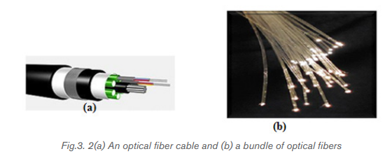
coating. The rest of the layers are provided in order to increase the flexibility,strength and protection from external stresses.
• Core:
Core is a thin glass/silica at the center of the optical fiber through which light
travels. A Glass material with high refractive index is used for this purpose. The core
of a fiber cable is a cylinder of plastic that runs all along the fiber cable’s length, and
offers protection by cladding. The diameter of the core depends on the application
used. Due to internal reflection, the light travelling within the core reflects from the
core, the cladding boundary. The core cross section needs to be a circular one formost of the applications.
• Cladding:
Core is surrounded by a medium, with lesser refractive index. Ray of light incident
on the core-cladding interface is reflected back into the core. Cladding ensures
that no light signal escapes from the optical fiber. Cladding is an outer optical
material that protects the core. The main function of the cladding is that it reflects
the light back into the core. When light enters through the core (dense material)
into the cladding (less dense material), it changes its angle, and then reflects backto the core.
• Coating:
The coating is the first non-optical layer around the cladding. The coating typically
consists of one or more layers of polymer that protect the silica structure againstphysical or environmental damage.
. Buffer Coating (Strength member)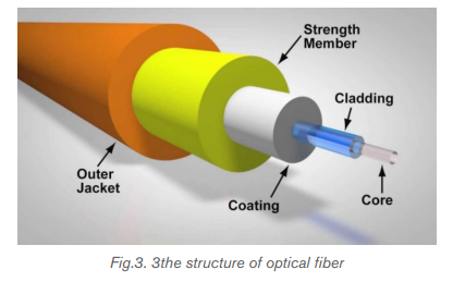
The entire structure is protected by a plastic coating. It is composed of multiple
layers and materials in order to protect from external shocks, moisture, surrounding
materials etc. The main function of the buffer is to protect the fiber from damage and
thousands of optical fibers arranged in hundreds of optical cables. These bundlesare protected by the cable’s outer covering that is called jacket.
• Outer Jacket
Fiber optic cable’s jackets are available in different colors that can easily make us
recognize the exact color of the cable we are dealing with. The color yellow clearly
signifies a single mode cable, and orange color indicates multimode.
The light is guided down the core of the fiber by the optical cladding which has a
lower refractive index. Remember that the refractive index is the ratio of the velocity
of light in a vacuum to its velocity in a specified medium. Then light is trapped in the
core through total internal reflection. The other outer parts that are the strength
member and the outer jacket, serve as protectors.
Connecting two optical fibers is done by fusion splicing or mechanical splicing.
It requires special skills and interconnection technology due to the microscopicprecision required to align the fiber cores.
3.1.2 Types of optical fiber
The properties of data transmission via a fiber optic depend on the core. Hence,
based on the differences in the structure of the core, there are three main types ofoptical fibers:
a. Single mode (monomode) fiber
To understand the behavior of electromagnetic waves in waveguides we use a
theory known as mode theory. The mode theory (this is a bit of an oversimplification)
essentially classifies electromagnetic waves on the basis of wavelengths into
different modes. A mode is a stable propagation state (stable operating points or
standing waves) in optical fibers.
Single-Mode Fibers: is a single stand of glass fiber with a diameter of 8.3 µm
to10 µm that has one mode of transmission. Single mode fibers are used to transmit
one signal per fiber; these fibers are used in telephone and television sets. Singlemode fibers have small cores.
As the name suggests, this type of optical fiber transmits only one mode of the light.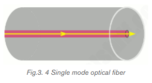
To put it another way, it can carry only one wavelength of light across its length.
This wavelength is usually 1310 nm or 1550 nm. Major reasons for this situationare as follows:
• The single mode optical fibers are much better than multimode optical
fibers as they have more bandwidth and experience fewer losses of the fiber
transmission. So the speed is unmatched.
• Interestingly, single mode fibers came into existence after multimode fibers.
They are more recent than the multimode cables.
• These cables can carry only one mode, physically, by having a tiny core. That
is to say that the diameter of the core is essentially of the same order as the
wavelength of the light passing through it.
• Only lasers are used as a light source. To point out, the light used in single
mode fibers are not in the visible spectrum.
• Since the light travels in a straight direction, there are fewer losses, and it can
be used in applications requiring longer distance connections.
• An obvious disadvantage of single mode fiber is that they are hard to couple.
• They have a superior transmission quality over other ber types because of
the absence of modal noise.
3.1.3 Multimode optical fiber
Multimode fibers are used to transmit many signals per fiber; these signals are usedin computer and local area networks that have larger cores.
• As the name implies, these types of optical fibers allow multiple modes of
light to travel along their axis.
• To explain physically, they can do this by having a thicker core diameter.
• The wavelengths of light waves in multimode fibers are in the visible spectrum
ranging from 850 to 1300 nm.
• The reflection of the waves inside the multimode fiber occurs at different
angles for every mode. Consequently, based on these angles the number of
reflections can vary.
• We can have a mode where the light passes without striking the core at all.
• We can have a slightly higher mode which will travel with appropriate internal
reflections.
• Since the basis of optical fiber communication is a total internal reflection,
all modes with incident angles that do not cause total internal reflection get
absorbed by the cladding. As a result, losses are created.
• We can have higher order modes, waves that are highly transverse to the axis
of the waveguide can reflect many times. In fact, due to increased reflections
at unusual angles, higher order modes can get completely lost inside the
cable.
• Lower order modes are moderately transverse or even completely straight
and hence fare better comparatively.
• There are two types of multimode optical fibers: stepped index and gradedindex.
a. Stepped index
In step-index multimode type, the core has the relatively large diameter of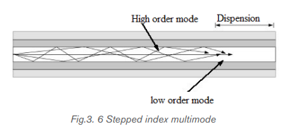
50 μm and the refractive index of the core is uniform throughout and undergoes an
abrupt change (or step) at the cladding boundary.
The wide core allows the infrared to travel by several paths or modes. Paths that
cross the core more often are longer, and signals in those modes are take longer to
travel along the fiber. Arrival times at the receiver are therefore different for radiation
of the same pulse, 30 ns km-1, being a typical difference. The pulse is said to suffer
dispersion, it means that it is spread out.
b. Graded-index multimode fiber
The core refractive index is made to vary as a function of the radial distance from
the center of the fiber.
If you look at the figure of stepped and graded index multimode fibers shown above,
you will notice that the waves in stepped index fiber arrive at the same point at
different times. This is because multiple modes have different velocities. As a result,
outputs are out of sync, and this reduces bandwidth. This is called intermodal
dispersion/distortion. However, this issue can be mitigated by using graded index
fibers. Since the refractive index changes radially the higher order modes are benttowards the lower order modes and as a result, they are synchronized in time.
In the graded index multimode type, the refractive index of the glass varies
continuously from a higher value at the center of the fiber to a low value at the
outside, so making the boundary between core and the cladding indistinct.
Radiation following a longer path, travel faster on average, since the speed of light
is inversely proportional to the refractive index. The arrival times for different modes
are the about the same (to within 1ns km-1) and all arrive more or less together at
the receiving end. Dispersion is thereby much reduced.
3.1.4 Micro structured optical fibers
These are the new types of optical fiber cables. In ordinary optical fibers mentioned
above, light travels due to total internal reflection and refractive indices of the
core and cladding. In micro structured optical fibers, the physical structure of the
waveguide is used at a nano-scale level to manipulate light.
Different types of micro structured optical fibers are constructed with a non
cylindrical core and/or cladding layer, usually with an elliptical or rectangular cross section.
These include:
• Polarization-maintaining fiber is a unique type of fiber that is commonly used
in fiber optic sensors due to its ability to maintain the polarization of the light
inserted into it.
• Photonic-crystal fiber is made with a regular pattern of index variation. It is
often in the form of cylindrical holes that run along the length of the fiber.
Such fiber uses diffraction effects instead of or in addition to total internal
reflection, to confine light to the fiber›s core.
• Air-clad or double clad fibers• Fresnel fibers
Application activity 3.1
1. Fiber optics is best known for its application in long-distance
telecommunications.
A. True
B. False
2. Choose the basic types of optical fiber:
A. Single-mode E. Multi-mode
B. X-mode F. A and C
C. Microwave-mode G. B and D
D. Graded-index mode H. A and E
3. Single-mode fiber has the advantage of greater bandwidth capability.
It has the disadvantage of:
A. Being harder to bend
B. Smaller mechanical tolerances in connectors and splices
C. Being difficult to couple light into
D. B and C
E. None of the above
4. Describe with the aid of simple ray diagrams:
A. The multimode step index fiber;
B. The single-mode step index fiber.
C. Compare the advantages and disadvantages of these two types of
fiber for use as an optical channel.5. Match the words of column A to their meaning of column B

3.2 PRINCIPLE OF OPERATION OF OPTICAL FIBRES
Activity 3.2
Given the illustration above, one can see different rays inside the optical fiber.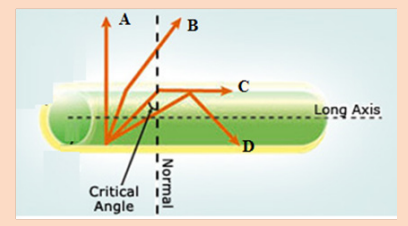
As the angle of incidence in the core increases, the angle of refraction increasesmore until it becomes right angle.
Discuss:
1. Name the rays A, B, C and D
2. As observed from the diagram, comment on the cause of different
directions of these rays.
3. Explain the scientific name of the angle for ray C to be in the direction
indicated in the figure.
4. Explain the scientific phenomenon represented by ray D.5. Explain any fields where the phenomenon stated in 4) above is applied.
3.2.1 Refractive index of light
When light fall at the interface (boundary) of two media, it is partially reflected andpartially refracted. As it passes from one medium to another it changes its direction.
The change in its direction is associated with the change in velocity. The ratio of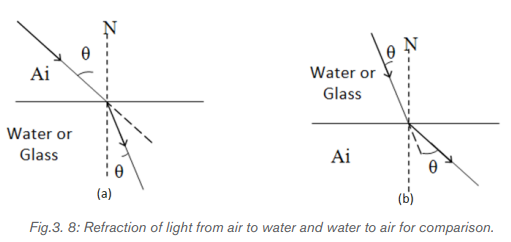
the speed of light in the vacuum c (or air) and that of light in a certain medium v iscalled the absolute refractive index n.
3.2.2 Total internal reflection
When light passes from one a medium of higher index of refraction into a medium
of lower refractive index the light bends away from the normal as indicated on
Fig.3.9. A weak internally reflected ray is also formed and its intensity increases asthe incident angle increases.
Increasing the angle of incidence increases the angle of refraction and at a particular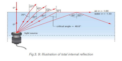
incidence, the angle of refraction reaches the 90°. This particular incident angle is
called the critical angle θ c . As the incident angle exceeds the critical angle, the
incident beam reflects on the interface between the 2 media and return in the first
medium. This effect is called total internal reflection.
For any two media, using Snell’s law the critical angle is calculated using theexpression
One of the applications of total internal reflection is optical fiber. An optical fiber is
basically made of 2 types of glass put together in a concentric arrangement so the
middle is hollow. The inner circle of glass also called the Core consists of a glass
of higher refractive index than the outside layer as indicated on Fig.3.10.
The outer layer of glass, which is also known as the optical cladding, does notcarry light but is essential to maintain the critical angle of the inner glass.
The underlying main physics concept behind the functioning of an optical fiber is a
phenomenon known as total internal reflection. Any light entering the fiber will
meet the cladding at an angle greater than the critical angle. If light meets the inner
surface of the cladding or the core - cladding interface at greater than or equal to
critical angle then total internal reflection (TIR) occurs. So, all the energy in the ray
of light is reflected back into the core and none escapes into the cladding. The ray
then crosses to the other side of the core and, because the fiber is more or less
straight, the ray will meet the cladding on the other side at an angle which again
causes the total internal reflection. The ray is then reflected back across the core
again and again until it reaches the end of the optical fiber.
A single optical fiber utilizes total internal reflection to transmit light, allowing bends
along its path. Minimal light loss during transmission allows optical fibers to transmit
light or data quickly over long distances. When bundled, fiberoptics can transmitlarge quantities of data for telecommunication applications.
Optical fibers use multiple total internal reflections to transmit light. They allow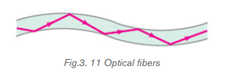
signals to travel for long distances without repeaters, which are needed to
compensate for reductions in signal strength. Fiber-optic repeaters are currently
about 100 km apart, compared to about 1.5 km for electrical systems.
Various consequences of Snell’s Law include the fact that for light rays traveling from
a material with a high index of refraction to a material with a low index of refraction,
it is possible for the interaction with the interface to result in zero transmission.
This phenomenon is called total internal reflection. As light signals travel down
a fiber optic cable, it undergoes total internal reflection allowing for essentially no
light lost over the length of the cable.Maximum angle of incidence
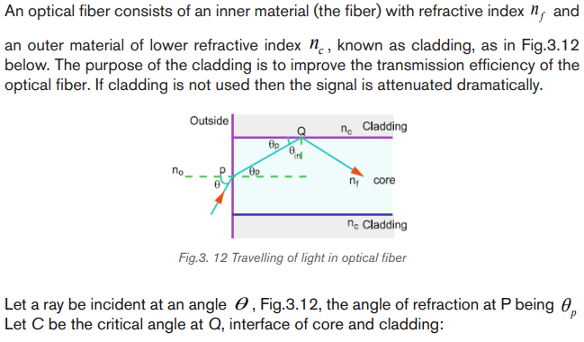
This the maximum angle of incidence in air for which all the light is total reflected
at the core-cladding.
This shows that there is a maximum angle of acceptance cone outside of which
entering rays will not be totally reflected within the fiber. For the largest acceptance
cone, it is desirable to choose the index of refraction of the cladding to be as small
as possible. This is achieved if there is no cladding at all. However, this leads to
other problems associated with the loss of intensity.
The transmission is reduced due to multiple reflections and the absorption of thefiber core material due to impurities.
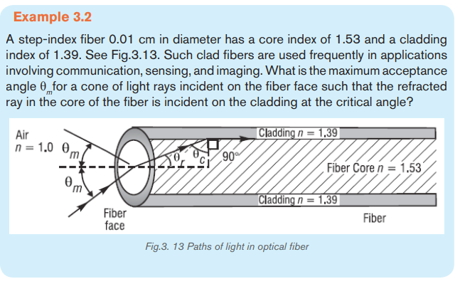
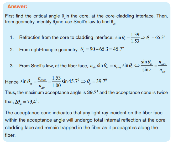
Application activity 3.2
1. Operation of optical fiber is based on:
A. Total internal reflection D. Einstein’s theory of reality
B. Total internal refraction E. None of the above
C. Snell’s law
2. When a beam of light passes through an optical fiber
A. Rays are continually reflected at the outside(cladding) of the fiber
B. Some of the rays are refracted from the core to the cladding
C. The bright beam coming out of the fiber is due to the high refractive
index of the core
C. The bright beam coming out of the fiber is due to the total internal
reflection at the core-cladding interface
E. All the rays of light entering the fiber are totally reflected even at verysmall angles of incidence
3. A laser is used for sending a signal along a mono mode fiber because
A. The light produced is faster than from any other source of light
B. The laser has a very narrow band of wavelengths
C. The core has a low refractive index to laser light
D. The signal is clearer if the cladding has a high refractive index
E. The electrical signal can be transferred quickly using a laser
4. Given that the refractive indices of air and water are 1 and 1.33
respectively, find the critical angle.
5. A beam of light is propagating through diamond, n = 2.42 and strikes
a diamond-air interface at an angle of incidence of 28°.
a) Will part of the beam enter the air or will the beam be totally refracted at
the interface?
b) Repeat part (a) assuming that diamond is surrounded by water, n = 1.336. A beam of light passes from water into polyethylene (n = 1.5). If the
7. (a) What is the critical angle when light is going from a diamond
(n = 2.42) to air?
(b) Using the answer to (a), what happens when:
i) The angle of incidence is less than that angle?ii) The angle of incidence is more than that angle?
3.3. MECHANISM OF ATTENUATION AND LIGHT SCATTERING
Activity 3.3
The image above is a section of optical fiber. Imagining that it is transmitting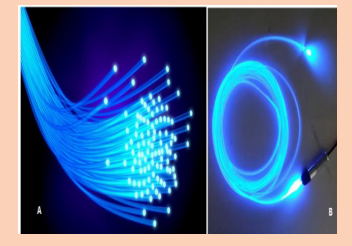
signal over a long distance,
a) Do you think all the fed signal can reach the destination? Defend your
reasoning.
b) If no, what do you think causes the loss in the signal transmission?
c) Suggest measures that should be done to minimize the energy lossduring transmission.
3.3.1 Mechanism of attenuation
Attenuation in fiber optics, also known as transmission loss, is the reduction in
intensity of the light beam (or signal) as it travels through the transmission medium.
Over a set distance, fiber optic with a lower attenuation will allow more power to
reach its receiver than a fiber with higher attenuation.
Attenuation can be caused by several factors both extrinsic and intrinsic:
• Intrinsic attenuation is due to something inherent to the fiber such as impurities
in the glass during manufacturing. The interaction of such impurities with light
results in the scattering of light or its absorption.
• Extrinsic attenuation can be caused by microbending and microbending. A
bent imposed on an optical fiber produce a strain in that region of the fiber
and affects its refractive index and the critical angle of the light ray in that
area. Microbending that is a large-scale bent and microbending which is
a small-scale bent and very localized are external causes that result in thereduction of optical power.
Attenuation coefficients in fiber optics usually are expressed decibels per kilometer
(dB/km) through the medium due to the relatively high quality of transparency ofmodern optical transmission media. It is observed that the attenuation is a function
Attenuation is an important limiting factor in the transmission of a digital signal across
large distances. Thus, much research has gone into both limiting the attenuationand maximizing the amplification of the optical signal.
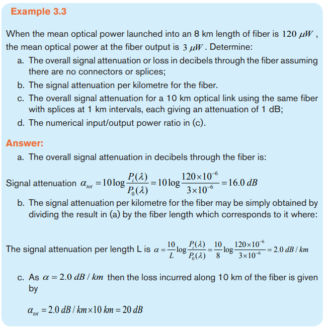

3.3.2 Light scattering and absorption
In the light transmission of signals through optical fibers, attenuation occurs due to
light scattering and absorption of specific wavelengths, in a manner similar to thatresponsible for the appearance of color.
a. Light scattering
The propagation of light through the core of an optical fiber is based on total internal
reflection of the light wave. Rough and irregular surfaces, even at the molecular
level, can cause light rays to be reflected in random directions as it is illustrated on
Fig.3.14. This is called diffuse reflection or scattering, and it is typically characterized
by wide variety of reflection angles.
Light scattering depends on the wavelength of the light being scattered. Thus, limits
to spatial scales of visibility arise, depending on the frequency of the incident light
wave and the physical dimension (or spatial scale) of the scattering center, which is
typically in the form of some specific micro-structural feature. Since visible light has
a wavelength of the order of one micrometer (one millionth of a meter) scattering
centers will have dimensions on a similar spatial scale. Thus, attenuation resultsfrom the incoherent scattering of light at internal surfaces and interfaces.
b. Light absorption
Material absorption is a loss mechanism related to the material composition and
fiber fabrication process. This results in the dissipation of some transmitted optical
power as heat in the waveguide. Absorption is classified into two basic categories:
Intrinsic and extrinsic absorptions. (John, 2009)
• Intrinsic absorption: is caused by basic fiber material properties. If an optical
fiber is absolutely pure, with no imperfections or impurities, ten all absorption
will be intrinsic. Intrinsic absorption in the ultraviolet region is caused bands.
Intrinsic absorption occurs when a light particle (photon) interacts with an
electron and excites it to a higher energy level.
• Extrinsic absorption is caused by impurities caused by impurities introduced
into the fiber material. The metal impurities such as iron, nickel and chromium
are introduced into the fiber during fabrication. Extrinsic absorption is caused
by electronic transition of these metal ions from one energy level to another
level.
3.3.3 Measures to avoid Attenuation
The transmission distance of a fiber-optic communication system has traditionally
been limited by fiber attenuation and by fiber distortion.
• Repeaters: Repeaters convert the signal into an electrical signal, and then
use a transmitter to send the signal again at a higher intensity than was
received, thus counteracting the loss incurred in the previous segment. They
mostly used to be installed about once every 20 km.
• Regenerators: Optical fibers link, in common with any line communication
system, have a requirement for both jointing and termination of the
transmission medium. When a communications link must span at a larger
distance than existing fiber-optic technology is capable of, the signal must
be regenerated at intermediate points in the link by optical communications
repeaters called regenerators. An optical regenerator consists of optical
fibers with special coating (doping). The doped portion is pumped with a
laser. When the degraded signal comes into the doped coating, the energy
from the laser allows the doped molecules to become lasers themselves.
The doped molecules then emit a new strong light signal with the same
characteristics as the incoming weak signal. Basically, the regenerator is a
laser amplifier for the incoming signal.
• Optical Amplifiers: Another approach is to use an optical amplifier which
amplifies the optical signal directly without having to convert the signal into
the electrical domain. It is made by doping a length of fiber with the rare
earth mineral erbium and pumping it with light from a laser with a shorter
wavelength than the communications signal (typically 980 nm). Amplifiershave largely replaced repeaters in new installations.
Application activity 3.3
1. For each of the statements below, indicate true if it is correct and
false if it is wrong
A. One of the reasons fiber optics hasn’t been used in more areas has
been the improvement in copper cable such as twisted pair.
B. With current long-distance fiber optic systems using wavelength-division
multiplexing, the use of fiber amplifiers has become almost mandatory.
C. Fiber optics has extraordinary opportunities for future applications
because of its immense bandwidth.
2. (a) What do we mean by attenuation in optical fibers?
(b) State two ways in which energy is lost in optical fibers.
(c) If a fiber loses 5% of its signal strength per kilometer, how much ofits strength would be left after 20 km?
3.4 OPTICAL FIBRE COMMUNICATION
Activity 3.4
With the basic information you know about the functioning process of optical
fiber, answer to the following questions:
1. Where does the light that is transmitted into the optical fiber core
medium come from?
2. What are the type compositions of the light signal propagating into
optical fiber?
3. Discuss and explain the function principle of signal generators andsignal receivers of light from optical fibers.
3.4.1 Basic Fiber Optic Communication System
Even without defining information formally, we intuitively understand that speech,
audio, and video signals contain information. We use the term message signals for
such signals, since these are the messages we wish to convey over a communication
system. In their original form both during generation and consumption these message
signals are analog: they are continuous time signals, with the signal values also
Iying in a continuum. When someone plays the violin, an analog acoustic signal
is generated (often translated to an analog electrical signal using a microphone).
Even when this music is recorded onto a digital storage medium such as a CD
(using the digital communication), when we ultimately listen to the CD being played
on an audio system, we hear an analog acoustic signal. The transmitted signals
corresponding to physical communication media are also analog. For example,
in both wireless and optical communication, we employ electromagnetic waves,
which correspond to continuous time electric and magnetic fields taking values in
a continuum.
In all of these instances, the key steps in the operation of a communication link are
as follows:
a. insertion of information into a signal, termed the transmitted signal,
compatible with the physical medium of interest;
b. propagation of the signal through the physical medium (termed the channel)
in space or time;
c. Extraction of information from the signal (termed the received signal)
obtained after propagation through the medium.
For gigabits and beyond gigabits transmission of data, the fiber optic communication
is the ideal choice. This type of communication is used to transmit voice, video,
telemetry and data over long distances and local area networks or computer
networks. A Fiber Optic Communication System uses light wave technology to
transmit the data over a fiber by changing electronic signals into light.
Some exceptional characteristic features of this type of communication system like
large bandwidth, smaller diameter, light weight, long distance signal transmission,
low attenuation, transmission security, and so on make this communication a major
building block in any telecommunication infrastructure. The subsequent information
on fiber optic communication system highlights its characteristic features, basic
elements and other details.
3.4.2 Block diagram of Fiber Optic Communication
Unlike copper wire based transmission where the transmission entirely depends
on electrical signals passing through the cable, the fiber optics transmission
involves transmission of signals in the form of light from one point to the other.
Furthermore, a fiber optic communication network consists of transmitting and
receiving circuitry, a light source and detector devices like the ones shownin the Fig.3.15.
The basic components are light signal transmitter, the optical fiber, and the photo
detecting receiver.
The additional elements such as fiber and cable splicers and connectors,
regenerators, beam splitters, and optical amplifiers, switches, couplers, multiplexing
devices, amplifiers and splices are employed to improve the performance of the
communication system.
When the input data, in the form of electrical signals, is given to the transmitter
circuit (Fig.3.15), it converts them into light signal with the help of a light source.
This source is of LED whose amplitude, frequency and phases must remain stable
and free from fluctuation in order to have efficient transmission. The light beam from
the source is carried by a fiber optic cable to the destination circuitry wherein the
information is transmitted back to the electrical signal by a receiver circuit.
Fiber optic communication system consists of transmitter, optical fiber, optical
regenerator and finally a receiver as shown in the following figure.
• Transmitter block
Transmitter is the first stage of the optical fiber communication system. It consists of
a light source which converts electric signals into light signals and a focusing lens
is used to focus the light beam into the optical fiber. Both Lasers and LEDs can be
used as a light source. Lasers have more power than LEDs, but its characteristics
vary with changes in temperature.
• Optical fiber
Light signal from the transmitter is given into the optical fiber. Signal is propagatedthrough it by multiple internal reflections.
• Optical regenerator
When light passes through the optical fiber, the signal may get distorted due
to the presence of impurities in the core. Distance to which the light signal can
propagate through the fiber depends on the purity of the glass and the wavelength
of the transmitted light. Therefore, to improve the transmission distance, Optical
regenerators must be used at regular intervals. One or more optical regenerators
are used to boost the degraded light signals in the optical communication system.
In certain systems, the feeble optical signals are converted back into electrical
signals and the optical data is reconstructed as in the case of a transmitter. Optical
regenerators are also called laser amplifiers. They are optical fibers with a special
coating (doping). When degraded signal comes into the doped coating, the energy
from the laser allows the doped molecules to become lasers themselves. Thus
degraded light signal will get amplified and propagate further.
• Optical receiver
Optical receiver receives light signals which it converts back to electrical signals.
Receiver uses a photocell or photodiode to detect the light and convert it to
proportional electric signals, which is capable of measuring magnitude, frequency
and phase of the optic field. This type of communication uses the wave lengths
near to the infrared band that are just above the visible range.
Two types of photo detectors are mainly used for optical receiver in optical
communication system: PN photo diode and avalanche photo diode. Depending
on the application’s wavelengths, the material composition of these devices varies.
These materials include silicon, germanium, InGaAs, etc
Both LED and Laser can be used as light sources based on the application.
• Compact Light Source
Depending on the applications like local area networks and the long haul
communication systems, the light source requirements vary. The requirements of
the sources include power, speed, spectral line width, noise, ruggedness, cost,
temperature, and so on. Two components are used as light sources: light emitting
diodes (LED’s) and laser diodes.
The light emitting diodes are used for short distances and low data rate
applications due to their low bandwidth and power capabilities. Two such LEDs
structures include Surface and Edge Emitting Systems. The surface emitting
diodes are simple in design and are reliable, but due to its broader line width and
modulation frequency limitation edge emitting diode are mostly used. Edge emitting
diodes have high power and narrower line width capabilities.
For longer distances and high data rate transmission, Laser Diodes are preferred
due to its high power, high speed and narrower spectral line width characteristics.But these are inherently non-linear and more sensitive to temperature variations.
Both these sources are modulated using either direct or external modulation
techniques.
3.4.3 Uses of optical fibers
a. Telecommunications Industry
Optical fibers offer huge communication capacity. The latest generations of optical
transmission systems are beginning to exploit a significant part of this huge capacity,
to satisfy the rapidly growing demand for data communications and the Internet.
Some of them are as follows
• Used in telephone systems
• Used in sub-marine cable networks
• Used in data link for computer networks, CATV Systems
• Used in CCTV surveillance cameras
• Used for connecting fire, police, and other emergency services.
The main advantages of using optical fibers in the communications industry are:
- A much greater amount of information can be carried on an optical fiber
compared to a copper cable.
- In all cables some of the energy is lost as the signal goes along the cable.
The signal then needs to be boosted using regenerators. For copper cable
systems these are required every 2 to 3km but with optical fiber systems they
are only needed every 50 km.
- Unlike copper cables, optical fibers do not experience any electrical
interference. Neither will they cause sparks so they can be used in explosive
environments such as oil refineries or gas pumping stations.
- For equal capacity, optical fibers are cheaper and thinner than copper cables
and that makes them easier to install and maintain.
b. Medicine Industry
The advent of practicable optical fibers has seen the development of much medical
technology. Optical fibers have paved the way for a whole new field of surgery,
called laproscopic surgery (or more commonly, keyhole surgery), which is usually
used for operations in the stomach area such as appendectomies. Keyhole surgery
usually makes use of two or three bundles of optical fibers. A “bundle” can contain
thousands of individual fibers”. The surgeon makes a number of small incisions in
the target area and the area can then be filled with air to provide more room.
One bundle of optical fibers can be used to illuminate the chosen area, and another
bundle can be used to bring information back to the surgeon. Moreover, this can
be coupled with laser surgery, by using an optical fiber to carry the laser beam to
the relevant spot, which would then be able to be used to cut the tissue or affect itin some other way.
3.4.4 Block Diagrams of Telecommunication
The elements of basic communication system are as follows
• Information or input signal:
Information is any entity or form that resolves uncertainty or provides the answer to
a question of some kind. It is thus related to data and knowledge, as data represents
values attributed to parameters, and knowledge signifies understanding of real
things or abstract concepts. The information can be in the form of sound signal
like speech or music or it can be in the form of pictures, words, group of words,
code, symbols, sound signal etc. However, out of these messages, only the desired
message is selected and communicated. Therefore, we can say that the functionof information source is to produce required message which has to be transmitted
A message is a term standing for information put in an appropriate form for
transmission. Each message contains information. A message can be either analog
message (a physical time variable quantity usually in smooth and continuous form)
or a digital message (anordered sequence of symbols selected from finite set of
elements) as shown in Fg.3.16.
A signal is a mathematical function representing the time variation of a physical
variable characterizing a physical process and which, by using various models, can
be mathematically represented. In telecommunication, the message is also known
as a signal and the signal is transmitted in an electrical or voltage form i.e. Signal
≈ Message
• Input Transducer.
A transducer is a device which converts one form of energy into another form. The
information in the form of sound, picture or data signals cannot the transmitted as it
is. First it has to be converted into a suitable electrical signal. The input transducers
commonly used in the communication systems are microphones, TV etc. For
example, in case of radio-broadcasting, a microphone converts the information ormassage which is in the form of sound waves into corresponding electrical signal.
• Transmitter:
The function of the transmitter block is to convert the electrical equivalent of the
information to a suitable form. It increases the power level of the signal. The power
level should be increased in order to cover a large range. The transmitter consists
of the electronics circuits such as:
- Mixer
- The oscillators are the sources of carrier signals which are used to modulate
and help the original signal to reach the destination
- Amplifiers: The signal normally, must be raised at a level that will permit it to
reach its destination. This operation is accomplished by amplifiers
- antenna
• Communication channel or medium:
The communication channel is the medium used for the transmission of electronic
signals from one place to another. The communication medium can be conducting
wires, cables, optical fibres or free space. Depending upon the type of the
communication medium, two types of the communication system will exist: Wire
communication or line communication and Wireless communication or radio
communication.
The term channel means the medium through which the message travels from
the transmitter to the receiver. In other words, we can say that the function of
the channel is to provide a physical connection between the transmitter and the
receiver.
There are two types of channels, namely point-to-point channels and broadcast
channels.
Examples of point-to-point channels are wire lines, microwave links and optical
fibres.
- Wire-lines operate by guided electromagnetic waves and they are used for
local telephone transmission.
- In case of microwave links, the transmitted signal is radiated as an
electromagnetic wave in free space. Microwave links are used in long distance
telephone transmission.
- An optical fiber is a low-loss, well-controlled, guided optical medium.
Optical fibers are used in optical communications.
Although these three channels operate differently, they all provide a physical
medium for the transmission of signals from one point to another point. Therefore,for these channels, the term point-to-point is used.
On the other hand, the broadcast channel provides a capability where several
receiving stations can be reached simultaneously from a single transmitter. An
example of a broadcast channel is a satellite in geostationary orbit, which covers
about one third of the earth’s surface.
During the process of transmission and reception the signal gets distorted due
to noise introduced in the system. Noise is an unwanted electrical signal which
gets added to the transmitted signal when it is travelling towards receiver. Due to
noise, the quality of the transmitted information will degrade. One added the noise
cannot be separated out from the information. Hence noise is a big problem in the
communication systems.
• Receiver
The reception is exactly the opposite process of transmission. The received signal
is amplified and demodulated and converted in a suitable form. The receiver
consists of the electronic circuits like mixer, oscillator, detector,amplifier and
antenna.The main function of the receiver is to reproduce the message signal in
electrical form from the distorted received signal. This reproduction of the original
signal is accomplished by a process known as the demodulation or detection.
Demodulation is the reverse process of modulation carried out in transmitter.
• Output Transducer
Destination is the final stage which is used to convert an electrical message
signal into its original form. Output Transducer consists of the electrical signal at
the output of the receiver back to the original form i.e. sound or TV pictures. The
typical examples of the output transducers are loud speakers, picture tubes etc.For
example, in radio broadcasting, the destination is a loudspeaker which works as
a transducer i.e. converts the electrical signal in the form of original sound signal.
• Types of Antenna
Antennas are essential components of all equipment which are used in radio. They
are used in broadcasting systems, broadcast television systems, two-way radio
systems, communications receiver’s systems, radar systems, cell phones systems,
and satellite communications systems, garage door openers systems, wireless
microphones systems, Bluetooth enabled devices systems, wireless computer
networks systems, baby monitors systems, and Radio Frequency Identification
(RFID) tags systems on merchandise etc.
An antenna or aerial is an electrical device connected (often through a
transmission line) to the receiver or transmitter which converts electric power into
radio waves, and vice versa. It is usually used with a radio transmitter or radio
receiver. In transmission, a radio transmitter supplies an oscillating radio frequency
electric current to the antenna’s terminals, and the antenna radiates the energy
from the current as electromagnetic waves (radio waves). In reception, an antenna
intercepts some of the power of an electromagnetic wave in order to produce a tinyvoltage at its terminals, which is fed to a receiver to be amplified.
In radio wave communication system, antennas are used at both transmitter and
receiver end. At the transmitter end, output from the transmitter is fed into the
antenna which launches the radio waves into space. At the receiver end, antenna
picks up as much of the transmitter’s power as possible. Size and construction of
antenna depends on the frequency that it deals with. An antenna consists of an
arrangement of metallic conductors. High frequency electric current fed to these
cause free electrons to vibrate at very high frequency resulting in the electromagnetic
radiation. There are a very large variety of antennas used in telecommunication.
Here we can discuss at least four types of antenna among others.
• Wire antennas
The wire antennas are dipole, monopole, loop antenna, helix and are usually usedin personal applications, automobiles, buildings, ships, aircrafts and super crafts.
• Aperture antennas
These are horn antennas and waveguide opening and they are usually used inaircrafts and space crafts because they are flush-mounted.
. Reflector antennas
These are parabolic reflectors and corner reflectors and they are high gain antennasusually used in radio astronomy, microwave communication and satellite tracking.
• Array antennas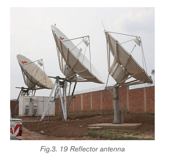
These are also called Yagi-Uda antennas or micro-strip patch arrays or aperture
arrays, slotted waveguide arrays. They are suitable for very high gain applicationswith added advantage, such as, controllable radiation pattern.
3.4.5 Advantages and disadvantages of optical fiber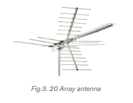
• Advantages of optical fibers are:
- The power loss is very low and hence helpful in long-distance transmissions.
- Fiber optic cables are immune to electromagnetic interference.
- These are not affected by electrical noise.
- The capacity of these cables is much higher than copper wire cables.
- Since these cables are di-electric, no spark hazards are present.
- These cables are more corrosion resistant than copper cables, as they are
bent easily and are flexible.
- The raw material for the manufacture of fiber optic cables is glass, which is
cheaper than copper.
- Fiber optic cables last longer than copper cables.
- Capacity: Optical fibers carry signals with much less energy loss than copper
cable and with a much higher bandwidth. This means that fibers can carry
more channels of information over longer distances and with fewer repeaters
required.
- Size and weight: Optical fiber cables are much lighter and thinner than
copper cables with the same bandwidth. This means that much less space
is required in underground cabling ducts. Also they are easier for installation
engineers to handle.
- Security: Optical fibers are much more difficult to tap information from
undetected; a great advantage for banks and security installations. They
are immune to electromagnetic interference from radio signals, car ignition
systems, lightning etc. They can be routed safely through explosive or
flammable atmospheres, for example, in the petrochemical industries or
munitions sites, without any risk of ignition.
- Running costs: The main consideration in choosing fiber when installing
domestic cable TV networks is the electric bill. Although copper coaxial cable
can handle the bandwidth requirement over the short distances of a housing
scheme, a copper system consumes far more electrical power than fiber,
simply to carry the signals.
• Disadvantages
Although there are many benefits to using optical fibers, there are also some
disadvantages as discussed below:
- Though fiber optic cables last longer, the installation cost is high.
- The number of repeaters is to be increased with distance.
- They are fragile if not enclosed in a plastic sheath. Hence, more protection is
needed than copper ones.
- Price: In spite of the fact that the raw material for making optical fibers,
sand, is abundant and cheap, optical fibers are still more expensive per meter
than copper. Having said this, one fiber can carry many more signals than a
single copper cable and the large transmission distances mean that fewer
expensive repeaters are required.
- Special skills: Optical fibers cannot be joined together (spliced) as an easily
as copper cable and requires additional training of personnel and expensiveprecision splicing and measurement equipment.
Application activity 3.4
1. Circle the three basic components in a fiber optic communications
system.
A. Telescope E. Maser fiber
B. Transmitter F. Optical fiber
C. Receiver G. Alternator
D. Surveillance satellites
2. Information (data) is transmitted over optical fiber by means of:
A. Light C. Acoustic waves
B. Radio waves D. None of the above
C. Cosmic rays
3. The basic unit of digital modulation is:
A. Zero D. A and B
B. One E. None of the above
C. Two
4. For each of the statements below, indicate true if it is correct and false
if it is wrong
A. Connectors and splices add light loss to a system or link.
B. The replacement of copper wiring harnesses with fiber optic cabling
will increase the weight of an aircraft.
5. 5. List two advantages and disadvantages of using optical fiber.
6. (a) Explain the optical transmitter and receiver in optical fiber
transmission system.
(b) Explain attenuation and state the measures to avoid it in optical fibertransmission system.
Skills Lab 3
In this activity you will demonstrate on how a signal can be transmitted
using water.It’s best to do it in a darkenedbathroom or kitchen at the sink or
washbasin.
You’ll need an old clear, plastic drinks bottle, the brightest flashlight (torch)
you can find, some aluminium foil, and some sticky tape.
a. Take the plastic bottle and wrap aluminium foil tightly around the sides,
leaving the top and bottom of the bottle uncovered. If you need to, hold
the foil in place with sticky tape.
b. Fill the bottle with water.
c. Switch on the flashlight and press it against the base of the bottle so the
light shines up inside the water. It works best if you press the flashlight
tightly against the bottle. You need as much light to enter the bottle as
possible, so use the brightest flashlight you can find.
d. Standing by the sink, tilt the bottle so the water starts to pour out. Keep
the flashlight pressed tight against the bottle. If the room is darkened,
you should see the spout of water lighting up ever so slightly. Notice
how the water carries the light, with the light beam bending as it goes!If you can’t see much light in the water spout, try a brighter flashlight.
End of unit 3 assessment
1. Which of the following is the most effective carrier of information?
A. Cables C . Microwaves
B. Radio waves D. Optical fibers
2. (a) An endoscope uses coherent and non-coherent fiber bundle
i) State the use of the coherent bundle and describe its
arrangement of fibers.
ii) State the use of the non-coherent bundle and describe its
arrangement of fibers.
(b) Each fiber has a core surrounded by cladding. Calculate the critical
angle at the core-cladding interface. Refractive index of core is1.52 and Refractive index of cladding is 1.
3. (a) Fig.3.21 shows a ray of light travelling through an individual fiber
consisting of cladding and a core. One part has a refractive index of1.485 and the other has a refractive index of 1.511.
i) State which part of the fiber has the higher refractive index and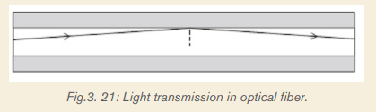
explain why.
ii) Calculate the critical angle for this fiber.
(b) The figure below shows the cross-section through a clad optical fiberwhich has a core of refractive index 1.50.
Complete the graph below to show how the refractive index changes with the
radial distance along the line ABCD in the figure above.
4. State and explain the measures to avoid the attenuation in optical fiber
transmission system
5. Estimate the length of time it would take a fiber optic systemto carry a signal from the UK to the USA under the Atlantic. (Take
a. Estimate the length of time it would take a microwave signal to travel
from the UK to the USA a satellite Enk. (Geosynchronous satellites orbit
at a height of about 86 000 km above the Earth’s surface.
b. Which would give less delay in a telephone conversation?
6. Write in your own words the description of step index fiber and graded
index fiber
7. One kind of optical fiber consists of two very thin rods one inside theother.
i) Explain why only a small amount of light is piped trough the fiber
in X.
ii) Why does the light travel along the fiber in Y without losing its
intensity.
iii) State how the inner and outer surfaces differ in their refractive
indices.
iv) Why is a fiber coated with a layer of plastic?
v) State two applications of optical fibers.8. Do fiber optics transmit radiations?
UNIT 4: NATURE OF PARTICLES AND THEIR INTERACTIONS
Key Unit Competence:Classify the nature of particles and their interactions
Introductory activity
In the study of matter description and energy as well as their interactions; the
fascinating thing of discovery is the structure of universe of unknown radius but
still to know the origin of matter one need to know about small and smallest
composites of matter.
a) Basing on the text above, what are the smallest particles in the universe
you know as of now?
b) Are those particles mentioned in a) above, have the same mass? If no,
what causes the difference in masses?
c) Basing on your submission in a) above do those particles interact? If not,
why not? If yes, how do they interact?d) Scientifically, how would you classify these particles?
4.1 FUNDAMENTAL PARTICLES
Activity 4.1
a) From your knowledge and understanding about atom in chemistry, can
you define an atom?
b) State all the particles you think make up an atom
c) Can you suggest a scientific name that may be used to describe theseparticles?
The idea that the world is made of fundamental particles has a long history. Inabout 400 B.C. The Greek philosophers Democritus and Leucippus suggested
that matter is made of indivisible particles that they called atoms, a word
derived from a (not) and tomos (cut or divided). At that time atoms were thought to
be indivisible constituents of matter. They were regarded as elementaryparticle.
This idea lay dormant until about 1804, when the English scientist John Dalton
(1766–1844), often called the father of Modern Chemistry, discovered that many
chemical phenomena could be explained if atoms of each element are the basic,
indivisible building blocks of matter.
Although Dalton had postulated that atoms were indivisible particles, experiments
conducted around the beginning of the last century showed that atoms themselves
consist of particles. These experiments showed that an atom consists of two kinds
of particles: a nucleus, the atom’s central core, which is positively charged and
contains most of the atom’s mass, and one or more electrons.
4.1.1 The Electron and the Proton
Some of the earliest evidence about atomic structure was supplied in the early
1800s by the English chemist Humphry Davy (1778–1829). He found that when
he passed electric current through some substances, the substances decomposed.
He therefore suggested that the elements of a chemical compound are held together
by electrical forces. In 1832–1833, Michael Faraday (1791–1867), Davy’s
student, determined the quantitative relationship between the amount of electricity
used in electrolysis and the amount of chemical reaction that occurs. Studies of
Faraday’s work by George Stoney (1826–1911) led him to suggest in 1874 that
units of electric charge are associated with atoms. In 1891, he suggested that they
be named electrons.
Rutherford’s experiments in 1910–1911 revealed that atoms consist of mostly
empty space with electrons surrounding a dense central nucleus made up of
protons and neutrons.
4.1.2 The Neutron
In 1930 the German physicists Walther Bothe and Herbert Becker and Irene
Joliot-Curie (1897–1956) observed that when beryllium, boron, or lithium was
bombarded by alpha particles, the target material emitted a radiation that had much
greater penetrating power than the original alpha particles.
Experiments done by the English physicist James Chadwick (1891–1974) in 1932
showed that the emitted particles were electrically neutral, with mass approximately
equal to that of the proton. Chadwick christened these particles neutrons(symbol

Elementary particles are usually detected by their electromagnetic effect for
instance, by the ionization that they cause when they pass through matter. (This
is the principle of the cloud chamber). Because neutrons have no charge, they
interact hardly at all with electrons and produce little ionization when they pass
through matter and so are difficult to detect directly.
However, neutrons can be slowed down by scattering from nuclei, and they can
penetrate a nucleus. Hence slow neutrons can be detected by means of a nuclearreaction in which a neutron is absorbed and an alpha particle is emitted. An example

The ejected alpha particle is easy to detect because it is charged. Later experiments

4.1.3 The Photon
Einstein explained the photoelectric effect in 1905 by assuming that the energy
of electromagnetic waves is quantized; that is, it comes in little bundles calledphotons with energy
4.1.4 The Neutrino
The Sun emits neutrinos copiously from the nuclear furnace at its core, and at night
these messengers from the center of the Sun come up at us from below, Earth
being almost totally transparent to them.4.1.5 The Positron and other antiparticles
Experiment and theory tell us that the masses of the positron and electron are
identical, and that their charges are equal in magnitude but opposite in sign. We
use the term antiparticle for a particle that is related to another particle as the
positron is to the electron. The positron is said to be the antiparticle to theelectron.
In 1955 the antiparticle to the proton, the antiproton which carries a negative
charge, was discovered at the University of California, Berkeley, by Emilio Segrè
(1905–1989) and Owen Chamberlain (1920–2006). A bar, such as over the p,is used to indicate the antiparticle.
Each kind of particle has a corresponding antiparticle. But a few, like the photon,
the and the Higgs, do not have distinct antiparticles we say that they are their
own antiparticles.
By the 1930s, it was accepted that all atoms can be considered to be made up
of neutrons, protons, and electrons. The basic constituents of the universe were
no longer considered to be atoms (as they had been for 2000 years) but rather
the proton, neutron, and electron. Besides these three “elementary particles,”
several others were also known by the 1950s and 1960: the positron (a positive
electron), the neutrino, and the γ particle (or photon), for a total of six elementary
particles.
4.1.6 Mesons and Beginning of Elementary Particle Physics
Elementary particle physics might be said to have begun in 1935 when the
Japanese physicist Hideki Yukawa (1907–1981) predicted the existence of a
new particle that would mediate the strong nuclear force the force that holds
nucleons together in the nucleus. Yukawa called this predicted particle meson(meaning medium mass).
Electrons contain no discernible structure; they cannot be reduced or separated into
smaller components. It is therefore reasonable to call them “elementary” particles,
a name that in the past was mistakenly given to particles such as the proton, which
is in fact a complex particle that contains quarks. The term subatomic particle
refers both to the true elementary particles, such as quarks and electrons, and tothe larger particles that quarks form.
By the term fundamental particle, we mean a particle that is so simple, so basic;
that it has no internal structure (is not made up of smaller subunits).
The science of the study of the particle is called Particle Physics,Elementary Particle Physics or sometimes High Energy Physics (HEP).
Application activity 4.1
1. The positron is called the antiparticle of electron, because it
A. Has opposite charge C. Collides with an electron
B. Has the same mass D. Annihilates with an electron
2. Beta particles are
A. Neutrons C. Electrons
B. Protons D. Thermal neutrons
3. The proton, neutron, electron, and the photon are called
A. secondary particles C. basic particles
B. fundamental particles D. initial particles
4. Particles that are unaffected by strong nuclear force are
A. protons C. neutrons
B. leptons D. bosons
5. The first antiparticle found was the
A. positron. C. quark.
B. hyperon. D. baryon.
6. The exchange particle of the electromagnetic force is the
A. Gluon. C. proton.B. Muon. D. photon.
4.2 CLASSIFICATION OF PARTICLES
Activity 4.2
a) Basing on what so-far you have studied, classify these elementary
particles?
b) Explain what you have based on to classify them.c) State the common characteristics/features for each group.
Today there are several hundreds of known particles. Naming them has strained the
resources of the Greek alphabet, and most are known only by an assigned number
in a periodically issued compilation. To make sense of this array of particles, we
look for simple physical criteria by which we can place the particles in categories.
The result is known as the Standard Model of particles. Although this model is
continuously challenged by theorists, it remains our best scheme of understanding
all the particles discovered to date.
To explore the Standard Model, we make the following three rough families of
the known particles: the photon: fermion or boson, hadron or lepton, particle orantiparticle?
As more and more particles were discovered, it became clear that they were not all
elementary particles (fundamental or basic particles). The suggestion was
made that the hadrons are made up of smaller, more elementary particles called
quarks. They are three families of quarks and three corresponding antiquarks, and
hadrons are constructed from combinations of these. Thus the quarks are elevated
to the status of elementary particles for the elementary particles for the family of
hadrons. The particles in the photon and lepton families are considered to be
elementary, and such they are not composed of quarks.
The fundamental particles were classified into two categories according to theirspin: Fermions and Bosons
4.2.1 Fermions
Particles with half-integer spin quantum numbers (like electrons) are called
fermions, after Fermi, who (simultaneously with Paul Dirac) discovered the
statistical laws that govern their behavior.They are two families of fermions (of spin ½): leptons and quarks
a. Leptons
Leptons (from the Greek leptos meaning small or light) are a group of particles
that participate in the weak nuclear force, they can exert gravitational, and they are
charged particles hence exert electromagnetic force on other particles. All leptons
have spin and thus are fermions.
Three pairs or families of leptons and their anti-particles exist as listed in thetable 4.1.
The six leptons are considered to be truly fundamental particles because they do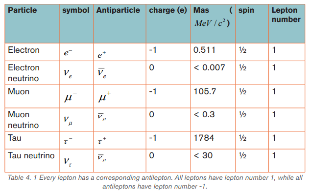
not show any internal structure, and have no measurable size.
b. Quarks
They are six quarks or flavors of quarks: up (u), down (d), strange (s), charm
(c),bottom (b)and top (t) quarks and they each have their partner anti-quarks(designated by a line over the letter symbol).


Quarks combine to form hadrons or meson. The hadrons are a composite particle
made of quarks (u, d, c, s, t, b) held together by the strong nuclear force. Hadrons
can also interact by weak nuclear force, gravitational force and electromagneticforces but at short distances
Hadrons are categorized into families distinguished by their masses and spins:
Hadrons and baryons
a. Mesons
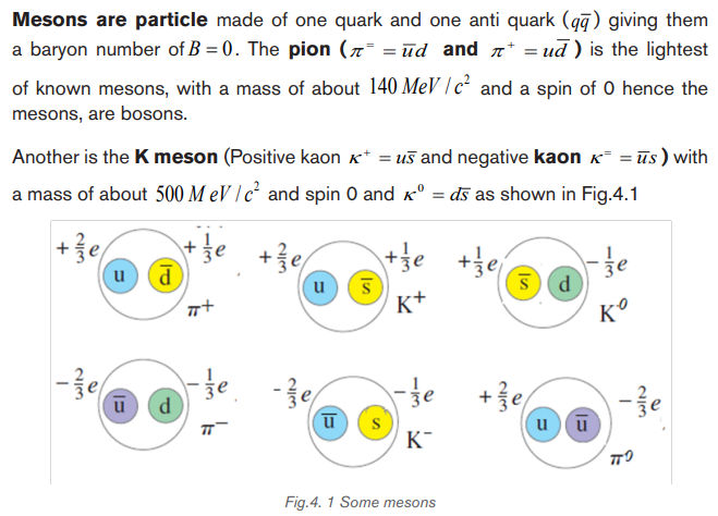
b. Baryons
The table below show gauge bosons
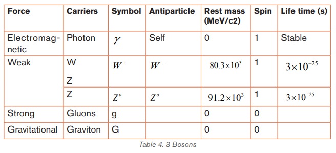
In summary, the following diagram shows some classes of elementary particles

Application activity 4.2
1. A proton is made up of
A. one up quark and two down quarks
B. an up quark and down antiquark
C. two up quarks and a down quark
D. strange quark and an anti-strange quark
2. Particles that are unaffected by strong nuclear force are
A. protons C. neutrons
B. leptons D. bosons
3. Particle which explains about mass of matter is called
A. Higgs boson C. protons
B. Leptons D. neutrons
4. Each hadron consists of a proper combination of a few elementary
components called
A. Photons C. quarks
B. Vector bosons D. meson-baryon pairs.
5. The proton, neutron, electron, and the photon are called
A. secondary particles C. basic particles
B. fundamental particles D. initial particles
6. The exchange particle of the electromagnetic force is the
A. gluon. C. proton.
B. muon. D. photon.
7. Particles that interact by the strong force are called
A. leptons C. muons
B. hadrons D. electrons
8. At the present time, the elementary particles are considered to be
the
A. Photons and baryons. C. Baryons and quarks.
B. Leptons and quarks. D. Baryons and leptons.
9. The electron and muon are both
A. Hadrons. C. Baryons.
B. Leptons. C. Mesons.
10. Particles that make up the family of hadrons are
A. Baryons and mesons. C. Protons and electrons.
B. Leptons and baryons. D. Muons and leptons.
11. Is it possible for a particle to be both:
A. A lepton and a baryon? C. A meson and a quark?
B. A baryon and hadron? D. A hadron and a lepton?
12. Distinguish between (a) fermions and bosons, (b) leptons and
hadrons and (c) mesons and baryon number
13. Which of the four interactions (strong, electromagnetic, weak, and
gravitational) does an electron take part in? A neutrino? A proton?
14. Describe the types and the characteristics of the quarks as well astheir interaction properties.
4.3 FUNDAMENTAL FORCES AND INTERACTIONS
Activity 4.3
From the previous lessons, you have learned that the particles have different
charges and masses. Basing on that, explain the magnitude of force that may
rise between these elementary particles depending on:
a) Masses (You can apply newton’s law of gravitation)b) Charges (You can use coulombs law of charges)
4.3.1 Antiparticle and antimatter
Antiparticles are produced in nuclear reactions when there is sufficient energy
available to produce the required mass, and they do not live very long in the
presence of matter.
Antimatter is a term referring to material that would be made up of “antiatoms” in
which antiprotons and antineutrons would form the nucleus around which positrons
(antielectrons) would move. The term is also used for antiparticles in general. Anti matter is a material composed of anti-particles.
We use the term antiparticle for a particle that is related to another particle as the
positron is to the electron. Each kind of particle has a corresponding antiparticle.
For a few kinds of particles (necessarily all neutral) the particle and antiparticle are
identical, and we can say that they are their own antiparticles. The photon is an
example; there is no way to distinguish a photon from an anti photon.
Pair production and pair annihilation
Positrons do not occur in ordinary matter. Electron–positron pairs are produced
during high-energy collisions of charged particles or γ -rays with matter.This process is called
 pair production.
When a particle meets its antiparticle, the two can annihilate each other and
release a large amount of energy. That is, the particle and antiparticle disappear
and their combined energies reappear in other forms. For an electron annihilatingwith a positron, this energy reappears as two gamma-ray photons:

4.3.2 Fundamental Interactions and Force Mediators
In nature they are two types of forces, fundamental and non-fundamental forces.
Fundamental (basic) forces are the ones that are truly unique, in the sense that all
other forces can be explained in terms of them.
By 1940, Physicists have long recognized for forces of nature (fundamental
forces):
• The gravitational force
The gravitational force is an inherent attraction between two masses.
Gravitational force is responsible for the motion of the planets and Stars in the
Universe. It is carried by Graviton but its existence has not been detected and it
may not be detectable. By Newton’s law of gravitation, the gravitational force is
directly proportional to the product of the masses and inversely proportional to
the square of the distance between them. Gravitational force is the weakest force
among the fundamental forces of nature but has the greatest large-scale impact
on the universe. Unlike the other forces, gravity works universally on all matter and
energy, and is universally attractive.
• The electromagnetic interaction
In Classical Physics we describe the interaction of charged particles in terms of
electric and magnetic forces. The electric force is a force between charges.
The magnetic force is a force between magnets or between magnetic body andferromagnetic body.
In quantum mechanics we can describe this interaction in terms of emission and
absorption of photons. Two electrons repel each other as one emits a photon and
the other absorbs it, just as two skaters can push each other apart by tossing aheavy ball back and forth between them (Fig. 4.5).
For an electron and a proton, in which the charges are opposite and the force
is attractive, we imagine the skaters trying to grab the ball away from each other
(Fig. 4.6). The electromagnetic interaction between two charged particles ismediated or transmitted by photons
In the 1860s, the Scottish physicist James Clerk Maxwell developed a theory that
unified the electric and magnetic forces into a single electromagnetic force.
Maxwell’s electromagnetic force was soon found to be the “glue” holding atoms,
molecules, and solids together. It is the force between charged particles such as
the force between two electrons, or the force between two current carrying wires.
It is attractive for unlike charges and repulsive for like charges. The electromagnetic
force obeys inverse square law. It is very strong compared to the gravitational force.It is the combination of electrostatic and magnetic forces.
• Strong nuclear force
In 1935 the Japanese physicist Hideki Yukawa suggested that a hypothetical
particle that he called a meson might mediate the strong nuclear force. The
strong nuclear force is the force that holds the protons and neutrons together
in the nucleus of an atom. It plays a primary role in stability of the nucleus of the
atoms. It is the strongest of all the basic forces of nature. It, however, has the
shortest range, of the order of 10−15 m (the size of the nucleus). This force only acts
on quarks. Quarks carry electric charge so they experience electric and magnetic
forces. It binds quarks together to form baryons and mesons such as protons and
neutrons. The strong force is mediated by Gluons. However, the force betweennucleons is more easily described in terms of mesons as the mediating particles.
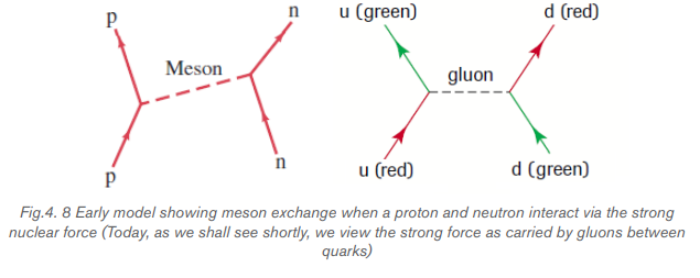
• The weak force
In the 1939, Physicists found that the nuclear radioactivity called beta decay could
not be explained by either the electromagnetic or the strong force. The strength
of this force is less than either the strong force or the electromagnetic force, so
this new force was named the weak force. Weak nuclear force is important in
certain types of nuclear process such as β-decay. This force is not as weak as
the gravitational force. The weak force acts on both leptons and quarks (and
hence on all hadrons).
Leptons – the electrons, muons and tau – are charged so they experience electric
and magnetic forces. Of these, our everyday world is controlled by gravity and
electromagnetism. The weak force is responsible for the radioactive decay ofunstable nuclei and for interactions of neutrinos and other leptons with matter.
By 1980, Scientists developed a theory that unifies electromagnetism and weak
force into electroweak force mediated by particles, the
Hence, our understanding of the forces of nature is in terms of three fundamentalforces: the gravitational force, the electroweak force, and the strong force.
The table below summaries the fundamental forces:
The intrinsic strengths of the forces can be compared relative to the strong
force, here considered to have unit strength(i.e., = 1). In these terms, the
electro magnetic force has an intrinsic strength of (1/137). The weak force
is a billion times weaker than the strong force. The weakest of them all is the
gravitational force. This may seem strange, since it is strong enough to hold
the massive Earth and planets in orbit around the Sun! But we know that that the
gravitational force between two bodies a distance r apart is proportional to the
product of the two masses (M and m) and inversely proportional to the distance rsquared:
We see now what is meant by intrinsic strength. It is given by the magnitude
of the universal force constant, in this case, G, independent of the masses ordistances involved.
In similar terms, the electromagnetic force between two particles is proportional tothe product of the two charges (Q and q) and inversely to the distance r squared:
We can compare the relative strengths of the electromagnetic repulsion and here the universal constant, k, gives the intrinsic strength.
the gravitational attraction between two protons of unit charge using the aboveequations.

Example 4.1
The 2 up quarks (u) in a proton are separated by These quarks
each have an electric charge so they repel each other through an electric
force obeying Coulomb’s law. They also attract each other through a gravitational
force. What is the ratio of the magnitude of the electric and gravitational forcesbetween these 2 quarks?
Answer
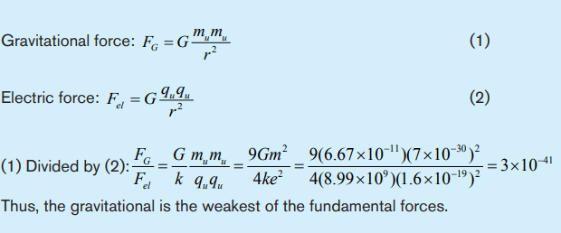
Application activity 4.3
1. If gravity is the weakest force, why is it the one we notice most?
Choose the letter correspond to the correct answer
A. Our bodies are not sensitive to the other forces.
B. The other forces act only within atoms and therefore have no effect on
us.
C. Gravity may be “very weak” but always attractive, and the Earth has
enormous mass. The strong and weak nuclear forces have very short
range. The electromagnetic force has a long range, but most matter is
electrically neutral.
D. At long distances, the gravitational force is actually stronger than the
other forces.
E. The other forces act only on elementary particles, not on objects our
size.
2. The two basic interaction forces that have finite ranges are:
A. electromagnetic and gravitational D. gravitational and weak
B. electromagnetic and strong E. weak and strong
C. electromagnetic and weak
3. Name the four fundamental interactions and the particles that
mediate each interaction.
4. State two differences between a proton and a positron.
5. Copy figure below into your notebook. Complete the diagram by
filling in the names of the fundamental forces and the names of theunification theories.

Skills Lab 4
In this activity, you make a comprehensive research about nature of particles
and their interactions.
Materials needed: Computer set, internet and reference books.
What to do.
a) Using internet, make general studies about the following
• Classification of particles and antiparticles.
• Properties of fundamental particles (Charges, spin, quark contents)
b) Compile your findings and make a report. Relate your findings to what
you have studied in this unit.
c) You can compare your report to your friends’ report and check whether
the information you have is related to your friends work.
d) You and your classmates may prepare a session of presentation so
that you can harmonize what you got in your research. Present using
PowerPoint presentations.
e) In case you find new points from other presentations, include it to yourreport.
End of unit 4 assessment
1. Which of the following are today considered fundamental particles
(that is, not composed of smaller components)? Choose as many
as apply.
A. Atoms. C. Protons. E. Quarks. G. Higgs boson
B. Electrons. D. Neutrons. F. Photon
2. The electron’s antiparticle is called the positron. Which of the
following properties, if any, are the same for electrons and positrons?
A. Mass. C. Lepton number
B. Charge. D. None of the above.
3. The strong nuclear force between a neutron and a proton is due to
A. The exchange of π mesons between the neutron and the proton.
B. The conservation of baryon number.
C. The beta decay of the neutron into the proton.
D. The exchange of gluons between the quarks within the neutron and
the proton.
E. Both (A) and (D) at different scales.
4. Which of the following will interact via the weak nuclear force only?
A. Quarks. C. Neutrons E. Electrons G. Higgs boson.
B. Gluons. D. Neutrinos. F. Muons.
5. Messenger particles of the weak interaction are called:
A. gluons D. gravitons
B. photons E. pions
C. W and Z
6. Messenger particles of the electromagnetic interaction are called:
A. gluons D. gravitons
B. photons E. pions
C. W and Z
7. The pair annihilation of an electron and a positron has been
investigated for many years at CERN in Switzerland. Two gamma-rayphotons are produced during this annihilation. What is a positron?
8. True or false: if the statement is true, explain why it is true. If it is
false, give a counterexample.
(a) leptons are fermions
(b) all baryons are hadrons
(c) all hadrons are baryons
(d) mesons are spin ½ particles
(e) leptons consist of three quarks
(f) the times for decays via the weak interaction are typically much
longer than those for decays via the strong interaction(g) the electron interacts with the proton via the strong interaction
UNIT 5:X-RAYS AND ITS EFFECTS
Key Unit Competence:Suggest and criticize possible effects of X-rays
Introductory activity
Technology has advanced and man has made all advancements to see that
human problems are solved with ease and using technology. Among other
areas where technology has been emphasized is in medicine (in hospitals).
For example, CT scans (Computerized tomography scans) and X-rays machines
are commonly used in hospitals to examine internal structures of a patient if
needed. When a person goes to the hospital with pain in her/his chest, or with
an internal fracture of the bone, physicians do normally recommend the patient
to pass by radiology service (Radiology means there are radiations).
1. Why do you think physicians recommend patients to pass by radiology
service?
2. Discuss different types of radiations that are found in there?
3. From your physics knowledge, how do you think these radiations
specifically X-rays are produced.
4. Like any other electromagnetic radiations, what do you think are some
of the properties of X-rays?
5. As seen from the statement X-rays are used in hospitals, other than
being used in medicine, discuss other areas/fields where X-rays are
applied.
6. What are the positive and negative effects of X-ray radiation on the
human body do you know?
7. Having seen that these radiations have negative effects on human body,
what are your recommendations to a technician works in areas that useX-rays.
5.1 PRODUCTION OF X-RAYS
Activity 5.1
X-rays are produced when the electrons are suddenly decelerated upon collision
with the metal target; these x-rays are commonly called “braking radiation”. If
the bombarding electrons have sufficient energy, they can knock an electronout of an inner shell of the target metal atoms.
Discovery of X-rays: Becquerel’s discovery wasn’t the only important
accidental one. In the previous year W.C. Roentgen unexpectedly discoveredX-rays while studying the behavior of electrons in a high-voltage vacuum tube.
In that instance, a nearby material was made to fluoresce. Roentgen named
them X- rays because he didn’t know what they were. Within twenty years of this
discovery, diffraction patterns produced using X-rays on crystal structures had
begun to show the finer structure of crystals while, at the same time,
giving evidence that X-rays had a wave nature. Since then, X-ray radiation hasbecome an indispensable imaging tool in medical science and other fields.
Questions:
1. According to the text, why do you think the electrons need to be
accelerated and decelerated to produce X-rays?
2. Imagine the energy of bombarding electrons is varied, do you think the
type X-ray emitted would remain same?
3. According to the text, do you think that it is possible to produce X-raysin our local laboratories? Defend your suggestion.
5.1.1 X-ray production
The above figure is an illustration of the Coolidge tube which is the most widely
used device for the production of X-rays. The electrons are produced by thermionic
effect from filament, which is the cathode of the tube, heated by an electric current.
These electrons are accelerated towards a metal target that is the anode due to the
high potential voltage between the cathode and the anode. The target metals are
normally Tungsten or Molybdenum and are chosen because they have high melting
point and higher atomic weights. The accelerated electrons interact with both
electrons and nuclei of atoms in the target and a mysterious radiation is emitted.This radiation was referred to as X-rays.
About 98% of the energy of the incident electron is converted into heat that isevacuated by the cooling system and the remaining 2% come out as X-rays.
5.1.2 Types of x-rays
Sometimes x-rays are classified according to their penetrating power. Two types
are mentioned:
- Hard X-rays: those are X-rays on upper range of frequencies or
shorter wavelength. They have greater energy and so they are more
penetrating.
- Soft X-rays: they are X-rays on lower range of frequencies or longer
wavelength. They have lower energy and they have very low penetrating
power. The Fig.5.2 below shows the relative location of the different types
of x-rays.
Hard x-rays are produced by high accelerating potential. They have high penetrating
power and short wavelength while soft x-rays are produced by lower acceleratingpotential, have relatively low penetrating power and relatively long wavelength.

Application activity 5.1
1. With the aid of a diagram, describe how X-rays are produced in a
laboratory.
2. a) Discuss the two types of X-rays and how they are produced.
b) In the two types of X-rays mentioned in b) above, which one can be
used to
i) Examine or kill cancer cells in a breast.ii) Examine minerals beneath a hard rock.
5.2 PROPERTIES OF X-RAYS AND CHARACTERISTICFEATURES OF X-RAY SPECTRUM
Activity 5.2
With reference to electromagnetic spectrum, what do you think are theproperties of X-rays?
5.2.1 Properties of x-rays
The following are the main properties of X-rays:
(a) X-rays can penetrate through most substances. However, their penetrating
power is different.
(b) X-ray can produce fluorescence in different substances.
(c) X-rays can blacken photographic plate. The degree of blackening depends
upon the intensity of x-rays incident upon the plate. Thus, X-ray intensity can
be measured with the help of photographic plates.
(d) X-rays ionize the gas through which they travel. The ionizing power depends
on the intensity of the x-ray beam. Thus, X-ray intensity can also be measured
by measuring their ionizing power.
(e) X-rays are not deflected by electric or magnetic fields. This proves that
unlike cathode rays or positive rays they are not a beam of charged particles.
(f) X-rays travels on a straight line like ordinary light.
(g) X-ray are both reflected and refracted.X-rays can be diffracted with the help of crystalline substances. They can
also be polarized
From the above characteristics it can be seen that X-rays have the properties that
are common to all electromagnetic radiations.
5.2.2 The origin and characteristic features of an x-ray spectrum
Variation of the X-ray intensity with wavelength
Depending on the accelerating voltage and the target element, we may find sharp
peaks superimposed on a continuous spectrum as indicated on Fig.5.3. These
peaks are at different wavelengths for different elements; they form what is called acharacteristic x-ray spectrum for each target element.
X-rays of different wavelengths are emitted from x-ray tube. If the intensity is
measured as a function of the wavelength and the variation is plotted graphically
then a graph of the nature shown on the figure above is obtained. The graph has
the following features,
(a)Minimum wavelength
(b)Continuous spectrum
(c)Characteristic peaks
Origin of the continuous spectrum
It is known that when charged particles such as electrons are accelerated or
decelerated, they emit electromagnetic radiation of different frequencies. In doing
so a part of their kinetic energy is transformed in the energy of the emitted radiation.
Electrons inside the x-ray tube decelerate upon hitting the target and as a result they
emit electromagnetic radiations with a continuous distribution of wavelength starting
from a certain minimum wavelength. This mechanism of producing electromagnetic
radiation from an accelerated or decelerated electron is called bremsstrahlung.The energy of the emitted photon is given by:
Where is the minimum wavelength, V is the potential difference between anode and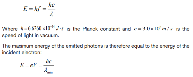
cathode and e the charge of the electron.
Origin of characteristic lines
The peaks observed in wavelengths distribution curves as shown in Fig. 5.3 are
spectral lines in the x-ray region. Their origin lies in the transition between energy
levels in the atoms of the target.
The electrons in the atoms are arranged in different atomic shell of these, the
first two electrons occupy the K-shell followed by 8 electrons in the L-shell, 18
electrons in the M-shell and so on until the electron in the target are used up. A
highly accelerated electron may penetrate atom in the target and collide with an
electron in K-shell. If such electron is knocked out it will leave an empty space that
is immediately filled up by another electron probably from the L-shell or M-shell. This
transition will be accompanied by the emission of the excess energy as a photon.
The energy of the emitted photon is a characteristic of the energy levels in theparticular atom and is given by:
For a transition between K and L-shells.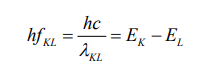
Thus, the energy of the emitted photon depends on the binding energies in the
K and L shells and hence the x-ray spectral lines have definite frequencies and
wavelengths which are characteristic of the target atom.
For a given target material more than one spectral lines are observed as transitionsmay occur between different energy levels.
The x-ray lines originating from the transition between the different electron levels
are usually labelled by the symbols α, β, γ, etc
From L-level to K-level transition produces Kα-line
From M-level to K-level transition produces Kβ –line
From M-level to L-level transition produces Lα –lineFrom N-level to L- level transition produces Lβ –line
Application activity 5.2
1. X-rays are electromagnetic waves produced when fast moving electrons
strike the matter. Discuss the properties of X-rays.
2. A plot of x-ray intensity as a function of wavelength for a particularaccelerating voltage and a particular target is shown in figure below.
a. There are two main components of this x-ray spectrum: a broad range of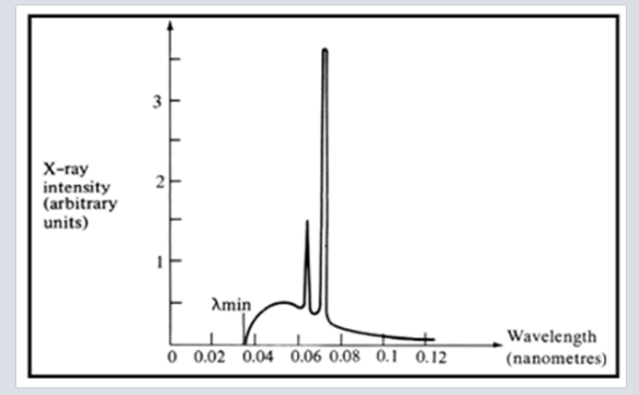
x-ray energies and a couple of sharp peaks. Explain how each of these
arises.
b. What is the origin of the cut-off wavelength λmin of the Figure shown
above? Why is it an important clue to the photon nature of x-rays?
c. What would happen to the cut-off wavelength if the accelerating voltage
was increased? What would happen to the characteristic peaks? Use a
sketch to show how this spectrum would look if the accelerating voltagewas increased
d. What would happen to the cut-off wavelength if the target was changed,
keep the same accelerating voltage? What would happen to the
characteristic peaks? Use a sketch to show how the spectrum would
look if some other target material was used, but the accelerating voltage
was kept the same.
3. Electrons are accelerated from rest through a potential difference of
10 kV in an x ray tube, calculate:
i) The resultant energy of the electrons in eV.
ii) The wavelength of the associated electron waves.iii) The maximum energy and the minimum wavelength of the x ray
5.3. APPLICATIONS AND DANGERS OF X-RAY
Activity 5.3
1. Basing on the nature and properties, what do you think are the uses of
X-rays in real life?
2. If during your internship as a student teacher in a certain primary school,
one of the pupils tells you that her father who is a medical doctor told her
that X-rays are useful and miss-used. That pupil seeks information from
you on how these dangers can be avoided. Provide relevant informationto him/her on how dangers caused by X-rays can be avoided.
5.3.1 Applications
X rays have many practical applications in medicine and industry. Because x-ray
photons are of such high energy, they can penetrate several centimetres of solid
matter. Hence, they can be used to visualize the interiors of materials that areopaque to ordinary light, such as broken bones or defects in structural steel.
a. In medicine
X-ray imaging utilizes the ability of high frequency electromagnetic waves to pass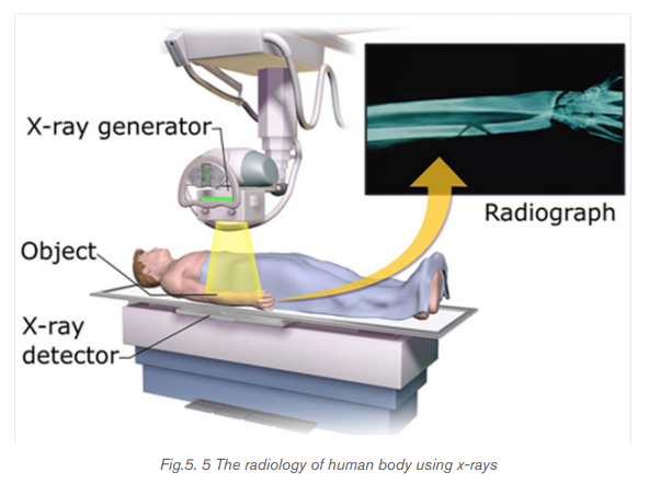
through soft parts of the human body largely unimpeded. For medical applications,
parts of the human body are exposed to moderated X-rays intensity and images
are produced in similar way as light on a photographic plate or digital recorder to
produce a radiograph (See Fig.5.5). By rotating both source and detector around
the patient’s body a “slice” image can be produced in what is called computerized
tomography (CT). Although CT scans expose the patient to higher doses of ionizing
radiation the slice images produced make it possible to see the structures of the
body in three dimensions.
In 1895, the Dutch Wilhelm Roentgen (See Fig.5.6) discovered that light energy
could be used to take photographs through substances such as paper, cloths
and wood. Roentgen also discovered that this invisible form of light energy, called
X-rays could be used to take the pictures of structures inside the body as shown in
Fig. 5.6. Bone tissue appears clearly on an X-rays.
The object to be visualized is placed between an x-ray source and an electronic
detector (like that used in a digital camera) or a piece of photographic film. The
darker area in the recorded images by such a detector, the greater the radiation
exposure. Bones are much more effective x-ray absorbers than soft tissue, so bones
appear as light areas. A crack or air bubble allows greater transmission and showsas a dark area.
A widely used and vastly improved x-ray technique is computed tomography; the
corresponding instrument is called a CT scanner. The x-ray source produces a thin,
fan-shaped beam that is detected on the opposite side of the subject by an array
of several hundred detectors in a line. Each detector measures absorption along
a thin line through the subject. The entire apparatus is rotated around the subject
in the plane of the beam, and the changing photon-counting rates of the detectors
are recorded digitally. A computer processes this information and reconstructs a
picture of absorption over an entire cross section of the subject. In the middle 1970,CT (Computer Tomography) scanning machines were introduced in medicine.
X-rays are also used in the following:
- Killing of cancerous cells
- Radiography is also used in industry for examining potentially damaged
machinery to ascertain the cause of damage and to verify castings or
welded joints
- X-rays are used to study the structure of crystals (crystallography).
When a handgun is fired, a cloud of gunshot residue (GSR) is ejected from the
barrel. The x-ray emission spectrum of GSR includes characteristic peaks from
lead (Pb), antimony (Sb), and barium (Ba). If a sample taken from a suspect’s skin
or clothing has an x-ray emission spectrum with these characteristics, it indicates
that the suspect recently fired a gun.
b. Examining luggage cargo and security.

X-rays are being used in airports to examine luggage for weapons or bombs.
Note that the metal detector that you walk through in the airport does not use x-rays
to examine you instead it uses magnetic waves to detect metal objects. X-rays are
also being used to examine cargo luggage for illegal or dangerous material as in
Fig.5.7.
c. In industry
They can be used to detect structural problems and cracks in metals that cannot
be seen from the outside. X-rays are used on commercial airplanes, bridges metals
and pipe lines, to make sure there are no stress fractures or other dangerous cracks
in the material.
d. In scientific research
• X-ray diffraction provides one of the most important tools for examining the
three-dimensional (3D) structure of biological macromolecules and cells.
• They are also used in crystallography, where X-ray diffraction and scatteredwaves show the arrangement of atoms in the crystal.
The array of spots formed on the film is called a Laue pattern and show the atom
structure of the crystal.
5.3.2 Dangers
• X rays cause damage to living tissues. As X-ray photons are absorbed
in tissues, their energy breaks molecular bonds and creates highly reactive
free radicals (such as neutral H and OH), which in turn can disturb the
molecular structure of proteins and especially genetic material. Young and
rapidly growing cells are particularly susceptible, which is why X-rays are
for selective destruction of cancer cells.
• Because X-rays can kill living cells, they must be used with extreme care.
When improperly used they can cause severe burns, cancer, leukemia, and
cataracts. They can speed aging, reduce immunity to disease, and bring
about disastrous changes in the reproductive cells.
• Lead screens, sheets of lead-impregnated rubber, and leaded glass are
used to shield patients and technicians from undesired radiation.
• The effect of X-ray radiations is cumulative. That is, many minor doses over
a number of years is equivalent to a large dose at one time.
• Unnecessary exposure to x-rays should be avoided. MRI (Magnetic
Resonance Imaging) uses magnets and sound energy to form pictures of
the internal organs without exposing patients to harmful X-rays.
• When they are used in hospitals, the sources should be enclosed in lead
shields.
A careful assessment of the balance between risks and benefits of radiation
exposure is essential in each individual case.
5.3.3 Safety precaution measures of dangers caused by x-rays
Medical and dental X-rays are of very low intensity, so that the hazard is minimized.
However, X-ray technicians who go frequently behind the lead shield while operating
X-rays need to be protected because of the frequency of exposure. A person can
receive many medical or dental X-rays in a year with very little risk of getting cancer
from it. In fact, exposure to natural radiation such as cosmic rays from space poses
a greater risk.
The following are some of the precautions:
i) Protective suits and wears such as gloves and eye glasses made of lead
are used always when handling these radiations. These shields protect the
workers from X-ray exposure.
ii) Workers who operate equipment’s that use X-rays must wear special
badges which detect the amount of radiation they are exposed to.
iii) Food and drinks are not allowed in places where X-radiations are present.
iv) Experiments that involve these radiations (X-rays) substances should be
conducted in a room surrounded by thick concrete walls or lead shields.
v) Equipment that use X-rays should be handled using remote-controlledmechanical arms from a safe distance.
Application activity 5.3
1. Using relevant examples, explain how X-rays are applied in different
fields.
2. Examine the dangers that may arise if these radiations are not handled
with care.
3. As a year III student- teacher, advise an internee having internship in
an area that has X-rays on what to do to avoid the dangers that may becaused by X-rays.
Skills Lab 5
In this activity you will visit a nearest Laboratory that uses X-rays. It may be a
hospital or an industry. In your visit, try to focus on the following
a. How do technicians obtain the x-rays?
b. Why is the room where radiology services done isolated?
c. What are some of the rules followed while in a room where radiology
services are provided?
d. How is X-ray machine operated to achieve results?
e. What are safety precautions to the dangers that may be due to exposure
of X-rays.
You can ask any question of your choice you think is relevant and can make
you understand this unit.
As you come back to the school, make sure you make a comprehensive report
on what you studied from the hospital. Compare the findings to what you
discussed in physics classes.
Present your final findings to the whole class and then finally to your physicstutor.
End of unit 5 assessment
Where necessary use the following constants.
1. Choose the letter that best matches the true answer:
(i) X-rays have
A. short wavelength C. both A and B
B. high frequency D. longest wavelength
(ii)If fast moving electrons rapidly decelerate, then rays produced are
A. alpha rays C. beta rays
B. x-rays D. gamma rays
iii) Energy passing through unit area is
A. intensity of x-ray C. wavelength of x-ray
B. frequency of x-ray D. amplitude of x-ray
iv) X-rays are filtered out of human body by using
A. cadmium absorbers C. copper absorbers
B. carbon absorbers D. aluminum absorbers
v) Wavelength of x-rays is in range
A. 10-8 m to 10-13 m C. 10-10 m to 10-15 m
B. 10-7 m to 10-14 m D. 10² m to 109 m
2. An x-ray operates at 30 kV and the current through it is 2.0 mA.
Calculate:
(i) The electrical power output
(ii) The number of electrons striking the target per second.
(iii) The speed of the electrons when they hit the target(iv) The lower wavelength limit of the x-rays emitted.
3. An x-ray machine can accelerate electrons of energies
The shortest wavelength of the x- rays produced by the machine is
found to be Use this information to estimate the value of
the plank constant.
4. You have decided to build your own x-ray machine out of an old
television set. The electrons in the TV set are accelerated through a
potential difference of 20 kV. What will be the λmin for this accelerating
potential?
5. A tungsten target (Z = 74) is bombarded by electrons in an x-ray tube.
The K, L, and M atomic x-ray energy levels for tungsten are -69.5, -11.3
and -2.30 keV, respectively.
a) Why are the energy levels given as negative values?
b) What is the minimum kinetic energy of the bombarding electrons
that will permit the production of the characteristic and lines of
tungsten?
c) What is the minimum value of the accelerating potential that will give
electrons this minimum kinetic energy?
d) What are the and wavelengths?
6. Using the following illustration, name each part marked by letter from Ato H and explain the function of each part A, B, C, D, E, F and H.

UNIT 6: LASER AND ITS EFFECTS.
Key Unit Competence:Point out effects of LASER beam.
Introductory activity
Man has tried all forms of advancements from traditional methods of solving
problems to advanced methods by use of different technologies. Among other
technological advancements, discovery of Laser that is a part of visible light
under electromagnetic waves has had a great impact in solving many of our
problems. In the picture above a doctor is examining internal part of a patient
using LASER beam. This work has been made easy by use of strong radiationslike LASER beam.
Note: LASER stands for Light Amplifier by Stimulated Emission of
Radiation.
a) From the diagram, what is the nature of LASER light?
b) What do you think are characteristics of a LASER beam as observed
from the figure above?
c) LASER beam is used in different fields. These include, industry,
Agriculture, Medicine and Scientific research. Can you explain how these
radiations are useful to these fields?
d) Though these radiations are useful in real life, but they are also dangerous
if miss-used. Can you guess some of the dangers of LASER beam?
e) With relevant examples, what do you think are dangers caused by theseLASER beam radiations if not well controlled?
6.1 PRODUCTION OF LASER
Activity 6.1
The laser is a special device that uses the ability of some substances to
absorb electromagnetic energy and re-radiate it, as a highly focused beam
of monochromatic and synchronized wavelength radiation. Laser has to be
produced in laboratory so that people benefit from its uses.
a) From your own understanding, explain how a LASER light can be
produced.
b) Does its production, need source of energy like electricity? Explain your
reasoning.
c) In energy levels, particles are either in ground or excited states. Is laser
formed when particles or electrons are in ground or excited states?
Explain your reasoning.
d) From question in c) it is clearly indicated that particles are either in
ground state or excited state. Explain the changes in number of particles(electrons) in each level as they energy is absorbed.
The acronym LASER stands for Light Amplifier by Stimulated Emission of
Radiation. This expression means that the light is formed by stimulating a material’selectrons to give out the laser light or radiation.
In 1953 Charles H. Townes, with graduate students James P. and Herbert
J., produced the first Microwave Amplifier by Stimulated Emission of
Radiation (MASER), as a device operating in the same way as a laser, but
amplifying microwave radiations. This system could release stimulated emissions
without falling to the ground state, and thus maintaining a population inversion. A
laser is a device that emits light through a process of optical amplification based
on the stimulated emission of electromagnetic radiation. That is, the laser is a light
source that produces a beam of highly coherent and very nearly monochromaticlight because of cooperative emission from many atoms.
6.1.1 Absorption, Spontaneous emission and Stimulatedemission
a. Absorption
During the process of absorption, a photon from the source is losingits entire
energy to the atom which was at the ground state and then the atom is promotedto the excited state.
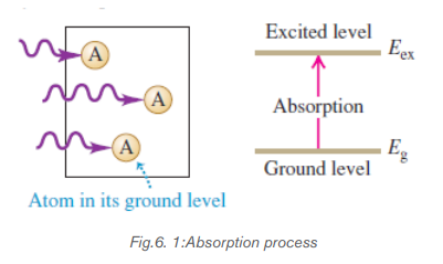
b. Spontaneous emission.
An atom or an electron can move from one energy level to another. A photon is
released when an electron moves from a higher energy level to a lower energy level.
The release of photon (a particle of light) is called spontaneous emission.
At the excited state, an atom will drop to a lower level by emitting a photon of radiation
in a process called spontaneous emission. It emits the photon spontaneously
after an average time τ called the spontaneous lifetime of the level. This
time depends on the atomic species; some levels have long lifetime measured in
seconds, whereas others are relatively short on the order of nanoseconds or less.
This lifetime determines the ability of the emitting atom to store energy and willaffect the efficiency of sources.

When an excited atom, depending on its lifetime at the higher energy level, comes
down to lower energy level, a photon is emitted, corresponding to the equation,
c. Stimulated emission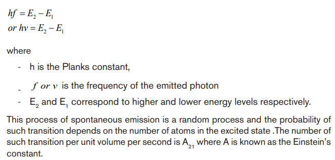
Stimulated emission occurs when a photon strikes an atom that is in excited state
and makes the atom emit another photon
In stimulated emission (Fig. 6.3), each incident photon encounters a previously
excited atom. A kind of resonance effect induces each atom to emit a second
photon with the same frequency, direction, phase, and polarization as the incident
photon, which is not changed by the process. For each atom there is one photon
before a stimulated emission and two photons afterthus the name light amplification.
Because the two photons have the same phase, they emerge together as coherentradiation.
6.1.2 Population inversion
Population inversion: This is the process of increasing excited electrons inhigher energy levels.
Normally, most atoms are in ground state, so most of the incident energy or photon
will be absorbed. To achieve a coherent light from stimulated emission, there are
some conditions that should be satisfied.
In the first case, the atoms must be excited to the higher state and so an inverted
population is produced. One in which more atoms are in upper state than in lower
one.
The emission of photons will dominate over absorption.
In the second case, the higher state must be a metastable state. This is a state
in which the electrons remain longer than usual so that transition to the lower stateoccurs by stimulated emission rather than spontaneously.
This is the redistribution of atomic energy levels that takes place in a system so that
laser action can occur.
There are different methods of achieving population inversion in atomic states that
is essential requirement to produce laser beam.
Normally, most of the atoms in a medium are in the ground state of energy There
There
are four different methods of making these atoms to get to excited states:
i) Excitation with the help of photons: If the atoms are exposed to an
electromagnetic radiation of high frequency, then there is selective absorption
of energy and thus atoms are raised to excited state.
ii) Excitation by using electrons: This method is used in some gas lasers.
Electrons are released from the atoms due to high voltage electric discharge
through a gas. These electrons are then accelerated to high velocities due to
high electric field inside a discharge tube. When they collide with neutral gasatoms, a fraction of these atoms are raised to excited state.
iii) Inelastic collision between atoms: If a gas contains two different kinds
of atoms X and Y, then during electric discharge through the gas some of the
atoms are raised to excited state.
iv) Excitation by chemical energy: Sometimes, an atom or a molecule can
be a product of a chemical reaction and can be produced in its excited state.
An example is hydrogen combining with fluorine to form hydrogen fluoride HFthat is in excited state.
6.1.3 Laser structure
In general case laser system consists of three important parts: Active medium or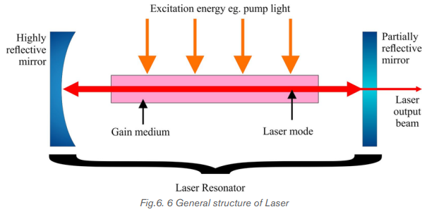
amplifying medium, the energy source referred as the pump or pump source
and the optical resonator consisting of mirrors or system of mirrors.
Pumping Mechanism
Pumping is the process of supplying energy to the laser medium to excite to the
upper energy levels. To have this mechanism, it depends on the existence of
interactions between light from pump source to constituents of active medium.
Usually, pump sources can be: electrical discharges, flash lamps, arc lamps, light
from another laser, chemical reactions and even explosive devices. Most common
lasers use electrical or optical pumping. The type of pump source used dependsessentially on the gain medium.
Active Medium
The active medium is the major determining factor of the wavelength of operation,
and other properties of the laser. The gain medium is excited by the pump source
to produce a population inversion, and it is where the spontaneous and stimulated
emission of photons take place, leading to the phenomenon of optical gain or
amplification. The gain medium may be a solid crystal like a ruby, a liquid dye, gases
like CO2 or He-Ne or semiconductors. The gain medium for some lasers like gas
lasers is closed by a window under the Brewster’s angle to allow the beam to leave
the laser tube.
Optical resonator or Optical cavity
The optical resonator or optical cavity is a system of two parallel mirrors placed
around the gain medium that provide reflection of the light beam. Light from the
medium produced by the spontaneous emission is reflected by the mirrors back
into the medium where it may be amplified by the stimulated emission. Mirrors are
required for most lasers to increase the circulating power within the cavity to the
point where gains exceed losses, and to increase the rate of stimulated emission.
One of the mirrors reflects essentially 100% of the light, while the other less than
100% and transmits the remainder. Mirrors can be plane, spherical or a combinationof both. Here represented are the common cavities configuration that can be used:
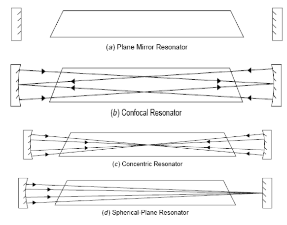
Application activity 6.
1. Write in full the acronym L.A.S.E.R
2. What do you understand by the term LASER?
3. In your own words, explain how laser light is produced.
4. Explain the meaning of population inversion and discuss how an atom
can be put into excited state.
5. a) What are the three major components of laser?
b) Using diagrams, explain all the types of optical cavity.
6. With the aid of diagrams explain the meaning of the following terms as
applied in LASERs
i) Stimulated Absorption
ii) Stimulated Emission
iii) Spontaneous Emissioniv) Population inversion
6.2 PROPERTIES OF LASER BEAM
Activity 6.2
Lasers emit light that is highly directional. Laser light is emitted as a relatively
narrow beam in a specific direction. Ordinary light, such as coming from the
sun, a light bulb, or a candle, is emitted in many directions away from the
source.
a) By analyzing how a laser pointer works, discuss characteristics of laser
light?
b) Do you think all different kinds of laser light have the same properties?Give reasons to support your answer.
The laser light is not like any other light emitted by usual sources found in nature. This
special light emitted by the laser, has three properties according to its usefulness inmany applications: Coherence, Monochromaticity and Collimation or Directionality.

6.2.1 Coherence
Coherence is the most interesting property of laser light. All photons emitted, are
exactly in the same phase, they are all crest and valley at the same time. It is
brought about by the mechanism of the laser itself in which photons are essentially
copied.
The good temporal coherence is essentially for Interferometry like in Holography.
Coherence is not trivial and is brought about by the amplification mechanism of the
laser.
6.2.2 Monochromaticity
Monochromaticity is the ability of the laser to produce light that is at one wavelength
λ. It is a requirement for coherence since photons of different wavelengths cannot
be coherent. When white light is dispersed through a prism, you note that it is
composed of an infinite number of wavelengths of light covering the entire visible
spectrum as well as into the UV and IR regions. However, no light source is perfectly
monochromatic. Lasers tend to be relatively monochromatic and this depends on
the type of laser. Monochromatic output, or high frequency stability, is of great
importance for lasers being used in Interferometry.
6.2.3 Collimation or Directionality
Collimation or directionality is the property of laser light that allows it to stay in one
direction at the strait line, confined beam for large distances. This property makes
it possible to use the laser as a level in construction or to pinpoint speeders on a
highway. This highly directional laser light is determined by the mechanism of thelaser itself.
Application activity 6.2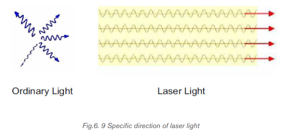
1. Choose the correct group of terms that are properties of laser light.
A. Coherent, unpolarized, monochromatic, high divergence
B. Monochromatic, low divergence, polarized, coherent
C. Polychromatic, diffuse, coherent, focused
D. Monochromatic, birefringent, nonpolarized, coherent
2. Which of the following properties of laser light enables us to use it to
measure distances with great precision?
A. All the light waves emitted by the laser have the same direction
B. The light waves are coherent
C. The light waves are monochromatic
D. The individual waves effectively work like a single wave with very
large amplitude.
3. Explain how coherence, monochromatic and collimation are
interconnected.
4. All light in laser light are produced and found to be in the same phase.
How does this help in the formation of 3D images?
5. Laser light can be used as a level. Which special feature that makes it
be used as a level. Explain your answer
6. Explain the meaning of the following properties of LASERS
i) Monochromaticity
ii) Coherenceiii) Directional/collimation.
6.3 APPLICATIONS AND DANGERS OF MISUSE OF LASER
Activity 6.3
You can use the figure above to answer the following questions
a) Starting from the figure indicated above, where do you think in real life
LASERS are helpful?
b) From your experience, have you ever used LASER light?
c) Other than using it by yourself, what are other places where laser light isapplied
6.3.1 Applications of lasers.
There are many interesting uses for lasers, depending on the special characteristic
being applied. Laser Diodes are used in a wide range of applications. Partial lists of
those applications include:
i) They are used in common consumer devices such as DVD players, bar code
scanners; CD ROM drivers; laser disc and other optical storage drivers; laser
printers and laser fax machines; sighting and alignment scopes; measurement
equipment; free space communication systems; pump source for other
lasers; high performance imagers; and typesetters. CD players have lasers.
Light from the laser in CD player reflects off patterns on CD’s surface. The
reflected light is converted to a sound wave.
ii) Laser beams can be used in diverse fields of science and technology. Like in
the control of motion of moving objects like aircrafts or missiles. This method
thus makes it possible for a missile to hit a certain target.
iii) Because of high directional property, lasers are used to measure distances
accurately. A laser beam is sent, and the time taken for it to be reflected back
is measured. Using this idea, the distance can thus be measured.
iv) Because laser beam can be focused into a small spot, it can thus be used to
cut minute holes onto a material.
v) The very high intensity of laser beam means that the amplitude of the
corresponding electromagnetic wave is very large. So it is possible to
investigate the nonlinear optical properties of different materials with the help
of laser light.
vi) Lasers are also used in industry for cutting materials such as metal and cloths.
and welding materials
vii) Doctors use lasers for surgery and various skin treatments
viii) They are used in military and law enforcement devices for marking targets
and measuring range and speed.
ix) Laser lighting displays use laser light as an entertainment medium.
x) Lasers also have many important applications in scientific research
In a tabular way, we can have a summary of different types of lasers and their
applications.The following are types of lasers and their applications
a. Gas Lasers:
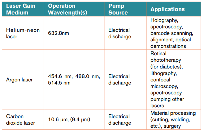
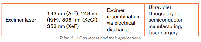
b. Solid State Lasers:

c. Metal-vapor Lasers:
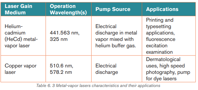
d. Other types of lasers:
6.3.2 Dangers of lasers
You should be careful when dealing with lasers, because they can have a negative
impact when exposed to your body. Among other negative effects, some of them
are discussed below
i) If directly exposed to our skin, it burns the skin
ii) When absorbed by skin, Laser light reacts with body cells causing cancer.
iii) Because of their high energy, it affects eyes if exposed to them
iv) Lasers can affect cells of a human being. This leads to mutation
Because of the negative effects of lasers, care must be taken to avoid all the risks
of being affected by lasers.
6.3.3 Precaution measures to avoid negative effects of lasers
The following are some of the measures one can take to avoid the negative effects
of lasers.
i) For any one working in places where there are incidences of being exposed
to laser light, one should wear protective clothes, glasses and shoes so that
there is no direct exposure of these radiations on to the body.
ii) One should minimize the time of working with lasers.
iii) Areas that are exposed to these radiations should be warning signs and labels
so that one can be aware of places/areas where laser light is used.
iv) Safe measures like Use of remote control should be used to avoid direct
exposure of these radiations (LASER light).
v) People should be given trainings on how to handle lasers.vi) There should also access restrictions to laboratories that use laser
Application activity 6.3
1. i) Discuss applications of Laser Light in daily life
ii) Though LASER light is important in day today life but if miss-used it
can be dangerous to our lives. Discuss dangers that may arise if it is
miss-used.
iii Using your personal judgment, which side outweighs the other? Give
scientific reasons.
3. Depending on your judgment in (2) do you think man should continue
using laser light?
4. Lasers are classified depending on either how they are produced or the
material that makes up laser. Discuss
i) Types of lasersii) Examples of lasers.
Skills Lab 6
This activity intends to make you analyze how LASER light is produced and
used.
What to do?
Visit a nearby hospital Laboratory where they do LASER surgery.
Note: Before you make a visit make sure that you inform them like a week
before through class leaders or your physics tutor.
In your visit focus on the following:
a. How laser light is produced
b. How LASER beam is used to do surgery.
c. Why do they adopt using this type of surgery but not other forms?For each case compile out your findings and a full report about lasers.
End of unit 6 assessment
Copy the questions below to your exercise books and chose the best
alternative that answers the question.
1) What does the acronym LASER stand for?
A. Light Absorption by Stimulated Emission of Radiation
B. Light Amplification by Stimulated Emission of Radiation
C. Light Alteration by Stimulated Emission of Radiation
2) The acronym MASER stands for?
A. Microwave Amplification by Stimulated Emission of Radiation
B. Molecular Absorption by Stimulated Emission of Radiation
C. Molecular Alteration by Stimulated Emission of Radiation
D. Microwave amplification by Stimulated Emission of Radio waves
3) Which of the following is true about population inversion
A. Number of electrons in excited levels reduces
B. Number of electrons in excited levels increase
C. Number of electrons in ground levels reduces
D. Electrons remain completely in ground state
4) Which of the following determines the color of light?
A. its intensity
B. its wavelength
C. its source
D. Some information missing
5) Among the three examples of laser listed below, which one is considered
“eye safe”?
A. Laser bar-code scanners
B. The excimer laser
C. Communications lasers
D. YAG
6) Why are lasers used in fiber optic communications systems?
A. The government has mandated it
B. They can be pulsed with high speed data
C. They are very inexpensive
D. They are not harmful
7) Lasers are used in CDs and DVDs. What type of laser is used in these
players?
A. Semiconductor
B. YAG
C. Alexandrite
D. All the above
8) The best reason why lasers used in “Laser Printers” is
A. They can be focused down to very small spot sizes for high resolution
B. They are cheap
C. They are impossible to damage
D. They are locally available
9) As wavelength gets longer, the laser light can be focused to…
A. Larger spot sizes
B. Smaller spot sizes
C. Large and small spot sizes
D. None of the above
10) Among the following, which color of laser has the shortest wavelength?
A. Yellow C. Blue
B. Red D. Green
11) What property of laser light is used to measure strain in roadways?
A. Intensity
B. Power
C. Coherence
D. All the above
12) What is the type of laser used most widely in industrial materials
processing applications?
A. Dye Laser C. YAG laser
B. Ruby Laser D. Carbon Dioxide Laser
13) Why are lasers used for cutting materials?
A. It never gets dull D. It has a small “heat affected zone”
B. Accuracy E. Smoother cuts
C. Repeatability F. All of the above
14) The Excimer laser produces light with what wavelength?
A. Visible
B. Ultraviolet
C. Infrared
D. All the above.
15) Most lasers are electrically inefficient devices.
A. True
B. False
16) Chemical lasers use………………. to produce their beams.
A. Excessive amounts of electrical power
B. Small amounts of electrical power
C. No electrical power
D. Other lasers
17) What type of laser could cause skin cancer if not used properly?
A. Red semiconductor laser C. Blue semiconductor
B. Excimer laser D. YAG laser.
18) a) what do you understand by term LASER?
b) Depending on the nature and what laser is made of, Laser is classified
into different types. Discuss at least 5 types of lasers
19) a) Laser light have been employed in different areas. This has helped
man in solving different problems. What are some of the areas where
laser light is employed?
b) Though laser light is very important in different activities, it can alsocause harm if miss-used in what ways is laser light harmful?
UNIT 7: MEDICAL IMAGING
Key Unit Competence:Generate the processes in medical imaging.
Introductory activity
Years ago, the only way to get information from inside of human bodies was
through surgery. In modern medicine, medical imaging has undergone major
advancements and this ability to obtain information about different parts of the
human body and has many useful clinical applications.
Observe and interpret, clearly the pictures above and answer to the following
questions:
1. Describe the phenomena happening in each figures above (A, B, C, D
and E)
2. Propose the technique that is being used for each image?
3. Suggest a detailed working principle of the mentioned techniques in
each image
4. Are there other techniques that are not indicated in the figure above? If
yes, state and explain them.
5. In your own view, what do you think are the effects of these techniquesin general?
7.1. CONCEPT OF MEDICAL IMAGING.
Activity 7.1
Imagine you were in internship in a certain primary school in your district and
while teaching in P3, a pupil happens to swallow a 20 Francs coin. You and
fellow teachers gave her first aid and later your Head teacher assigned you to
take her to the nearest hospital.
a) In your own view, how do you think a doctor will be able to locate the
swallowed coin without operating her?
b) Explain using scientific reasoning, why a doctor needs to use method(s)you suggested in (a) above.
The technique and process of producing visual representations of the interior areas
inside the human body (function of some organs or tissues) to diagnose medical
problems and monitor treatment is known as Medical imaging.
There are many types of medical imaging, and more methods for imaging are being
invented as technology advances. The main types of imaging used in modern
medicine include
• Radiography.
• Mammography
• Magnetic resonance imaging.
• Nuclear medicine.
• Ultrasound.
• Endoscopy
• Elastography.
• Photoacoustic imaging.
• Tomography.
• Echocardiography, etc
In this unit, we shall focus on only the following: Radiography and
Mammography, Magnetic Resonance Imaging, Ultrasound and Endoscopy.
SPECIFIC PURPOSES OF IMAGING TECHNIQUES
Each technique is used in different conditions. For example:
• Ultrasound is used to study the development of fetus in the mother’s womb
and to take images of internal organs when high resolution is not needed.
• Radiography is often used when we want images of bone structures to look
for breakages.
• MRI scanners are often used to take images of the brain or other internal
tissues, particularly when high-resolution images are needed.
• Nuclear medicine is used when you need to look inside the digestive or
circulatory systems, such as to look for blockages. It uses radioactive materialsthat are injected or swallowed.
Application activity 7.1
1. Explain the meaning of medical imaging
2. Medical imaging being a new technique of examining internal parts of
a body under examination, has been emphasized and used in different
medical places. What are different methods of medical imaging you
know and where are they used?
3. Highlight the purpose(s) of each of the methods mentions in question2 above.
7.2 ULTRASONIC IMAGING
Activity 7.2
Mutesi is a young mother who was pregnant used to feel pain in her lower
abdomen and she happened to go to the hospital for medical checkup. As she
reached the hospital, she underwent a medical testing and her doctor referred
her to undergo ultrasound scan.
What she noticed was a doctor moving a piezoelectric crystal on her abdomen.At the end, she was given image of affected part.
a) As a physics student, explain why the technique used is regarded as
ultrasound?
b) From your understanding, how the emitted rays are used to capture the
image of intended part and feed it back to the computer.
c) If you were a doctor, would you advise someone to always visit ultrasound
scans every time he/she feels pain? If No explain why? If yes, defend youropinion.
7.2.1 Interaction of sound waves with different structure inside
the body
Ultrasound imaging uses ultra-high-frequency sound waves to produce cross
sectional images of the body. Ultrasound is actually sound with a frequency in
excess of 20 kHz, which is the upper limit of human hearing. The frequency range
of normal person hearing is between 20 Hz to 20 KHz. In fact, the frequencies
used in medicine are much higher than this, typically between 1 MHz and 15 MHz
like all sound waves, ultrasound consists of longitudinal, elastic or pressure waves,
capable of traveling through solids, liquids and gases. This makes them ideal for
penetrating the body, unlike transverse mechanical waves, which cannot travel to
any great extent through fluids.
Different tissues in a human or animal body alter the ultra sound waves in different
ways. Some waves are reflected directly while others scatter the waves before they
return to the transducer as echoes. The reflected ultrasound pulses detected by
the transducer need to be amplified in the scanner or ultrasonic probe. The echoes
that come from deep within the body are more attenuated than those from the more
superficial parts and therefore required more amplification. When echoes return to
the transducer, it is possible to reconstruct a two-dimensional map of all the tissues
that have been in the beams. The information is stored in a computer and displayed
on a video (television) monitor. Strong echoes are said to be of the high intensity
and appear as brighter dots on the screen.
Normally in medicine, Doctors commonly use ultrasound to study a developing
fetus (unborn baby), a person›s abdominal and pelvic organs, muscles and tendons,or their heart and blood vessels
7.2.2 Reflection of ultrasound
When the pulse of ultrasound is sent into the body and meets a boundary between
two media, of different specific acoustic impedance Z, the sound wave needs to
change gear in order to continue. If the difference in Z across the boundary is large
the wave cannot easily adjust: there is an “acoustic mismatch”. Most of the waveis reflected and a strong echo is recorded. The fraction of intensity reflected back
 at the normal incidence, is known as the intensity of
 reflection coefficient α
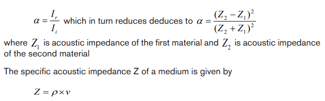
Where ρ is the density of the medium and v is he velocity of sound in medium.

Table 7. 1 Values ρ,v and Z for various substances
Note that large differences in Z give rise to large values for intensity of reflectioncoefficient (α), producing strong echoes.
7.2.3 Attenuation of ultrasound
The combined effect of scattering and absorption is called attenuation. The
attenuation of the waves describes the reduction in its intensity as they travel
through a medium. This loss is due to a number of factors:
• The wave simply “spreads out” and suffers an “inverse square law type”
reduction in intensity.
• The wave is scattered away from its original direction
• The wave is absorbed in the medium.
The amount of absorption of ultrasound beam in a medium is described by the
absorption coefficient, which is intensity level per unit length. It is expressed in
decibels per cm and it firstly depends on the type of medium the wave is propagating
into. As example whilst water absorbs very little ultrasound, bone is a strong
absorber, putting it at risk, for example, during high- power ultrasound therapy.
Secondly, higher frequencies suffer greater absorption. In fact if the frequency is
doubled, the absorption increases by the factor of four.
7.2.4 Ultrasonic imaging techniques
The basic component of the ultrasound probe is the piezoelectric crystal. Excitation
of this crystal by electrical signals causes it to emit ultra-high-frequency sound
waves; this is the piezoelectric effect. The emitted ultrasound waves are reflected
back to the crystal by the various tissues of the body. These reflected sound waves
also called the “echoes” act on the piezoelectric crystal in the ultrasound probe to
produce an electric signal, again by the piezoelectric effect. It is this electric signal
which is analysed by a computer produces a cross-sectional image.
The process of imaging is the same as the echo-locating sonar of a submarine or
a bat. The observer sends out a brief pulse of ultrasound and waits for an echo.
The pulse travels out, reflects off the target and returns. The ultrasound machine
uses pulses because the same device acts as both transmitter and receiver. If it
continually sent out sounds, then the receiver would not hear the much softer echoover the louder transmission.
Sonar is an acronym for Sound Navigation and Ranging. It relies on the reflection of
ultrasound pulses. A short pulse of ultrasound is directed towards the object interest,
which then reflects it back as an echo. The total time between transmission of pulse
and reception of an echo is measured, often using a cathode ray oscilloscope(CRO). The sonar principle is used to estimate the depth of a structure, using
Where t is the time taken to go and back and is the velocity of ultrasound in the
medium.
The factor of 2 is necessary because the pulse must travel “there and back”
An ultrasound beam structure is directly into the body. The reflection or echoes
from different body structure are then detected and analyzed, yielding information
about the locations. For example if the time delays between the reception of echo
pulse1 and 2 (Figure below) is, then the diameter of the baby’s head can befound using the above formula.
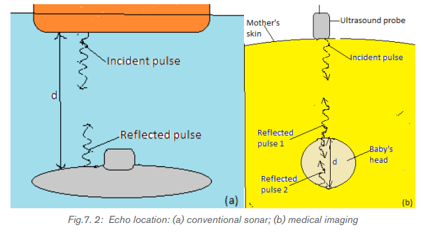
As ultrasound imaging uses sound waves to produce pictures of inside of the body.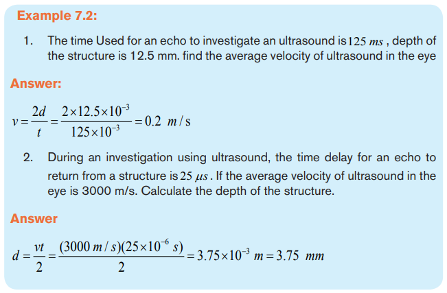
It is used to help diagnose the cause of pain, swelling and infection in the body’s
internal organs and to examine a baby in pregnant woman and the brain and hips in
infants. It is also used to help guide biopsies, diagnose heart conditions and assess
damage after a heart attack.
Ultrasound examinations do not use ionizing radiation (x-rays), there is no radiation
exposure to the patient. Because ultrasound images are captured in real time, they
can show the structure and movement of the body’s internal organs, as well asblood flowing through blood vessels.
7.2.5 Risk and benefit associated to ultrasounds
Ultrasound imaging uses high-frequency sound waves, not used x-ray and no
radiation exposure to the patient does not mean the at ultrasound is mostly safe. It
has some dangers.
Some of them are:
• Cannot penetrate bone, so the adult skeletal system and head cannot be
imaged.
• Clarity of image is poorer than in many other techniques.
• It cannot be used in areas that contain gas (such as lungs)
• Scan can take a long time and demand greater skills and experience to
produce a clear result.Etc.
Apart from its dangers, ultrasound is helpful because:
No known harmful effects of diagnostic ultrasound.
Clear examination of soft tissues, e.g.Obstetric and abdomen studies
More cost effective than other imaging modalities
Real time imaging means required quick procedure.
It is noninvasive (of medicine procedures not involving the introduction of instruments
into the body)
• Lack of ionizing radiation.• Equipment is safe, easy to handle, can be operated and be portable. Etc
Application activity 7.2
1. Calculate the percentage of incidence intensity reflected back at:
a) Air per soft tissues boundary
b) Bone per softy tissues boundary.
2. Discuss on the purpose of using ultrasound in medicine
3. Outline the application of ultrasound scan?4. Why ultrasound is performed
7.3 X-RAY IMAGING.
Activity 7.3
1. The figure below is what a Doctor got after X-rays scan in order tocheck a problem that was suspected to be in ribs.
a) Using the picture above, how do you think the doctor was able to get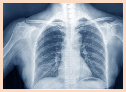
the image
b) Why do you think devices like cameras cannot give such images?
2. One day a girl suffering from the breast tells her mother about the
problem. And her mother advises her to go to the hospital to consult
a doctor. After examined by the doctor, she was referred to the X-ray
room for further checking.
a) Imagine you are the doctor operating the X-ray device, explain all that
you can do to detect the problem a girl had.
b) There are many methods of X-ray imaging techniques that may be
used. Can you suggest one that can be used to examine breastproblems? Defend your suggestion.
7.3.1 Interaction of X-rays with matter.
a. Introduction
In unit 5, we learnt that X-rays are electromagnetic radiation produced by focusing
a beam of high energy electron on a target material in x-ray tube. Since the major
part of the energy of the electrons is converted into heat in the target (only about
1% will appear as X-rays), the target material should have a high melting point and
good heat conduction ability. To get a high relative amount of X-ray energy, the
anode material should be of high atomic number. Tungsten is the dominating anodematerial and is in modern X-ray tubes often mixed with Rhenium.
In X-ray diagnostics, radiation that is partly transmitted through and partly
absorbed in the irradiated object is utilized. An X-ray image shows the variations
in transmission caused by structures in the object of varying thickness, density or
atomic composition.
b. Attenuation and Absorption of X-rays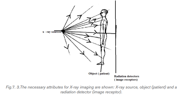
There are principally two interaction processes that give rise to the x-ray attenuation
(variation in photon transmission) through the patient which is the basis of X-rayimaging. These are photoelectric absorption and scattering processes.
A photon which has experienced an interaction process has either been absorbed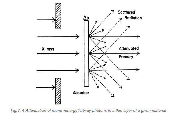
or has changed its energy and/or direction of motion. A photon that changes its
direction of motion is called a scattered photon. For mono-energetic x-ray photons,
the number of photons that experience such interactions and therefore removed
from the primary beam when this is incident on a thin layer of material is proportional
to the number of incident photons (N) and the thickness of the layer (dx) followingthe expression :
where µ is a constant of proportionality called the linear attenuation coefficient.
Integrating the above equation will result in
Where
 is the initial number of photons in the incident beam.
It can be seen that the incident beam photons (or the beam energy) is attenuated
exponentially as the x-rays travel through the material. The different interaction
processes involved, that are absorption, coherent and incoherent scattering andpair production, add their contributions to the total linear attenuation coefficient
Where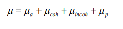
 and µ p are the contributions to the attenuation from
photoelectric absorption, coherent scattering, incoherent scattering and pairproduction.
7.3.2 X-rays Imaging Techniques
a. Conventional Radiography
X-rays are able to pass through the human body and produce an image of internal
structures. The resulting image is called a radiograph, more commonly known
as an ‘X-ray’ or ‘plain film’. The common terms ‘chest X-ray’ and ‘abdomen
X-ray’ are widely accepted and abbreviated to CXR and AXR.
As a beam of X-rays passes through the human body, some of the X-rays photons
are absorbed or scattered producing reduction or attenuation of the beam with the
internal human structure acting as contrasting details. Therefore, tissues of high
density and/or high atomic number cause more X-ray beam attenuation and are
shown as lighter grey or white on radiographs. Less dense tissues and structures
cause less attenuation of the X-ray beam, and appear darker on radiographs
than tissues of higher density. The figure below shows the typical conventionalradiograph of a human body.
Five principal densities are easily recognized on this plain radiograph due to the
increase in their densities:
• Air/gas appears as black, e.g. lungs, bowel and stomach
• Fat is shown by dark grey, e.g. subcutaneous tissue layer, retroperitoneal fat
• Soft tissues/water appears as light grey, e.g. solid organs, heart, blood
vessels, muscle and fluid-filled organs such as bladder
• Bone appears as off-white
• Contrast material/metal: bright white.
In the past, X-ray films were processed in a darkroom or in freestanding daylight
processors. In modern practice, radiographic images are produced digitally using
one of two processes, computed radiography (CR) and digital radiography (DR).
DR uses a detector screen containing silicon detectors that produce an electrical
signal when exposed to X-rays. This signal is analyzed to produce a digital image.
Digital images obtained by CR and DR are sent to viewing workstations for
interpretation. Images may also be recorded on X-ray film for portability and remoteviewing.
The image given by a computer radiography may be reviewed and reported on
a computer workstation. This allows various manipulations of images as well as
application of functions such as measurements of length and angles measurements.
The relative variance of the shadows depends upon the density of the materials
within the object or body part. Dense, calcium – rich bone absorbs X-rays to a
higher degree than soft tissues that permit more X-rays to pass through them,
making X-rays very useful for capturing images of bone.
In projection radiography, there is much room for adjusting the energy level of the
X-rays depending on the relative densities of the tissues being imaged and also
how deep through a body the waves must travel in order to achieve the imaging.
• Images of bones (for instance, to examine a fracture or for diagnostic measures
related to bone conditions like osteoarthritis or certain cancers) require high
energy X-rays because of the high density of bone.
• Images of soft tissues like lungs, heart and breasts (both chest X-rays and
mammography are very common diagnostic applications of X-rays) require
relatively less energy from the X-rays in order to penetrate properly and
achieve excellent images.
• In order to achieve these different energies, technologists use X-ray generatorsof different voltages and equipped with anodes made of different metals.
Advantages and disadvantages of conventional radiography.
b. Mammography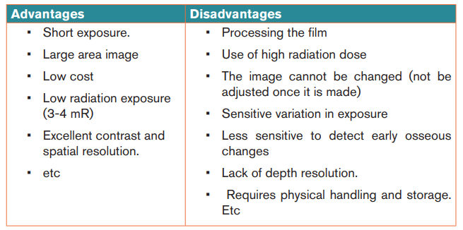
Mammography is a specialized medical imaging that uses low-dose X-rays to
investigate the internal structure of the breast. A mammography exam, called a
mammogram, helps in the early detection and diagnosis of women’s breast diseases
such as breast cancer before even experiencing any symptom. Below is a typical
mammography test showing the presence of abnormal areas of density, mass, orcalcification that may indicate the presence of cancer.
A mammography unit is a rectangular box that houses the tube in which X-rays are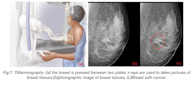
produced. The unit is used exclusively for X-ray exams of the breast, with special
accessories that allow only the breast to be exposed to the X-rays. Attached to the
unit is a device that holds and compresses the breast and positions it so imagescan be obtained at different angles.
In conventional film and digital mammography, a stationery X-ray tube
captures an image from the side and an image from above the compressed breast.
Breast tomosynthesis, also called three-dimensional (3-D) mammography and
digital breast tomosynthesis (DBT), is an advanced form of breast imaging where
multiple images of the breast from different angles are captured and reconstructed
(“synthesized”) into a three-dimensional image set. In this way, 3-D breast imaging
is similar to computed tomography (CT) imaging in which a series of thin “slices”
are assembled together to create a 3-D reconstruction of the body.
Advantages and disadvantages of mammography
Advantages are:
• Non-invasive procedure
• Minimum hazard of radiation.
• Increase in cancer detection rate.
• Improved positive predictive values for recall and biopsy.
• Etc.
Disadvantages are:
• May increase radiation dosage patient receivers.
• May require new equipment / training for techs and radiologist
• Is inconclusive in women under 35 years old due to dense breast tissue.
• Etc.
c. Computer Tomography scan (CT scan)
i) CT terminology
In 1970s, a revolutionary new X-ray technique was developed called Computer
tomography (CT), which produce an image of a slice through the body. The word
tomography comes from the Greek: tomos =slice, graph= picture.)
A computed tomography scan also known as CT scan, makes use of computer
processed combinations of many X-ray measurements taken from different angles
to produce cross-sectional (tomographic) images (virtual “slices”) of specific areas
of a scanned object, allowing the user to see inside the object without cutting it.
Other terms include computed axial tomography (CAT scan) and computer aided
tomography
The term “computed tomography” (CT) is often used to refer to X-ray CT, because
it is the most commonly known form but many other types of CT exist.
CT is an imaging technique whereby cross-sectional images are obtained with the
use of X-rays. In CT scanning, the patient is passed through a rotating gantry that
has an X-ray tube on one side and a set of detectors on the other. Information from
the detectors is analysed by computer and displayed as a grey-scale image. Owing
to the use of computer analysis, a much greater array of densities can be displayed
than on conventional X-ray films. This allows accurate display of cross-sectional
anatomy, differentiation of organs and pathology, and sensitivity to the presence of
specific materials such as fat or calcium. As with plain radiography, high- density
objects cause more attenuation of the X-ray beam and are therefore displayed aslighter grey than objects of lower density.
ii) Principle behind of computer tomography scan (CT scan).
Computer Tomography is shown in below figure: a thin collimated beam of X- ray (“
to collimate” means to “make straight”) passes through the body to a detector that
measures the transmitted intensity. The apparatus is rotated slightly about the body
axis and again scanned; this is repeated at . The intensity of the
transmitted beam for the many points of each scan, and for each angles, are sent
to a computer that reconstructs the image of the slice. Note that the imaged slice is
perpendicular to the long axis of the body. For this reason, CT is sometimes calledcomputerize axial tomography.
The use of single detector would require a few minutes for many scans needed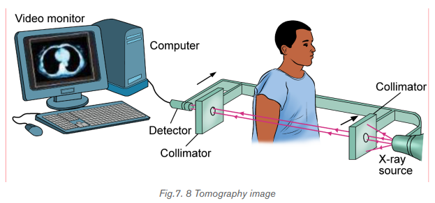
to form a complete image. Much faster scanner use a fan beam in which passing
through the entire cross section of the body are detected simultaneously by many
detectors. The x-ray source and the detectors are rotated about the patient and
an image requires only few seconds to be seen. This means that rays transmitted
through the entire body are measured simultaneously at each angle where thesource and detector rotate to take measurements at different angles.
CT images of internal organs, bones, soft tissue, and blood vessels provide greater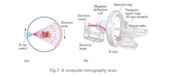
clarity and more details than conventional X-ray images, such as a chest X-Ray.
iii) Function of CT scan
• A motorized table moves the patient through a circular opening in the CT
imaging system.
• While the patient is inside the opening, an X-ray source and a detector
assembly within the system rotate around the patient. A single rotation
typically takes a second or less. During rotation the X-ray source produces
a narrow, fan-shaped beam of X-rays that passes through a section of the
patient’s body.
• Detectors in rows opposite the X-ray source register the X-rays that pass
through the patient’s body as a snapshot in the process of creating an image.
Many different “snapshots” (at many angles through the patient) are collected
during one complete rotation.
• For each rotation of the X-ray source and detector assembly, the image data
are sent to a computer to reconstruct all of the individual “snapshots” into one
or multiple cross-sectional images (slices) of the internal organs and tissues.
Note that, it is advisable to avoid unnecessary radiation exposure; a medically
needed CT scan obtained with appropriate acquisition parameter has benefits that
outweigh the radiation risks.
iv) Advantages and disadvantages of CT-Scan
CT-Scan has Advantages and disadvantages.
Disadvantages include:
• Risk to the patient because of the high radiation dose.
• Very expensive.
• Not commonly used to image painful joints modality
• Poor soft-tissue contrast.
• Higher radiation exposure.
• Involves exposure to ionising radiation(gamma-rays)
• Radiation material may cause allergic injection-site reactions in some people.
• etc
Advantages include:
• Images can be scored in a computer memory.
• The computer can also be used to construct a slide in a different plane using
other visual data.
• Widely available
• Quick exam.
• CT-Scan give a good contrast images
• High spatial resolution (bone/lung).
• Unlike most other imaging types, can show how different parts of the body
are working and can detect problem earlier.
• Can check how far a cancer has spread and how well treatment is working.• etc
Application activity 7.3
1. Outline the advantages and disadvantages of CT scan
2. Explain the types of x-ray imaging used in mammography.
3. In mammography exams, is the breast compression necessary? Why
4. A beam of X-rays passes through the human body of tissues with
different densities; explain the production of X-rays on less dense
tissues?
5. If you are getting a mammogram for the first time, what are the specific
questions you are expected to be asked by a doctor.
6. What does a biopsy mean?
7. Explain reasons why people do not attend breast screening (screeningmammography)
7.4 ENDOSCOPY
Activity 7.4
The picture below show the procedure that enables doctor to examine the
lining of esophagus and stomach. Examine it well and answer the followingquestion.
1. Name parts labelled letters A, B, C and D.
2. How do you call the examination technique taken by a doctor?
3. How can we examine inside the stomach by using light rays?
4. How is endoscopy performed?
5. What do you think are the advantages and disadvantages of thistechnique?
7.4.1 Description
Endoscopy is a nonsurgical procedure used to examine a person’s digestive tract.
Using an endoscope, which is a flexible tube with a light and camera attached to
it, the specialist can view pictures of your digestive tract on a monitor. During an
upper endoscopy, an endoscope is easily passed through the mouth and throat
and into the esophagus, allowing the specialist to view the esophagus, stomach,
and upper part of the small intestine. Similarly, endoscopes can be passed into thelarge intestine (colon) through the rectum to examine this area of the intestine.
7.4.2 Upper endoscopy
Upper Endoscopy (also known as gastroscopy, EGD, or
esophagogastroduodenoscopy) is a procedure that enables your surgeon to
examine the lining of the esophagus (swallowing tube), stomach and duodenum
(first portion of the small intestine). A bendable, lighted tube about the thickness ofyour little finger is placed through your mouth and into the stomach and duodenum.
How is the upper endoscopy performed?
Upper endoscopy is performed to evaluate symptoms of persistent upper abdominal
pain, nausea, vomiting, difficulty swallowing or heartburn. It is an excellent method
for finding the cause of bleeding from the upper gastrointestinal tract. It can be
used to evaluate the esophagus or stomach after major surgery. It is more accurate
than X-rays for detecting inflammation, ulcers or tumors of the esophagus, stomach
and duodenum. Upper endoscopy can detect early cancer and can distinguish
between cancerous and noncancerous conditions by performing biopsies ofsuspicious areas.
A variety of instruments can be passed through the endoscope that allows the
surgeon to treat many abnormalities with little or no discomfort, remove swallowed
objects, or treat upper gastrointestinal bleeding. Safe and effective control ofbleeding has reduced the need for transfusions and surgery in many patients.
7.4.3 Advantages and disadvantages of endoscopy
Advantages
• Complete visualization of the entire stomach or digestive tract.
• It is very safe and effective tool in diagnosis
• Does not leave any scar because it uses natural body openings.
• It is cost effective and has low risk
• They are generally painless.
• Can do therapeutic interventions
• Allows for sampling/biopsying of small bowel mucosa
• Allows for resection of polyps.
Overall, endoscope is very safe; however, the procedure has a few potential
complications which may include:
- Bleeding
- Perforation (tear in the gut wall)
- Infection
- Reaction to sedation (action of administering a sedative drug to produce a
state of calm or sleep.
- Technically difficult procedure
- Very time consuming (Procedure can take > 3 hours)
- Patient may need to be admitted to the hospital
- Higher risk of small bowel perforation
- Case reports of pancreatitis and intestinal necrosis
- Reported incidents of aspiration and pneumonia
Application activity 7.4
1. What are instruments used to view the oesophagus, stomach and
upper small intestine of human
2. Discuss different functions of endoscope in medicine.
3. Compare and contrast colonoscopy and gastroscopy4. What are some of negative effects of using endoscopy?
7.5 MAGNETIC RESONANCE IMAGING (MRI)Activity 7.5
i) The diagram above is a Magnetic Resonance Imaging (MRI) machine.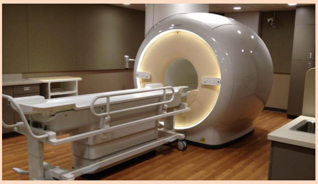
Basing on its name, explain what it does.
ii) Comparing it to other imaging techniques, explain how this machine is
different from other imaging machines.
iii) Would you advise a pregnant woman to always use this machine for a
medical checkup? Explain your view
iv) From your reasoning in iii) above, suggest advantages and disadvantagesof using MRI machine
Historically, Magnetic Resonance Imaging as with all medical imaging techniques, isa relatively new technology with its foundations beginning during the year of 1946.
Felix Bloch and Edward Purcell independently discovered the magnetic resonance
phenomena during this year, Up until the 1970s MRI was being used for chemical
and physical analysis. Then in 1971 Raymond Damadian showed that nuclear
magnetic relaxation times of tissues and tumors differed motivating scientists to
use MRI to study disease. MRI began in the central nervous system, but it has
now extended to all regions of the body. It is involving three very complex topics in
physics like: Nuclear, Magnetic and Resonance (NMR). In MRI, a particular type of
nucleus is selected and its distribution in the body is monitored. Hydrogen is the
most commonly imaged element, not only due to its abundant in the body, but also
because it gives the strongest MRI signal.
7.5.1. Concepts MRI
Magnetic Resonance (MRI) Imaging has become the dominant clinical imaging
modality with widespread, primarily noninvasive, applicability throughout the body
and across many disease processes. The progress of MR imaging has been rapid
compared with other imaging technologies and it can be attributed in part to physics
and in part to the timing of the development of MR imaging, which corresponded to
an important period of advances in computing technology.
Initially let us described how magnetic resonance can be demonstrated with a pair
of magnets and a compass. If a compass happens to find itself near a powerful
magnet, the compass needle will align with the field. In a normal pocket compass,
the needle is embedded in liquid to dampen its oscillations. Without liquid, the
needle will vibrate through the north direction for a period before coming to rest.
The frequency of the oscillations depends on the magnetic field and of the strengthof the magnetic needle.
Let us focus on what made the needle oscillate. It was the small movements of the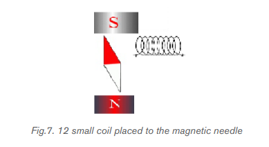
magnet, back and forth, or more precisely the oscillation of a weak magnetic field
perpendicular to the powerful stationary magnetic field caused by the movement
of the magnet. But oscillating magnetic field is what we understand by “radio
waves”, which means that in reality, we could replace the weak magnet with other
types of radio wave emitters. This could, for example, be a small coil subject to an
alternating current, as shown in figure above. Such a coil will create a magnetic
field perpendicular to the magnetic needle. The field changes direction in synchronywith the oscillation of the alternating current, so if the frequency of the current is
adjusted to the resonance frequency of the magnetic needle, the current will set
the needle in motion. This is also applied in an MR scanner. In summary, the needle
can be set in motion from a distance by either waving a magnet or by applying an
alternating current to a coil. In both situations, magnetic resonance is achieved
when the magnetic field that motion or alternating currents produce, oscillates at
the resonance frequency. When the waving or the alternating current is stopped,
the radio waves that are subsequently produced by the oscillating needle willinduce a voltage over the coil.
MRI uses the magnetic properties of spinning hydrogen atoms to produce images.
The first step in MRI is the application of a strong, external magnetic field. For this
purpose, the patient is placed within a large powerful magnet. Most current medical
MRI machines have field strengths of 1.5 or 3.0 Tesla. The hydrogen atoms within
the patient align in a direction either parallel or antiparallel to the strong external
field. A greater proportion aligns in the parallel direction so that the net vector of
their alignment, and therefore the net magnetic vector, will be in the direction of
the external field. This is known as longitudinal magnetization. A second magnetic
field is applied at right angles to the original external field. This second magnetic
field is known as the radiofrequency pulse (RF pulse), because it is applied at a
frequency in the same part of the electromagnetic spectrum as radio waves. A
magnetic coil, known as the RF coil, applies the RF pulse. The RF pulse causes
the net magnetization vector of the hydrogen atoms to turn towards the transverse
plane, i.e. a plane at right angles to the direction of the original, strong external field.
The component of the net magnetization vector in the transverse plane induces an
electrical current in the RF coil. This current is known as the MR signal and is the
basis for formation of an image. Computer analysis of the complex MR signal fromthe RF receiver coils is used to produce an MR image.
7.5.2. The magnetism of the body
Let‘s see how magnet needles with and without spin are affected by radio waves,
we now turn to the “compass needles” in our own bodies.
a) Most frequently, the MR signal is derived from hydrogen nuclei (meaning the
atomic nuclei in the hydrogen atoms). Most of the body’s hydrogen is found
in the water molecules. Few other nuclei are used for MR.
b) Hydrogen nuclei (also called protons) behave as small compass needles that
align themselves parallel to the field.
c) The compass needles (the spins) are aligned in the field, but due to movements
and nuclear interactions in the soup, the alignment only happens partially.
d) The nuclei in the body move among each other (thermal motion) and the net
magnetization in equilibrium is thus temperature dependent.
e) Due to the number of hydrogen nuclei (about 27 10 ) found in the body, the net
magnetization still becomes measurable. It is proportional to the field: A large
field produces a high degree of alignment and thus a large magnetization andbetter signal to noise ratio.
7.5.3 Magnetic Resonance Imaging (MRI).
The hydrogen nucleus is the most use in MRI.The technique uses a very powerful
magnet to align the nuclei of atoms inside the body, and a variable magnetic field that
causes the atoms to resonate, a phenomenon called nuclear magnetic resonance.
The nuclei produce their own rotating magnetic fields that a scanner detects and
uses to create an image.
MRI is used to diagnose a variety of disorders, such as strokes, tumors, aneurysms,
spinal cord injuries, multiple sclerosis and eye or inner ear problems. It is also widelyused in research to measure brain structure and function, among other things.
An MRI scan can be used to examine almost any part of the body, including the:
• brain and spinal cord
• bones and joints
• breasts
• heart and blood vessels
• internal organs, such as the liver, womb or prostate gland ,etc
The results of an MRI scan can be used to help diagnose conditions,
plan treatments and assess how effective previous treatment has been.
7.5.4. Functional of MRI Scan
There are many forms of MRI, some of them are:
a) Diffusion weighted imaging.
Diffusion-weighted imaging (DWI) is sensitive to the random Brownian motion
(diffusion) of water molecules within tissue. The greater the amount of diffusion,
the greater the signal loss on DWI. Areas of reduced water molecule diffusion
show on DWI as relatively high signal. Diffusion-weighted imaging is the
most sensitive imaging test available for the diagnosis of acute cerebral
infarction. With the onset of acute ischaemia and cell death there is increased
intracellular water (cytotoxicoedema) with restricted diffusion of water molecules.
An acute infarct therefore shows on DWI as an area of relatively high signal.
b) Perfusion-weighted imaging
In perfusion-weighted imaging (PWI) the brain is rapidly scanned following
injection of a bolus of contrast material (gadolinium). The data obtained may be
represented in a number of ways including maps of regional cerebral blood volume,
cerebral blood flow, and mean transit time of the contrast bolus. PWI may be used
in patients with cerebral infarct to map out areas of brain at risk of ischaemia that
may be salvageable with thrombolysis.
c) Magnetic resonance spectroscopy
Magnetic resonance spectroscopy (MRS) uses different frequencies to identify
certain molecules in a selected volume of tissue, known as a voxel. Following data
analysis, a spectrographic graph of certain metabolites is drawn. Metabolites of
interest include lipid, lactate, NAA (N-acetylaspartate), choline, creatinine, citrate
and myoinositol. Uses of MRS include characterization of metabolic brain disorders
in children, imaging of dementias, differentiation of recurrent cerebral tumour from
radiation necrosis, and diagnosis of prostatic carcinoma.
d) Blood oxygen level-dependent imaging
Blood oxygen level-dependent (BOLD) imaging is a non-invasive functional MRI
(fMRI) technique used for localizing regional brain signal intensity changes in
response to task performance. BOLD imaging depends on regional changes in
concentration of deoxyhemoglobin, and is therefore a tool to investigate regional
cerebral physiology in response to a variety of stimuli. BOLD fMRI may be used prior
to surgery for brain tumor or arteriovenous malformation (AVM), as a prognosticindicator of the degree of postsurgical deficit.
7.5.5 Advantage and disadvantages of MRI.
Advantages of MRI in clinical practice include:
1. Excellent soft tissue contrast and characterization
2. Lack of ionizing radiation.
3. Noninvasive machine.
4. Lack of artefact from adjacent bones, e.g. pituitary fossa
Disadvantages of MRI:
1. High capital and running costs.
2. Image selected and interpretation is complex.
3. Examination can be difficult for some people who are claustrophobic
4. The examination is noisy and takes long.
5. Hazards with implants, particularly pacemakers.6. Practical problems associated with large superconducting magnets.
In additional to that, out of imaging the techniques discussed above there other
imaging techniques called “radionuclide imaging/nuclear medicine/ scintigraphy”.
A radionuclide is used to collect in areas where there is a lot of bone activity.
This method uses gamma radiation to form images by injection of various
radiopharmaceuticals. The most commonly used radionuclide in clinical practice
is technetium, written in this text as where m stands for metastable.
Other commonly used radionuclides include gallium citrate
indium
Application activity 7.5
1. With clear explanations, explain the benefit and limitation of MRI
machine.
2. What is meant by relaxation in the context of MRI?
3. Give the reasons why the hydrogen nucleus is most used in MRI.
4. What does NMR stand for? Explain carefully the role of the three terms
involved
5. With the aid of drawing, discuss the basic steps in the formation ofMRI image.
Skills Lab 7
In this activity you will make a visit to the nearest hospital.
Note: Make inquiry either through your class leaders or class tutor to know
whether the hospital has the following.
• Ultrasound machine
• X-ray machine
• Endoscopy
• Magnetic Resonance Imaging (MRI) machine.
The main aim of the visit is to understand how these machines work.
The following are guiding questions you may ask either laboratory technician
or the doctor.
a. What is the main objective(s) of using these machines?
b. What does one need to do to use these machines?
c. What are the precautions that must be taken before using any of these
machines?
d. Are there regulations guiding any person working in the rooms where
these machines are installed?
e. Could there be negative effects of these machines on human body if
used regularly?
You can also ask any question you feel can make you understand this concept
better.
Make sure you note down something as the doctor or laboratory technician
explains the asked questions.
After leaving the hospital, Make a comprehensive report and compare the
information you got from the hospital to the one learnt in this unit.Present your findings (in the report) to your class and to your tutor.
End of unit 7 assessment
I-5: Choose the correct answer.
1. One of the medical imaging using X-ray is:
A. thermography
B. CT Scan
C. endoscopy
D. both of them
2. Magnetic Resonance Imaging uses:
A. x-rays
B. Light
C. Magnetization
D. both of them
3. The medical imaging techniques used injection of various
radiopharmaceuticals is:
A. Mammography
B. Radiography
B. Radionuclide.
D. Endoscopy
4. Mammography is used to detect:
A. Brain diseases
B. Baby diseases
C. Breast diseases
D. None of them
5. A radionuclide scan may be done for one reason:
A. A radionuclide is used to collect the areas where the infrared are
synchronized.
B. A radionuclide is used to collect in areas where there is a lot of bone
activity.
C. A radionuclide is use to collect the areas where gamma camera are
produced image
D. A radionuclide is used to collect information from the exam of lining
of esophagus.
6. Write the missing word or words on the space before each number.
A. The best human ears can respond to frequencies from about 20Hz
to almost 20 000Hz. This frequency is called the ……….
B. A variety of instruments can be passed through the endoscope
that allows the surgeon to treat many abnormalities with little or no
discomfort, remove swallowed objects
C. Equipment is safe, easy to handle, can be operated and be portable.
This in one of the ……. of ultrasound
D. When the pulse of ultrasound is sent into the body and meets a
boundary between two media, most of the wave is reflected and a
strong …… is recorded.
E. Transducers used are different depending on …… of a patient, one
has 5 MHz and other 3.5 MHz.
F. Hydrogen nuclei (also called protons) behave as small ............ that
align themselves parallel to the field.
G. In…………………. there are appearance three words: nuclear,
magnetic and resonance.
H. Lack of ……. radiation is one of the advantages of MRI.
7. Answer by True if it is true and by False if it is False
A. The use of gamma radiation to form images following the injection of
various radiopharmaceuticals is known as Scintigraphy.
B. This decision to scan or not to scan a normal pregnancy must be
made only by the photographer. There are universally accepted
guidelines at present.
C. Tissue in the body absorbs and scatters ultrasound in the same
ways. Lower frequencies are more rapidly absorbed (attenuated)
than higher frequencies.
D. Upper endoscopy uses light and camera to view the esophagus,
stomach, and upper part of the small intestine.
E. Ultrasound is both generated and detected through high frequency
oscillations in piezoelectric crystals so there is ionizing radiationexposure associated with ultrasound imaging.
iii) Determine the values of intensity of reflection coefficient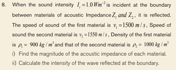
iv) Calculate the intensity of the wave transmitted into the second
material.
9. The distance between pulse representing ultrasonic reflections fromopposite sides of a fetus head was recorded on a screen of a cathode
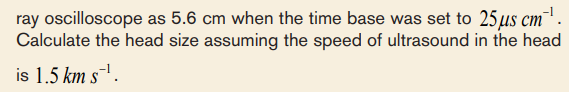
10. Compare and contrast endoscopy imaging and radionuclide imaging
11. What are the advantages of MRI in clinical practice?
12. Why areas of the body can be imaged by ultrasound??
13. In mammography exams, is the breast compression necessary? Why
14. Historically, MRI began in the central nervous system, but it is now
extended to all regions of the human body. The excellent resolution and
contrast available in any chosen plane in the body, makes the MRI an
invaluable diagnostic tool with which to study body structure, functionand chemistry, as well as disease. Discuss the application of MRI.
UNIT 8: RADIATIONS AND MEDICINE
Key Unit Competence:Categorize hazards and safety precautions of radiation in medicine
Introductory activity
Radiation has always been present and is all around us. Life has evolved in a
world containing significant levels of ionizing radiation. Our bodies are adapted
to it.
People are constantly exposed to small amounts of ionizing radiation from the
environment as they carry out their normal daily activities; this is known as
background radiation. We are also exposed to radiations through some medical
treatments and through other activities involving radioactive substances.
The figure above identifies four major sources of public exposure to natural
radiation: cosmic radiation, terrestrial radiation, inhalation and ingestion.Brainstorm and try to answer the following questions:
a) From your own understanding, how is artificial source of radiation different
from natural source of radiation?
b) Using your physics knowledge, what do you think are major sources of
radiation that are mostly preferred to be used in medicine? Defend your
opinion.
c) Do you think exposure to heavy ions at the level that would occur during
deep-space missions for a long duration pose a risk to the integrity and
function of the central nervous system? Explain to support your idea.
d) As a physics student-teacher, what do you think are the symptoms,effects and jeopardy of radiation exposure to human body?
8.1. RADIATION DOSE
Activity 8.1
Radiation is the emission of particles or electromagnetic waves from a source.
Radiation from radioactive materials has the ability to interact with atoms and
molecules of living objects.
a) From your understanding, what makes these radiations able to penetrate
matter?
b) Do you think any amount of radiation should be applied to human body in
case it is to be used to examine a certain part under study or investigation?
Defend your reasoning.
c) Using your prior knowledge about use of radiation in hospitals, what are
common used radiations?
d) Suggest the possible side effects of these radiations to human body.
e) From your suggestions in (d) above, what do you think are precaution
measures one should take to avoid dangers that may be caused by theseradiations?
8.1.1 Ionization and non-ionization radiations.
Radiation is the emission of particles or electromagnetic waves from a source.
Also it is amount of energy deposited in a given mass of medium by ionization
radiation. Radiation from radioactive materials has the ability to interact with atoms
and molecules of living objects.
In a neutral atom, the positive charge of the nucleus is equal and opposite to the
total negative charge of the orbital electrons. If such an atom loses or gains an
electron, it becomes an ion. The atom will now have a net positive or negative
charge and is called an ion. This process is called ionization, and the radiation
responsible for it is called ionizing radiation. When discussing the interaction of
radiations with matter in particularly in relation to health,
two basic types of radiation can be considered.
a. Ionizing radiation.
Ionization radiation refers to a radiation that carries sufficient energy to release
electrons from atoms or molecules, in that way ionizing them. It is made up of
energetic subatomic particles,ion or atoms that moving at high speeds. As the more
powerful form of radiation, ionizing radiation is more likely to damage tissue than
non-ionizing radiation. The main source of exposure to ionizing radiation is the
radiation used during medical exams such as X-ray radiography or computed
tomography scans.
However, the amounts of radiation used are so small that the risk of any damaging
effects is minimal. Even when radiotherapy is used to treat cancer, the amount of
ionizing radiation used is so carefully controlled that the risk of problems associated
with exposure is tiny.
All forms of living things emit a certain amount of radiation, with humans, plants and
animals accumulating radioisotopes as they ingest food, air and water. Some forms
of radiation such as potassium-40 emit high-energy rays that can be detected using
measurement systems. Together with the background radiation, these sources of
internal radiation add to a person’s total radiation dose.
Background radiation is emitted from both naturally and man-made sources. Natural
sources include cosmic radiation, radon radiation in the body, solar radiation and
external terrestrial radiation. Man-made forms of radiation are used in cancer
treatment, nuclear facilities and nuclear weapons.
Globally, the average exposure to ionizing radiation per year is around 3 milli
Sieverts (mSv), with the main sources being natural (around 80%). The remaining
exposure is due to man-made forms such as those used in medical imaging
techniques. Exposure to man-made forms of ionizing radiations is generally much
higher in developed countries where the use of nuclear imaging techniques is
much more common than in developing countries.
b. Non-ionizing radiations
Non-ionizing radiationis any type of electromagnetic radiation that does not carry
enough energy to ionize atoms or molecules. Examples of non-ionizing radiations
include visible light, microwaves, ultraviolet (UV) radiation, infrared radiation, radio
waves, radar waves, mobile phone signals and wireless internet connections.
Although UV has been classified as a non-ionizing radiation but it has been
confirmed. High levels of UV-radiation can cause sunburn and increase the risk ofskin cancer developing.
Scientific investigations suggest that the use of telecommunications devices such
as mobile phones may be damaging, but no risk associated with the use of these
devices has yet been identified in any scientific studies. This energy is emitted bothinside the body and externally, through both natural and man-made processes.
8.1.2 Radiation penetration in body tissue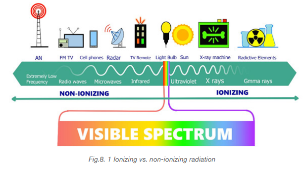
Radiation cannot be spread from person to person. Small quantities of radioactive
material occur naturally in the air, drinking water, food and our own bodies. People
can come into contact with radiation through medical procedures. An important
characteristic of the various ionizing radiations is how deeply they can penetrate
the body tissues. X-rays, gamma rays, and neutrons of sufficient energy described
below can reach all tissues of the body from an external source.
• Alpha Radiation
Alpha radiation refers to a form of particle radiation that occurs when an atom
undergoes radioactive decay. They consist of two protons and two neutrons
(essentially the nucleus of a helium-4 atom). Due to their charge and mass, alpha
particles interact strongly with matter, and can only travel a few centimeters in air.
A thin sheet of paper, on the other hand, stops alpha particles. Alpha radiation can
only penetrate the outer layers of human skin. Therefore, radionuclides that emit
only alpha particles are harmless unless you take them into the body. This you might
do by inhalation (breathing in) or ingestion (eating and drinking).
• Beta Radiation
Beta radiation occurs when radioactive atomic nuclei emit electrons (negatively
charged) or frequently positron (positively charged particles with the same mass of
electron).Due to their smaller mass, they are able to travel further in air, up to a few
meters, and can be stopped by a thick piece of plastic, or even a stack of paper
i.e can penetrate the skin a few centimeters to metres in air and few millimetres to
centimetre in soft tissue and plastic. Posing somewhat of an external health risk.
The depth to which beta particles can penetrate the body depends on their energy.
High-energy beta particles (several MeV) may penetrate a centimeter of a tissue,
although most are absorbed in the first few mm. As a result, beta emitters outside
the body are hazardous only to surface tissues such as the skin or the lenses of the
eye. When you take beta emitters into the body, they will irradiate internal tissues
and then become a much more serious hazard.
• Gamma Radiation
In the case of gamma radiation, energy is transferred as an electromagnetic wave.
Electromagnetic radiation can be described in terms of its frequency or wavelength
( the high frequency and the shorter the wavelength, the more energetic radiation).
Gamma radiation is at high energy end of electromagnetic spectrum. Gamma
radiation, unlike alpha or beta, does not consist of any particles, instead consisting
of a photon of energy being emitted from an unstable nucleus. Having no mass or
charge, gamma radiation can travel much farther through air than alpha or beta,
losing (on average) half its energy.
Gamma waves can be stopped by a thick or dense enough layer material, with high
atomic number. Materials such as lead can be used as the most effective form of
shielding.
• X-Rays
X-rays are similar to gamma radiation, with the primary difference being that they
originate from the electron cloud. This is generally caused by energy changes in
an electron, such as moving from a higher energy level to a lower one, causing the
excess energy to be released. X-Rays are longer-wavelength and (usually) lower
energy than gamma radiation, as well.
• Neutron Radiation
Neutron radiation consists of a free neutron that is mainly released in nuclear
fission. They are able to travel hundreds or even thousands of meters in air, they are
however able to be effectively stopped if blocked by a hydrogen material, such as
concrete or water.
Neutron radiation occurs when neutrons are ejected from the nucleus by nuclear
fission and other processes. The nuclear chain reaction is an example of nuclear
fission, where a neutron being ejected from one fission atom will cause another
atom to fission, ejecting more neutrons. Unlike other radiations, neutron radiation isabsorbed by materials with lots of hydrogen atoms, like paraffin wax and plastics.
8.1.3 Radiation dosimetry
Just as for drugs, the effect of radiation depends on the amount a person has
received. Therefore, amounts of radiation received are referred to as doses, and
the measurement of such doses is known as dosimetry.
A radiation dosimeter refers to a device the measures dose uptake of external
ionizing radiation. Dosimeters are used to monitor your occupational dose from
radioactive material or radiation-producing equipment. Most individuals working
with X-ray producing equipment in the hospital will be issued with a dosimeter.
For those individuals working in the research laboratory setting, dosimeters will be
issued based on the nuclide and total activity that will be used.
Dosimeters are integrating detectors; that is, they accumulate the radiation dose
and give off an amount of light which is proportional to that dose.
The energy absorption properties of dosimeters are designed to be very similar to
tissue, so they are very effective as personnel dosimeters. These devices are used
to measure exposures from x-ray, gamma ray and high energy beta particles.
Dosimeters are not suitable for measuring exposures to low energy beta particles
or alpha particles.
8.1.4. Radiation exposure.
Exposure is a measure of the ionization produced in air by X-rays or gamma rays,
and it is defined in the following manner. A beam of X-rays or gamma rays is sent
through a mass m of dry air at standard temperature and pressure
In passing through the air, the beam produces positive ions whose total charge is q.
Exposure is defined the total charge per unit mass of air. The SI unit for exposure
is coulomb per unit mass . The unit for exposure E is the roentgen(R). 1R
is the amount of electromagnetic radiation which produces in one gram of air ( ) at normal temperature (22 and pressure (760mmHg) conditions.
Since the concept of exposure is defined in terms of the ionizing abilities of X-rays
and gamma rays in air, it does not specify the effect of radiation on living tissue. For
biological purposes, the absorbed dose is more suitable quantity, because it is theenergy absorbed from the radiation per unit mass of absorbing material.
Long-term exposure to small amounts of radiation can lead to gene mutations
and increase the risk of cancer, while exposure to a large amount over a briefperiod of time can cause burns or radiation sickness.
Radiation sickness is a damage human body caused by a large dose
of radiation often received over a short period of time (acute). It isn’t caused by
common tests that use low-dose radiation such as x-rays or CT-Scans. Radiationsickness also called acute radiation syndrome or radiation poisoning.
8.1.5. Absorbed radiation dose.
Radiation dose is a quantity of the energy measured which is deposited in matter by
ionizing radiation per unit mass. A similar approach is used in radiation protection
measurements, where the unit of absorbed dose is specified in terms of the
amount of energy deposited by radiation in 1 kg of material. International system of
unit for radiation measurement is the “gray” (Gy) and “sievert’’ (Sv). These units
can be expressed into others like “rad”, “rem” or roentgen(R). An absorbed
radiation dose of 1 Gray corresponds to the deposition of 1 joule of energy in 1kg of material (air, water, tissue or other).
It describes the physical effect of the incident radiation, but it tells us nothing about
the biological consequences of such energy deposition in tissue. Studies have
shown that alpha and neutron radiation cause greater biological damage for a given
energy deposition per kg of tissue than gamma radiation does. A person who has
absorbed a whole body dose of 1 Gy has absorbed one joule of energy in each
kg of its body tissue. Absorbed dose is used in calculation of dose uptake in livingtissues in both radiation protections.
In other words, equal doses of, say, alpha and gamma radiation produce unequal
biological effects. This is because the body can more easily repair damage from
radiation that is spread over a large area than that which is concentrated in a small
area. Because more biological damage are caused for the same physical dose.
When we analyze the effect of radiation on human being is not so much the total
dose to the whole system but the dose per kg. That’s why a doctor will recommendsmaller doses of medicine for children than for adults.
8.1.6 Quality factors
Quality factors are used to compare the biological effects from different types
of radiation. For example, fast neutron radiation is considered to be 20 times as
damaging as X-rays or gamma radiation. You can also think of fast neutron radiation
as being of “higher quality”, since you need less absorbed dose to produce
equivalent biological effects. This quality is expressed in terms of the Quality Factor
(Q). The quality factor of a radiation type is defined as the ratio of the biological
damage produced by the absorption of 1 Gy of that radiation to the biological
damage produced by 1 Gy of X or gamma radiation.
The Q of a certain type of radiation is related to the density of the ion tracks it leavesbehind it in tissue; the closer together the ion pairs, the higher the Q.
8.1.7 Equivalent dose
The measure of biological damage that is calculated by multiplying absorbeddosebyquality factor for the type of radiation involved is known as equivalent dose.
The unit of equivalent dose H is the Sievert (Sv). An equivalent dose of one Sievert
represents that quantity of radiation dose that is equivalent, in terms of specified
biological damage, to one gray of X or gamma rays. Normally, we use the millisievert
(mSv) and microsievert (µSv). Few other instruments can read in mGy or µGy, butthey measure only gamma radiation.
The Calculation of Equivalent Dose and Effective dose is given by:
The effective dose is a measure of cancer risk, it adjusts the equivalent dose based
on the susceptibility of the tissue exposed to the radiation. It is expressed in Sv andmSv.
8.1.8 Radiation protection
The effects of radiation at high doses and dose rates are reasonably well
documented. A very large dose delivered to the whole body over a short time willresult in the death of the exposed person within days.
We know from these that some of the health effects of exposure to radiation do
not appear unless a certain quite large dose is absorbed. However, many other
effects, especially cancers are readily detectable and occur more often in those
with moderate doses. At lower doses and dose rates, there is a degree of recoveryin cells and in tissues.
Radiation protection sets examples for other safety disciplines in two unique
respects:
• First, there is the assumption that any increased level of radiation above
natural background will carry some risk of harm to health.
• Second, it aims to protect future generations from activities conducted today.
The use of radiation and nuclear techniques in medicine, industry, agriculture, energy
and other scientific and technological fields has brought tremendous benefits to
society. The benefits in medicine for diagnosis and treatment in terms of human
lives saved are large in size.
No human activity or practice is totally devoid of associated risks. Radiation should
be viewed from the perspective that the benefit from it to mankind is less harmfulthan from many other agents.
Note: The optimization of patients’ protection is based on a principle that the dose
to the irradiated target (tumor) must be as high as it is necessary for effectivetreatment while protecting the healthy tissues to the maximum extent possible.
Application activity 8.1
1. a) How ionization differs from non-ionization radiations
b) Give any two examples of each.
2. What does the following terms mean in medical treatment?
a) absorbed dose
b) radiation dose
c) The quality factor
3. In the application of radiation in medicine, we use the statement “Ameasure of the risk of biological harm”. Clearly explain this statement
8.2. HAZARDS AND SAFETY PRECAUTIONS WHEN HANDLING
RADIATIONS
Activity 8.2
1. The picture below show doctors’ meeting and they are discussing on
a therapeutic treatment due to the wrong exposure to radiation that
normally occur in their hospital. These radiations happened in unintendedevent occurring as a radiation accident.
a) In your own words, what does radiation accident mean?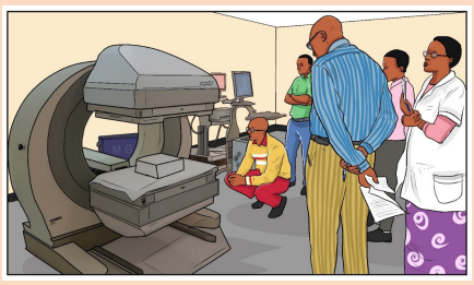
b) What do you think are the radiation accident (unintended events)
which may happen due to wrong exposure radiation?
c) That radiation exposure may be computed in fewer and greater
amount. What do you think are the negative effects that may be as a
result of exposure of these amounts of radiations?
d) Based on unintended event you think might have happened in (b)
above, what do you think are preventive measures that should be
taken to reduce or stop the occurrence of unintended radiationaccident?
2. You happen to interact with a man who was diagnosed and found to
have cancer cells in one of his fingers. He was advised by the doctor
that the cells can be killed by X-rays’ radiations. He had previously
been told that X-rays have a lot of negative effects if exposed to human
body. He at first resisted and was given 2 days to decide. It’s one day
remaining and you happen to interact with him and he is seeking advicefrom you. Advise this man on what do.
8.2.1 Deterministic and stochastic effects:
Effects of radiations due to cell killing have a practical threshold dose below
which the effect is not evident but in general when the effect is present its severityincreases with the radiation dose.
The threshold doses are not an absolute number and vary somewhat by individual.
Effects due to mutations (such as cancer) have a probability of occurrence thatincreases with dose.
a. Deterministic effects:
These effects are observed after large absorbed doses of radiation and are
mainly a consequence of radiation induced cellular death. They occur only if a large
proportion of cells in an irradiated tissue have been killed by radiation, and the losscan be compensated by increasing cellular proliferation.
b. Stochastic effects:
They are associated with long term, low level (chronic) exposure to radiation. They
have no apparent threshold. The risk from exposure increases with increasing dose,
but the severity of the effect is independent of the dose.
Irradiated and surviving cells may become modified by induced mutations (somatic,
hereditary). These modifications may lead to two clinically significant effects:
malignant neoplasm (cancer) and hereditary mutations.
In evaluating biological effects of radiation after partial exposure of the body further
factors such as the varying sensitivity of different tissues and absorbed doses
to different organs have to be taken into consideration. The lifetime value for the
average person is roughly a 5% increase in fatal cancer after a whole body dose
of 1 Sv. It appears that the risk in fetal life, in children and adolescents exceeds
somewhat this average level (by a factor of 2 or 3) and in persons above the age of
60 it should be lower roughly by a factor of ~ 5.
Animal models and knowledge of human genetics, the risk of hereditary deleterious
effects have been estimated to not be greater than 10% of the radiation induced
carcinogenic risk.
All living organisms on this planet, including humans, are exposed to radiation from
natural sources. An average yearly effective dose from natural background amounts
to about 2.5 mSv. This exposure varies substantially geographically (from 1.5 to
several tens of mSv in limited geographical areas).
Various diagnostic radiology and nuclear medicine procedures cover a wide dose
range based upon the procedure. Doses can be expressed either as absorbed
dose to a single tissue or as effective dose to the entire body which facilitates
comparison of doses to other radiation sources (such as natural background
radiation. Quality assurance and quality control in diagnostic radiology and nuclear
medicine play also a fundamental role in the provision of appropriate, sound
radiological protection of the patient.
There are several ways that will minimize the risk without sacrificing the valuable
information that can be obtained for patients’ benefit. Among the possible measures
it is necessary to justify the examination before referring a patient to the radiologist
or nuclear medicine physician. Failure to provide adequate clinical information at
referral may result in a wrong procedure or technique being chosen by radiologist
or nuclear medicine specialist.
An investigation may be seen as a useful one if its outcome - positive or negative
- influences management of the patient. Another factor, which potentially adds
to usefulness of the investigation, is strengthening confidence in the diagnosis.
Irradiation for legal reasons and for purposes of insurance should be carefully
limited or excluded.
While all medical uses of radiation should be justified, it stands to reason that
the higher the dose and risk of a procedure, the more the medical practitioner
should consider whether there is a greater benefit to be obtained.Among these
special position is occupied by computed tomography (CT), and particularly its
most advanced variants like spiral or multi slice CT.
If an examination is typically at the high end of the diagnostic dose range and the
fetus is in or near the radiation beam or source, care should be taken to minimize
the dose to the fetus while still making the diagnosis. For children, dose reduction in
achieved by using technical factors specific for children and not using routine adult
factors, because children are small, in nuclear medicine the use of administered
activity lower than that used for an adult will still result in acceptable images and
reduced dose to the child.
The most powerful tool for minimizing the risk is appropriate performance of the test
and optimization of radiological protection of the patient. These are the responsibility
of the radiologist or nuclear medicine physician and medical physicist.
The basic principle of patients’ protection in radiological X-ray investigations and
nuclear medicine diagnostics is that necessary diagnostic information of clinically
satisfactory quality should be obtained at the expense of a dose as low as reasonablyachievable, taking into account social and financial factors.
8.2.2 Effects of radiation exposure
Some effects may occur immediately (days or months) while others might take tens
of years or even get passed to the next generation. Effects of interest for the person
being exposed to radiation are called somatic effects and effects of interest that
affect our children are called genetic effects.
i. Radiation Health Effects
Ionizing radiation has sufficient energy to cause chemical changes in cells and
damage them. Some cells may die or become abnormal, either temporarily or
permanently. By damaging the genetic material (DNA) contained in the body’scells, radiation can cause cancer.
Fortunately, our bodies are extremely efficient at repairing cell damage. The extent
of the damage to the cells depends upon the amount and duration of the exposure,
as well as the organs exposed.
Exposure to an amount of radiation all at once or from multiple exposures in a
short period of time. In most cases, a large acute exposure to radiation causes
both immediate (radiation sickness) and delayed effects (cancer or death), can
cause sickness or even death within hours or days. Such acute exposures areextremely rare.
ii. Chronic Exposure
With chronic exposure, there is a delay between the exposure and the observed
health effect. These effects can include cancer and other health outcomes such asbenign tumors, cataracts, and potentially harmful genetic changes.
a. Low levels of radiation exposure
Radiation risks refer to all excess cancers caused by radiation exposure (incidence
risk) or only excess fatal cancers (mortality risk). Risk may be expressed as a
percent, a fraction, or a decimal value.
For example, a 1% excess risk of cancer incidence is the same as a 1 in a hundred
(1/100) risk or a risk of 0.01. However, it is very hard to tell whether a particular
cancer was caused by very low doses of radiation or by something else.
While experts disagree over the exact definition and effects of “low dose.”
Radiation protection standards are based on the premise that any radiation
dose carries some risk, and that risk increases directly with dose.
Note:
• The risk of cancer from radiation also depends on age, sex, and factors such
as tobacco use.
• Doubling the dose doubles the risk.
Acute health effects occur when large parts of the body are exposed to a large
amount of radiation. The large exposure can occur all at once or from multiple
exposures in a short period of time. Instances of acute effects from environmentalsources are very rare.
8.2.3 Safety precautions for handling radiations
Shortening the time of exposure, increasing distance from a radiation source and
shielding are the basic countermeasures (or protective measures) to reduce dosesfrom external exposure.
Note: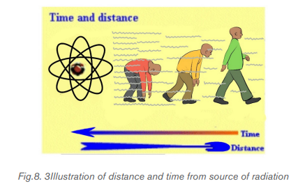
• Time: The less time that people are exposed to a radiation source, the less
the absorbed dose.
• Distance: The farther away that people are from a radiation source, the lessthe absorbed dose.
Note: Shielding: Barriers of lead, concrete or water can stop radiation or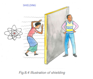
reduce radiation intensity.
There are four main factors that contribute to how much radiation a person absorbs
from a source. The following factors can be controlled to minimize exposure toradiation:
i. The distance from the source of radiation
The intensity of radiation falls sharply with greater distance, as per the inverse
square law. Increasing the distance of an individual from the source of radiation
can therefore reduce the dose of radiation they are exposed to. For example, such
distance increases can be achieved simply by using forceps to make contact with
a radioactive source, rather than the fingers.
ii. Duration of exposure
The time spent exposed to radiation should be limited as much as possible. The
longer an individual is subjected to radiation, the larger the dose from the source
will be. One example of how the time exposed to radiation and therefore radiation
dose may be reduced is through improving training so that any operators who need
to handle a radioactive source only do so for the minimum possible time.
iii. Reducing incorporation into the human body
Potassium iodide can be given orally immediately after exposure to radiation. This
helps protect the thyroid from the effects of ingesting radioactive iodine if an
accident occurs at a nuclear power plant. Taking Potassium iodide in such an eventcan reduce the risk of thyroid cancer developing.
iv. Shielding
Shielding refers to the use of absorbent material to cover the source of radiation,
so that less radiation is emitted in the environment where humans may be exposed
to it. These biological shields vary in effectiveness, depending on the material’s
cross-section for scattering and absorption. The thickness (shielding strength) of
the material is measured in Any amount of radiation that does penetrate the
material falls exponentially with increasing thickness of the shield.
Some examples of the steps taken to minimize the effects of radiation exposure are
described below;
- The exposed individual is removed from the source of radiation.
- If radiation exposure has led to destruction of the bone marrow, the number
of healthy white blood cells produced in the bone marrow will be depleted.
- If only part of the body has been exposed to radiation rather than the whole
body, treatment may be easier because humans can withstand radiation
exposure in large amounts to non-vital body parts.
In every medicine there is a little poison. If we use radiation safely, there are benefits
and if we use radiation carelessly and high doses result, there are consequences.
Ionizing radiation can change the structure of the cells, sometimes creating
potentially harmful effects that are more likely to cause changes in tissue. These
changes can interfere with cellular processes so cells might not be able to divideor they might divide too much.
Radioactive rays are penetrating and emit ionizing radiation in the form of
electromagnetic waves or energetic particles and can therefore destroy living
cells. Small doses of radiation over an extended period may cause cancer and
eventually death. Strong doses can kill instantly. Marie Curie and Enrico Fermi died
due to exposure to radiation.
Several precautions should be observed while handling radioisotopes. Some of
these are listed in the following:
• No radioactive substance should be handled with bare hands. Alpha and
beta emitters can be handled using thick gloves. Gamma ray emitters must
be handled only by remote control that is by mechanical means. Gamma rays
are the most dangerous and over exposure can lead to serious biological
damage.
• Radioactive materials must be stored in thick lead containers.
• Reactor and laboratories dealing with and conducting experiments with
radioactive metals must be surrounded with thick concrete lined with lead.
• People working with radioactive isotopes must wear protective clothing
which is left in the laboratory. The workers must be checked regularly with
dosimeters, and appropriate measures should be taken in cases of overdose.
• Radioactive waste must be sealed and buried deep in the ground.
Rules to remember when working with radiation
Everyone must take radiation overexposure seriously. Hence, preventive measures
and rules must be strictly followed to avoid critical health conditions.
b. Acquire adequate training to better understand the nature of radiation hazards.
a. Reduce handling time of radioactive materials and equipment.
b. Be mindful of your distance from sources of radiation. Increase distance as
much as possible.
c. Use proper shielding for the type of radiation.
d. Isolate or contain harmful radioactive materials properly.
e. Armor yourself with appropriate protective clothing and dosimeters.
f. Conduct contamination surveys in the work area.
g. Do not eat, drink, smoke, or apply cosmetics in an area where unsealed
radioactive substances are handled.
h. Observe proper radioactive waste disposal.i. Conduct usual radiation safety self-inspection
8.2.4 Concept of balanced risk
a. Risks of ionizing radiation in medical treatment
Risk in the area of radiation medicine has several dimensions that are less common
in other areas of medicine. First, there may be risks from overexposure that do
not cause immediate injury. For example, the causal connection, if any, may be
difficult or impossible to verify for a malignancy that surfaces several years after
an inappropriate exposure. Second, the risks associated with the medical use of
ionizing radiation extend beyond the patient and can affect health care workers
and the public.
In amplifying these and other aspects of the risks that attend medical uses of
ionizing radiation, the discussion addresses the following issues: human error and
unintended events; rates of misadministration in radiation medicine; inappropriate
and unnecessary care; and efforts that reduce misadministration and inappropriate
care.
b. Human Error and Unintended Events
Errors occur throughout health care: A pharmacist fills a prescription with the wrong
medicine; an x-ray technician takes a film of the wrong leg; a surgeon replaces
the wrong hip. The advent of complex medical technology has increased the
opportunity for error even as it has increased the opportunity for effecting cures.
By educating health care workers, and by circumscribing their actions, human error
may be minimized. However, some number of mistakes will always, unavoidably, be
made, and no amount of training or double-checking can erase that fact.
c. Comparison of risks in the use of ionizing radiation
The comparison of relative risks of misadministration in by-product radiation
medicine to error rates and events in other medical practice settings, as well
as the comparison of disease and death rates with the risks of the therapeutic
administration itself, help to some extent to place ionizing radiation use in a broader
context.
To achieve this success requires the highest standards of performance (accuracy
of delivered dose), both when planning irradiation for an individual patient and in
actual delivery of the dose.
In a large number of cases, decreasing the dose to the target volume is not possible
since it would unacceptably decrease the cure rate. In these cases present
technological developments aim at optimizing the patients’ protection, keeping
the absorbed tumor dose as high as is necessary for effective treatment while
protecting nearby healthy tissues.
It should be remembered that successful eradication of a malignant tumor by
radiation therapy requires high-absorbed doses and there is a delayed (and usually
low) risk of late complication. The above mention techniques are used to provide
the best benefit/risk ratio.
A malignant tumor in a pregnant woman may require radiotherapy in attempt to
save life of the patient. If a tumor is located in a distant part of the body, the therapy
- with individually tailored protection of the abdomen (screening) may proceed.
When thyroid cancer with metastases is diagnosed in a pregnant woman, treatment
with 131 I is not compatible with continuation of the pregnancy. The treatment should
then be delayed until delivery if doing so wouldn’t put the mother’s life in danger.
Medical radiation can be delivered to the patient from a radiation source outside the
patient. Regardless of how much dose the patient received, they do not become
radioactive or emit radiation.
Balancing risks are often summarized in the following:
• The demand for imaging, especially computed tomography, that has increased
vastly over the past 20 years
• An estimated 30% of computed tomography tests that may be unnecessary
• Ionizing radiation that may be associated with cancer.
• The risks of radiation exposure that is often overlooked and patients are
seldom made aware of these risks
• The requesting doctor who must balance the risks and benefits of any
high radiation dose imaging test, adhering to guideline recommendations if
possible
• Difficult cases that should be discussed with a radiologist, ideally at a clinicradiological or multidisciplinary team meeting.
Application activity 8.2
1. How do you understand by the term balance risk?
2. What is magnitude of the risk for cancer and hereditary effects?
3. Is ionizing radiation from medical sources the only one radiation for
which people are expected to be exposed?
4. What are typical doses from medical diagnostic procedures?
5. Can radiation doses in diagnosis be managed without affecting the
diagnostic benefit? Explain to support your decision.
6. Explain clearly how radiation can be reduced by three principles for
radiation safety: time, distance and shielding
8.3. BASICS OF RADIATION THERAPY FOR CANCER
TREATMENT
Activity 8.3The figure below shows the radiotherapy of breast cancer treatment
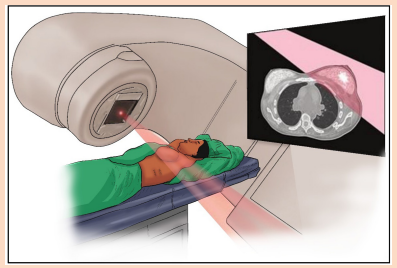
Use the diagram above to answer the following questions.
i) Use a pencil, re-draw the picture in your notebook and locate points
that may be affected by cancer cells.
ii) From your reasoning, does the breast cancer affect only women?
Support your answer.
iii) In Medicine, the concern of breast tissue cancer can be solved by
radiation therapy. It should be delivered in two ways i.e. External andinternal, why do think a doctor may opt one method over another?
8.3.1. Background of Radiation therapy
Radiation therapy plays an important role in curative treatment of many cancers. It
can be used alone or in conjunction with the surgery, chemotherapy or both in order
to eradicate cancer.
Cancer is the name given to a range of diseases where there is malignant tumour.
A malignant tumour may grow slowly for a time and then faster; it infiltrates
surrounding structure and will destroy them. Many cancers are treated successfully
with radiation.
Radiation therapy (also called radiotherapy) refers to the cancer treatment which
uses high dose of radiation to kill cancer cells and tumors. It can be used to cure
cancer, control the growth or spread of cancer and to provide comfort by alleviating
thesymptoms cancer can sometimes cause.The specification for the radiotherapylead to the complicated cancer like: painful bone and soft tissue metastases,
hemoptysis, dyspnea, dysphagia, brain metastases, and spinal cord compression,
etc.
Long exposure of radiation or spent the total dose of radiation over time, allow
tissue cells to be destroyed and be damaged by cancer cells. This is not a big issue
for palliative radiotherapy, but is critical for curative treatment.
Radiotherapy consists/ focuses of treating cancer without removing organs and
tissues. It can be used alone or in conjunction with the surgery and systemic
therapies(e.g., chemotherapy, hormones). The intent is either to cure with radical
radiotherapy or to control symptoms with palliative radiotherapy.
Radiotherapy is usually given over several minutes and is similar to having an
x-ray examination. Patients need to be cooperative and able to lie still for 10 to 15
minutes. As it is a localized treatment, benefits and side effects are generally limitedto the areas being treated.
Radiation therapy had the following types:
• 3D conformal radiation therapy
• Intensity-modulated radiation therapy(IMRT)
• Volumetric-guided radiation therapy(VGRT)
• Image-guided radiation therapy(IGRT)
• Stereotactic radiosurgery(SRS)
• Brachytherapy
• Superficial x-ray radiation therapy(SXRT)
• Intraoperative radiation therapy (IORT)
8.3.2 Cancer treatment
a. Destruction
Radiation damages cells through ionization. This may bea direct ionization of
important molecules such as DNA, in the cell nucleus (shown in below figure) orindirect action through ionization of the more abundant water within the cell.
DNA is a complex responsible for protein synthesis and growth pattern. In some
case, the cells begin to grow uncontrollably (cancer), whilst in others its ability toproduce is destroyed(sterilization).
The ionization of water results in the formation of free radicals H and OH. These
are very reactive and potentially damaging, often leading to cell death or onset of
mutation. Cells are most vulnerable to radiation damage when they are reproducing,
so that fast growing cells are very radiation sensitive, for example the developing
fetus, the reproductive organs and bone marrow. In contrast, brain and bone tissues,which do not replace themselves rapidly, are least affected.
b. The cure
Cancerous cells tend to reproduce more rapidly than normal cell, making them
relatively more radiation sensitive and capable of being selectively destroyed
through ionization. The target is always the DNA within the nucleus: breaks in the
DNA stands can result in cell death or loss of reproductive capacity either of which
stops the spread of the disease. Healthy cells recover from irradiation more quickly
than cancer cells. In order to achieve the greatest destruction of cancer cells, with
the least damaged to surrounding healthy tissue, the radiation should therefore be
delivered in short treatment or fractions of relative high doses over a period of time.
A typical fractionation scheme might be involved daily treatment for five days in fiveweeks.
c. The care
Certain organisms in the body are very weak to radiation damage and during
therapy, it is important to keep dose delivered to these tissues to a minimum. Such
critical organism include the:
• Eye(cataracts)
• Spinal cord(paralysis)
• Reproductive organs(sterility)
• Kidney, liver, rectum.
The treatment depends on the nature of the tumor and its location. There are four
basic methods and treatment for any one patient may involve two or more of them.
• Surgery: if the tumor is easily located, it may simply be removed.
• Chemotherapy: the patient is given dose of cell destroying drugs.
• Hormone therapy: some hormone dependent tumor can be treated by altering
the hormone balance within the body.
• Radiotherapy: tumor cells are destroyed with high-energy radiation, eithergamma-rays from a radioactive source or x-rays.
There are three steps to follow radiotherapy treatment:
The first step in radiotherapy is to meet with a radiation oncologist so that an informed
decision can be made regarding the overall prognosis and goals of treatment and
so that patients and physicians can proceed with planning treatment.
The next step is to determine the area to be treated. This process is called
simulation. The simulation is done with fluoroscopy, x-ray films, CT-Scan and
MRIs can.
The third step is treatment. Radiation treatments are usually given 5 days a week
over several weeks.
During the treatment planning, the doctor or radiotherapist analyses the information
about the size and position of the tumors using various imaging techniques available
like x-ray films, CT-Scan and MRI scan, even ultrasound imaging sometime can be
applied for example in assessing the thickness of the chest wall when planning
breast treatment.
The total quantity of radiation required to destroy the tumors depends on the many
factors, such as:
• Types of cell irradiated(some cancer cells are more radiation-sensitive than
others)
• Environment of the cell(its blood and oxygen supply are important)
• Extent of cancer
• Fractionation scheme selected (a large total dose is needed for more, smallerfractions).
Treatment for certain condition
a. spinal cord compression
Spinal cord compression coming from tumor growth is an oncologic emergency that
should be treated in 24hours of diagnosis with aim of maintaining patient’s ability to
walk, continence and comfort. People with spinal cord compression (about 95%)
had back pain and neurologic signs and symptoms including weakness, paresthesia,
Incontinence, spasticity and hyperreflexia.
Patients’ neurologic deficits sometimes increase rapidly, and early detection is of
highest importance. Magnetic resonance imaging is the modality of choice for this.
A radiation oncologist should be consulted on an emergency basis for spinal cordcompression.
Prognosis is largely dependent on a patient’s overall condition, pretreatment ability
to walk, rate of symptom progression, and the extent of the block. Most patient’s
ambulatory at diagnosis of spinal cord compression remain ambulatory if treated
promptly; only half of those who can move their legs but are not walking become
ambulatory after treatment.
Ambulatory means able to walk but ambulatory care or outpatient care is medical
care provided on an outpatient basis, including diagnosis, observation, consultation,
treatment, intervention, and rehabilitation services. This care can include advanced
medical technology and produces even when provided outside of hospitals.
b. Superior vena cava obstruction
Superior vena cava obstruction caused by cancer also requires urgent, though not
emergency, treatment. Patients with superior vein cava obstruction present with
neck and facial swelling, dilated neck veins, orthopnea, and shortness of breath,
and sometimes progress to headaches and cerebral edema. The treatment usually
varies within 1 to 2 weeks depending on the severity of presenting symptoms. Some
chemotherapy-responsive malignancies, such as lymphomas and small cell lung
cancers, can also cause superior vena cava obstruction and are primarily treatedwith chemotherapy.
c. Bone metastasis
Bone metastases are usually sign for palliative radiotherapy. About 80% of patients
who receive radiation therapy for bone pain experience fewer symptoms; maximum
effect is noticed on average 1 to 3 weeks after treatment. Breast, prostate and
lung are common primary cancer places for bone metastases. Diagnosis is usually
made using bone scans and plain x-ray films, but occasionally magnetic resonanceimaging or computed tomography scans are needed.
d. Brain metastasis
Brain metastases occur around 10% to 30% to all cancer patients. Patient with
brain metastases present the symptoms like: headache, cognitive dysfunction,
neurologic deficits, and seizures. The diagnosis duration given over 1 to 2 weeks
to the entire brain, can improve symptoms and extend survival. Contrast-enhanced
computed tomography (CT-Scan) or magnetic resonance imaging (MRI) scans are
used to diagnose brain metastases.
Conclusion
Radiotherapy has fundamental role in both curative and palliative management
of cancer patients. So that family physicians will be better aware of the appropriateness
of referring patients for such treatment and participating in care of cancer patient
can help facilitate for radiotherapy when they encounter patients with oncologicproblems or complications amenable to radiotherapy treatment.
Application activity 8.3
1. What does a radiation therapy mean?
2. What is radiotherapy used for?
3. How long does it take for radiation therapy treatment to work?4. At what stage of cancer is radiotherapy used?
Skills Lab 8
In this activity you will invite a medical doctor that has expertise in radiationand medicine.
What to do?
• Invite the doctor (using a written letter).Your class tutor or class leaders
may help you in doing this. You may target different doctors so that if
disappointed by one, you do not miss it all. Remember these doctors are
always busy at their work.
• When he/she comes, make sure you give him points of discussion.
These may include: Radiation and dosimetry, balanced risk, Hazards and
safety precautions while handling radiations, and radiation therapy for
cancer treatment. You can still send him/her these topics before so that
he/she can do enough preparations.
• While he/she is presenting, make sure you note down important
information in your notebooks.
• You may ask questions in case you do not understand what the doctor
is explaining.
• Compare what the doctor explained to what you have been discussing
in this unit.
• Develop a comprehensive report including all what you have been
studying and information from the doctor.• Submit your report to your tutor for marking or checking.
End of unit 8 assessment
1. The large amount of radiation absorbed by the body can lead to the
radiation sickness. What do you think is the symptoms and complications
of the radiation sickness?
2. Cleary explain what kind of radiation causes radiation sickness.
3. Is it possible that radiation spread from person to person?
4. What are the risks associated with radiation from diagnostic X-ray
imaging and nuclear medicine procedures?
5. Does receiving external-beam radiation make a person radioactive or
able to expose others to radiation?
6. Is there any risk that internal radiation implants (brachytherapy) will leak
or break free from where they are placed and move around my body?
7. I’m having an imaging test using radioactive materials. Will I be
radioactive after the test?
8. Are there situations when diagnostic radiological investigations shouldbe avoided? Explain to support your decision.
BIBLIOGRAPHY
1. Abbot, A. F., & Cockcroft, J. (1989). Physics (5 ed.). Heinemann: Educational
Publishers.
2. Atkins, K. R. ( 1972). Physics-Once over Lightly. New York : New York.
3. Avison, J. (1989). The world of PHYSICS. Cheltenham: Thomas Nelson and
Sons Ltd.
4. AVISON, J. (1989). The world of PHYSICS. Cheltenham: Thomas Nelson
and Sons Ltd.
5. BIPM. (2006). The International System of Units (SI). (8 ed.). Sevres,
France: International Bureau of Weights and Measures.
6. Breithaupt, J. (2000). Understanding Physics For Advanced Level. (4 ed.).
Ellenborough House, Italy: Stanley Thorners.
7. Chand, S., & S.N., G. S. (2003). Atomic Physics (Modern Physics) (1 ed.).
India.
8. CPMD. (2015). Advanced Level Physics Sylabus. Kigali: REB.
9. Cunningham, & William, P. (2000). Environmental science (6 ed.). Mc
Graw-Hill.
10. Cutnell, J. D., & Johnson, K. W. (2006). Essentials of Physics. USA: John
Wlley &Sons, Inc.
11. Cutnell, J. D., & Johnson, K. W. (2007). Physics. (7 ed.). USA: John Wiley;
Sons, Inc.
12. Cuttnell, J. D., & kennety, W. J. (2007). Physics (7 ed.). United State of
America: John Willey & Sons . Inc.
13. David, A. D. (2012). Child’s thigh demonstrate the five basic radiographic
densities: Imaging for students (4 ed.).
14. Douglass, C. G. (2014). PHYSICS, Principles with applications. (7 ed.).
Pearson Education.
15. Douglass, C. G. (2014). PHYSICS, Principles with applications. (8 ed.).
Pearson Education.
16. Duncan, T., & Kennett, H. (2000). Advanced Physics (5 ed.). London, UK:
Holder Education.
17. eschooltoday. (2008-2018). Retrieved February 19, 2018, from natural
diseasters: http
18. eschooltoday. (2008-2018). Retrieved February 19, 2018, from Climate
change: http
19. Giancoli, D. (2005). PHYSICS: Principles with applications. New Jersey:
Pearson Education, Inc.
20. Giancoli, D. C. (2005). Physics principals with application. Upper Saddle
River, NJ 07458: Pearson Education, Inc.
21. Giancoli, D. C. (2005). Physics: principals with application. Upper Saddle
River, NJ 07458: Pearson Education, Inc.
22. Giancoli, D. C. (2005). Physics: Principles with applications. New Jersey:
Pearson Education, Inc.
23. Glencoe. (2005). Physics - Principles and Problems [textbook]. McGraw.
24. Haber-Schaim, U., Cutting, R., Kirkesey, H. G., & Pratt, H. A. (2002). Force,
Motion, and Energy. USA: Science Curriculum, Inc.
25. Halliday, D., Resneck, R., & Walker, J. (2014). Fundamentals of Physics. (10
ed.). USA: John Wiley; Sons,Inc.
26. Halliday, Resneck, & Walker. (2007). Fundamentals of Physics. (8 ed.).
Wiley.
27. Hewitt, P. G., SUCH0CKI, J., & Hewitt, L. A. (1999). Conceptual Physical
Science. (2 ed.). Addison Wesley Longman.
28. Hirsch, A. S. (2002). Nelson Physics 12. Toronto: University Preparation.
29. http://www.threastafrican.co.ke. (2017). Retrieved from rwanda/Business/
kigali.
30. Hugh, D. Y., & Roger, A. F. (2012). University Physics with Modern Physics
(13 ed.). San Francisco, USA: Pearson Education, Inc.
31. IPCC. (1996). Economics of Greenhouse Gas limitation, Main report
“Methodological Guidelines.
32. John, M. (2009). Optical Fiber Communications, Principals and Practice
(3rd Ed.). London: Pearsnon Prentice Hall.
33. Jones, E. R., & Childers, R. L. (1992). Contemporary College Physics. (2
ed.). USA: Addison-Wesley Publishing Company.
34. Kansiime, J. K. (2004). Coumpound Physical Geography: Morphology,
Climatology, Soils and Vegetation. uganda.
35. Linda, W. (2004). Earth Sceience demystified a self-teaching guide. USA:
McGraw-Hill Campanies, inc.
36. Michael, E. B. (1999). Schaum’s outline of Theory and Problems of Physics
for Engineering and Science. USA: McGRAW-HILL Companies, Inc.
37. Michael, J. P., Loannis, M., & Martha, C. (2006). Science Explorer, Florida
Comprehensive Science. Boston: Pearson Prentice Hall.
38. MIDIMAR. (2012). Disaster High Risk Zones on Floods and Landslide.
Kigali: MIDMAR.
39. Nagashima, Y. (2013). Elementary Particle Physics. Osaka University:
Deutsche Nationalbibliothek.
40. Nelkon, M., & Parker, P. (1997). Advanced level Physics. (7 ed.). Edinburgh:
Heinemann.
41. Nelkon, M., & Parker, P. (2001). Advanced Level Physics (7 ed.). Edinburgh
gate: Heinemann.
42. Office, U. M. (2011). Warming: A guide to climate change. U.K.: Met Office
Hadley Centre.
43. Orazio, S. (2010). Principles of Lasers (5 ed.). Milan, Italy: Springer.
44. Patrick, T. (2004). Mathematics standard level. (3 ed.). Victoria: Ibid Press.
45. Pope, J. (1998). Medical Physics Imaging. Heinemann.
46. R.B., B. (1984). Physical Geography in diagrams for Africa. Malaysia:
Longmann Group Limited.
47. Randall, D., & Knight. (2004). Physics for scientists and engineers: Stategic
approach (Vol. 2). San Fransisco: Pearson Education.
48. Randall, D., & Knight. (2004). Physics for scientists and engineers: Stategic
approach. (Vol. 3). San Fransisco: Pearson Education, Inc.
49. Randall, D., & Knight. (2008). Physics for scientists and engineers: Stategic
approach. (2 ed., Vol. 3). San Fransisco: Pearson Education, Inc.
50. Randall, D., & Knight. (2008). Physics for scientists and engineers: Stategic
approach. (2 ed., Vol. 3). San Fransisco: Pearson Education, Inc.
51. REMA. (n.d.). Rwanda Second National Communication Under the
UNFCCC. KIGALI: MINISTRY OF NATURAL RESOURCES,RWANDA.
52. Science, G. (2006). Florida Physical Science with Earth Science. USA: Mc
Graw Hill Glencoe Companies, Inc.
53. Serway, R. A. (1986). Physics for Scientists and Engineers (2 ed.). Saunders
College Publishing.
54. Serway, R. A. (1992). Principles of Physics. Orlando, Florida: Saunders
College Publishing.
55. Serway, R. A., & Jewett, J. J. (2008). Physics for Scientists and Engineers.
(7 ed.). USA: Thomson Learning, Inc.
56. Serway, R. A., & Jewett, J. J. (2010). Physics for Scientists and Engineers
with Modern Physics. (8 ed.).
57. Silver, B., & Ginn, I. (1990). Physical Science. Unit States of America.
58. Stephen, P., & Whitehead, P. (1996). Physics. (2 ed.). School Edition.
59. Stephen, P., & Whitehead, P. (1996). Physics. (2 ed.). School Edition.
60. Strassler, M. (2011, September 25). What’s a Proton, Anyway? Retrieved
March 05, 2018, from www.profmattstrassler.com: https://profmattstrassler.
com/articles-and-posts/largehadroncolliderfaq/whats-a-proton-anyway/
61. Subranya, K. (1993). Theory and applications of fluid mechanics. Tata
McGraw: Hill Companies.
62. Taylor, E., & Wheeler, J. A. (1992). Spacetime Physics: Introduction to
Special Relativity. (2 ed.). San Francisco: W.H.Freeman & Company,
Publishers.
63. Taylor, E., & Wheeler, J. A. (1992). Spacetime Physics: Introduction to
Special Relativity. (2 ed.). San Francisco: W.H.Freeman & Company,
Publishers.
64. Tipler, P. A. (1991). Physics for Scientists and Enginners. (3 ed., Vol. 2).
USA: Worth Publishers, Inc.
65. Tipler, P. A. (1991). Physics for Scientists and Enginners. (3 ed., Vol. 1).
USA: Worth Publishers, Inc.
66. Tom, D. (2000). Advanced Physics (5 ed.). H. Kennett.
67. Toyal, D. C. (2008). Nuclear Physics (5 ed.). Himalaya Publishing House.
68. Uichiro, M. (2001). Introduction to the electron theory of metals . Cambridge
University Press .
69. Weseda, Y., Mastubara, E., & Shinoda, K. (2011). X-rays properties-google
search. Retrieved 03 06, 2018, from www.springer.com: http://www.
springe.com/978-3-642-26634-1
70. Wysession, M., Frank, D., & Yancopoulos, S. (2004). Physical Science.
Boston, Massachusetts, Upper Saddle River, New Jersey: Pearson PrenticeHall