General
- Physics SME Y3 TG File Uploaded 1/11/21, 16:40
UNIT 5:X-RAYS AND ITS EFFECTS
Key Unit Competence:Suggest and criticize possible effects of X-rays
Introductory activity
Technology has advanced and man has made all advancements to see that
human problems are solved with ease and using technology. Among other
areas where technology has been emphasized is in medicine (in hospitals).
For example, CT scans (Computerized tomography scans) and X-rays machines
are commonly used in hospitals to examine internal structures of a patient if
needed. When a person goes to the hospital with pain in her/his chest, or with
an internal fracture of the bone, physicians do normally recommend the patient
to pass by radiology service (Radiology means there are radiations).
1. Why do you think physicians recommend patients to pass by radiology
service?
2. Discuss different types of radiations that are found in there?
3. From your physics knowledge, how do you think these radiations
specifically X-rays are produced.
4. Like any other electromagnetic radiations, what do you think are some
of the properties of X-rays?
5. As seen from the statement X-rays are used in hospitals, other than
being used in medicine, discuss other areas/fields where X-rays are
applied.
6. What are the positive and negative effects of X-ray radiation on the
human body do you know?
7. Having seen that these radiations have negative effects on human body,
what are your recommendations to a technician works in areas that useX-rays.
5.1 PRODUCTION OF X-RAYS
Activity 5.1
X-rays are produced when the electrons are suddenly decelerated upon collision
with the metal target; these x-rays are commonly called “braking radiation”. If
the bombarding electrons have sufficient energy, they can knock an electronout of an inner shell of the target metal atoms.
Discovery of X-rays: Becquerel’s discovery wasn’t the only important
accidental one. In the previous year W.C. Roentgen unexpectedly discoveredX-rays while studying the behavior of electrons in a high-voltage vacuum tube.
In that instance, a nearby material was made to fluoresce. Roentgen named
them X- rays because he didn’t know what they were. Within twenty years of this
discovery, diffraction patterns produced using X-rays on crystal structures had
begun to show the finer structure of crystals while, at the same time,
giving evidence that X-rays had a wave nature. Since then, X-ray radiation hasbecome an indispensable imaging tool in medical science and other fields.
Questions:
1. According to the text, why do you think the electrons need to be
accelerated and decelerated to produce X-rays?
2. Imagine the energy of bombarding electrons is varied, do you think the
type X-ray emitted would remain same?
3. According to the text, do you think that it is possible to produce X-raysin our local laboratories? Defend your suggestion.
5.1.1 X-ray production
The above figure is an illustration of the Coolidge tube which is the most widely
used device for the production of X-rays. The electrons are produced by thermionic
effect from filament, which is the cathode of the tube, heated by an electric current.
These electrons are accelerated towards a metal target that is the anode due to the
high potential voltage between the cathode and the anode. The target metals are
normally Tungsten or Molybdenum and are chosen because they have high melting
point and higher atomic weights. The accelerated electrons interact with both
electrons and nuclei of atoms in the target and a mysterious radiation is emitted.This radiation was referred to as X-rays.
About 98% of the energy of the incident electron is converted into heat that isevacuated by the cooling system and the remaining 2% come out as X-rays.
5.1.2 Types of x-rays
Sometimes x-rays are classified according to their penetrating power. Two types
are mentioned:
- Hard X-rays: those are X-rays on upper range of frequencies or
shorter wavelength. They have greater energy and so they are more
penetrating.
- Soft X-rays: they are X-rays on lower range of frequencies or longer
wavelength. They have lower energy and they have very low penetrating
power. The Fig.5.2 below shows the relative location of the different types
of x-rays.
Hard x-rays are produced by high accelerating potential. They have high penetrating
power and short wavelength while soft x-rays are produced by lower acceleratingpotential, have relatively low penetrating power and relatively long wavelength.

Application activity 5.1
1. With the aid of a diagram, describe how X-rays are produced in a
laboratory.
2. a) Discuss the two types of X-rays and how they are produced.
b) In the two types of X-rays mentioned in b) above, which one can be
used to
i) Examine or kill cancer cells in a breast.ii) Examine minerals beneath a hard rock.
5.2 PROPERTIES OF X-RAYS AND CHARACTERISTICFEATURES OF X-RAY SPECTRUM
Activity 5.2
With reference to electromagnetic spectrum, what do you think are theproperties of X-rays?
5.2.1 Properties of x-rays
The following are the main properties of X-rays:
(a) X-rays can penetrate through most substances. However, their penetrating
power is different.
(b) X-ray can produce fluorescence in different substances.
(c) X-rays can blacken photographic plate. The degree of blackening depends
upon the intensity of x-rays incident upon the plate. Thus, X-ray intensity can
be measured with the help of photographic plates.
(d) X-rays ionize the gas through which they travel. The ionizing power depends
on the intensity of the x-ray beam. Thus, X-ray intensity can also be measured
by measuring their ionizing power.
(e) X-rays are not deflected by electric or magnetic fields. This proves that
unlike cathode rays or positive rays they are not a beam of charged particles.
(f) X-rays travels on a straight line like ordinary light.
(g) X-ray are both reflected and refracted.X-rays can be diffracted with the help of crystalline substances. They can
also be polarized
From the above characteristics it can be seen that X-rays have the properties that
are common to all electromagnetic radiations.
5.2.2 The origin and characteristic features of an x-ray spectrum
Variation of the X-ray intensity with wavelength
Depending on the accelerating voltage and the target element, we may find sharp
peaks superimposed on a continuous spectrum as indicated on Fig.5.3. These
peaks are at different wavelengths for different elements; they form what is called acharacteristic x-ray spectrum for each target element.
X-rays of different wavelengths are emitted from x-ray tube. If the intensity is
measured as a function of the wavelength and the variation is plotted graphically
then a graph of the nature shown on the figure above is obtained. The graph has
the following features,
(a)Minimum wavelength
(b)Continuous spectrum
(c)Characteristic peaks
Origin of the continuous spectrum
It is known that when charged particles such as electrons are accelerated or
decelerated, they emit electromagnetic radiation of different frequencies. In doing
so a part of their kinetic energy is transformed in the energy of the emitted radiation.
Electrons inside the x-ray tube decelerate upon hitting the target and as a result they
emit electromagnetic radiations with a continuous distribution of wavelength starting
from a certain minimum wavelength. This mechanism of producing electromagnetic
radiation from an accelerated or decelerated electron is called bremsstrahlung.The energy of the emitted photon is given by:
Where is the minimum wavelength, V is the potential difference between anode and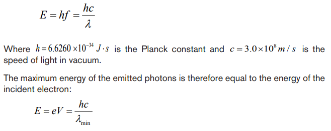
cathode and e the charge of the electron.
Origin of characteristic lines
The peaks observed in wavelengths distribution curves as shown in Fig. 5.3 are
spectral lines in the x-ray region. Their origin lies in the transition between energy
levels in the atoms of the target.
The electrons in the atoms are arranged in different atomic shell of these, the
first two electrons occupy the K-shell followed by 8 electrons in the L-shell, 18
electrons in the M-shell and so on until the electron in the target are used up. A
highly accelerated electron may penetrate atom in the target and collide with an
electron in K-shell. If such electron is knocked out it will leave an empty space that
is immediately filled up by another electron probably from the L-shell or M-shell. This
transition will be accompanied by the emission of the excess energy as a photon.
The energy of the emitted photon is a characteristic of the energy levels in theparticular atom and is given by:
For a transition between K and L-shells.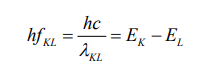
Thus, the energy of the emitted photon depends on the binding energies in the
K and L shells and hence the x-ray spectral lines have definite frequencies and
wavelengths which are characteristic of the target atom.
For a given target material more than one spectral lines are observed as transitionsmay occur between different energy levels.
The x-ray lines originating from the transition between the different electron levels
are usually labelled by the symbols α, β, γ, etc
From L-level to K-level transition produces Kα-line
From M-level to K-level transition produces Kβ –line
From M-level to L-level transition produces Lα –lineFrom N-level to L- level transition produces Lβ –line
Application activity 5.2
1. X-rays are electromagnetic waves produced when fast moving electrons
strike the matter. Discuss the properties of X-rays.
2. A plot of x-ray intensity as a function of wavelength for a particularaccelerating voltage and a particular target is shown in figure below.
a. There are two main components of this x-ray spectrum: a broad range of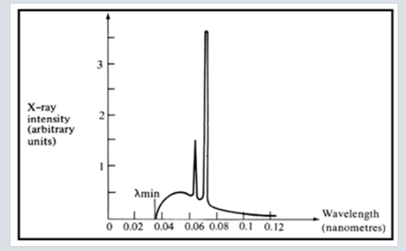
x-ray energies and a couple of sharp peaks. Explain how each of these
arises.
b. What is the origin of the cut-off wavelength λmin of the Figure shown
above? Why is it an important clue to the photon nature of x-rays?
c. What would happen to the cut-off wavelength if the accelerating voltage
was increased? What would happen to the characteristic peaks? Use a
sketch to show how this spectrum would look if the accelerating voltagewas increased
d. What would happen to the cut-off wavelength if the target was changed,
keep the same accelerating voltage? What would happen to the
characteristic peaks? Use a sketch to show how the spectrum would
look if some other target material was used, but the accelerating voltage
was kept the same.
3. Electrons are accelerated from rest through a potential difference of
10 kV in an x ray tube, calculate:
i) The resultant energy of the electrons in eV.
ii) The wavelength of the associated electron waves.iii) The maximum energy and the minimum wavelength of the x ray
5.3. APPLICATIONS AND DANGERS OF X-RAY
Activity 5.3
1. Basing on the nature and properties, what do you think are the uses of
X-rays in real life?
2. If during your internship as a student teacher in a certain primary school,
one of the pupils tells you that her father who is a medical doctor told her
that X-rays are useful and miss-used. That pupil seeks information from
you on how these dangers can be avoided. Provide relevant informationto him/her on how dangers caused by X-rays can be avoided.
5.3.1 Applications
X rays have many practical applications in medicine and industry. Because x-ray
photons are of such high energy, they can penetrate several centimetres of solid
matter. Hence, they can be used to visualize the interiors of materials that areopaque to ordinary light, such as broken bones or defects in structural steel.
a. In medicine
X-ray imaging utilizes the ability of high frequency electromagnetic waves to pass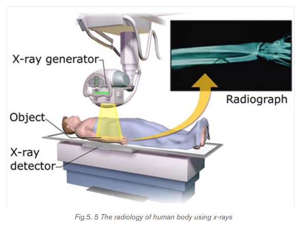
through soft parts of the human body largely unimpeded. For medical applications,
parts of the human body are exposed to moderated X-rays intensity and images
are produced in similar way as light on a photographic plate or digital recorder to
produce a radiograph (See Fig.5.5). By rotating both source and detector around
the patient’s body a “slice” image can be produced in what is called computerized
tomography (CT). Although CT scans expose the patient to higher doses of ionizing
radiation the slice images produced make it possible to see the structures of the
body in three dimensions.
In 1895, the Dutch Wilhelm Roentgen (See Fig.5.6) discovered that light energy
could be used to take photographs through substances such as paper, cloths
and wood. Roentgen also discovered that this invisible form of light energy, called
X-rays could be used to take the pictures of structures inside the body as shown in
Fig. 5.6. Bone tissue appears clearly on an X-rays.
The object to be visualized is placed between an x-ray source and an electronic
detector (like that used in a digital camera) or a piece of photographic film. The
darker area in the recorded images by such a detector, the greater the radiation
exposure. Bones are much more effective x-ray absorbers than soft tissue, so bones
appear as light areas. A crack or air bubble allows greater transmission and showsas a dark area.
A widely used and vastly improved x-ray technique is computed tomography; the
corresponding instrument is called a CT scanner. The x-ray source produces a thin,
fan-shaped beam that is detected on the opposite side of the subject by an array
of several hundred detectors in a line. Each detector measures absorption along
a thin line through the subject. The entire apparatus is rotated around the subject
in the plane of the beam, and the changing photon-counting rates of the detectors
are recorded digitally. A computer processes this information and reconstructs a
picture of absorption over an entire cross section of the subject. In the middle 1970,CT (Computer Tomography) scanning machines were introduced in medicine.
X-rays are also used in the following:
- Killing of cancerous cells
- Radiography is also used in industry for examining potentially damaged
machinery to ascertain the cause of damage and to verify castings or
welded joints
- X-rays are used to study the structure of crystals (crystallography).
When a handgun is fired, a cloud of gunshot residue (GSR) is ejected from the
barrel. The x-ray emission spectrum of GSR includes characteristic peaks from
lead (Pb), antimony (Sb), and barium (Ba). If a sample taken from a suspect’s skin
or clothing has an x-ray emission spectrum with these characteristics, it indicates
that the suspect recently fired a gun.
b. Examining luggage cargo and security.

X-rays are being used in airports to examine luggage for weapons or bombs.
Note that the metal detector that you walk through in the airport does not use x-rays
to examine you instead it uses magnetic waves to detect metal objects. X-rays are
also being used to examine cargo luggage for illegal or dangerous material as in
Fig.5.7.
c. In industry
They can be used to detect structural problems and cracks in metals that cannot
be seen from the outside. X-rays are used on commercial airplanes, bridges metals
and pipe lines, to make sure there are no stress fractures or other dangerous cracks
in the material.
d. In scientific research
• X-ray diffraction provides one of the most important tools for examining the
three-dimensional (3D) structure of biological macromolecules and cells.
• They are also used in crystallography, where X-ray diffraction and scatteredwaves show the arrangement of atoms in the crystal.
The array of spots formed on the film is called a Laue pattern and show the atom
structure of the crystal.
5.3.2 Dangers
• X rays cause damage to living tissues. As X-ray photons are absorbed
in tissues, their energy breaks molecular bonds and creates highly reactive
free radicals (such as neutral H and OH), which in turn can disturb the
molecular structure of proteins and especially genetic material. Young and
rapidly growing cells are particularly susceptible, which is why X-rays are
for selective destruction of cancer cells.
• Because X-rays can kill living cells, they must be used with extreme care.
When improperly used they can cause severe burns, cancer, leukemia, and
cataracts. They can speed aging, reduce immunity to disease, and bring
about disastrous changes in the reproductive cells.
• Lead screens, sheets of lead-impregnated rubber, and leaded glass are
used to shield patients and technicians from undesired radiation.
• The effect of X-ray radiations is cumulative. That is, many minor doses over
a number of years is equivalent to a large dose at one time.
• Unnecessary exposure to x-rays should be avoided. MRI (Magnetic
Resonance Imaging) uses magnets and sound energy to form pictures of
the internal organs without exposing patients to harmful X-rays.
• When they are used in hospitals, the sources should be enclosed in lead
shields.
A careful assessment of the balance between risks and benefits of radiation
exposure is essential in each individual case.
5.3.3 Safety precaution measures of dangers caused by x-rays
Medical and dental X-rays are of very low intensity, so that the hazard is minimized.
However, X-ray technicians who go frequently behind the lead shield while operating
X-rays need to be protected because of the frequency of exposure. A person can
receive many medical or dental X-rays in a year with very little risk of getting cancer
from it. In fact, exposure to natural radiation such as cosmic rays from space poses
a greater risk.
The following are some of the precautions:
i) Protective suits and wears such as gloves and eye glasses made of lead
are used always when handling these radiations. These shields protect the
workers from X-ray exposure.
ii) Workers who operate equipment’s that use X-rays must wear special
badges which detect the amount of radiation they are exposed to.
iii) Food and drinks are not allowed in places where X-radiations are present.
iv) Experiments that involve these radiations (X-rays) substances should be
conducted in a room surrounded by thick concrete walls or lead shields.
v) Equipment that use X-rays should be handled using remote-controlledmechanical arms from a safe distance.
Application activity 5.3
1. Using relevant examples, explain how X-rays are applied in different
fields.
2. Examine the dangers that may arise if these radiations are not handled
with care.
3. As a year III student- teacher, advise an internee having internship in
an area that has X-rays on what to do to avoid the dangers that may becaused by X-rays.
Skills Lab 5
In this activity you will visit a nearest Laboratory that uses X-rays. It may be a
hospital or an industry. In your visit, try to focus on the following
a. How do technicians obtain the x-rays?
b. Why is the room where radiology services done isolated?
c. What are some of the rules followed while in a room where radiology
services are provided?
d. How is X-ray machine operated to achieve results?
e. What are safety precautions to the dangers that may be due to exposure
of X-rays.
You can ask any question of your choice you think is relevant and can make
you understand this unit.
As you come back to the school, make sure you make a comprehensive report
on what you studied from the hospital. Compare the findings to what you
discussed in physics classes.
Present your final findings to the whole class and then finally to your physicstutor.
End of unit 5 assessment
Where necessary use the following constants.
1. Choose the letter that best matches the true answer:
(i) X-rays have
A. short wavelength C. both A and B
B. high frequency D. longest wavelength
(ii)If fast moving electrons rapidly decelerate, then rays produced are
A. alpha rays C. beta rays
B. x-rays D. gamma rays
iii) Energy passing through unit area is
A. intensity of x-ray C. wavelength of x-ray
B. frequency of x-ray D. amplitude of x-ray
iv) X-rays are filtered out of human body by using
A. cadmium absorbers C. copper absorbers
B. carbon absorbers D. aluminum absorbers
v) Wavelength of x-rays is in range
A. 10-8 m to 10-13 m C. 10-10 m to 10-15 m
B. 10-7 m to 10-14 m D. 10² m to 109 m
2. An x-ray operates at 30 kV and the current through it is 2.0 mA.
Calculate:
(i) The electrical power output
(ii) The number of electrons striking the target per second.
(iii) The speed of the electrons when they hit the target(iv) The lower wavelength limit of the x-rays emitted.
3. An x-ray machine can accelerate electrons of energies
The shortest wavelength of the x- rays produced by the machine is
found to be Use this information to estimate the value of
the plank constant.
4. You have decided to build your own x-ray machine out of an old
television set. The electrons in the TV set are accelerated through a
potential difference of 20 kV. What will be the λmin for this accelerating
potential?
5. A tungsten target (Z = 74) is bombarded by electrons in an x-ray tube.
The K, L, and M atomic x-ray energy levels for tungsten are -69.5, -11.3
and -2.30 keV, respectively.
a) Why are the energy levels given as negative values?
b) What is the minimum kinetic energy of the bombarding electrons
that will permit the production of the characteristic and lines of
tungsten?
c) What is the minimum value of the accelerating potential that will give
electrons this minimum kinetic energy?
d) What are the and wavelengths?
6. Using the following illustration, name each part marked by letter from Ato H and explain the function of each part A, B, C, D, E, F and H.
