General
- Biology S2 SB File Uploaded 24/01/22, 13:23
- S2: Biology TG File Uploaded 10/08/22, 16:27
UNIT 10 : Excretion in humans
Key unit competency
To be able to describe the structure and function of the excretory organs and suggest good practices for healthy kidneys.
Learning objectives
After studying this unit, I should be able to:
• Define and explain the need for excretion.
• Describe the role of the liver in excretion.
• Name excretory organs and excretory products.
• Outline the structure of kidney and describe a nephron.
• Describe the process of urine formation.
• Use a dissected mammal to identify parts of the urinary system.
• Develop good habits to maintain healthy urinary system.
Introduction
We use fuel, for example, diesel and petrol to run machines such as engines of vehicles. Are there waste products produced by a running engine? Look at the picture below.

What is happening in the picture? What are the components of the smoke? What do you think would happen to the engine of the vehicle if the products being removed were left to accumulate? From your answers above what do you think you will learn in this unit?
10.1 Need for excretion
Just as vehicles remove substances that can damage their engine if left to accumulate, human beings also remove toxic waste products from their bodies. This happens through a process called excretion. Therefore, we can define excretion as the process by which organisms remove waste products of metabolism from the body. Through excretion, organisms control osmotic pressure. It also enables them to promote homeostasis; that is, the balance of the organism’s internal body environment.
Discussion corner
1. Discuss the following with a classmate.
(a) The importance of excretion.
(b) The health implications of not emptying the bladder and bowels promptly.
2. Research on human excretory organs. Use your answers to fill the table below.

3. Compare your findings with that of your classmates.
4. Share your work with the rest of the class.

Excretion is one of the main characteristics of living things. Every living cell carries out life processes which involve chemical reactions. In some of these reactions, complex materials are chemically broken down and in others simple materials are used to make new compounds. All these reactions are collectively referred to as metabolism. During metabolism, waste products are formed. If waste products accumulate in the cell or even in its surrounding, they can reach very high concentrations which affect the normal cell function and could even kill the cell, for example:
1. Carbon dioxide is a by-product of respiration. If it accumulates in the cell it changes the pH of the cell. It then interferes with the functioning of certain enzymes. If this situation is not corrected, the affected cells eventually die.
2. The body has no mechanism of storing excess amino acids from the digestion of proteins. Excess amino acids are therefore taken to the liver where they are broken down in a process called deamination to urea and glycogen. Small amounts of uric acids are also formed from the breakdown of proteins. Urea and uric acids comprise the nitrogenous waste products which are excreted from the body through urine.
Many other reactions in cells also produce by-products that could be toxic. Therefore it is necessary that these waste products be eliminated from the cells. The waste products if not removed can accumulate to toxic levels resulting to ill health and eventually death.
Unlike plants, animals have difficulty getting rid of waste substances for several reasons:
• Animals are more active than plants. Therefore their metabolic processes take place at a higher rate, producing larger quantities of waste products.
• Animals do not put most of their waste products to other uses like it happens in plants.
• Animals take in certain substances in their food in excess of their needs. These extra substances, for example proteins, are broken down with the formation of toxic substances such as ammonia.
For these reasons, animals have a more complex excretory system than plants.
10.2 The role of the liver in excretion
The table below summarises the excretory organs and their waste products in human beings.
Discussion corner
1. Discuss the following questions with a classmate.
(a) What are the functions of the liver in excretion?
(b) What are the homeostatic functions of the liver.
(c) Why is ammonia produced in the liver not excreted in that form but is first converted to urea?
2. Share your work with the rest of the class.

The liver is the second largest organ in the body after the skin. It plays a very important role in the body. It is described as the metabolic centre. The liver has many functions including maintenance of a constant internal environment (homeostasis) and excretion. The excretory functions of the liver are described below.

(a) Deamination
The body cannot store proteins or amino acids. This is because their breakdown forms toxic substances in the body. Amino acids are the end products of protein digestion. Surplus amino acids are broken down in the liver in a process called deamination. From each amino acid, the amino group (NH2)) is converted to ammonia (NH3). Ammonia should not be allowed to accumulate in the body because it is highly toxic. The remainder of the amino acid molecule is changed to glycogen or fat for storage.
The ammonia produced from the amino group is quickly converted to a less toxic substance called urea. During this conversion, ammonia is first combined with carbon dioxide through a series of enzyme-catalysed reactions. The resulting urea is taken to the kidney in blood and is eliminated from the body in urine.
(b) Detoxification
The liver removes harmful substances such as drugs and hormones from the blood. Within the liver, these substances are converted into inactive or less dangerous forms, for instance, hydrogen peroxide, a highly toxic by-product of certain metabolic process is rapidly split into water and oxygen by enzyme catalase in the liver.

Thus, the liver purifies or detoxifies blood. The inactive substances formed in the liver are returned to the bloodstream and are finally excreted from the body by the kidneys.
(c) Elimination of Haemoglobin
Haemoglobin from old worn out red blood cells is broken down by the liver cells into pigments. These pigments are further broken down and eliminated in the bile, giving urine its characteristic yellow colour.
(d) Elimination of sex hormones and cholesterol After sex hormones have performed their functions, some are modified chemically by the liver cells. Others are sent to the kidney for renal excretion while others are expelled in bile.
Excess cholesterol is also excreted in bile. If there is a considerable excess amount of cholesterol in the blood, some may be deposited in the walls of blood arteries obstructing flow.
Self-evaluation Test 10.1
1. Undigested and indigestible waste products are not considered as excretory wastes, explain.
2. Copy and fill the blank spaces in the passage that follows.
Excretion involves the removal of _______ products from the body. Urea is produced by the ______ during while carbon dioxide is a waste product of _______ . Nitrogenous waste products are excreted by the _______ while carbon dioxide is excreted by the ________.
3. Proteins are very important in our bodies. However, excess amino acids from protein digestion cannot be stored. Study the diagram below and answer the questions that follow.

(i) Name the process illustrated in the diagram.
(ii) In which organ does the process you have named in (i) above occur?
(iii) Name compound Z.
(iv) Compound W is used as source of energy, name compound W.
4. Other than urea, name two other waste products excreted by the liver.
5. Describe how urea is transported in blood to the kidneys.
10.3 Structure of the human urinary system
Activity 10.1: Identifying parts of the human urinary system
Requirements
• Manila papers and marker pens
• Charts, blackboard diagrams and photographs of the human urinary system.
• A drawing of the internal structure of the mammalian kidney
Procedure
1. Study the chart provided and use them to draw the human urinary system on the Manila paper and label the kidneys, ureters, bladder and urethra.
2. Draw a labelled diagram of the internal structure of the mammalian kidney.
3. In your groups, discuss the positions of the cortex, medulla, pyramid and pelvis.
4. Share your findings with other class.

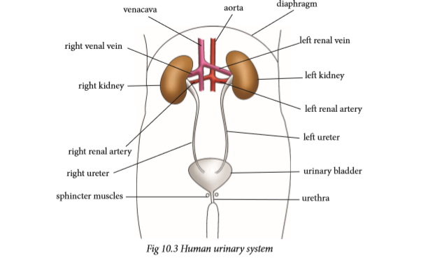
The human urinary system is made up of two kidneys, urinary bladder, two ureters and a single urethra. Kidneys are involved in filtering blood and separating waste metabolic substances from it. The main purpose of the urinary system is to remove urine from the body. The draining of urine from the bladder, through the urethra and out of the body is known as urination.
Function of parts of the urinary system
The urinary system keeps everything in balance by removing waste, such as urea, extra salt, extra water and other things the body does not need. Urea is produced when protein, found in meats, is broken down in the body.
The kidneys
There are a pair of kidneys that are red-brown and are located below the ribs in the middle of the back. Their function is to:
• Remove waste from the blood in the form of urine.
• Keep substances stable in the blood.
• Make erythropoietin, a hormone which helps make red blood cells.
• Make vitamin D active.
• Regulate blood pressure.
Ureter
Each kidney has a narrow tube called a ureter, which carries urine from the kidney to the bladder. Muscles in the ureter walls tighten and relax forcing urine down this tube, away from the kidneys.
Bladder
The bladder is a triangle-shaped, hollow organ located in the lower abdomen. It is held in place by ligaments attached to the pelvic bones. The bladder's walls relax and expand to store urine, and contract and flatten to empty urine through the urethra.
Sphincter muscle
Circular muscles that help keep urine from leaking by closing tightly like a rubber band around the opening of the bladder.
Urethra
Urethra is the tube that allows urine to pass outside the body. The brain signals the bladder muscles to tighten, which squeezes urine out of the bladder. At the same time, the brain signals the sphincter muscles to relax to let urine exit the bladder through the urethra. When all the signals occur in the correct order, normal urination occurs.
The kidneys
Activity 10.2: To examine the external and internal structure of a mammalian kidney
Requirements
• Fresh kidney of sheep, goat or cow
• Sharp razor, knife or scalpel
• Small dissecting board
• Hand lens or compound microscope
Procedure
1. Examine the whole kidney. Note the various tubes attached to it.
• What is the outer colour of the kidney?
2. Draw and label the external structure of the kidney.
3. Place the kidney on the dissecting board.
4. Use the scalpel, razor or knife to cut the kidney along its length at the middle.
5. Use a hand lens to identify the following parts: cortex, medulla, pelvis, renal artery, renal vein and urethra.
6. Draw and label the internal structure of the kidney.
Study questions
1. What are nephrons?
2. How are nephrons structured?
3. What is the function of the nephrons?

Kidneys are bean-shaped and are redbrown in colour. They lie near the back of the abdominal cavity about the level of the waistline. Each kidney weighs approximately 142.5 g. It is about the size of a clenched fist. The right kidney is slightly lower than the left. The kidney is surrounded by a layer of fat which helps to cushion it from mechanical or physical injury.
The kidney is supplied with blood from the general circulatory system via the renal artery which branches off from the aorta. Blood from the kidney goes back to the general circulation through the renal vein which joins the vena cava. A tube called the ureter connects each kidney to the bladder located in the lower abdomen.
The bladder is stretchy to hold large quantities of urine. It stores urine temporarily. From the bladder, another tube called the urethra opens to the exterior of the organism. Two rings of sphincter muscles encircle the urethra. They control the emptying of the bladder.
Internal structure of the kidney
A frontal section through the kidney reveals three main regions. The outer part called cortex, inner part called medulla and the pelvis.

1. Cortex
This is the outer part which is dark in colour. It contains a dense network of blood capillaries that form the glomeruli of nephrons. Nephron is the functional unit of the kidney.
2. Medulla
This part is pale red in colour and lies between the cortex and the pelvis. It contains several cone-like extensions called pyramids.
3. Pelvis
This part is white in colour. It narrows to form the ureter. Pelvis is a collecting space leading to the ureter, which takes urine to the bladder.
The nephron
The most important function of the kidney as an excretory organ is to filter wastes from the blood. This takes place in tiny units called nephrons or renal tubules. A nephron is therefore referred to as the functional unit of the kidney. Each kidney has about 1.25 million nephrons. One part of the nephron is in the cortex and the other part in the medulla.

The nephron has three distinct coiled parts:
• The proximal convoluted tubule
• A U-shaped loop of Henle
• A distal convoluted tubule
Both the proximal convoluted tubule and the distal convoluted tubule are located in the cortex. The loop of Henle is in the medulla. One end of the nephron is modified to form a cup shaped structure called the Bowman’s capsule.
The nephron is supplied with an extensive network of blood capillaries. In the Bowman’s capsule, the capillaries form a knot called the glomerulus (plural glomeruli) which branches from an afferent arteriole that originates from the renal artery. The glomeruli capillaries reunite to form an efferent arteriole which channels blood away from the glomerulus. Thus, on one end of the nephron is blood supply from the artery, and on the other end is blood supply to the vena cava.
Urine formation
Excretion in the nephron is carried out in two stages: ultrafiltration and selective reabsorption. Blood coming into the kidney from the artery contains both waste substances and useful substances. Both substances must enter the nephron, where separation takes place by ultrafiltration. The body must not lose the useful substances. Therefore useful substances must be taken back into the blood so that they are not lost. This process is known as selective reabsorption.
(a) Ultrafiltration
Ultrafiltration takes place in the glomerulus.
Note: The afferent arteriole that takes blood to the glomerular capillaries has a wider lumen than the efferent arteriole that takes blood away from it.
Due to the difference in afferent and efferent arteriole size, a high pressure of blood is created in the glomerulus. This pressure forces water, mineral ions and small molecules like glucose, amino acids and urea out of the glomerulus. These pass through the tiny pores in the walls of the glomerular capillaries into the Bowman’s capsule. The liquid collected in the Bowman’s capsule is called glomerular filtrate.
The larger molecules in the blood, like blood proteins, white blood cells, red blood cells and platelets cannot pass through the capillary walls of the glomerulus. These remain in the blood and continue to flow to the efferent arteriole. The glomerular filtrate flows down the nephron where re-absorption will take place as it flows along.
(b) Selective reabsorption
As the glomerular filtrate passes along the nephron, some substances that are useful to the body are selectively taken back or reabsorbed into the blood capillaries network surrounding the nephron.
• All amino acids and glucose are reabsorbed by active transport in the proximal convoluted tubule.
• Some salts and water are reabsorbed depending on how much of them the body still needs. Water is absorbed by osmosis and salts by active transport. Salts are absorbed mainly in the distal convoluted tubule. Water is reabsorbed in both the proximal and distal convoluted tubules. However, most of the water is reabsorbed in the region of the collecting duct.
• No urea is reabsorbed into the blood.
By the time the filtrate from the glomerulus completes its movement down the nephron, it has a high concentration of urea, some salts and water. The liquid is now called urine. Several nephrons empty into one collecting duct, and all the collecting ducts of a kidney empty into the ureter. The process of urine formation is a continuous one, and the ureter continuously receives small amounts of urine. The bladder stores the urine until it is full, then one begins to experience an un-comfortable feeling. The sphincter muscles must then be relaxed in order to empty the bladder.
Research project
Using reference materials and internet, find out why an individual may pass much dilute urine or less but concentrated urine.
Factors that affect urine production
The volume, colour, odour of urine and frequency of urination is affected by many factors. They include:
1. Amount of fluids taken
Large intake of fluids lowers the osmotic pressure of blood. This leads to reduced reabsorption of water in the kidney tubules resulting in the production of large amounts of dilute urine.
2. Amount of salt taken
Intake of a salty meal raises the osmotic pressure of blood. This leads to increased reabsorption of water in the kidney tubules resulting in the production of coloured, little and smelly urine.
3. Weather
In hot and dry weather conditions, the body tends to lose a lot of water through sweating thereby raising the osmotic potential of blood. In this case a lot of water is reabsorbed resulting in coloured, little and smelly urine.
During cold weather the frequency of urination increases because sweating is so minimal.
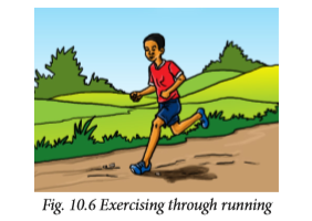
4. Physical activity
During an exercise like running, jumping and playing, we sweat a lot. The kidney reabsorbs more water resulting in little, coloured and smelly urine.
5. Diseases
Certain diseases that affect the secretion of hormones that control reabsorption of water in the kidney tubules can either lead to production of large or small amounts of urine. An example is diabetes insipidus.
Self-evaluation Test 10.2
1. The diagram below shows the mammalian urinary system. On it, indicate the renal artery, urethra, ureter and left kidney.

2. Given the following parts of the nephron:
(i) Bowmans capsule
(ii) Loop of Henle
(iii) Distal convoluted tubule
(iv) Collecting duct
(v) Proximal convoluted tubule
Arrange the parts in order beginning with the region of the nephron from where the filtrate begins to flow.
3. A sea salmon swims up a river. Explain how this would affect the quantity and concentration of its urine.
10.4 Practices that maintain healthy urinary system
Discussion corner
1. Discuss with a classmate, the good habits that enhance healthy urinary system. 2. Present your work to the rest of the class.

The best way to prevent urinary tract infection is to keep the kidneys and the entire urinary system healthy. This can be done through proper diet and nutrition. Some methods that keep the urinary system and kidneys functioning normally include:
(i) Drinking a lot of water, at least 10 glasses of water a day to flush out toxins in the body.
(ii) Exercising regularly to keep fit. Maintain a healthy weight according to your age to avoid putting excess strain on all bodily systems.
(iii) Avoid taking too many drugs especially pain killers. Stick to prescriptive drugs from a qualified medical officer.
(iv) Visit a doctor (urologist) regularly to check the health of the urinary system.
(v) Eat healthy by avoiding junk food. Eat more fresh fruits and green vegetables. Choose foods low in sodium, sugar and fats but high in fiber content.
(vi) Be informed about the causes and prevention methods of kidney diseases and urinal track infection causes.
(vii) Avoid smoking and alcohol intake.
Health Check!
It is good practice to empty your bladder in toilets, latrines or urinals. Urinating in public places like bus stops, by the roadside, in water bodies or on walls is extremely unhygienic. In addition to polluting the environment, the urine may also contain disease-causing micro-organisms that can contaminate drinking water and vegetables.
Self-evaluation Test 10.3
1. Suggest reasons why people who take alcohol tend to urinate a lot.
2. Why is it important to take only prescribed medicine from a qualified doctor?
3. Drinking a lot of water is healthy. Explain.
Unit summary
• Excretion is the process by which waste substances from cells, tissues or blood of organisms are removed from the body. Egestion is getting rid of undigested or unabsorbed wastes from an organism.
• Excretion is important as it enables the elimination of urea and carbon dioxide.
• The excretory substances in animals include ammonia, urea, uric acid, carbon dioxide and mineral salts.
• If waste substances accumulate in the cell or even in its surroundings, they kill the cell. Therefore they should be excreted.
• In multicellular organisms such as human beings, excretion occurs through excretory organs such as skin, lungs, liver and kidneys.
• The major component of the kidney is the cortex, medulla, pelvis and ureter.
• The nephron consists of Bowman’s capsule, proximal convoluted tubule, Loop of Henle, distal convoluted tubule and the collecting duct.
• Urine is produced by the processes of ultrafiltration and selective reabsorption.
• Urine is transported through the ureters to the bladder where it is stored temporarily before it is removed from the body through the urethra.
• The volume and concentration of urine is affected by water intake, temperature and exercise.


