Topic outline
General
- Biology S2 SB File Uploaded 24/01/22, 13:23
- S2: Biology TG File Uploaded 10/08/22, 16:27
Unit 1: Classification of Kingdom Animalia
Key unit competency: To be able to classify animals into their main groups based on their external features.
Learning objectives
After studying this unit, I should be able to:
• State the characteristics of all animals.
• Identify the common features of chordates.
• Explain the economic importance of arthropods to humans.
• State other phyla of kingdom Animalia and give examples of each.
• Distinguish different groups of animals using observable features.
• Appreciate the existence of animal diversity and the need for classification
of animals.Introduction
In Senior 1, you learnt about biodiversity and classification of organisms in the
environment. Can you recall the five kingdom system of classification and the main
features of each Kingdom?
Now, look at the picture below. Which animals can you see? Write down their
external features.
Fig. 1.1: Animal diversity
With a friend think about how the features can be used to classify the animals. Try putting the animals into various groups. Do you now have an idea of what you will learn in this topic?
1.1 General characteristics of animals
There are many species of animals living on land (about 6.5 million), in water (about 2.2 million) and approximately 10,500 species in the air. However, only a few of these animals are clearly understood. It is therefore important to study and describe the characteristics of the main groups of the animal kingdom. This study enables us to be familiar with our environment as we interact with these animals in our day to day lives. How are animals different from other organisms and from each other?
Activity 1.1: To identify the main characteristics of kingdom Animalia Requirements
Obtain the following for collection of specimen:
• Specimen bottles
• Pair of forceps
• Gloves
• Sweep nets
• Pooter
Procedure
1. With the guidance of your teacher, go to the school field and collect animal specimen of different types.
• For crawling animals use a pooter.
• Use a sweep net to collect hopping
animals such as grasshoppers.
Caution: Some animals are harmful in some ways. Care should be taken when making collections.2. Put on gloves and use a pair of forceps to transfer the collected animals into specimen bottles.
3. Carry the collected animals to the Biology laboratory.
4. Observe the animals collected and note down their external features.
Caution! Some animals can bite or sting. Be careful when handling them.
5. Your teacher will also show you pictures and photographs of different animals.
• Can you identify the animals?
6. Keenly observe the animals you collected and do the following:
• Count the number of legs and wings (if present) in each animal.
• How many wings does each animal have?
• Touch the outer covering of each animal. Describe it.
7. Discuss with your classmate the external features you observed.Study questions
(a) Do all animals have the same number of legs and wings?
(b) What are the features used to classify animals?
(c) What is the meaning of the following terms?
• Eukaryotic
• Multicellular
• Heterotrophic
I have discovered that…….
The main features used to classify animals include:
• Presence or absence of appendages (An appendage is a projection from the body of an organism), their type and number.• The body form; whether segmented or unsegmented.
• Presence of skeleton and its type; exoskeleton or endoskeleton.
• Type of body symmetry; whether bilateral or radial.
The facts Animals have the following general
characteristics:
1. They are multicellular organisms.
2. They have eukaryotic cells.
3. Their cells are differentiated into tissues and organs.
4. They are all heterotrophic, meaning they depend on other organisms for food.
5. Their cells lack cell walls, cell sap and chloroplasts. They only have cell membranes and this makes their cell to be irregular in shape.
6. Most animals are able to move the whole body from one place to another (locomote).
7. Reproduction in most animals takes place through fusion of gametes.
8. They respond to external stimuli.Files: 41.2 Phylum Chordata
Discussion corner
1. Look up the meaning of the following words; backbone, notochord, exoskeleton and endoskeleton from reference materials provided.
2. Write the meaning of the words in your notebook.
3. Share your findings with classmates.
4. Did you all get the meanings right? I have discovered that …. The term ‘chordata’ comes from theord chorda which means chord. Organisms in this phylum, at one time in their life have a chord-like structure called notochord. The facts We differ from other organisms because we have a backbone that enables us to stand upright. The main characteristics of organisms in phylum Chordata are:
1. Presence of a chord like structure called notochord. The notochord supports the body in lower
chordates while in higher chordates (vertebrates) it is present only during embronic stages. It is replaced by a vertebral column.
2. Presence of a vertebral column which forms part of an internal skeleton. Vertebral column is a bony structure made up of vertebrae. It protects the spinal cord.
3. Presence of a nervous system with a brain which is connected to a hollow nerve tube or a single tubular nerve cord. The nerve tube runs along the back and forms a brain anteriorly.4. Bilateral symmetry: this means that the body can be divided along one plane into two equal halves that are roughly mirror images of each other.Look at these pictures below. Can you state all the similarities and differences among the organisms. All the animals in the picture belong to phylum chordata. Based on this, what can you say about phylum chordata? Organisms in the phylum Chordata can further be subdivided into 5 different
classes. These are:
• Pisces (fishes)
• Amphibia
• Reptilia
• Aves (birds)
• MammaliaClass Pisces
Latin word ‘Piscis’ which means fish. Therefore, the class pisces is made up of the fish family. The class consists of all type of fish such as:
• Tilapia • Nile perch
• Cod fish • Shark
• Ray fish • Mud fishActivity 1.2: To examine external features of fish
Requirements
• Tilapia or any other type of fish (freshly killed)
• Hand lenses
• White tiles
• Gloves
• A pair of forceps
Procedure
1. Using the hand lens, observe the body surface of the fish and note the arrangement of the scales.
2. Put on the glove on one hand and place the hand on the anterior part; slowly move your hand over the body surface of the fish towards the tail region. How do you feel?
3. Using a pair of forceps, gently lift the flap-like structure (operculum) covering the gill chamber to expose the gills.
4. Note the shape of the body of the fish.Study questions
Discuss the following questions in your groups then present to the rest of the class.
(a) State the observable external features which all fish possess.
(b) Draw the arrangement of scales on fish.
(c) Draw the structure of a gill.
(d) What is the function of the gills bserved in the gill chamber?The facts
Main characteristics of animals in class Pisces
1. All fishes are aquatic. They live in places such as seas, lakes, oceans, rivers and dams.
2. The skin of the fish is covered with scales which overlap backwards.
3. Fish have gills which are used for gaseous exchange. The gills are located in a space called gill cavity on the side of the head. The number of gills in a fish ranges from 4 - 7 pairs.
4. Fish have fins that aid in movement. The fins on the sides of the body are usually found in pairs, for example, the pelvic and pectoral fins. The dorsal, ventral and tail fins are usually unpaired.
5. Fish are poikilothermic: Their body temperature is dependent on environmental temperature because they cannot regulate their own body temperature.
6. Fish exhibit external fertilisation where eggs are first laid by the female then the male sheds sperms over them.
7. Fish have a lateral line on their body for sensitivity.
8. Fish have a single circulatory system with a two chambered heart. Note: Fish use gills for gaseous exchange except lung fish that lives in oxygen deficient swamps and use lungs for breathing.
Adaptation of fish to aquatic environment How does fish survive in water?
Research Activity
1. Using reference materials such as textbooks and the internet, research on the adaptive features
of a fish. Use the following questions as a guideline.
a) What kind of body symmetry does fish have?
b) Describe how the following structures enable fish to live in water:
i) Body shape
ii) Arrangement of scales
iii) Fins
iv) Gills
v) Scales
2. Compare your finding to the ones below.The facts
The following features enable fish to survive in water.
1. Gills Fish use gills to breathe under water. Most fish have to swim constantly. This enables water to pass through the gills to allow for gaseous exchange.
2. Streamlined body The body shape of a fish is well suited to its particular habitat. Most fish have
a streamlined body to allow water to easily pass over them, reducing friction (resistance) as they swim.3. Fins and tails
Fins and tails allow fish to move through water. The tail propels the fish while fins guide their movement in water by controlling their direction and balance.
4. Lateral lines
The lateral line allows fish to detect vibrations in water, alerting them of predators.
5. Huge number of eggs
A single fish can lay more than a million eggs, which can all be fertilised. However, a lot of eggs are eaten by predators while others are washed away by water currents. A large number of eggs ensures that at least some will survive to maturity.Class Amphibia
The word amphibia comes from the word ‘amphi’ which means ‘dual’ or ‘two’. This class of chordates can live both on land and in water. Most adult amphibians live on land. However, they go back to the
water to breed. Examples are:Activity 1.3: To examine external features of amphibians
Requirements
• Preserved specimen of toads and frogs or freshly killed.
• Photos and illustration of newts and salamanders. Procedure
1. Examine the external features of the specimen provided. Note the following:
(a) The nature of the skin.
(b) Presence or absence of scales.
(c) Number of legs.
2. Draw a labelled diagram of a toad, a newt and a frog. Caution: Do not touch the warty skin of toads. It contains glands which produce poison. Study questions
a) What is the difference between a toad and a frog?
b) Do these amphibians have scales on their skins?
c) How many legs do these amphibians have?
d) Share your findings with the rest of the class.
The facts
Main characteristics of animals in class Amphibia
1. They have mucus glands under the skin to keep it moist.
2. The skin has no scales unlike that of fish and reptiles. (Reptiles will be,discussed later in this unit).3. Adult amphibians use the lungs, moist skin and mouth cavity for gaseous exchange. However, their
young ones use external gills for gaseous exchange while in water.
4. The adult female amphibians always lay their eggs in water.
5. They exhibit external fertilisation.
6. They have two pairs of limbs.Class Reptilia
The term ‘Reptilia’ comes from a latin word, ‘Reptilis’ which means ‘crawl’. The animals in this class move by creeping or crawling. Unlike amphibians, reptiles do not breed in water because they lay
eggs with leathery shells; hence cannot dry out. Examples of reptilia are; snakes, turtles, tortoises, crocodiles and lizards Activity 1.4: To examine external features of reptiles
Requirements
• Live specimens of non-poisonous reptiles such as lizards
• Preserved specimens
• Photos and pictures
• Films and videos on lives of reptilesProcedure
1. Observe the specimens, photos and illustrations provided carefully.
2. Do the following:
(a) Note the nature of the skin.
(b) Note the presence and number of limbs.
(c) Count the number of limbs and the terminating digits.
Caution: Do not attempt to catch live animals like snakes or even go close to them because they are
poisonous and dangerous. Study questions
a) How are the scales attached onto the skin?
b) Is the skin of reptiles moist or is it dry?
c) Compare the scales of reptiles to those of fish.
d) Which reptile does not have legs?
e) How many legs do the other reptiles have?
f) What is the habitat of each of the reptiles studied?The facts
Main characteristics of animals in Class Reptilia
1. They have a dry scaly skin. Some like the tortoise have scales which have hardened to form a shell.
2. They are mostly terrestrial with a few being partially aquatic.
3. They undergo internal fertilisation, where the male introduces sperms into the female body. The eggs laid thereafter are covered with a shell.4. Most of them have two pairs of legs except the snake.
Class Aves (birds)
The term ‘aves’ comes from a Latin word ‘Avis’ which means bird. Animals in this class consist of birds such as the humming bird, ostrich, fowl, sparrow, hawk, eagle, sea gull, parrot, crow and ibis.
Look at the following pictures. Have you ever see them? Activity 1.5: To examine external features of birds
Requirements
• Photos and illustrations of different kinds of birds.
• Live specimen of caged or domestic birds.
Procedure
1. Examine the birds, photographs and illustrations carefully.
2. Note the following:
(a) The nature of the skin
covering on their body.
(b) The skin on their legs.
(c) Number of limbs.
(d) Adaptation of the limbs to their function.
(e) The nature of the feathers.
3. Draw and label different parts of the bird. Main characteristics of Class Aves
1. Their bodies are covered with feathers.
2. Their legs are covered with scales.
3. Their front limbs are modified to form wings. The hind legs are used for walking, running, swimming mamong other uses.
4. They have hollow bones that make them light for flight.
5. They have toothless jaws covered by a horny beak or bill.
6. They lay eggs with a hard shell madeof calcium.
7. They carry out internal fertilisation. Adaptations of birds to their environment
Discussion corner
1. Using text books and the internet find out the adaptive features of birds.
2. Answer these questions:
a) Why do birds have wings?b) What is the importance of birds having a streamedlined body?
c) Why do birds have hollow bones?
d) Why do birds have beaks?1. The forelimbs of birds are modified to form wings for flight. The sternum of pectoral girdle is expanded for attachment of flight muscles.
2. Flight birds are light in weight. Their bones are air-filled and therefore have low density to enable ease of flight.
3. Birds lay eggs with hard calcareous shells to avoid drying out.
4. The beaks of birds are modified for different modes of feeding, for example:
• Seed eaters – like sparrows have short thick conical bills for cracking seeds.
• Flesh eating birds – like hawks have sharp curved beaks for tearing meat.
• Nectar eating birds – like humming birds have long slender beaks to probe the flowers.
• Insect eaters – like bee-eater have thin pointed beaks.
• Filter feeders – have serrated beaks to filter food from muddy water. An example is the duck and flamingoes.
• Water plant eaters – like ducks have flat beaks to strain small plants and animals the water.Activity 1.6: To Identify the mode of feeding of the birds whose beaks are shown below.
Use a table like the one given below.
Table 1.1: Birds and their mode of feeding
 5. Birds have different types of feet to them to their different environments. These include:
• Feet for grasping – like those of a kingfisher. They are large and curved to grasp a prey tightly.• Feet for scratching – like those of chickens. They have nail-like toes to scratch the soil for food.
• Swimming birds – like ducks have webbed feet used like paddles.
• Perching feet – like that of a robin. They have long back toes to grab and perch tightly on a tree branch or bark.
• Feet for running – like those of ostrich. They have three toes to enable stability when running.Activity 1.7: Identify the adaption of the bird's feet shown below. Use a table like the one given below.
Table 1.2: Birds and their feet adaptation
 2. Draw the expected type of feet to match the type of beak in birds.
Use a table like the one given below.
Table 1.3: Matching birds beaks to feet

Class Mammalia
The term Mammalia is derived from the Latin word ‘mammalis’ which means ‘mammal’ or ‘mamma’ which means milk secreting organ of female mammals. Most mammals are terrestrial except a few like dolphins and whales which are aquatic.
• Name some organisms in this class.Activity 1.8: To examine theexternal features of mammalsRequirements
• Live specimen of animals in class Mammalia in the school compound such as rabbits or rats.
• Photos of aquatic mammals.
• Illustrations and videos of other members of class Mammalia.
Procedure
1. Examine the animals of class Mammalia found in your school compound or in the school
surroundings.2. Recall a goat or a cow in your home or at your neighbour.
3. Note the following:
• Presence of mammary glands.
• Presence of hair or fur on the skin.
• The nature of the ears.
4. Locate a mammal with young one(s). For instance a rabbit with its litter.
• What is the behaviour of the mother towards its young ones?Study questions
(a) What is the importance of mammary glands in a mammal?
(b) What is the covering on the skin of mammals?
(c) Describe the ears of mammals.
Main characteristics of animals in Class Mammalia
1. They have mammary glands. They suckle and take care of their young ones.
2. Their bodies are covered with fur, hair or wool.
3. They give birth to young ones except the duck-billed platypus and spiny ant-eater which lay eggs.
4. They have external ears. These arethe only class of chordates that have the external ears.
5. They exhibit internal fertilisation.
6. They are Homeothermic. Their body temperatures do not depend on the environment. It ismaintained constantly.
7. They have differentiated teeth (i.e incisors, canines, pre-molars and molars) each with different function. They are therefore referred to as heterodonts. Others with uniform teeth differing only in size are homodonts.


1.3 Phylum Arthropoda
Activity 1. 9: To examine features
of animals in phylum Arthropoda
You will be provided with pictures
or photographs of animals in
phylum Arthropoda.
1. Observe the external features of
the animals.
2. Discuss with your class members
the features observed.
3. Write down the common features
possessed by the different groups
of organisms.
4. Note the number of body parts,
legs, antennae and eyes.
5. Summarise the features of the
different groups of organisms in
a table.What are arthropods? How do they differ from chordates?
Look at the pictures in fig 1.10. Can you identify the animals in the pictures? Come up with a list of the differences between these animals and chordates.
The word arthropod comes from two words ‘Arthros’ meaning ‘jointed’ and‘poda’ meaning ‘leg’ or ‘foot’. Therefore arthropods are animals with jointed appendages. It is the largest phylum in the animal kingdom. The animals in this group inhabit land, water and soil. Some arthropods are useful in many ways, for example:
• Butterflies and bees act as pollinators of flowering plants.
• Bees make honey.
• Lobsters are used as food. Some arthropods are harmful to other living organisms, for instance:
• Ticks transmit diseases in animals.
• Mosquitos transmit malaria.
• Tsetse flies transmit trypanosomiasis.
• Aphids destroy crops such as maize, coffee and cassava.Main characteristics of organisms in phylum Arthropoda
1. They have jointed legs (appendages).
2. They have a tough coat or covering made of chitin. This coat forms an outer skeleton known as the
exoskeleton. The tough coat protects the internal organs against damage. The exoskeleton does not grow. It is usually shed in a process known as moulting to allow the organism to grow.
3. They have bilateral symmetry. This means that they can be cut into two similar halves in only one way. Each half is a mirror image of the other. Fig 1.11: Bilateral symmetry in arthropods4. Muscles for movement are attached on the cuticle or exoskeleton, forexample in insects.
5. They have a fluid filled body cavity called haemocoel for example in earthworms.
6. Their bodies are segmented.Activity 1. 9: To examine features of animals in phylum Arthropoda You will be provided with pictures
or photographs of animals in phylum Arthropoda.
1. Observe the external features of the animals.
2. Discuss with your class members the features observed.
3. Write down the common features possessed by the different groups of organisms.
4. Note the number of body parts, legs, antennae and eyes.
5. Summarise the features of the different groups of organisms in a table.
I have discovered that… Organisms in phylum Arthropoda can further be grouped into various classes.
The organisms are grouped on the basis of:
• Number of legs
• Presence and absence of antennae
• Number of antennae
• Number of body parts
• Type of eyes
The 5 classes of phylum Arthropoda are:
a. Insecta
b. Arachnida
c. Crustacea
d. Diplopoda
e. ChilopodaClass Insecta
This is the largest class in the phylumn Arthropoda. The term ‘insecta’ comes from the word ‘incised’ which means ‘cut.’ The body of organisms in this class are divided into three distinct parts; that
is head, thorax and abdomen. look at the pictures below. Can you identify the different insects?
Activity 1.10: To observe the external features of insects
Requirements
• Sweep nets
• Hand lenses
• Glass jars
• Gloves
Procedure
1. Collect a large variety of insects from the school compound.
Caution: Avoid catching dangerous insects like wasps and bees as they can sting you.
2. Use sweep nets to catch flying or jumping insects like houseflies and grasshoppers.
3. Put them in glass jars and take them back to the laboratory for observation.
Note: To make observation easier, some insects can be immobilised using chloroform.
Precaution: Do not inhale chloroform. It is toxic.
4. Examine the specimen carefully and note the following features.
(a) The number of body parts.
(b) The types of eyes: simple or compound.
(c) Presence or absence of antennae.
(d) Presence or absence of wings
(e) Number of legs.
5. Draw and label clear diagrams of the specimen.Study questions
(i) Insects have ……………..body parts?
(ii) Insects have…………….pairs of legs?
(iii) Draw well labelled parts of an insect.My environment, my life!
Do not kill the insects. It is important to release insects after observation while they are still alive.
Main characteristics of organisms in class Insecta
1. They have three distinctive body parts; head, thorax and abdomen.
2. They have a pair of long antennae.
3. They have three pairs of jointed legs, which are attached to their thorax.
4. They have a pair of large compound eyes.
5. Some have one or two pairs of wings that are attached to the thorax.
6. They breathe by means of spiracles,which are found on the sides of the abdomen and thorax.
Class Arachnida
This class of arthropods includes spiders, mites, ticks and scorpions.

Activity 1.11: To examine the main features of class Arachnida
Requirements
• Hand lens
• Live or preserved specimen or photographs of any of the following organisms: spiders, ticks or mites.
• White tiles, papers or petri dishes Caution: Some animals in this class like spiders can be poisonous.
Procedure
1. Using a hand lens, examine the specimen provided.
2. Note the following
• Number of body parts.
• Number of legs.
• Presence or absence of antennae.
• Presence or absence of wings.
• Number and type of eyes.Study Questions
1. How many body parts can be observed in the organisms?
2. How many legs do they have?
3. Do they have any antennae?
4. Describe the type and number of eyes present in the organisms.
5. Do the organisms have wings?
Main features of organisms in class Arachnida
1. The body is divided into two parts: cephalothorax and abdomen. The head and thorax are joined to form a cephalothorax.
2. They have four pairs of jointed legs attached to the cephalothorax.
3. They have simple eyes about 8 in number.
4. They do not have antennae. Instead they have a pair of pedipals.
5. They posses a pair of poison glands called chelicerae.Class Crustacea Have you come across a crab, wood louse or water flea? All these are Crustaceans. Other animals in this class include lobsters, crayfish, shrimps and barnacles.
The term crustacea is derived from “crusta” which means a ‘hard shiny coat’. They are aquatic arthropods except for wood lice which are the only fully terrestrial crustaceans.
Activity 1.12: Investigating external features of class Crustacea
Requirements
• Live specimen, preserved specimen or pi c tures or illustrations of crustaceans like crayfish, crabs, prawns, lobsters and shrimps.
• Hand lens
Procedure
1. Identify the specimens you have been given.
2. Examine the specimens and note the following for each specimen:
• Number of body parts.
• Number of antennae.
• Position of the eyes.
• Type of eyes.
• Types and number of appendages present.
• Nature of the carapace.Study questions
(a) How many body parts are observed on each of the specimen given?
(b) Comment on the number and nature of antennae.
(c) How many walking legs can you observe on the specimen?
(d) Are the eyes on the specimens simple or compound?
(e) How many types and number of appendages are present on the organisms?
(f) What is the nature of the carapace?
Main characteristics of organisms in class Crustacea
1. Their body is divided into two parts: the cephalothorax and the abdomen. The head and the thorax are joined to form the cephalothorax. The cephalothorax is covered by a shiny coat known as carapace.
2. They have different types of appendages. Their appendages are modified to form legs for walking, feeding, protection, and for swimming.
3. They have two pairs of antennae.
4. They have a pair of compound eyes at the end of stalks.
5. They have ten or more legs. The walking legs are usually located on the cephalothorax while the
swimming legs are located in the abdomen.
6. They breathe by use of external gills on the underside of the carapace. Class Diplopoda Diplopoda means arthropods wi th two pairs of legs per segment. They also have elongated cylindrical bodies. Diplopods are herbivores and mainly feed onvegetables and dead organic matter. This class c onsists of millipedes. Millipedes are of various sizes. There are giant millipedes and small sized millipedes.Activity 1. 13: Investigating the characteristics of class Diplopoda
Requirements
• Hand lenses
• White tile or a white piece of paper
• Petri dish
• Millipedes (live specimens, pictures, illustrations or preserved specimen)
Procedure
1. Place the specimen on the petri dish or white tile.
2. Examine the specimen carefully using a hand lens.
3. Observe the following features:
• The number of legs on the organism.
• Number of legs per segment, and position of legs on the segment.
• Number of body parts.
• Presence or absence of antennae.
• Number of antennae.
• Type of eyes.
Study questions
a) Millipedes have ……………..legs per segment?
b) What type of eyes does a millepede have?
c) Millipedes have …………body parts.Main characteristics of organisms in class Diplopoda
1. They have elongated and cylindrical bodies.
2. Their bodies are divided into two main parts; the head and a segmented trunk. The number of body segments ranges from 25 – 100.
3. Each body segment has two pairs of legs except the first thoracic segments that have one pair of leg each.
4. They have a pair of antennae.
5. They may have simple or compound eyes, or in some cases no eyes are present.
6. They breathe through spiracles found on the sides of the body segments.
7. Millipedes roll their bodies when disturbed. After coiling, they produce a stinking substance from their stink glands. This is self- protective behaviour against enemies and preators.
Class Chilopoda
This class is composed of centipedes. They are found in virtually all habitats but unlike the millipedes, they have less
number of legs. Centipedes have only one pair of legs per segment.
Activity 1.14: To investigate the characteristics of centipedes
Requirements
• Preser ved specimen of a centipede, illustration or picture of a centipede.
• White tile or paper
• Hand lens
Caution: Centipedes have poison claws and should never be handled when alive.
Procedure
1. Place the specimen on the white tile or white paper or petri dish (in the case of preserved specimen).
2. Examine the specimen carefully using a hand lens.
3. Note the following observablefeatures:
• The number of legs on the organism.
• The number of legs per segment and position of legs on segment.
• Presence or absence of wings.
• Number of body parts.
• Presence or absence of antennae.
• Number of antennae.
• Type of eyes.
4. Study the first segment after the head.Note the large structure with a claw at its end, projecting from the first segment. It is called a poison claw.Study questions
(a) How many legs does the centipede have in a segment?
(b) Does it have wings?
(c) What type of eyes does it have?
(d) How many body parts can be seen?
(e) Does it have antennae? If so, how many?
(f) Where do centipedes live?
(g) In what way are they similar to diplopods?
Main characteristics of organisms in class Chilopoda
1. They have flat bodies with the legs positioned on either side of the body.
2. They have a pair of legs in each segment.
3. Their body is divided into a head and a segmented trunk. However, they have 15 -21 segments, which are fewer than those of animals in the class Diplopoda.
4. They are carnivorous. They use their poison claws to kill their prey.
5. They may have compound eyes, simple eyes or no eyes.6. They breathe through spiracles found on each side of the body seents.

Economic importance of arthropodsArthropods have both advantages an ddisadvantages.
Activity 1.15: Discussing the importance of Arthropods Team up with your class member and
come up with a table like the one shown below.
Table 1.5 Economic importance of arthropods

1. Some arthropods are useful to us in many ways. They include:
• Butterflies and bees act as pollinators of flowering plants.
• Bees make honey.
• Lobsters and prawns are used as food.
2. However, most arthropods are harmful to other living organisms.For instance:
• Ticks transmit diseases in animals; for example East Coast Fever
• Mosquitos transmit malaria.
• Tsetse flies transmit trypanosomiasis in human beings and Nagana in cattle.• Aphids destroy crops such as maize, coffee and cassava in the fields while weevils destroy stored grains.
3. Sme arthropods cause harm and injury to human beings as well. Some are poisonous if they bite, for example, spiders, wasps, centipede and crabs.

1.4 Other Phyla belonging to Kingdom Animalia
Apart from the two phyla; Chordata and Arthropoda, Kingdom Animalia comprises of other lower level organisms that belong to different phyla. These phyla include:
• Platyhelminthes
• Nematoda
• Annelida
• Mollusca
• Coelenterata• Cnidaria
• Porifera/sponges
• Echinodermata
These animals are complex and therefore are not studied at this level in details.
The main features that are used to group animals further into other phyla include;
• Body symmetry
• Type of skeleton
• The body formTable 1.6 The different phyla, their main characteristic and examples of organisms in each.


Further Activity 1.16: Observing organisms in Kingdom Animalia and Classifying them
Requirements
• Specimen bottles or jars
• Forceps
• Pr e s e r ved sp e cimen of roundworm (Ascaris) and tapeworm in different petri dishes.
• Petri dishes
• Labels
• Gloves
Procedure
1. Visit a damp place around the school compound. Carry with you specimen bottles and petri dishes.
2. With your gloves on and using a pair of forceps, collect the following specimen; snail and earthworm.
3. Put the snail in a specimen bottle and the earthworm in a petri dish.Label them and take them to the laboratory for examination.
4. Examine the specimens carefully with a hand lens.
5. Note the external features for each organism.
6. Construct a dichotomous key using the features you have noted in (5) to place each organism in the correct phylum.
7. Discuss the steps you followed to identify each organism and present your work to the rest of the class.Self-evaluation Test 1.3
1. Some of the impor t ant characteristics of some phyla belonging to the Kingdom Animalia are listed below. Against each characteristic, write the name of the phylum.
(i) Animals with a two layered body wall enclosing a single cavity .
(ii) Animal whose gut has only one opening .
(iii) Simplest multicellular animals with a tube-like body and pores in the body wall .
(iv) Unsegmented sea animals with an exoskeleton of spines .
(v) Animals with unsegmented bodies with tapering ends
2. Match the animals in column A with the phylum they each belong in column B.


unit2: Introduction to Environmental Biology
Key unit competency: To be able to explain the concepts applied in environmental Biology including interaction and interdependence of organisms.
Learning objectives
After studying this unit, I should be able to:
• Define terms used in ecology.
• Construct and interpret simple food chains, food webs, pyramids of biomass
and numbers.
• Appreciate the interdependence of living organisms.
• Appreciate the role of green plants and interdependence of living organisms.Introduction
Look at the picture below. Can you explain what is happening? What is the importance
of the action in the picture? Supposing one of the animals in the picture becomes
extinct, what would happen?
Talk to your friend about how such relationships in your area are regulated. What does this tell you about what you will learn in this topic?
2.1 Concepts of ecology
An old saying goes ‘no man is an island’. This seems to apply to all other organisms on earth. Organisms do not exist in isolation. They depend on each other for survival. Plants obtain their energy from the sun. Herbivores like cows eat the plants for survival. On the other hand, carnivores like lions feed on herbivores if they are to survive. Through this interdependence, organisms have developed feeding relationships. The scientific study of these relationships between organisms
and their environment is called ecology. Therefore, ecology seeks to explain the connection between plants and animals and their surroundings. It also provides information about the benefits of the
ecosystem and how we can use the Earth’s resources efficiently. The study of ecology enables us to appreciate the environment and leave it healthy for future generations.
Research Activity
1. Using text books and the internet research on the meaning of the following words:
• Biodiversity
• Ecosystem
• Population
• Community
• Habitat
• Niche
• Biotic and abiotic factors
• Food chains and food webs
2. Discuss the meaning of these words with a classmate.
3. Note the meaning of the words in your notebooks.
During the study of ecology, the following terms are widely used.
• Ecosystem: This is a stable unit of nature consisting of all communities interacting with each other and their surrounding physical environment. Examples of ecosystem include a pond, a grassland and desert.
• Biosphere: This is the part of the Earth and its atmosphere capable of supporting life. It is an area where organisms live, including the ground and the air.
• Producer: This refers to all green plants (for example, beans, mango tree and pine) which manufacture their own food by the process of photosynthesis.
• Habitat: An area in which an organism lives. The habitat for a leopard should have the right amount of food (gazelle, rabbits and impala) water (a lake, river or spring), and shelter (trees or dens
on the forest floor).
Fig. 2.2: Flamingoes in their habitat
• Community: A combination of different species of organisms living together in an area. An example is a forest of trees and undergrowth plants, inhabited by animals and rooted in soil containing bacteria and fungi.
• Population: This is the total number of organisms of the same species living together in a specific area at a certain time.
• Biotic factors: These are the living components of an ecosystem. These may be plants, animals, fungi, and any other living things.
• Abiotic factors: These are the nonliving components of an ecosystem. They include pH, sunlight intensity and temperature.
• Niche: This is the position that an organism occupies in a habitat. It includes where it lives, its role and feeding habits, for example, a garden spider is a predator that hunts for prey among plants, while an oak tree grows to dominate a forest canopy, turning sunlight into food.
• Carrying capacity: The maximum size of a population that a given areacan support without straining.
• Edaphic factors: These are all nonliving
components of an ecosystemboth chemical and physical. They are related to abiotic factors.
• Environment: This is a combination of all factors that affect the life of an organism.
• Biodiversity: This is the variety of life in the world or in a particular habitat or ecosystem.• Biome: This is a large naturally occurring community of animals (fauna) and plants (flora) occupying
a major habitat. Examples include: forest, grassland, freshwater, marine and desert.
• Biomass: This is the total mass of organisms in a given area or volume.
Activity 2.1: To investigate various concepts of ecology
Procedure
1. Take a walk in the school compound or the surrounding area.
2. Look for the various organisms e.g: termites, cockroaches, birds, grasshoppers, earthworms and
grasses. Alternatively, look at these animals:
3. Find out the following for each of the organisms.
• Where it lives.
• Its role in the environment
• The factors in its surroundings (habitat) that attract it to this specific area.4. Give a rough estimate of the number of some of the organisms in your environment.
• Are the numbers of these organisms influenced by the availability of what they eat?
That is the amount of grass and number of grasshoppers noticed.
5. Record your observations.
6. Share your findings with the rest of the class. Study questions
(a) What role does each of the or g a n isms pl ay i n the environment?
(b) State at least two physical aspects that attract the organisms to this particular habitat.
(c) Which organisms are abundant in the area studied?
(d) Where are the grasshoppers more concentrated in relation to the amount of grass in the area?
Explain.Ecosystems
Ecosystems are also known as biomes. They are made up of both the living component (biotic factors) and the non-living component (abiotic factors).
There is a close interaction between the two components whereby each affects the other. Ecosystems are broadly subdivided into two: terrestrial and aquatic ecosystems.Activity 2.2 Class Activity
1. You will be provide with pictures or photographs of different ecosystems by your teacher.
• Study the pi c tures and photographs carefully.
• Identify the different ecosystems shown in the photographs.
• Identify both biotic and abiotic factors in the photographs and pictures.
2. Can you identify in your locality where the ecosystems seen in (1.) above are found?
3. Suggest how important wetlands are to the community.
• What problems will the community face when wetlands are interfered with? Aquatic ecosystem
Aquatic biomes or ecosystems form the largest part of the biosphere. Since water bodies contain different amounts of salt in them, they are classified into two: marine and fresh water ecosystem.
(a) Marine ecosystems
These are aquatic ecosystems made up of water with high salt concentration. Oceans, seas and all organisms in them form part of such ecosystems. This covers the largest part of the earth’s
surface.
Fig 2.4: A mangrove forest forms part of the marine ecosystem
(b) Freshwater ecosystems These are made up of water with a less salt concentration. Lakes, wetlands and rivers with all organisms in them fall under this category of ecosystems.

Fig 2.5: Fresh water ecosystem
Terrestrial ecosystems This is the land ecosystem. Terrestrial ecosystems are always named according to the climate and physical features that define them. They include savanna
grasslands, deserts, temperate grasslands and forests. Terrestrial biomes usually cross cut into each other without clear boundaries.
Fig 2.6: Terrestrial ecosystem
Biotic and abiotic factors
Discussion corner
1. Talk to a classmate about the following:
• Non–living things that you interact with in the environment.
• Ways in which non-living things are important to living organisms.
2. Share your findings in class. Communities and their physical surroundings constitute an ecosystem.
The environmental conditions that affect a community are temperature, light, pH, wind, salinity and atmospheric pressure. These physical factors that affect the type of organisms and their distribution
are called abiotic factors. Communities include living organisms that is plants and animals. These are known as biotic factors.Abiotic factors
These are the environmental factors in an ecosystem. They describe the physical conditions in the ecosystem.
These factors influence the type of organisms living in an ecosystem. They also influence the distribution of the organisms in that ecosystem. The abiotic factors in an ecosystem include
the following:
(a) Light - The sun is the main source of light energy in all ecosystems on earth. Light is important in any ecosystem because plants use it to make their own food.
(b) Temperature – Temperature variation in an ecosystem affects the type and distribution of organisms found in it. Some organisms prefer high temperatures while others prefer low temperatures. Therefore they will inhabit different parts of the ecosystem.
(c) Atmospheric pressure – This is the pressure exerted by air in the atmosphere. Atmospheric pressure affects the amount of oxygen in the air. At the sea level, air pressure is high. This reduces at higher altitudes. Therefore different organisms will occupy different altitudes based on their endurance.
(d) Salinity – This is the degree of salt concentration in water. This is a factor that is mainly found in aquatic habitats. Some organisms live in salty environments while others live in fresh water environments.
(e) Humidity – This is the amount of water vapour in the atmosphere. It influences evaporation and
transpiration rates.(f) pH– This is the degree of acidity or alkalinity. The pH of an ecosystem affects the type of distribution of organisms found in it.
(g) Wind – This is moving air. Air moves in different directions and speed. Wind influences environmental factors such as temperature and humidity.Biotic factors
There are many different people who form the school community. There are teachers, parents, students, secretaries, cooks and watchmen. The school cannot be able to run efficiently if all these
people do not perform the jobs or tasks they are supposed to perform. If the night guard slept while on duty, then robbers might get into the school and steal all the students' books. This would affect the learning process. With the interelationship among different people, the school runs smoothly. A
similar form of interelationship exist among organisms in any ecosystem.
Biotic factors are the entire living components in an environment. They affect each other either positively or negatively.
Autotrophic organisms such as plants use energy from the sun and simple elements to make food. Without them other species of organisms in the area cannot survive. Herbivores eat food from plants; they affect the population of the plants and at the same time support other organisms that depend on them. Organisms in an ecosystem interact in many ways.Activity 2.3: Class Activity
1. Your teacher will show you a film or video on wildlife interdependence.
2. Watch the film or video carefully.
3. From the film or video you have watched.
• Can you identify the biotic and abiotic factors in the film?
4. Share your findings with the class.Self-evaluation Test 2.1
1. Match the following terms and their definition.

2. Non-living things affect the life of living things. Explain.
2.2 Energy flow in ecosystems
Its common knowledge that a bicycle cannot move unless the person ridding it peddles! Human beings cannot perform their daily activities like talking and moving if they do not eat good food. Where do living things obtain energy from? How is energy transfered from one organism to another?
The primary source of energy is the sun. Energy enters ecosystems in form of sunlight. Part of it is then converted into chemical energy by the green plants (producers) through the process of photosynthesis. For this reason they are known as Autotrophs.
Plants pass on this energy to the grazers who feed on plants for food. These organisms that feed on plants cannot manufacture their own food. They are therefore known as heterotrophs. Those organisms that feed on plants directly are known as herbivores. Herbivores fix part of the energy into their bodies; the rest is lost into the surroundings. They are also in turn eaten by the carnivores who take in part of this energy losing the largest part in form of heat and waste excreta.
The carnivores are grouped into first level, second level up to the topmost level. The top most carnivores receive the least portion of energy as most of it has been lost. The last trophic level in an ecosystem is that of the decomposers. They decompose organic matter thus allowing the recycling of nutrients. Decomposers are saprophytic organisms. They act on the dead remains of organisms in all the other levels.
It should be noted that sometimes in an ecosystem, organisms can feed on both plants and animals. In this case, they are known as omnivorous organisms. Examples of such organisms are pigs and human beings.
In ecology, a trophic level refers to the position an organism occupies in a food chain. Green plants form the first trophic level known as the producers. Some organisms such as herbivores feed directly on the plants to obtain energy. Other organisms obtain this energy indirectly by feeding on the herbivores. Collectively, these organisms are called consumers because they cannot make their own food. Herbivores like grasshoppers form the second trophic level. First level carnivores like the lizards form the third trophic level, followed by snakes on the fourth level and then eagles come on the fifth level as top level carnivores. Some consumers called the detrivores derive their energy from non-living organic material such as feaces and fallen leaves.
The table below gives a summary of organism we expect in each trophic level
Table 2:1: Organisms in different trophic levels

Food chain
A food chain is a linear representation of how organisms eat each other before they are eaten in return. It is a sequence describing a feeding relationship between producers and consumers. When the feeding relationship is put down schematically, what is formed is known as a food chain.
Energy in an ecosystem can be passed on from the sun through several organisms. Insects like grasshoppers feed on grass. The grass is the producer because it makes its own food. The grasshoppers are the primary consumers because they obtain chemical energy directly from the plant. The insects are eaten by birds which obtain chemical energy from them. The birds are secondary consumers. If a bird is eaten by a wild dog, then the wild dog is quarternary consumer. This is an example of food chain as shown below.

Food web
It is rare in an ecosystem to find that organisms eat only one type of food. They always have other options in case one is not in place to be consumed. A food web is therefore a complex series of interconnected food chains. It shows feeding relationships between various species of organisms in a given community. A food web is composed of all possible food chains in any given ecosystem. When a schematic representation is made to show other options an organism has for food, a food web is obtained.

In the food web identify as many food chains as possible. Note that the higher organisms such as the snakes and owls depend on more than one particular food. Also note that decomposers such as the bacteria and fungi are involved in all parts of the food web. The food web comprises the relationship within a community. It is easily made by arranging organisms in their trophic levels. The arrows in the food chain indicate the flow of energy from the food web. One organism can provide energy to more than one trophic level.
Activity 2.4: Investigating trophic levels occupied by organisms
1. Take a walk in the school compound or to an area around the school.
2. List all the organisms found in the school compound.
3. Group the organisms according to their trophic levels.
4. Record your work in a table.
5. Construct several food chains using the organisms you have listed.
6. Model a simple food web from the organisms above.
Study questions
(a) How are the organisms in one trophic level related to those in the next trophic level?
(b) From the food chains, identify organisms that can be eaten by more than one organism.
(c) Discuss in groups the importance of each tropic level in a food chain.
(d) Discuss the effect of removing one tropic level from the food chain.
How energy flows in an Ecosystem
Study the diagram below. What does it show?

From the diagram above, we can see that energy from the sun flows through producers to the consumers. It also shows that not all energy from the producers is transferred to the consumer.
Energy flows into a food chain from the sun to plants through the process of photosynthesis. It then moves up the food chain to higher trophic levels. Since the transfer of energy from one trophic level to the next is inefficient, less energy enters higher trophic levels.
• What brings about the loss of energy at each trophic level?
Several factors account for the loss of energy as one moves from one trophic level to the next. They include the following:
• Respiration
• Part of the energy is lost as undigested food matter.
• The rest is lost as excretory products.
Self-evaluation Test 2.2
1. Consider this food chain
Grass ->Grasshopper->Birds.
(a) The birds in this food chain are:
A. Producers
B. Primary consumers
C. Secondary consumers
D. Tertiary consumers
(b) If a disease killed all the birds, what effect will it have in this food chain?
2. A vulture feeds on dead bodies of animals. The vulture is best described as:
A. An omnivore
B. A carnivore
C. A decomposer
D. A scavenger
3. An ecologist carried out a survey to estimate the number of organisms in a certain dam. The following table shows the record of the survey.

(a) Which of the above organisms are consumers of the last order?
(b) Which organism is likely to get finished first in the dam? Give a reason to support your answer.
(c) Draw a possible food chain that shows the energy flow in the dam.
4. Explain why food chains are not endless; they are limited to four or five and rarely six organisms.
2.3 Ecological pyramids
What does the word pyramid reminds you of? The Egyptian pyramids probably? Look at the picture below, what shape does it have?
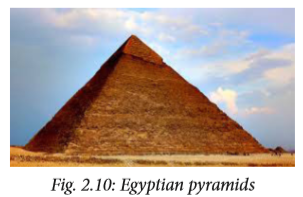
Compare the shape of the Egyptian pyramid above and the ecological pyramids discussed below.
Ecological pyramids are diagrams that show how important factors in an ecosystem such as energy, biomass and population size change at each trophic level. Traditionally, these diagrams place the primary producers (green plants) at the bottom. The highest trophic levels are placed at the top. The size of the portion of the diagram associated with each trophic level shows the amount of factor in consideration. A food chain can be expressed in a measurable way by using pyramid of numbers or pyramid of biomass.
Pyramid of biomass
This is a diagrammatic representation of mass or weight of organisms in each trophic level in a food chain. If the dry mass of all organisms at each trophic level of a food chain can be weighed, the mass can be used to draw a type of diagram called pyramid of biomass.
Biomass means the dry mass of any living material at any trophic level in a food chain. Biomass reduces as one moves from the producers to the various levels of consumers. This is the same trend observed with the amount of energy.
A pyramid of biomass is a chart drawn to scale showing the amount of biomass at each stage in a trophic level. The bars become narrower from the base to the top of the pyramid.

It is always hard to construct a pyramid of biomass because of the following reasons
• Measuring biomass often means death of the organism.
• Organisms may belong to more than one trophic level in an ecosystem. It is therefore not accurate to represent it with only one bar.
Note: When making a pyramid of biomass, you must use a scale.
Pyramid of numbers
The number of all organisms at each trophic level of a food chain can be counted. These numbers can be used to draw a diagram called pyramid of numbers. The number of organisms in each level can be obtained by totalling the population of all the species making up that level for instance:
• Total number of plants. (Producers)
• Total number of herbivores. (Primary consumers)
• Total number of carnivores. (Secondary consumer)
The pyramid is therefore a diagramatic representation of numbers of organisms in each trophic level in a food chain. In this way, it is possible to know the number of organisms that are capable of transferring energy from one trophic level to the next. The Figure below shows pyramid of numbers. Its shape as we can see is like that of a pyramid. This is called an upright pyramid.

The pyramid indicates that organisms transferring energy to the next energy levels decrease as we rise up. Sometimes the pyramid is not upright, for instance, if you were to construct pyramid of numbers using the tree as a habitat. It would be the only producer and the consumers such as caterpillars and birds would be many. If we were to construct pyramid of numbers, the smallest box would represent the tree. It would be at the bottom and not at the top. The shape of this pyramid would be inverted as can be seen in the figure below.


Self-evaluation Test 2.3
1. Plot the following data by placing the producers at the bottom. Use the scale 1cm = 50g/m2 for length and 1 cm height for each level.

(a) What does this pyramid indicate?
(b) Explain why the dry weight of the carnivores must be low.
2. If the total weight of producers in an ecosystem is 1000 kg, what would you expect the total weight of secondary consumers to be?
3. Study the food chain given below Oak tree -> insect larvae -> insectivorous birds
The food chain can be represented in _______ diagram.

(a) Which of the pyramids above (A, B, C and D) best represents this food chain?
(b) Identify the producer in the food chain.
(c) Name the source of energy entering this food chain.
(d) Name two main groups of organisms referred to as decomposers.
4. Do you agree with the statement below? ‘All food chains begin with green plants’ Explain your answer.
Unit summary
• Ecology is a branch of Biology that deals with the relationship between organisms and how they relate with their physical surroundings.
• Biotic factor is any living component that affects an organism.
• Abiotic factors are non-living conditions which influence where plants or animals live.
• A food chain is a linear sequence that shows how organisms dependent on one another for food.
• A food web is a system of interlocking and interdependent food chains.
• Trophic levels are the several hierarchical units in an ecosystem, comprising of organisms that share the same function and nutritional relationship in the food chain.
• The sun is the main source of energy in the ecosystem.
• Green plants are the producers in the ecosystem. They are able to manufacture their own food through the process of photosynthesis.


UNIT 3 : Passive movement of substances across a cell membrane
Key unit competency
To be able to explain the processes involved in the movement of water molecules and ions into and out of a cell.
Learning objectives
After studying this unit, I should be able to:
• Define diffusion and osmosis.
• Describe the importance of diffusion and osmosis.
• Explain turgor pressure.
• Investigate diffusion and osmosis through experiments.
• Appreciate the importance of turgidity in plant cells.
Introduction
Look at the diagrams below. Can you tell what is happening in flasks A, B, C and D?

In real life situations, what can we compare the observations above to? What does this tell you what this topic is about?
Our bodies are made of cells that carry out several metabolic and physiological processes. In order to carry out these life processes, a cell needs to take in various substances. It also produces certain substances, some of which are waste products which may be toxic and can harm the organism, hence need to be removed from cells.
Other products are useful to cells within the tissue. These useful substances are transferred to cells where they are needed for important metabolic processes like respiration.
Therefore substances are always moving into and out of the cells. The way substances move into and out of the cells depends on certain properties of the substances, for example, size of the molecule and the type of substance. There are three main physiological processes by which substances move in and out of cells. These are diffusion, osmosis and active transport.
3.1 Diffusion of gases and solutes
When a drop of ink is placed into a glass of water, the ink particles spread in the water until all the water is uniformly coloured.
You are also able to smell perfume that other people have worn because the particles of perfume diffuse from them through the air to our organs of smell, the nose.
Activity 3.1:
To investigate diffusion using copper (II) sulphate solution
Requirements
• Crystals of copper (II) sulphate
• 250 ml beaker
• Water
• Glass rod (open ended)
Procedure
1. Insert the open ended glass rod into the empty beaker.
2. Drop a crystal of potassium permanganate to the bottom of the beaker through the upper end.

3. With the glass rod still intact, pour water to fill the beaker.
4. Gently remove the glass rod so as not to tamper with the potassium permanganate crystal.
5. Make your observations.
6. Share your findings with the rest of the class.
Study questions
(a) What observation did you make when the glass rod is lifted from the beaker?
(b) Describe the movement of particles of the copper (II) sulphate crystals.
(c) Why does the movement occur?
(d) Other than using the above experiment, describe how you would demonstrate diffusion using perfume.

Diffusion involves movement of particles (ions or molecules) from a region of high concentration to a region of low concentration. This process continues until the particles are uniformly distributed throughout the system or until equilibrium is reached. Diffusion is a product of constant random motion (kinetic energy) of all atoms, molecules, or ions in a solution. The area with higher concentration of the particles has more random motion resulting to the net movement of the particles to the area with lower concentration.
Net movement of particles will always take place whenever there is a difference in concentration of particles between two regions. This difference is known as concentration gradient. Diffusion is important since it enables useful molecules to enter the cell and waste products to be removed.
Factors that affect the rate of diffusion
Discussion corner
1. What do you think affects the rate of diffusion?
2. Are the factors that affect the rate of diffusion related?

The rate of diffusion of particles refers to the time taken for the particles to move within an available space (fixed) until they are evenly distributed. Several factors affect the rate of diffusion. They include:
a. Temperature
When the temperature of particles is high, their kinetic energy increases and the particles move faster. Therefore, the higher the temperature, the faster the particles will diffuse while the lower the temperature the lower the rate of diffusion.
b. Concentration gradient
This is the difference in the amount of particles present in two regions. A greater difference in concentration of particles between two regions, results in a steeper concentration gradient which causes diffusion rate to be faster. When the concentration gradient is low, diffusion rate is also slower.

c. Size of molecules
Larger molecules are heavier and will diffuse at a slower rate compared to smaller molecules which are lighter.
d. Diffusion distance
The rate of diffusion depends on the distance that particles have to travel in order to be evenly distributed within the available space. An even distribution of particles is reached faster when the distance involved in diffusion is small compared to longer distances. It takes a longer time for molecules to diffuse across a thick membrane while It takes less time for molecules to diffuse across a thin membrane.
(e) Surface area to volume ratio
When the surface area to volume ratio is large, more of the substance diffuses across it than when it is small. This takes place as long as the concentration and temperature of the diffusing molecules remain the same.
Note: A larger surface area to volume ratio does not increase the speed of diffusion of the particles. It simply enables more particles to diffuse across it in a given time.
The figures below show two cubes, calculate the surface area (S.A) of the two cubes).

Also calculate the volume (V) of each cube. Divide the surface area (S.A) with the volume to obtain the surface area to volume ratio (SA/V).
• Which is bigger A or B?
• How will this affect the rate of diffusion?
Activity 3.2: Relating surface area to volume ratio with the size of an organism
Requirements
• Potato
• Razor blade
• Ruler
Procedure
1. Using the razor blade, cut five cubes of potato each of sides 1cm long. Name this cube X.
2. Cut another five cubes each of sides 3 cm long. Name this cube Y.
3. Calculate the surface area to volume ratio of the cubes.
Study questions
(a) Which cube has a smaller surface area to volume ratio?
• Calculate the surface area (S.A) of the two cubes.
• Also calculate the volume (V) of each cube.
• Divide the surface area (S.A) with the volume (V) to obtain the surface area to volume ratio (S.A)/(V) .
• Which is bigger X or Y? How will this affect the rate of diffusion?
(b) What is the significance of surface area to volume ratio?

Small organisms such as Amoeba have a greater surface area compared to volume than larger organisms, for example human beings. Therefore, diffusion of substances into and out of smaller organisms is faster than in larger organisms. Such small organisms can absorb oxygen and other materials from the environment much more rapidly than large ones. They also can excrete waste products at a faster rate than large organisms. In human beings and other larger organisms, diffusion of substance into their bodies would be slow. Therefore, their bodies have developed a complex system of transport called the blood circulatory system.
Discussion corner
1. Discuss with a classmate the importance of diffusion. Use the following guidelines:
• Gas exchange
• Excretion
• Absorption of materials in both plants and animals
2. Share your work with the rest of the class.
Importance of diffusion in plants and animals
(a) Plants absorb water, mineral salts and oxygen from the soil through the root hairs by diffusion.
(b) Digested food such as glucose and amino acids move from the small intestine into the blood of animals by diffusion. These substances move from the blood to the cells and tissues by diffusion as well.
(c) Cells and unicellular organisms such as Amoeba get rid of waste substances by diffusion.
(d) Diffusion is involved in exchange of gases in stomata, skin of frogs and in the lungs of animals.
Self-evaluation Test 3.1
1. Diffusion is a passive process, explain.
2. State the condition that must be in place for diffusion to take place.
3. The rate of diffusion increases if the
A. Temperature of solution decreases
B. Concentration gradient decreases
C. Viscosity of solution decreases
D. All of the above.
4. Concerning the process of diffusion, at equilibrium ______
A. Random movement of molecules continues.
B. The concentration of particles is equal throughout the solution.
C. Net movement of particles either side is equal.
D. The diffusion gradient increases.
3.2 Osmosis
Osmosis is the movement of water molecules from a region of high water potential (dilute solution) to a region of low water potential (concentrated solution) through a partially permeable membrane.
Activity 3.3: To demonstrate osmosis using visking tubing
Requirements
• 10 cm long visking tubing
• Distilled water
• Concentrated salt solution
• Capillary tube
• Two pieces of strings each measuring 30 cm
• 250 ml beaker
Procedure
1. Tie one end of the visking tubing using a string.
2. Open the other end of the visking tubing and half-fill it with the salt solution.
3. Tightly tie the open end of the visking tubing and allow part of the string to hang.
4. Fill the beaker with distilled water.
5. Gently put the visking tubing containing the salt solution into the beaker with distilled water.
6. Tie the hanging end of the string on the visking tubing onto the capillary tube and inverse the visking tubing into the distilled water
7. Support the capillary tube using a rhetort stand as shown below.

Note: The capillary tube level of the solution in the visking tubing and the beaker at the beginning of the experiment.
8. Leave the set-up undisturbed for at least 30 minutes.
9. Remove the visking tubing from the beaker and make your observations at the end of the experiment.
Study questions
(a) State three observations you made at the end of the experiment.
(b) Account for your observations in (a) above.
(c) Discuss your findings with your class members.
Since the concentration of solutions is defined in terms of solute concentration and not in terms of water content; water molecules diffuse from less concentrated solution (fewer solutes, more water) to a more concentrated solution (more solute, less water).
Activity 3.4: To demonstrate osmosis in a tuber
Requirements
• Fresh arrow roots, cassava, sweet potatoes or Irish potatoes
• Strong salt solution
• Distilled water
• Scalpel
• Large beaker or small basin
Procedure
1. Using a scalpel, peel a large Irish potato. You can also use an arrow root, cassava or sweet potato.
2. Cut off a piece so that it stands at least 6 cm high.
3. Cut and scoop out a deep hollow portion in its middle and pour strong salt solution halfway up the hollow portion.
4. Mark the level of the salt solution using a scalpel.
5. Place the potato in a petri-dish containing distilled water. Let it stand for several hours then note the level of solution in potato.
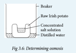
6. Repeat the experiment with boiled pieces of Irish potato, sweet potato, cassava or arrow roots.
Study questions
(a) Draw a diagram to represent the results of the experiment.
(b) Is the level of the salt solution still the same at the end of the experiment?
(c) Explain what brings about the change in level of the salt solution.
(d) Compare these results with those when boiled Irish potato is used.
Activity 3.5: To investigate osmosis in a plant tissue
Requirements
• Tubular part of a pumpkin leaf or black jack stem
• Distilled water
• 5% sucrose solution
• Pair of scissors
• Ruler
• Three Petri-dishes
• Two 200 ml beakers
• Labels • Pair of forceps
Procedure
1. Using a ruler measure 4 cm of the tubular part of the leaf or stem and cut using a pair of scissors.
2. Repeat the procedure (1) to obtain two other pieces of the leaf or stem.
3. Make four cuts, each 2 cm long in each of the cut pieces of the leaf or stem.
4. Put 100 cm3 distilled water into a petri-dish and label water.
5. Put 100 cm3 of 5% sucrose solution into another petri-dish and label sucrose solution.
6. Put one of the cut pieces into the petri-dish containing water, another in the petri-dish containing sucrose solution and the last one in an empty Petridish labelled air.
7. Leave the set-up undisturbed for 25 minutes.
8. Remove the pieces of the tubular leaf from their respective conditions using a pair of forceps.
9. Hold the pieces from distilled water and sucrose solution between your fingers and record your observations in a table below.
Table 3.1: Results for osmosis in plant tissues

11. Share your findings with the rest of the class.
Study questions
(a) Draw the pieces as they appear after the experiment.
(b) Account for any differences that you have observed for the three pieces of leaves.
(c) What was the purpose of putting one piece in the petri-dish exposed to air?
Types of solutions
From your knowledge of chemistry; can you recall what the word solution means? What about solute and solvent?
When a solid is dissolved in water, we get a solution. The solid that is dissolved in this solution is called the solute. The liquid that dissolves the solid is known as the solvent.
Solute + Solvent = Solution
Concentration of a solution depends on the amount of solute dissolved. A dilute solution has more water molecules compared to solute molecules whereas a concentrated solution has more solute molecules than water molecules.

Suppose a dilute solution is separated from a concentrated solution by a semi-permeable membrane as shown in figure 3.8. Water molecules will move from the dilute solution to the concentrated solution. This is because the dilute solution has more water molecules than the concentrated one. Water molecules are very small and pass easily through the channels or pores of the cell membrane. On the other hand, solute molecules are too large to pass through the pores.

Note:
• The concentration of water in a solution containing mixtures of different solute molecules depends only on the total solute concentration and not the types of solutes.
• If the total concentration of all ions and solutes on both sides of a membrane are the same, there will be no osmosis.
It is important to first understand the terms used to describe the solutions of different concentration with respect to cells function.

Water relations in plant cells
Discussion corner
1. What will happen to an animal cell if placed in:
• a hypertonic solution?
• a hypotonic solution?
• isotonic solution?
2. Compare the structures of an animal cell and a plant cell in the cases above.
3. Draw the structure of turgid plant cell and plasmolysed plant cell.
4. Share your findings with other class members.
Living cells are surrounded by a fluid medium which may be isotonic, hypertonic or hypotonic to the cell contents. If the fluid is isotonic, there will be no net movement of water into or out of the cell. If the external fluid is hypertonic to the cell contents, then, water leaves the cell. If it is hypotonic, then water enters the cell. The movement of water into and out of the cell and the effects that such movements have on the cell may be described as water relations in the cell.
In the next section, we find out how various solutions affect how plant cells behave.
(a) Plant cells in hypotonic solutions
Plant cells have a large vacuole that contains a fluid called cell sap. The sap contains salt and sugar molecules. If a plant cell is surrounded by a hypotonic solution, water molecules move from the surrounding fluid through the cell wall and cell membrane into the vacuole by osmosis.
As it receives water, the vacuole, swells and pushes the cytoplasm and nucleus outwards against the cell wall. This pressure exerted by the cell contents against the cell wall is called turgor pressure. As turgor pressure increases due to intake of more water by osmosis, the cell wall exerts a pressure that is equal to turgor pressure on the protoplasm called wall pressure.
A point will reach when no more water can enter the plant cell. At this point, the wall pressure is equal to turgor pressure but opposite in direction. Because the cell wall is made of rigid cellulose material, it does not stretch very much and the cell does not burst.

However, turgor pressure causes the cell to become stiff or firm. Such a cell is described as being turgid. A plant in which all the cells are turgid always appears firm and erect.
Turgidity in plant cells is important because the stiff cells give support to the soft tissues such as petals and sepals. Turgor pressure enables soft, nonwoody plant stems to remain upright despite the downward force of gravity. If there is inadequate water in the environment, turgor pressure cannot be maintained in a plant.
(b) Plant cells in hypertonic solutions
A plant cell that is surrounded by a hypertonic solution will lose water. Water is lost from the cytoplasm then from the vacuole. The turgor pressure in the cell begins to decrease. If this continues, the cell membrane and cytoplasm shrink away from the cell wall. The vacuole in turn reduces in size.
The moving of the cell membrane and cytoplasm away from the cell wall is called plasmolysis. The cell is said to be plasmolysed. When a plasmolysed cell is placed in distilled water, it will become turgid again. The cell is said to undergo deplasmolysis. The point at which plasmolysis just begins is called incipient plasmolysis. At this point, the cell protoplasm that is, cell membrane and cytoplasm no longer exerts any pressure against the cell wall. The turgor pressure is zero, and the cell therefore loses its turgidity. It is now a flaccid cell.

Plants that are deprived of water for several days have their leaf cells plasmolysed. The overall effect is a drooping plant which is said to be wilting. If this persists for long, the plant cells become flaccid. The plant then dies. However, if the plant is well watered before the cells become flaccid, its turgidity is restored.
Note:
Turgidity in plant cells is caused by osmotic flow of water from an area of low solute concentration to outside the cell into the cell vacuole.
The cell vacuole has a higher solute concentration. Healthy plant cells are turgid. Plants rely on turgidity to maintain rigidity and stand upright.
Activity 3.6: To investigate turgor pressure in plant cells
Requirements
• Raw Irish potatoes
• Boiled Irish potato
• Distilled water
• Sucrose
• Spatula
• Glass rod
• Cork borer
• 10 cm ruler
• Four 200 ml beakers
• Labels
• Scapel
Procedure
Your teacher will help you to prepare these solutions:
1. Prepare 5% sucrose solution by dissolving 17.1 g of sucrose in 100 ml of water in a beaker. Stir the solution well until all the sucrose dissolves. Label this 5% sucrose.
2. Prepare 20% sucrose solution by dissolving 68.4g of sucrose in 100 cm3 of water in a beaker.Stir the solution well until all the sucrose dissolves. Label this 20% sucrose.
3. Put 100 cm3 of distilled water in the other empty beaker and label it distilled water.
4. Peel three Irish potatoes; insert the cork borer through one end of the potato and press hard so it comes out through the other end of the potato to produce a cylinder. Cut off the rough end of the potato cylinder and measure 6 mm, cut off the remaining part.
5. Repeat procedure (4) above so as to obtain at least six potato cylinders.
6. Put 2 potato cylinders in each of the three solutions in the beakers.
7. Put the boiled potato tissue in the fourth beaker containing distilled water.
8. Leave the set-ups uninterrupted for 45 minutes.
9. Prepare a table like the one shown below to record your observations.

10. Remove the cylinders from the different solutions and measure their final length using the 10 cm ruler. Record your results in the table you have prepared.
11. Compare your findings with those of other class members.
Study questions
(a) Work out the change in length of the potato cylinders in:
• Distilled water with raw potato tissue.
• 5% sucrose solution.
• 20% sucrose solution.
• Distilled water with boiled potato tissue.
(b) Account for the change in length in (a) above.
(c) Why was it necessary to include the set up with boiled potato tissue in the experiment?
Role of osmosis in plants and animal cells
Discussion corner
1. Discuss the following with a classmate.
• How are plants supported by turgor pressure?
• What is the importance of osmosis in plant and animal cells?
2. Share your findings with other members of your class.

1. Uptake of water by roots Plants absorb water from the soil inner cells in the root.

Fig. 3.11: Movement of water from cell to cell in tissues
2. Opening and closing of stomata
When guard cells gain water by osmosis from the surrounding cells, they become turgid. This changes their shape, making the stomata to open. As a result:
by osmosis. The cell sap in root hair cells usually has a higher solute concentration than water in the soil. Therefore, root hair cells are able to take up water from the soil by osmosis.
3. Movement of water from cell to cell in tissues
When a cell takes up water by osmosis, its solute concentration becomes lower (it becomes more dilute) compared to the cell adjacent to it. As a result, water moves from it to the adjacent cell whose solute concentration is higher (more concentrated). In this way, water moves from the root hair cells to the
• Gaseous exchange takes place through the open stomata.
• Water vapour also exits through the open stomata.
When guard cells lose water through osmosis to surrounding cells, their size and shape changes. This makes the stomata to close.

4. Feeding in insectivorous plants
Insectivorous plants such as Pitcher plant prey on insects.

They trap insects when there is a sudden change in their turgor pressure when disturbed by the insect.
Self-evaluation Test 3.2
1. Which one of the following describes osmosis.
A. Movement of sugar molecules.
B. Movement of water molecules,
C. Movement of sugar and salt molecules.
D. Evaporation of water.
2. What will happen to an animal cell when it is placed in an isotonic solution?
A. Shrink
B. Burst
C. Remain the same
D. Expand
3. What kind of a membrane is partially permeable?
A. A membrane made of plant tissue.
B. A membrane that is made up of water molecules.
C. A membrane that allows certain substances to go into and out of cells.
D. A membrane that surrounds a food vacuole.
4. A learner set-up an experiment shown below to investigate osmosis.

(a) Draw the diagram to show the results of this experiment.
(b) Account for observations you made in (a) above.
(c) What does the visking tubing represent in a plant cell?
5. An animal cell bursts when placed in water but a plant cell does not. Explain
6. Using your knowledge of plant and animal cell structure. Explain why plant cells have regular shapes while animal cells have irregular shapes.
Unit summary
• Passive movement of substances into and out of cells involves diffusion and osmosis. These processes involve kinetic energy of the molecules involved.
• Diffusion is the movement of particles from an area of higher concentration to one of lower concentration along a concentration gradient. These particles move by their natural kinetic energy.
• A concentration gradient is the difference in solute concentration between two points separated by a distance.
• The end result of diffusion is a uniform distribution of the particles (molecules or ions).
• Factors that influence diffusion include: temperature, concentration gradient, surface area and distance.
• Osmosis involves movement of water molecules from a region of their higher concentration to a region of their lower concentration across a semi-permeable membrane.
• Osmotic pressure is a force generated to prevent movement of water molecules from a solution across a partially permeable membrane.
• Plant and animal cells behave differently when placed in an isotonic solution, hypertonic and hypotonic solution.
• Osmosis enables uptake of water into the plant through the roots, turgidity in plant cells and opening and closing of stomata.
• Turgidity in plant cells provides support to plant tissues.


UNIT 4 : Active transport
Key unit competency
To be able to analyse and interpret the process of active transport and its significance to living organisms.
Learning objectives
After studying this topic, I should be able to:
• Define active transport.
• Compare passive and active transport.
• Appreciate the importance of active transport.
• Describe how carrier proteins move particles across membrane during active transport.
Introduction
Study the pictures A and B below.

What is going in the pictures? Which is easier, climbing uphill or going downhill? Why is that the case? How do actions in the pictures relate to active transport? Think about how things are transported in our bodies. Are there special things that help in the transportation process? What do they do?
4.1 Active transport and its importance
The types of movement of molecules and ions that we have considered in Unit 3 involved molecules moving down or along a concentration gradient. These movements are passive hence name passive transport. Molecules and ions can also move from an area of low concentration to that of higher concentration. They are said to move against a concentration gradient. Such a process requires the use of energy and is called active transport.
Discussion corner
1. You will be provided with Manila paper, marker pens and cello tape.
2. Prepare a table on the Manila paper.
3. Discuss and write the differences between passive and active transport.
4. Share your findings with the rest of the class.

The table below shows the difference between passive and active transport.

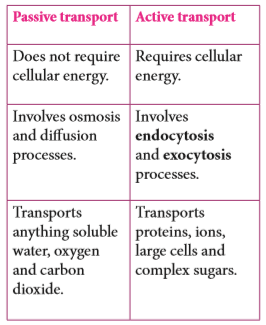
Active transport is the movement of particles through the cell membrane from a region of low concentration to a region of high concentration using energy. This process requires the expenditure of energy since particles move against a concentration gradient.
The role of proteins in active transport
The movement of substances across a membrane by active transport requires carrier protein molecules. They pick up molecules of a substance from one side of the cell membrane and transport them across.
Carrier protein molecules have binding sites for the molecules. The molecules to be transported bind to the carrier protein using energy. The carrier protein changes shape and the particles are transported to the other side of the membrane; and they diffuse away from the carrier protein.

Activity 4.1 Demonstrating active transport
You will be provided with charts and animated pictures involving active transport.
1. Observe the charts and animations carefully.
• Can you tell what is taking place in the pictures?
• Which part of the body is the process taking place?
• What is the role of proteins in active transport?
2. Record what you see in the pictures.
3. Discuss with your partner the importance of the process involved.
Study questions
(a) What is the role of active transport in organisms?
(b) Which factors affect active transport?

In active transport, the cell must use its own energy to move the molecules against a concentration gradient. Such energy is supplied by the cell’s mitochondria. Therefore active transport takes place only in living cells.
Role of active transport in living organisms
1. Through active transport, root hair cells in plants absorb mineral salts and ions from the surrounding soil. This takes place even when the concentration of these mineral salts is already higher in the cells than in the soil.
2. The cells lining the human small intestines continue to absorb food molecules by active transport even when the concentration of these molecules is higher in the cells than in the intestinal lumen.
3. Nerve cells need sodium ions and potassium ions to function. The concentration of sodium ions outside a nerve cell is higher than the concentration on the inside. The concentration of potassium ions on the outside is lower than on the inside. The nerve cells maintain these concentrations in this way by active transport.
4. When urine is first formed in the kidney, it contains useful substances like glucose in addition to the waste substances. The useful substances are reabsorbed into the blood by active transport.
5. Active transport is involved in accumulation of substances in the body of some organisms to offset osmotic imbalances in arid and saline environments.
Factors that affect active transport
Any factor that affects energy production also affects active transport.
a. Concentration of oxygen
At higher oxygen concentration, the cells produce more energy. This leads to rapid active transport by the protein carriers.
b. Concentration of glucose
The amount of energy produced by a cell depends on the amount of glucose available. When more glucose is available, the cells produce more energy. This leads to rapid active transport.
c. Temperature
High temperatures can destroy (denature) the protein carriers in the cell membrane. This stops active transport. Active transport works best under normal temperature which in human beings is 37ºC.
d. Enzyme inhibitors
An enzyme is a substance which speeds up reactions taking place in the cell. Enzyme inhibitors are chemical substances which ‘poison’ or make an enzyme inactive. If enzymes involved in the release of energy during respiration are affected, there will be lack of energy. This will prevent active transport from taking place.
e. pH
Abnormal changes in pH in the environment of the cell can alter the structure of the protein carriers. This will reduce their ability to transport molecules across the cell membrane.
Self-evaluation Test 4.1
1. Absorption of mineral salts from the soil to the root hair requires energy. Which of these describe the process used?
A. Diffusion
B. Osmosis
C. Active transport
D. Respiration
2. Which one is a function of the proteins in the cell membrane?
A. Communication
B. Cellular respiration
C. Cell transport
D. Cell repair
3. The root of a certain plant was treated with a respiratory poison.
(a) State the effect of this to the uptake of mineral ions.
(b) Explain the effect you have stated in (a) above.4.2 Endocytosis and Exocytosis
The movement of macromolecules such as proteins or polysaccharides into or out of the cell is called bulk transport. There are two types of bulk transport, exocytosis and endocytosis, and both require the expenditure of energy.
Activity 4.2: Investigating the process of endocytosis and exocytosis
You will be provided with charts, computer simulations and animations.
1. Use the charts, computer simulations and animations to illustrate the process of endocytosis and exocytosis.
2. Discuss with your group members the process involved.
3. Share your findings with the rest of the class.
Study questions
(a) Differentiate between phagocytosis, exocytosis and pinocytosis.
(b) How important are these processes in a cell.
Endocytosis
This is the in folding of the cell surface membrane to form a vesicle or vacuole. A portion of the membrane wraps around the particle and fuses. Thereafter the particle is surrounded by the plasma membrane. The portion of the membrane containing the particle then pinches off leaving the plasma.
Endocytosis involves the bulk uptake of materials through the plasma membrane. It takes place in two ways: phagocytosis and pinocytosis.
(i) Phagocytosis
Phagocytosis means ‘cell eating.’ This is a process by which plasma membrane extend to form a depression. The depression encloses solid foreign particles. The food vacuole formed moves towards the interior of the cell. The process is selective and specific to solid materials engulfed. An example is white blood cells that engulf bacteria in our bodies.

(ii) Pinocytosis
Pinocytosis means ‘cell drinking.’ This process is similar to phagocytosis only that the cells take in materials in liquid form. In this case molecules dissolved in liquid are taken up by formation of smaller vesicles compared to phagocytic vesicles.

Exocytosis
This is the process by which materials are removed from the cells. Materials removed include solid particles, undigested remains from food vacuoles among others. The molecules or cell secretions accumulate in secretory vesicles, which then moves and fuses with the plasma membrane.
The contents of the vesicle are then expelled out of the cell.
Examples of exocytosis:
• Secretion of digestive enzymes by pancreas.
• Secretion of milk from mammary glands.
• Secretion of mucus by salivary glands.
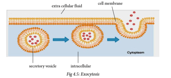
Self-evaluation Test 4.2
1. State one difference and one similarity between endocytosis and active transport.
2. A white blood cell ingests solid particles by forming vesicles. This describes which process?
3. Given these characteristics:
(i) Requires carrier proteins
(ii) Requires energy
(iii) Requires membrane channels
(iv) Requires vesicles Choose the characteristics that define exocytosis.
Unit summary
• Active transport involves the use of energy to move molecules across the cell membrane. During active transport, molecules are moved against a concentration gradient.
• Factors affecting active transport include: concentration of oxygen and glucose, temperature, enzyme inhibitors and pH.
• Protein carrier molecules, which are part of the cell membrane, are involved in active transport.
• Endocytosis is the process of capturing a substance or particle from outside the cell by engulfing it with the cell membrane, and bringing it into the cell.
• Exocytosis is a form of active transport in which a cell transports molecules out of the cell by expelling them through the plasma membrane.


UNIT 5 : Identification of food components
Key unit competency
To be able to carry out chemical tests on a variety of foods to identify the nature of food substances.
Learning objectives
After studying this topic, I should be able to:
• State the chemical reagents used in the identification of each of the classes of food.
• Carry out tests to identify food substances in a given sample.
• Appreciate the importance of different classes of food in one food substance.
Introduction
Have you ever visited a tea or coffee processing factory? What happens during grading of coffee or tea? Look at the picture below. What is the person using to tell whether the coffee of great quality?

Coffee or tea is tasted to determine its quality during processing. The fragrance of the brewed tea or coffee, the deep flavours when drinking and the pleasant after-taste are used to tell the quality.
Based on this, do you think it is possible to know the components of the various foods that we eat by tasting? Why? What can we use to do this?
5.1 Components of food substances
The foods that we eat contain different types of nutrients. These nutrients serve different functions in the body. It is therefore essential that we know the components of the food that we eat in order to live healthy lives.
Drinks and foods that are manufactured in factories need to be tested as well. This is done to confirm the type and percentage of nutrients they contain. This enables the consumer to be informed on the product contents.
Food contains mainly two classes of nutrients; organic and inorganic nutrients. The inorganic nutrients include mineral salts like calcium, phosphorous and others like water. The organic nutrients include proteins, carbohydrates, lipids and vitamins.
Chemical test for food substances
The process by which foods and drinks are taken to the laboratory to confirm the type of food components in them is called food testing. Food testing is done to confirm the presence of starch, reducing sugars, proteins, fats and vitamin C in a food sample.
5.2 Testing for starch and reducing sugars
a) Test for starch Starch
is a polysaccharide comprising of glucose monomers linked together. Starch is insoluble in water. It does not have a sweet taste and on hydrolysis, it readily converts back to monosaccharides. Some foods containing starch include sweet potatoes and cassava.

Activity 5.1 Testing for starch
Requirements
• Iodine solution (brown)
• Test tubes and test tube rack
• Droppers • Water
• Starch solution (sweet potato, or plantain extracts)
• Glucose solution
• Measuring cylinder
Procedure
1. Using the measuring cylinder, measure about 2 cm3 of the following solutions into separate test tubes: starch solution, glucose solution and water.
Precaution: Each time, rinse the measuring cylinder thoroughly before use.
2. To each test tube, add a few drops of iodine solution and note any colour change.

3. Record your results in the table below


Starch is the storage form of glucose in plants. Each starch molecule has about 300 - 1000 glucose units. Most starch in plants is found in seeds and storage organs like potato tubers. Starch gives a blue-black colour with Iodine in potassium iodide solution. This is the laboratory test for starch. The colour of Iodine turns from brown to blue-black if starch is present. If there is no starch in the food sample, the brown colour of iodine persists.
Money matters!
You should not waste laboratory reagents because they are usually very expensive.
b) Testing for sugars
Based on their reaction with Benedict’s solution sugars are grouped into two.
• Reducing sugars
• Non-reducing sugars
Reducing sugars
Some simple sugars, for example glucose can be made to reduce copper in blue copper sulphate to red copper oxide. This reaction can be used as a test for some sugars. Copper sulphate is mixed with other chemicals to be used as Benedict’s solution which is used in the test. Other examples of reducing sugars
include: maltose, lactose, fructose and galactose.
All monosaccharides are reducing sugars, they taste sweet and are soluble in water.
Activity 5.2: Test for reducing sugars
Requirements
• Benedict’s solution
• Test tubes and test tube rack
• Glass rod and a spatula
• Dropper
• Labels
• Conical flask (100 cm³) or small beaker
• Motor and a pestle
• Source of heat
• Food extracts, for example, banana juice
• Glucose, starch and sucrose solutions
• Distilled water
Procedure
1. If the food to be tested is in liquid form, go straight to procedure (2). If it is solid do the following to form an extract:
(a) Grind, crush (using a motor and pestle) or chop the solid food subtance.
(b) Scoop a small amount using a spatula and put into a test tube to a depth of about 2cm.
(c) Add a similar amount of distilled water and stir with a glass rod.
(d) Allow to stand for a few minutes.
2. Using a dropper put 2 cm3 of each of the solutions to be tested into separate test tubes.
3. Label the test tubes with the solutions in them.
4. To each test tube, add 2 cm3 of Benedict’s solution.

5. Heat for about two minutes and observe the colour change.
6. Record your observations in the table below.


Benedict’s solution is the chemical reagent used to test for the presence of reducing sugars. When boiled with glucose, which is a reducing sugar, an orange precipitate forms.
A change in colour from blue to green, yellow and finally red indicates the presence of reducing sugar. The colour changes depend on the amount of reducing sugars present.

• Green colour indicates small amounts of reducing sugars.
• Yellow/orange shows high levels of reducing sugars.
When Benedict’s solution, which is blue in colour, is boiled with substances with non-reducing sugar, its blue colour persists.
Note: Water acts as the control.
Health Check!
• Wear eye protection for eye safety.
• Take care while heating and handling test tubes. This minimises risk of laboratory accidents.
• Do not consume any food provided in the laboratory, it may be poisoned or contaminated.
Non-reducing sugars
Some sugars such as sucrose are unable to reduce the copper ions in Benedict’s solution. This makes the colour of Benedict’s solution to persist when these sugars are boiled with it. Such sugars are called non-reducing sugars. Most nonreducing sugars are disaccharides. This means they contain two single sugar units.
Activity 5.3: Testing for nonreducing sugars
Requirements
• Benedict’s solution
• Dilute hydrochloric acid
• 10% sucrose solution
• Sodium hydrogen carbonate solid
• Four test tubes and a test tube rack
• Water
• Means of labelling
• Measuring cylinder
• Dropper
Procedure
1. Arrange the four test-tubes in a test tube rack and label them 1 - 4.
2. Put 2 cm3 of sucrose solution into the test tube labelled 1 and add 2cm3 of Benedict’s solution then boil.
• Note down any observations.
3. Measure 2 cm3 of dilute hydrochloric acid and pour it into the test tube labelled 2. Add to it 2 cm3 of Benedict’s solution and boil.
• Note down any observations.
4. Follow the following procedure for test tube labelled 3.
(i) Measure 2 cm3 of sucrose solution into the test tube. Add a few drops of the dilute hydrochloric acid and boil.
(ii) Allow the contents of the test tube to cool, and then add a little sodium hydrogen carbonate slowly until the fizzing stops.
Note: The sodium hydroxide helps to neutralise the excess acid in solution.
(ii) Measure 2cm3 of Benedict’s solution and add it into the test tube then boil.
• Note down your observations.
5. Compare the colour of the contents of test tube 3 with that in test tube 1 and test tube 2.
6. Record your result in a table similar to table below.


There are no special reagents used to test for non-reducing sugars. The same reagents used for reducing sugars are instead applied. This time hydrochloric acid is also added to hydrolyze these non-reducing sugars to reducing form. Later when Benedict’s solution is added, the test becomes positive.
There is no change in the colour of Benedict’s solution when it is heated with sucrose solution. This is because sucrose is not a reducing sugar. Benedict’s solution changes colour only in the presence of a reducing sugar. Boiling the sucrose solution with dilute hydrochloric acid hydrolyses sucrose to glucose and fructose. Both of these are reducing sugars and as such, the colour of the Benedict’s solution changes from blue to orange/brown precipitate.
Any excess acid is neutralised by the sodium hydrogen carbonate. Boiling is a requirement for the hydrolysis to occur. Heating the Benedict’s solution with the acid is to rule out the possibility of the acid being the one changing the colour of Benedict’s solution from blue to orange.
Note: Any excess hydrogen carbonate does not affect Benedict’s solution.
Self-evaluation Test 5.1
1. With examples, distinguish between reducing sugars and non-reducing sugars.
2. What is the purpose of sodium hydrogen carbonate in the test for non -reducing sugars?
3. Starch is stored in seeds, stems and roots among many other places in plants. Where is starch stored in animals?
5.3 Testing for proteins
Proteins are essential nutrients for the human body. They are one of the building blocks of the human tissue and can also serve as a fuel source. They contain as much energy as carbohydrates but less than lipids. Amino acids are the building blocks of proteins. Most of the foods we eat contain these amino acids. Milk, eggs, meat, beans and peas are examples of foods that contain proteins.

There are two major ways in which scientists test for the presence of proteins in food. They are:
• Millon’s test
• Biuret test
(a) Millon’s test
In this test a reagent known as Millon’s reagent is used to test for proteins.
Activity 5.4: To test for the presence of proteins using Millon’s reagent
Requirements
• Test tubes and test tube rack
• Source of heat
• Dropper
• Measuring cylinder
• Syringes
• Cooking oil
• Egg albumen
• Distilled water
• Milk
• Millon’s reagent
Procedure
Carry out the following procedures and record your observations and deductions in the table below.


The table below shows the results that may be obtained during a Millon’s test

Health Check!
Care should be taken while boiling to avoid laboratory accidents. Millon’s reagent is very poisonous! Be careful when handling it
Quality Check!
Buying quality apparatus reduces the risk of breaking and therefore it is cost effective.
b) Biuret test
The Biuret’s test uses several reagents that are mixed in equal proportions. Copper (II) sulphate and sodium hydroxide make up the reagent. The normal colour of the reagent is blue. The reagent turns violet in the presence of peptide bonds.
These bonds hold amino acids together to form a protein.
Activity 5.5 Test for proteins using Biuret reagent
Requirements
• Soya bean solution
• Distilled water
• 10% sodium hydroxide solution,
• 1% copper sulphate solution (Biuret reagent)
• Four test tubes
• Means of labelling the test tubes
Note: No heating is involved during the Biuret test.
Procedure
1. Place 2 cm3 of soya bean, distilled and water and orange juice into three separate test tubes.
2. To each of these test tubes, add 2 cm3 of sodium or potassium hydroxide solution.
3. Add 1cm3 of 1% copper sulphate solution to each test tube then shake.

4. Record your observations in a table similar to the one given below.

Study questions
(a) What colour change was observed in each of the test tubes?
(b) What is the role of the test tube with distilled water?

Copper sulphate is pale blue in colour. In the presence of a protein, its colour changes to purple. The appearance of purple colour, therefore, is a confirmation for the presence of protein in a food sample.
Self-evaluation Test 5.2
1. The _________ test uses several reagents. The reagents are mixed in equal proportions. _________ and __________ make up this reagent.
2. The bonds that hold protein structure are known as _________
3. To test for proteins, two reagents are used. Millon’s reagent and Biuret reagent. Which of these two tests does not involve heating?
5.4 Testing for lipids
Lipids are composed of fats and oils. Fats are solid at room temperature while oils are liquid at room temperature. Cooking oil, animal fats, groundnuts, meat among others, all contain lipids.

Two types of tests can be carried out on a food sample to identify if it contains lipids. These are:
• The ethanol emulsion test
• The translucent paper mark test
(a) Ethanol emulsion test
In this procedure ethanol is used as the reagent. Water is also added. A white emulsion confirms the presence of lipids in the food sample.
Activity 5.6: Testing for presence of lipids using the ethanol emulsion test
Requirements
• Test tubes and test tube rack
• Droppers
• Ethanol
• Distilled water
• Cooking oil
• Groundnut extract solution
Procedure
Carry out the following tests and record your observations and deductions in the table below.

Study Questions
(a) Which food components are found in cooking oil?
(b) Why is it important to eat foods containing lipids?
(c) Which health problems are related to too much lipids in the diet?

An emulsion forms when the solution of oil and alcohol is shaken with water. This is because the oil does not dissolve in water. A cloudy or white liquid is formed. If a food sample is used, the alcohol extracts the oil from the food by dissolving it.
(b) The translucent spot test
Activity 5.7: Testing for presence of lipids using the translucent spot test
Requirements
• Vegetable oil or cooking fat
• Blotting or absorbent paper
Procedure
• Put a drop of oil or fat on some absorbent paper.
• Hold it up against light from the window and note what you observe.

A translucent spot indicates presence of oil or fat.
5.5 Testing for vitamin C
A reagent known as Dichlorophenol indophenol (DCPIP) is used to test for vitamin C. DCPIP is a deep blue reagent in colour. When vitamin C is present in a food sample, the blue colour disappears (decolourised). Fresh fruits and green vegetables contain vitamin C. Table 5.8 shows the test for vitamin C using DCPIP.

Activity 5.8

Further Activity 5.9
You are provided with the following:
• Specimen S which is an orange, • Millon’s reagent • DCPIP
• Specimen J which is milk • Source of heat • Scalped • Test tubes and test tube rack
• Distilled water • Droppers • Syringe
• Iodine solution • A stirrer
Procedure
1. Cut a transverse section of specimen S and obtain about 10 cm3 of juice from the specimen by squeezing into a test tube.
2. Mix the orange juice in 1 above with the milk to form a solution. Label the solution Extract A.
3. Carry out the following tests on this extract A using the reagents provided and record your observations in table like the one shown below:

Study questions
(a) From your observations name the food substance(s) present in the extract.
(b) What is the biological significance of the food substance named in (a) above?
Self-evaluation Test 5.3
1. Name diseases that result due to lack of vitamin C in the diet.
2. Why is it not advisable to take too much food containing lipids?
3. Which of the following substances is the most abundant compound in a living cell?
A. Fats
B. Water
C. Proteins
D. Carbohydrates
Unit summary
• Starch is a polysaccharide that functions as a carbohydrate store and is an important constituent of the human diet.
• Food testing is the process that is carried out to determine the food components in a food sample.
• Starch is tested in a food sample by using Iodine solution. If it turns blue-black, it indicates the presence of starch.
• Benedict’s solution is used to test for the presence of reducing sugars. Reducing sugars change the colour of Benedict’s solution from blue to orange.
• Non- reducing sugars do not change the colour of Benedict’s solution. Unless they are first boiled with dilute hydrochloric acid to hydrolyse them.
• Presence of proteins in a food sample can be tested by Biuret test or Millon’s reagent.
• The alcohol –emulsion test is used to test for presence of lipids in a food sample.
• Presence of vitamin C is tested by a chemical called DCPIP.


UNIT 6 : Enzymes
Key unit competencyTo be able to explain the role of enzymes in living organisms and how they are affected by temperature.
Learning objectives
After studying this unit, I should be able to:
• Define the term catalyst and enzyme.
• Describe why enzymes are important in all living organisms.
• Explain the factors that affect enzyme activity.
• Draw and interpret graphs for the rate of enzyme activity.
• Appreciate the importance and specificity of enzymes in speeding up reactions.
Introduction
Look at the photographs below. Can you identify what is in the photographs? What is the relationship between them?

Try washing off the stain in a piece of cloth with water only, then using the detergent. Where was it easy to remove the stain?
Detergents which are used to remove stains act as enzymes. During the washing process, water and dirt are quickly combined together when the detergent is available to form a new product. This new product is easily removed from the cloth as we wash. On the other hand, water without detergent or soap can take a long time to remove the stains.
Most metabolic reactions in the body are catalysed by enzymes; hence increasing efficiency of these processes in living organisms. It is important therefore to understand the nature, properties of enzymes and how they function.
Enzymes are biological catalysts that are protein in nature. They speed up or slow down the rate of chemical reactions in the body without being used up themselves.
6.1 Types of enzymes
Some enzymes are used within the cells that produce them. These are called intracellular enzymes, for example, respiratory enzymes and catalyse enzyme in the liver cells.
Other enzymes are transported from the cells that produce them to be used in other cells. These are called extracellular enzymes. Some examples of extracellular enzymes are those secreted into the alimentary canal to help with digestion.
There are also different types of enzymes based on the foods they act on, for example:
• Peptidases: Break down proteins
• Lipases: Break down lipids
• Carbohydrases: Break down carbohydrates
Activity 6.1: To investigate the presence of enzymes in living tissues
Requirements
• Samples of liver, potato or yeast
• 8 cm3 volume hydrogen peroxide
• Forceps, pestle and mortar
• Wooden splint
• Matchbox
• Fine sand
• Test tubes
• Labels
Procedure
1. Label 4 test tubes as A, B, C and D.
2. Measure 2cm3 of hydrogen peroxide solution and pour into each of the labelled test tubes.
3. Into test tube A, do not add anything.
• Note any changes or observations.
4. Into test tube B, add some sand. Shake the test tube and note down any changes.
5. Into test tube C add some pieces of potato that have been ground into a paste (using a pestle and mortar).
• Shake and observe what happens.
• Test for oxygen with a glowing splint at the mouth of the test tube.
6. Repeat procedure (5) in test tube D using some liver which has been cut up then pounded in a mortar using a pestle.
7. Record your observations in a table like the one below.


Living tissues contain an enzyme called catalase which breaks down hydrogen peroxide to water and oxygen.

The gas produced relights a glowing splint as seen in procedure 5 and 6. No gas is produced in Test tube 1 and 2 which do not contain living tissue.
6.2 Characteristics of enzymes
Discussion corner
1. Discuss with a classmate the characteristics of enzymes. Use the following questions as your guidelines:
• What are enzymes made of?
• What factors affect enzyme action?
• Do enzymes act on all substrates?
• Do enzymes change after a reaction?
2. Share your findings with other class members.

1. Enzymes are protein in nature: all enzymes are made up of proteins.
2. Enzymes are affected by temperature. They work best at specific temperatures; for example, enzymes found in human bodies work best at 37oC. This is called the optimum temperature.

• Very low temperatures inactivate enzymes. Therefore enzymes are not able to catalyse reactions.
• High temperatures beyond the optimum temperature denature enzymes. The structure of the protein molecule is destroyed by heat.
3. Enzymes work best at specific pH. Different enzymes have a given specific pH at which they act best.
This pH is called optimum pH. Some enzymes work best at low pH (acidic medium) while others work best at high pH (alkaline medium). Most enzymes in the human body for instance work best at neutral or slightly alkaline pH. Examples are: lipases, peptidases and amylase. A few enzymes like pepsin that digests proteins in the stomach works best at an acidic pH of 2.

4. Enzymes remain unchanged after catalysing a reaction. Enzymes are catalysts and can therefore be used over and over again in small amounts without being changed.
5. Enzymes catalyse reversible reactions. This means that they can change a substrate into products and the products back to the original substrate.

6. Enzymes are substrate-specific. This means that an enzyme can only catalyse one reaction involving a particular substrate. This is because they have active sites which can only fit to a particular substrate whose shape complements the active site. For example, pepsin works on proteins but not on fats or starch.
7. Enzymes work rapidly. Enzymes work very fast in converting substrates into products. The fastest known enzyme is catalase, which is found in both animal and plant tissues.
8. Enzymes are efficient. This is best described by the fact that:
• They are required in very small amounts.
• They are not used up in a reaction and can therefore be used repeatedly
Activity 6.2: To investigate the effect of amylase on starch
Requirements
• 1% amylase solution
• Starch solution
• Distilled water
• Three boiling tubes
• Iodine solution
• Labels
Procedure
1. Put 2 cm3 of starch solution in a test tube and add 5 cm3 of 1% amylase solution to it. Label it A.
2. In the other test tube, put 2 cm3 of distilled water and add 5 cm3 of 1% amylase solution. Label it B.
3. Put the two test tubes in a water bath at 35oC and leave the set up undisturbed for 20 minutes.
4. Carry out a starch test on contents of test tube A and B.
5. Record your observations in a table.
6. Account for your observations.
7. Share your work with the rest of the class.
Self-evaluation Test 6.1
1. Distinguish between enzymes and other catalysts.
2. Explain the meaning of the following terms with reference to enzymes:
(a) Denaturation
(b) Inactivation
3. The reaction below illustrates an enzyme catalysed reaction.

State two characteristics of enzymes illustrated in the reaction.
4. Explain why enzymes are said to be substrate specific.
6.3 Factors that affect enzyme activity
As mentioned earlier, enzymes are protein in nature and are very sensitive. Any change in the structure of an enzyme interferes with its ability to catalyse a reaction. We therefore need to investigate the factors that affect enzyme activity. They include: temperature, enzyme concentration, substrate concentration and presence of inhibitor.
a) pH
Activity 6.3: To investigate the effect of pH on action of salivary amylase on starch
Requirements
• 1% amylase solution
• Starch solution
• Iodine solution
• Benedict's solution
• 0.1M Hydrochloric acid (HCL)
• Distilled water
• 0.1% sodium hydroxide (NaOH)
• Metal test tube rack
• Boiling tubes
• Labels
• Source of heat
• Water bath at 37o C
• Thermometer
Procedure
1. Take a sample of starch solution and test for the presence of starch, and reducing sugars. Divide the remaining solution into three test tubes and label them as A, B and C.
2. To test tube A, add 1cm3 of dilute hydrochloric acid and shake. Add 1cm3 of the enzyme amylase and shake.
3. To test tube B, add 1cm3 of dilute sodium hydroxide solution then 1cm3 of the enzyme amylase and shake.
4. To test tube C, add 1cm3 of distilled water into the test tube then add 1cm3 of the enzyme amylase and shake.
5. Place the labelled test tubes into a water bath maintained at 37ºC for 20 minutes.
6. Test the contents of the test tubes for the presence of starch and reducing sugars.
7. Record your results and conclusions in a table like table 6.2.

8. Discuss your findings with a classmate.
9. Share your findings with the rest of the class.
10. Account for your results in the experiment above.

Different enzymes have different pH at which they work best. Most enzymes in the human body however, work best at pH 7 (neutral pH). Some digestive enzymes have different optimum pH though. For instance pepsin digests proteins in the stomach at a pH of 2 while trypsin digests proteins in the duodenum at a pH of 9. Extreme pH causes enzymes to denature and permanently lose their function.

b) Temperature
Activity 6.4: To investigate the effect of temperature on action of ptyalin
Requirements
• 1% Amylase solution
• Starch solution
• Iodine and Benedict’s solution
• Metal rack
• Source of heat
• Pair of tongs
• 3 boiling tubes
• Water bath at 37o C
• Labels
• 2 beakers
• Ice cubes and cold water
Procedure
1. Place 2 cm3 each of starch solution into three different test tubes labelled A, B and C.
2. To each test tube, add 1cm3 of amylase enzyme.
3. Immerse test tube A into a beaker of cold water (preferably with icecubes).
4. Put test tube B in a water bath maintained at 37ºC.
5. Boil the contents of test tube C.
6. Test the contents of each test tube for the presence of starch and reducing sugars.
7. Record your results in a table like table 6.3.


Enzyme activity is higher at high temperature since molecules gain more kinetic energy. There is therefore a higher chance of substrates bumping into enzyme’s active site. The temperature at which enzymes work best is called optimum temperature. Temperatures lower than the optimum cause enzymes to be inactive. Therefore they work at a slow rate.
Higher temperatures than the optimum temperature destroy enzymes. This is because such temperatures alter or change the structure of the enzyme. This destroys the binding site; enzymes are therefore said to be denatured. A denatured enzyme cannot function. Most enzymes begin to denature at 40oC. However, enzymes of bacteria found in hot springs may begin to denature at temperatures higher than 80o C.

Note: Other factors of that affect enzyme activity are: substrate concentration, enzyme concentration, presence of inhibitors and co-factors.
Self-evaluation Test 6.2
1. Which of the following has an active site?
A. Product
B. Substrate
C. Enzyme
D. Enzyme substrate complex
2. Name two factors that destroy the shape of the active site of an enzyme.
3. Enzyme pepsin is a protease that digests proteins in the stomach.
(a) Suggest the optimum pH for this enzyme.
(b) Name the liquid in the stomach with the optimum pH for this enzyme.
(c) Suggest the optimum temperature for this enzyme.
(d) Explain why the rate of an enzyme-catalysed reaction slows down as temperature increases beyond optimum.
4. An enzyme Z was extracted from the alimentary canal of a certain mammal. The effect of pH on the activity of this enzyme was investigated at 38o C. The results are shown in the figure below.

(a) Why was the investigation carried out at 38oC?
(b) Describe the effect of increasing pH on rate of reaction of enzyme Z
(c) Explain what would happen to enzyme Z if the reaction was carried out at pH 2.
6.4 Mode of enzyme action
Activity 6.5: To investigate the fitting of substrate to enzyme active site
Requirements
• A set of 15 keys
• 10 different padlocks
Procedure
• Working in groups, each member of the group should try to find the right key for the two padlocks.
Study questions
(a) Were you able to find out the right key?
(b) What conclusion can you make from the activity?
(c) What do the following correspond to with regard to enzymes and substrates?
• Key • Padlock hole

Enzymes are very specific in nature. This means that they can only catalyse a specific reaction. The shape of the substrate must complement the shape of the enzyme’s active site. This enables fitting of the substrate into the enzyme active site and enables the substrate to be changed into products. The 'groove' in an enzyme where the substrate fits during the reaction is called active site. When the substrate fits into the active site, an enzyme-substrate complex is formed. This reaction complex can be explained by a key and lock mechanism as illustrated below.

Further Activity 6.6: To investigate the action of enzyme amylase
Requirements
• 5 cm3 of three different amylase solution concentrations. P1 (1.0%), P2 (0.5%) and P3 (0.25%)
• 3 separated small circles of filter paper measuring 10 mm in diameter
• Blunt ended forceps
• Pair of scissors
• Clock
• Three Petri-dishes
• Teat pipette
• 50 cm3 tap water in a beaker
• Plain piece of photocopy paper.
• Labels
• Iodine solution
Procedure
1. Label three different petri dishes as P1, P2 and P3
2. Put the different concentrations of amylase in the three different Petri-dishes.
3. Put a small disc of filter paper in each of the amylase solutions in the petri-dishes. 3. Cut a circular piece from the plain photocopy paper that can fit at the bottom of a petri-dish.
4. Place the circular paper at the bottom of an empty petri-dish and irrigate it with iodine solution.
5. Pour away excess iodine solution.
6. Using the teat pipette, pour water on the circular paper and rinse it.
7. Using the blunt forceps, remove the small discs of filter paper from the respective solutions and place them on the stained piece of circular paper in the petri-dish.
8. Cover the petri-dish and leave the set up for 15 minutes.
9. Using the forceps, gently lift the circular pieces of paper from the petri-dish one at a time taking care not to tear the paper beneath.
10. Observe the circular portions where you removed the pieces of filter papers.
11. Record your observations in a table.
Study questions
(a) Explain the observations you have recorded for the stained piece of paper where the small disc of filter paper was placed.
(b) Suggest three ways you could improve this investigation.
Self-evaluation Test 6.3
1. Amylase is a digestive enzyme found in the saliva. Which of the following food substances does it breakdown?
A. Glucose
B. Starch
C. Protein
D. Carbohydrate
2. What does the key and lock hypothesis refer to with regard to enzyme action?
3. Draw a graph that shows the effects of increasing temperature in an enzyme catalysed reaction.
Unit summary
• An enzyme is a substance produced by a living organism that acts as a catalyst to bring about a specific biochemical reaction.
• A catalyst is a substance that increases the rate of a chemical reaction without itself undergoing any permanent chemical change.
• Enzymes are characterized by their specificity, sensitivity to temperature and pH change, and working very fast.
• Factors affecting enzyme activity include pH and temperature range.
• The substrate must fit into the active site of an enzyme for a reaction to take place. This forms the key and lock mechanism.


UNIT 7 : Photosynthesis
Key unit competency
To be able to explain the process of photosynthesis and how various environmental factors affect the rate at which photosynthesis occurs.
Learning objectives
After studying this topic, I should be able to:
• Define and state the word equation for photosynthesis.
• Recall the location of plastids and chloroplasts in a plant cell.
• Identify the products of photosynthesis.
• Explain the importance of nitrates and magnesium in chlorophyll synthesis.
• Explain how the internal and external structures of a leaf are adapted for photosynthesis.
• Describe the uses and dangers of fertilisers.
• Carry out experiments to test for starch in green leaves.
• Appreciate the process of photosynthesis.
Introduction
By now, you must have heard about photosynthesis. What does the word photosynthesis mean? How does it occur? What does it require in order for it to take place?
Look at the plant leaves below. Which one do you think is best suited to carry out the process of photosynthesis? Why?

Discuss the above questions with your friend. From your discussion, what do you think you will learn in this unit?
Plants and certain types of bacteria are autotrophs. This means that they can synthesise their own food. Plants use energy from the sun, carbon dioxide and water as raw materials to make food in a process known as photosynthesis. They have chlorophyll which traps light energy from the sun. In the process, oxygen is given off as a by-product. The process of photosynthesis can be summarised as follows:

The chlorophyll are found in chloroplasts. Chloroplast is an example of a plastid. It is the organelle in a plant cell where photosynthesis takes place. Chloroplasts are found in the cytoplasm of the cells found in either palisade cells mesophyll, spongy mesophyll and guard cells in a leaf. Cells that have chloroplasts are called photosynthetic cells.
To find out whether the leaf is the site for photosynthesis, we test for the presence of starch in the leaf.
Activity 7.1: Testing for the presence of starch in leaves
Requirements
• Leaves
• Beaker with water
• Boiling tube
• Tripod stand and wire gauze
• Methylated spirit or alcohol
• Iodine solution
• Pair of forceps
• Source of heat
Procedure
1. Half-fill a beaker with water.
2. Boil the water in the beaker.
3. Dip a leaf into the boiling water for 2 to 3 minutes.

4. Put a boiling tube half-filled with methylated spirit into the boiling water (water bath).
Caution: Do not expose methylated spirit to a naked flame.
5. Take the leaf and dip it into the boiling methylated spirit.
6. Leave it in the hot methylated spirit until all the chlorophyll is removed.
7. Take the leaf, which is now white in colour, and dip it once again in the boiling water.
8. Take the leaf using a pair of forceps and spread it out carefully onto a white tile.
9. Using a dropper, place a few drops of iodine solution onto the leaf and note the colour of iodine.
Study questions
(a) Give reasons why the leaf was boiled in water?
(b) What is the significance of boiling the leaf in ethanol using a water bath rather than directly heating with an open flame.

Change in colour of iodine solution from brown to blue-black shows presence of starch. This means that the part of the leaf tested has starch and therefore photosynthesis must have taken place in that part.
For photosynthesis to take place certain conditions are necessary. These conditions include the presence of:
• Light energy
• Carbon dioxide
• Water
• Chlorophyll
7.1 Necessity of chlorophyll, light energy and carbon dioxide for photosynthesis
Activity 7.2: To investigate necessity of chlorophyll for photosynthesis
Requirements
• Potted plant with variegated leaves
• Ethanol or methylated spirit
• 2 beakers (250 ml)
• Iodine solution
• White tile
• Source of heat
• Water in a wash bottle
Procedure
1. Expose the variegated potted plant (that had previously been put in darkness for 48 hours - a process called destarching) to sunlight for about 3 hours.
2. Detach a variegated leaf from the plant and draw it.
3. Label the green part and the white part.
4. Test the leaf for starch. (Refer to activity 7.1)
5. Draw the leaf after the test and label the brown parts and the blue-black parts.
Study questions
(a) In the test for starch, what is the colour of the part of the leaf that was:
(i) Green?
(ii) Yellow?
(b) Explain the results that you get on testing the variegated leaf for starch.
(c) Which part of the leaf acts as a control?
Health Check!
• Put on eye protection.
• Ethanol is highly flammable; therefore remember to turn out the flame before putting the boiling tube with the ethanol into the hot water.

A variegated leaf is one whose surface shows two colours, for example, green on some parts and white on others.

The green part of the leaf has cells with chlorophyll. Therefore photosynthesis will take place and form starch. Starch present in this part will turn iodine from brown to blue-black colour. The yellow part has cells that do not contain chlorophyll. These cells will not carry out photosynthesis; therefore no starch will be formed. The starch test will be negative. The green part of the leaf acts as a control experiment because it has all the conditions required for photosynthesis.
Activity 7.3: To investigate necessity of light in photosynthesis
Requirements
• A potted plant
• Aluminium foil
• Paper clips
• 250ml beakers
• Water bath
• Water in a wash bottle
• Forceps
• White tile
• Iodine solution
• Ethanol
Procedure
1. Cut out a star shape or circle in the middle of an aluminium foil or black carbon paper that is big enough to cover a big portion of a leaf.
2. Use a paper clip to attach this paper securely round one of the green leaves of a potted plant as shown in figure 7.4. Cover the leaf with aluminium foil only leaving out a star shaped space that exposes part of the leaf.
3. Expose the plant to sufficient light for about 3 hours.
4. Pick the leaf that had been covered and one other uncovered leaf and carry out a starch test on the leaves.

5. Record your results.
6. Account for the observations.
7. Discuss with a classmate your findings.
Study questions
(a) What do you observe when you test the leaf for starch?
(b) Draw the different parts of the leaf as observed in the experiment.
(c) Which part of the leaf acts as a control?
(d) Explain your answer in (c) above.

The part of the plant not covered with the foil had all conditions needed for photosynthesis. Therefore, on testing with iodine, it appeared blue-black. This is because starch was present. This part of the leaf acts as a control set-up.
When a test for starch was carried on the part covered with aluminium foil, the colour of iodine persisted. This shows that starch was absent. The foil prevented sunlight from reaching this part of the leaf and hence photosynthesis did not take place.
Activity 7.4: To investigate the necessity of carbon dioxide in photosynthesis
Requirements
• Two potted plants growing in small plastic containers kept in the dark for 24 hours.
• Transparent plastic bags
• Sodium hydroxide (soda lime) or potassium hydroxide (potash) solution
• Rubber bands to fit around plastic containers for the solutions
• Methylated spirit
• Beaker with water
• Boiling tube
• Sodium hydrogen carbonate (sodium bicabornate).
Procedure
Set up the two potted plants as follows:
Plant A
1. Put some sodium hydroxide or potassium hydroxide into the small plastic container. Place the container carefully on the soil holding the plant.
2. Take the transparent polythene bag and cover the whole plant with it. Secure the bottom by tying it with one or two elastic bands.

Plant B
3. Repeat the procedure but place sodium hydrogen carbonate (sodium bicarbonate) in the plastic container.

4. Leave the set-up in a well-lit part of the laboratory or in sunlight for several hours or 1 to 2 days.
5. Detach leaves from each setup and test for the presence of starch.
Study questions
(a) What is the role of the sodium hydroxide (potassium hydroxide) in set-up A?
(b) What did you find out when you tested for starch in leaves from set-up A and B?
(c) What conclusion can you make from the results of set-up A and B?
(d) Between set-up A and B which one acts as the control experiment?
(e) What is the role of sodium bicarbonate in set-up B?

As mentioned earlier, photosynthesis involves synthesis of organic food substances by plants. The overall photosynthesis equation is:

End products of photosynthesis
Glucose: This is the main product. It is used in respiration to release energy. Excess glucose is stored as starch or oil.
Oxygen: Some of the oxygen is used during respiration while the rest is released to the atmosphere during gaseous exchange.
Activity 7.5: To show that oxygen is produced during photosynthesis
Requirements
• Two large beakers
• Two funnels (glass)
• Two test tubes
• Water with sodium hydrogen carbonate dissolved in it
• Splints
• Match box
• Water weed e.g. Elodea or Spirogyra
Procedure
1. Prepare two set-ups of apparatus as shown below.

Note: Set up A placed in a dark cupboard Set up B placed in a bright sunshine.
3. Observe the set-up in the dark cupboard.
• What did you notice?
4. Observe the set-up in the bright sunshine.
• What do you notice?
5. Test any gas produced using a glowing splint.
Study questions
(a) What is the role of the sodium hydrogen carbonate (sodium bicarbonate) dissolved in the water?
(b) What happens to the glowing splint when it is exposed to the gas in the test tubes? • What is your conclusion from the observation?
(c) What was the role of the setup that was placed in the dark cupboard?

In presence of sunlight, photosynthesis takes place producing oxygen as a by-product. Oxygen relights a glowing splint. In darkness no photosynthesis takes place. The set up in the dark cupboard therefore does not produce any oxygen.
Self-evaluation Test 7.1
1. Name the raw materials required for photosynthesis to occur.
2. Below is an incomplete symbolic equation of photosynthesis.

(a) What does Y represent? (b) What is product X?
3. Plants convert glucose into _______ and store it in _______, _________ and ________.
4. The figure below shows an experiment that was carried out on two green leaves. The mid-rib of leaf M was severed at point x while still attached to the tree in an evening of a sunny day. The mid-rib of leaf N was left intact. The two leaves were detached from the plant and tested for starch.

(a) Account for the distribution of starch in part of leaf M.
(b) There was complete absence of starch in leaf N. Account for this.
7.2 Limiting factors of photosynthesis
A limiting factor is a variable which limits the rate of photosynthesis.
The process of photosynthesis does not always take place efficiently. Sometimes there are some factors that hinder its progress. Photosynthesis may be limited by a shortage in supply of one or more raw materials or other factors necessary for photosynthesis.
Discussion corner
1. Discuss with your classmates the effects of the following on the process of photosynthesis under the following conditions:
(i) Very low temperatures
(ii) Cloudy conditions
(iii) Very high light intensity
(iv) Low carbon dioxide
2. What does this tell you about the process of photosynthesis

Some of these factors are given below.
a. Light intensity
The intensity of sunlight varies with time of the day, season and position of the plants on the earth’s surface. Light is a necessity for photosynthesis to occur. In darkness, plants cannot photosynthesise at all. In low light intensity, the rate of photosynthesis is low. But as light intensity increases, the rate of photosynthesis also increases. There reaches a point where the plant cannot photosynthesise any faster even with further increase in light intensity. At this point, any other factor affecting the rate of photosynthesis hinders the rate of photosynthesis.

• Between points A and B; rate of photosynthesis is affected by the amount of sunlight. Increase in light intensity leads to an increase in the rate of photosynthesis. Light is therefore a limiting factor.
• Between B and C however, the plant cannot photosynthesise any faster even with increase in light intensity. At this point, light is no longer a limiting factor. Instead, other factors limit the rate.
b. Carbon dioxide concentration
The amount of carbon dioxide in the atmosphere is quite low (0.03%). Therefore it can also be a limiting factor to photosynthesis. Increase in carbon dioxide increases the rate of photosynthesis. But this continues only to a certain point where rate of photosynthesis does not increase further with more carbon dioxide since other factors affecting photosynthesis become limiting.

AB: Increase in concentration of carbon dioxide causes a rise in rate of photosynthesis.
BC: Limiting factors set in and a further rise in carbon dioxide concentration does not cause a corresponding increase in rate of photosynthesis.
c. Temperature
Photosynthesis is an enzyme- catalysed reaction and is therefore affected by temperature. If temperatures are low, plants photosynthesise very slowly; but as temperature increases, the rate of photosynthesis also increases. Rate of photosynthesis is highest at optimum temperature. Further increase in temperature above optimum results to a decrease in rate of photosynthesis since enzymes are denatured.
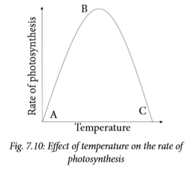
AB – Increase in temperature causes a corresponding increase in the rate of photosynthesis. The rate of photosynthesis is highest at B.
BC – Any further increase in temperature denatures the enzymes. Therefore the rate of photosynthesis declines.
d. Water
Only about 1% of the water taken in by plants is used for photosynthesis. Water shortage only indirectly affects the rate of photosynthesis, for instance, when water is in short supply, the stomata close. This lowers the exchange of gases between the leaf and the atmosphere. As a result, less carbon dioxide diffuses via the stomata into the leaf. This reduces the rate of photosynthesis.
Self-evaluation Test 7.2
1. Which of the following is not a limiting factor in photosynthesis?
A. Amount of oxygen
B. Concentration of chlorophyll
C. Light intensity
D. Temperature
E. Carbon dioxide concentration
2. The graph below shows the effect of increasing carbon dioxide on the rate of photosynthesis.

Explain why the rate of photosynthesis does not continue to increase with increase in carbon dioxide concentration.
3. Temperature is a factor that limits the rate of photosynthesis. Explain why the rate of photosynthesis starts to decrease as temperature increases beyond 40oC and eventually stops beyond 60oC.
4. The atmospheric air contains 0.03% carbon dioxide. What will happen to the rate of photosynthesis if carbon dioxide level rises to 4%?
7.3 Internal structure of the leaf and its adaptaions to photosynthesis
The leaf is the main organ for photosynthesis. However, other green parts of the plant carry out photosynthesis as well. The structure of the leaf is well suited for photosynthesis. Look at the diagram below. It shows the internal parts of the leaf.

The leaf has cuticle which covers both upper and lower surfaces of the leaf. Immediately after the cuticle is the upper and lower epidermis respectively. The palisade mesophyll which contains palisade cells that carry out photosynthesis is located below the upper epidermis. Its is followed by the spongy mesophyll layer which has cells with large air spaces between them. On the lower epidermis are tiny openings called stomata. They are surrounded by special cells known as guard cells.
Research work
1. Using text books and the internet research on the adaptations of the leaf for photosynthesis.
2. Write down short notes regarding your findings.
3. Discuss your findings with your class members

The following are some of the adaptations of the leaf to photosynthesis :
• The leaf blade is broad and flat to provide a large surface area for absorption of sunlight and carbon dioxide.
• Most leaves are thin. This reduces the distance across which carbon dioxide has to diffuse from the stomata to reach the photosynthesising cells.
• Leaves have vascular bundles that contain structures (xylem and phloem) which supply the cells with water and mineral salts. The vascular bundles also transport manufactured food to the other parts of the plant.
• The leaf cuticle and epidermis are transparent and thin to allow easy penetration of light.
• Presence of stomata on the leaves allows easy diffusion of carbon dioxide.
• The leaves are well arranged to avoid overlapping and overshadowing. This ensures maximum exposure to light. • The spongy mesophyll layer has cells that are irregular in shape and are loosely arranged hence have large air spaces between them. This allows gases to circulate freely thereby enhancing gaseous exchange between the cells and the air surrounding them.
• Palisade cells are closely packed. Further, they are elongated and lie at right angles to each other. They also contain many chloroplasts hence absorb maximum sunlight required for photosynthesis.
Self-evaluation Test 7.3
1. Use the diagram of the cross-section through the leaf to answer the questions that follow.

(a) Indicate on the diagram the following: guard cells, spongy mesophyll layer, palisade layer and vascular bundles.
(b) Which cell type absorbs most carbon dioxide during the day?
(c) State the role of the part labelled D in photosynthesis.
(d) Describe ways in which cell type B are suited for photosynthesis.
(e) Of what importance is the shape of cell type C in photosynthesis?.
2. Differentiate between the following.
(a) Epidermal cell and guard cell.
(b) Palisade layer and spongy layer.
3. Explain the role of vascular bundles in photosynthesis.
4. Describe how each of the following helps a leaf to photosynthesise.
(a) Chlorophyll molecules in the chloroplast.
(b) Lack of chloroplasts in the epidermal cells.
(c) Transparent cuticle.
7.4 Importance of photosynthesis
Discussion corner
1. Discuss with a friend the following questions:
(a) How would life be on earth without photosynthesis?
(b) What do you think is the importance of photosynthesis?
2. Share your findings with the class.

All life on earth depends on photosynthesis directly or indirectly for the following reasons.
a. As a source of energy
We are already aware that plants make their own food through photosynthesis. Animals too depend directly or indirectly on plants for their food. Food contains energy from the sun stored as chemical energy. This energy is necessary for the normal life processes to take place.
b. Provides oxygen in air
Oxygen is a by-product of photosynthesis. This oxygen replaces oxygen in air which is continuously used up by all living things during respiration.
c. Makes carbon available to plants and animals
Carbon is a major and important component of chemicals in cells. During photosynthesis, the carbon part of carbon dioxide from the air is incorporated into synthesised food. This way the carbon is made available to living things.
d. Prevents accumulation of carbon dioxide in the air
Some of the carbon dioxide in the air is used up during photosynthesis. This reduces the amount of carbon dioxide in the atmosphere. A reduced level of carbon dioxide in the atmosphere prevents global warming. Global warming is the increase in global temperature caused by increasing levels of carbon dioxide in the atmosphere among other factors.
e. It is responsible for the energy stored in coal and petroleum
Plants and animals that existed on earth millions of years ago were converted into fossils. The energy they contained, which they obtained as a result of photosynthesis is stored in their fossil. Their fossils contain energy inform of fuels such as petroleum and coal.
Class Activity: Debate
Organise a class debate with a motion: Deforestation is the main cause of global warming.
Self-evaluation Test 7.4
1. Describe the role of plants in an ecosystem.
2. Suggest conservation methods you can initiate in your village to prevent environmental degradation.
7.5 Mineral requirements for plant growth
Plants require essential mineral ions such as magnesium and nitrate ions for proper growth.
Research work
1. Using soilless cultures, devise experiments to investigate the effect of nitrate and magnesium ion deficiency on plant growth.
2. Tabulate your results using the following guidelines:
• For magnesium ions, record number of leaves that are green, green with yellow patches and yellow.
• For nitrate ions, record the height of plant, stem diameter, size of leaves and number of leaves with yellowish colour.
3. Account for the observations you make.
• What are the dangers of applying excess fertilisers to crops?
4. Present your work to the rest of the class.

a. Magnesium ions
Magnesium ions are required for the synthesis of chlorophyll, the pigment required in photosynthesis. They are also needed for activation of many enzymes involved in the energy transfer processes.
Deficiency of magnesium ions affects plant growth and development asphotosynthesis is directly affected. Leaves start to change colour from green to yellow. The result of this is less photosynthesis occurs hence poor growth.
(b) Nitrate ions
Nitrate ions are required by plants for protein synthesis. Nitrogen is a constituent of amino acids, proteins, coenzymes, and chlorophyll.
Deficiency of nitrate ions results to poor synthesis of chlorophyll hence leaves become yellowish. This causes less photosynthesis to occur resulting to stunted growth.
Uses and dangers of nitrogen fertilisers
Research Activity
1. Using text books and internet, research on the following:
(a) The effects of increased soil acidity due to high nitrate content.
(b) Meaning of term eutrophication and its effects.
2. Present your findings to the rest of the class.

Soils naturally contain many nutrients like nitrogen, phosphorous, calcium, and potassium. These nutrients enable plants to grow healthy. When soil nutrients are missing or in short supply, plants suffer from nutrient deficiency. Their growth and development is affected hence the plant cannot function properly to produce food.
In nutrient deficient soils, fertilisers are applied. Fertilisers add plant nutrients such as nitrogen, phosphorus, and potassium that are lacking in the soil. Fertilisers are simply nutrients applied to agricultural fields to supplement required elements missing in the soil. Nitrogen, naturally comes from the atmosphere, and enters the soil through rainfall and lightning. It can also be found in beneficial microorganisms (some types of bacteria), which live in the soil.
It is the misuse and overuse of nitrogen fertilisers that has severely affected the Earth’s natural ecosystem. It has also depleted minerals from the soils.
Dangerous levels of nitrates in the environment; due to overuse of nitrogen fertiliser is linked to diseases such as diabetes, cancer, and Parkinson’s disease.
Consumption of ground water containing high amounts of nitrate may lead to health problems in young children.
Harmful effects of nitrogen fertilisers to the environment
(a) Nitrogen fertilisers aid plants in their growth. However, weeds and non-native plants tend to grow more readily with additional nitrogen supplies.

(b) Excess nitrogen in the soil creates an imbalance of nutrients. This causes a depletion of other important minerals such as calcium, phosphorus and magnesium.
(c) High amounts of nitrates in the soil results to increased acidity. This affects plants that require alkaline conditions to grow.
(d) Nitrogen-polluted air, caused by nitrates from automobiles and industrial plants, results in acidification of the soil when acid rain falls.
(e) Surface run-offs containing high amounts of nitrates enter into water bodies resulting to eutrophication. When nitrogen levels in rivers and streams increase, they aid in algae overgrowth. As algae dies and decompose, organic matter in water increases. This process uses up the available oxygen, causing levels to drop. Without oxygen, fish, crabs and other aquatic organisms die.
(f) Nitrogen is a soluble substance. It soaks deeply into the soil after a rainstorm or irrigation. It may reach ground water and nearby wells hence contaminating them.
My environment, my life!
All of us have a role to play in ensuring that the environment is conducive for humans and other living organisms. This preserves biodiversity for future generations.
Self-evaluation Test 7.5
1. Describe the symptoms shown by plants due to deficiency of:
(a) Magnesium ions
(b) Nitrate ions
2. A student carried out an investigation to show how magnesium affected the growth of a water plant, Elodea. The student prepared five dishes containing 20 plants of the water plant with different concentration of magnesium salt. After 30 days, the student counted the number of plants that had grown and recorded their appearance. The results are shown in the following table.

(a) Describe the effect of decreasing the concentration of magnesium salt on the growth of Elodea.
(b) How does magnesium deficiency affect the growth and appearance of Elodea?
Unit summary
• Photosynthesis is a process used by plants and other organisms to convert light energy, normally from the sun, into chemical energy that can be later released to fuel the organisms’ activities.
• Plants use sunlight energy, carbon dioxide and water as raw materials to make glucose as product and oxygen as a by-product.
• Chlorophyll is a green pigment, present in all green plants and in cyanobacteria, responsible for the absorption of light to provide energy for photosynthesis. Its molecule contains a magnesium atom.
• The limiting factors of photosynthesis include carbon dioxide concentration, light intensity and temperature.
• Plant leaf is structurally modified to carry out the process of photosynthesis efficiently.
• Photosynthesis is important to organisms as it helps to cycle oxygen and carbon dioxide in the environment.
• For plants to grow healthy they require mineral salts.
• Lack of mineral salts in the soil especially nitrates and magnesium leads to poor plant growth.
• Mineral salts can be replaced in deficient soils through application of fertilisers.
• Overuse of fertilisers have adverse effects to organisms and the environment.


UNIT 8 : Transport of water, minerals and organic foods in plants
Key unit competency
To be able to explain the process of uptake and transport of mineral and organic saps, transpiration and translocation, and their roles in plants.
Learning objectives
After studying this unit, I should be able to:
• Identify xylem and phloem tissues from transverse sections and state their functions.
• Explain the mechanism by which water moves upwards in the xylem.
• Explain the adaptations of plant leaves to controlling water loss.
• Describe transpiration and its effects.
• Explain the adaptations of desert plants.
• Explain how translocation takes place.
• Use a potometer to measure the rate of water uptake in a given plant.
• Appreciate the importance of absorption and transport of water in plants.
Introduction
Plants manufacture food in the leaves. They absorb water and mineral salts which they use as raw materials from the soil. How then do these important plant requirements reach other parts of the plant from their source? Look at the tree alongside. What is going on in the tree?
The tree is big and complex. How will food manufactured in the leaves reach its stem and roots? By what process will water absorbed in the soil reach the stem and leaves? What does this tell you about what you will learn in this unit?

In unicellular organisms, diffusion is enough to transport materials into and out of the body of the organisms. However, plants are complex multicellular organisms. Diffusion alone is therefore, not enough to transport materials within it. This is because they have a lower surface area to volume ratio. Their cells are far away from the outside environment where these materials are located.
This necessitates a transport system to enable movement of materials in plants.
8.1 Transport system in plants
All living organisms are made up of cells. In order to stay alive, these cells take up useful substances from their environment. They also produce and release waste substances.
Water and mineral salts are transported from the soil through roots to the leaves and other parts of the plant. Manufactured food is also transported from the leaves to other parts of the plant.
A transport system in living organisms is made up of specialised tissues and organ systems. In plants, the transport system is made up of specialised tissues called vascular bundles. The vascular bundles contain two types of tissues: xylem and phloem.
Substances that need to be transported in plants are:
• Water required in photosynthesis.
• Mineral salts used in various plant processes.
• Organic substances manufactured mainly by photosynthesis taking place in leaves.
Discussion corner
1. Discuss the following questions with a friend.
(a) Where are the photosynthetic cells located in a plant?
(b) How does water and mineral salts reach the photosynthetic cells?
(c) Where do plants obtain water and mineral salts from?
(d) State how the products of photosynthesis are distributed to other parts of the plant.
2. Share your work with the rest of the class.

Phloem and xylem tissues are found in the root, the stem and the leaves. Phloem tissues transport food substances such as glucose and amino acids from the leaves to other plant tissues where they are used or stored. The xylem tissues transport water and mineral salts absorbed by the roots to different parts of the plant.
Water and mineral uptake
Water and mineral salts are absorbed from the soil through the root hair cells found in the roots. In order to understand how this takes place, we will first look at root hairs and the internal structure of the root.
Activity 8.1: Observing permanent slides of dicotyledonous and monocotyledonous roots
Requirements
• Microscopes
• Permanent slides of transverse sections of:
• Dicotyledonous roots
• Monocotyledonous roots
Procedure
1. Place a prepared slide of a dicotyledon root under the microscope. Observe under low power and high power objective lenses.
2. Note the different tissues present and their location.
3. Draw a plan diagram to show the position and layout of different layers of tissue. Do not draw any cells or shade.
4. Compare your diagram with that of the plan diagram of the dicotyledonous root section in fig 8.2 (a) and use it to identify the tissues.
5. Repeat this procedure for the monocotyledon root and compare your drawing with that in fig 8.2 (b).
Study questions
(a) Describe the pattern in the arrangement of xylem in relation to phloem in the
• Dicotyledonous root
• Monocotyledonous root
(b) What is the difference in distribution of tissues between the dicot root and the monocot root?

The distribution of tissues in a transverse section of the dicotyledonous root is not the same as that in the monocotyledonous root. In both the monocot and dicot roots the vascular bundle occupies a central position.
In the dicotyledonous root, the xylem occupies the centre where it forms a star shape. The phloem is found in between the two rays of the star. The vascular bundles are arranged in a ring.
In the monocotyledonous root, the xylem and phloem are arranged to form a ring in which xylem tissue alternates with the phloem tissue.

The root hair
Discussion corner
You will be provide with a chart showing the structure of root hair. Study it and do the following.
1. Discuss the following questions with a friend.
(a) Describe the appearance of the root hair cells.
(b) What do you think is the function of the root hair cell?
(c) What is the significance of
(i) The number of the root hair cells found on each root?
(ii) The size of the root hair cells?
(d) What do you think is the importance of the structure of the root hair?
2. Share your findings with other class members.

Root hair cells are found growing from the outer surface of the root towards the end but not at the tips. Their function is to absorb water and mineral salts.
Adaptations of the root hair cells
(i) They are numerous so as to increase the surface area over which absorption of water and mineral salts occurs.
(ii) They are thin and fine so that they can penetrate the spaces in between the soil particles where water is found.

Absorption of water
The soil particles are usually surrounded by a film of water except when there is drought. Root hair cells absorb water from the soil by osmosis.

The cell sap in the vacuole of the root hair cell has a high concentration of salts and sugars. It is therefore hypertonic to the water found between the soil particles. Due to this concentration gradient, water molecules move by osmosis from the soil through the semi-permeable membrane of root hair cells into the cell sap. Root hair cells will take up water as long as their concentration of salts is higher than that in the soil and the cell wall pressure is not large enough to prevent osmosis.
Within the roots, water moves from cell to cell by the process of osmosis. This is because adjacent cells contain different water quantities.
Different theories have been put up to try to explain the movement of water in plants. They include capillarity, root pressure, transpiration pull and cohesion tension theory.
a. Capillarity
In plants, xylem form narrow tubes through which water moves. Water rises in the xylem because of strong forces of attraction between the water molecules and the cell walls of the tubes or xylem vessels hence capillarity.
b. Root pressure
Root pressure is responsible for the rising of water up the stem. When water finally finds its way into the xylem vessels of the stem, two other processes take over the transportation process. Root pressure is demonstrated when a tree trunk is cut and water oozes out as shown in Fig 8.5 alongside.

c. Transpiration pull and cohesion tension theory
Transpiration pull is as a result of water being lost at the leaves. Leaves contain small pores called stomata. Stomata continuously lose water vapour. Since water molecules stick together, a pull is created known as transpiration pull. It allows lost water to be compensated for.
Since the xylem vessels are dead and narrow, the adhesive forces between water molecules and the walls of the xylem vessels allows water to climb up. Cohesive forces allow a stream of water which is unbroken to ascend up the plant. The continuous movement of water in columns in the root xylem, stem xylem and leaf xylem, to the air spaces in the leaf is referred to as the transpiration stream.

Once water and mineral salts are absorbed into the root hair cells, they need to reach the xylem tissue which is located at the centre of the root. It is at the xylem where water and mineral salts are distributed to other parts of the plant. Once in the root xylem, water moves up into the stem xylem and eventually reaches the leaf xylem.
Activity 8.2: Demonstrating movement of dyed water up the xylem
Requirements
• Boiling tube
• Dilute solution of red ink or dye
• Cotton wool
• Microscope or hand lens
• Oxalis latifolia
Procedure
1. Set up the apparatus as shown below. Colour the solution with red ink or eosin stain.

2. Leave the set up for three or four hours then remove the plant and cut sections of the root, stem and leaf.
3. Examine the sections under low power of the microscope or with a hand lens.
• What do you observe in the cut section?
Study questions
(a) Which tissues are stained red by the dye?
(b) Suggest a control for this experiment.
Active uptake of mineral salts
Soil water contains dissolved mineral ions such as potassium, magnesium, nitrates and phosphates. This means that a root hair must take up these mineral ions against their concentration gradient. Mineral salts are taken up by plants in ion form, for example, calcium as Ca2+ and magnesium as Mg2+.
Roots have higher concentrations of ions as compared to the soil, yet they continue taking in the mineral salts. They do this through active transport which requires the expenditure of energy. The energy is obtained from the process of respiration.
Mineral salts find their way into the xylem vessels, where they move together with water in form of a solution up the plant.
Transport of water takes place within the xylem vessels. Xylem is composed of a system of interconnected tubes which run all the way from the roots to leaves. In leaves they are in form of veins. Xylems contain two types of modified cells namely tracheids and vessel elements. Xylems are hollow tubes which act like pipes allowing water and dissolved minerals to flow through them. The cell walls in xylem vessels contain a substance called lignin. Lignin strengthens the cells and gives them structural support.

Activity 8.3: Observing permanent slides of dicotyledonous and monocotyledonous stems
Requirements
Permanent slides of dicotyledonous and monocotyledonous stems
Procedure
1. Place a prepared slide of a dicotyledonous stem on the stage of a microscope. Observe under low power and high power objective lenses.
2. Note the different tissues present and their location.
3. Draw a plan diagram to show the position and layout of different layers of tissues. Do not draw any cells.
4. Compare your diagram with that of the plan diagram of the dicotyledonous stem section in a plant in Fig. 8.9 and use it to identify the tissues.
5. Repeat this procedure for the monocotyledonous stem and compare your drawing with that in Fig. 8.10.
Study questions
a) Is the distribution of tissues in the cross (transverse) section of a dicotyledonous stem the same as that in a monocotyledonous stem?
b) Describe the pattern in the arrangement of xylem and phloem.

The distribution of tissues in a transverse section of the dicotyledonous (dicot) stem is not the same as that in the monocotyledonous (monocot) stem. In the dicotyledonous stem the vascular bundles which contain both xylem and phloem are arranged to form a ring as shown in Fig. 8.9. In the monocotyledon stem the vascular bundles appear scattered in the stem as shown in Fig. 8.10


Most of the tissues in the root and stem are similar. This is because these tissues are continuous from the root into the stem. The stem has additional tissue known as pith. Pith is the central part of the roots of monocots and dicots and the stem of dicots. It is lacking in the stem of monocots. It is made up of parenchyma cells.
Self-evaluation Test 8.1
1. Why should plants have an elaborate transport system?
2. Differentiate between monocotyledonous and dicotyledonous roots.
3. Come up with a diagram to show how mineral salts move from the roots to the leaves.
8.2 Transpiration
Transpiration is the evaporation of water from the plant surface mainly through the leaf. Much of the water that plants take up through their roots is lost to the atmosphere by evaporation. Through transpiration, plants are able to maintain a steady supply of water since the lost water has to be compensated for. If plants lose a lot of water than they can gain, they wilt. If this continues for a long period of time, they may die. Thus transpiration is always referred to as a necessary evil.
Leaves contain small pores called stomata on their surfaces, which open and close. This allows the exchange of respiratory gases as well as the loss of water in form of vapour. This is known as stomatal transpiration. Most water in plants is lost through this way.

Other than the stomata, there are other ways through which plants can lose water. They include;
Lenticular transpiration: This is the loss of water through the lenticels found in woody stems. Because of the limited distribution of lenticels, this type of transpiration accounts for less than 1% of the total loss of water by a plant.
Cuticular transpiration: This is the loss of water through the cuticle in herbaceous stems. Leaf surfaces and stems are normally covered with a waxy substance called cuticle. It accounts for up to 10% of the total water loss by the plant.
Activity 8.4: To demonstrate the process of transpiration
Requirements
• Potted plants (one with leaves, the other with its leaves removed)
• Two polythene bags
• Strings
Procedure
1. Set up the potted plants and cover each with a polythene bag. Tie the polythene bag round the stem as shown below.

2. Leave the two set-ups in a sunny place.
3. Collect and test any liquid which collects in the plastic bag with anhydrous copper (II) sulphate or anhydrous cobalt chloride paper.
Study questions
(a) What observation was made in set-ups A and B after several hours?
(b) What conclusion can be made from the above observations?
(c) Which is the control experiment and why?
(d) What changes are observed on the anhydrous cobalt chloride paper?
Factors that affect the rate of transpiration
A plant is always absorbing water from the soil and losing it into the atmosphere by the process of transpiration. If the water is not replaced as fast as it is lost, wilting of the plant takes place. If this situation continues for a long time then the plant can die due to environmental or structural factors. The environmental factors affecting transpiration are:
• Temperature
• Wind
• Humidity
• Light intensity
Temperature
This is the factor that affects transpiration the most. The temperature surrounding a plant indicates the amount of heat present around the plant. On a hot day, the temperature is high because there is a lot of heat in the atmosphere.
This causes faster evaporation of water from the leaf and therefore the rate of transpiration is high. On cold days the temperature is low because there is very little heat in the environment. As a result of this, little evaporation of water from the leaf occurs. This causes the rate of transpiration to decrease.
Humidity
Humidity is the amount of water vapour in the air. If this amount of water is a lot then humidity is high. If it is little, then the humidity is described as low. When humidity is very high, air around the plant becomes saturated with water vapour. Under these conditions the water vapour gradient is low. This means that transpiration reduces or even stops. However, when the air is dry that is humidity is very low, there is a high water vapour gradient between the inside of the leaves and the surrounding environment. Therefore the rate of transpiration is high.
Wind
Wind is moving air. Wind carries with it moisture that has evaporated from the leaf surface creating room for more to occupy. This prevents the air surrounding the stomata from becoming saturated with water vapour from the leaf. The faster the air moves, the faster the rate of transpiration. Plants will lose a lot of water during windy conditions as compared to calm conditions.
Light intensity
Light intensity affects transpiration because it has an effect on the opening and closing of the stomata. The rate of transpiration is high when there is high light intensity because the stomata open
more. When the intensity of light is low, the rate of transpiration is reduced because stomata opens less. Stomata close in darkness, therefore at night very minimal amounts of water is lost.All these factors discussed above are called external factors since they affect transpiration from the outside. Other factors affect the rate of transpiration from within the plant. These are called internal factors that affect transpiration. They include; stomatal distribution, leaf surface area, presence of a cuticle and the number of stomata on a leaf.
Potometer
Transpiration is measured using an instrument known as a potometer. This works on the principle that the amount of water lost is equal to the amount of water taken up by the plant.

Vaseline is applied around the rubber bungs to ensure an airtight seal, thus the only water loss from the apparatus is via transpiration. The function of the reservoir is to allow the air bubble to travel back to the start of the measuring scale on repeating the experiment. As water moves up through the plant the air bubble moves along the scale giving a measure of water absorbed by the plant over time and hence the transpiration rate.
Activity 8.5: Measuring the rate of transpiration
Requirements
• Leafy plant freshly uprooted or a freshly cut leafy branch e.g. a tomato plant, bean or any other suitable plant
• Basin of water
• Scalpel
• Means of timing, for example, stop watch or wristwatch
• Potometer
• Polythene bag
Procedure
1. Immerse the potometer in a basin of water making sure it is completely filled with water.
2. Put the plant into the water and cut through the stem under water.
• Why do you think the leafy stem is cut under water?

3. Attach the freshly cut end of the stem into the potometer still under water.
4. Remove the plant and potometer from the water and mount them in a fixed position. The end of the capillary tube should rest in a beaker of water.
5. Setting up the potometer as shown below.

6. Carry out the following activities:
(a) (i) Place the potometer with the shoot in a windy place outside the classroom.
(ii) Introduce an air bubble into the capillary tube by removing the beaker of water at the end of the tube
• What happens to the bubble?
(iii) Measure the distances moved by the bubble for five minutes and record your results in the table as shown below.

(b) Repeat the above procedure but place the plant where air is still. Record the distance covered by the bubble. Calculate the rate of water uptake by the same shoot.
(c) (i) Place the set-up outside in the hot sun and again in a cool place.
(ii) Calculate the rate of water uptake in each case using the procedure described in (a) above.
(d) (i) Put the plant in a humid environment. By covering the leaves with a polythene bag and leaving it without the polythene bag to compare.
(ii) Calculate the rate of water uptake in each case.
Study questions
1. What does the potometer measure:
(i) Directly
(ii) Indirectly? Explain?
2. What conclusion can you draw from your results when the following environmental conditions were investigated?
(i) Wind
(ii) Humidity
(iii) Temperature?
3. Evaporation alone cannot account for the movement of water through a plant? What other forces might be involved?

The potometer measures directly the rate of uptake of water. It also indirectly measures the rate of transpiration. Evaporation of water from the leaf leads to the replacement of this water by its uptake.
Self-evaluation Test 8.2
1. Explain why plants growing in an enclosed environment (greenhouse) have a lower rate of transpiration than plants growing in the open field?
2. Explain the following observation. A freshly cut stump of a tree will continue releasing water for some time.
8.3 Adaptations of plants to different environmental conditions
Characteristics that enable a plant to survive in its specified habitat are called adaptations.
Plants that grow in dry areas are called xerophytes, whereas others that grow in or near water are called hydrophytes.
Plants that grow in areas that are neither too dry nor too wet are called mesophytes, whereas plants that grow in saline habitats, for example, sea or ocean are called halophytes.
Activity 8.6: To examine the adaptive features of different plants
You will be provided with samples, pictures and photographs of different plants.
Procedure
1. Study each of the plants provided and note the following for each plant.
(a) Their leaves, stems and roots
(b) Where they are found
(c) Their sizes
2. Why do you think the plants have the features above? Find out from textbooks or the internet.
3. Share your findings with other class members.

Plants are structurally adapted to reduce the rate of transpiration. This depends on the environment they live in. The following are some of the ways through which plants growing in different habitats reduce transpiration rate.
a. Xerophytes
Xerophytes are plants that have characteristics suited to areas with very little water and very high temperatures. These plants grow in dry areas such as the arid and semi-arid areas.
Adaptations of xerophytes to their habitat
1. They have a high ability to absorb water from the soil. Their roots are usually more developed and grow deep into the soil and extend over a wide area.
2. They have water storage tissues. Many xerophytes have swollen stems or leaves which contain special water storage tissues. Such plants are called succulent plants.
Examples of succulent plants are Bryophyllum, sisal, cactus, Euphorbia, and Aloe vera. A baobab tree has a thick stem for water storage.

3. Xerophytes reduce water loss through transpiration in many ways:
(a) They have a thick waxy cuticle which prevents excessive water loss through the leaf by evaporation.
(b) They have hairs that trap damp air near the leaf surface causing saturation.
(c) In some xerophytes for example Nerium oleander, the stomata are found sunken in pits. The rate of transpiration is reduced because there is a space containing moist air between the stomata and the atmosphere.
(d) Many xerophytes have small leaves to reduce surface area for transpiration.
(e) Some xerophytes have very few stomata which are located on the lower epidermis away from the direct heat of the sun. Others have reversed stomata rhythm. Their stomata opens during the night and close during the day unlike ordinary plants.
4. Some xerophytes have life cycles that enable them to evade dry seasons, for example, some shed their leaves during the dry season.
b. Mesophytes
Mesophytes are plants that grow under average conditions of water supply and temperature. These plants grow very well on land and develop into forests and grasslands.

Adaptations of mesophytes to their habitats
1. They have thin leaves, which ensure rapid diffusion of gasses from the stomata to the photosynthetic cells.
2. They have broad and flat leaf blades that provide a large surface area for absorption of light and carbon dioxide.
3. Mosaic arrangement of leaves on the plant to make sure that each leaf receives maximum amount of sunlight.
4. Presence of stomata on the upper and lower leaf epidermis to allow for efficient gaseous exchange and also for transpiration.
5. Internal structures of their leaves have air spaces that allow free circulation of gases.
6. Their leaves have cells with chlorophyll so that photosynthesis takes place.
7. They have thick transparent cuticles to prevent water loss.
8. They have a well developed root system with long tap or fibrous roots.
c. Hydrophytes
Hydrophytes are plants that live in water or in very wet places.
Examples are Nymphaea and water hyacinth. There are three types of water plants:
(i) Emergent plants – they have roots and part of stem under water. While their leaves are above water. They have a problem of taking in excess water, for example, reeds.
(ii) Floating plants – they float on the water surface with roots in water. Water lilies is an example.

(iii) Submerged plants - these are found completely under water, for example, spirogyra.

(a) Their cuticle is thin or lacking. This permits the plant to absorb water, minerals and carbon dioxide over its whole surface.
(b) Since some hydrophytes absorb water over their whole body surface, their roots are not well developed. The roots may be used for anchorage, for example, in water lily or used for absorption of nutrients.
(c) The presence of many air spaces in the stem and leaf tissue; a special tissue called aerenchyma which makes the plants buoyant for support and for gaseous exchange.

(d) They contain little xylem and support tissue. They are supported by aerenchyma and the buoyancy of the water.
(e) Submerged leaves do not have stomata and floating types have many on their upper surface.

d. Halophytes
These are plants that grow in salty places such as rocky shores, seas and sand dunes which occur along coastal regions. Some halophytes such as Atriplex and mangroves grow near sea water.

Therefore they have a problem of taking up water from their salty surroundings. They have cells that absorb salt. As a result, they create a higher osmotic pressure which enables the plant to absorb water. Because of taking much salt, they excrete excess salts using salt glands.
The salt is washed from the plant surface by rain. Some halophytes absorb salt from their habitats and remove it by shedding leaves that have accumulated salt.
Self-evaluation Test 8.3
1. What is the significance of modified leaves in xerophytes?
2. Halophytes are found in salty environments. How do they avoid having too much salt in their tissues?
3. Mutuyimana found a plant that did not have stomata. Where do you think she got the plant from?
Translocation
Discussion corner
1. Discuss with a classmate the meaning of the following terms
• Source
• Sink
• Photosynthates
• Translocation
2. What structures are responsible for translocation in a plant?.

• The products of photosynthesis are called photosynthates. They are usually in the form of simple sugars, such as sucrose. Photosynthates are produced by sources and are translocated to sinks. The points of sugar delivery, such as roots, young shoots, and developing seeds, are called sinks. Seeds, tubers and bulbs can be either a source or a sink, depending on the plant’s stage of development and the season.
• The movement of organic products of photosynthesis from leaves to other parts of the plant is called translocation.
• Sugars are produced in the leaves (source) but other nonphotosynthetic parts of the plant like roots and stems also need part of this food. For this reason, organic food products are translocated form the source to the sink.
• Some important sinks are roots, flowers, fruits, stems and developing leaves. Leaves are particularly interesting in this regard because they are sinks when they are young and become sources later, when they are about half grown.
• Organic products of photosynthesis (photosynthates) are translocated through the phloem tissue.

• Photosynthates are directed primarily to the roots during early development, to shoots and leaves during vegetative growth, and to seeds and fruits during reproductive development. The products from the source are usually translocated to the nearest sink through the phloem.
The high percentage of sugar in phloem sap causes water to move from the xylem into the phloem. This increases water pressure inside the phloem, causing the sap to move from source to sink.
Activity 8.7: The ringing experiment
Requirements
• Tree or shrub with many branches
• Sharp knife
Procedure
1. Remove completely a ring of bark with its phloem from two branches. The xylem tissue which makes up the bulk of the stem is left intact.

2. The setup is left undisturbed for four weeks.
Study question
(a) What observations were made in the stem after four weeks?
Explain these observations.
(b) Discuss with your friends how the bark of medicinal trees can be harvested without killing these important trees.

When the ring of bark is removed, the phloem beneath it is also removed. After several weeks, swelling above the cut ring is noted.

The swelling is due to the accumulation of food substances. They were being transported from the leaves but could not get across the debarked part of the stem. That is why there is no swelling on the lower part of the ring.
My environment, my life!
This ringing procedure is sometimes employed to kill some unwanted trees, before they are cut down. Do not use it on a tree that has economic value to your community.
Self-evaluation Test 8.4
1. Differentiate between a source and a sink?
2. The movement of organic products of photosynthesis from________ to other parts of the plant is called_____________.
Unit summary
• Transport in plants involves movement of materials in and out of the cells. It also involves materials being taken away from the cells to outside of the organism.
• Transport is necessary because all cells in living organisms need food substances and oxygen from their surroundings. They produce waste substances that need to be eliminated from their bodies.
• Plants have a transport system for the moving substances. They use two different types of transport tissue.
• Xylem transports water and solutes from the roots to the leaves.
• Phloem transports food from the leaves to the rest of the plant.
• Water and mineral salts move through the xylem by the forces of cohesion, adhesion, capillarity, root pressure and transpiration pull.
• Transpiration is the process by which a plant loses water in form of water vapour.
• Transpiration is important because it cools the plant on hot days. It also assists the movement of water and mineral salts up the plant.
• Environmental factors that affect transpiration are humidity, temperature, wind and light intensity.
• Structural factors that affect transpiration include size of leaf and number of stomata.
• Organisms have different adaptations that enable them survive in their habitats. Some plants live in or around wet places (hydrophytes), others live in dry areas (xerophytes) and others live in areas which are neither too wet nor too dry (mesophytes). There are also some that live in salty places (halophytes).
• Translocation is the movement of materials from leaves to other tissues throughout the plant.


UNIT 9 : Gaseous exchange in humans and plants
Key unit competency
To be able to explain gaseous exchange in plants and human beings.
Learning objectives
After studying this unit, I should be able to:
• Explain the characteristics and adaptive features of gaseous exchange surface. • Define gaseous exchange and why it is an important process.
• Describe gaseous exchange in the lungs and alveolus.
• Demonstrate the process of inspiration and expiration.
• Explain the process of gaseous exchange in plants.
• Identify common respiratory diseases and suggest their prevention and treatment.
• Dissect mammalian lung and relate its features to gaseous exchange surfaces.
• Construct bell-jar model from locally available materials.
• Appreciate learning through using locally available materials.
Introduction
In senior one, you learnt about gaseous exchange in human beings. How did you define gaseous exchange? What did you learn as the importance of gaseous exchange?
Look at the cycle below.

What is the importance of the cycle shown in the diagram? Can you identify gases X and Y? What is their importance to each of the organisms represented in the cycle? Based on the diagram, predict what you will learn in this unit.
9.1 The concept of respiration and respiratory surfaces
Respiration is the process where food taken in by organisms is burnt to produce energy required for their body functions. During respiration, oxygen is used and carbon dioxide is removed.
Cellular respiration takes place constantly in all living cells. It creates a constant demand for oxygen and a need to eliminate carbon dioxide gas. Gaseous exchange is the biological process through which these gases are transported through the body of an organism across a specialised respiratory surface. Organisms such as plants need to take in or release oxygen and carbon dioxide at one time or another during respiration and photosynthesis. Animals, on the other hand, always take in oxygen and release carbon dioxide during respiration. Gaseous exchange therefore is:
(i) Exchange of respiratory gases in animals.
(ii) Exchange of photosynthetic and respiratory gases in plants.
Therefore an efficient system for the exchange of gases is extremely important in living organisms. Gaseous exchange is necessary because organisms are able to obtain useful gases from their environment and get rid of waste gases into the environment.
The environments that organisms exchange gases with include: air for some organisms and water for others. Air is the main source of oxygen and carbon dioxide. Organisms that live on land exchange these gases directly with air. Oxygen and other gases from the air diffuse into lakes, rivers and oceans. The air dissolved in water is used for gaseous exchange by organisms that live in water.
Discussion corner
1. Find out the following with a friend.
(a) Respiratory surfaces.
(b) The gaseous exchange surface in man.
(c) The difference between cellular respiration and gaseous exchange.
2. Write down answers in your note books.
3. Discuss your findings with fellow classmates.
In large multicellular animals, the surface area to volume ratio is small. Many cells are deep inside the body of the animal, away from the surface. Diffusion of gases alone is not efficient enough in moving gases to and from all the cells. Therefore, large multicellular animals have specialised structures or organs with special surfaces over which gaseous exchange takes place. These special surfaces are called respiratory surfaces.
Examples of specialised structures for gaseous exchange in animals include the following:
• Cell membrane
• Tracheal system in insects
• Buccal cavity in frogs
• Skin in frogs
• Gills in fish
• Lungs in mammals, birds, reptiles and amphibians.
Gaseous exchange takes place over the respiratory surfaces. A respiratory surface has a number of characteristics that make it efficient for gaseous exchange. Some of these characteristics include:
(i) Thin walls for faster diffusion of gases across it.
(ii) It is moist to dissolve gases as they diffuse across it.
(iii) It has a large surface area for maximum gaseous exchange.
(iv) In animals with a transport system, the respiratory surface has a rich supply of blood capillaries (highly vascularized) to quickly transport gases to and from the cells.
Not all respiratory surfaces are in direct contact with the medium through which gaseous exchange occurs, such as water or air around the organism. Therefore, there is need for a process that can ensure a continuous supply of fresh water or air to and from the respiratory surface. This is achieved by the process of ventilation which continuously brings water or air containing more oxygen to the respiratory surface. It also removes water or air containing a lot of carbon dioxide from it. Ventilation, therefore, is important because it maintains a high diffusion gradient at the respiratory surface. It also ensures a high rate of gaseous exchange. Breathing is an example of ventilation.
9.2 The mechanism of breathing in humans
In human beings, the process of breathing (ventilation) is the first part of the gaseous exchange processes. The second part is the exchange of these gases between the lungs and blood. Breathing provides a continuous supply of fresh air to the gas exchange surface. It also helps to maintain a large diffusion gradient across the gas exchange surface. The volume of gases exchanged during breathing, changes according to physiological demands placed on the body, for example, during an exercise. The breathing rate is controlled by the respiratory center of the brain.

Activity 9.1: To observe the movement of the chest during breathing
Working in pairs, carry out the following exercise.
1. Ask your partner to breathe in deeply then hold their breath.
2. Describe what you see happening to the chest as your partner breathes in.
• Does it remain in the same position?
3. Let your partner now breathe out.
• What do you see happening to the chest?
4. Record the form of movements noticed.

The process that brings air into the lungs and removes it again is known as breathing. Breathing involves two phases called inhalation and exhalation. Since the lungs contain no muscle tissue, they are not capable of independent movement. However, they are elastic, and during breathing they are forced to expand or contract as a result of pressure changes around them. These pressure changes are caused by the movement of the muscular diaphragm, ribs and intercostal muscles (rib muscle), and by the force of atmospheric pressure.
Activity 9.2: Construction and use of a model to demonstrate breathing mechanism
Requirements
• Bell jar
• Two balloons
• Rubber stopper with a hole
• Y-Shaped glass tube
• Rubber sheet and rubber band
• String
Procedure
1. Assemble the materials as shown below.

2. Study the model and state what the following parts represent in the human respiratory system
• Bell jar
• Balloon
• Rubber sheet
3. Pull down the rubber sheet at the bottom of the bell jar.
• What happens to the balloons?
4. Now push up the rubber sheet.
• What happens to the balloons?
Study questions
(a) Draw sketches to show the appearance of the balloons in (2) and (3) above.
(b) Explain your observation.
(c) Explain how the model is similar to the working of the thorax in humans beings during breathing.
a) Inhalation (breathing in)
Inhalation is also known as inspiration. This is the active phase of breathing which draws air into the lungs. During inhalation, the diaphragm muscles contract causing it to flatten, look at Fig. 9.4. In the ribs region, the external intercostal muscles contract while the internal intercostal muscles relax. This causes the rib cage to move upwards and outwards.

The contraction of the diaphragm and external intercostal muscles increases the volume in the chest cavity. However, it causes a decrease in the pressure of air inside compared to atmospheric air. Air rushes through the air passages into the lungs, forcing them to expand.
b. Exhalation (breathing out)
Exhalation is also known as expiration. This is the phase of breathing, which expels air out of the lungs. During exhalation, the diaphragm muscle relaxes making it to move upward and regain its dome shape. The external intercostal muscles relax and the internal intercostal muscles contract. This causes the rib cage to move downward and inwards. The volume of the chest cavity decreases and the pressure increases compared to the atmospheric air. Increased pressure forces air out of the lungs, which become deflated.

Table 9.1: Differences between inhalation and exhalation

Activity 9.3: Dissection of a small mammal to display the structures of the respiratory system
Requirements
• Dissection board
• Pins
• Scissors
• Scalpels
• Cotton wool
• Rat or rabbit (freshly killed)
• Forceps
• String
• Rubber pipette
• Gloves
Procedure
1. Pin the animal onto the dissection board with the ventral side up.
2. Open the thorax by cutting with scissors along the mid section from the neck to the hind legs.

3. Cut the skin further to expose the abdomen as shown in Figure 9.6 above.
Note the following:
• Muscles of the diaphragm.
• Intercostal muscles between the ribs.
• Lungs.
4. Remove the lungs out and place it on the board.
• How does it feel to touch?
5. Cut through the lungs.
• What do you see?
6. Identify the main airways.
7. Note your observations.
8. Share your findings withother class.
Gas exchange in the alveoli
Air passes the nose or mouth as it moves down the trachea. The trachea is divided into the left and right bronchi. Each bronchus is divided into smaller bronchioles and each bronchiole is in turn attached to numerous alveolar sacs. The groupings of alveoli take the same shape like a bunch of grapes.

Gaseous exchange at the alveolus takes place between the phases of inhalation and exhalation. The alveolus is a suitable point for gaseous exchange because:
• It is supplied with blood which carries the gases being exchanged.
• It has a very thin wall across which gases diffuse between it and the blood.
• It is lined with a thin film of moisture to dissolve the diffusing gases.
• A ventilation process brings in and takes away air containing the gases being exchanged.
• It has a very large number of alveoli to increase their surface area for gaseous exchange.
Gas exchange between the air within the alveoli and the pulmonary capillaries occurs by diffusion. Oxygen in air, in the alveolar space is at a higher concentration than that in the blood capillaries. It therefore first dissolves in the water layer in the alveolar lining then diffuses across the alveolus and then the capillary walls into the red blood cells. This becomes oxygenated blood which is carried to the heart by the pulmonary vein.
Carbon dioxide in the blood diffuses across the capillary and alveolus walls into the alveolar space and is eventually expelled during exhalation.

A diffusion gradient is essential for rapid gaseous exchange in the alveolus. The following factors contribute towards maintaining this diffusion gradient.
(a) Lung ventilation: Breathing movements transport respiratory gases to and from the alveolus.
(b) Blood flow: This constantly replaces oxygenated blood with deoxygenated blood. The pulmonary artery brings blood low in oxygen concentration and high in carbon dioxide concentration. The pulmonary vein takes away blood high in oxygen concentration and low in carbon dioxide concentration.
(c) Haemoglobin: It quickly combines with oxygen and prevents its accumulation in the alveolus.
Activity 9.4: To analyse the gas present in exhaled air
Requirements
• Conical flask
• Couch
• T-tube
• L-tube
• Limewater
• Delivery tube
Procedure
1. Set up the apparatus as shown in the diagram below.

2. Breath in and out through the mouth-piece.
3. What do you notice in the lime water in:
• Flask A? • Flask B?
4. Explain your observation in
• Flask A • Flask B


Task: From the table above, explain the difference in composition of gases in inhaled and exhaled air.
Self-evaluation Test 9.1
1. What role does the nasal passages and the diaphragm play in gaseous exchange?
2. Describe the characteristics of a respiratory surface.
3. Describe the mechanism by which carbon dioxide in the blood capillaries around the alveoli passes into the alveoli and finally is exhaled.
9.3 Respiratory diseases and smoking
Research Activity
1. Using handouts, textbooks and internet, research on the following:
(a) Respiratory diseases
(b) Causes of respiratory diseases in man.
(c) Prevention and treatment of respiratory diseases.
2. Share your findings with the class.

The respiratory system is affected by many diseases and disorders. Some of these are caused by micro-organisms while others are genetic. The most common respiratory diseases are:
• Tuberculosis
• Asthma
• Pneumonia
• Bronchitis
• Whooping cough
• Common cold
• Influenza
1. Asthma This is a disease that comes about when the air passages in the lungs suddenly narrow as a result of contraction of their smooth muscles. It is also characterised by an inflow of mucus which clogs the narrow passages even more. Some substances can trigger an asthma attack. They are called allergens. They include pollen grains, some type of proteins in milk, pet hairs, dust and even flavours in food. It can also caused by stress and anxiety. In some families the disease is inherited.
Symptoms of asthma
• Difficulty in breathing. Breathing can feel so difficult or quick that the patient can faint.
• Wheezing sounds when breathing.
Prevention and treatment
Asthma is treated by two types of medication: long-term control and quick-relief medicines that help reduce airway inflammation and prevent asthma symptoms.
• Quick-relief medicines relieve asthma symptoms that flare up.
• People with asthma should avoid an environment that is likely to bring on asthmatic attack. This is by avoiding contact with allergens.
• Asthma patients are advised to carry inhalers that contain a drug which pacifies the condition.

2. Bronchitis This is an infection of the inner walls of the bronchi. It is caused by bacteria or air pollutants such as smoke in inhaled air. The infection causes the mucous membrane in the respiratory tract to produce excess mucus. This causes the cells lining the bronchi and bronchioles not to function properly. As a result, the air passage to the alveoli becomes blocked with mucus. Severe coughing occurs in an attempt to get rid of the excess mucus. Breathing also becomes difficult. Bronchitis may be acute or chronic.
Acute bronchitis starts quickly and stops after a few days. The symptoms of acute bronchitis are like those of a cold.
Chronic bronchitis starts slowly and lasts for a long time. It is a more serious kind of infection. It is commonly caused by smoking and air pollution.
Symptoms of bronchitis
• Secretion of excess mucus
• Coughing
• Difficulties in breathing.
Prevention and treatment
• Acute bronchitis is treated by simple measures that include: getting plenty of rest, drinking lots of fluids and taking a cough syrup.
• People with chronic bronchitis should take antibiotics every time they have a cold with a fever.
• A doctor should be consulted at the early stages of bronchitis.
• Avoid smoking whether directly or passively.
• Avoid polluted air.
3. Emphysema
This results from long untreated bronchitis where the bronchioles in the lungs become blocked. This causes damage to delicate walls of the alveoli due to high pressure when coughing. This leads to reduced surface area for gaseous exchange. The lungs become distended and inelastic that gases cannot be exchanged efficiently. The patient becomes weak due to insufficient oxygen supply to tissues. Running and walking can prove to be hard when one has this condition.
Prevention and treatment
Emphysema is treated according to the severity of symptoms. Bronchodilators are normally given to help relieve coughing, shortness of breath and breathing problems.
Early treatment of bronchitis with antibiotics to prevent secondary infection can help to prevent emphysema.
4. Pneumonia
Pneumonia is an infection of the lungs. It is caused by bacteria called Pneumococcus that spreads through the air. It can also be caused by a virus or a fungi. Infection proceeds from the mouth down into the lungs. As a result of the infection, a fluid is produced which collects in the alveoli. The lungs become solid and have no air. This prevents exchange of gases in the lungs.
Signs and symptoms
• Sudden chills and high fever.
• Rapid shallow breathing and sometimes wheezing.
• Cough with yellow, greenish colour or mucus with some blood.
• Chest pains.
Prevention and treatment
• Overcrowded places should be avoided and good ventilation in living rooms should be provided.
• Treatment of pneumonia involves curing the infection and preventing complications. It also depend on the causative agent: bacterial, viral or fungal.
• Bacterial and fungal pneumonia are treated with drugs while viral pneumonia clears by itself.
5. Tuberculosis
Tuberculosis (TB) is caused by bacteria called Mycobacterium tuberculosis. The source of infection may be droplets containing bacteria sprayed from the air passages during breathing or sneezing. It can also be caused by infected dry sputum in particles of dust. Tuberculosis bacteria may attack any part of the body, but they usually invade the lungs, causing pulmonary tuberculosis. Another source of infection is by drinking raw milk from a cow suffering from bovine tuberculosis.
Signs and symptoms
• Tuberculosis of the lungs starts with a dry cough followed by the spitting of blood, fever and sweating at night as the infection proceeds.

• If there is no treatment, loss in weight occurs and finally death of the patient.
In addition to tuberculosis of the lungs, there are other forms of the disease in which bacteria attack the lymphatic glands, bones and other parts of the body.
Prevention and treatment
• The patient should consult a doctor for adequate treatment. Treatment for TB will usually involve a long course of antibiotics lasting 6-9 months.
• Overcrowding increases the risk of spread of tuberculosis.
• Avoid taking raw milk. Boil all milk or drink pasteurised milk.
• Immunisation with B.C.G. vaccine in children.
• Isolating patients
6. Whooping cough
Whooping cough is caused by bacteria called Bordetella pertussis. The mode of infection is from one person to another through inhalation of infected droplets.
Signs and Symptoms
• Whooping cough starts like a cold with fever, running nose and cough.
• Two weeks later, the whooping begins. The patient coughs rapidly many times without taking a breath, until one coughs up a mass of sticky mucus, and the air rushes back into the lungs with a loud whoop sound. After the “whoop”, the patient may vomit. Between coughing bouts the patient seems fairly healthy.
Prevention and treatment
1. Patients should consult a doctor for adequate treatment. Treatment for whooping cough involves taking antibiotics early before coughing fits begin.
2. Patients should be isolated from contact with other people.
3. Immunisation with vaccines against whooping cough. In infants, the vaccine against whooping cough is usually combined with those against diphtheria, tetanus and poliomyelitis.
7. Common cold
A common cold is an illness caused by a virus infection located in the nose. Colds also involve the sinuses, ears and bronchial tubes. Colds last on average for one week. Mild colds may last only 2 or 3 days while severe colds may last for up to 2 weeks. A cold is a milder illness than influenza.
Nasal secretions containing cold viruses contaminate the hands of people with colds as a result of nose blowing, covering sneezes and touching the nose. Cold viruses may contaminate objects and surfaces in the environment of a patient.
Note: Young children are prone to colds.
Cold virus, which is expelled into the air in coughs and sneezes, may land in the nose or eye of another person and cause infection. Hence transmission.
Signs and symptoms
• Sneezing
• Runny nose
• Nasal obstruction
• Sore or scratchy throat
• Cough
• Hoarseness
• Mild general symptoms like headache, feverishness, chilliness, and not feeling well in general.
Prevention and treatment
Cold is caused by a virus, therefore it will clear after several days. Treatment is mainly to lessen the symptoms. If a cold persists seek medical advice.
To prevent catching a cold, limit contact with known cold patients, especially during the first three days of their illness. Practice preventive measures which keep cold virus from entering the nose:
• Wash hands after contact with cold sufferers and objects and surfaces they may have contaminated.
• Keep fingers out of the eyes and nose.
• Avoid having cold patients cough and sneeze on you or in your direction.
8. Influenza
Influenza or 'the flu' is a highly contagious disease caused by infection from influenza type A or B (or rarely C) virus. These viruses infect the upper airways and lungs. The flu is highly contagious.
Flu is not the same as a common cold, and can be a serious illness. For some people, such as the elderly and those with underlying medical conditions, the flu can cause serious complications which require hospitalisation. It can sometimes lead to death.
Flu is usually spread through infected people coughing and sneezing, which temporarily contaminates the surrounding air and surfaces with infected droplets
Signs and Symptoms
Symptoms usually appear 1–3 days after being infected. A person can spread flu to others 1–2 days before they become unwell and up to 5 days after symptoms develop. The symptoms of influenza can include:
• fever
• dry cough
• muscle and joint pain
• tiredness or extreme exhaustion
• headache
• sore throat
• Stuffy nose
Prevention and treatment
1. Generally, uncomplicated flu is managed by simply resting in bed, drinking plenty of fluids (particularly water) and taking over-the-counter medication to help relieve the symptoms.
2. Antiviral medications reduce the length of time symptoms last and help people infected return to their daily routines earlier.
3. Good hygiene is essential to protect yourself and others
4. You can reduce the risk of infection by getting vaccinated.
Health Check!
It is unhygienic to cough and spit carelessly in public places. This can easily spread disease. It exposes other members of the public to infections. Besides it is also ugly.
Effect of smoking on the respiratory system
The respiratory system is not designed to cope with tobacco smoke. Tobacco smoke paralyses the cilia in the respiratory tract and stops their movement.
In addition, tobacco smoke increases the production of mucus in the air passages. A cough by a smoker is an attempt to remove the excess mucus from the respiratory system. Besides irritating the trachea and bronchi, smoke particles interfere with the uptake of oxygen in the air sacs.
When cigarette smoke is inhaled, about one-third of the particles remain in the alveoli. Phagocytic cells called macrophages can slowly remove many of the particles. However, an excess of particles from smoking or from other sources of air pollution breaks down the walls of the air sacs and causes the formation of inelastic tissue. This reduces the functional area of the respiratory surface and in severe cases may lead to a disease called emphysema. In some cases, lung cancer also develops.
Health Check!
Cigarette smoking is addictive. Once you are an addict, it is hard to stop. Cigarette smoke also pollutes the environment in public places such as buses, shops and hotels. It also forces nonsmokers to inhale the smoke and become passive smokers.
Self-evaluation Test 9.2
1. Suppose you are nursing a patient suffering from a respiratory disease. How would you prevent contracting the disease?
2. Is it proper to avoid friends who smoke?
3. Why is it not advisable to take antibiotics without prescription from a doctor?
9.4 Gaseous exchange in plants
The principal gaseous exchange surfaces for plants are the leaves. Plant leaves have stomatal pores on their surface where gaseous exchange occurs. Plants do not have a specialised respiratory system like animals. This is because they are metabolically less active than animals.
Activity 9. 5: To demonstrate the presence of stomata in leaves
Requirements
• Plant leaves (e.g. hisbiscus plant leaves)
• A bottle of clear or colourless nail polish
• A clear cellophane tape (or clear packing tape)
• Microscope slides
• Scissors
• Microscope
Procedure
1. Paint a 1 cm 2 (or larger) square of thick nail polish on the underside surface of the leaf being studied. Allow the nail polish to dry fully.
2. Once the patch of nail polish is dry, tape a piece of clear cellophane tape to the patch on the leaf.
3. Carefully pull on a corner of the tape and gently peel the nail polish off the leaf. The layer of cells that sticks on the nail polish is what you will examine under the microscope.
4. Tape the peeled nail polish and its layer of cells to a clean microscope slide, using scissors to trim any excess tape.
- The teacher will help you predict and explain the different types of cells that you see and how to find the stomata.
5. Place the slide on the microscope stage. Using the low power objective lens, focus the slide until the stomata are visible.
Each stoma is bordered by two sausage-shaped cells, called guard cells, which are normally smaller than epidermal cells. Unlike other cells in the epidermis, guard cells contain chloroplasts.
6. Sketch what you observe under the microscope, labelling the stoma, guard cells, epidermal cells, and chloroplasts.
7. Count the number of stomata in your field of view, then estimate the number of stomata on the sample being examined.

Stomata are pores between guard cells. They are found on the upper or lower epidermis or both. Stomata allow:
• Entry of carbon dioxide into the leaf for photosynthesis.
• Exit of oxygen.
• Evaporation of water.
Stomata are the main structures for gaseous exchange in leaves of plants. They are tiny openings found in the epidermis of leaves. With the exception of submerged plants, stomata are present in all the leaves of plants. Most land plants have stomata on the lower surface of the leaf.
Stomata allow gaseous exchange to take place in leaves. Let us examine the structure of stomata to understand how gaseous exchange takes place through them.

The guard cells control the opening and closing of each stoma.
Discussion corner
1. Discuss the following questions with some of your classmates:
a. How gaseous exchange occurs in plants?
b. Relate gaseous exchange in plants and animals.
2. Share your findings with the rest of the class.

Gas exchange in plants
When the stomata open, gases come into the leaf while others come out. Oxygen which is a product of photosynthesis diffuse out of palisade cells to the air spaces. It eventually comes out through the stomata to the atmosphere. On the other hand, carbon dioxide from the atmosphere enters into the leaf through the stomata into the airspaces. It then diffuses from the air spaces into palisade cells where it is used during photosynthesis.
When the stomata are open, air from the surrounding enters the leaf and occupies the air spaces. Oxygen and carbon dioxide diffuse into or out of the leaf cells along their concentration gradient.
During photosynthesis, carbon dioxide is used up by the palisade cells, and oxygen is produced.
This means that the concentration of carbon dioxide in the palisade cells becomes lower than in the air spaces outside the cells. Therefore during photosynthesis carbon dioxide diffuses into the palisade cell from the airspaces. On the other hand, photosynthesis produces oxygen whose concentration becomes higher inside the palisade cells than in the air spaces surrounding the cells. Oxygen therefore diffuses out of the palisade cells into the air spaces. This is the process of gaseous exchange in leaves.
Gases first dissolve in the film of moisture surrounding the cell, before they diffuse into or out of the cell. Note that when air is entering or leaving the plant through stomata, the process is not gaseous exchange, but diffusion. Gaseous exchange refers to the movement of gases between the cells and their surroundings.

Stems of woody terrestrial trees and shrubs have areas of loosely arranged cells with large air spaces between them. These cells together form a structure called a lenticel. Lenticels are formed when the epidermis is replaced by the bark. Lenticels appear scattered on the surface of the stem as small raised openings. They allow gaseous exchange of oxygen and carbon dioxide between the atmosphere and the internal tissues of the stem.
Self-evaluation Test 9.3
1. What are the structural differences between guard cells and other epidermal cells?
2. Although the leaves are the main organs of gaseous exchange in plants, the roots also absorb oxygen in the soil. Why is this necessary?
Unit summary
• Gaseous exchange is the process by which animals exchange respiratory gases while plants exchange both respiratory and photosynthetic gases.
• Gaseous exchange between a living cell and its surrounding always takes place by diffusion across a moist cell membrane.
• The surface over which gaseous exchange takes place is called a respiratory surface.
• All respiratory surfaces have a large moist surface, basically a mechanism of transporting gases to and from the respiratory surface.
• The direction that a gas takes during diffusion depends on the concentration of the gas on either side of the respiratory surface.
• In large multicellular organisms such as human beings, most cells are deep inside the body. Diffusion alone is not enough to move gases across the large number of cells that lies inside.
• Large multicellular organisms have special respiratory structures for gaseous exchange. These include the tracheal system in insects; the skin, buccal cavity and lungs in frogs; the gills in fish and the lungs in humans.
• In humans beings, breathing is controlled by medulla oblongata in the brain which is sensitive to the concentration of carbon dioxide. When this exceeds a certain level the medulla oblongata stimulates the ribs and diaphragm to contract more rapidly.
• Plants also require carbon dioxide for photosynthesis and produce oxygen as a waste product.
• In plants, gaseous exchange occurs through stomata in leaves, lenticels in the stem and roots and through the epidermis of the root.
• Certain diseases of the respiratory system such as asthma, bronchitis, pulmonary tuberculosis, pneumonia and whooping cough interfere with the functioning of the respiratory structures.


UNIT 10 : Excretion in humans
Key unit competency
To be able to describe the structure and function of the excretory organs and suggest good practices for healthy kidneys.
Learning objectives
After studying this unit, I should be able to:
• Define and explain the need for excretion.
• Describe the role of the liver in excretion.
• Name excretory organs and excretory products.
• Outline the structure of kidney and describe a nephron.
• Describe the process of urine formation.
• Use a dissected mammal to identify parts of the urinary system.
• Develop good habits to maintain healthy urinary system.
Introduction
We use fuel, for example, diesel and petrol to run machines such as engines of vehicles. Are there waste products produced by a running engine? Look at the picture below.

What is happening in the picture? What are the components of the smoke? What do you think would happen to the engine of the vehicle if the products being removed were left to accumulate? From your answers above what do you think you will learn in this unit?
10.1 Need for excretion
Just as vehicles remove substances that can damage their engine if left to accumulate, human beings also remove toxic waste products from their bodies. This happens through a process called excretion. Therefore, we can define excretion as the process by which organisms remove waste products of metabolism from the body. Through excretion, organisms control osmotic pressure. It also enables them to promote homeostasis; that is, the balance of the organism’s internal body environment.
Discussion corner
1. Discuss the following with a classmate.
(a) The importance of excretion.
(b) The health implications of not emptying the bladder and bowels promptly.
2. Research on human excretory organs. Use your answers to fill the table below.

3. Compare your findings with that of your classmates.
4. Share your work with the rest of the class.

Excretion is one of the main characteristics of living things. Every living cell carries out life processes which involve chemical reactions. In some of these reactions, complex materials are chemically broken down and in others simple materials are used to make new compounds. All these reactions are collectively referred to as metabolism. During metabolism, waste products are formed. If waste products accumulate in the cell or even in its surrounding, they can reach very high concentrations which affect the normal cell function and could even kill the cell, for example:
1. Carbon dioxide is a by-product of respiration. If it accumulates in the cell it changes the pH of the cell. It then interferes with the functioning of certain enzymes. If this situation is not corrected, the affected cells eventually die.
2. The body has no mechanism of storing excess amino acids from the digestion of proteins. Excess amino acids are therefore taken to the liver where they are broken down in a process called deamination to urea and glycogen. Small amounts of uric acids are also formed from the breakdown of proteins. Urea and uric acids comprise the nitrogenous waste products which are excreted from the body through urine.
Many other reactions in cells also produce by-products that could be toxic. Therefore it is necessary that these waste products be eliminated from the cells. The waste products if not removed can accumulate to toxic levels resulting to ill health and eventually death.
Unlike plants, animals have difficulty getting rid of waste substances for several reasons:
• Animals are more active than plants. Therefore their metabolic processes take place at a higher rate, producing larger quantities of waste products.
• Animals do not put most of their waste products to other uses like it happens in plants.
• Animals take in certain substances in their food in excess of their needs. These extra substances, for example proteins, are broken down with the formation of toxic substances such as ammonia.
For these reasons, animals have a more complex excretory system than plants.
10.2 The role of the liver in excretion
The table below summarises the excretory organs and their waste products in human beings.
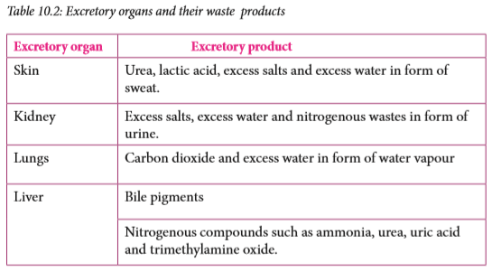
Discussion corner
1. Discuss the following questions with a classmate.
(a) What are the functions of the liver in excretion?
(b) What are the homeostatic functions of the liver.
(c) Why is ammonia produced in the liver not excreted in that form but is first converted to urea?
2. Share your work with the rest of the class.

The liver is the second largest organ in the body after the skin. It plays a very important role in the body. It is described as the metabolic centre. The liver has many functions including maintenance of a constant internal environment (homeostasis) and excretion. The excretory functions of the liver are described below.

(a) Deamination
The body cannot store proteins or amino acids. This is because their breakdown forms toxic substances in the body. Amino acids are the end products of protein digestion. Surplus amino acids are broken down in the liver in a process called deamination. From each amino acid, the amino group (NH2)) is converted to ammonia (NH3). Ammonia should not be allowed to accumulate in the body because it is highly toxic. The remainder of the amino acid molecule is changed to glycogen or fat for storage.
The ammonia produced from the amino group is quickly converted to a less toxic substance called urea. During this conversion, ammonia is first combined with carbon dioxide through a series of enzyme-catalysed reactions. The resulting urea is taken to the kidney in blood and is eliminated from the body in urine.
(b) Detoxification
The liver removes harmful substances such as drugs and hormones from the blood. Within the liver, these substances are converted into inactive or less dangerous forms, for instance, hydrogen peroxide, a highly toxic by-product of certain metabolic process is rapidly split into water and oxygen by enzyme catalase in the liver.

Thus, the liver purifies or detoxifies blood. The inactive substances formed in the liver are returned to the bloodstream and are finally excreted from the body by the kidneys.
(c) Elimination of Haemoglobin
Haemoglobin from old worn out red blood cells is broken down by the liver cells into pigments. These pigments are further broken down and eliminated in the bile, giving urine its characteristic yellow colour.
(d) Elimination of sex hormones and cholesterol After sex hormones have performed their functions, some are modified chemically by the liver cells. Others are sent to the kidney for renal excretion while others are expelled in bile.
Excess cholesterol is also excreted in bile. If there is a considerable excess amount of cholesterol in the blood, some may be deposited in the walls of blood arteries obstructing flow.
Self-evaluation Test 10.1
1. Undigested and indigestible waste products are not considered as excretory wastes, explain.
2. Copy and fill the blank spaces in the passage that follows.
Excretion involves the removal of _______ products from the body. Urea is produced by the ______ during while carbon dioxide is a waste product of _______ . Nitrogenous waste products are excreted by the _______ while carbon dioxide is excreted by the ________.
3. Proteins are very important in our bodies. However, excess amino acids from protein digestion cannot be stored. Study the diagram below and answer the questions that follow.

(i) Name the process illustrated in the diagram.
(ii) In which organ does the process you have named in (i) above occur?
(iii) Name compound Z.
(iv) Compound W is used as source of energy, name compound W.
4. Other than urea, name two other waste products excreted by the liver.
5. Describe how urea is transported in blood to the kidneys.
10.3 Structure of the human urinary system
Activity 10.1: Identifying parts of the human urinary system
Requirements
• Manila papers and marker pens
• Charts, blackboard diagrams and photographs of the human urinary system.
• A drawing of the internal structure of the mammalian kidney
Procedure
1. Study the chart provided and use them to draw the human urinary system on the Manila paper and label the kidneys, ureters, bladder and urethra.
2. Draw a labelled diagram of the internal structure of the mammalian kidney.
3. In your groups, discuss the positions of the cortex, medulla, pyramid and pelvis.
4. Share your findings with other class.

The human urinary system is made up of two kidneys, urinary bladder, two ureters and a single urethra. Kidneys are involved in filtering blood and separating waste metabolic substances from it. The main purpose of the urinary system is to remove urine from the body. The draining of urine from the bladder, through the urethra and out of the body is known as urination.
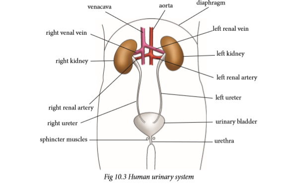
Function of parts of the urinary system
The urinary system keeps everything in balance by removing waste, such as urea, extra salt, extra water and other things the body does not need. Urea is produced when protein, found in meats, is broken down in the body.
The kidneys
There are a pair of kidneys that are red-brown and are located below the ribs in the middle of the back. Their function is to:
• Remove waste from the blood in the form of urine.
• Keep substances stable in the blood.
• Make erythropoietin, a hormone which helps make red blood cells.
• Make vitamin D active.
• Regulate blood pressure.
Ureter
Each kidney has a narrow tube called a ureter, which carries urine from the kidney to the bladder. Muscles in the ureter walls tighten and relax forcing urine down this tube, away from the kidneys.
Bladder
The bladder is a triangle-shaped, hollow organ located in the lower abdomen. It is held in place by ligaments attached to the pelvic bones. The bladder's walls relax and expand to store urine, and contract and flatten to empty urine through the urethra.
Sphincter muscle
Circular muscles that help keep urine from leaking by closing tightly like a rubber band around the opening of the bladder.
Urethra
Urethra is the tube that allows urine to pass outside the body. The brain signals the bladder muscles to tighten, which squeezes urine out of the bladder. At the same time, the brain signals the sphincter muscles to relax to let urine exit the bladder through the urethra. When all the signals occur in the correct order, normal urination occurs.
The kidneys
Activity 10.2: To examine the external and internal structure of a mammalian kidney
Requirements
• Fresh kidney of sheep, goat or cow
• Sharp razor, knife or scalpel
• Small dissecting board
• Hand lens or compound microscope
Procedure
1. Examine the whole kidney. Note the various tubes attached to it.
• What is the outer colour of the kidney?
2. Draw and label the external structure of the kidney.
3. Place the kidney on the dissecting board.
4. Use the scalpel, razor or knife to cut the kidney along its length at the middle.
5. Use a hand lens to identify the following parts: cortex, medulla, pelvis, renal artery, renal vein and urethra.
6. Draw and label the internal structure of the kidney.
Study questions
1. What are nephrons?
2. How are nephrons structured?
3. What is the function of the nephrons?

Kidneys are bean-shaped and are redbrown in colour. They lie near the back of the abdominal cavity about the level of the waistline. Each kidney weighs approximately 142.5 g. It is about the size of a clenched fist. The right kidney is slightly lower than the left. The kidney is surrounded by a layer of fat which helps to cushion it from mechanical or physical injury.
The kidney is supplied with blood from the general circulatory system via the renal artery which branches off from the aorta. Blood from the kidney goes back to the general circulation through the renal vein which joins the vena cava. A tube called the ureter connects each kidney to the bladder located in the lower abdomen.
The bladder is stretchy to hold large quantities of urine. It stores urine temporarily. From the bladder, another tube called the urethra opens to the exterior of the organism. Two rings of sphincter muscles encircle the urethra. They control the emptying of the bladder.
Internal structure of the kidney
A frontal section through the kidney reveals three main regions. The outer part called cortex, inner part called medulla and the pelvis.

1. Cortex
This is the outer part which is dark in colour. It contains a dense network of blood capillaries that form the glomeruli of nephrons. Nephron is the functional unit of the kidney.
2. Medulla
This part is pale red in colour and lies between the cortex and the pelvis. It contains several cone-like extensions called pyramids.
3. Pelvis
This part is white in colour. It narrows to form the ureter. Pelvis is a collecting space leading to the ureter, which takes urine to the bladder.
The nephron
The most important function of the kidney as an excretory organ is to filter wastes from the blood. This takes place in tiny units called nephrons or renal tubules. A nephron is therefore referred to as the functional unit of the kidney. Each kidney has about 1.25 million nephrons. One part of the nephron is in the cortex and the other part in the medulla.

The nephron has three distinct coiled parts:
• The proximal convoluted tubule
• A U-shaped loop of Henle
• A distal convoluted tubule
Both the proximal convoluted tubule and the distal convoluted tubule are located in the cortex. The loop of Henle is in the medulla. One end of the nephron is modified to form a cup shaped structure called the Bowman’s capsule.
The nephron is supplied with an extensive network of blood capillaries. In the Bowman’s capsule, the capillaries form a knot called the glomerulus (plural glomeruli) which branches from an afferent arteriole that originates from the renal artery. The glomeruli capillaries reunite to form an efferent arteriole which channels blood away from the glomerulus. Thus, on one end of the nephron is blood supply from the artery, and on the other end is blood supply to the vena cava.
Urine formation
Excretion in the nephron is carried out in two stages: ultrafiltration and selective reabsorption. Blood coming into the kidney from the artery contains both waste substances and useful substances. Both substances must enter the nephron, where separation takes place by ultrafiltration. The body must not lose the useful substances. Therefore useful substances must be taken back into the blood so that they are not lost. This process is known as selective reabsorption.
(a) Ultrafiltration
Ultrafiltration takes place in the glomerulus.
Note: The afferent arteriole that takes blood to the glomerular capillaries has a wider lumen than the efferent arteriole that takes blood away from it.
Due to the difference in afferent and efferent arteriole size, a high pressure of blood is created in the glomerulus. This pressure forces water, mineral ions and small molecules like glucose, amino acids and urea out of the glomerulus. These pass through the tiny pores in the walls of the glomerular capillaries into the Bowman’s capsule. The liquid collected in the Bowman’s capsule is called glomerular filtrate.
The larger molecules in the blood, like blood proteins, white blood cells, red blood cells and platelets cannot pass through the capillary walls of the glomerulus. These remain in the blood and continue to flow to the efferent arteriole. The glomerular filtrate flows down the nephron where re-absorption will take place as it flows along.
(b) Selective reabsorption
As the glomerular filtrate passes along the nephron, some substances that are useful to the body are selectively taken back or reabsorbed into the blood capillaries network surrounding the nephron.
• All amino acids and glucose are reabsorbed by active transport in the proximal convoluted tubule.
• Some salts and water are reabsorbed depending on how much of them the body still needs. Water is absorbed by osmosis and salts by active transport. Salts are absorbed mainly in the distal convoluted tubule. Water is reabsorbed in both the proximal and distal convoluted tubules. However, most of the water is reabsorbed in the region of the collecting duct.
• No urea is reabsorbed into the blood.
By the time the filtrate from the glomerulus completes its movement down the nephron, it has a high concentration of urea, some salts and water. The liquid is now called urine. Several nephrons empty into one collecting duct, and all the collecting ducts of a kidney empty into the ureter. The process of urine formation is a continuous one, and the ureter continuously receives small amounts of urine. The bladder stores the urine until it is full, then one begins to experience an un-comfortable feeling. The sphincter muscles must then be relaxed in order to empty the bladder.
Research project
Using reference materials and internet, find out why an individual may pass much dilute urine or less but concentrated urine.
Factors that affect urine production
The volume, colour, odour of urine and frequency of urination is affected by many factors. They include:
1. Amount of fluids taken
Large intake of fluids lowers the osmotic pressure of blood. This leads to reduced reabsorption of water in the kidney tubules resulting in the production of large amounts of dilute urine.
2. Amount of salt taken
Intake of a salty meal raises the osmotic pressure of blood. This leads to increased reabsorption of water in the kidney tubules resulting in the production of coloured, little and smelly urine.
3. Weather
In hot and dry weather conditions, the body tends to lose a lot of water through sweating thereby raising the osmotic potential of blood. In this case a lot of water is reabsorbed resulting in coloured, little and smelly urine.
During cold weather the frequency of urination increases because sweating is so minimal.
4. Physical activity
During an exercise like running, jumping and playing, we sweat a lot. The kidney reabsorbs more water resulting in little, coloured and smelly urine.
5. Diseases
Certain diseases that affect the secretion of hormones that control reabsorption of water in the kidney tubules can either lead to production of large or small amounts of urine. An example is diabetes insipidus.
Self-evaluation Test 10.2
1. The diagram below shows the mammalian urinary system. On it, indicate the renal artery, urethra, ureter and left kidney.

2. Given the following parts of the nephron:
(i) Bowmans capsule
(ii) Loop of Henle
(iii) Distal convoluted tubule
(iv) Collecting duct
(v) Proximal convoluted tubule
Arrange the parts in order beginning with the region of the nephron from where the filtrate begins to flow.
3. A sea salmon swims up a river. Explain how this would affect the quantity and concentration of its urine.
10.4 Practices that maintain healthy urinary system
Discussion corner
1. Discuss with a classmate, the good habits that enhance healthy urinary system. 2. Present your work to the rest of the class.

The best way to prevent urinary tract infection is to keep the kidneys and the entire urinary system healthy. This can be done through proper diet and nutrition. Some methods that keep the urinary system and kidneys functioning normally include:
(i) Drinking a lot of water, at least 10 glasses of water a day to flush out toxins in the body.
(ii) Exercising regularly to keep fit. Maintain a healthy weight according to your age to avoid putting excess strain on all bodily systems.
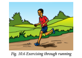
(iii) Avoid taking too many drugs especially pain killers. Stick to prescriptive drugs from a qualified medical officer.
(iv) Visit a doctor (urologist) regularly to check the health of the urinary system.
(v) Eat healthy by avoiding junk food. Eat more fresh fruits and green vegetables. Choose foods low in sodium, sugar and fats but high in fiber content.
(vi) Be informed about the causes and prevention methods of kidney diseases and urinal track infection causes.
(vii) Avoid smoking and alcohol intake.
Health Check!
It is good practice to empty your bladder in toilets, latrines or urinals. Urinating in public places like bus stops, by the roadside, in water bodies or on walls is extremely unhygienic. In addition to polluting the environment, the urine may also contain disease-causing micro-organisms that can contaminate drinking water and vegetables.
Self-evaluation Test 10.3
1. Suggest reasons why people who take alcohol tend to urinate a lot.
2. Why is it important to take only prescribed medicine from a qualified doctor?
3. Drinking a lot of water is healthy. Explain.
Unit summary
• Excretion is the process by which waste substances from cells, tissues or blood of organisms are removed from the body. Egestion is getting rid of undigested or unabsorbed wastes from an organism.
• Excretion is important as it enables the elimination of urea and carbon dioxide.
• The excretory substances in animals include ammonia, urea, uric acid, carbon dioxide and mineral salts.
• If waste substances accumulate in the cell or even in its surroundings, they kill the cell. Therefore they should be excreted.
• In multicellular organisms such as human beings, excretion occurs through excretory organs such as skin, lungs, liver and kidneys.
• The major component of the kidney is the cortex, medulla, pelvis and ureter.
• The nephron consists of Bowman’s capsule, proximal convoluted tubule, Loop of Henle, distal convoluted tubule and the collecting duct.
• Urine is produced by the processes of ultrafiltration and selective reabsorption.
• Urine is transported through the ureters to the bladder where it is stored temporarily before it is removed from the body through the urethra.
• The volume and concentration of urine is affected by water intake, temperature and exercise.


UNIT 11 : Joints And Movement
Key unit competency
To be able to describe types of joints and relate the structure of joints to their functions.
Learning objectives
After studying this unit, I should be able to:
• Differentiate between hinge joint and ball and, socket joint.
• Outline the function of bones, ligaments, tendons, cartilage, nerves and synovial fluid in the joint.
• Analyse the structure of immovable and movable joints.
• Demonstrate how antagonistic muscles bring about movement at the hinge joint.
• Appreciate the link between skeletal muscles and bones in movement.
• Develop good habits that maintain safety of the body joints.
Introduction
In senior one, you learnt about skeletal systems of organisms. What did we say is the importance of the skeleton in our body? Can you give examples of movable and fixed joints?
Look at the pictures below.

What is the role played by joints in pictures A and B above? We are able to move different parts of our body because of joints that occur between different bones.
However, there are joints that are not movable at all. In this unit, you shall learn the different types of joints, their functions and how to keep our joints healthy.
11.1 Types of joints
A joint is a point where a bone meets another bone or bones. Except for pelvic, sacral, skull and sternal bones, joints allow movement and provide mechanical support. Muscles pull on bones to make joints move. Therefore, without joints, movement would not be possible. Most joints only allow slight movement while other joints allow no movement at all.
Joints develop between adjacent bones. However, movement is important in determining the type of joint that develops. Restricted movement even in a freely movable joint can transform it to an immovable joint.
Activity 11.1: Identifying the location of the movable and immovable joints in the body
What to do
1. Try to locate the position of the movable and immovable joints in your body and note them down.
2. Write down the examples of each of the following categories of joints;
(a) Immovable joints
(b) Slightly movable joints
(c) Freely movable joints
3. Try to demonstrate while your partner watches the 360o movement of the shoulder joint.
Study question
Look for the meaning of the following terms: bone, ligament, muscle, cartilage and tendon.

Bone – is a hard, tough connective tissue composed of mineral salts such as calcium and phosphate. It is abundant in all animal skeletons.
Cartilage – this is a skeletal connective tissue which is softer than a bone. It supports the trachea, nose, oesophagus and pinna of the ear.
Ligament-this is a fibrous tissue which join one bone to another. They are elastic to allow movement at a joint.
Tendon –a tough connective tissue which attaches a muscle to a bone. They are inelastic.
Muscle- this is a contractile tissue specialised for contraction and relaxation. They cover the skeleton. Muscles are responsible for locomotion and other type of movement in animals.
Joints are classified structurally depending on how the bones articulate or functionally depending on the degree of movement between the articulating bones.
i. Structural classification
(a) Cartilaginous joint- the bones are connected by cartilage.
(b) Fibrous- the bones are connected by dense fibrous tissue rich in collagen.
(c) Synovial- there is a space between the bones called synovial cavity that is filled with a fluid known as synovial fluid.
ii. Functional classification
(a) Movable joints- allow some degree of movement. There are two categories;
• Those which allow slight mobility.
• Those which are freely movable.
(b) Immovable joints-permit very little or no mobility.
Fixed or immovable joints
These are joints that do not allow any movement, for example, joints in the cranium commonly known as sutures. These joints have seams between the bones of the skull. They are not smooth but rather have interlocking finger-like processes that increase stability. Between the bones are dense fibrous tissues.

Fixed joints are also found in the pelvic girdle.
Movable joints
These are joints that allow movement of body parts to take place. Some movable joints allow only a small degree of movement while others allow a wide range of movement. There are several movable joints which include:
(a) Gliding joints
These joints consist of two opposing flat surfaces that allow slight amount of gliding motion. They occur between the vertebrae. They have no fluid between them but instead have a larger cartilage between them known as intervertebral disc which reduces friction during movement. They are also found in the wrist and ankle.

(b) Synovial joints
These joints are classified depending on the shape of the adjoining articular surface.
They include:
(i) Saddle joints - comprises of two saddle-shaped articulating surfaces that are oriented at right angles to each other. Example is the joint of the thumb.

(ii) Hinge joints
Activity 11.2: Determining the position of hinge joint
1. When you open and close a door, how does it move?
2. Ask a friend to open and close a door as you watch.
• What do you notice?
You may have realised that the door can move only towards you or away from you. Notice too that the door is held in place by hinges which allow this movement.
3. Is there any other possible way the door can move? Try it and find out.
The movement of the door can be described as a back and forth movement only. When something held at a point, moves in such a way, it is said to move along one plane only.
4. Now, try and move different parts of your arm or leg. At which joints do you think there is movement in one plane only?
5. Name the position of these joints.
6. You can use an arm model with a rubber band to demonstrate movement of joints.

Hinge joints allow movement in one plane. The joints at the elbow, knee and finger joints show this back and forth movement as the only type of movement.

(iii) Pivot joints
It consists of a cylindrical like bony process of one bone that rotates within a ring partially composed of bone and cartilage. Examples include articulation of the axis with the atlas and the articulation between the head of radius and the proximal end of the ulna.
(iv) Ball and socket joint
Move your arm at the elbow. Then move your arm at the shoulder in as many ways as possible. What was the difference in the type of movements at the elbow joint and at the shoulder joint?
These types of joints allow the greatest flexibility of movement of all joints. It consists of a ball-shaped head end of one bone that fits into the cavity (socket) of an adjacent bone. This type of joint allows wide range of movement in almost any direction. The shoulder joint is an example of a ball and socket joint. The other is the hip joint. Bones at hinge joint and ball and-socket joint are held in place by ligaments which join them together.

Structure of a synovial joint
The articular surfaces of bones at synovial joint are covered with articular cartilage that provides a smooth surface where the bones meet. The adjoining bones are enclosed within a synovial cavity that is surrounded by a fibrous capsule that holds the bones together. Some portions of the capsule may thicken to form ligaments.
A synovial membrane lines the synovial cavity. The membrane produces synovial fluid that is a complex mixture of cells and nutrients such as proteins, polysaccharides and fats. The synovial fluid covers the surfaces of the joint and provides a lubricating film. Examples include: hip joint, shoulder joint and knee joint.

Self-evaluation Test 11.1
1. Assume that a sharp object penetrated a synovial joint. From the list of structures write the order in which they would most likely be penetrated.
1 Muscle or tendon
2 Ligament
3 Articular cartilage
4 Fibrous capsule
5 Skin
6 Synovial membrane
A. 5,1,2,4,3,6
B. 5,1,2,4,6,3
C. 5,1,2,6,4,3
D. 5,2,1,4,3,6
2. Which two joints move in the same way?
A. Knee joint and shoulder joint
B. Knee joint and elbow joint
C. Neck joint and knee joint
D. Neck joint and ankle joint
3. Which of the following is used to classify joints in the body?
A. The number of bones that articulate.
B. The embryonic tissues formed at the joint.
C. The connective tissues that bind the bones together.
D. The degree of movement at the joint.
E. Both C and D.
11.2 Action of antagonistic muscles in the movement of a hinge joint
Roll up your sleeves. Make a fist with your hand tightly. Bend your arm at the elbow. Observe the inner part of the upper arm. You may have noted that the inner muscles of your upper arm bulge. What do you think causes the bulge?
The biceps and triceps are referred to as antagonistic muscles. They are found on the upper part of the forearm. To lift the arm, the biceps contracts while the triceps relaxes. Joints provide a fulcrum point for muscles to act. The joint action in the upward movement of the arm is flexion resulting to the decreasing of the joint angle. Biceps muscles are therefore called flexor muscles. To straighten the arm, triceps muscles contract while biceps relaxes; the joint action is extension that results to increase in the joint angle.
Antagonistic muscles occur in pairs and oppose a specific movement such that when one muscle contracts, the other relaxes. This means that they never contract or relax at the same time. When the lower arm is straightened at the elbow, we say that the arm is extended. When the arm is bent at the elbow, we say the arm is flexed. When the arm is extended, the triceps muscles are in a contracted state and so, they are tight and short in length. The bicep muscles on the other hand are relaxed and therefore stretched.
Basically the movement of the arm is purely brought about by the opposing actions of the biceps and triceps.


Self-evaluation Test 11.2
1. Suppose the biceps muscles are paralysed, which arm movement will not be possible?
2. Why must muscles work in antagonistic pairs at joints?
11.3 Practices that promote healthy bones and joints
Discussion corner
1. What practices can cause bone fractures?
2. To maintain healthy bones and enhance quick recovery from fractures we should take food rich in calcium, zinc ions and magnesium, ions.
3. Sunlight enhance strong bones.
4. Elderly people develop a bent back.
5. Present your work to the rest of the class.

Keeping bones and joints healthy becomes more important as we age. Serious conditions such as osteoporosis and arthritis can make it hard to move around. They may lead to even more medical problems. There are simple things that you can do to reduce your likelihood of developing these conditions or at least prevent them from becoming worse.
Simple lifestyle changes can help to protect bones and joints. They include:
1. Eating healthy food appropriate for bone health, for example:
(a) Incorporating more calcium rich foods into the diet. Getting enough calcium is essential in keeping the bones healthy and strong.
Calcium rich foods include:
(b) Eating foods that contain Vitamin D. Foods that contain• Low-fat dairy products such as milk.
• Green leafy vegetables, such as Dodo or cabbages.
• Foods fortified with calcium, such as cereals and bread.
vitamin D include: egg yolk and beef liver. Vitamin D is also sythesised when our skin get exposed to the sun.(c) Getting enough of Vitamin C, which is necessary in repairing tissues, including the cartilage in joints. Foods that are rich in vitamin C include: fruits, such as oranges, lemons, watermelon, sweet potatoes and tomatoes.2. Avoiding alcohol consumption. Taking alcohol interferes with the body’s ability to absorb vitamins and minerals. It also results in an increase in hormones that deplete bone density.
3. Weight bearing exercises which helps the body to build more bone mass and maintain the bone density. Some examples of weight bearing exercises include:
• Walking
• Running
• Dancing
• Playing soccer
• Playing basketball
• Playing tennis
4. Avoiding smoking. Smoking has been shown to cause bone mass depletion. Smoking also causes many other serious health problems as well.

5. Maintaining a healthy body weight and posture. It is important to maintain a healthy body weight to keep your bones and joints healthy. If you are underweight, you are at a higher risk of bone loss. If you are overweight, you may be causing extra stress on your joints. This may put you at risk of developing osteoarthritis.
Self-evaluation Test 11.3
1. Explain why it is important to maintain a healthy body weight.
2. Give reasons why we should avoid alcohol and smoking.
3. Give the benefits of putting on the right size of shoes or non-fitting clothes.
Unit summary
• A joint is a point where two bones meet (articulate).
• Joints can be classified according to function, type of connective tissue that binds them or presence or absence of fluid.
• Fibrous joints allow little or no movement; cartilaginous joints are slightly movable while synovial joints allow considerable movement.
• The shoulder joint is a ball and socket joint between the head of humerus and the glenoid cavity of scapula. This joint allows a wide range of movement.
• Synovial joints include: hinge joints, ball and socket joints and pivot.
• The elbow joint is a compound hinge joint between the humerus, sigmoid notch of ulna and radius. Movement at this joint is limited to flexion and extension.
• Antagonistic muscles include the biceps which is the flexor muscle while triceps is the extensor muscle.
• Good health habits, for example, eating a balanced diet rich in calcium and vitamin D, and plenty of physical activity is important in keeping bones healthy.


UNIT 12 : Infectious diseases
Key unit competency
To be able to identify symptoms of common infectious diseases and their prevention and treatment.
Learning objectives
After studying this unit, I should be able to:
• Define pathogen, transmissible disease and host.
• Explain how the body’s defence mechanism can be enhanced by vaccination.
• Explain the symptoms, prevention and control of malaria, Ebola and HIV and AIDS.
• Apply practices of hygiene and sanitation to control the spread of diseases.
• Organise community campaigns on prevention and treatment of diseases.
Introduction
Infectious diseases are also known as communicable diseases. They are diseases that are transmitted from one person to another. These diseases are usually caused by harmful micro-organisms. Examples of micro-organisms include viruses, fungi, protozoa and bacteria. Look at the picture below. Can you tell what will happen to the other person who is not coughing?

Can the action by the person in the picture lead to transfer of disease or not? If so, can all diseases that we know be transmitted in such a manner? Why? What does this tell you about the diseases that you will learn about in this unit?
12.1 Transmission of infectious diseases
Infectious diseases are capable of spreading from one person to another either directly or indirectly.
Research Activity
1. Using text books, handouts and internet, research on the following terminologies:
• Pathogen
• Epidemic
• Pandemic
• Host
• Infectious disease
• Vector
2. How does the body defend itself against diseases.
3. Write your findings in your notebook.

Pathogen: This is any disease-causing microorganism. Bacteria, protozoa and viruses that cause disease are called pathogens. Pathogens are also known as germs.
Transmissible disease: These are diseases capable of spreading from one person to another, for example, common cold.
Host: This is an organism that harbours a disease-causing micro-organism, for example, water snails are hosts to schistosomes that causes bilharzia.
Vector: This is a living organism that can transmit infectious diseases between human beings or from animals to human beings. Female anopheles mosquito is a vector to the plasmodium parasite malaria.
Carrier: This is a person who has been infected but develops no signs or symptoms. This person is capable of transmitting the disease to other people.
Signs: These are visible expressions brought about by a disease, for example, red spots on the body.
Symptoms: A physical or mental feature that is regarded as indicating a condition of disease, particularly such a feature that is felt by the patient, for example, nausea, dizziness and headache.
Epidemiology: The study of all factors that contribute to the appearance of a particular disease.
Activity 12.1 Visit to a health centre
1. Prepare a questionnaire by formulating a series of questions to ask a health worker about transmission of infectious diseases.
You may include the following questions.
• How is Ebola spread from one person to another?
• Who are the most vulnerable people to be infected with malaria?
• Why is tuberculosis a very dangerous disease?
• Is smoking related to tuberculosis?
• What has the government done to control HIV and AIDS in the Rwandan population?
• What is the role of pathogens in spreading diseases?
2. Hand over in your research to the teacher for assessment.

Infections can be spread in several different ways. Some of them are listed below:
1. Diseases that are transmitted through the air are referred to as air-borne diseases. Tuberculosis and common cold are examples.
2. Some diseases are transmitted through the consumption of contaminated water and food. They are referred to as waterborne or foodborne diseases respectively. Examples are cholera and typhoid fever.
3. Through blood transfusion or contact with contaminated blood. Examples of diseases that can be spread through contaminated blood are HIV and AIDS and malaria.

4. Physical contact with an infected person can transmit a disease. These infectious diseases are termed as contagious. They include ringworms, scabies and Ebola.
5. Communicable diseases are also spread by vectors. They are therefore known as vector-borne diseases, for example, sleeping sickness is spread by tsetse flies, malaria is spread by female anopheles mosquito. Yellow fever on the other hand is spread by black fly and some species of mosquitoes.
6. Some infections can also spread through unprotected sexual intercourse with an infected person. These diseases are known as sexual transmitted infections (STIs), for example, HIV and AIDS.
7. Germs can also spread from a mother to her unborn child, usually through blood or other body fluids. They can also pass through contact with skin or mucous membranes during delivery.
Self-evaluation Test 12.1
1. Which one of the following is a vector for sleeping sickness?
A. Tsetse fly
B. Mosquito
C. Black fly
D. Trypanosome
2. If you came into contact with a person who was coughing, which diseases are you likely to catch?
3. Name two diseases transmitted through transfusion of contaminated blood.
12.2 Defense against infection
Research Activity
1. Using text books and internet research on the ways by which the body defends itself from infections.
2. Share your findings with other class members.
Immunity is the ability of the body to resist disease infection. This means a person can be exposed to factors that can cause a disease, yet they do not become sick. The body’s defense mechanism protects an organism from infections.
If pathogens do not encounter resistance from the body’s defense mechanism, almost all diseases would be fatal. Organisms must find a way of defending themselves against harmful micro-organisms like viruses, bacteria and fungi.
The body has many ways of defending itself from invasion by harmful microorganisms. In most cases the body’s defense mechanism prevents this from happening. Some of these defense mechanisms are made in a way to block pathogens from entering into the body.
Other mechanisms eliminate pathogens that are already in the body. There are also defence mechanisms that attack micro-organisms when they persist inside the body. The ability of the body to prevent itself from diseases using its lines of defense is called resistance.
Body defence mechanisms can either be first line or second line.
(a) Body’s first line of defence
This line of defence prevents harmful micro-organisms from entering the body. It is the external defence system of the body. It includes both physical and chemical defence lines. The body does this using various barriers that intercept micro-organisms hence prevent them from entering the body.

Example of the body defence mechanism include:
(i) The skin creates a physical barrier that protects the cells inside the body against the entrance of pathogens. A healthy skin is rarely penetrated by pathogens. Certain chemical secretions produced by the skin helps to stop the growth of bacteria and fungi.
(ii) The nose and passages leading to the lungs are lined with cells that produce sticky fluid called mucus that traps invading microbes and dust. Tiny hairs called cilia lines the trachea. They can move back and forth in a wave-like motion. Cilia trap microbes and dust particles and prevent them from entering the lungs. These particles then combine with mucus and are either coughed, sneezed out or swallowed and then passed out of the body in faeces.

(iii) The stomach produces hydrochloric acid which destroys many of the microbes that enter the body in food and drinks we take.
(iv) Tears act as a barrier to pathogens. Tears are a watery secretion produced by tear glands located in the outer eye. They contain a powerful enzyme that can digest and breakdown harmless substances. Therefore, harmful microorganisms cannot enter the body through the opening of the eye.
(v) Clotting of blood occurs when an open cut or wound exposes blood to air. Such a cut causes a break in the skin exposing the body to harmful micro-organisms. When a clot forms, it seals the opening and enables the wound to heal.
(b) Body’s second line of defence
When pathogens are able to get past the first line of defence, for example, through a cut in the skin, a disease develops. This prompts the second line of defence to become active. This happens through a sequence of steps called the immune response. The immune system attacks the pathogens. The body has a range of defence mechanisms that operate to prevent or destroy pathogens. These responses only react to the presence of any pathogen or foreign material.
White blood cells known as phagocytes are adapted to fight these disease causing micro-organisms in the following ways:
(i) They move by amoeboid action. They send out cell extensions called pseudopodia which allow them to surround invading microbes and engulf them.
(ii) Phagocytes release digestive enzymes which break down the trapped microbes before they can do any harm. This process is called phagocytosis.
(iii) They also have the ability to change their shape. This enables them fit in the narrow capillaries hence reach the pathogens

Immunity
This is the ability of an organism to resist a particular infection or toxin by the action of specific antibodies or some white blood cells.
An antibody is a protein produced by the body’s immune system when it detects harmful substances, called antigens. Examples of antigens include micro-organisms such as bacteria, fungi, parasites, viruses, and chemicals. Each type of antibody is unique and defends the body against one specific type of antigen.
Antibodies detect and neutralises the antigen by binding to it. This signals the production of other cells of the immune system to get rid of the invading microbes before it spreads or attacks the body.
(a) Natural immunity

Natural and artificial immunity
Natural immunity comes from the body itself. It is divided into two: active and passive natural immunity.
(i) Active natural immunity may develop after recovering from a disease. The organism makes its own antibodies as a result of contact with antigen from disease-causing pathogen. Once the organism recovers from the disease, it can produce antibodies very quickly should the pathogens invade again. This makes the organism immune to the invading micro-organism. If someone gets a measles attack and gets healed, chances of the disease recurring is minimal.
(ii) Passive natural immunity is immunity acquired by the foetus from the mother through the placenta and also through breast milk. This type of immunity is short-lived.
(b) Artificial immunity is obtained by introducing antigens into the body of an organism to protect the organism from a disease. It is divided into two: active and passive artificial immunity.
(i) Active artificial immunity is induced by introducing antigens into the body of an organism through the use of a vaccine. A vaccine contains antigens composed of living, dead or weakened pathogens. They are used to stimulate the body to recognise certain disease antigens and respond to them. Vaccines usually do not cause the disease.
(ii) Passive natural immunity is the transfer of immunity in the form of ready-made antibodies. This is immunity that comes from using antibodies produced in one organism to protect another organism from a specific disease. These antibodies are usually extracted from the serum (plasma without soluble proteins) of an animal that has recovered from the disease. Such immunity does not last long.

12.3 Symptoms, prevention, control and treatment of common infectious diseases


a) Malaria
Malaria is a protozoan disease caused by a parasite called Plasmodium. The parasites are spread through the bites of infected female Anopheles mosquitoes, which are the malaria vectors. There are several species of plasmodium parasite that cause malaria in human beings.
They include P. falciparum and P. vivax. Malaria poses the greatest threat to human health.
Malaria affects some groups in the population more than it does to others. These groups are prone to developing a severe form of the disease, than others. They include infants, children under 5 years of age, pregnant women and patients suffering from HIV and AIDS.
Symptoms of malaria
(i) High fever and periodic chills that may be accompanied with sweating.
(ii) Frontal headache.
(iii) Pain in the joints.
(iv) General body weakness.
(v) Lack of appetite and nausea that may be accompanied by vomiting.
If malaria is not treated within 24 hours it can progress to severe illness, often leading to death. Children with severe malaria frequently develop severe anaemia and cerebral malaria. In adults, it may involve several malfunctioning of organs.
Prevention and treatment of malaria
Mosquito control is the main way to prevent and reduce malaria transmission. The following are some of the ways mosquitos can be controlled.
(i) Clearing bushes around homes.
(ii) Draining marshes, pools and stagnant water around homes. This destroys breeding places for mosquitos.
(iii) Spraying light used oil on stagnant water that cannot be drained. Oil sprayed on stagnant water kills mosquito larvae by suffocating them.
(iv) Spraying the inside walls of our houses with insecticides.
(v) Closing doors and windows during the evening time to prevent mosquitoes from entering. Windows can also be screened with window proof wire mesh.
(vi) Sleeping under a treated mosquito net.

(vii) Removing old tins, broken pots, polythene bags and other things that can hold water from the compound. This prevents mosquitoes from breeding.
(viii) Patients can be isolated during the infection period to prevent transmission to others.
(ix) Fish-eating mosquitoes can be introduced into ponds and pools to feed on mosquito larvae and pupae.
(x) Preventative medicines can be taken regularly but upon prescription from a qualified medical doctor.
b) Cholera
Cholera is an acute diarrhoeal infection caused by ingestion of food or water contaminated with the bacterium Vibrio cholerae. It has a short incubation period of 2 hours to 5 days, a factor that triggers its pattern of outbreaks. Cholera is endemic in poor countries and strikes areas with poor sanitary conditions and where drinking water is not well treated. Cholera affects both children and adults and can cause death within hours.
Most people infected with cholera do not develop any symptoms. The bacterium is present in their faeces for 1-10 days after infection and is shed back into the environment, endangering the health of other people.
Most people who develop symptoms, have mild or moderate symptoms, while others develop acute watery diarrhoea with severe dehydration (lack of water in the body). This can lead to death if left untreated.
Symptoms of cholera
(i) Severe watery diarrhoea (rice water stool) and vomiting.
(ii) General body weakness.
(iii) Low blood pressure.
(iv) The patient loses weight rapidly.
(v) The patient develops wrinkled skin and sunken eyes because of dehydration.
Control and treatment of cholera
(i) Drinking water should be treated or boiled and stored properly.
(ii) Proper disposal of human wastes. A well-managed sewage disposal system in towns and use of latrines. in the rural areas.
(iii) Washing hands before and after eating and after visiting the toilet.

(iv) Covering cooked food to avoid contamination by house flies.
(v) Vaccination during epidemics or when one is visiting an infested area.
(vi) Cholera is treated using antibiotics. Prevention of dehydration also helps in the cure.
c) HIV and AIDS
The Human Immunodeficiency Virus (HIV) attacks the immune system. This weakens a person’s defence mechanism against infections and some types of cancer. As the virus destroys and impairs the function of immune cells, infected individuals gradually become immunedeficient. Immune function is typically measured by number of white blood cells in the body (CD4 cell count).
Immunodeficiency results in increased susceptibility to a wide range of infections. These infections (opportunistic infections) are diseases that people with healthy immune systems can fight off. At this stage, the patient is said to be suffering from AIDS HIV and AIDS is transmitted from one person to another through various ways. They include:
(i) By birth, from an infected mother to her unborn baby.
(ii) By having unprotected sexual intercourse with an infected person.
(iii) By transfusion of infected blood.
(iv) By sharing of unsterilised surgical and skin piercing instruments with infected people.
Symptoms of HIV and AIDS
The symptoms depend on the stage of infection. People living with HIV and AIDS tend to be most infectious in the first few months. The first few weeks after initial infection, individuals may experience no symptoms or an influenza-like illness including fever, headache, rash or sore throat.
As the infection progresses, the immune system weakens. An individual can develop other signs and symptoms, such as swollen lymph nodes, weight loss, fever, diarrhoea and cough. Without treatment, they could also develop severe illnesses such as tuberculosis, meningitis, and cancers such as lymphomas and Kaposi’s sarcoma, among others.
Prevention and control
The most effective way to prevent the spread of AIDS is through health and sex education. Everyone has to be enlightened on the risks of acquiring the virus through careless lifestyles.
Patients of HIV and AIDS should take anti-retroviral drugs (ARVs) which prolong their life span. To prevent AIDS one also needs to avoid the ways through which it is transmitted. The following are the ways through which a person can prevent himself or herself from contracting the disease.
i. Abstaining from sex till marriage.
ii. Being faithful to one partner.
iii. Using condoms during sex.
iv. Mothers should give birth in hospitals and go for regular medical checkup.
v. Blood should be screened before transfusion.
vi. Avoid sharing piercing and cutting instruments like razorblades and safety pins.
d.) Tuberculosis
Tuberculosis (TB) is caused by bacteria called Mycobacterium tuberculosis that most often affects the lungs. TB is spread from person to person through the air. When a person with pulmonary TB coughs, sneezes or spits, they spread germs into the air. If a healthy person nearby inhales these germs they can become infected.
People with low immune systems, such as people living with HIV and AIDS, malnutrition, diabetes, or people who smoke, have a much higher risk of being infected.
Symptoms of pulmonary TB
Common symptoms of active pulmonary TB are:
(i) Cough with sputum and blood at times
(ii) Chest pains
(iii) General weakness
(iv) Weight loss
(v) Fever
(vi) Night sweats
When a person develops active tuberculosis, the symptoms; cough, fever, night sweats and weight loss may be mild for many months. This can lead to delays in seeking medical care, resulting in transmission of the bacteria to other people.
Prevention and treatment of tuberculosis
(i) Isolate patients to prevent transmission of the disease to other people.
(ii) Immunise children with tuberculosis vaccine known as BCG. This may prevent the spread of tuberculosis. But the vaccine does not necessarily protect against pulmonary tuberculosis.
(iii) Avoiding overcrowded places. Opening windows in overcrowded rooms to allow free circulation of air.
(iv) Drinking boiled milk. Some types of tuberculosis is transmitted from bovine.
(v) Once infected, tuberculosis is treated with antibiotics. Standard anti-TB drugs have been used for some time, and resistance to the medicines is widespread.
e) Ebola
Activity 12.3: Watching a film about ebola
You will observe videos showing doctors treating ebola patients. Observe the protective gear worn by the doctors.
Study questions
(a) What causes Ebola?
(b) How is Ebola transmitted to the human population?
(c) What are some of the signs and symptoms of the Ebola virus disease?
(d) What can you do, to prevent contracting the Ebola disease?
(e) From the video you have watched, why are doctors in protective gear while handling the patients?
(f) Why do you think the ebola patients are in isolated wards?

The Ebola virus causes an acute, serious illness which usually leads to death if untreated. Ebola virus is introduced into the human body through contact with body fluids of infected animals such as chimpanzees, gorillas, fruit bats, monkeys, forest antelope and porcupines found ill or dead in the forest.
Ebola then spreads through humanto-human transmission via direct contact (through broken skin or mucous membranes) with the blood, secretions, organs or other bodily fluids of infected people and with surfaces and materials such as bedding and clothing contaminated with these fluids.
Medical workers too, become infected while caring for patients suffering from ebola. Burial ceremonies in which mourners have direct contact with the body of the dead patient can also play a role in the transmission of Ebola. People remain infectious as long as their blood contains the virus.

Symptoms of Ebola virus disease
The incubation period; the time interval from infection with the virus to onset of symptoms is 2 to 21 days. Human beings are not infectious until they develop symptoms.
(i) First symptoms are the sudden onset of fever, fatigue, muscle pain, headache and sore throat.
(ii) This is followed by vomiting, diarrhoea, rash, symptoms of impaired kidney and liver function.
(iii) Internal and external bleeding with blood oozing from the gums, ears and eyes.
Prevention of Ebola
(i) Avoid direct contact with wild animals like bats and chimpanzees.
(ii) Avoid direct contact with people showing signs of Ebola virus.
(iii) Use gloves and other protective clothing when handling Ebola patients.
(iv) Wash your hands regularly with soap and water.
(v) Couples who have just recovered from the virus should abstain from sexual intercourse or use condoms.
(vi) Burial of the dead patients should be done by health workers.
f) Salmonellosis
This is an infection caused by Salmonella bacteria. The elderly, infants, and those with impaired immune systems are more likely to develop severe form of illness.
Salmonella infection usually results from ingestion of the bacteria from contaminated food or water. Eggs, milk, meat or poultry are particularly high risk foods. Fruit and vegetables may also be contaminated, especially if manure has been used as fertiliser. People may become infected if they transfer animal faeces containing Salmonella bacteria from their hands to their mouths, for example, eating after touching animals and failing to wash hands.
Person-to-person spread may occur when hands, objects or food become contaminated. In most cases, the illness lasts for between 4 to 7 days. Most people recover without treatment. In some cases, the diarrhea may be so severe that the patient becomes dangerously dehydrated and must be hospitalised.
Symptoms of salmonellosis
Most people infected with salmonella experience diarrhoea, chills with fever, nausea with or without vomiting, headache, blood in the stool and abdominal cramps 12 to 72 hours.
Prevention and treatment of salmonellosis
(i) People with salmonella infection should not be allowed to handle food or take care of children.
(ii) Food should be well cooked.
(iii) Do not purchase dirty or cracked eggs.
(iv) Boil milk thoroughly before drinking.
(v) Always wash your hands with soap and clean water.
(vi) Wash fruits (including melons) and vegetables before eating.
(vii) Swimming pools should be disinfected regularly.
(viii) Recovery from Salmonella infection usually occurs within a week and antibiotic treatment is not normally required.
g) Typhoid
Typhoid fever is a bacterial infection. It is caused by a bacterium called Salmonella typhi, which only lives in human. It is related to the bacteria that cause salmonella food poisoning. It can spread throughout the body, affecting many organs. Without prompt treatment, it can cause serious complications and can be fatal.
Typhoid fever is highly contagious. A small number of people (carriers) recover from typhoid fever but continue to have the bacteria. Both ill people and carriers shed the bacteria in their faeces and urine. The bacteria is spread by eating food or drinking water contaminated with faeces or urine of patients and carriers.
Typhoid is common in areas where hygiene standards are poor and water is likely to be contaminated with sewage waste.

Symptoms of typhoid
(i) Sustained high fever
(ii) Continuous headache
(iii) Malaise (feeling of being unwell)
(iv) Decreased appetite
(v) Enlarged spleen which may cause abdominal discomfort
(vi) Dry cough in the early stage of the illness
(vii) A flat rose-coloured rash which may be visible on the trunk (vii) Constipation or diarrhoea – constipation occurs more often than diarrhoea in adults
(viii) Illness varies from mild with lowgrade fever, to severe with multiple complications
Prevention and treatment of typhoid
(i) Isolation of the patients to avoid spread of the disease by contact and sterilising clothes used by patients.
(ii) Food handlers in institutions, like hospitals, schools and restaurants should go for regular medical checkups and treatment if found infected.
(iii) Proper disposal of faeces in toilet and in pit latrines.
(iv) Water treatment and purification to kill the bacteria.
(v) Boiling drinking water and cooking of food properly to kill the bacteria.
(vi) Practice good hygienic methods like washing hands properly after visiting the toilet.
(vii) Washing fruits and vegetables with clean water before eating.
(viii) Vaccinations in the case of outbreaks of the disease in epidemics.
(ix) Seek proper treatment from qualified medical doctor in case of an infection. Antibiotic treatment helps reduce the time a person is infectious.
Self-evaluation Test 12.3
1. Which disease among the following is the most highly contagious?
A. Malaria
B. Ebola
C. HIV and AIDS
D. Typhoid
2. What is the simplest thing you can do to protect yourself from contracting a disease?
3. State ways you can use to sensitize your community about infectious diseases?
Unit summary
• Pathogen is a bacterium, virus, or other micro-organism that can cause disease.
• Infectious diseases are transmitted through: air, contact, blood or other body fluids, for example, urine, saliva, breastmilk, semen and vaginal secretions.
• First line of defence is a combination of physical and chemical barriers that prevent all types of foreign agents from penetrating the outer layer of the body. It includes the skin, mucous membranes, hair and cilia, gastric juices, tears, sweat and saliva.
• The second line of defence is a group of cells, tissues and organs that work together to protect the body. This is the immune system.
• Immunity is the ability of an organism to resist a particular infection or toxin by the action of specific antibodies or sensitised white blood cells.
• Vaccination is the injection of a killed or muted microbe in order to stimulate the immune system against the microbe, thereby preventing disease.
• Some unhygienic human activities, such as improper disposal of sewage and other wastes, cause diseases. These diseases include cholera, typhoid and salmonella.
• Malaria is caused by a protozoan parasite that invades the red blood cells. The parasite is transmitted by mosquitoes in many tropical and subtropical regions.
• Ebola is an infectious and generally fatal disease marked by fever and severe internal bleeding, spread through contact with infected body fluids.
• Acquired immune deficiency syndrome (AIDS) is caused by the human immunodeficiency virus (HIV). It damages the immune system and interferes with the body’s ability to fight the organisms that cause disease. HIV is a sexually transmitted infection.


UNIT 13 : Immunity and vaccination
Key unit competency
To be able to describe natural and artificial methods that fight against infection.
Learning objectives
After studying this unit, I should be able to:
• Explain how each pathogen has its own antigens.
• Define active immunity, pathogen and antibody production in the body.
• Demonstrate the importance of passive immunity for breastfed infants.
• Use a diagram to illustrate antibody-antigen reaction sites.
• Observe and differentiate the immune responses.
• Advocate for vaccination and breastfeeding as a sustainable disease prevention method.
Introduction
When pathogens find their way into the body, they can cause diseases. However, the body has its own mechanisms of dealing with such foreign materials. The Ability of the body to defend itself against foreign bodies and disease-causing microorganisms is known as immunity. The system that is responsible for defending the body against diseases is known as the immune system. Look at the picture below. What is happening in the picture?

Talk to your friend about the importance of this.
13.1 Antibodies and antigens
Activity 13.1: Investigating antibodies and antigens
i) A chart showing an antigen and antibody will be displayed on the wall.
ii) Observe the diagrams on the chart carefully and identify the following:
• Shape of the antigen and that of the antigen-binding site on the antibody.
• The reaction centers of antibodies.
Study questions
a) Why are antibodies specific to only one type of antigen?
b) Draw an antibody.
c) Describe the antibody-antigen complex.

An antibody also known as an immunoglobulin is a large Y-shaped protein. It is produced mainly by plasma cells. It is used by the immune system to identify and neutralise pathogens such as bacteria and viruses. The antibody recognises a unique molecule of the harmful agent, called an antigen. The antibody then binds to this antigen. Antibodies are specific to their antigens.

Antigen
This is any substance that is capable of stimulating an immune response, specifically activating the body to produce antibodies. In general, two main divisions of antigens are recognised: foreign antigens and self-antigens.
• Foreign antigens come from outside the body. All pathogens are examples of foreign antigens. Viruses and micro-organisms such as bacteria and protozoa, and certain proteins in foods all lie under this group.
• Self-antigens, on the other hand, come from within the body. Normally, the body is able to distinguish self from non-self. However, in individuals with autoimmune disorders, normal body substances provoke an immune response, leading to the generation of auto-antibodies.
Antigen–antibody reaction
An antigen and antibody reaction works like a lock and key mechanism. Antibodies themselves do not destroy antigens. They inactivate and tag antigens for destruction by phagocytes.
All antibodies form an antigen-antibody (immune) complex When they combine with antigens.

Each antigen stimulates the production of a specific antibody. All antibodies are Y-shaped. They however, differ at the antigen binding site. Each antibody has a site complementary to a certain antigen. This means that each antibody can only bind to a specific antigen.
In response to the antigen, the antibody wraps its two sites like a ‘lock’ around the ‘key’ of the antigen sites to destroy it. An antibody's mode of action varies with different types of antigens. With its two-armed Y-shaped structure, the antibody can attack two antigens at the same time with each arm.
Defensive mechanisms used by antibodies are neutralisation, agglutination, precipitation and plasma complement system.
i) Neutralisation – antibodies bind to and block specific sites on antigens; viruses and bacteria. This prevents antigens from binding to sites called receptors on tissue cells. They are later destroyed by phagocytes, for instance, if the antigen is a toxin produced by pathogenic bacteria that cause an infection like diphtheria or tetanus, the binding process of the antibody will neutalise the antigen's toxin.
ii) Agglutination – antibodies bind the same determinant on more than one antigen. An example is when an antibody surrounds a virus, such as one that causes influenza, it prevents it from entering other body cells.
iii) Precipitation – soluble molecules are cross-linked into large insoluble complex. After which they fall out of solution, and are phagocytised.
iv) Plasma complement system- The antibodies coat infectious bacteria and then white blood cells will complete the job by engulfing the bacteria, destroying them, and then removing them from the body.
Self-evaluation Test 13.1
1. Draw a key and lock and relate it to antibody- antigen reaction.
2. How does the body defend itself from infections?
13.2 Immune response
Activity 13.2: Observing immune response
You will be provided with computer aided simulations of the immune response of organisms.
1. Observe the simulations carefully.
2. Differentiate immune responses of organisms.
3. Share your findings with the class.

Immune response refers to the process by which the body recognises and defends itself against bacteria, viruses and substances that appear foreign and harmful. The immune system is a collection of cells, tissues and molecules that protect the body from numerous pathogens and toxins in our environment.

This defense against microbes has been divided into two general types of reactions:
• Reactions of innate immunity
• Reactions of adaptive immunity.
a) Innate immunity
Innate immunity also called nonspecific immunity is the body defense system that one is born with. It protects against all antigens. Innate immunity involves barriers that keep harmful materials from entering your body. It consists of cells and proteins that are always present and ready to fight microbes at the site of infection.
The main components of the innate immune system are:
i. Physical epithelial barriers
ii. Phagocytic leukocytes
iii. Dendritic cells
iv. A special type of white blood cells called a natural killer (NK) cell
v. Circulating plasma proteins
Innate immunity includes the components of the body’s first line of defence. These are enzymes in tears and skin oils, mucus, skin and acid in the stomach.If an antigen gets past these barriers, it is attacked and destroyed by other parts of the immune system.
b) Adaptive immunity
Adaptive immune system also known as acquired immunity is immunity that develops after exposure to various antigens. The immune system builds a defense against that specific antigen. It is called into action against pathogens that are able to evade or overcome innate immune defenses. Components of the adaptive immune system are normally silent; however, when activated, these components “adapt” to the presence of infectious agents by creating mechanisms for neutralising or eliminating the microbes.
The immune system includes certain types of white blood cells, for example, lymphocytes. There are B and T type lymphocytes.
• B lymphocytes are cells that produce antibodies. Antibodies attach to a specific antigen and present it to other immune cells for destruction.
• T lymphocytes are cells that attack antigens directly and help control the immune response.
During growth and development of white blood cells, they always learn to differentiate between individuals own body tissues and foreign substances. Once B cells and T cells are formed, a few of those cells will multiply and provide "memory" for the immune system. This allows the immune system to respond faster and more efficiently the next time an individual is exposed to the same antigen. In many cases it will prevent you from getting sick, for example, a person who has had measles or has been immunised against measles is immune from getting it again.
Active immunity
This is the immunity that results from the production of antibodies by the immune system in response to the presence of an antigen. Active immunity is either natural or artificial.
• Naturally acquired active immunity occurs when a person is exposed to a live pathogen. The individual develops the disease, and becomes immune as a result of the primary immune response.
• Artificially acquired active immunity can be induced by a vaccine, a substance that contains alternated form of the antigen. A vaccine stimulates a primary response against the antigen without causing symptoms of the disease.
Passive immunity
This is a short-term immunity that results from the introduction of antibodies from another person or animal. Passive immunity is either artificial or natural.
• Artificially acquired passive immunity is a short-term immunisation. It is achieved by the injection of antibodies that are not produced by the recipient's cells.
• Naturally acquired passive immunity occurs during pregnancy, in which certain antibodies are passed from the maternal body into the foetal bloodstream.
Passive immunity for breast–fed infants
Discussion corner
1. Discuss with a classmate the following:
i. Importance of vaccination and its application.
ii. The importance of breastfeeding newborns.
iii. Why we should participate in a vaccination campaign.
2. Share your findings with the class.

Infants have passive immunity because they are born with antibodies that are transferred through the placenta from their mothers. They also get some antibodies from the mother’s breast milk. These antibodies disappear between ages 6 and 12 months. Passive immunisation may also be due to injection of antiserum, which contains antibodies that are formed by another person or animal. It provides immediate protection against an antigen, but does not provide longlasting protection. BCG given to a child at birth to protect it from TB is an example of passive immunisation.

13.3 Immunisation and autoimmunity
Immunisation is the process by which an immune response is triggered by the administration of a vaccine towards an infectious disease. Small amounts of an antigen, such as dead viruses, are given to activate immune system "memory". Memory allows your body to react quickly and efficiently to future exposure. Such small doses of antigens are referred to as vaccines.
Immunisation is an important method of disease prevention. By receiving a vaccination, usually in the form of an injection, a person can be 'immunised' against a disease, and reduce their likelihood of developing the illness. Immunisation can also reduce the spread of the disease in the population.
Self-evaluation Test 13.2
1. Differentiate between active and passive immunity.
2. What is immune system made up of?
3. Outline the importance of breastfeeding.
Discussion corner
1. Discuss with a classmate how a person’s own immune system can bring about Type 1 diabetes.
2. Share your findings with the class.

Autoimmunity is a condition whereby the immune system attacks and kills own cell. Any disease that results from such an immune response is termed an autoimmune disease.
An efficient immune response protects against many diseases and disorders. Wrong immune response causes immune system disorders. An overactive immune response can lead to the development of autoimmune diseases, in which antibodies form against the body's own tissues.
Complications from altered immune responses include:
i. Allergy or hypersensitivity
ii. Autoimmune disorders
iii. Immunodeficiency disorders
iv. Serum sickness
v. Transplant rejection
Type 1 diabetes and the immune system
Type 1 diabetes was initially known as juvenile diabetes or insulin-dependent diabetes mellitus. It is a chronic condition in which the pancreas produces little or no insulin. Insulin is a hormone needed to enable sugar (glucose) to enter cells to produce energy.
The pancreas is the organ responsible for controlling the amount of sugar (glucose) in the body. Specific cells in the pancreas known as the beta cells are responsible for manufacturing insulin. Insulin released into blood regulates the amount of sugar in the body keeping it at normal levels.

When a virus attacks the body, T- cells start producing antibodies to fight against this virus. However, the virus can have same antigens like those of the beta cells in the pancreas. In this case, the T-cells mistakenly recognise beta cells as foreign to the body. They attack these cells hence destroying them. Once the beta cells of the body are destroyed, no insulin is produced. This leads to an increase in blood sugar level and therefore increases the possibility of having diabetes.
Not every virus can trigger the T cells to turn against the beta cells. The virus must have antigens that are similar enough to the antigens in beta cells, and those viruses include:
• German measles
• Mumps
• Rotavirus (which generally causes diarrhoea)
Note: Type 2 Diabetes mellitus occurs when the body cells becomes resistant to insulin or when less insulin is produced by the body.
Self-evaluation Test 13.3
1. What can make antibodies turn against oneself?
2. Compare type 1 diabetes and type 2 diabetes.
Unit summary
• An antibody is a protein produced by the body's immune system when it detects harmful substances, called antigens. Examples of antigens include micro-organisms (bacteria, fungi, parasites, and viruses) and chemicals.
• An antigen is any substance that triggers the immune system to produce antibodies that fight and destroy the antigen.
• Each pathogen has its own antigens that have specific binding sites that are only recognised by certain antibodies.
• Antibodies lock on to antigens, thereby marking them for destruction by phagocytes.
• Immune response is the body’s mechanism of defending itself against bacteria, viruses, and substances that appear foreign and harmful.
• Naturally acquired active immunity occurs when the person is exposed to a live pathogen, develops the disease, and becomes immune as a result of the primary immune response. Artificially acquired active immunity can be induced by a vaccine, a substance that contains the antigen.
• Passive immunity is a short-term immunity that results from the introduction of antibodies from another person or animal.
• Immunisation is the process whereby a person is made immune or resistant to an infectious disease, typically by the administration of a vaccine.
• Autoimmunity is a condition in which the immune system invades and kills own cells and tissues. Any disease that results from such a response is termed an autoimmune disease.


UNIT 14 : Sexual behaviour and sexual response
Key unit competency
To be able to explain safe sex, sexuality and sexual behaviours and argue for control of sex violations.
Learning objectives
After studying this unit I should be able to:
• Describe male and female responses to sexual stimulation.
• Explain different ways couples can show love and affection.
• Outline responses to sexual attraction and stimulation at puberty by boys and girls.
• Share experience and emotional attraction to sexuality.
• Recognise that sexual relationships require emotional and physical maturity.
• Be sensitive to others feelings, beliefs and attitudes towards sex.
• Develop self-confidence and control towards sexual thoughts and feelings.
Introduction
You may have read about adolescence in primary school science. What does the word mean? What are some of the risks which we encounter during adolescence? What should we do to avoid them? Now look at the picture below. What is going in the picture? Is it good or bad?

Suggest activities the two could engage in that are more constructive other than what is happening.
14.1 Male and female sexual responses
Discussion corner
1. Discuss with your classmates about the following:
• Sexual responses in males and females.
• Sexual stimulation for physical response.
2. Note down your findings.
3. Share your findings with the rest of the class members.

The human sexual response is a series of changes that take place in the body in response to sexual stimulation. These changes take a different form in men and women. The stages are in four phases as below.
i. Excitement phase
ii. Plateau phase
iii. Orgasmic phase
iv. Resolution phase
1. Excitement phase
This is the initial stage of stimulation that gets the body ready for sex. The stimulation may be due to kissing, seeing an attractive person or soft touches. This stage is accompanied by a rise in heart rate, breathing rate and blood pressure. The phase can last for a few minutes to several hours.
2. Plateau phase
This is a phase that prepares both man and woman for orgasm. It is characterised by increased heartbeat, muscle tension and blood pressure.
3. Orgasmic phase
The orgasm is the climax of the sexual response cycle. It is the shortest of the phases and generally lasts only a few seconds. General characteristics of this phase include: Blood pressure, heart rate and breathing which are at their highest rates, accompanied by a rapid intake of oxygen.
This phase is accompanied with sexual pleasure and satisfaction with release of sexual tension.
4. Resolution phase
During resolution, the body slowly returns to its normal level of functioning. This phase is marked by a general sense of well-being, enhanced intimacy and, often, fatigue.
Sexual stimulation
This is any stimulus whether physical or mental that leads to sexual arousal. These stimulations may lead to orgasm if they are sufficient. The term sexual stimulation often implies stimulation of the genitals, but may also include stimulation of other areas of the body, stimulation of the senses (such as sight or hearing) and mental stimulation (from reading or fantasising).
Sexual arousal may arise without physical stimulation but achieving orgasm usually requires physical sexual stimulation.
Puberty in boys and girls
Activity 14.1: Investigating puberty
1. In groups share experiences you have on the emotional attraction to sexuality.
2. You will watch a film about physical and emotional changes during puberty.
3. Role play boy-girl relationship.
4. Share your experience with the rest of the class.

Puberty is a period during which adolescents reach sexual maturity and become capable of reproduction. Numerous changes take place both in males and females at this stage. Puberty comes as a result of hormones that originate from the gonads: testicles in men and ovaries in females.
Teenagers normally face challenges during this period of growth and development. The greatest challenge is increase in sexual desire in both females and males. These challenges include unwanted pregnancies and sexually transmitted diseases. Normally there is no big physical difference between males and females before puberty apart from the sexual organs.
Puberty in boys
Puberty in males takes place between the ages of 11 and 13 on average. When a boy reaches puberty at an early stage, he may have benefits. The benefits include popularity with his fellow peers because of being tall and therefore a leader. Late onset of puberty can lead to low self-esteem in males and such may also face problems of anxiety and fear. Sexual desire is at its peak. Boys start dating girls at this stage.
This is a critical time where boys need guidance and counseling because most of them face problems; associated with increased body activity and sexual desire. The problems faced at this stage include:
• Abuse of drugs and alcohol
• Lack of respect for authority
• Fighting among peer groups
• Parental hatred
• Poor hygiene and general body cleanliness.
Puberty in girls
On average, girls begin puberty at an early age of about 10-11years. Puberty in girls occurs fast as compared to boys. Menstruation in girls is a characteristic that shows the onset of puberty. This is brought about by the growth and development of the ovaries.
Early onset of puberty in girls is detrimental, causing low self-esteem and stress. This is due to breast enlargement and a lot of fat deposition on the body that causes different body shapes between the girl and her peers. Early onset is caused by a number of factors which include diet and environmental exposure.
Puberty in girls is more challenging to parents. Girls tend to become more private and sensitive emotionally. Parents should therefore be more careful. Social behaviours of girls also tend to change. Girls become more attracted to males than females. The main challenge that girls face during puberty is unwanted pregnancies. Other challenges include rejection from peers, clothes to wear and physical appearance.

Sexuality education
• Girls should be aware of the biological importance of growth and development. They should be reminded of the sexual dangers that exist at this stage of development; the risk of contracting STDs and unwanted pregnancies. It should also be a point to these girls to maintain general cleanliness and hygiene especially for a healthy life.
• Both girls and boys should be reminded that sex is not the only way to express love. They should abstain from sex until marriage.
Self-evaluation Test 14.1
1. Describe emotional changes that occur in boys and girls during puberty.
2. Draw a step diagram to show sexual response in males and females.
3. Is there a way to avoid unnecessary sexual stimulation?
14.2 People and sexual relationships
Discussion corner
1. Discuss the following questions with your classmates.
• What is the meaning of love?
• What are some of the things we do to show love?
• Does love go hand in hand with sex?
• How can one show love to a neighbour?
• What is sexuality and life skills education?
• Why do we need this education now?
2. Note down your findings.
3. Share your findings with the rest of the class members.

People develop different types of relationships ranging from acquaintances to friendships to romantic relationships. Most people form romantic relationships. This involves falling in love and making a commitment. Intimate relationships bring a sense of stability and security. Partners in a healthy relationship should have the following characteristics; they should:
• Treat each other with love and respect.
• Be honest to each other.
• Spend time with each other.
• Take special interest in activities that each likes.

• Respect each other’s emotional, physical and sexual limits.
• Speak honestly about their feelings.
It is important to note that sexual relationships between close members of a family like father and daughter is unacceptable. It is considered a taboo in the society and is also punishable by law. This form of sexual relationship is called incest. If it happens, children born normally suffer from genetic disorders and lack of vigor. The following are forms of sexual relationships:
a) Exogamy- since incest is a taboo in the Rwandan society, individuals are advised to find their mates outside their family circles. This is known as exogamy. Endogamy is the opposite of exogamy where individuals are allowed to marry their distant cousins.
b) Monogamy: This is a longterm relationship between a man and a woman in which none has other sexual partners. In the Rwandan society, monogamy is the legal form of sexual relationship.
c) Polygamy: This is a long term sexual relationship of having more than one wife or husband at the same time. An example is a man having more than one wife or a woman with more than one man. The Rwandese government is not in support of polygamous sexual relationships. These relationships are a source of sexually transmitted diseases and also contribute to poor family planning that hinders development in families.
d) Casual relationships: This is an act of having sex with someone you do not have an attachment to. This is the case when one engages with a sex worker. Casual relationships are dangerous since they can be sources of sexually transmitted diseases including HIV and AIDS and other forms of sexual and physical abuse.
e) Dating relationships: This is when two people (a boy and a girl) go out together. It leads to engagement and later marriage. Dating should be done carefully. You should avoid sexual intercourse until at appropriate time e.g. after marriage. Sexual intercourse can lead to unexpected pregnancies or sexually transmitted diseases such as HIV and AIDS.

Sexual problems and disappointment
A sexual problem or sexual dysfunction, refers to a problem experienced during any phase of the sexual response cycle. Such a problem prevents the individual or couple from experiencing satisfaction from the sexual activity. The following are some of the sexual disappointments.
In males
a) Premature ejaculation: This is a condition whereby a man cannot delay ejaculation long enough to satisfy the woman.
b) Impotence: This is the inability to produce or maintain an erection. Impotence occurs as a result of fear, anxiety, disease such as diabetes and drug abuse.
c) Inflammation: Blockage of the urethra occurs as a result of an infection or due to swelling of the prostrate gland.
In females
a) Vaginal discharges: This is any abnormal discharge from the vagina. It can be a white discharge with irritating smell especially after sexual intercourse.
b) Functional disorders: This is the inability of a woman to experience sexual pleasure and satisfaction. This results from emotional problems, feeling of guilt and trauma.
Sexuality, age and culture
Sexuality refers to anything to do with being male or female. Human beings have engaged in sexual relationships for long. Each society, however, interprets sexuality and sexual activity in different ways. Human sexuality is part of social life.
The society dictates the norm; what is considered acceptable or unacceptable. Sexual behaviors are based on the morals and values of the particular society. The following sexual behaviors are unacceptable in the Rwandan culture:
i) Incest: This is the act of having sex with a close relative.
ii) Homosexuality: This is where people of the same sex are engaged in a sexual relationship.
iii) Masturbation: The act of selfstimulation that leads to sexual satisfaction.
iv) Anal sexual intercourse: This is a type of sexual activity in which the penis is inserted into the anus.
v) Rape: The act of forcing some one into sexual intercourse without consent.
vi) Adultery: Having sex outside wedlock. vii) Fornication: Engaging in sex before marriage.
viii) Sex slavery: The act of taking advantage of vulnerable individuals in a forced commercial sex.
ix) Prostitution: Engaging in sexual relationships for financial gains.
Ageing is process of growing old. The ageing process does not reduce the need or desire for sex. For many older adults, the concept of sexuality includes practices such as kissing, hugging and fondling. Such activity may replace actual intercourse and becomes increasingly important for those who no longer desire or are in capable of sexual intercourse.
Ageing in men
In an ageing male, the most common sexual dysfunction is erectile dysfunction. This can be due to hormonal changes as part of normal ageing or due to underlying conditions such as diseases. Testosterone hormones in men decline with age. This hormone is produced by the testis in man. It increases sexual urge and libido.
Ageing in women
The most common sexual dysfunction in women, is lack of desire and arousal. This is due to decrease in hormones, which begins at menopause and continue as women age. This decrease in hormones may also produce reduced muscles, loss of bone mass and declining energy. Sexually, it may result in vaginal dryness, decreased libido, and decreased ability to achieve orgasm.
Violation of human rights
Look at the picture below. What is happening? In your opinion, should children be treated this way? Suggest better methods of discipline.

Human right is a right that is believed to belong justifiably to every person. This right is supposed to be enjoyed by each and every one irrespective of their position in the society. Violation of human rights includes: sexual harassment or abuse, coercion and human trafficking.
Child sexual exploitation
Child sexual exploitation is a type of sexual abuse in which children are sexually exploited for money, power or status.
Children or young people are tricked into sexual relationships by being offered money. They might be invited to parties and given drugs and alcohol. They may also be recruited online.
Some children and young people are illegally taken into foreign countries for the purpose of sexual exploitation. This is part of human trafficking.
Sexual assault
Sexual assault is any forced or coerced sexual contact or behavior that happens without consent. Sexual assault includes rape and attempted rape, child molestation, and sexual harassment or use of threats.
Self-evaluation Test 14.2
1. What are the effects of sex outside marriage?
2. Suggest factors that propagate sexual abuse of children.
3. How does culture affect sexuality?
Unit summary
• Sexual response in both male and female has four phases: excitement, plateau, orgasm and resolution.
• Sexual stimulation is any stimulus including bodily contact that leads to, enhances and maintains sexual arousal, and may lead to orgasm.
• Puberty is the period during which adolescents reach sexual maturity and become capable of reproduction.
• Affection is a physical expression of feelings. It is usually associated with love and long-term relationships.
• Sexual problems and disappointments are difficulties experienced by an individual or a couple during any stage of a normal sexual activity, including physical pleasure, desire, preference, arousal or orgasm.
• Sexuality is a person's sexual orientation or preference.
• Human rights are the rights a person has simply because he or she is a human being. Human rights are held by all persons equally, universally, and forever.
• Violation of human rights include: sexual harassment, coercion and human trafficking.

UNIT 15 : Pregnancy prevention
Key unit competency
To be able to apply the knowledge of pregnancy prevention in sexual and reproductive decisions.
Learning objectives
After studying this unit I should be able to:
• Identify effective ways of preventing unintended pregnancy.
• Outline forms of contraceptive methods and their working mechanisms.
• Demonstrate confidence in discussing different contraceptive methods.
• Design and illustrate a model that shows efficient use of male and female condoms.
• Appreciate the importance of making informed choices about reproduction and family size.
• Recognise the benefits of child spacing.
Introduction
From what you learnt in Unit 14 can you list some of the things that entail good sexual behaviour? Assuming that you have a friend who engages in bad sexual activities, which are some of the risks that face them? How can they avoid such risks? Look at the pictures below. What are the things in the picture used for? Are they good or bad?

15.1 Contraceptives
Pregnancy can occur when it is not planned for or expected. This is always the case, for example, during rape.
Research Activity
1. Using textbooks and the internet research on the following:
a. What contraceptive is a. Contraceptive methods
b. Common myths about contraceptives
c. Importance of use of contraceptives
2. Share your findings with the rest of the class.

Contraceptives are the things that are used to prevent occurrence of pregnancies.
Common myths about contraceptives
i. Birth control pills make women gain weight and can cause cancer.
ii. The birth control pill has to be taken at the same time every day.
iii. Mothers who breast feed do not need to be on birth control pill.
iv. Being on the pill for a long time makes it harder to get pregnant later.
v. Newer forms of birth control methods are not as safe as the older ones.
vi. Intra uterine device (IUD) should be used by women who already have children.
vii. It is unhealthy to use birth control to skip your period.
viii. A girl cannot get pregnant when she has sex for the first time.
Importance of using contraceptives
Family planning involves using birth control methods to decide how many children to have and when to have them. It allows couples to have their desired number of children, and control the spacing and timing of their births.
Maternal health refers to the health of women during pregnancy, childbirth and after delivery. A woman's ability to space and limit her pregnancies has a direct impact on her health and well-being, as well as on the outcome of each pregnancy. By reducing unintended pregnancies and abortions, and facilitating family planning or spacing of births, effective contraception provides both health and social benefits to mother and her children
Discussion corner
1. Discuss the following with your classmates.
a) Different forms of contraception.
b) Advantages and disadvantages of using each contraceptive method.
c) Effective ways of preventing unintended pregnancy.
d) The working mechanisms of contraceptives in the body.
2. Write your findings in your notebook.
3. Share your findings with the rest of the class.
15.2 Natural contraceptives
Natural family planning methods relies on knowledge of the menstrual cycle of the woman. This knowledge enables couples to avoid having sex when the woman is fertile. Natural birth control methods teach women to determine the fertile phase of their menstrual cycle. To avoid pregnancy, women avoid intercourse on their fertile days. In order to effectively use the method, a woman needs to accept responsibility for charting and interpreting her fertility on a day-to-day basis.
Advantages of natural contraception
a) Effective method of birth control.
b) Have no negative health side effects.
c) An alternative for women who cannot or do not want to use hormonal methods.
d) Promotes positive body awareness.
e) Consistent with many religious beliefs and lifestyles.
f) Alerts women to reproductive health and fertility concerns.
g) Fosters communication between partners and encourages male involvement.
Disadvantages of natural contraception
a) Provides no protection from sexually transmitted infections.
b) Often difficult to find trained instructors.
c) Requires time to learn (usually 3 to 6 cycles).
d) Requires discipline and commitment to chart fertility signs and follow the rules to avoid pregnancy.
e) Times of abstinence from intercourse may be a challenge for some couples.
Types of natural contraception
a) Abstinence
Sexual abstinence is the avoidance of vaginal intercourse. It is very effective for preventing unwanted pregnancy and sexualy transmitted diseases. It allows couples to engage in other forms of sexual expression.

Advantages of abstinence
a) Minimal risk of misuse.
b) Prevents transmission of STIs including HIV and AIDS.
c) No physical side effects.
d) No need to visit a health care provider.
e) No cost involved .
There are no disadvantages of abstinence so long as a couple is able to maintain a fulfilling relationship without the need for penetrative sex.
b) Lactation Amenorrhea Method (LAM)
Lactation Amenorrhea Method (LAM) is used by a mother who has just given birth and is exclusively breastfeeding. This method is highly effective for the first six months after childbirth. The mother has to breastfeed the baby at least every four hours during the day and every six hours through the night. She also has to be aware of her menstrual period. After six months fertility may return at any time.
c) Temperature method
Temperature method is also known as the basal body temperature method. It is a type of natural family planning where a woman measures her basal body temperature: temperature when fully at rest especially after waking up. It is believed that ovulation may cause a slight increase in basal body temperature. Elevated waking temperature for three days in a row is considered confirmation of the post-ovulatory less fertile phase.
d) Calendar method
Calendar method is also known as Rhythm method or cycle beads. It is a much less effective natural birth control method. It predicts a woman’s fertile days using calculations based on the length of past cycles and not daily observations of fertility signs.

f) The Billings Ovulation Method
This is a natural method of fertility management. It enables a woman to recognise her body's natural signal of fertility through production of cervical mucus. The cervix produces different types of mucus in response to changing hormone levels. Once a woman is familiar with these changes they are able to identify their patterns of fertility and infertility in their cycle.

Advantages
It allows a woman to understand her fertile and infertile days and helps to safeguard the woman’s reproductive health.
Disadvantages
It is not an effective method since the body system can fail.
Self-evaluation Test 15.1
1. _________ is the avoidance of vaginal intercourse. It is the most effective _________ family planning method.
2. List down the misconceptions you have heard about birth control methods.
15.3 Artificial contraceptives
Activity 15.1: Observing artificial contraceptives
1. You will be given models of artificial contraceptives.
2. Observe the models.
• Can you identify the models?
• How are the models used in relation to real life situation?
3. Share your findings with the rest of the class.

Artificial birth control is any product, procedure or practice that uses artificial or unnatural means to prevent pregnancy. Barrier methods such as condoms and diaphragms, hormonal methods such as the pill and IUDs, and surgical sterilisation procedures such as vasectomy or hysterectomy are all artificial birth control methods. Most types of artificial contraceptives work by:
• Preventing an egg from being released every month (hormones).
• Preventing sperms from reaching the egg (barrier and some IUD methods).
• Blocking the reproductive function in men or women (sterilisation).
• Preventing a fertilised egg from implanting in the uterus (hormones).
Barrier methods
a) The male condom
This is a thin rubber sheath worn by a man over an erect penis. It collects semen at its tip so that they are not released into the vagina. Condoms are usually made of latex. It is the most used contraceptive.

Advantages
(i) It is easy to use.
(ii) Affordable
(iii) It offers the protection against sexually transmitted infections.
(iv) It can be bought over the counter without prescription from a doctor.
Disadvantages
(i) A new condom is required every time one engages in sex.
(ii) If used improperly it can bust, this could lead to unwanted pregnancy or transmission of STIs.
b) The female condom
This is a loose rubber fitting with two flexible plastic rings on either ends. It is inserted into the vagina before sexual intercourse.

It offers 95% effective protection against pregnancy, as well as some protection against STIs. Female condoms are generally more expensive than the male ones but they are less likely to burst. They can be inserted up to eight hours before sex.
c) Spermicide
Spermicide is a birth control method that contains chemicals that stop sperms from fertilising the ova. Spermicides are available in different forms, including creams, film, foams and gels. Spermicide can be used alone, or it can be used with other birth control methods such as diaphragm, to make them more effective.
Spermicide does not always offer the best protection against pregnancy. It has very few associated side-effects, but it does not protect against STIs.
d) The Diaphragm

This is a rubber cap inserted into the vagina to cover the cervix. It prevents sperms from entering the uterus. Diaphragm should be used together with spermicides which kill the sperms. It is inserted at least six hours before sex and it needs to be removed after 24 hours for cleaning. Depending on the material and type of the diaphragm, it can be reused many times.
The diaphragm does not protect against STIs and a doctor is needed to direct on how it should be used.
Hormonal based contraceptives
This is the use of a mixture of artificial hormones resembling oestrogen and progesterone. They are administered in three ways:
• Orally (oral contraceptives)
• Through injections
• Administration of implants
a) The Pill
It is the most popular form of female contraception. It is taken orally in form of a tablet, every day at almost the same time.
The contraceptive pill will prevent pregnancy 95%. It comes close to providing 99% protection if one pill is taken every day as prescribed.
The pill does not provide any protection against STIs and a doctor's prescription is required to buy it.

b) Contraceptive Injections
This is an injection that contains artificial progesterone (progestin). One shot of hormones lasts in the body for 8 to 12 weeks (3 months) and has the same effect as the pill.
Injections are about 99% effective. It fails if the user forgets to renew the contraceptive shot in time. However, once the shot is given, it cannot be reversed. One becomes effectively infertile for the next three months. Just like the pill, contraceptive injections do not protect against STIs.
c) The contraceptive implant
The implant offers long term protection. It lasts for about three years on average. The contraceptive implant contains progestin (progesterone), the same hormone as the contraceptive pill. The hormone is released into the body at a steady, slow pace for three years, producing the same effects as the pill.
The implant is inserted in the arm by a doctor and must be removed after three years. Since the risk of human mistake is ruled out, the implant has a much higher effectiveness rate than the pill around 99.99%. The implant does not protect against STIs.
d) The vaginal ring
The vaginal contraceptive ring is a small, transparent plastic ring that is inserted in the vagina and kept for three weeks. It is removed during monthly periods and replaced with a new one after that.
The ring contains the same hormones as the contraceptive pill (progesterone and oestrogen), therefore providing the same kind of effective protection. A doctor's prescription is required and just like other hormonal contraception, the vaginal ring does not protect from STIs.
e) The contraceptive patch
The contraceptive patch is exactly the same thing as the contraceptive pill but in the form of a patch. It provides the same effective protection against pregnancy. It does not protect from STIs. There is however a risk of skin irritation, and a (rare) chance that the patch accidentally comes off.

f) Emergency contraception
Emergency contraception assists to stop pregnancy in cases of eventualities such as rape or unprotected sex. This method is for one-off occasions and is not recommended for daily use. After 72 hours (3 days) the effectiveness drops to below 50%.
Non – hormonal based contraception methods
a) The Intrauterine Device (IUD)
An intrauterine device (IUD or coil) is a small contraceptive device, often 'T'shaped. It often contains either copper or hormones. It is inserted into the uterus. It offers long-acting reversible contraception. Therefore it is the most effective type of reversible birth control. It is kept inside the vagina for up to five or ten years.

The effectiveness rate for IUDs is above 99%. However, it provides no protection against STIs. It requires a doctor to properly insert it and to make periodic follow ups.
b) Permanent contraception methods
Sterilisation
Sterilisation is a procedure that closes or blocks fallopian tubes in women and sperm duct in men. This prevents movement of the ova and sperm respectively. Sterilisation is an option available to both men and women.
i) Vasectomy
This involves tying off and cutting the tubes that carry sperms; the vas deferens. It provides no protection against STIs and the effects are permanent. In very rare cases (less than 1%), the tubes can grow back, making pregnancy a risk.

ii) Tubal ligation
• Surgical sterilisation: This involves cutting and tying the fallopian tubes so that they cannot link the ovaries with the uterus any more. The effects are permanent.

• Non-surgical sterilisation: This involves placing a coil in each fallopian tube through the vagina and uterus. Scars appear and eventually block each tube completely. The scars may take up to 3 months to completely block the tubes, so you need to use another method of contraception in the meantime.
Both options also offer more than 99% of protection against pregnancy. In rare cases the blocked tubes can grow back and reconnect. However the method offers no protection against STIs.
Discussion corner
1. Discuss with your classmates about abortion and menopause in relation to pregnancy conception.
2. Share your findings with the rest of the class.
Abortion
Abortion is the deliberate process of prematurely terminating a pregnancy usually before the embryo or foetus is capable of independent life. Depending on the number of weeks it has, a pregnancy can be terminated either by taking medication or through a surgical procedure.
The following are some of the reasons why a woman might decide to have an abortion:
• Personal circumstances including risk to the wellbeing of existing children.
• Health risk to the mother.
• A high chance that the baby will have a serious abnormality either genetic or physical.
Menopause
This is a term used to describe that state when a woman stops having monthly periods and is no longer able to get pregnant naturally. Monthly periods become less frequent over a few months or years before they stop altogether. In some women they stop abruptly.
Most women will experience menopausal symptoms. Some of these can be quite severe and have a significant impact on a woman’s everyday activities. Common symptoms include:
a. Hot flushes
b. Night sweats
c. Vaginal dryness and discomfort during sex
d. Difficulty in sleeping
e. Low mood or anxiety
f. Reduced sex drive (libido).
g. Problems with memory and concentration
Menopausal symptoms can begin months or even years before the periods stop. They can last around four years after the last period, although some women experience them for much longer.
Self-evaluation Test 15.2
1. Should abortion be allowed for unwanted pregnancies in teenagers?
2. Why are spermicides not good for preventing pregnancy?
3. The following are artificial contraceptives.
Male and female condoms, diaphragm, implants, the pill and spermicides
Identify the ones that require a doctor’s prescription.
Unit summary
• Contraceptive is a device or drug that serves to prevent pregnancy.
• Sexual abstinence is a choice not to participate in any sexual contact. The choice is usually made for a specific moral, religious, legal or health reason.
• There are a number of birth control methods that are highly effective in preventing pregnancy. There is also a lot of misinformation about how to use birth control, as well as some methods that simply do not work.
• Natural family planning methods work by observing and recording the body’s different natural signs or fertility indicators on each day of the menstrual cycle.
• Natural family planning methods include: calendar methods, lactation amenorrhea, billings and withdrawal method.
• Artificial birth control methods include barrier methods, hormonal based, non-hormonal and permanent contraceptive methods.
• Abortion is the deliberate termination of pregnancy, most often performed during the first 28 weeks of pregnancy.
• Menopause is the cessation of menstruation. It is the period in a woman's life (typically between 45 and 50 years of age) when this occurs.


UNIT 16 : Reducing Risk of STIs and HIV na AIDS
Key unit competency
To be able to apply the knowledge of STI and HIV transmission, prevention and treatment in sexual decision.
Learning objectives
After studying this unit, I should be able to:
• Explain how STIs and HIV are transmitted, treated and prevented.
• State that abstinence is the most effective protection against HIV and other STIs.
• Explain that sexual health services can help people access personal risks and perceived vulnerability about safer sexual practices.
• State that culture, gender and peer norms can influence decision making about sexual behaviour.
• Recognise symptoms and complications of STIs and HIV.
• Demonstrate communication skills in negotiating safer sex and refusing unsafe sexual practices.
• Explain how culture and gender affect personal decision making regarding sexual relationship.
• Appreciate behaviours that reduce the risk of STIs and HIV transmission.
Introduction
You may have fallen sick and was taken to a health facility. Did you like being there? What could you have done to prevent such a situation?
Now, look at the picture alongside. What massage is being relayed in the picture? Which other ways can we use to reduce HIV and AIDS? How about other Sexually Transmitted Infections (STI's)?

16.1 Transmission of STIs
Safe sex practices can help us prevent contracting sexually transmitted diseases. On the other hand, irresponsible sexual behaviour increases chances of individuals contracting sexually transmitted infections.
Most sexually transmitted diseases can be treated and cured. This becomes easy when diagnosis is done early and treatments starts during the early stages of the disease. It is therefore important for sexually active persons to visit health centres regularly for check ups. Though there is still no cure for HIV, early detection of the disease makes its management easy. This enable patients to live a normal life.
Activity 16.1: Transmission of chlamydia, gonorrhea, syphilis and HIV infection
What to do
1. Work in groups.
2. Each group will discuss only one disease under the following headings;
• Causative agent (include scientific name)
• Incubation period
• Mode of transmission
3. Write your findings in a Manila paper.
4. Present your work to the rest of the class.

Sexually transmitted infections (STIs) are transmitted by infected persons to healthy persons during sexual intercourse.
Examples of these diseases are chlamdia, gonorrhoea, syphillis and, HIV and AIDS. They are discussed below:
1. Chlamydia
Chlamydia is caused by the bacterium Chlamydia trachomatis. The disease is spread by oral, vaginal or anal sex, and also through touch, for example, touching the eyes with a contaminated hand, may lead to conjunctivitis. Chlamydia can also be passed to the infant during birth.
It causes inflammation of the cervix in women, urethra and rectum in both men and women. Occasionally, other parts of the body like eyelids and throat may be affected. Other complications like pelvic inflammatory disease (PID) or infertility may occur from this infection. Any sexually active person is at risk of contracting the disease. However, it is more common in young people. The disease is known as a ‘silent’ infection because it is mainly asymptomatic, thus the symptoms can be mild or be confused with gonorrhea.
Signs and symptoms of chlamydia
In males
• Pain when passing out urine.
• White discharge from the penis.
• The testicles may be painful or swollen.
• Swelling of skin around the anus.
In females
• Painful and frequent urination.
• Smelly yellowish and abnormal vaginal discharge.
• Pain in the lower abdomen.
• Swollen skin in the vagina or around the anus.
Treatment
Chlamydia is easily treated using antibiotics.
2. Gonorrhoea
Gonorrhoea is transmitted through sexual contact with the penis, vagina, mouth or anus of an infected partner. Gonorrhoea can also be spread from mother to baby during childbirth.
Gonorrhoea is caused by a bacterium called Neisseria gonorrhoeae. The bacteria attaches on the epithelial cells of the vagina or male urethra. This results in inflammation and discharge of pus. If left untreated, the infection spreads to the other reproductive parts and may eventually block the passages resulting to infertility.
Signs and symptoms of gonorrhoea
Some men with gonorrhea may have no symptoms at all. However, men who do have symptoms may have:
• A burning sensation when urinating.
• A white, yellow, or green discharge from the penis.
• Painful or swollen testicles (although this is less common).
Most women with gonorrhea do not have any symptoms. Even when a woman has symptoms, they are often mild and can be mistaken for a bladder or vaginal infection. Women with gonorrhea are at risk of developing serious complications from the infection, even if they do not have any symptoms. Symptoms in women can include:
• Painful or burning sensation when urinating.
• Increased vaginal discharge.
• Vaginal bleeding between periods.
Treatment
Gonorrhea can be treated using antibiotics like penicillin.
3. Syphilis
Syphilis is transmitted from person to person by direct contact with a syphilitic sore, known as a chancre. Chancres occur mainly on the external genitals, vagina, anus or in the rectum. Chancres also can occur on the lips and in the mouth. Transmission of syphilis occurs during vaginal, anal or oral sex.
Syphilis is caused by a bacteria called Treponema pallidum. The bacterial infection progresses through several stages.
• In the primary stage, small hard painless sores develop at the site of infection usually the penis and the vagina.
• The disease enters secondary stage several weeks later characterised by rashes on the skin and mild fever. These symptoms subside after a few weeks followed by a latent asymptomatic period.
• In the tertiary stage, lesions develop and cause extensive tissue damage that may lead to paralysis, insanity, blindness and eventually death.
Treatment
Antibiotics like penicillin, erythromycin or tetracycline are used to treat syphilis although some strains can be resistant to certain antibiotics.
Activity 16.2: Interactive talk
1. Your teacher will organise for a medical personnel to talk to you about transmission of HIV.
2. Prepare questionnaires you will use.
3. Engage the medical officer during the talk.
4. Write short notes during the presentation.
5. Share your finding with the rest of the class.
Transmission of HIV (Human immunodeficiency virus) infection
HIV is the virus that causes AIDS (Acquired immune deficiency syndrome). It is mainly transmitted through:
a) Certain body fluids: blood, semen, pre-seminal fluid, rectal fluids, vaginal fluids and breast milk from a person who has HIV. These fluids must come in contact with a mucous membrane or damaged tissue or be directly injected into the bloodstream (from a needle or syringe) for transmission to occur. Mucous membranes are found inside the rectum, vagina, penis and mouth.
b) Having anal or vaginal sex with someone who has HIV without using a condom or taking medicines to prevent or treat HIV. Anal sex is the highest-risk sexual behavior. Vaginal sex is the second-highestrisk sexual behavior.
c) Sharing needles or syringes or other equipment used to prepare drugs for injection with someone who has HIV.
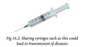
d) Mother to child during pregnancy, birth or breastfeeding. Although the risk can be high if a mother is living with HIV and not taking medicine.

e) Being stuck with an HIVcontaminated needle or other sharp object. This is a risk mainly for health care workers.
Note: In some rare cases, HIV has been transmitted through:
a) Receiving blood transfusions, blood products, organ or tissue transplants that are contaminated with HIV.
b) Eating food that has been prechewed by an HIV-infected person. The only known cases are among infants.
c) Being bitten by a person with HIV. There is no risk of transmission if the skin is not broken.
d) Contact between broken skin, wounds or mucous membranes and HIV-infected blood or bloodcontaminated body fluids.
e) Deep, open-mouth kissing if both partners have sores or bleeding gums. HIV is not spread through saliva.
f) Oral sex.
Sexual education
• Be aware of sugar mummies and sugar daddies. They lure young people with money and promise good life in exchange for sexual favours. Such acts could lead to transmission of STIs and, HIV and AIDS.
• Abstinence is the most effective protection against STIs and, HIV and AIDS.
Self-evaluation Test 16.1
1. Which is the most effective protection against HIV and other STIs?
2. Given the following STIs: Gonorrhoea, syphilis, chlamydia and HIV. Choose the ones that can be prevented?
3. What is the common transmission mode for all the STIs?
16.2 Ways of reducing STIs and HIV infection
Discussion corner
1. Discuss the following:
• Ways of reducing STIs and HIV infection.
• Ways of prevention and treatment of STIs and HIV infection.
• Role of artificial contraceptives (barrier method- male and female condoms) and their correct use.
2. Present your work to the rest of the class

The following are ways of reducing STIs and HIV infection:
a) Abstinence is the only sure way to prevent STIs.
b) Being faithful to one trusted partner.
c) Using condoms every time when engaging in sexual intercourse. Condoms are not 100% effective at preventing disease or pregnancy. However, they are extremely effective if used properly.
d. Reduce the number of sexual partners.
e. Avoid sharing towels or underclothing.
f. Get a vaccination for hepatitis B.
g. Get tested for HIV.
h. Avoiding alcohol consumption and abuse of drugs. Individuals who are drunk or on drugs often fail to have safe sex.
Activity 16.3:
Role play
1. Discuss with your classmates the following:
• Refusal skills that can be employed regarding sex.
• Harmful effects of consuming alcohol and drugs.
• The importance of voluntary counselling and testing (VCT) centres.
• How culture, gender and peer norms can influence decision making about sexual behaviour.
2. Prepare a skit to dramatise your content and present it to the rest of the class.
Treatment of STIs and HIV
Activity 16.4: Visit to a health centre
1. Your teacher will organise for a visit to a health centre.
2. Prepare a questionnaire to use during the visit.
3. Engage the officer in charge with the following guiding questions
• Does the centre offer sexual health services?
• Do they have a VCT centre that offers pre- and post – test counselling?
• How do they handle People living with HIV and AIDS (PLWHA)?
4. Write a report and present your work in class.
Treatment of STIs and HIV is based on their causative agents.
1. Bacterial STIs can be cured using antibiotics if treatment begins early enough. Antibiotics, often in a single dose, can cure many sexually transmitted bacterial and parasitic infections, including gonorrhoea, syphilis, chlamydia and trichomonasis. Treatment of gonorrhoea and chlamydia is always done at the same time because the two infections often appear together.
2. Viral STIs cannot be cured, but the symptoms can be managed with medications. For instance there is a vaccine against hepatitis B, but it will not help if you already have the disease.
3. HIV cannot be cured but can be managed. A person may choose to visit a voluntary counselling and testing (VCT) centre to undergo HIV and AIDS counselling. After conselling he or she can make an informed decision about whether to be tested for HIV or not.

At the VCT centre pre-test and posttest counselling is offered to assist people to learn more about their status. Counselling involves getting information and learning how to live positively with the virus. This means learning to accept the fact that one is HIV-infected, seeking emotional support, eating a healthy diet, learning how to control the amount of stress in our life, making sure there is no re-infection and planning for the future.
Sex education
Post-Exposure Prophylaxis (PEP) or short-term anti-retroviral therapy (ART) can reduce the likelihood of HIV infection after potential exposure.
Antiretroviral therapy (ART) is the use of HIV medicines to treat HIV infection. People on ART take a combination of HIV and AIDS drugs every day. People infected with HIV and AIDS should start ART as soon as possible. ART cannot cure HIV and AIDS, but helps people infected to live longer and healthier lives. ART also reduces the risk of HIV and AIDS transmission.
Potential risks of ART include unwanted side effects from HIV and AIDS drug interactions. This includes not taking drugs every day as required. It could lead to drug resistance and treatment failure.
Health Check!
• Do not try to treat a sexually transmitted disease on your own. These diseases are contagious and serious. You must see a doctor.
• Antibiotics that are used to treat STIs should never be shared. It is also important to take the entire drug prescribed, even if the symptoms go away.
• Never, take someone else's medication to treat your infection; it may make it more difficult to treat.
• If someone is pregnant and have an STI, prompt treatment can prevent or reduce the risk of infection of your baby.
Self-evaluation Test 16.2
1. What happens in a VCT centre?
2. What do you understand by the term ART?
Unit summary
• Sexually transmitted infections are also known as sexually transmitted diseases (STDs) or venereal diseases (VD) and are commonly spread through sexual contact.
• Bacterial STIs include: Chlamydia, gonorrhoea, syphilis among others.
• Viral STIs include genital herpes, genital warts and HIV among others.
• Abstinence is the most effective way of preventing STIs; some vaccines are however, used to reduce the risk of infections such as hepatitis B and some strains of HPV (human papiloma virus)
• Bacterial infections can be easily cured using antibiotics but viral infections like HIV, genital warts and HPV are controlled but cannot be cured. • Post –exposure prophylaxis can reduce the likelihood of HIV infection.


Glossary
A
Abiotic - physical factors in the environment that affect living organisms. They include; wind, humidity, sunlight, soil and atmospheric pressure among others.
Abortion - deliberate physical or chemical termination of human pregnancy.
Aboral - relating to or denoting the side or end that is furthest from the mouth, especially in animals that lack clear upper and lower sides, such as echinoderms.
Abstinence - act of restraining oneself from indulging or doing something; for example, restraining oneself from having sex.
Active transport - movement of particles against a concentration gradient.
Active site- a region on an enzyme that binds to a protein or other substance during a reaction.
Aerencyma tissue - A spongy tissue with large intercellular air spaces that is found in aquatic plants. It provides buoyancy and allows the circulation of gases.
Aerosol - a substance enclosed under pressure and able to be released as a fine spray.
Agglutination - the clumping together in suspension of antigen-bearing cells, micro-organisms or particles in the presence of specific antibodies.
Amenorrhea - absence of menstruation for at least three months
Appendage - a structure that attaches to another part of the body.
Antagonistic muscles - a muscle that opposes the action of another, for example, the biceps and triceps.
Antibody - proteins found in plasma that are responsible for mediation of immunity in body fluids.
Antibiotic - a drug derived from bacteria or fungus and is used to treat bacterial infections.
Anti-retroviral (ARV) - drugs to maximally suppress the HIV virus and stop the progression of HIV disease.
Antigen - any substance that stimulates response of a specific immune system.
Articulate - meet at a point to form a joint.
Arthritis – a condition that causes pain and inflammation in a joint.
Autoimmune - a condition arising from an abnormal immune response to a normal body part.
B
Biotic - activities of living organisms that affect an ecosystem.
Bilateral symmetry - symmetrical arrangement of an organism or part of an organism along a central axis.
Bilirubin - a yellow pigment produced from the breakdown of heme in the worn out red blood cell.
Biodiversity - variety and variability of life forms on earth.
Biome - large ecological areas on the earth's surface, with fauna and flora (animals and plants) adapting to their environment.
Biuret test- a test that uses a reagent (a solution of copper sulfate (CuSO4) and sodium hydroxide (NaOH)) used to determine the presence of peptide bonds in protein.
Bowman’s capsule - the filtration unit of the glomerulus and has tiny slits in which filtrate may pass through into the nephron.
Bone – a rigid body tissue consisting of cells compressed into hard intercellular material.
C
Capillarity - the ability of a narrow tube to draw a liquid upwards against the force of gravity.
Carrier protein - a protein that transports specific substance across the cell membrane.
Cartilage - tough elastic tissue.
Catalyst - a substance that causes a chemical reaction to occur but is not itself involved in the reaction.
Cephalothorax – a term referring to the fused head and thorax occurring in many arthropods, particularly crustaceans and arachnids.
Chaetae - bristles made of chitin that are found on bodies of annelids.
Cholesterol - a fatty substance made in the body and found in certain foods. Some cholesterol are harmful others are useful to the body.
Carapace - hard structure made of chitin that covers the dorsal part of an animal and protects internal organs.
CD4 cell - white blood cells that play an important role in the immune system.
Cheliserae - a pair of appendages modified to form poison glands or mouthparts.
Chitin - a tough, semitransparent substance that is the main component of the exoskeletons of arthropods.
Concentration gradient - the gradual difference in concentration of a dissolved substance in a solution between a region of high density and one of lower density.
Contraceptives - a device or drug that prevents pregnancy.
Cortex - the outermost (or superficial) layer of an organ.
Crustaceans - animals that usually have a hard covering, or exoskeleton, and two pairs of antennas, for example, crabs and lobsters.
D
Denature - change the original or natural structure.
Destarch - process of eliminating starch from leaves by allowing the plant to use up the starch previously synthesized.
Deamination - the removal of an amino group from an amino acid or other compound.
Detrivours - an organism (as an earthworm or a fungus) that feeds on dead and decomposing organic matter.
Detoxification - the metabolic process by which toxins are changed into less toxic or more readily excretable substances.
Diffusion - the movement of molecules from areas of higher concentration to areas of lower concentration.
DCPIP – (Dichlorophenolindophenol) a reagent used to measure the amount of ascorbic acid (vitamin C) in fruits and plant material.
E
Edaphic - Relating to soil, especially as it affects living organisms.
Endocytosis - the transport of solid matter or liquid into a cell by means of a vacuole or vesicle.
Endoskeleton - internal support structure made of bone or cartilage.
Enzyme – substrate complex- intermediate formed when a substrate molecule interacts with the active site of an enzyme.
Epidermis - protective outermost portion of the skin.
Epidemiology - the study of how often diseases occur in different groups of people and why.
Exoskeleton - external support structure that protects the internal organs.
Exocytosis - is a process by which a cell transports secretory products through the cytoplasm to the plasma membrane.
Eukaryotic – containing nucleus bound by a nuclear membrane.
Exhalation - the act of breathing out air.
F
Flaccid - soft, flabby and weak.
Food testing - a process used to check that a food is safe and that it does not contain harmful contaminants.
Flexion - the action of bending, especially the bending of a limb or joint.
G
Glomerula filtrate - the renal fluid in the blood filtered across the capillaries of the glomerulus.
H
Haemocoel - a body cavity (as in arthropods or some molluscs) that contains blood or hemolymph and functions as part of the circulatory system.
Halophytes - A plant that can tolerate a high concentration of salt in the soil.
Homeotherm - an organism that maintains its body temperature at a constant level, usually above that of the environment, by its metabolic activity.
Host - an organism that harbours a parasitic.
Hydrophytes – aquatic plants.
Hypotonic - solution that has a lower osmotic pressure than another solution.
Hypertonic – a solution where the concentration of solutes is greater outside the cell than inside it.
I
Immunisation - the process whereby a person is made immune or resistant to an infectious disease, typically by the administration of a vaccine.
Immune response - any reaction by the immune system.
Inhalation - the action of breathing in.
Insulin - a hormone made by the pancreas that allows the body to regulate glucose.
Isotonic - two solutions having the same osmotic pressure across a semipermeable membrane.
J
Joint - A point of articulation between two or more bones.
K
Key and lock mechanism - the hypothesis that helps explain some of the ways that enzymes work.
L
Lateral line - a visible line along the side of a fish consisting of a series of sense organs that detect pressure and vibration.
Lesion - injured or diseased structure or part of tissue or organ.
Lignin - an organic substance in plants binding the cells, fibres and vessels which forms wood.
Ligament - a short, flexible, fibrous connective tissue that connects two bones or cartilages or holds together a joint.
M
Menopause – a period when a woman stops having periods and is no longer able to get pregnant naturally.
Mesophyte - a plant growing under conditions of well-balanced moisture supply.
Metabolism - all chemical reactions involved in maintaining the living state of the cells and the organism.
Moulting - periodic shedding of the cuticle in arthropods or the outer skin in reptiles.
Millon’s test – a chemical test that detect the presence of proteins in a food sample.
Multicellular - an organism that is made up of many cells.
Muscle - a tissue composed of cells or fibers, the contraction of which produces movement in the body.
N
Nephrone - the basic structural and functional unit of the kidney.
Notochord - a flexible rod-like structure that exists in bodies of vertebrates at some point in their lives.
Non-reducing sugars – sugars such as sucrose that cannot be oxidized.
O
Oral - relating to the mouth.
Omnivorous - an animal that eats food from both plants and animals.
Osmosis - the movement of water from a less concentrated solution to a more concentrated solution through a partially permeable membrane.
Osteoporosis - condition that weakens bones, making them fragile and more likely to break.
P
Passive - allowing what happens, without active response or resistance.
Pathogen - a parasite that causes disease.
Pedipals - segmented appendages attached to the cephalothorax of arachnids.
Pinocytosis - the ingestion of liquid into a cell by the budding of small vesicles from the cell membrane.
Pith - the soft, spongy tissue in the centre of the stems of most flowering plants, gymnosperms and ferns.
Potometer - a device used for measuring the rate of water uptake of a leafy plant shoot.
Phagocytosis - the process by which a cell engulfs material.
Plastids – are organelles that are the main site of photosynthesis in eukaryotic cells.
Pseudopodia - temporary or semipermanent extension of the cytoplasm, used in locomotion and feeding in some protozoa.
Pooter - a device used to pick up small invertebrates without harming them.
Poikilotherm - an organism that cannot regulate its body temperature.
Plasmolysis - the contraction of the protoplasm of cells within plants due to the loss of water through osmosis.
Phloem - part of a vascular bundle that conducts sugars and other metabolic products downward from the leaves.
R
Receptor - a protein molecule that receives chemical signals from outside a cell.
Reducing sugars - any sugar such as glucose which is capable of being oxidised.
Respiratory surfaces - a special area that is developed in order to satisfy the requirements for gaseous exchange in larger organisms.
S
Septa - walls that divide internal body cavities or chambers.
Sessile – an organism, fixed in one place (immobile).
Selective reabsorption - The absorption of some of the components of the glomerular filtrate back into the blood as the filtrate flows through the nephrons of the kidney.
Sexual response - is the arousal of sexual desire, during or in anticipation of sexual activity.
Sexuality - the whole way a person goes about expressing himself or herself as a sexual being.
Source – the sites in a plant where net fixation of carbon dioxide occurs.
Sign - evidence of disease as seen by the examining physician. Sink-the sites in a plant where assimilates are stored or used.
Spermicide – This comprise of creams, gels, foams and other suppositories that prevent sperm from moving.
Substrate - the substance on which an enzyme acts.
Sutures - a seam like immovable junction between two bones, such as those of the skull.
Synovial - a lubricating fluid secreted by certain membranes, for example, in joints.
Symptoms - evidence of disease perceived by the patient.
T
Tentacles - a slender, flexible limb or appendage in an animal.
Tendon - A band of tough, fibrous, inelastic tissue that connects a muscle to a bone.
Transpirational pull - a mechanism by which trees draw water through their roots.
Translocation - the movement of materials from leaves to other tissues throughout the plant.
Tracheid - a type of water-conducting cell in the xylem that lacks perforations in the cell wall.
Turgor pressure - the pressure of water against the inside wall of a plant cell.
U
Ultrafiltration – a process in the kidney in which urea, salt, water and glucose are taken out of the blood.
Urologist - a physician who specialises in diseases of the urinary tract and the male reproductive system.
V
Vaccine - preparation of weakened form of a pathogen such as killed microbe, altered microbes or derivative form of pathogens.
Vasectomy - a permanent method of birth control where sperm ducts are cut and the ends are sealed to prevent sperm from entering the ejaculate.
Vascular bundles - Xylem and phloem tissues.
Vector – an organism that transmits a disease or parasite from one animal or plant to another.
Verigated – having different colours.
Ventilation - the movement of air between the environment and the lungs via inhalation and exhalation.
Vessel element - An elongated, waterconducting cell in xylem.
W
Wall pressure - pressure exerted by the flow of water through a semipermeable membrane separating two solutions with different concentrations of solute.
Wilting - loss of turgidity and rigidity that occurs when the rate of water loss is greater than the rate of water uptake.
X
Xylem – a vessel in plants that transport water from roots to shoot and leaves, it also transports some nutrients.
Xerophytes – plants that grow in areas with very little water.
References
• J. Akatsa, H mwaura (2008); Longhorn secondary Biology, Longhorn Publishers; Nairobi, Kenya
• J.Mwaniki , G Geoffrey (2013); Fundamentals of Biology, Longhorn Publishers: Dar es Salaam, Tanzania
• D. Nserebo et al (2015); Excel & Succeed Biology, Longhorn Publishers: Malawi
• D.G. Mackean (2012); GCSE Biology (Third edition); CGP Books
• Michael Roberts (1986); Biology - A Functional Approach (Fourth Edition); Oxford publishers
• Rod Seeley, Stephens Tate; (2002) Anatomy and physiology; McGraw-Hill Global Education Holdings