Topic outline
General
- Biology S4 SB File Uploaded 28/01/22, 10:40
- S4: Biology TG File Uploaded 10/08/22, 16:31
UNIT 1: INTRODUCTION TO BIODIVERSITY
Key Unit Competence
Explain how diversity is threatened by climate change and human activitiesLearning objectivesBy the end of this unit, I should be able to:
–– Define the terms: species, ecosystem and niche.
–– Explain that biodiversity is considered at three different levels
–– Evaluate the consequences of loss of biodiversity.
–– Characterize the biotic and abiotic components that define Rwanda’s ecosystems (example: freshwater, marine, and terrestrial).
–– Apply Simpson’s Index of Diversity.
–– Explain the importance of random sampling in determining the biodiversity of an area.
–– Use suitable survey methods such as frame quadrats, line and belt transects to assess the distribution and abundance of organisms in a local area.
–– Use Pearson’s linear correlation to analyze the relationships between the
distribution and abundance of species and abiotic or biotic factors.
–– Recognize that the biodiversity of the earth is threatened by human activities and climate changeIntroductory activity: Biodiversity of RwandaRead the following text and answer the questions that followRwanda is located at the heart of the Albertine Rift eco-region in the western arm of the Africa’s Rift Valley. Habitats of Rwanda are equally varied, ranging from Afro-Montana ecosystems in the northern and western regions to lowland forests, savannah woodlands and savannah grasslands in the southern and eastern regions. There are other habitats around volcanic hot springs and old lava flows, especially in the northern and western part of the country.Rwanda also has several lakes and wetlands which are rich in different species. Though not yet well surveyed, all these ecosystems host a rich variety of fauna and flora and micro-organisms. This rich biodiversity is mainly conserved in protected areas including three national parks, natural forests and wetlands. These cover almost 10 percent of the national territory while the rest of the country is densely populated (507 people per square kilometer in 2018).Many tourists visit Rwanda for its beautiful environment and biodiversity made of different species of plants and animals such as Aloe vera (Igikakarubamba), Muringa oleifera (Muringa), Phaseolus vulgaris (common bean), Nymphaea thermarum (Endemic plant species that cannot be met elsewhere in the world, only found in Mashyuza minor locality harbors),Colobus polykoma (White-black colobus monkey), Gorilla gorilla (mountain
gorilla) bird Laniarius mufumbiri (Bird species mainly found in Rweru- Mugera wetland),etc.The most attracting species in Rwanda is Gorilla gorilla whose habitat is the mountains of Birunga where they make a large population. Another natural forest, Nyugwe National Park is a terrestrial ecosystem that contains a large community of different plants and animals.Rwanda also has different lakes such as Muhazi and Rumira. They are aquatic ecosystems made of few species of fish, such as tilapias. Tilapias from Lake Muhazi are small, black and bony fish while those from Lake Rumira look red, big and soft. Tilapias from both lakes still belong in the same species but show variations.Many species of animals and plants have been discovered in Rwanda but some species also disappeared. Today the big garden snails known as Achatina achatina have become rare in Bugesera. Other people poached Rhinoceros alba living in Savanah of Akagera National Park.Honey bees, butterflies and grasshoppers are small in size but still important for different ecosystem services. Each organism is important for its niche in ecosystem. We need to identify and protect the biodiversity of our ecosystem. Many tourists enjoy visiting Rwanda for its biodiversity.
1. Name the species not found elsewhere that attract the tourists and locate where it is found.
2. Mashyuza is a minor locality in western province in Rusizi district that contributes to biodiversity of Rwanda. Give any other two locations.
3. Define each of the following biological terms and give an example from the text
abovea) Species (b) Population (c) Community (d) Habitat (e) Ecosystem
(f )Variation (g) Niche
4. What causes some species to become extinct?
5. What can be the consequences of the loss of some species from our
biodiversity?
6. Do you support tourism in Rwanda? Give a reason to justify your answer.1.1 Meaning of key ecological terms and biodiversity
Activity 1.1
1. What do you understand by the following terms: biodiversity, species, niche, population, and community?
2. Differentiate between ecological niche and habitat.1.1.1 Key ecological terms
Species is a group of closely related organisms which are capable of interbreeding to produce fertile offspring. Occasionally two organisms which are genetically closely related but not of the same species can interbreed to produce infertile offspring. For example:
–– A cross between a donkey and a horse produces a mule, which is infertile. Thus, a donkey and a horse do not belong to the same species
–– Lions and tigers belonging to different species. However, when a male tiger mates with a female lion they can have fertile offspring called tiglons, although the offspring of female tigers and male lions called ligers are not fertileNote that normally, tigers are forest dwellers and lions are plains dwellers and they are ecologically isolated. Breeding has only been observed in captivity. An ecological population is a group of individuals of the same species which live in a particular area at any given time.An ecological community consists of populations of different species which live in the same place at the same time, and interact with each other. A habitat is a specific area or place in which an individual organism lives. When a habitat is very small it is regarded as a microhabitat. Most ecosystems contain several habitats, and one species can have more than one habitat constituting its geographic range.An ecological niche is the status or the role of an organism in its habitat or the mode of life of an organism within its habitats. For example, insects are pollinating agents and preys of insectivores.Abiotic factor are non-living physical aspects of the environment such as the sunlight, soil, temperature, wind, water, and air. Biotic factors are the living organisms in the environment. They include organisms and their interactions with each other.An ecosystem is a natural unit consisting of biotic and abiotic factors through which energy flows and nutrients recycle. In an ecosystem, nutrients pass between different organisms in definite pathways. For example, nutrients in the soil are taken up by plants, which are then eaten by herbivores, which in turn may be eaten by carnivores and recycled by decomposers.A biome is a group of ecosystems that have the same climate and similar dominant communities. The highest level of organization is the entire biosphere.The Biosphere is the whole of the earth’s surface, the sea and the air that is inhabited by living organisms. The biosphere is made up of all ecosystems.1.1.2. Biodiversity
 Biodiversity is defined as the full range of variety and variability within and among living organisms and the ecological complexes in which they occur.Self-assessment 1.1
1. Describe the two main components of an ecosystem.
2. Hippopotamus has different habitats. It was found that the resting habitat is different from the mating habitat, and these two habitats are different from the area where this animal gets food. Explain the ecological term given to this set of habitats.1.2 Identification of biodiversity
Activity 1.2
Use books or other sources of information to answer the followings questions:
1. What kinds of initiatives and incentive mechanisms are put in place by the Government of Rwanda to motivate local community in biodiversity conservation?
2. Describe different ways used to identify biodiversity.
3. Discuss the values of biodiversity and ecosystem services in Rwanda.
4. Evaluate the contribution of biodiversity to human well-being.1.2.1. Categories of biodiversity
Biodiversity can be categorized into three groups:
–– Genetic diversity: the combination of different genes found within a population of a single species, and the patterns of variation found within different populations of the same species.
–– Species diversity: the variety and abundance of different types of organisms which inhabit an area.
–– Ecosystem diversity: the variety of habitats that occur within a region, or within the mosaic of patches found within a landscape.1.2.2. Importance of biodiversity
Biodiversity contributes to ecosystem goods and services. The ecosystem goods and services include:
–– Provision of food, air, fire wood, medicines(Fig.1.2), energy, fresh water.
–– Nutrient cycling such carbon, water and nitrogen cycles by microorganisms and primary production by photosynthesis.
–– Cultural or aesthetic service recreation, ecotourism, cultural andreligious inspiration.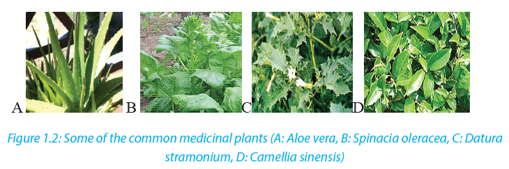 1.2.3. The threats and consequences of biodiversity loss
1.2.3.1. Causes of biodiversity loss
The main causes of biodiversity loss can be attributed to the influence of human activities on ecosystems. Threats to biodiversity include:a. Habitat loss and the degradation of the environmentThe habitat loss and the degradation of the environment occur in different ways.The most occurring, are tree cutting, agriculture and fires (Figure1.3). These human activities lead to the alteration and loss of suitable habitats for biodiversity. As a consequence, there is a loss of plant species as well as the decrease in the animal species associated to this plant diversity.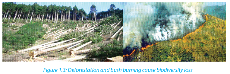 b. Introduction of invasive alien species and genetically modified organismsSpecies originating from a particular area are harmful to native species also called endemic species when they are introduced into new natural environments. They can lead to different forms of imbalance in the ecological equilibrium, so that endemic species may fail to compete with introduced species, and they may affect the abundance and distribution in natural habitat.c. Pollution
Human activities such as excessive use of fertilizers, and increased pollutants from industries and domestic sewage affect biodiversity. They contribute to the alteration of the flow of energy, chemicals and physical constituents of the environment and hence species may die as a result of toxic accumulation.d. Overexploitation of natural resources
Increased hunting, fishing, and farming in particular areas lead to the decrease and loss of biodiversity due to excessive and continuous harvesting without leaving enough time for the organisms to reproduce and stabilize in their natural habitat.e. Climate change
This is a change in the pattern of weather, related changes in oceans, land surfaces and ice sheets due to global warming resulting from man’s activities. Increasing global temperatures have resulted into melting of icebergs raising sea levels and so flooding coastal areas eventually affecting the niche.1.2.3.2. Consequences of loss of biodiversity
They are various consequences of loss of biodiversity that include:
–– Desertification, is thought by scientists to be a consequence of climate change, has been considered to be related to deforestation. Disrupting water cycles and soil structure results into less rainfall in an area.
–– Floods as a result of rising sea levels
–– Habitat destruction for extensive farming, timber harvesting and infrastructure and settlement
–– Decrease in food production as result of change in pattern of weather that affects productivity
–– Large scale deforestation has a negative effect on nutrient recycling and can accelerates soil erosion
–– Diseases that come as effects of floods and malnutrition due to famineSelf-assessment 1.2
1. Define the term Extinction.
2. Suggest the causes of extinction of species in Rwanda.
3. Discuss the benefits of biodiversity to humans
4. Discuss the major factors leading to the degradation of ecosystems in Rwanda
5. Discuss the contribution of ecosystems to cultural traditions in Rwanda.
6. In Rwanda different plants are used in traditional medicine to treat different diseases. Conduct a research and list at least 20 medicinal plants and the diseases they treat. From the list above describe at least one medicinal plant and get ready to present your work. The project work should include: written content of 2 pages in minimum and 4 pages in maximum, a testimony of people that have used plant species.
7. Pollution is one of the causes of aquatic biodiversity loss.
a. What do you understand by water pollution?
b. Outline human activities that contribute to water pollution
c. Discuss how polluted water affects aquatic living organisms?1.3 Calculation of Simpson’s index
Activity 1.3
A survey on tree species was conducted in Gako forest by a group of students. Five tree species (A to E) were identified and counted. The numbers found during this exercise are summarized in the following table: 1. Describe the relative abundance of species A to E.
2. Based on the data in the above table, suggest how species diversity of tree species can be calculated.There are many ways to measure diversity. The Simpson diversity index among indices used to measure diversity. It is expressed in three related indices namely Simpson index, Simpson index of diversity and Simpson reciprocal index.a. Simpson index D
Simpson index D can be expressed in two ways and takes into consideration the total number of organisms of a particular species and the total number of organisms of all species. It is calculated as follows: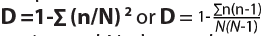 with n: the total number of organisms of a particular species and N: the total number of organisms of all species. When the index equals or is nearby 0 there is an infinite diversity of considered species. When it equals or is nearby 1, this means that there is no diversity. The bigger the value of D, the lower the diversity and small is D, the bigger is the diversity.b. Simpson index of diversity 1 – DThe value of this index ranges between 0 and
1, but now, the greater the value, the greater the sample diversity. This makes more sense. In this case, the index represents the probability that two individuals randomly selected from a sample will belong to different species.c. Simpson reciprocal index 1 / D
Another way of overcoming the problem of the counter-intuitive nature of Simpson’s index is to take the Simpson’s reciprocal index 1 / D. The value of this index starts with 1 as the lowest possible figure. This figure would represent a community containing only one species. The higher is the value of Simpson reciprocal index, the greater the biological diversity.Examples
1. In woodland, a quadrat was sampled for ground vegetation. Data collected were recorded in the table 1.3.2. Find out the value of the Simpson index and draw the conclusion about the biological diversity of the sampled area.
Table 1.3.2: Recorded data on the vegetation from a woodland Solution: Putting the figures into the formula for Simpson’s Index:
 Based on the meaning of Simpson index, the quadrat presents a low diversity because the value of D is near zero and zero and below 0.5.
2. Calculate the value of Simpson’s Diversity Index (D) for a single quadrate sample of ground vegetation in woodland from which the following sampling date was obtained:
 Self-assessment 1.3
1. Differentiate between species richness and species evenness
2. Suggest precautions taken when measuring populations of aquatic animals or plants.
3. Explain why a habitat with high diversity tends to be more stable than one with lower diversity.
4. In a survey of trees in a tropical forest, students identified five tree species (A to E).
They counted the numbers of trees in an area 100 m × 100 m and found these results: Calculate the Simpson’s Index diversity for identified species and explain the advantage of using data on species diversity and abundance when calculating an index of diversity.5. The Simpson’s Index of diversity for vegetation in an open area inhabited by grasslands was 0.8. For a similar sized area of vegetation beneath some conifer trees it was 0.2. What do you conclude from these results?1.4 Sampling techniques to assess the distribution and abundance of organisms
Activity 1.4From your school garden, sample different flowering plant species and answer the following questions:
1. Specify the techniques used for collecting flowers of different species.
2. What are the advantages of the technique you used for data collection?
3. Move around the school garden and collect different specimens of plant species. Name the collected species by using their names. In case you don’t know their names, use letters A, B, C ….Calculate Simpson index D, Simpson index of diversity and Simpson reciprocal index.To calculate Simpson’s index for a particular place:
–– Identify the habitat to be studied.
–– The number of individuals sampled for each species must be recorded.
To analyze the distribution and abundance of organisms in an area of study, there are different sampling methods.
Note that, sampling only one quadrat would not give reliable estimate of the diversity of the ground flora in the wood.a. Random sampling method
A random sampling method is a sampling method where samples are taken from different positions within a habitat and those positions are chosen randomly.b. Quadrat sampling method
A quadrat is a square area that is marked using a pre-made square of plastic, or stakes and string and it can range in size. Different species and their numbers within the quadrat are counted. Counting is repeated many times in different places in the habitat to get an accurate representation of biodiversity.c. Frame quadrats
Frame quadrats are small plot used to isolate a standard unit of area for the study of the distribution of an item over a large area. While originally rectangular, modern quadrats can be rectangular, circular, and /or irregular. The quadrat is suitable for sampling plants, slow-moving animals such as millipedes and insect and some aquatic organisms.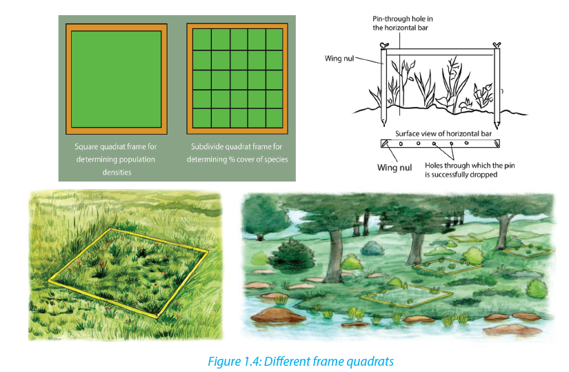 d. Transect sampling
Transect sampling is done using a transect line, which is usually a rope or measuring tape that has been marked at set intervals, such as every meter. The line is unrolled within the habitat. At every interval, the type and number of species along the line are recorded. A measured line is laid across the area in the direction of the environmental gradient. The species touching the line can be recorded along the whole length of the line (continuous sampling) or at specific points along the line (systematic sampling). e. Belt transects method
Belt transects method is the same as the line transects but widens the sampling area. The samples are taken and the abundance, percentage cover in a defined area determined. Samples can be taken within the belt.
 f. Netting
Netting is a sampling method where fine mesh nets are used to capture different organisms that include insects, birds and bats. The technique is also used for sampling small aquatic organisms like daphnia, and water boatman.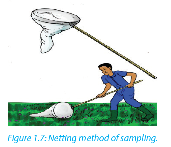 g. Capture -recapture techniqueThis method is useful for sampling non-fixed population and is suitable for animal such as fishes, birds, lizards and insects. A sample of the population to be studied is first captured and each individual is marked with a spot for identification. These are then released and given enough time to mix up with the rest of the members in the habitat. After a certain period of time, another sample is taken. During the mark-release-recapture technique, the total population can be estimated by the use of the formula:
 wheren1 is a number caught and marked in first sample,
n2 is a number caught in second sample
n3 is a number in the second sample that had been marked.
To understand this application, let us use the following examples:1. A team of students used a sweep net to sample brown grasshoppers
and each collect insect was marked with a very small spot of non-toxic waterproof paint and then they were released in the field. The next day, a second large sample was conducted and data were recorded as follows: number of caught and marked in first sample (n1) = 247, number of caught in second sample (n2) = 269, and the number in the second sample that had been marked (n3) = 16. What is the number of estimated population? 2. A student collected 16 butterflies which he marked and released. For a second time he collected 18 butterflies among which 12 were already marked from the first sampling. Estimate the population size of butterflies in that area.
 Self-assessment 1.4
1. Explain the advantages of the random sampling techniques.
2. Use suitable methods, such as frame quadrats, line transects, and belt transects, to assess the distribution and abundance of insect species in a school garden. Record your data and use the Simpson index ofdiversity (D) to calculate the diversity of collected insects.
3. Suggest the benefi ts of using the following sampling techniques:
a. Quadrats
b. Transect
c. Mark-capture-recapture
4. State the conditions in which quadrats, transect and mark recapture are
suitable sampling methods.1.5 Pearson’s linear correlation
Activity 1.5
Some of the following fi gures indicate a positive, negative or non-correlation. 1. What do you understand by the term correlation?
2. Categorize the graphs given as positive, negative or weak or no correlation
3. In which conditions results can indicate a positive correlation?
4. Conclude about your results when there is no correlation.
To decide if there is an association between collected data, a correlation coeffi cient is calculated and plot scatter graph drawn in order to make a judgment. The strongest correlation is present for studied items when all the points lie on a straight line. In this case, there is linear correlation, and the correlation coeffi cient equals1. If a given variable X increases so does another variable Y, the relationship is a positive correlation. If a variable X increases while the variable Y decreases, then the relationship is a negative correlation. A correlation coefficient of 0 means there is no correlation at all. These correlation coefficients are ways to test a relationship observed and recorded to see if the variables are correlated and, if so, to find the strength of that correlation.a. Pearson’s correlation coefficient
Pearson’s correlation coefficient can only be used where there might be a linear correlation and when there are collected quantitative data as measurements (for example, length, height, depth, and light intensity, mass) or counts (for example number of plant species in quadrats). The data must be normally distributed.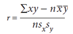 Where:
r is the correlation coefficient
x is the number of species in a quadrat
y is the number of species in the same quadrat
n is the number of readings (From1 to n)
x is the mean number of species
y is the mean number of species
sx is the standard deviation for x
sy is the standard deviation for ySelf-assessment 1.5
Use Pearson’s linear correlation to analyze the relationships between the distribution and abundance of species and abiotic or biotic factors.End of unit assessment 1Section A: Answer as true or false
1. Abiotic factors are the non-living physical aspects of the environment.
2. Capture –recapture is a method used to integrate the numbers of mobile animals in a particular place.
3. A correlation coefficient of 0 means that there is no correlation at all.4. A sample is a portion, piece, or segment that is representative of a whole area of study.
5. In the Simpson’s index, Nrepresentsthetotal number of organisms of a particular speciesSection B: Long and short answer based questions
1. What do you understand by the term biodiversity?
2. What do you think would happen to plants if there were no insects?
3. Suggest different ways to conserve our forests.
4. A student has randomly collected 5 types of species at the following frequencies. Calculate the Simpson’s diversity index of this community.
5. A team of students conducted the capture- recapture sampling method of tilapia from lake Muhazi at different times of the day as recorded in the data below: a. Plot the graph for the date provided and describe the shape of the graph.
b. From the graph, determine the appropriate time to have the most catch.
6. What do you understand by term endangered species?
7. Describe how diversity is threatened by climate change and human activities.URLs: 2Files: 2UNIT 2: INTRODUCTION TO CLASSIFICATION
Key Unit Competence
Apply the basic knowledge of classification to group living organisms into the three domains.Learning objectives–– Describe the classification of species into the taxonomic hierarchy of domain, kingdom, phylum, class, order, family, genus and species.
–– Outline the characteristic features of the three domains Archaea, Bacteria and Eukarya.
–– Draw and label the structure of a typical bacterial cell.
–– Identify common bacterial diseases in plants and animals.
–– Outline the characteristic features of the kingdoms Protoctista, Fungi, Plantae and Animalia.
–– Explain why viruses are not included in the three domain classification.
–– Outline how viruses are classified limited to type of nucleic acid and their host.
–– Describe the role of bacteria in the production of dairy products.
–– Describe methods of preventing common bacterial diseases.
–– Construct a dichotomous key for a group of organisms.
–– Recognize that microorganisms can survive in hot springsIntroductory activityCollect different fruits such as oranges, lemons, avocado, green paper, red paper, bananas, mangoes and tomatoes.
1. Observe each of the above fruits and group them based on their external features.
2. Based on groups made, which fruits are most closely related?
For more than 3.5 billion years, life on earth has been constantly changing. Natural selection and other processes have led to a staggering diversity of organisms. A tropical rain forest, for example, may support thousands of species per meter square.Recall that a species is a population of organisms that share similar characteristics and breed with another to produce fertile offspring. Biologists have identified and named about 1.5 million species so far, and they estimate that between 2 and 100 million additional species have yet to be discovered.2.1 Taxonomic hierarchy
Activity 2.1
You are provided with cards written on a list of words such as continent, district,
country, cell, province, sector, village and family.
1. Arrange the above words in increasing size
2. What is your opinion about the people of the same family and those in the
whole country?
3. Compare your arrangement above with 8 groups of the biological
taxonomic hierarchy.Taxonomy is the study and practice of classification, which involves placing organisms in a series of taxonomic units, or taxa (singular: taxon). In biological classification, these taxa form a hierarchy. Each kind of organism is assigned to its own species, and similar species are grouped into a genus (plural: genera). Similar genera are grouped into a family, families into an order, orders into a class, classes into a phylum (plural: phyla) and phyla into a kingdom. The domain is at the top of this hierarchical system.The hierarchy classification starts from the largest group, the domain. The eight levels of classification are known as taxa (taxon in singular), these include: Domain, Kingdom, phylum, class, order, family, genus and species. As one moves down the taxonomic hierarchy, it follows that the number of individuals decreases but the number of common features increases. For example, there are numerous individuals in the domain Eukarya, with very few features in common.Binomial nomenclature
When precision is not required one generally reverts to common names. The trouble is that an organism may be known by different common names, and sometimes the same name may be given to two quite different organisms because common names are not internationally recognized and they change from one region to another one, or from one country to another one. To solve this problem, the binomial system also known as scientific name was introduced and it was pioneered by the Swedish naturalist Carl Linnaeus (1707-1778).In this system, each organism is given two Latin names: a generic name beginning with a capital letter and a specific name beginning with a lowercase letter based on the physical characteristics of studied species. The scientific name is in italic when printed otherwise it is underlined, when hand written.For example, many cats belong to the genus Felis but there are many species of cats:
A wild cat is Felis sylvestris while a domestic cat is Felis domesticus. These names are in
italic because this book was written by the use of computer. Hierarchy taxonomy of human, earthworm and hibiscus plant are given in the table 2.1.Table 2.1 Taxonomic classification of human being, earthworm and hibiscus Scientific names present more advantages than common names.
–– They are necessary whenever precise identification is required, and they enable scientists to communicate accurately with each other.
–– They are used worldwide and have the merit that every biologist knows exactly which organism is being referred to.
Currently, with DNA technology, it is possible to investigate relationships based on genes or DNA structure. As this new technology comes to greater use, it is possible to find that some species had to be reclassified into different taxa.Self-assessment 2.1
1. An African bush elephant belongs to order Proboscidae and family Elephantae. Its scientific name is Loxodonta africana.
a. Make a table indicating the hierarchy classification of African bush elephant
b. Use the examples from table 2.1 to define the term “taxon”
2. Classify each of the following organisms under the following kingdom, phylum and class taxa: honey bee, cockroach, maize, and spider.
3. Describe the system of naming species that Linnaeus developed.2.2 Three domains: Archaea, Bacteria and Eukarya.
Activity 2.2.
Using text books and other sources identify the characteristics of each of the three biological domains
Three domains are used by biologists to divide organisms into three large groups based on their cell structure. The domain is the highest taxon in the hierarchy. The prokaryotes are divided between the domains Bacteria and Archaea, while all the eukaryotes are placed into the domain Eukarya.a. Domain Bacteria
Domain bacteria include prokaryotic organisms as their cells have no true nucleus. They are all microscopic that vary in size between 0.2 to 10 micrometres. The characteristic features of bacteria are:
–– Cells with no true nucleus
–– DNA exists in circular chromosome and does not have histone proteins associated with it
–– No membrane-bound organelles (mitochondria, endoplasmic reticulum, Golgi body, chloroplasts)
–– Contain mesosomes as infolding of membrane and acts as sites for respiration.
–– Ribosomes (70 S) are smaller than in eukaryotic cells
–– Cell wall is always present and contains peptidoglycans in place of cellulose
–– Cells divide by binary fission
–– Usually exist as single cells or coloniesb. Domain Archaea
This contains bacteria that live in extreme environments where few other organisms can survive. They are classified according to the environments they live in;
–– Methanogenic bacteria that live in habitats deprived of oxygen and give off methane as a product of metabolism for example those that live in the guts of ruminant animals
–– Halophilic bacteria live only in salty conditions
–– Thermoacidophilic bacteria tolerate extreme acid and temperature that exceed boiling point of water and a pH below 2.c. Domain Eukarya
All the organisms classified into this domain have cells with nuclei and membrane bound
organelles. Their characteristic features are:
–– Cells with a nucleus and membrane-bound organelles
–– linear DNA associated with histones arranged within a chromosome in the nucleus
–– Ribosomes (80S) in the cytosol are larger than in prokaryotes, while chloroplasts and mitochondria have 70S ribosomes, like those in prokaryotes.
–– Chloroplast and mitochondrial DNA is circular as in prokaryotes suggesting an evolutionary relationship between prokaryotes and eukaryotes
–– A great diversity of forms: unicellular, colonial and multicellular organisms
–– Cell division is by mitosis
–– Many different ways of reproduction including asexually and sexually.Self-assessment 2.2
1. What are the three domains of living things?
2. Describe the ways in which a domain differs from a kingdom?
3. It is confirmed that: “Some bacteria can survive in extreme temperatures such
as hot springs”. Justify this statement.
4. How is the information about evolutionary or phylogenetic relationships useful in classification of the living things?2.3 Five kingdoms of organisms
Activity 2.3.
Collect organisms from a habitat near your school including a housefly, spider, frog, gecko, bean/maize plant, moulds/mushroom, spirogyra (algae) and a hen. If there are small rapidly moving land animals such as insects, anaesthetise them by placing them in an ether/ethanol bottle for few seconds. Preserve the collected specimens for future use
1. Examine each organism, using a hand lens.
2. Make a table of the features observed and identify the kingdom to which each organism belongs.
There are different ways of classifying the living world into kingdoms but the most common and recommended is the five kingdom classification. According to Kent (2000) the kingdoms are:
–– Kingdom Monera or prokaryote
–– Kingdom Protoctista
–– Kingdom Fungi or kingdom mycota
–– Kingdom Plantae
–– Kingdom Animalia2.3.1 Kingdom Protoctista
This kingdom is made up of a very diverse range of eukaryotic organisms, which
includes those that are often called protozoans and algae. Any eukaryote that is not
a fungus, plant or animal is classified as a protoctist. The characteristic features of
protoctists are listed according to the different phyla due to their diverse range:
–– Rhizopods that have pseudopodia for locomotion. Example, amoeba
–– Flagellates which are hereorophic organisms with at least one flagellum for locomotion. Example, trypanosoma.
–– Sporozoans which are mainly parasitic organisms that reproduces by multiple fission. Example plasmodium.–– Ciliates which are organisms with cilia. Example paramecium
–– Euglenoid flagellates which are organisms with flagella but with a biochemistry quite distinct from that of flagellates. Example Euglena
–– Oomocytes which are similar to fungi except that they have cell wall with cellulose. Example Phytopthora infestans; potato blight
–– Green algae which are photsynthetic organisms with chlorophyll pigments similar to the ones of plants. Example chlorella
–– Red aglae which are photosynthetic organisms with organelles with red pigment as well as chlorophyll. Example, chondrus
–– Brown algae which are photsynthetic organisms with organelles which contain brown pigments as well as chlorophy. Example Fucus, sea weed Living things such as paramecium (a), amoeba (b), euglena (c) and plasmodia belong to the kingdom Protoctista.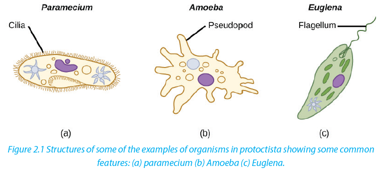 2.3.2 Kingdom Fungi
Fungi have some similarities with plants, but none of them is able to photosynthesise. They are all heterotrophic, obtaining energy and carbon from dead and decaying matter or by feeding as parasites on living organisms. There is a vast range in size from the microscopic yeasts to what may be the world’s largest organisms. Other characteristic features of fungi are:
–– Heterotrophic nutrition – they use organic compounds made by other organisms as their source of energy and source of molecules for metabolism
–– Reproduce asexually by means of spores and sexually by conjugation
–– Simple body form, which may be unicellular or made up of long threads called hyphae (with or without cross walls).
–– Large fungi such as mushrooms produce large compacted masses of hyphae known as fruiting bodies to release spores
–– Cells have cell walls made of chitin or other substances 2.3.3 Kingdom Plantae
Plants are all multicellular photosynthetic organisms. They have complex bodies that are often highly branched both above and below the ground. Characteristic features of plants are:
–– Multicellular eukaryotes with cells that are differentiated to form tissues and organs.
–– Few specialized cells
–– Cells have large and often permanent vacuoles for support with cell walls made of cellulose
–– Most plants store carbohydrates as starch or sucrose2.3.4 Kingdom Animalia
Animals (Fig 2.3) are multicellular organisms that are all heterotrophic with different methods of obtaining their food. Organisms in this kingdom have other additional features.
–– Different types of specialized cells
–– Cells do not have chloroplasts and cannot photosynthesize (although some,
such as coral polyps have photosynthetic protoctists living within their tissues)
–– Cell vacuoles are small and temporary (for example lysosomes and food vacuoles)
–– Cells do not have cell walls
–– Communication is by the nervous system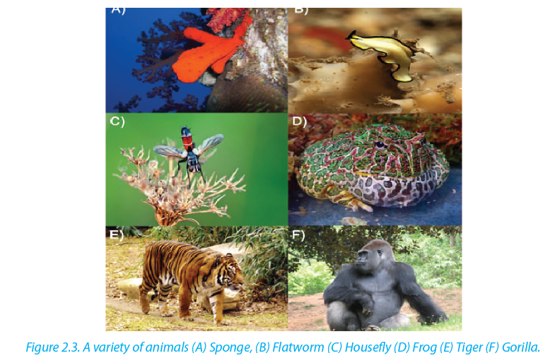 Activity 2.3
Which feature do all animals (except sponges) have that distinguishes them from plants and fungi?2.3.5. Kingdom Monera
Organisms in this kingdom have single cells that do not have a nucleus. They are prokaryotic. They are the smallest and simplest organisms. Examples are bacteria which form a diverse group with members that range widely in size and shape. Some of them stick together to form chains or clusters while others are single cells. The figure below (Figure 2.4) shows a typical structure of a bacterial cell which contains all the main features of prokaryotes Self-assessment 2.4
1. The kingdom protoctista contains groups which do not appear to show an evolutionary relationship. On this basis, is the five kingdom classification a natural or artificial classification?
2. What are the three methods that protists use to obtain food?
3. Identify three characteristics of protists
4. The following is a list of organisms belonging to various kingdoms: housefly (Musca domestica), maize (Zea mays), Frog (Rana spp), Bat and Eagle.
a. Classify these organisms into their kingdoms
b. Name any two organisms that are not closely related and give a reason.
5. How are fungi different from members of kingdom plantae?2.4 Economic importance of bacteria
Activity 2.4
“Bacteria are both useful and harmful to humans”. Discuss the validity of the statement.
Bacteria are economically important because they are essential in many beneficial biological and industrial processes. There exist some examples of bacteria that are pathogens as they cause disease and spoilage of food..2.4.1 Useful bacteria
a. Biotechnology
Bacteria are used in biotechnology and industry. They are used to manufacture products such as ethanol, acetone, organic acid, enzymes, and perfumes. In the chemical industry, bacteria are most important in the production of pharmaceuticals. For example, E. coli is used for commercial preparation of riboflavin and vitamin K.b. Genetic engineering
Bacteria are used in genetic engineering through the manipulation of genes, also called recombinant DNA technology. In this case, bacterial cells are transformed and used in production of commercially important products for example, production of human insulin used in treatment of diabetes.c. Decomposition
In addition, bacteria are important in decomposition of dead organisms and animal wastes such as feces to form organic matter. This process improves soil fertility and plays an important role in mineral recycling in an ecosystem.d. Fibre retting
Some bacteria including Clostridium butyricum are used to separate fibres in a process called retting. In this process, fibres are formed to make ropes and sacks.e. Nitrogen fixation
Some other bacteria are used to fix nitrogen in form of nitrates into the soil. For example, Rhizobium bacteria which live in root nodules of leguminous plants. Such bacteria help in improvement of soil fertility.f. Digestion
Some bacteria living in the gut of ruminant animals such as cattle, horses and other herbivores secrete cellulase, an enzyme that helps in the digestion of cellulose of plant cell walls. Another example is Escherichia coli that live in the human large intestine which synthesizes vitamin B and releases it for human use.g. Biological control
Some bacteria are used as biological agents in biological pest control such as Bacillus thuringiensis (also called BT) instead of pesticides. Because of their specificity to the
host, these bacteria are regarded as environmentally friendly, with little effect on humans, wildlife, pollinators, or other beneficial insects.2.5 Common bacterial diseases in plants and animals
Activity 2.5
Suppose there is cholera outbreak in your village and the executive secretary invited you to sensitize people about preventive measures against cholera. Prepare a brief presentation for this purpose.The bacteria that cause diseases are harmful to humans and other animals and are referred to as pathogenic bacteria. The body is a home to many millions of bacteria both useful and harmful to humans.
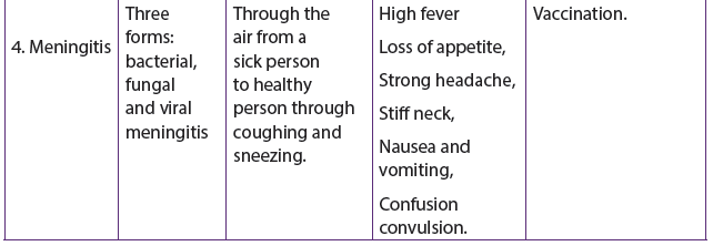
A bacterial disease is caused by entry of bacteria into a host where theycan grow, flourish then causing harm to the host. Bacteria e diseases include cholera, tuberculosis (TB), typhoid fever, pneumonia, tetanus, and diphtheria, and bacterial meningitis, tooth decay in humans and anthrax in cattle.Table 2.2. Common bacterial diseases in humans
2.5.1 Common Bacterial Diseases in PlantsThe table 2.3 common bacterial diseases in plants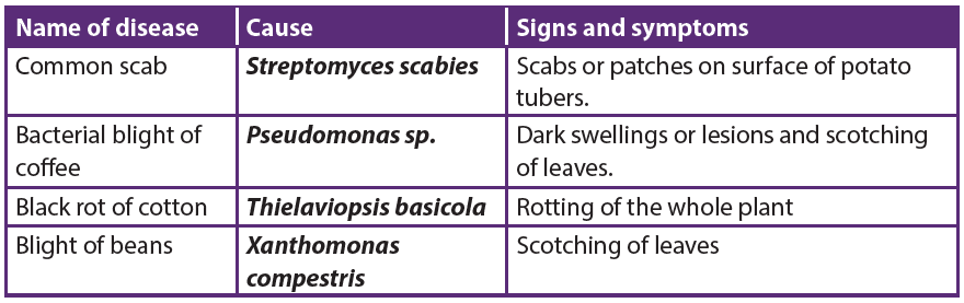 Self-assessment 2.5
Mr. Green lives in one of the slums in a certain city. He prepares and sells chapattis on street. He is usually very clean, but one morning, he is late for work so he does not bother to wash his hands after visiting the toilet. That day he prepares 400 chapattis all of which are sold. Few hours later, his customer Sandra suffered from a disease with the following signs and symptoms: severe diarrhea, excessive loss of water leading to dehydration, and vomiting. Five dayslater, all his customers were rushed and admitted in hospital due to the same problem.
1. Suggest the disease that Mr. Green’s customers were suffering from and what caused the disease
2. Name three ways this disease might be spread around city.
3. After reading this scenario, what message do you have for people who are like Mr. Green?
4. Suppose you were the health officer for the area in town with such a
problem. What steps would you take to prevent the disease from spreading further?
5. House flies are described as vectors. Describe how houseflies transmit diseases to humans.2.6 Structure and classification of Viruses
Activity 2.6
Discuss the reasons why viruses are not classified in any of the five kingdoms of
living organisms.
Viruses are microorganisms whose structure is only visible with electron microscopes. Viruses are acellular and lack cellular structure. Viruses have none of the features that we traditionally use for classification. They are particles made of proteins and nucleicacids that are found in all cellular organisms, but show metabolism only once inside the host cell.
When they infect cells, they use biochemical machinery and proteins of the host cell to copy their nucleic acids and to make proteins coats often leading to destruction of the host cells. The energy for these processes is provided by the ATP from the host cell.2.6.1. Structure of a virus
A typical virus consists of DNA or RNA within a protective protein coat called capsid. The shape of the capsid may vary from one type of virus to another, as shown in Figure 2.5 below. Some viruses have an envelope of phospholipids and proteins. The envelope is made
from portions of the host’s cell membrane. It surrounds the capsid and helps protect the virus from the host’s immune system. The envelope may also have receptor molecules that can bind with host cells and facilitate the virus to infect the cells.2.6.2. Characteristics of viruses
An individual virus is called a virion. It is a tiny particle much smaller than a prokaryotic cell. Because viruses do not consist of cells, they also lack cell membranes, cytoplasm, ribosomes, and other cell organelles. Without these structures, they are unable to make proteins or even reproduce on their own.
Instead, they must depend on a host cell to synthesize their proteins and to make copies of themselves. Viruses infect and live inside the cells of living organisms. They are also regarded as parasites since they depend entirely on living cells for their survival. Although viruses are not classified as living things, they share two important traits with living things: They have genetic material, and they can evolve.2.6.3. Classification of viruses
Viruses can be classified according to:
–– Type of nucleic acid molecules of DNA or RNA, forming the core of the capsid:
Most animal viruses contain RNA while plant viruses contain DNA
–– Type of host cell: plant or animal viruses as they are specific to their hosts
–– Presence or absence of the envelope: Plant viruses’ bacteriophage are nonenveloped
while animal viruses like HIV and influenza virus are enveloped.2.6.4. Viruses and human disease
When viruses infect cells of their host, they cause disease. Examples of diseases caused by viruses include HIV/AIDS, influenza (flu), chicken pox, and the common cold. The human immunodeficiency viruses that causes AIDS is a retrovirus. Other viral diseases include rabies, measles, diarrheal diseases, hepatitis A, B and C, polio, and cold sores. One-way virus cause disease is by causing host cells to burst open and die. Viruses may also cause disease without killing host cells. They may cause illness by disrupting homeostasis in host cells.
Some viruses live in a dormant state inside the body. The virus that causes chicken pox may infect a young child and causes the short-term disease chicken pox. Then the virus may remain latent in nerve cells within the body for decades. The virus may re-emerge later in life as the disease called shingles, where the virus causes painful skin rashes with blisters. Some viruses can cause cancer. Examples include the human papillomavirus (HPV) causing cancer of the cervix in females. Hepatitis B virus causes cancer of the liver. A viral cancer is likely to develop only after a person has been infected with a virus for many years.Self-assessment 2.6
1. What is meant by the term virus?
2. State the main components of a virus.
3. Describe the two ways how viruses cause an infection.
4. Differentiate between a bacteriophage and a retrovirus?
5. Do you think viruses should be considered as a form of life? Give reasons for your answer.2.7 Dichotomous key of identification of organism
Activity 2.7
The figure below represents different types of plant leaves. Make a classification of these plants based on the external structure of the leaves.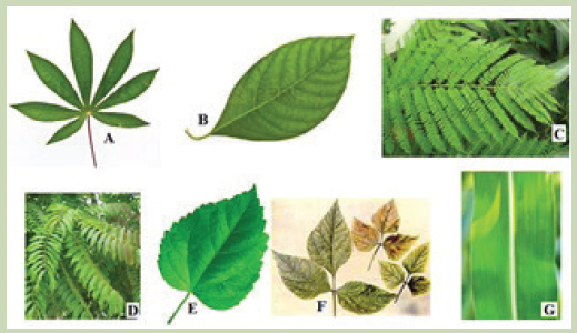 The dichotomous key is also referred to as biological identification key. It is made up of a series of contrasting statements called leads indicated by the numbers 1, 2, 3… where each lead deals with a particular observable characteristic. The characteristics used in keys should be readily observable morphological features which may be either qualitative, such as shape of abdomen, nature of legs, or quantitative, such as number of antennae, number of pairs of legs and length of the antennae in case of arthropods. It is essential to note that size and color are often less considered as both can be influenced by the environment, the season, the age or state of the organism at the time of identification.2.7.1. Guidelines used in construction of a dichotomous key:
The following guidelines must be considered while constructing a dichotomous key.
–– Use morphological characteristics which are observable as much as possible such as leaf venation, nature of margin, apex, lamina and nature or length of the petiole (leaf stalk).
–– Start with a major characteristic that divide the organism or the specimen into two large groups such as the type of a leaf.
–– Select a single characteristic at a time and identify it using a number for
example: simple leaf………go to 2, compound leaf………go to 5. This means that in 2 you will deal with only simple leaves and 5 only compound leaves.
–– Use similar forms of words for two contrasting statements for example at 2:
leaf with parallel venation …………go to G and leaf with network venation ………go to 3.
–– The first statement should always be positive.
–– Avoid generalizations or overlapping variations, be specific and precise to the point.Example
–– Collect leaves from the following plants: cassava, avocado, jacaranda, cassia, hibiscus bean, maize or paspalum grass,
–– Label different leaves collected as, A, B, C, D, E, F and G
–– Observe and familiarize with the specimens before starting the experiment to minimize errors during the identification process
–– Make a table summarising the specimens and steps followed to identify each
of them.
–– Construct a dichotomous key based on the observable features (characteristics) and table of steps followed.Solution: The dichotomous key of specimens A, B, C, D, E, F and G.
1. a. Simple leaves ---------------------------------------------------------------------go to 2
b. Compound leaves ---------------------------------------------------------------go to 5
2. a. Parallel venation ------------------------------------------------------------------------G
b. Network venation --------------------------------------------------------------go to 3
3. a.Simple digitate ---------------------------------------------------------------------------A
b. Non simple digitate -------------------------------------------------------------go to 4
4. a. Leaf with serrated margin -------------------------------------------------------------E
b. Leaf with smooth margin -------------------------------------------------------------B
5. a.Leaf with three leaflets (compound trifoliate)-------------------------------------F
b. Leaves with more than three leaflets --------------------------------------go to 6
6. a. Pinnate leaf ------------------------------------------------------------------------------ D
b. Bipinnate leaf --------------------------------------------------------------------------- C2.7.2. Common features used for identification of animals
Animals are classified based on the following features:
–– Locomotory structures such as legs, wings and fins
–– Antennae (presence, nature and number)
–– Presence or absence of eye and eye type
–– Number of body parts for example insects have three body parts
–– Body segments (nature and number)
–– Body surface structures such as fur, hair, feathers and scales
–– Feeding structures such as mouth parts in arthropods for example in insects
–– Type of skeleton present such as endoskeleton, exoskeleton and hydrostatic2.7.3. Common features used for identification of plants
Plants can be classified basing on the following features:
–– The leaf structure such as nature of apex, margin, venation, lamina and petiole
–– The flower structure including inflorescence type, flower shape and number of floral parts
–– The type of stem (woody, fleshy and herbaceous), shape (rectangular,
cylindrical) and texture of the stem (smooth, spiny and thorny) …
–– The type of root system, tap root, storage root, fibrous roots… Precaution
–– Care must be taken while collecting and handling some organisms because
some are poisonous, have thorns and others are able to sting
–– Collection of specimen should be done a day or few days before the experiment
depending on nature of the experiment
–– Avoid and try to minimize where possible, uprooting, cutting down or plucking
and pruning of plants as this may threaten the biodiversity as well as result
into environmental degradationActivity 2.8
Construct and interpret a dichotomous key of arthropods listed below.
–– Collect the following litter arthropods: honey bee, spider, millipede, butterfly,
sugar ant, centipede and mosquito and label each specimen as A, B, C, D, E, F and G respectively
–– Observe and familiarize yourself with the specimens before starting the experiment.
–– Use sharply contrasting external features of collected specimens /diagrams to construct a dichotomous key.Self-assessment 2.7
Read and interpret the dichotomous tree below and use it to answer the following questions. 1. Specify the phylum of kingdom animalia represented by the above dichotomous tree?
Give one observable reason to support your answer.
2. According to this dichotomous tree, which characteristic feature was used to classify different insects?
3. Which observable characteristic feature distinguishes between a spider and a mosquito?
4. How does a millipede differ from a centipede?
5. To which classes do a millipede and a centipede belong?
6. Which class of arthropods is not represented on the dichotomous tree?d. Phylum
3. Which one of the following is not a kingdom of living organisms?
a. Monera
b. Animalia
c. Annelida
d. Protoctista
4. Which one of the following is a characteristic feature common to fish, reptiles and birds but absent in mammals?
a. Possession of scales
b. Has no limbs
c. Possession of feathers
d. Undergo internal fertilization
5. Which one of the following statements about fish is not correct?
a. Fish live both in water and on land and undergo external fertilization.
b. Most fish have bones while others are cartilaginous
c. Most fish have streamlined body, lateral line and swim bladder.
d. Gills are organs for gaseous exchange in fish
6 Which one of the following is not a characteristic of all insects?
a. They have three body parts namely head, thorax and abdomen.
b. They have three pairs of jointed legs attached on segment of the thorax.
c. They have four pairs of jointed legs
d. They have a pair of antennae attached on the head.
7. The following are characteristics of all mammals except;
a. They have mammary glands to secrete milk feed their young ones.
b. Their skin is covered with hair.
c. Undergo internal fertilization and internal development of the embryo.
d. They have a pair of wings made up feathers.
8. The point where the leaf joins the stem is called;
a. Apex
b. Margin
c. Leaf base
d. Lamina
e. Length of petiole.
9. Which of the following is less considered while identifying feature to construct
a dichotomous key of leaves?End of unit assessment 2
1. Which one of the following living organisms belongs to domain bacteria?
a. Euglena
b. Vibrio cholerae
c. Paramecium
d. moulds
2. The group of classification where organisms resemble one another and are
capable of interbreeding together to produce viable offspring is known as:
a. Species
b. kingdom
c. Genusa. Nature of margin
b. Nature of apex
c. Size and color of leaf
10. The following are characteristics of arachnids except;
a. Four pairs of jointed legs
b. Two body parts
c. Three body parts
d. Do not have wings
11. Match the structures with the organisms which possess them. 12. A group of S4 students drew a Venn diagram below to summarize the five kingdoms into which organisms are classified. Study the diagram and answer the questions that follow:
 a. Which kingdoms are represented by the letters x and y?
b. State one characteristic that organisms of x may share with:
i. Prokaryotes
ii. Fungi
iii. Plantae13. Complete the table to summarize the characteristics of each class of phylum Arthropoda.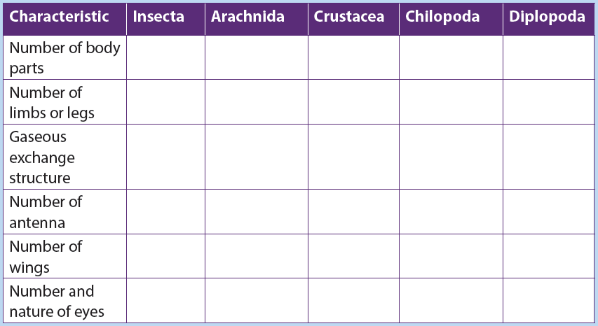 14. What is the significance of classification of living organisms?
15. The binomial system of naming a blue monkey, Cercopithecus mitis, is provided below;
Complete the table by filling the missing words.
UNIT 3: MICROSCOPY
Distinguish between the types of microscopes and their principal uses.Learning objectivesBy the end of this unit, I should be able to:
–– Describe the main features and functions of the components of a compound light microscope.
–– Manipulate a compound light microscope to observe prepared slides.
–– Show perseverance when using light microscopes.
–– Pay attention when using a compound light microscope to avoid damage of the lenses, mirrors and slides.
–– State that magnification is the increase in the apparent size of the object.
–– State that resolution is the ability of the microscope to show two objects as separate.
–– Appreciate the importance of magnifying instruments in Biology.
–– Use of a microscope to determine the relationship between actual size of the specimen and the image.
–– Calculate the approximate size of different biological structures using an appropriate unit of measurement
–– State the advantages and disadvantages of using an electron microscope.
–– State the principles and limitations of TEM (Transmission Electron Microscopy).
–– State the advantages and disadvantages of using SEM (Scanning Electron Microscopy).
–– Compare light and electron microscopes
–– Acknowledge the use of electron microscopes in modern science with reference to electron micrographs.
–– Observe and draw biological specimens under a light microscope.
–– Prepare temporary slides for observation under light microscopes using different objective lenses
–– Appreciate the importance of magnifying instruments in BiologyIntroductory activityPoint out scientific activities that require the use of microscope in our daily lives.
A microscope is used to produce a magnified image of an object or specimen.Anton Van Leeuwenhoek (1632-1723) was the first to invent a microscope powerful enough to explore the world of microbes. His discoveries stimulated an explosion of interest in scientific use of microscopes. Since the 18th century, many new types have been invented of which the most commonly used today are the compound light microscope and the electron microscope.1 (Kent, 2000, p. 58)).3.1 Compound Light Microscope
Activity 3.1
Some of the living things including protoctista and fungi have small size to be observed by naked eyes. Discuss the ways used by biologists to observe and identify different parts of these living organisms.
The optical microscope, often referred to as light microscope is a type of microscope which uses visible light and a system of lenses to magnify images of small samples. The different parts of light microscope are described below:
–– Base: supports and stabilizes the microscope on the table or any other working place
–– Light source: It is made by lamp or mirror which provides light for viewing the slide.
–– Stage: is a platform used to hold the specimen in position during observation.
–– Stage clips: are pliers used to fix and hold tightly the slide on stage.
–– Arm: supports the body tube of microscope
–– Body tube: maintains the proper distance between the objective and ocular lenses
–– Arm: used for holding when carrying the microscope and it holds the body tube which bears the lenses.
–– Coarse focus adjustment: moves stage up and down a large amount for coarse focus
–– Fine focus adjustment: moves stage up and down a tiny amount for fine focus
–– Objective lenses: focuses and magnifies light coming through the slide–– Revolving nosepiece: rotates to allow use of different power objectives
–– Slide: is a transparent pane on which a specimen is placed.
–– Eye piece/ocular lens: magnifies image produced by objective lens
–– Condenser: It will gather the light from the illuminator and focus it on the specimen lying on the stage. The function of the condenser is to focus the light rays from the light source onto the specimen.
–– Iris diaphragm lever: This allows the amount of light passing through the condenser to be regulated to see the object.Activity 3.2
Using the light microscope
a. To observe under low power and low magnification, proceed as follows:
–– Objects (specimens) to be observed under the microscope are first placed on a glass slide and covered with a cover slip.
–– Place the specimen on the stage of your microscope; in other words, arrange it so that the specimen is exactly at the center of the hole at the stage.
–– Fix the slide in place with two clips.
–– Rotate the nosepiece so that small objective lens is immediately above the specimen.
–– Set the angle of the reflector mirror so that light is directed up through the microscope.
–– Look down the microscope through the eye piece. Adjust the iris diaphragm so that the field of vision is bright and not dazzling.
–– Turn the course adjustment knob until the tip of the objective lens is close to the slide.
–– Now look down the microscope again. Slowly turn the course adjustment knob in the other direction, so the tube gradually moves upwards. The specimen on the slide should eventually come into view.
–– Use the course and fine adjustment knobs to focus the object as sharply as possible.
–– If necessary readjust, the iris diaphragm so the specimen is correctly illuminated. You will get a much better image if you don’t have too much light coming through the microscope.
b. To observe under high power at a greater magnification, proceed as
follows:
–– Rotate the nosepiece so that the large objective lens (with higher magnifying power) is immediately above the specimen. The nosepiece should click into position, as before.
–– If the specimen is not in focus, focus it with fine adjustment knob. Be careful that the tip of the objective lens does not touch the slide.
–– Readjust the illumination if necessary.Microscope uses transmitted light for observation. However, microscope uses specific light characteristics for specific samples, such as transparent specimens and samples that do not pass light. All parts of a microscope work together, the light from the illuminator passes through the aperture, through the slide, and through the objective lens, where the image of the specimen is magnified. Then the magnified
image continues up through the body tube of the microscope to the eyepiece, which further magnifies the image the viewer can see.
Light from the source is focused on the specimen by the condenser lens. It then enters the objective lens, where it is magnified to produce a real image. The real image is magnified again by the ocular lens to produce a virtual image that is seen by the eye.Care of the compound microscope
The microscope is an expensive instrument that must be given proper care. Always
general instructions have to be respected when using a microscope. These include:
–– Carry the microscope with both hands, one hand under the base, and the other on the arm.
–– When getting ready to put the microscope away, always return it to the low power or scanning power setting.
–– When setting the microscope on a table, always keep it away from the edge.
–– It is generally better to clear your lab table of items that are not being used.
–– Never clean lenses with anything other than lens paper, don’t use towels and
other paper tissues because they scratch the lens.
–– Inform the instructor or the biology lab technician if there is any microscope
damage or irregularity in its operation as soon as possible. Do not return a
faulty microscope without first informing the instructor or lab technician.
–– You are responsible for the microscope while using it treat it with care!Self-assessment 3.1
1. Complete the table below: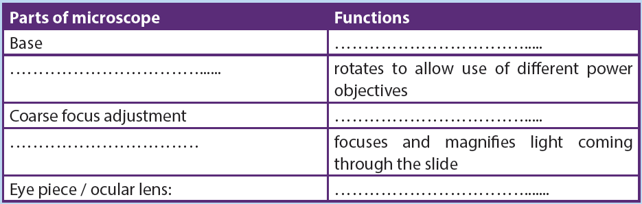 2. What is the importance of a light microscope?
3. Suggest a reason why it is not advisable to clean the objective and eye piece
lens with a wet cloth or towel?3.2 Magnification and resolution of a compound light microscope.
Activity 3.2
Work out the following equivalent measurements:
1. 1 millimetre (mm) =........... metre (m)
2. 1micrometre (μm) =............mmetre (m)
3. 1 nanometre (nm) =..............metre (m)
4. 1 metre (m) = .............mm =.......... μm =........nm,
5. 1 kilometre (km) = .............ma. Magnification
Magnification refers to increase in the apparent size of the object, while resolution of a microscope is the ability to show two close objects as separate. The maximum magnification of an ordinary light microscope is about x1500. Magnification must be written on the right side and below the biological drawing and it does not have units. The size of the image is measured in mm but converted into micrometres or nanometres to work out the actual size. It is calculated as follows: Example
Calculate the magnification if the actual size is 5μm and the measured image of the specimen has the size of 40mm.Answer:
–– Make the size of the image and the actual size in the same units by converting mm in μm. This is done by multiplying 40mm by 1000 so that 40mm = 40000 μm Note that the magnification of the specimen under a light microscope is calculated by multiplying the magnification of the objective used to that of the eyepiece.For example: 10x (objective) 10x (eyepiece) = x100.b. Microscopic observation
Activity 3.3
Using prepared slides of microorganisms such as a bacterium, amoeba, and paramecium. Observe, draw and label the visible parts under a light microscope. Avail these materials before you start: Petri-dishes, plate covers, pencil, transparent tape, microscope, agar powder, and permanent slide of bacteria, amoeba, and paramecium, Bunsen burner or any other source of heat.Procedure
–– Prepare agar medium by boiling a mixture of 10g of agar powder with 50ml of water
–– Label a control and exposed petri dishes in which you pour prepared agar medium.
–– Cool both plates for 20 minutes until the medium hardens.
–– Tape closed the cover of the control plate and removes the cover of the exposed plate.
–– Leave both plates for 5 minutes, and do not touch or breathe on the agar. After five minutes, tape closed the lid of the exposed plate and store both plates upside down in a warm place and draw your observations
–– Repeat the observation by using mounted slides of amoeba and paramecium and make a comparison between bacteria, amoeba and paramecium: what is your conclusion?
For this experiment, light microscope allows to observe organisms of small size including bacteria, amoeba and paramecium. Some other parts of macroscopic organisms such as cells and tissues of plants and animals or some parts of these living organisms such as stems and roots can also be observed under light microscope.
Some specimens can be observed directly after collection and preparation. However, some of the details might not be clearly observed because specimens are not colored. Also, some material distorts when you try to cut the specimen into thin sections. To overcome this challenge, slides can be prepared in advance by the use of the following steps:–– Staining: colored stains are chemicals that bind to chemicals on or in the specimens. This allow the specimen to be seen. Some stains bind to specific cell structures. For example, acetic orcein stains DNA dark red, while gentian violet stains bacterial cell walls.
–– Sectioning: specimens are embedded in wax, where thin sections are then cut without distorting the structure of the specimen. This is particularly useful for making sections of soft tissue, such as brain. Safety measures might be taken. Make sure that hands are washed with soap and warm water after theexperiment. Use a disinfectant to wipe down all surfaces where bacteria may
have been deposited for example. Be sure that some substances and animals
might be harmful to the life.Activity 3.4
Preparing of temporary slides and observation under light microscopeMake temporary preparation of slides of epidermis of onions young stems by fixing, staining and mounting. Observe under low and high power of a light microscope.Preparation and procedures
–– Add a drop of water at the center of the microscopic slide to flatten the membrane
–– Pull of a thin membrane from the onion layer and lay it at the center of the microscopic slide
–– Add a drop of iodine solution or methylene blue on the onion membrane
–– Gently lay a microscopic cover slip on the membrane and press it down gently using a needle to remove air bubbles.
–– Touch a blotting paper on one side of the slide to drain excess iodine/water solution,
–– Place the slide on the microscope stage under low power to observe.
–– Adjust focus for clarity to observe.
–– Make another slide without adding the stain to see the difference between a stained slide and a non- stained slide.
–– Draw and label the observed parts of each of the two slides and compare a drawing of a stained slide and that of a non-stained slid.c. Measuring cells
Cells and organelles can be measured with a microscope by means of an eyepiece called graticule. This is a transparent scale, usually having 100 divisions (Figure 3.4, A). The eyepiece graticule is placed in the microscope eyepiece so that it can be seen at the same time as the object to be measured (Figure 3.4, B). At this figure (Figure 3.4, B), the cell lies between 40 and 60 on the scale, so that it measures 20 eyepiece units in diameter (60 – 40 = 20).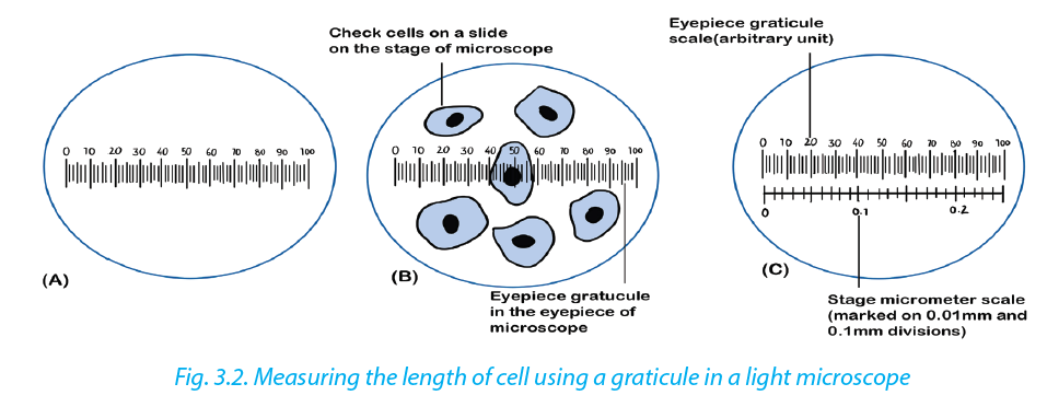 To calibrate the eyepiece graticule scale, a miniature transparent ruler called a stage micrometer scale is placed on the microscope stage and is brought into focus. This scale may be fixed onto a glass slide or printed on a transparent film. It commonly has subdivisions of 0.1 and 0.01 mm. The images of the two scales can then be superimposed (Figure 3.4, C). If in the eyepiece graticule, 100 units measure 0.25
mm, the value of each eyepiece unit equals By converting mm to μm, the value of eyepiece equals
  The diameter of the cell shown superimposed (Figure 3.4, B) measures 20 eyepiece units. Its actual diameter equals 20 × 2.5 μm = 50 μm. This diameter is greater than that of many human cells because the cell is a flattened epithelial cell.
Use the following instructions to measure the length of one cell–– Measure the distance in millimetre from the start of one cell to the end of 10 cells
–– Divide by 10 to find the length of one cell in the specimen.
–– Convert this length in millimetre to micrometer by multiplying by 1000.
–– Find the actual length of a cell by dividing this length by the magnification of thespecimen got from the product of eye piece and objective lens used.Self-assessment 3.2.
1. Calculate the magnification of an image with 50mm, and the objectmeasuring 5μm. in length.
2. If a nucleus measures 100mm on a micrograph, with a magnification of X10 000, what is the actual size of the nucleus?3.3 Electron microscopes
Activity 3.3
Suggest the form and source of energy used by electron microscope. How does this differ from that used by a compound microscope?An electron microscopes use a beam of accelerated electrons as a source of illumination.
Electron beams have a much smaller wave length than light rays and therefore have greater resolving powers and can produce higher effective magnifications than light microscopes. There are two types of electron microscopes;
–– Transmission electron microscope (TEM)
–– Scanning electron microscope (SEM)
Electron microscopes are used to study the details of internal structures (the ultrastructures)
of cells. Most modern TEMs can distinguish objects as small as 0.2nm.
This means that they can produce clear images magnified up to 250,000 times. Formation of an image by the TEM:
–– Extremely thin samples of the specimen are needed and are cut by using diamond or glass knives as they are supported in resin block to prevent them from collapsing
–– The section is then impregnated with a heavy-metal stain
–– As the beam passes through the specimen, electrons are absorbed by the heavily stained parts but passes readily through the lightly stained parts.
–– Electro magnets bend the electron beam to focus an image onto the florescent screen or photographic film to form an electron micro graph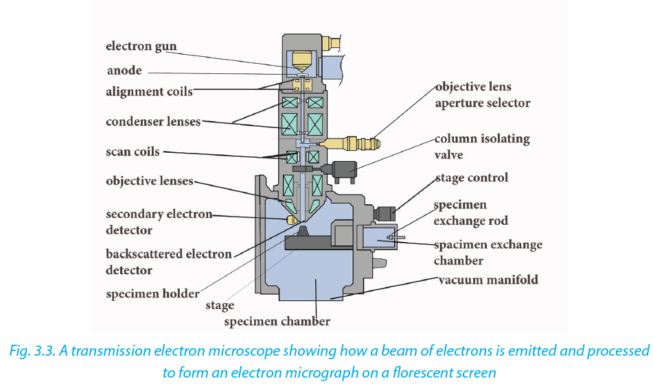 Scanning electron microscope (SEM)
The SEM is used to produce 3D images of surfaces of the specimens. Electrons are reflected from the surface of the specimen stained with a heavy metal. This enables the SEM to produce images of all specimens, cells, tissues, or even organisms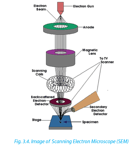 a. Advantages of the electron microscope over light microscope
Electron microscope has a higher resolution and istherefore able of a higher effective magnification estimated at up to 250,000 million times compared to the light microscope which can show a useful magnification only up to 1000-2000 times. This is because a light microscope uses a beam of light with a longer wave length while Electron microscopes use a beam of electrons that have a short wave length.b. Disadvantages of electron microscope
Despite the advantages, electron microscope presents a number of setbacks and limitations.
–– They are extremely expensive and the maintenance costs are high.
–– Sample preparation is often much more technical requiring special training.
–– Samples must be dead, exposed to high radiation and are placed in a vacuum so that it is impossible to observe living specimens
–– It is not possible to observe colors because electrons do not possess a color. The image is only black-white, even if sometimes the image is colored artificially to give a better visual impression.
–– They require more training and experience in identifying artifacts that may have been introduced during the sample preparation process.c. Comparison between light and electron microscopes
Light and electron microscope presents the following similarities and differences. The following are some of the similarities:–– Both light and electron microscopes form larger (magnified) and more detailed (highly resolved) images of small objects or small areas of larger objects
–– Both light and electron microscopes are used in biology study, research and medical sciences particularly histology, material sciences such as metallurgy and other aspects of science.
–– Specimens must be carefully prepared using techniques appropriate for both the equipment and the sample including slicing, staining, and mounting.Despite the similarities, light and electron microscope presents differences such as these summarized in the following table:
Table 3.1. Differences between light and electron microscopic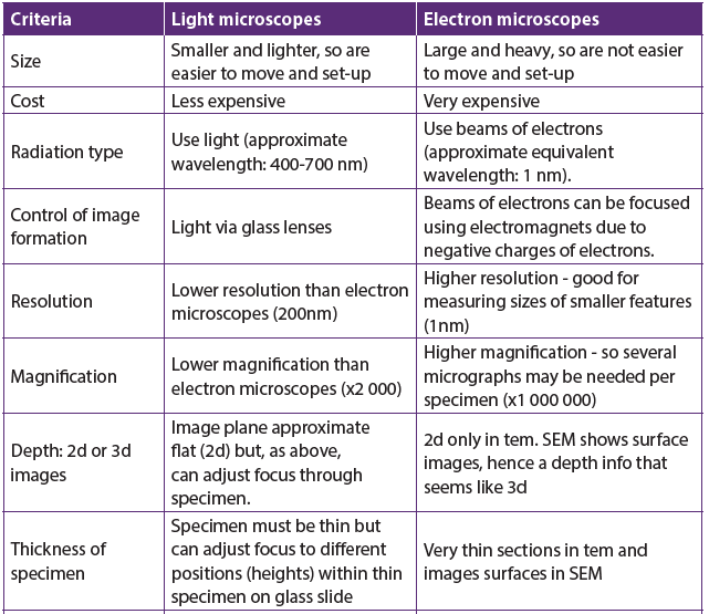

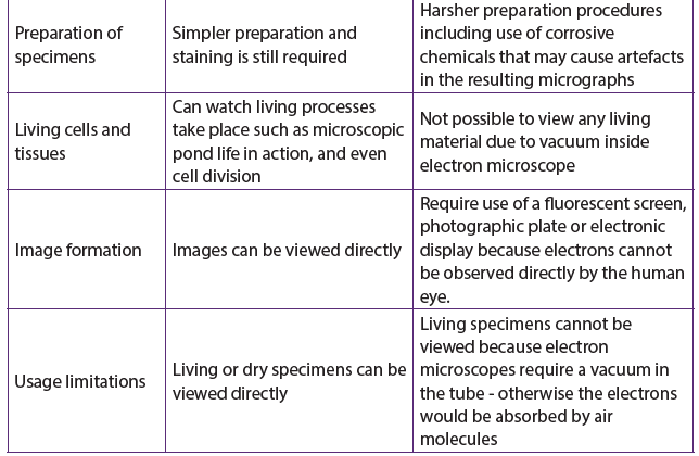 Self-assessment 3.3
1. How is magnification varied in;
a. A light microscope
b. An electron microscope?
2. Why is the resolving power of an electron microscope such better than that of a light microscope?
3. Make a comparison between light and electron microscope, highlighting the advantages and disadvantages for each type of microscope. Summarise the similarities and differences between light and electron microscopesEnd of unit assessment 3
Section A. Multiple choice questions
1. Which ranges can be viewed using a light microscope? a. 4 only
b. 1 and 2 only
c. 2 and 3 only
d. 3 and 4 only
2. The figure below shows a mitochondrion drawn from an electron micrograph. Study it carefully and answer the following questions. If the length of the mitochondrion line X Y is 3000 nm. What is the magnification of the drawing of the mitochondrion?
a. ×100
b. ×1000
c. ×10 000
d. ×100 000
3. A light microscope is used to observe two membranes that are 200 nm apart. How far apart are the membranes when the objective lens is changed from low power (×40) to high power (×400)?
a. 2 μm
b. 20 μm
c. 200 nm
d. 2000 nm
4. The electronmicrograph below is that of a chloroplast. The length of the chloroplast along the line shown is 80 mm. The actual length of the chloroplast is 10 μm. What is the magnification of the chloroplast?
a. ×8 × 10²
b. ×8 × 10³
c. ×8 × 10⁴
d. ×8 × 10⁶
5. The following diagram below is drawn from an electron micrograph of an animal cell. Which represents the same cell, seen under a light microscope at ×400 magnification?
 Section B:6.
 The micrograph above shows a transverse section of part of an animal cell.
a. Identify organelles labelled X and Y
b. The maximum actual diameter of Y is 2um. Calculate the magnification of this organelle in the electron micrograph.
c. Determine with two reasons whether the cell is prokaryotic or eukaryotic.
d. From evidence in the electron micrograph, deduce two substances that were being synthesised by large quantities by this cell.
e. The dark granules in the cell are glycogen. Explain the conclusion that you draw from this informationUNIT 4: CELL STRUCTURE AND SPECIALIZATION
Key Unit Competence
Describe the structure and function of cells in an organism.Learning objectives
By the end of this unit, I should be able to:
–– Identify plant and animal cell structures visible under a light microscope.
–– State functions of cell structures as seen under an electron microscope.
–– Describe the nature of artefacts.
–– State the importance of freeze fracturing for examining membrane structure.
–– Explain how cell organelles can be isolated by cell fractionation.
–– List the functions of cell membranes.
–– Describe the fluid mosaic structure of cell membranes.
–– Explain the role of the different components of a cell membrane.
–– Explain cell specialization as the differentiation of a cell or process to do a particular function.
–– Interpret charts and micrographs to relate the structure of specialized cells to their functions.
–– Prepare, observe and draw diagrams for specimens on temporary slides for:
Wandering Jew, in plants and cheek cells under a light microscope.
–– Distinguish between ultra-structures of plant cells and animal cells.
–– Compare ultra-structures of prokaryotic and eukaryotic cells
–– Show resilience and be aware of artefacts when preparing temporary slides.
–– Appreciate the importance of cell specialization in multicellular organisms.Introductory Activity
1. Differentiate between prokaryotic and eukaryotic cells.
2. By using charts for the two cells, identify different organelles of eukaryotic cell that may perform functions similar to those of a prokaryotic cell.Cytology is the study of the structure and function of cells. A Cell is the basic unit of life. All living organisms are made up of cells.
Living organisms are classified into:
–– Unicellular organisms are made of only one cell, such as bacteria,
–– Multicellular organisms are animals and plants composed of many cells. In
multicellular organisms, cells divide and then undergo differentiation or
specialisation for specific functions.Cell theory.
The cell theory states that all living organisms are made up of cells, and cells are the basic unit of structure function in all living organisms.
The main principles of cell theory are based on the following ideas.
–– All known living organisms are made up of one or more cells,
–– All cells come from pre-existing cells by division
–– Cells contain the hereditary information that is passed from cell to cell during
cell division.
–– Metabolism takes place in cells
–– Given suitable conditions, cells are capable of independent existence4.1 Ultra-structure of a cell
Activities 4.1
1. Observe the chart given for Ultra structure of a cell and identify parts that are easily recognizable when compared with a photomicrograph form a light microscope.
2. Identify the se mitochondria and ribosomes and state their roles in the life of the cell.
When viewed under light microscope, the most obvious features observed are the very large nucleus and a clear cytoplasm surrounded by a cell membrane. However, under electron microscope, it is possible to identify a range of organelles in plant and animal cells. Ultrastructure is the detailed of cell as revealed by the electron microscope.
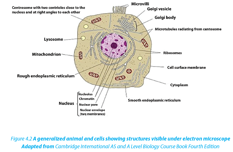
4.1.2 Similarities between animal cell and plant cell
–– Both have a cell membrane, a cytoplasm and a nucleus.
–– Both animal and plant cells have mitochondria, Golgi apparatus, Reticulum endoplasmic, lysosome, big ribosomes (80S), peroxisome, microtubules.
Table 4.1: Differencied between animal and plant cell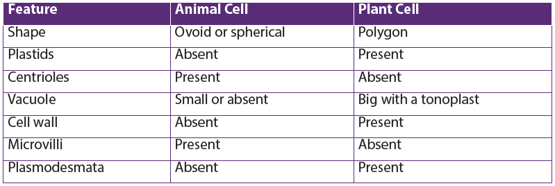
Self-assessment 4.1
1. What structures do both animal and plant cells have in common?
2. State any five principles of the cell theory.
3. Give the major difference between a plant and animal cell. Which organelles does this difference relate to?4.2 Prokaryotic cells
Activities 4.2
Under microscope, observe mounted slides of bacteria, and plant cells. Draw and label the parts that are common in both plant and bacterial specimens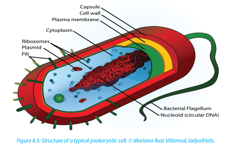
A typical bacterial cell has a cell surface membrane enclosing the cytoplasm that contains enzymes, ribosomes and food granules. The membrane is surrounded by the cell wall and this may in turn be enclosed in a capsule. A bacterial cell lacks high level of organization compared to animal or plant cell. It has no Golgi apparatus or endoplasmic reticulum. The genetic material is a single strand of DNA usually
coiled up into the center of the cell to form a nucleoid. This nucleoid has no double membraned nuclear envelope so is often described as an ‘ill-defined nucleus’.
–– Some bacterial cells contain plasmids with additional DNA.
–– Respiration generally takes placein mesosomeswhich is an in-folding of the
cell surface membrane but lack mitochondria
–– Photosynthesizing bacterial cells such as cyanobacteria(blue green algae) have a special form of chlorophyll but it I not enclosed in a double membraned chloroplast4.4.1. Comparison between prokaryotic and eukaryotic cells
Table 4.2 Comparison between prokaryotic and eukaryotic cells
Self-assessment 4.2
Organisms such as bacteria are known as prokaryotes.
1. Which structure in a bacterial cell resembles a nucleus?
2. How does it differ from the nucleus of eukaryotic cells?4.3 Cell organelles
Activities 4.3
By using iodine solution, methylene blue, a piece of onion leaf, a scalpel, forceps, light microscope, slides and cover slips, clean cotton wool bud, and onion bulbs. Observe cells from onion epidermis under light microscope. Observation of a plant cell
–– Add a drop of diluted iodine solution on the slide.
–– Remove a transparent layer of onion epidermis from the inner side that you will mount on the slide and add iodine solution.
–– Cover your preparation with a cover-slip and mount it on the stage.
–– Observe the preparation under the low power and thereafter under high magnification.
Why did you use iodine solution in this experiment?
What main parts of a plant cell are easily observed from a light microscope?Observe animal cells from mouth cheek epithelium
–– By using a clean cotton wool bud, wipe over inside of your cheek.
–– Smear cells over surface of a clean grass microscope slide containing a drop of methylene blue stain
–– Carefully put the cover-slip on the preparation and mount it on the stage to observe.
Draw both plant and animal cell and label the cell wall, nucleus and vacuole.
4.3.1Nucleus

The cell nucleus contains nearly all the cell’s DNA with the coded instructions for making proteins and other important molecules. The nucleus is surrounded by a double nuclear envelope, which allow materials to move into and out of the nucleus through nuclear pores. The granules found in the nucleus are called chromatin whichconsist of DNA bound to protein. When a cell divides, the chromatin condenses into chromosomes containing the genetic information. The nucleus contains a dense spherical structure called nucleolus in which assembly of ribosomes occurs.
4.3.2 Endoplasmic reticulum (ER)
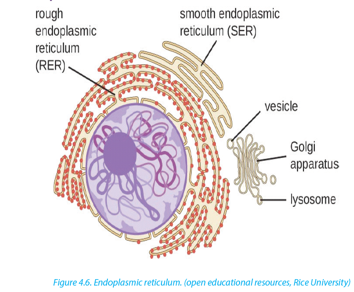
The ER consists of a series of flattened membrane-bound sacs called cisternae. The rough ER is surrounded with ribosomes. The rough ER transports proteins made on attached ribosomes. The smooth ER is made of tubular cavities lacks ribosomes, and it involves in synthesis of lipids that the cell needs. The number and distribution of the ER relates to the functions of the cell; glandular cells are seen to have several RER for synthesis of hormones and enzymes. Examples include liver cells, plasma cells, and pancreatic cells.
4.3.3 Golgi apparatus
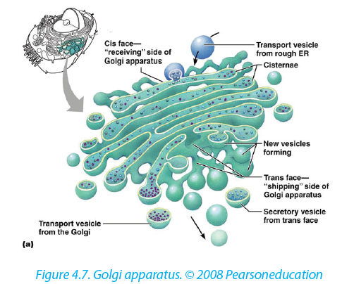
The Golgi apparatus is a stack of membrane-bound, flattened sacs, which receives proteins from the ER and modifies them. It may add sugar molecules to them to form glycoproteins or lipids to form glycolipids. The Golgi apparatus then packages the modified substances into vesicles that can be transported to their final destinations throughout the cell or outside of the cell by exocytosis.
4.3.4 Mitochondria

Mitochondrion have two membranes separated by a fluid-filled intermembrane space. The inner membrane is highly folded to form cristae that plays a big role in aerobic respiration.The central part of the mitochondrion is called matrix. The mitochondria are the site where Adenosine triphosphate (ATP) is produced during aerobic respiration.
4.3.5 Chloroplasts

Chloroplasts are the site of photosynthesis in plant cells. These are found in plant cells and in cells of some protoctists. They also have two membranes separated by a fluid-filled space, circular DNA as in mitochondria. The inner membrane is continuous, with thylakoids. A stalk of thylakoids is called a granum (plural: grana). Chlorophyll molecules are present on the thylakoid membranes.
4.3.6 Lysosomes
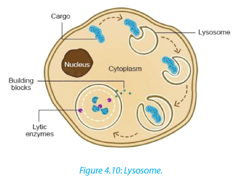
These are spherical sacs surrounded by a single membrane. They contain powerful digestive enzymes. Their role is to break down materials such as worn out cell organelles, and destroy foreign microorganisms that enter the body. In acrosome, lysosomes help the sperm to penetrate the egg by breaking down the material surrounding the egg. Lysosomes are also involved in autolysis, breakdown of dead tissues or harmful objects inside the cell. Therefore, lysosomes are referred to as ‘suicide bags’
4.3.7 Ribosomes
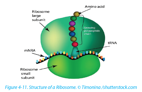
Ribosomes appear as dark granules in the cytoplasm and are not surrounded by a membrane. They have the same size as those found attached to the rough endoplasmic reticulum- about 20nm in diameter and known 80S. Free ribosomes make proteins that are as enzymes or in other forms in the cytoplasm. Ribosomes are made in a region of the nucleus called the nucleolus.
4.3.8 Centrioles
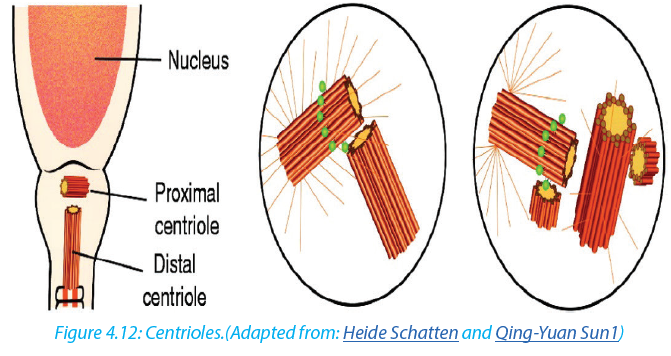
Centrioles are small tubes of protein fibers called microtubules which have many roles including moving chromosomes during nuclear division. Animal cells have structures called centrioles which consist of two groups of nine triple microtubules. Centrioles form an anchor point for microtubules during cell division.
4.3.9 Vacuole
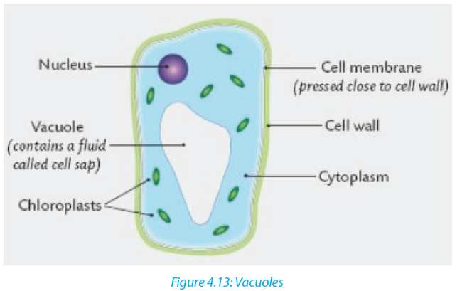
A vacuole is a saclike structure that stores materials such as water, salts, proteins, and carbohydrates. In many plant cells there is a single and large central vacuole filled with liquid. The pressure in the cells of central vacuole makes it possible for plants to support heavy structures like leaves and flowers. Some animals and unicellular organisms contain contractile vacuoles which contract to pump excess water out
of the cell.self-assessment 4.3
1. Explain why muscle cells contain several mitochondria compared to fat storage cells
2. What kind of information is contained in chromosomes?
3. Describe the functions of the endoplasmic reticulum, Golgi apparatus, chloroplasts, mitochondria and nucleus in the cell.
4. The diagram below shows the 3D structures which would be visible in ultrastructure of a plant cell.
Identify the parts labeled in this plant cell and:
a. State one function for 1, 2, 3, 7, and 10
b. What are parts 4 and 5 made of?
c. What are two functions of the cytoskeleton?4.4 Membrane structure
Activity 4.4
Learners mix a portion of cooking vegetable oil with water and shake the mixture vigorously and leave it to settle. Note the way water and oil are distributed within the mixture and suggest a possible explanation for your observation. Cell membranes cover surfaces of every cell. Some organelles in cytoplasm are
enveloped by membranes. The cell membranes ultrastructure is not easily visible under a light microscope but is studied by electron microscopes, freeze structuring and other modern techniques which reveal complex structures A detailed study of a cell membrane reveals that it is 7-8nm wide and is made of a phospholipid bilayer.
–– Lipid component makes up 45% protein and 10% carbohydrate. Most of the lipids are phospholipids
–– Each molecule of phospholipid consists of a hydrophobic tail of two fatty acids and a hydrophilic phosphate head. They arrange themselves in phospholipids bilayer with their tails pointing inward away from the water both inside and outside the cell
In 1972, Jonathan singer and Garth Nicolson proposed the fluid mosaic model of the cell membrane structure. This was done after realizing that membranes must have a complex structure to carry out a variety of activities. In their model;
–– Individual protein molecules shift and move on a fluid bilayer of phospholipids;
some spanning the width of the membrane (intrinsic proteins), others confined to the outer or inner surface (extrinsic protein)
–– Protein molecules are variable in structure and function but they all contribute to the mechanical strength of membranes
The membrane is referred to as;
–– A fluid because it appears to have the properties of a fluid rather than a solid as the major constituent,lipids and proteins move about the structure
–– Mosaic because protein and lipid components form a pattern of parches model4.4.1 Properties of the cell membrane
–– It is mainly made of lipids, proteins and carbohydrates.
–– It is semi-permeable or partially permeable to allow some substances to pass through but prevents others to cross depending on their size, charges and polarity.
–– It is positively charged outside and negatively charged inside and has a hydrophilic pole and a hydrophobic pole
–– It is a bilayered sensitive and flexible.It has inorganic ions and its proteins and lipids may be mobile and contains different types of enzymes and coenzymes.
–– It is perforated of pores and recognizes chemicals messengers including hormones and neurotransmitters.4.4.2 Roles of different components of cell membrane
a. Cholesterol
–– Gives the membranes of some eukaryotic cells the mechanical stability.
–– It fits between fatty acid tails and helps make the barrier more complete, so substances like water molecules and ions cannot pass easily and directly through the membrane.b. Channel proteins
–– Allow the movement of some substances across the membrane.
–– Large molecules like glucose enter and leave the cell using these protein channels.c. Carrier proteins
–– Actively move some substances across the cell membrane. For example, magnesium and other mineral ions are actively pumped into the roots hair cells from the surrounding soil.
–– Nitrate ions are actively transported into xylem vessels of plantsd. Receptor sites
–– Allow hormones to bind with the cell so that a cell response can be carried out.
–– Glycoproteins and glycolipids may be involved in cells signaling and they allow the immune system to recognize foreign objects to the cells.
–– Some hormone receptors are glycoprotein and some are glycolipid.e. Enzymes and coenzymes
–– Some reactions including metabolic processes in photosynthesis take place in membranes of chloroplasts.
–– Some stages of respiration take place in membranes of mitochondria, where Enzymes and coenzymes may be bound to these membranes.
–– The more membrane there is, the more enzymes and coenzymes it can hold and this helps to explain why mitochondrial inner membranes are folded to form cristae, and why chloroplasts contain many stacks of membranes called thylakoids.4.4.4. Functions of a cell surface membrane
–– The cell membrane acts as a selective barrier at the surface of the cell, and controls the exchange between the cell and its environment.–– Glycoproteins and glycolipids are involved in the cell protection, the process by which cell adhesions are brought about and in the cell recognition.
–– Receptor sites for hormones and neurotransmitters
–– Transmission of nerve impulses
–– Insulation of nerves to improve transmission speeds. Internal membranes:
–– Act as reaction surfaces
–– Act as an intra cellular transport system
–– Providing separate intra cellular compartment, isolating different chemical reactions as in organelles.Self-assessment 4.4
1. What is meant by the fluid mosaic model of the cell membrane?
2. State at least three properties of the cell membrane.
3. Describe at least 4 types of the proteins in the cell membrane and their roles.
4. What is a partially permeable membrane?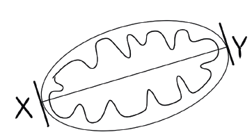
5. What do the words hydrophilic and hydrophobic mean?
6. The diagram below shows the structure of a cell membrane. Study it carefully and answer the following questions.
a. Name parts labelled A, B, C and D and give the function of the part B.
b. What types of molecule are likely to be involved in?
i. Cell signaling and recognition
ii. Allowing small charged molecules to pass through the cell membrane
iii. Site metabolic reactions
7. What is the difference between rough and smooth endoplasmic reticulum?
8. Describe the role of cytoskeleton9. The photograph in the figure below shows an organelle of the living cell.

a. Name this organelle.
b. What is the function of this organelle?
c. In which ways is this organelle similar to a chloroplast?4.5 Specialized cells
Activity 4.5
By using the diagrams below, relate the structure of specialized cells to their functions.
Differentiation refers to the changes occurring in cells of a multicellular organism so that each different type of cell becomes specialized to perform a specific function. In animals, the first type of cells in the developing embryo is stem cells. These are unspecialized cells that go on to form all the different types of cells in adult. Cell can differentiate in many ways, with changes to the shape of the cell, the number of particular organelles and the content of the cell.
4.5.1 Specialized animal cells and their functions
4.5.1.1 Red blood cells
All blood cells are produced from undifferentiated stem cells in the bone marrow but the cells destined to become erythrocytes (red blood cells) lose their nucleus, mitochondria, Golgi apparatus and rough endoplasmic reticulum. They are packed full of the protein called haemoglobin. The shape of this cells change so that they become biconcave discs, and they are then able to transport Oxygen in the body.
4.5.1.2 Sperm cell

Sperm cells are specialized to fertilize the egg. Its specialization involves many
changes in shape and organelles content.By shape: the sperm cells are very small, long and thin to help them to move easily, and they have a flagellum which helps them to move up the uterine tract towards the egg.
By organelles content: sperm cells contain numerous mitochondria which generate much energy for their movement. Their acrosome has specialized lysosomes containingmany enzymes that are released on the outside of the egg. These enzymes lyse the wall of the egg, and facilitate the sperm nucleus to penetrate easily. In content, the sperm cell nucleus contains the half number of chromosomes of the germ cell in order to fulfil its role as a gamete of fertilizing the egg.
Did you know: As a sperm fuses with an ovum to form a zygote which grows into an individual, in the same way: a man maries a woman to form a couple which will produce children and form a family.
4.5.1.3 Nerve cells
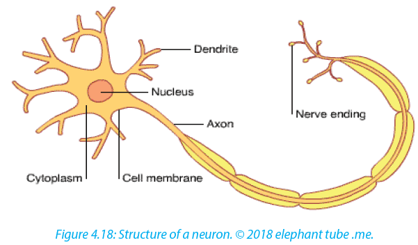
Nerve cells also known as neurons are specialized cells to carry nervous impulses in the body. These signals between neurons occur via specialized connections called synapses. Specialized animal cells have different functions. Some of them are summarized in the following table.
Table 4.3: Specialized animal cells and their functions.
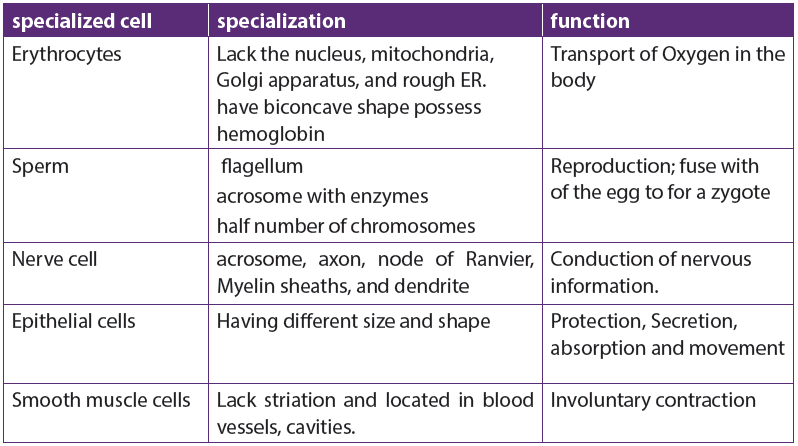
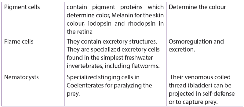
4.5.2 Specialized plant cells and their functions
4.5.2.1 Root hair cells
The root hair cells have hair-like projection from their surface out into the soil. This increase the surface area of root available to absorb water and minerals from the soil.
4.5.2.2 Palisade cells
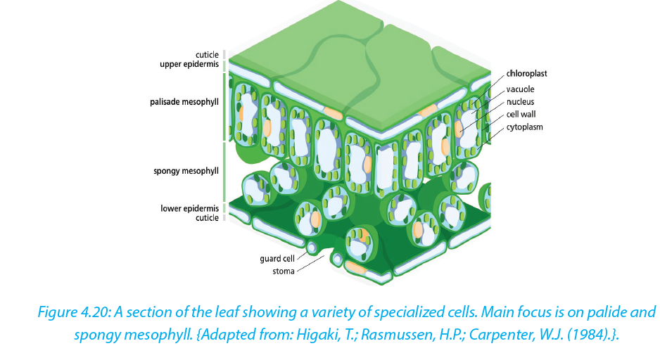
Palisade cells are in leaves, right below the upper epidermis. They are vertically elongated, a different shape from the spongy mesophyll cells beneath them in the leaf. Their large numbers of chloroplasts allow them have several chloroplasts used in photosynthesis.
Parenchyma cells
Parenchyma is composed of relatively simple and undifferentiated parenchyma cells. They function in storage, photosynthesis. In most plants, metabolic activity such as cell division, respiration, and photosynthesis occurs in these cells because they retain their active cytoplasm. .4.5.2.3 Guard cells

Guard cells are cells surrounding each stoma. Guard cells are specialized cells in the epidermis of leaves, stems and other organs that are used to control gas exchange. They are produced in pairs with a gap between them that forms a stomatapore. Guard cells have the following feature:
–– Un even thick walls
–– Possess chloroplasts; they are the epidermal cell that have chloroplasts an adaptive feature in controlling pore opening.Self-assessment 4.5
1. Explain why differentiation to produce erythrocytes involves a change in shape.
2. Red blood cells cannot divide as they have no nucleus. State two other
biological processes that red blood cells cannot carry out.
3. Describe how the following are specialized for their roles:
a. Neutrophil
b. Sperm cellRoot hair cell
3. Explain why photosynthesis is carried out in palisade mesophyll more than
in spongy mesophyll.
4. In what kinds of organisms is cell specialization pronounced characteristic?
5. Discuss the advantages of cell specialization in living thingsEnd of unit assessment 4
Section A. Multiple choice questions
1. Which organelle converts the chemical energy in food into a form that cells can use?
a. Chromosome
b. Chloroplast
c. Nucleus
d. Mitochondrion
2. The cell membranes are constructed mainly of:
a. Carbohydrate gates
b. Protein pumps
c. Lipid bilayer
d. Free-moving proteins
3. In many cells, the structure that controls the cell’s activities is the:
a. Nucleus
b. Nucleolus
c. Cell membrane
d. Organelle
4. Despite differences in size and shape, all cells have cytoplasm and a
a. Cell wall
b. Cell membrane
c. Mitochondria
d. Nucleus
5. If a cell of an organism contains a nucleus, the organism is a (an)
a. Plant
b. Eukaryote
c. Animal
d. Prokaryote
6. Match each part of the cell (left column) to corresponding statement (right column):
Nucleus controls movement of substances in and out of the cell Mitochondrion where photosynthesis takes place Chloroplast where aerobic respiration takes place Smooth ER controls the activity of the cell
Ribosomes where lipids including steroids are madeSection B: Questions with short answers
1. How does a cell membrane differ from a cell wall?
2. Name the structures that animal and plant cells have in common, those found in
only plant cells, and those found only in animal cells.
3. List:
a. Three organelles each lacking a boundary membrane
b. Three organelles each bounded by a single membrane
c. Three organelles each bounded by two membranes (an envelope)
4. Identify each cell structure or organelle from its description below.
a. Manufactures lysosomes and ribosomes
b. Site of protein synthesis
c. Can bud off vesicles which form the Golgi body
d. Can transport newly synthesized protein round the cell
e. Manufactures ATP in animal and plant cells
f. Controls the activity of the cell, because it contains the DNA
g. Carries out photosynthesis
h. Can act as a starting point for the growth of spindle microtubules during
cell division
i. Contains chromatin
j. Partially permeable barrier only about 7 nm thick
k. Organelle about 25 nm in diameter
l. Which two organelles other than the nucleus contain their own DNA?Section C: Essay questions
1. Describe the structure and function of the cell membrane and cell wall.
2. Describe the basic structure of the cell membrane.
3. Explain two common characteristics of chloroplasts and mitochondria.
Consider both function and membrane structure.
4. The diagram below shows the structure of a liver cell as seen using an electron microscope.
a. Name the parts labelled A, B, C and D.
b. The magnification of the diagram above is x12 000. Calculate the actual length of the mitochondrion labelled M, giving your answer in μm. Show your working.
c. Explain the advantage to have a division of labor between different cells in the body.UNIT 5 :DIVERSITY OF SPECIALIZED TISSUES
Key Unit Competence
Describe different specialized plant and animal cells and adaptation of tissues.Learning objectives
By the end of this unit, I should be able to:
–– Define a tissue as a group of cells with similar structure working together for a function.
–– Name the main types of animal and plant tissues.
–– Define an organ as a structure made up of a group of tissues with related
functions working together to perform bodily functions.
–– Explain how epithelial tissues are adapted to perform a diversity of functions in the body.
–– State the advantages and disadvantages of being unicellular.
–– Observe and draw plant and animal tissues as seen under a light microscope.
–– Interpret photomicrographs of plant and animal tissues
–– Acknowledge the relationship between levels of organization
–– Recognize the efficiency shown by multicellular organisms to explore more modes of life that are not available to single celled organisms that show little or no specializationIntroductory activity
Read the following passage and use it to answer the following questions:In an anthill, there are different groups of termites such as a queen, workers and soldiers. Each group has a specific role to play in the colony. The structure termites of each group is related to their role for example soldiers that protect the colony have mouth parts shaped like a pair of scissors building and a slightly larger abdomen for storing water. The queen is the largest of all and has a role of laying eggs. Workers have mouth parts for cutting and chewing food or soil particles. Some members of workers are in charge of caring for the young while others find food and defend the colony or remove dead members. Their specialization and division of labor bring about efficiency in the colony.
1. Specify the message addressed by the above paragraph.
2. Explain how is the structure of termites related to their functions?
3. What is the significance of specialized tissues in multicellular organisms like plants and animals?
The study of tissues is known as Histology. A tissue is a group of associated, similarly structured cells that perform specialized functions for the survival of the organism. In histology, differentiation is the process by which structures become modified and specialized to perform specific functions. Differentiation is also known as ‘specialization’. In animals, the first type of cells in the developing embryo is stem cells. These are unspecialized cells that go on to form all the different types of cells in adult.5.1 Specialized plant tissues
Activity 5.1.1
–– Remove an epidermis layer from the ventral side of an onion leaf.
–– Mount it on the slide containing a drop of iodine solution
–– Observe your preparation under a light microscope
–– Draw, label and describe your findings.
–– From your discussion:
1. What is a tissue?
2. What is the role of epidermis in onion?5.1.1. Plant tissues
Activity 5.1.2
The following figure represents the flow chart of subdivisions of plant tissues. Use it to answer the following questions.
1. How do meristems differ from permanent tissues?
2. Plant tissues are classified into ground tissues and vascular tissues as shown in the figure above. What is meant by the term vascular tissues?
3. How is the structure of the xylem and phloem vessels related to their function?
4. From the flow diagram above, identify three types of ground tissues.5. Write down short notes on each of the following types of meristems.
a. Apical meristems.
b. Lateral meristems
c. Intercalary meristems
Plant tissues can be divided into two main groups, Meristematic tissues (apical, lateral, and intercalary meristems) and Permanent tissues (ground tissues and vascular tissues).5.1.2. Meristem tissues
Meristem tissue is a group of cells which retain the ability to divide by mitosis. Meristematic tissues are specialized to carry out specific functions such as reproduction, growth, photosynthesis and replacement of old or damage tissues. The cells making a meristem tissue are small, have a central large nucleus and dense cytoplasm, thin-walled, with no or small vacuole, and no specialized features. The cells are rectangular and closely packed with no intercellular air spaces.
Types of meristematic tissues
Meristematic tissues are subdivided into apical meristems, lateral meristems (cambium) and intercalary meristemsa. Apical meristems
They are located in the root and shoot apex (at the growing points of roots and stems). They are responsible for primary growth, leading to the increase of primary plant body.b. Lateral Meristems (cambium)
Lateral meristems are in lateral parts of the plant, where they are responsible for Biology Senior Four Student’s Book 87 secondary growth. The cambium gives rise to secondary vascular tissues (secondary
xylem and secondary phloem) in dicotyledonous plants.c. Intercalary meristems
These are found in the region of permanent tissues like at nodes of monocotyledonous plants (e.g. sugar cane). It allows growth in length to occur between internodes. Functions of meristematic tissues
–– The main function of meristematic tissue is to produce new cells by mitosis.
The cells elongate and differentiate to form new cells for primary growth of shoot and root.
–– Vascular cambium produces new cells to increase the diameter of stems and roots during secondary growth.
–– Cork cambium called (phellogen) produces the outer cork layer called phellem which consists of suberized cells. The cork layer reduces water evaporation from the plant and protects the plant against the entry of pathogens.
–– The intercalary meristems allow growth and increase in length in regions other than the tips.5.1.3 Permanent tissues
Permanent tissues consist of two groups of tissues such as: ground and vascular tissues.5.1.4. Ground tissues
The ground or fundamental tissues are plant tissues which function in storage, metabolism and support. There are three types of ground tissues: parenchyma, collenchyma and sclerenchyma tissues.5.1.5. Parenchyma tissues
Parenchyma is a soft plant tissue made up of thin-walled cells that forms the greater part of leaves, stem pith, roots, and fruit pulp. They are the main sites for physiological and biochemical processes in the plants including photosynthesis, protein synthesis and storage of starch and mineral ions. Parenchyma tissues can be found in epidermis, mesophyll, endodermis, pericycle, aerenchyma and secretory cells.Characteristics
–– Parenchyma tissues consist of large living cells, with relatively thin wall containing cellulose, pectin and hemicellulose.
–– Parenchyma tissues consist of cells, usually having a large central vacuole. They are often partially separated from each other.
–– Spongy cells present intercellular spaces that intervene in gaseous exchange and transpiration through stoma. They are usually stuffed with plastids.
–– Parenchyma tissues consist of cells with polygonal and spherical shapes in the leaf. They form the mesophyll, and are located between upper and lower epidermises. They are responsible for photosynthesis.Functions of parenchyma tissues
–– In the leaves, parenchyma tissues form the mesophyll and are sites for photosynthesis, gaseous exchange and transpiration.
–– They store food substances such as starch, proteins and lipids
–– They can be modified to form specialized cells to carry out other function in epidermis, endodermis, pericycle, parenchyma, and secretory cells.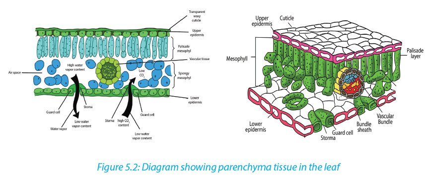
Adaptations of parenchyma for its function
–– Parenchyma tissues are made of unspecialized cells with variety of functions:
–– Parenchyma can become specialized to carry out specific functions e.g. mesophyll has cells with many chloroplasts, and aerenchyma which has air spaces. All of these adaptations help in photosynthesis and gas exchange.
–– They have isodiametric cells and function as packing tissue and storage tissue.
–– Cells are loosely packed with many large intercellular spaces. This permits diffusion of gases.
–– They have thin cellulose cell wall which is permeable so that it permits passage of materials.
–– The walls are transparent and permit entry of light in photosynthesis cells.
–– Large cells with large vacuoles provides space for storage of substances, where the entry of water causes vacuole to expand and cells become turgid
–– Leucoplasts act as storage of starch while chromoplasts present in some cells
e.g. in petals attract insects for pollination.5.1.6. Collenchyma tissues
Their cells are elongated with irregularly thickened cell walls that provide structural support, particularly in growing shoots and leaves. Their thick cell walls are composed of cellulose and pectin. These cells are often found under the epidermis, or the outer layer of cells in young stems and in leaf veins.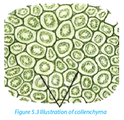
5.1.7. Sclerenchyma tissues
Sclerenchyma is found in hard parts of the plant body. They are very common in roots, stems, leaves and petioles. They may be present in patches, groups or layers. The cells of the sclerenchyma are dead, they are elongated, narrow, and thick walled and lignified. They are pointed at both ends where it gives strength, rigidity and flexibility to the plant body. They consist of fibres and sclereids. Fibres are long,
narrow, thick and liquefied cells usually tapering at both ends. Sclereids cells are normally short with very thick walls, irregular and not tapering at the ends.
Table 5.1: Comparison between collenchyma and sclerenchyma tissues
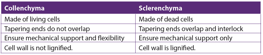
5.1.8. Vascular tissues
The vascular tissue system consists of two kinds of conducting tissues: the xylem responsible for conduction of water and dissolved mineral nutrients, and the phloem responsible for conduction of elaborated food.a. Xylem
The xylem tissues are made of dead cells which have the cell walls removed at the end of the cells, forming tubes through which the water and dissolved mineral ions can flow. Xylem vessels are involved in the movement of water through a plant - from its roots to its leaves via the stem. During this process water is absorbed from the soil through root hair cells, moves by osmosis from root cell to root cell until it
reaches the xylem, and finally it is transported through the xylem vessels up the stem and then to the leaves.
Xylem vessels are hollow tubes or lumen with a thick strengthened cellulose cell wall. The hollow tubes act like pipes allowing water and dissolved minerals to flow through them. They develop from cylindrical cells arranged end to end, in which the cytoplasm dies and the cell walls between adjoining cells breaks down leaving a dead empty tube. The cell walls in xylem vessels contain a substance called lignin
which strengthens the cells and gives structural support.
b. Phloem
Phloem vessels are involved in translocation of elaborated substances. Dissolved sugars, produced during photosynthesis, and other soluble food molecules are moved from the leaves to growing tissues such as the tips of the roots and shoots and storage tissues such as in the roots. In contrast to xylem, phloem consists of columns of living cells. The cell walls of these cells do not completely break down,
but instead form small holes at the ends of the cell. The ends of the cell are referred to as sieve plates. The connection of phloem cells effectively forms a tube which allows dissolved sugars to be transported.Phloem tubes carry food substances like sugar and amino acids produced in leaves during photosynthesis to every part of the plant. The movement of food substances through the plant is called translocation.
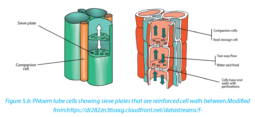
Table 5.2: Comparison between Xylem and Phloem tissues
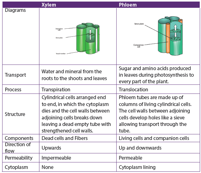

Self-assessment 5.1
1. State where in a flowering plant you would find:
a. Lateral meristem
b. Intercalary meristem
c. Apical meristem
2. Give characteristics of meristematic cells.
3. What do you understand by the following terms?
a. Differentiation
b. Cambium
c. Wood
d. Meristem
4. Differentiate between Collenchyma and sclerenchyma
5. State the main structures (components) that make up a xylem and phloem tissues.
6. Explain how the structure of Parenchyma and Xylem tissues are suitable to their functions.
7. The diagram below shows a longitudinal section of two cells of phloem tissue in a plant stem.
a. Name the cells labelled A and B on the diagram.
b. State the function of phloem in a plant.5.2 Animal tissues
Activity 5.2.1
Conduct a research by using different sources of information to find out the structures and the main functions of the following four groups of animal tissues: epithelial, connective, muscular and nervous tissues. There are four basic types of animal tissues such as epithelial tissue, muscle tissue, nervous tissue, and connective tissue.5.2.1. Epithelial tissue
Epithelial tissue consists of closely packed cells arranged in single or multilayered sheets. It is made up of layers of tightly packed cells that form the external surfaces of the body and cover the outer and the inner surfaces of the organs. Some are specialized to form glandular tissues (glands). The epithelium lining the inside of the heart, blood vessels and lymph vessels is referred to as endothelium. Two criteria for classifying epithelia are: the number of cell layers and the shape of cells on the free surface. The following are the types of epithelium tissues:a. Simple cuboidal epithelium
This is a tissue with cells that are cubical in shape. Cuboidal cells are specialized for secretion and they make up the epithelia of kidney tubules and many glands including salivary glands, and thyroid gland.
b. Simple squamous epithelium
It is thin,leakyand functions in the exchange of material by diffusion. This type of epithelium lines blood vessels and the air sacs of lungs, where diffusion of nutrients and gases is critical.
c. Simple columnar epithelium
These are columnar in shape with free surface containing extensions of micro villi. It lines the intestines. This epithelium secretes digestive juices for the final stages of digestion and absorbs nutrients to blood stream.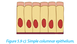
d. Pseudo-stratified ciliated columnar epithelium
It forms a mucous membrane that lines the nasal passages of many vertebrates. The beating cilia move the film of mucus along the surface.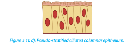
e. Stratified squamous epithelium
Itv regenerates rapidly by cell division near the basal lamina. The new cells are pushed outward to , replacecells that are sloughed off. This epithelium is commonly found on surfaces subject to abrasion, such as the outer skin and lining of the esophagus, anus, and vagina.
Figure 5.11 e) Stratified squamous epithelium.
f. Transitional epithelium
In this type of stratified epithelium, the surface cells change their shape from round to squamous. Transitional epithelium lines urinary bladder. When the bladder is empty, the surface cells are rounded. As the bladder fills urine, these cells become flattened. Transitional epithelium enables the bladder to fill and stretch without tearing the lining.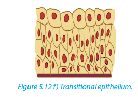
g. Stratified columnar epithelium
It is a rare type of the epithelial tissue composed of column shaped cells arranged in multiple layers. They are found in the conjunctiva or the eye, in parts of the pharynx, anus, uterus, the male urethra and vas deferens.
h. Stratified cuboidal epithelium
It is a type of epithelial tissue composed of multiple layers of cube-shaped cells. Only the most superficial layer is made up of cuboidal cells and the other layers can be cells of other type. It has several locations in the body including sweat gland ducts, egg-producing vesicles and ovaries.
5.2.2. Main characteristics of epithelial tissues
a. Polarity
All epithelia have a free surface and a lower attached basal surface that differ in structure and function. For this reason, epithelium is described as showing polarity.b. Supported by connective tissue
All epithelia are supported by connective tissue. For instance, deep to the basal lamina is reticular lamina, anextracellular material containing collagen protein fiber which forms the basement membrane. The basement membrane reinforces the epithelium and helps it to resist stretching and tearing.
c. They are avascular; have no blood vessel in them. Nutrients and gases are supplied by blood through the connective tissue by simple diffusionc. Regeneration
Epithelium have a high regenerative capacity and can reproduce rapidly as long as they receive adequate nutrition.Functions of epithelium
–– Epithelium forms a protective layer: The epithelium of the skin protects the body from mechanical damage, entry of pathogens, ultraviolet rays and dehydration. Epithelium lining the respiratory air passages secretes mucus which traps inhaled dust particles and microbes.
–– The ciliated epithelium cells have cilia that propel the mucus and trapped particles to the throat.
–– Glandular tissues secrete the digestive enzymes, hormones, mucus, sweat and sebum.
–– Acts as a barrier and regulates movement of substances across kidney
–– Some epithelial cells can divide mitotically producing new cells to replace damaged or dead cells.
–– Some epithelial cells such as taste buds and retina cells are specialized to form sensory receptors.5.2.3 Muscular tissues
Muscle tissues consist of elongated cells held together by connective tissue. Muscle cells are highly specialized in that they are able to shorten to a half or even a third of their resting length by the process of contraction. The contraction is caused by two types of fibrous proteins: myosin and actin.Muscles in the body provide the necessary force for the motion and they convert chemical energy into kinetic or mechanical energy. There are three types of muscle tissue:
–– Smooth muscle which is found in the inner linings of organs;
–– Skeletal or striated muscle, which is attached to bone and helps in movement
of the body;
–– Cardiac muscle which is found only in the heart.
Smooth and cardiac muscles are involuntary muscles whereas skeletal muscles are called voluntary muscles because they are under voluntary (conscious) control.a. Smooth Muscle
Smooth muscle is also called unstriated, unstriped, involuntary or visceral muscle.
It is found in the walls of the hollow internal organs such as blood vessels, intestinal tract, urinary bladder, and uterus. Smooth muscles have the following features;
–– It is under control of the autonomic nervous system; they cannot be controlled consciously, so they are also called involuntarily muscle.They do not have striations.
–– Smooth muscle cells contract slowly and rhythmically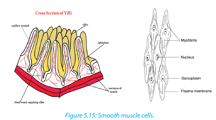
b. Cardiac tissue
Cardiac tissue (figure 5.10 a) is found in the walls of the heart and it is under control of the autonomic nervous system. Cardiac muscle has the flowing basic features.
–– It contracts and relaxes continuously.
–– It is branched and connected to other cardiac muscle fibers through intercalateddiscs(Figure 5.16 b), which are reinforced membranes that hold the cells together during contractions. These interconnections or intercalated discs between the fibers ensure a rapid and uniform spread of excitation throughout the wall of the heart which in turn ensures a synchronous contraction.
–– They are myogenic (their contraction originate from within the heart itself ).
c. Skeletal Muscle
Skeletal muscle is also called striated, striped, or voluntary. They are attached to bone, and are responsible for body movements and body posture. There are approximately 639 skeletal muscles in the human body.
Characteristics of skeletal muscles:
–– They are under control of voluntary nervous system–– They are attached to bone and this is the reason why they are called skeletal muscles.
–– They are made of elongated and cylindrical muscle fibres
–– They appear under microscope to have alternate light and dark bonds and this is why they are called striated muscles.
–– Their muscle fibres are multinucleated (many nuclei per cell)
–– These muscle cells also contain light and dark stripes called striations
General functions of muscle
The main function of muscle is its contribution to motion, where body movements such as walking, breathing, and speaking, as well as movements associated with digestion and the flow of fluids take place. Muscles contribute to the heat production, maintenance of posture and body support and communication through facial expression, writing and speech.5.2.4. Nervous tissue
Nervous tissue is composed principally of densely packed cells called the nerve cells (neurons) that together form the nervous system including the brain and spinal cord. Neurons are specialized for transmitting electrical nerve impulses.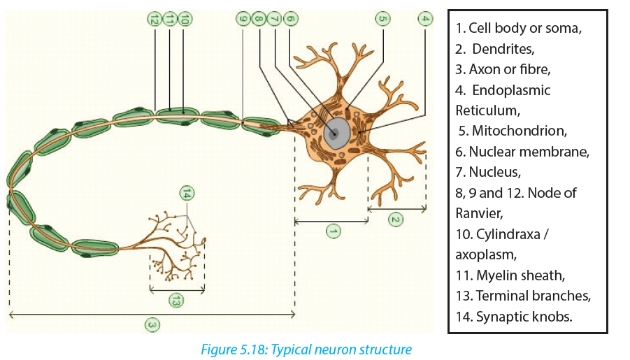
A typical neuron has three main parts: Cell body, dentrites and axon.
a. The cell body or soma
–– It is the main part from which, extensions derive (Axon and Dendron).
–– It is made of a great spherical nucleus, granular cytoplasm and controls
all nerve cell activities.b. Dendrites (Dendron when single): small branches attached to the cell body and receive nerve impulse from other neurons
c. Axon or cylindrax:
–– It is the thinner nerve fibre that carries messages away from the cell body and can be as long as 1 m. In some neurones, the axons have a fatty myelin sheath formed by Schwann cells which wrap themselves around the axon to increase the speed of impulse transmission.5.2.5. Connective tissues
Connective tissue is made up of many different types of cells that are all involved in structure and support of the body. Bone, blood, fat, and cartilage are all connective tissues. Connective tissues can be densely packed together, as bone cells are or loosely packed, as adipose tissue (fat cells) are. A connective tissue is made up of a variety of cells embedded in a large amount of intracellular substance called matrix
and fibers which are non-living products of the cells.a. Common functions of connecting tissues:
–– Connective tissues protect and support the body and internal organs.
–– They act as connecting systems, binding all other tissues together.
–– They also form surrounding sheaths to separate the various organs.b. Cells of connective tissue
The specialized cells of the various connective tissues produce the extracellular matrix. The names of the cells end with suffixes that identify the cell functions as blasts, cytes, or clasts. Blasts create the matrix, cytes maintain it, and clasts break it down for remodeling. For example: Fibroblasts are cells that form fibrous connective tissue and fibrocytes maintain it, chondroblasts form cartilage and chondrocytes
maintain it, and osteoblasts form bone, osteocytes maintain it, and osteoclasts break it downAdipose, or fat cells, also called Adipocytes, contain large amounts of lipid. The lipid pushes the rest of the cell contents to the periphery, so that each cell appears to contain a large and centrally located lipid droplet with a thin layer of cytoplasm around it. Adipose cells are rare in some connective tissue types like cartilage but they are abundant in others like loose connective tissue, and they are predominant in adipose tissue.
Mast cells are commonly found beneath membranes in loose connective tissue and along small blood vessels of organs. They contain chemicals such as heparin, histamine and proteolytic enzymes. These substances are released in response to injury such as trauma and infection and play important roles in inflammation.
White blood cells continuously move from blood vessels into connective tissues. The rate of movement increases dramatically in response to injury or infection. In addition, accumulations of lymphocytes, a type of white blood cell, are common in some connective tissues, such as in the connective tissue beneath the epithelial lining of certain parts of the digestive system.
Macrophages are found in some connective tissue types. They are derived from monocytes, a white blood cell type. Macrophages are either fixed and do not move through the connective tissue in which they are found or are wandering macrophages and move by amoeboid movement through the connective tissue. Macrophages phagocyte foreign or injured cells, and they play a major role in providing protection against infections.
Note that there are three structural major components of the extracellular matrix of connective tissue such as fluid, ground substance consisting of non-fibrous protein and other molecules and protein fibers. The structure of the matrix gives connective tissue types most of their functional characteristics, such as the ability of bones and cartilage to bear weight, tendons and ligaments to withstand tension, and dermis of the skin to withstand punctures, abrasions, and other abuses.
c. Fiber connective tissues
Another type of connective tissues consists of fibers. Fibers are of different types including:
–– Connective tissue fibers: which are made of protein and are of three kinds:
collagenous, elastic and reticular fibers.
–– Collagenous fibers: These provide strength combined with flexibility. They are made up of collagen, probably the most abundant protein in the animal kingdom.
–– Elastic fibers: These are easily stretched but are also resilient, snapping back to their original length when tension is released. Shaped as long threads, elastic fibers are made of a protein called elastin.
–– Reticular fibers: These are thin collagen fibers coated with glycoprotein. They are very short, thin fibers that branch to form a network and appear different microscopically from other collagen fibers. Reticular fibers are not as strong as most collagen fibers, but networks of reticular fibers fill space between tissues and organs.d. Loose connective tissue
This is also called areola connective tissue and is the most widespread connective tissue in all animal tissues. It binds epithelial tissues to underlying tissues and functions as packing material, holding organs in place. Loose Connective tissue has the following main components;–– Fibers: collagen, elastic and reticular.
–– Cells; fibroblasts and macrophages. Fibroblasts secrete the protein ingredients of the extracellular fibers. Macrophages are cells that roam the maze of fibers, engulfing both foreign particles and the debris of dead cells by phagocytosis.e. Fibrous connective tissue
Fibrous Connective tissue is dense with collagenous fibers. The fibers form parallel bundles, which maximize non-elastic strength. Fibrous Connective tissue is found in tendons, which attach muscles to bones, and ligaments, which connect bones at joint.f. Adipose tissue
Adipose tissue is a specialized form of loose connective tissue that stores fats in adipose cells distributed throughout its matrix. Adipose tissue consists of adipocytes, or fat cells, which contain large amounts of lipid. Unlike other connective tissue types, adipose tissue is composed of large cells and a small amount of extracellular matrix that consists of loosely arranged collagen and reticular fibers with some scattered elastic fibers. Blood vessels form a network in the extracellular matrix. The fat cells
are usually arranged in clusters or lobules separated from one another by loose connective tissue. Adipose tissue functions as:–– An insulator against heat loss
–– A protective tissue to delicate internal organs
–– A site of energy storage in the form of fat.g. Bone and Cartilage tissue
Cartilage has an abundance of collagenous fibers embedded in a rubbery matrix made of a protein-carbohydrate complex called chondroitin sulfate. Cartilage is composed of specialized cells, called chondrocytes, surrounded by a gelatinous matrix of collagen, a tough protein. The cartilage surface is covered by a membrane known as the perichondrium. There are three types of cartilage (hyaline cartilage, yellow elastic and white fibrous cartilage.)–– Hyaline cartilage is semi-transparent and is often stained light blue or pink in tissue sections. It is extremely very strong but very flexible and elastic. Hyaline cartilage occurs in the trachea, larynx, tip of the nose, connection between the ribs and the breastbone; and at the ends of bones where they form joints. It also forms much of the fetal skeleton.
–– Elastic cartilage is similar to hyaline cartilage, but in addition to the collagenous fibers.The matrix of the elastic cartilage also contains an abundant network of branched elastic fibers. This type of cartilage is found in the lobe of the ears, the epiglottis and in the parts of the larynx. They provide flexibility and elastic support.
–– Fibro-cartilage(White fibrous cartilage) is an extremely tough tissue. It is found as discs between the vertebrae, bones, anterior joint between the two halves of pelvic girdle and at points where tendons inserted on bones near hyaline cartilage. It resists compression and absorbs shock in some joints.Bone tissue
This is a firmer and denser material that has the following features:
–– Hard and compact
–– Has many collagen fibres
–– Its matrix has inorganic salts such is calcium carbonate and calcium phosphate
–– Has few cells located in the lacunae in the matrix
–– Has osteoblasts as mature and non-dividing cells
–– Have a harversian canal
–– Consists of irregular cylinder with layer of matrix call lamellae The following are the main functions of bone tissue:
–– Structural support of the body
–– Protection of internal organs, heart and lungs.
–– Attachment of the muscles to effect movement
–– Production of blood cellsh. Blood tissue
Blood is a flowing made up of particles suspended in a fluid composed of fluid called plasma, and several kinds of cells. Within the blood plasma, there are erythrocytes (red blood cells), leukocytes (white blood cells), thrombocytes (platelets) and other substances. Blood performs the following important functions:Transport
–– Blood transports absorbed substances such as glucose, amino acids, mineral ions and vitamins from the small intestine.
–– Blood transports the respiratory gases (Oxygen and Carbon dioxide).
–– Blood transports the excretory wastes such as urea, uric acid to excretory organs to be removed out of the body.
–– Blood transports hormones e.g. insulin from pancreas to the liver where it is stored.Homeostasis
Na+ affects the water potential of the blood and regulates the diffusion of water between blood and tissues. Hydrogen carbonates help to maintain the pH of the blood.Protection
–– Leucocytes such as neutrophils and macrophages engulf pathogens e.g. bacteria
–– B-lymphocytes produce antibodies to destroy pathogens or to neutralize toxins.
–– T-lymphocytes destroy infected cells.
–– Platelets, fibrinogen and prothrombin play an important role in blood clotting to reduce blood loss and the entry of pathogens.Activity 5.2.3
You are provided with photomicrographs or slides of different plant and animal tissue. Study them carefully and answer questions that follow. Identify the different tissues provided and where they are located. One of the images is a blood smear. Draw a well labeled diagram of this tissue5.3 Levels of organization: cell, tissue, organ and system
Activity 5.3
Visit a classroom block, administration block or any building in school which is constructed with bricks and use it to answer the following questions.
1. What is the smallest unit or component of the classroom block?
2. How are bricks arranged?
3. Do you think the brick has other smaller particles in it?
4. How many bricks does a classroom block have?
5. How are walls, classrooms, washrooms and other apartments of the block formed?
6. Arrange the following in their ascending order of size (from the smallest to the largest); whole block, wall, a brick, a room, course (a line of bricks).
7. Relate the above arrangement of a building to levels of organization in multicellular organisms, beginning with a cell and ending with an organism The human body is organized into structural and functional levels of increasing complexity. Each higher level incorporates the structures and functions of the previous level. The simplest is the cells, organized into tissues, organs, and organ systems. All of the levels of organization of the human body are represented in the following figure.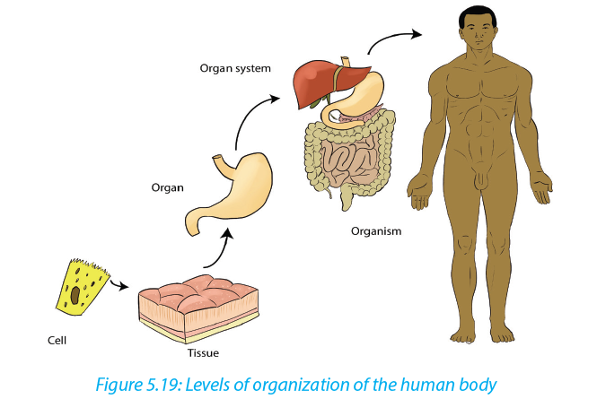
5.3.1 Cells
The smallest structural and functional living units of living things are cells. There are many different types of human cells, though they all have certain similarities. Each type of cell is made of chemicals and carries out specific chemical reactions.5.3.2 Tissues
A tissue is a group of cells with similar structure and function. There are four groups of tissues (Epithelial tissues, Connective tissues, Muscle tissues, Nerve tissue)5.3.3 Organs
An organ is a group of tissues precisely arranged so as to accomplish specific functions. Examples of organs are the kidneys, individual bones, the liver, lungs, and stomach. The kidneys contain several kinds of epithelial or surface tissues, for their work of absorption. The stomach is lined with epithelial tissue that secretes gastric juice for digestion. Smooth muscle tissue in the wall of the stomach contracts to mix food with gastric juice and propel it to the small intestine. Nerve tissue carries impulses that increase or decrease the contractions of the stomach.5.3.4 Organ systems
An organ system is a group of organs that all contribute to a particular function. Examples are the urinary system, digestive system, and respiratory system. For example, the urinary system consists of the kidneys, ureters, urinary bladder, and urethra. These organs all contribute to the formation and elimination of urine.
The Human body has 11 organ systems: circulatory, digestive, endocrine, and
excretory (urinary), the lymphatic, integumentary, muscular, nervous, reproductive,
respiratory, and skeletal systems.
Table 5.4: Major organ systems of the human body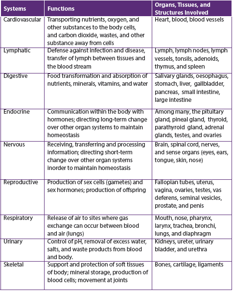
Self-assessment 5.3
1. Answer by true or false
a. Organic chemicals are often very complex and always contain the element carbon only.
b. A tissue is a group of cells with similar structure and function.
c. Integumentary organ system plays the role in protection of the human
body from injury and fluid loss.
d. An organ system is a group of organs that all contribute to a particular function.
2. Explain why the cell as level of organization of human body is said to be:
a. Basic unit of human body
b. Structural unit of human body
c. Functional unit of human body5.4 Advantages and disadvantages of being Unicellular or Multicellular
Activity 5.4
Discuss the advantages and disadvantages of an organism being unicellular or Multicellular5.4.1 Advantages of unicellular organisms
–– Unicellular organisms need fewer nutrients and can survive in unfavorable conditions.
–– Some of the organisms can generate energy through photosynthesis.
–– Sometimes different bacteria work together to work to their advantages.
–– Unicellular organisms can multiply quickly and have less energy/resource demands.5.4.2. Disadvantages of unicellular organisms
Unicellular organisms only have one cell that is used to function their entire being. This is a disadvantage compared to multicellular organisms, which have many cells and function more easily and properly.5.4.3 Advantages of a multicellular state of an organism
–– Multicellular organism usually has a wider range of functions because of the aggregation of different types of cells.
–– Multicellular organisms have many more necessities and can only survive in certain conditions.
–– Multicellular organisms such as animals are unable to make their own food so
they survive by eating living things such as vegetables, fruits, and meat. They can also eat things that are produced by other living things such as eggs, milk, and honey.Self-assessment 5.4
1. Give the advantages and disadvantages of being Unicellular organisms.
2. Describe how unicellular organisms perform their functions.End of unit assessment 5
1. Which type of tissue forms glands?
a. Epithelial
b. Connective
c. Nervous
d. Muscles
2. What are the four types of animal tissues?
a. Epithelial, squamous, muscular, connective
b. Epithelial, connective, muscular, cardiac
c. Connective, muscular, epithelial, nervous
d. Cuboidal, ciliated, glandular, columnar
3. Which type of the tissues form glands
a. Epithelial
b. Connective
c. Nervous
d. Muscle
4. Describe how epithelial tissues have adapted to their functions
5. Describe the three main functions of the bloo
6. Complete the following table by filling in the examples of the respective tissues:
UNIT 6: TESTING FOR BIOLOGICAL MOLECULES
Key unit competence
Test for biological molecules in a variety of contexts, such as identifying the contentsof mixtures of molecules and to follow the activity of digestive enzymesLearning objectives
By the end of this unit, I should be able to:
–– Write out procedures in the identification of biological molecules
–– Explain the importance of the reagents used in the identification of biological molecules.
–– Carry out tests for the identification of biological molecules
–– Compare reducing and non-reducing sugars
–– Appreciate the importance of identification of food values in the food industry and in processing and packaging.
–– Show resilience making observations on color changes during food testsIntroductory activity
You are given solutions containing different food stuffs including maize flour, vegetable cooking oil, and egg white sugar cane liquid and passion fruit. Using prior knowledge of biological molecules to suggest the type of biological molecule in each one of them. Suggest the chemical tests used to identify each
of the molecules.6.1 Test for carbohydrates
Activity 6.1
Materials required:
Starch powder, Irish potatoes juice, prepared porridge, Iodine solution, beakers, droppers, source of heat and test tubesa. Test for starch
Procedure
–– Mix 1g of starch powder with 100ml of water
–– Boil the mixture while stirring; then cool the solution
–– Boil the mixture while stirring; then cool the solution
–– Put 2ml of starch solution in a test tube labeled 1, 2ml of Irish potato juice in a test tube labeled 2 and 2ml of prepared porridge in a test tube labeled 3
–– In each test tube put 2 drops of Iodine solution and shake
–– Record your observation and draw a conclusion
b. Test for reducing sugar
Requirements
Glucose powder, beaker and test tube, Benedict solution, Bunsen burner, droppers Procedures
–– In the beaker mix 1cm3 of water and 1g of glucose powder.
–– Pour the prepared solution of glucose in a test tube and
–– Add 2ml of benedict’s solution and heat
–– Record your observation.
Biological molecules are grouped into organic molecules including carbohydrates, proteins, lipids, nucleic acids and vitamins. They also contain inorganic molecules such as minerals and water. The first four organic molecules are called macromolecules because they are required in organism in large quantity. Carbohydrates including starch, reducing and non-reducing sugars appear in this category and are the main energy producers in the organisms. Others, including lipids and proteins are needed for building organisms while vitamins protect the organisms against diseases. We need to ensure that what we take from diet have all required biological molecules.a. Test for starch.
Carbohydrates such as starch are tested by mixing a sample with 2-4 drops of iodine or Lugol’s solution. If the sample contains starch the solution will turn from a yellowbrown color to a dark purple/dark blue (Figure 6.1). The color change is due to a chemical reaction between the large carbohydrate molecule and the iodine ions. If the sample does not contain starch the solution remains yellow-brown.
b. Testing for reducing and non-reducing sugar
The presence of reducing sugar can be tested by using benedict reagent. Benedict solution has copper ions that have a light blue color. When this solution is heated in the presence of simple reducing sugars such as glucose, the blue color of copper ions changes from a light green color to rusty orange-brown color (Figure 6.2).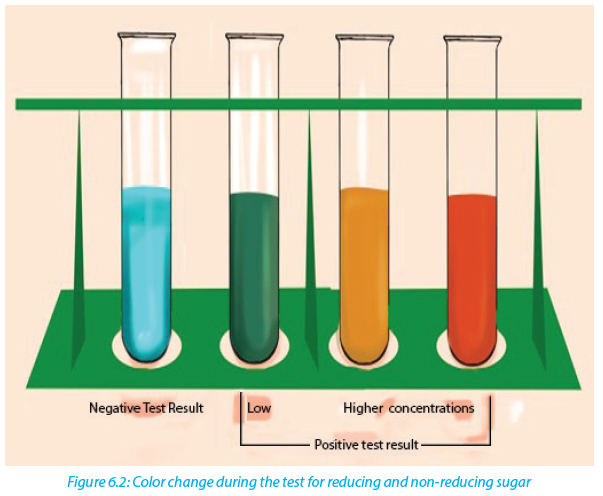
If the color of Benedict reagent persists, the sugar tested is not a reducing sugar. Note that there is no special reagent to test for non-reducing sugar, but by the addition of HCl, non-reducing sugars can be hydrolyzed to reducing sugars. To test the presence of reducing sugars, a solution of sodium hydroxide is needed to neutralize the acidity because Benedict reagent works better in neutral solution
Self-assessment 6.1
A student prepared carbohydrate solution labeled C. Perform the following experiment to confirm whether C1 is starch, reducing sugar, or non-reducing sugar.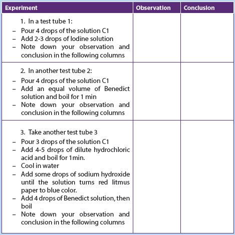
a. Is this solution a carbohydrate?
b. Specify the role of Hydrochloric acid and sodium hydroxide used at stage 3 in the table above.6.2 Test for proteins
Activity 6.2
Materials required
Milk, eggs, soybeans, test tubes, beakers, mortar for crushing beans, 1% NaOH or
1% KOH solution, 0.1M of CuSO4 solution and Millon’s reagent.Procedure
–– Extract the white fluid from an egg
–– Prepare an extra of soya bean and 10ml of fresh milk
–– Put 2ml of albumen solution in a test tube labelled A1 and 2ml in A2
–– Put 2ml of milk solution in a test tube labelled B1 and 2ml in B2
–– Put 2ml of soya bean solution in a test tube labelled C1 and 2ml in C2
–– Put 1ml of KOH or NaOH solution in each of the test tubes A1, B1, and C1. Shake the mixture and add 1ml of CuSO4 solution in each (A1, B1, and C1)
test tube
–– Put 1ml of Millon’s reagent in each of test tubes (A2, B2, and C2). Shake the
mixture and thereafter boil the three test tubes (A2, B2, and C2).
The Biuret reagent is used to test for the presence of proteins. It contains copper ions with blue characteristic color. During the copper ions react with protein molecules and causes the biuret solution to turn from a light blue color to purple if proteins are present.
The test can also be done by using Millon’s reagent, which in the presence of proteins, the Millon reagent changes from colorless to pink.
Self-assessment 6.2
1. You are provided with the sample of the substance M and A. Carry out the following experiments and complete the table below.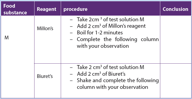
2. Carry out the same experiment using the substance A and compare your findings with M.
3. Which of the substance A and M contain proteins?6.3 Test for lipids
Activity 6.3
Materials required - Olive oil, test tubes, ethanol, water, sudan III solutionProcedure:
Use olive oil to carry out the following experiments Add 2 cm3of olive oil in the test tube:
–– Add 5 cm3 of ethanol followed by 5 drops of water.
–– Shake the mixture and record your observation.
–– To another test tube containing 2 cm3 of olive oil:
–– add 5 drops of Sudan III solution
–– Shake thoroughly and examine the mixture in the test tube after few minute and record your observations The presence of lipids can be determined by using Sudan III indicators, which are fat-loving
molecules that are colored. During the test for a solution containing lipids, two results are likely to be found: there is either the separation of layers indicating the levels of water and lipid, or the dye migrates toward one of the layers. If the mixtureis composed of water, the conclusion is that the lipids are not present. In this case, the Sudan III indicator will form small micelles/droplets and disperse throughout the solution. A positive result indicates the lipid layers sitting on top of the water layer with a red-orange color. When using ethanol for testing lipids the presence of the color changes from colorless to milky (emulsion test).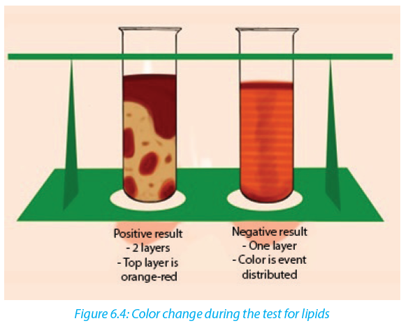
Self-assessment 6.3
You are provided with a solution X. Use Sudan III indicator to test the presence of lipids in the solution X.6.4 Test for vitamin C (Ascorbic Acid).
Activity 6.4
Squeeze the orange fruits to extract the juice and carry out the following test.
Which of the two solutions give a positive solution for DCPIP?
Vitamin C is tested by using DCPIP (Dichlophenol Indophenol). Its positive (presence of vitamin C) test decolorizes DCPIP, while the negative (absence of vitamin C) test is indicated by the persistence blue color of DCPIP.
Self-assessment 6.4
In this experiment you are to press a tomato fruit (s) to get juice out of it. Use the juice to carry out the test for vitamin C Draw a table of results that includes the procedure, observation and conclusion.End of unit assessment 6
1. Biological molecules are divided into:
a. Organic molecules and inorganic molecules
b. Carbohydrates and starch
c. Lipids, carbohydrates and water
d. Carbohydrates, food and potatoes
2. Name the reagents that are used to test for the following food substances
a. Lipids
b. Starch
c. Reducing sugar
3. You are provided with the following specimen:
Specimen A: Sorghum
Specimen B: Irish potatoes
Specimen C: Oranges
Specimen D: Sunflower seeds
a. Carry out chemical tests to determine the composition of the above seed to
tell whether they are composed of proteins, fats, starch or vitamin C.
b. Draw the table of used reagent, procedure and observation in (a)
4. Some drops of fresh pineapple juice are added drop by drop to DCPIP solution.
The deep blue color of the DCPIP quickly fades.
a. Explain why the blue colour disappeared?
b. What is the importance of this food substance to the human body?
5. The result of food tests on unknown sample are shown below. Copy and complete the table to show the conclusions which could be drawn from these tests.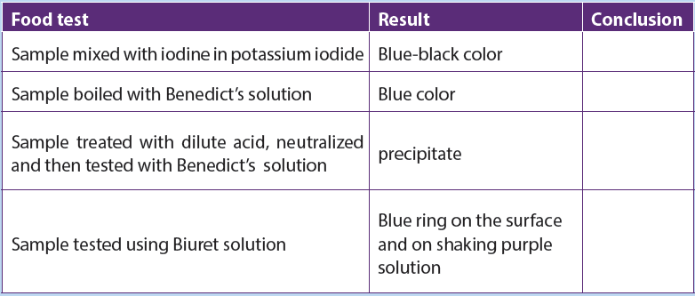
6. This is a practical question to be conducted using provided materials and reagents to determine the food nutrients in each solution: You are provided with the following solutions, A (sucrose 0.5%), B (1%starch), C (dilute hydrochloric acid) and D (sodium hydroxide) and 6 test tubes labeled 1 to 6. Use the reagents provided to determine the chemical nature of the substance present in the solutions. Indicate your observations and conclusions in the table below:

a. Why was it necessary to boil solutions A and B with solution C in test (3) and (6)?
b. Why was solution D added to test tubes 3 and 6?UNIT 7: CARBOHYDRATES AND LIPIDS
Key Unit Competences
Explain the important roles of carbohydrates and lipids in the provision and storage of energy and for a variety of other functions.Learning objectives
By the end of this unit, I should be able to:
–– State the roles of carbohydrates and lipids.
–– Recall the elements that make up carbohydrates and lipids.
–– Explain the proportion of hydrogen in carbohydrates and lipids and relate this to the amount of energy released when oxidized.
–– Define the terms monomer, polymer, macromolecule, monosaccharide, disaccharide and polysaccharide.
–– Describe the ring forms of α-glucose and β-glucose structure.
–– Explain the formation of glycosidic bonds.
–– Describe the structure of phospholipids and relate to their functions in living organisms.
–– Describe the molecular structure and formation of triglycerides and phospholipids, and give their functions in living organisms.
–– Demonstrate that phospholipids have a hydrophilic head and hydrophobic tails using a heterogeneous mixture made up of water and cooking oil.
–– Interpret the charts and illustrations of molecular structure and the formation of maltose and triglycerides.
–– Demonstrate through a process of combustion that sugars and lipids are biological fuel
–– Differentiate between starch and cellulose.
–– Appreciate the importance of carbohydrates and lipids in organisms.
–– Be aware of the other roles of lipids in the formation of soap and with carbohydrates and syrups in medicineIntroductory activity
1. In the previous unit (test for biological molecules), we tested carbohydrates, starch, reducing sugar, lipids, proteins, and vitamins. Where do you classify monosaccharide, disaccharides and polysaccharides in the above tested biochemical compounds?
2. Sometimes people say that fatty persons do not feel cold. What could be the reasons?
7.1 Classes of monomers
Activity 7.1
1. Give the description of the term monomer
2. Where can we find monomers?
3. What is the biological importance of monomers?
A monomer is a molecule that can combine with others of the same kind to form a polymer. A polymer is a large molecule or macromolecule composed of many repeated sub-units (monomers). Because of their broad range of properties, both synthetic and natural polymers play essential and ubiquitous roles in everyday life. Polymers make up many of the materials in living organisms including proteins,
cellulose, and nucleic acids. Glucose molecules for example, are monomers that combine to form the polymer cellulose. The examples of monomers are summarized in the table 7.1.Table 7.1: Biological molecules and their monomers

Carbohydrates comprise a large group of organic compounds which contain carbon, hydrogen and oxygen. The word carbohydrate suggests that these organic compounds are hydrates of carbon. Their general formula is Cx (H2O) y. In carbohydrates the ration hydrogen-oxygen is usually 2:1. Carbohydrates are divided into three groups including the monosaccharide (single sugars), disaccharides (double sugars) and polysaccharides (many sugars). The most common monosaccharide of carbohydrates is glucose with molecular formula C6H12O6.
Self-assessment 7.1
1. What are some examples of polymers and monomers?
2. How are monomers, polymers and macromolecules related?7.2 Ring form of α-glucose and β-glucose
Monosaccharides are group of sweet and solubleActivity7.2
1. Based on the knowledge acquired during the lesson of monomers and further information from books and internet:
a. What are the examples of monosaccharide?
b. Give the molecular formula of each of the monosaccharide stated above
c. Use the books to illustrate the structural formula of each of the monosaccharide stated abovecrystalline molecules of relatively low molecular mass. They are named with the suffix –ose. The general formula of a monosaccharide is (CH2O) n, with n the number of carbon atoms. The simplest monosaccharide has n=3 and it is a triose sugar. When n = 5, this is a pentose sugar, and when n = 6, this is a hexose sugar. The two common pentose sugars are ribose and deoxyribose, while the most known hexose is glucose. Its molecular formula is C6H12O6. It is the most important simple sugar in
human metabolism called simple sugar or monosaccharide because it is one of the smallest units which has the characteristics of this class of carbohydrates.Monosaccharides can exist as isomers. The isomer is defined as each of two or more compounds with the same formula but a different arrangement of atoms in the molecule and different properties. For example, glucose, fructose and galactose share the same molecular formula which is C6H12O6. However, they differ by their structural formulae as follow:

One important aspect of the structure of pentoses and hexoses is that the chain of carbon atoms is long enough to close up on itself and form a more stable ring structure. This can be illustrated using glucose as an example. When glucose forms a ring, carbon atom number 1 joins to the oxygen on carbon atom number 5 (Figure 7.2).
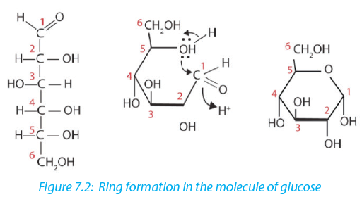
All hexoses sugars can exist as straight-chain structures but they tend to form ring structures. Glucose, fructose, galactose can exist in ring structures (Figure 7.3).

Ring monosaccharides are said to be alpha (α) if the -OH group located on carbon 1 is below the ring and beta (β) when the -OH group is above the ring. The molecule of glucose for example can exist as alpha and beta glucose denoted by α-glucose and β-glucose (Figure 7.4)
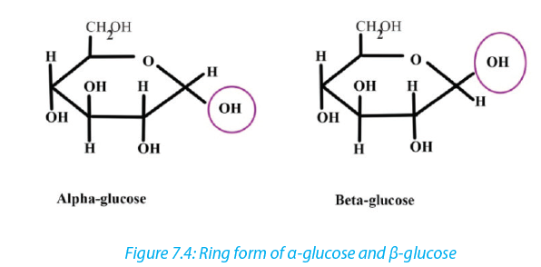
Self-assessment 7.2
1. How do we call the monosaccharide with 3, 5 and 6 carbon atoms?
2. Differentiate between α and β glucose
3. What are the properties of glucose?7.3 Formation and breakdown of glycosidic bonds
Activity 7.3
1. Monomers are joined to form polymers, use a point as a monomer to illustrate how a polymer can be formed
2. How do you call joining structures between atoms?
3. Use books or other sources to show how monosaccharide form a
disaccharide.7.3.1 Monosaccharides
Monosaccharides may combine together in pairs to give a disaccharide (doublesugar).
The union involves the loss of a single molecule of water and is therefore a condensation reaction. The bond which is formed is called a glycosidic bond. It is usually formed between carbon atom1of one monosaccharide and carbon atom 4 of the other, hence it is called a -1, 4- glycosidic bond. Any two monosaccharides may be linked together to form a disaccharide of which maltose, sucrose and lactose
are the most common.
The addition of water under suitable conditions is necessary if the disaccharide is to be split into its constituent monosaccharide. This is called hydrolysis waterbreakdown or more accurately, breakdown by water.
7.3.2 Disaccharides
These are carbohydrates made of two monosaccharides. They include maltose (glucose + glucose), sucrose or saccharose (glucose +fructose), and lactose (glucose+ galactose). The maltose is the sugar from the germinating seeds, sucrose or saccharose is the common table sugar obtained from sugarcane, while lactose is the sugar from the milk. In addition, sucrose is a non-reducing sugar.
Table 7.2: Types of disaccharides and their monomers
In maltose ring, the two ring of glucose are bonded by the -1, 4-glycosidic bond while in sucrose the glucose and fructose are bonded by -1, 2-glycosidic bond.

All the disaccharides are non-reducing sugar, except maltose which behaves in the same as a reducing sugar with benedict’s solution. All monosaccharides and disaccharides have the following characteristics: sweet taste, soluble in water and lower molecular mass.
Self-assessment 7.3
1. Write the molecular structure of sucrose
2. How is the glycosidic link is formed
3. Sucrose is formed when two monosaccharide are assembled together:
a. Name those two monosaccharides.
b. Using the ring form of these monosaccharide named above to explain and
show sucrose formation?7.3.4 Polysaccharides: starch, glycogen and cellulose
Activity 7.4
1. Based on the meaning of monosaccharide, what is a polysaccharide?
2. Classify the following compound into polysaccharide, monosaccharide and disaccharide
a. Glucose, fructose and galactose
b. Lactose, sucrose, and maltose
c. Starch, cellulose and glycogen
3. Use glucose to form any polysaccharide of your choice In the same way that two monosaccharides may combine in pairs to give a disaccharide, many monosaccharides may combine by condensation reactions to form a polysaccharide.The number of monosaccharides that combine is variable and the chain produced may be branched or unbranched. Polysaccharide are many but the most known are starch, glycogen and cellulose.
a. Starch
Starch is made up of two components: amylose and amylopectin. Amylose is a linearunbranched polymer of 200 to 1500 α-glucose units in a repeated sequence of α-1,4-glucosidic bonds. The amylose chain coils into helix held by hydrogen bonds formed between hydroxyl groups. A more compact shape is formed. The amylose helices are entangled in the branches of amylopectin to form a complex compact
three dimensional starch molecule.Amylopectin is a branched polymer of 200 to 200,000 α-glucose units per starch molecule. The linear chains of α-glucose units are held together by α-1, 4-glucosidic bonds. Branches occur at intervals of approximately 25 to 30 where α-1, 6-glucosidic bonds occur. Starch grains are found in chloroplast, potato tubers, cereals and legumes. Starch is insoluble in cold water. It is digested by salivary amylase and pancreatic amylase into maltose and the latter is hydrolyzed by maltase enzyme to form glucose. Therefore, diabetic people should avoid tubers since they are rich in starch which in turn gives glucose (Figure 7.8).
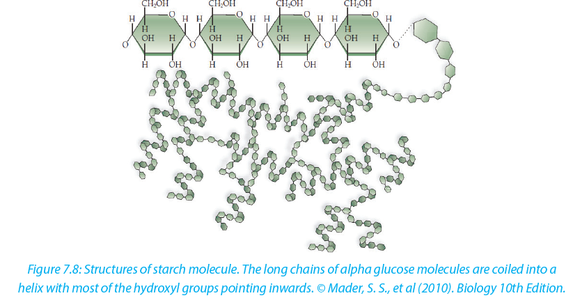
b. Glycogen
Glycogen is often called animal starch because it is a major polysaccharide storage material in animals and fungi. The brain and other tissues require constant supply of blood glucose for survival. Some tissues particularly the liver and skeletal muscles store glycogen in the form that can be rapidly mobilized to form glucose. Like starch, glycogen is made up of α-glucose and exists as granules. It is similar to amylopectin in structure but it has shorter chains (10-20 glucose unit) and is more highly branched.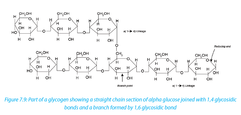
c. Cellulose
Cellulose is the structural polysaccharide in plant cell wall. It is found in vegetables and fruits but it cannot be hydrolyzed by enzymes in the human digestive system. Cellulose is composed of long unbranched chains of up to 10,000 β-glucose units linked by β-1,4-glucosidic bonds. Each β-glucose unit is related to the next by a rotation of 180 ͦ C with OH groups projecting outwards on either side of the chain.Cellulose chains run parallel to one another. Unlike amylopectin and glycogen molecules, there are no side chains (no branch) in the cellulose. This allows the linear chains to lie close together. Many H-bonds are formed between the OH groups of adjacent chains. The chains group together to form microfibrils arranged in larger bundles of macrofibrils. The fibrils give the plant cell their high tensile strength and rigidity. The layers of fibrils are permeable to water and solutes.
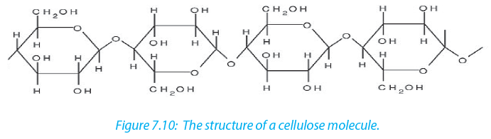
Cellulose is formed from ß - glucose units linked by 1,4 glycosidic bonds. The hydroxyl groups alternate on either side of the molecule forming straight chains giving cellulose a fibrous structure. Cellulose are strengthened further by hydrogen bonds that link adjacent chains.
d. Chitin
Chitin is one of naturally occurring Polymers. It forms a structural component of many animals such as exoskeleton in arthropods. Chitin is a polymer of glucose although in its structure a molecule of amino acid is added to each glucose. The digestion of chitin yields simple sugars and ammonia.Self-Assessment 7.4
1. What type of reaction is involved in the formation of glucose from starch?
2. Use the type of reaction above to form glucose from sucrose molecule
3. What are the 2 main components of starch? Give the difference between them7.3.5 Lipids
Activity 7.5
1. List the monomers that are present in lipids
2. Where can we find lipids?
3. Discuss the reasons why animals like pig do not like hot weather.
Lipids are a broad group of naturally occurring molecules which include fats, waxes, sterols, fat soluble vitamins (such as vitamins A, D, E and K), monoglycerides, diglycerides, Phospholipids and others. Lipids are grouped into fats which are solid at room temperature and oils which are liquid at room temperature. Lipids are made by carbon, hydrogen and oxygen, but the amount of oxygen in lipids is much smaller than in carbohydrates. Lipids are made by two components namely glycerol and fatty acids. The chemical formula for glycerol is C3H8O3 with structural formula as shown in the figure 7.11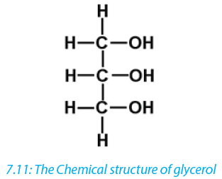
In all lipids glycerol do not show any variation while fatty acids vary. Therefore, the nature of lipid depends on the fatty acid it contains. There are two types of fatty acids: unsaturated fatty acid characterized by the chain of hydrocarbon containing one or more double and triple bonds; and saturated fatty acid characterized by the chain of hydrocarbon without any double or triple bond.
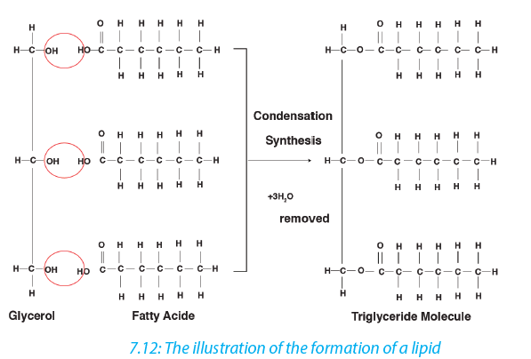 Lipids are of different types as it is summarized in the following table (Table 7.3)Table 7.3: Types of lipids, their structure, main role and features
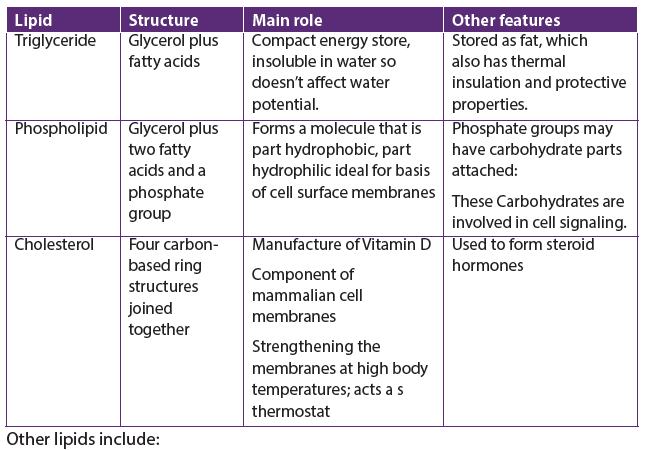 a. Waxes
Waxes are similar to triglycerides but contain fatty acids bonded to log chain alcohol rather than to glycerol. Waxes form the cuticle that protects the leaves and surfaces of insects against the loss of water
b. Steroids
A steroid is an organic compound with four rings of carbon and hydrogen atoms with various side chains. Steroids have several functions. It is a component of most animal hormones like estrogen, testosterone.General functions of lipidsLipids perform a number of functions within living organism:
–– Lipids are source of energy: due to the presence of C-H bond, lipids can generate more ATP compared to the carbohydrates of the same mass
–– Lipids are storage of energy in adipose cells forming adipose tissue in fat of organism
–– Lipids act as insulators of the organism. For example, they reduce heat loss.
Lipids also are electrical insulators around the nerve cells, the Myelin sheath
–– Lipids have a role of protection, in the cuticle of plant leaves against drying, in exposed organ like hand and knees
–– Synthesis of hormones such as steroid hormones (most of sex hormones) are made by lipids
–– Lipids are used in production of soap by saponification reactionSelf-assessment 7.6
1. Name the small units found in lipids
2. Differentiate between fats and oilsEnd of unit assessment 7
1. Write the formula of a monosaccharide with 3 atoms of carbon
2. Compare the structure of fat(triglycerides)and the phospholipids
3. Give two examples of how carbohydrates are used in the body.
4. The formula for a hexose is C6H12O6 or (CH2O)6. What would be the formula of?
a. Triose
b. Pentose
5. The general formula of a monosaccharide is (CH2O) n where n is any number between 3 and 9. What would be the formula of a pentose sugar where n is 5?
6. What type of chemical reaction would be involved in the formation of glucose from starch or glycogen?
7. Distinguish between:
a. Alpha glucose and beta glucose
b. Glycogen and cellulose
c. Amylopectin and amyloseUNIT 8: PROTEINS AND WATER
Key Unit Competence
Describe how protein structure is related to function and the role of water as a special molecule with extrardinary properties that make life possible.Learning objectivesBy the end of this unit, I should be able to:
–– Describe the structure of an amino acid and the formation and breakage of a peptide bond.
–– Describe the primary, secondary, tertiary and quaternary structure of proteins.
–– Describe the molecular structure of hemoglobin as an example of a globular protein.
–– Describe the functions with an emphasis on iron in the hemoglobin molecule.
–– Explain the effect of heat, pH and chemicals on protein structure.
–– Explain how hydrogen bonding occurs between water molecules and relate the properties of water to its roles in living organisms.
–– Devise an experiment to investigate the effect of temperature, pH and chemicals on the structure of protein.
–– Relate the structure of globular and fibrous proteins to their functions.
–– Investigate the effect of lowering temperature on water.
–– Distinguish between collagen molecules and collagen fibres
–– Appreciate the importance of globular and fibrous proteins in biological processes such as the transport of gases and providing support for tissues.
–– Express that protein structure is central to many aspects of biology, such as enzymes, antibodies and muscle contraction.
–– Acknowledge that water is a special molecule with extraordinary properties that make life possible on this planet.Introductory activity1. What are proteins?
2. What do you understand by universal solvent in living organisms?8.1 ProteinsProteins are organic compounds of large molecular mass. For example, the hemoglobin has a molecular mass of 64500. In addition to carbon, hydrogen and oxygen, proteins always contain nitrogen, usually sulphur and sometimes phosphorus. Proteins are polymers of amino acids and they are not truly soluble in water, but form colloidal suspensions.8.1.1. Amino acidsAmino acids are group of over a hundred chemicals of which around 20 commonly occur in proteins. They always contain a basic group, the amine group (-NH2) and a carboxylic acid group(-COOH) together with -R group or side chain (Figure 8.1). All the amino acid differs one to another by the structure of their side chain. Amino acids are divided into two categories: essential amino acid and non-essential amino acid. Essential amino acids are those amino acids which cannot be synthesized by the body. Non –essential amino acids are synthesized by the organism. All 20 amino acids can be found in diet from plant and animal tissues.
 Amphoteric nature of an amino acid
When an amino acid is exposed to basic solution, it is deprotonated (release of a proton H+) to became negative carboxylate COO -while in acid solution it is protonated (gains of a proton H+) to became ammonium positive ion -NH3 +(Figure 8.1.3.a and Figure 8.1.3.b).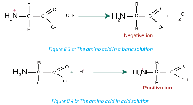 At a physiological pH, usually around 7, the amino acid exists as ZWITTERION (from German means hermaphrodite) it is a molecule with two different charges (positive and negative) at the same time (Figure 8.1.4).
 8.1.2. Formation and breakage of peptide bond
The formation of peptide bond follows the same pattern as the formation of glycosidic bond in carbohydrates and ester bond in fats. A condensation reaction occurs between the amino group of one amino acid and the carboxyl group of another, to form a dipeptide (fig 8.5).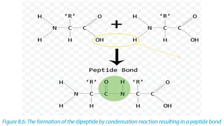 A peptide bond is formed between two amino acids to form a dipeptide molecule. If three amino acids are assembled togetherthey form a tripeptidewhile four amino acids form a tetrapeptide and so on. A long chain of amino acid it is called a polypeptide. The polypeptide chain or oligopeptide comprise more than 50 amino acids joined together by peptide bonds.During digestion, proteins are hydrolyzed to give amino acids that can be diffuse across the wall of intestine into blood stream. In hydrolysis the peptide bond breaks down by the addition of a water molecule (Figure 8.5).
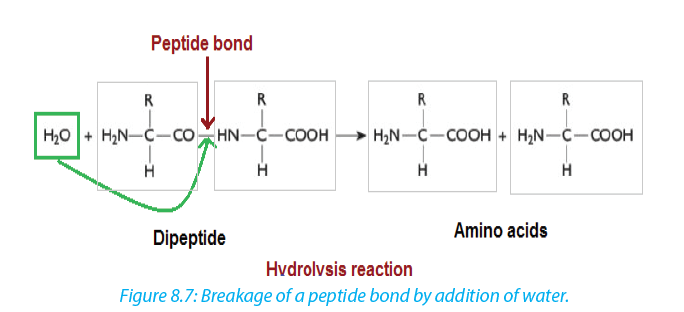 Self-assessment 8.1
1. Explain what are essential amino acids?
2. Describe the formation of a peptide bond?
3. At physiological pH, the amino acid exists as zwitterions. What is a zwitterion?
4. Alanine is an amino acid with -CH3 as a side chain. Write its structural formulae.
5. Most plants lack one or more of the essential amino acids needed by the body. Explain how a vegetarian can obtain the essential amino acids.8.2 Structure and denaturation of proteins
Activity 8.2
1. From the books make a research on proteins and answer to the following questions:
a. What are different structures of proteins?
b. Differentiate globular proteins and fibrous proteins.
2. Take a plastic rope cord, create the nodes bulk on it and suppose that those are
monomers of a long chain of polymer (the whole cord). Heat it using a Bunsen
burner or another source of fire. Discuss the change that takes place.8.2.1. Structure of proteins
The long chain of polypeptide can take different forms according to its molecular weight and the types of bond that hold together atoms and molecules.
a. Primary structure of proteins
Primary structure of a protein is the sequence of amino acid thatis made up of the polypeptide chain or chains. Ribonuclease is an enzyme found in pancreatic juice, which hydrolyses (digests) RNA. Notice that at one end of the amino acid chain there is an –NH3+ group, while at the other end there is a –COO− group. These are known as the amino and carboxyl ends, or the N and C terminals, respectively. (Adapted from Cambridge International AS and A Level Biology Course Book Fourth Edition)b. Secondary structure of proteins
The regular arrangement of amino acids in primary structure can induce the interaction of the back bone of the polypeptide chain (side chain) by hydrogen bonds. Those side chains are coiled and folded in the patterns that contribute to the protein’s overall shape. One such secondary structure is α-helix and sometime β-pleated sheet (Figure 8.8).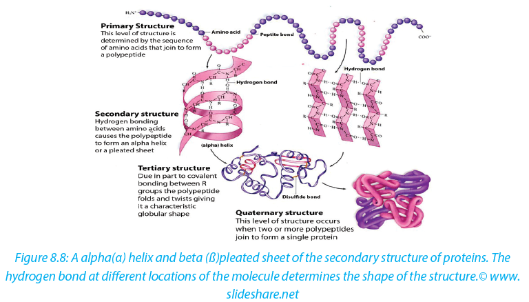 c. Tertiary structure of proteins
In addition to hydrogen and peptide bond in primary and secondary structure, the tertiary structure of protein has other types that include:
–– Hydrophobic interaction
–– Ionic bond between positively and negatively charged r groups.
–– Disulfide bridges (-s-s)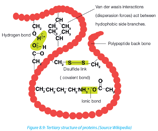 d. Quaternary structure of proteins
Quaternary structure involves more than one polypeptide chain chemically bonded to each other. The quaternarystructure refers to the way in which these polypeptide chains are arranged in the protein. Examples, Hemoglobin that is composed of:
–– Four polypeptide subunits, two α- chains and two β- chains. Both α and β subunits primarily are α helical secondary structure polypeptide chain with 140 amino acids.
–– Haeme composed of iron that binds with oxygen.
Collagen: this is a fibrous protein consisting of three helical polypeptides that are supercoiled to form a rope like structure of great strength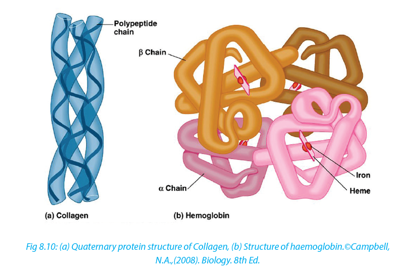 Globular protein
These are polypeptide chains that are tightly folded to form a spherical shape. Many globular proteins are folded so that their hydrophobic groups are on the inside of the molecule and the hydrophilic groups face outwards making these proteins soluble in water.
Properties of globular protiens:
–– They are spherical in shape
–– Physiologically active
–– Soluble in water.
–– May contain prosthetic group for example the iron (haeme)
–– Examples include hemoglobin and enzymes.8.2.2. Protein denaturation
Protein denaturation is a process by which protein changes shape due to breakage
of bonds holding the polypeptide chains. Protein denaturation may be temporary or permanent.
The agent of denaturation can be caused by;–– Extremely high temperatures beyond optimum,
–– changes in pH,
–– Ultra Violet (UV) rays,
–– High salt concentration and heavy metals. 8.2.3. Functions of proteins.
–– Proteins such as lipase, pepsin and protease act as enzymes as they play a crucial role in biochemical reaction where they act as catalysts.
–– Proteins play an important role in coordination and sensitivity (hormones and pigments).
–– Proteins have a transport functions. Example: Haemoglobin transport oxygen
–– Proteins in the cell membrane facilitate the transport of substance across the cell membrane.
–– Proteins provide a mechanical support and strength.
–– Proteins such as myosin and actin are involved in movement.
–– Proteins play the role of defense of the organisms. Example: Antibodies are proteins8.3 Water
Activity 8.3
1. What is the medium of reaction in the organisms?
2. If two people are boiling the same quantity of cooking oil and water, which one could evaporate first? Explain your choice.
Living organisms contain between 60% and 90% of water, the remaining being the dry mass. The function of water is defined by its physical and chemical properties that differ from those of most liquids and make it effective in supporting life.8.3.1 Biological significance of the physical properties of water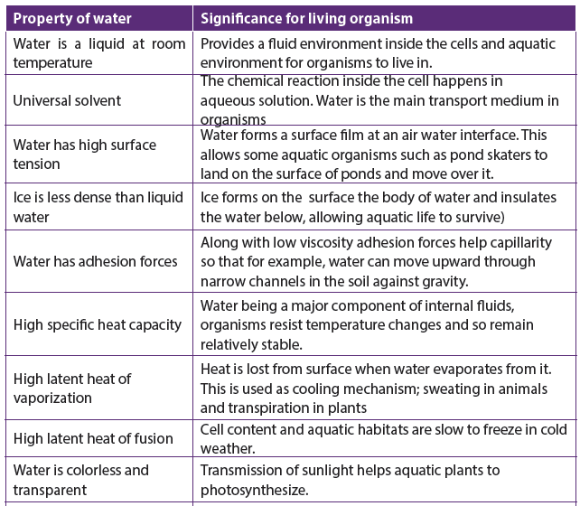

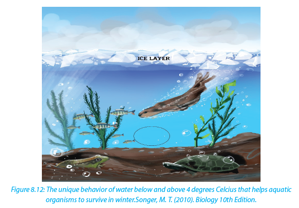 Functions of water
–– Turgidity of plant cell which increases their size is due to the availability of water.
–– The transport of substances (minerals, nutrients in plant and animals) that are dissolved in water.
–– Excretion of waste product
–– Support for hydrostatic skeleton.
–– Temperature regulation in plant and animals
–– Seed germination by breaking down the seed coat
–– Medium for biochemical reaction.Self-assessment 8.3
1. State the functions of water in animals
2. What do you understand by heat capacity?
3. Relate the high heat capacity of water to its biology functions.
4. Describe and explain how aquatic organisms live below frozen water
bodies
End of unit assessment 8
1. Certain drugs can break the covalent bond between two sulfur atoms of nonadjacent amino acids. Which level of protein that can be affected most if the drug is mixed with primary,secondary,tertiary and quaternary structure of proteins?
2. Complete the following statements by appropriate terms:
a. The formation of large molecules from small repeating units is called ………reaction.
b. A carbohydrate(polysaccharide)that is formed by the plant as a reserve food supply and made up of only glucose molecules covalently bonded
together is……...
3. State the property of water that allows each of the following to take place. In each case,explain its importance:
a. The cooling of skin during sweating
b. The transport of glucose and ions in a mammal
c. Much smaller temperature fluctuations in lakes and oceans than in terrestrial (land-based) habitats.
4. Construct a three column table and relate the following terms with arrows to
indicate the correct match.
Phosphodiester linkages Monosaccharide Polypeptides
Peptide bonds Nucleotides Triacylglycerol
Glycosidic linkages Amino acids Polynucleotides
Ester linkages Fatty acids Polysaccharides
5. Explain what happens during protein denaturation?UNIT 9: VITAMINS AND MINERALS
Key Unit Competence
Discuss the roles of minerals and vitamins in dietLearning objectives
At the end of this unit you be able to:
–– State the mineral requirements for bodily functions.
–– Identify the symptoms of mineral and vitamin deficiency.
–– Outline the need for consumption of minerals and vitamins in small amounts.
–– Organize a list of foods that are good sources of vitamins and mineral salts.
–– Recognize the signs and symptoms of scurvy, night blindness, goiter, and anaemia.
–– Differentiate between water soluble and lipid soluble vitamins.
–– Analyze one’s eating habits and suggest improvements
–– Appreciate the importance of a balanced diet in relation to health and economic prosperity.
–– Advocate for healthy feeding methods.Introductory activity
1. From the different food stuffs in our community, make a list of food stuffs that are good sources of minerals and vitamins.
2. Using text books and other resources, make a list of e vitamins and mineral deficiency diseases9.1 Mineral nutrients in humans
Activity 9.1
Use textbooks and internet to list mineral nutrients found in human diet Mineral nutrients are sometimes called mineral salts or just minerals. Mineral salts are essential nutrients that our body needs. They are called essential not because they are more important than other substances in our body but because our
bodiescannot produce them. They include the inorganic substances found in daily diet. They are dissolved in body fluids.They are found in human body as ions (cations and anions). Organic food like proteins, carbohydrates and fats provide the body with carbon, hydrogen, oxygen, nitrogen, sulfur and phosphorus. Butthere are several more elements that the body needs and occur as salts in the food we eat. They constitute about 1% of an organism by weight. Even though they are required in a very small amount, they are nonetheless essential for body processes.
Some mineral nutrients are required by animals, plants, a few by both. Humans require a number of minerals for the good functions of their bodies. Those are: Calcium (Ca2+), phosphorus (H2PO-4), Nitrogen(NO-3), Sulfur (SO2- 4) Potassium (K+), Sodium (Na+), Iron (Fe2+ or Fe3+), Magnesium (Mg2+), Iodine (I-), Chloride (Cl-), Manganese (Mn2+), Fluoride (F-), zinc (Zn2+), Cobalt (Co2+), Chromium (Cr2+ or Cr3+) and molybdenum (MoO-4)Self-assessment 9.1
1. Outline ten mineral nutrients required in human diet.
2. Answer by true or false and justify your answer: “Minerals are called essential nutrients because they are more important than others”.9.2 Classification of mineral nutrients
Activity 9.2
Iodized table salt is advised to prevent goiter. In 100g of table salt there is 99% of NaCl, and only 1% of iodine. Refer to the notes below to find the reason behind
this ratio.The classification of minerals is based upon their requirement rather than on their relative importance. Mineral nutrients are needed in a precise small amount. The five major minerals needed in human body include calcium (Ca2+),phosphorus (H2PO-4), potassium (K+), sodium
(Na+) and magnesium (Mg2+). Mineral nutrients are grouped into two groups: the macronutrients
or major elements and the micronutrients or trace elements.
Macronutrient or major elements are minerals needed by humans in a relative large amount (greater than 200 mg/day). Their examples include nitrogen(NO3-) , phosphorus (H2PO-4), sulfur(SO2-4) , calcium (Ca2+), sodium (Na+), chlorine (Cl-), magnesium (Mg2+), and iron (Fe2+ or Fe3+).Micronutrients or trace elements are those which are needed in minute amount (a few parts per million). Examples
include manganese (Mn2+), iodine (I-), zinc (Zn2+), molybdenum(MoO- 4)and fluorine (F-).Self-assessment 9.2
1. Categorize mineral nutrients according to their amount in human body.
2. Distinguish the two categories of mineral nutrients needed by the human body.
3. From the minerals listed here, identify the five major minerals in the human body: Sulfur (S), Potassium (K), Sodium (Na), Iron (Fe), Magnesium (Mg), Iodine (I), Chloride (Cl), zinc (Zn), Cobalt (Co), chromium, Calcium (Ca), phosphorus (P), Nitrogen (N).
9.3 Sources, functions and deficiency symptoms of mineral nutrients in humans
Activity 9.3
Use your textbook to answer the following questions:
Hereunder is a variety of food stuffs: Banana, cassava, wholegrain, oranges, pumpkin, potato, beans, water melon, green leafy vegetables, poultry, eggs, liver, and milk. Choose the food stuffs which are good sources of minerals. Human body requires mineral nutrients to survive and to carry out daily functions
and processes. Minerals keep humans healthy and have key roles in several body functions. Humans receive minerals by eating plants that absorb minerals from the soil and by eating meat and other products from animals, which graze on plants. The deficiency of mineral nutrients results into body functional disorders and diseases. Most are found in the blood and cytoplasm of cells, where they assist basic functions. For example, calcium and potassium regulate nerve and muscle activity

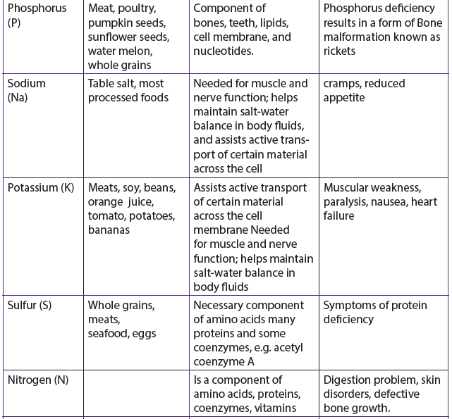

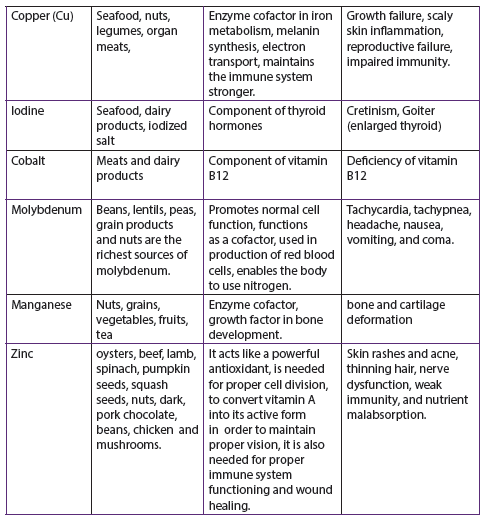
Self-assessment 9.3
1. Match the mineral nutrients with its function
a. Iodine 1. make bones hard
b. Fluorine 2. maintains the immune system stronger
c. Phosphorus 3. component of hemoglobin
d. Iron 4. prevents tooth decay
e. Copper 5. used in synthesis of thyroid hormone (thyroxin)
2. In a tabular form, identify the major dietary sources, the functions in human bodies and the deficiency diseases of the following minerals: Ca, I, P, zinc, and Cu
3. Choose the best answer.
i. They are the minerals we need a lot in every day diet. How are they called?
a. Macronutrents
b. Micronutrients
c. Giant minerals
d. Monster minerals
ii. This mineral helps to build up strong teeth and bones. How is it called?
a. Calcium
b. Iron
c. Zink
d. potassium
iii. What are foods that are natural good source of iron?
a. Roast Beef
b. Macaroni and cheese
c. Baked beans
d. Water melon
iv. Select 2 that are natural good sources of calcium?
a. Milk and cheese
b. Whole-wheat bread
c. Iceburg lettuce
d. Scanned salmon
v. The mineral that helps in oxygen transport to lungs is?
a. calcium
b. iron
c. zinc
d. potassium
vi. Which foods are good sources of zinc?
a. Milk and cheese
b. Lamb and pork
c. Macaroni and cheese
d. Peanuts and lentils
vii. Bananas are great source of this mineral, which helps our muscles and nervous
system to maintain your right water levels. What is it called?
a. Calcium
b. Iron
c. Zinc
d. Potassium
viii. Which mineral is important and needed by our body to fight off infection?
a. Calcium
b. Iron
c. Zinc
d. Potassium
ix. Which of the following mineral are needed in large amount every day?
a. Zinc
b. Iron
c. Calcium
d. selenium
x. Which is the type of mineral that keep your nervous system health?
a. Calcium
b. Iron
c. Zinc
d. Potassium
4. From the diseases listed below, what are those caused by the deficiency of minerals?
Goiter, malaria, diabetes, rickets, beriberi, scaly skin, night blindness, anemia, impaired immunity, diarrhea9.4 Vitamins and the classification of vitamins
Activity 9.4
Two students with different complains went to consult a medical doctor. Student A says to the doctor that whenever he/she bleeds whenever she /he brushes teeth. Student B doesn’t see well objects around him/her, The results from the doctor showed that they all have lack some vitamins.
1) What kind of vitamins that each student needs to take?
2) Use your student textbook to explain your answer
Like minerals, vitamins are also essential for the human body. They are required for metabolism, protection health and growth. Vitamins also assist in formation of hormones, blood cells and genetic material. Vitamins are directly absorbed from the small intestine into the blood stream. Water –soluble vitamins are absorbed in the ileum while fat-soluble vitamins are absorbed in jejunum. Features shared by all vitamins:
–– They are not digested or broken down for energy
–– They are not synthesized into the body structures
–– Most are rapidly destroyed by heat.
–– They are essential for good human health and needed in a very small amount
–– They are required for chemical reactions in cells, working in association with enzymes.There are thirteen vitamins required by human body. They are classified by their solubility, whether they dissolve in water or in fats. Water-soluble vitamins including vitamins C and B complex, and fat-soluble vitamins including vitamins A, D, E and K (Table 9.2). Excess water- soluble vitamins are simply excreted in urine, while fatsoluble vitamins are stored in body fatty tissues to be used later if there is deficient
in diet. Excess intakes of these vitamins are stored in fatty tissues of the body, where they can build up to toxic levels, especially if they are taken improperly in supplements.Table 9.2 Water-soluble and fat-soluble vitamins
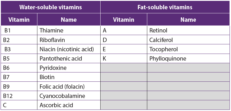
Self-assessment 9.4
1. How many vitamins does the human body needs to function properly?
2. Describe the classification of vitamins.9.5 Sources, functions and symptoms of vitamin deficiency
Activity 9.5
Here is a number of foodstuffs rich in vitamins.
From the list of provided food stuffs (Banana, cassava, wholegrain, oranges, pumpkin, potato, beans, water melon, green leafy vegetables, and milk). Can you give some foods that are good sources of vitamins?
Some vitamins, including some vitamin B complex and Vitamin K are produced by bacteria that normally live in the intestines, where they help to digest food. Vitamin D is synthesized in the skin when it is exposed to UV radiation in sunlight.Vitamins and their derivative are coenzymes; note that a coenzyme is an organic molecule that combines temporaly with enzymes making them more efficient. For example, Niacin or vitamin B3 is an essential component of coenzymes NAD and NADP involved in lipid metabolism. It inhibits production of cholesterol and catabolism of triglyceride. Thiamin or vitaminB1 is a coenzyme for many different enzymes that break complex molecules such as carbohydrates to produce ATP.
Thiamin deficiency results into Beriberi anemia and stunted growth in children. Vitamin K is an essential coenzyme for synthesis of several blood clotting factors. Several vitamins, including vitamins C and E, act as antioxidants. An antioxidant is a compound that neutralizes chemicals called free radicals. Free radicals are produced naturally during cellular activities and may cause some types of cancer. Neutralizing free radicals makes them harmless.
The table: 9.3. The major dietary sources, functions and possible symptoms of vitamin deficiency


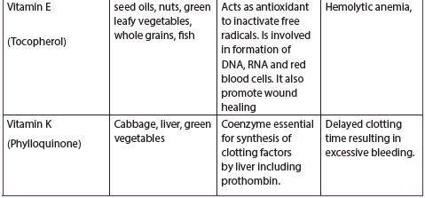
Many vitamin supplements are available in the market. However, it is always advisable to obtain them from their natural sources by eating food rich in vitamins daily. Possible symptoms of vitamins deficiency are shown by the following pictures:

End of unit assessment 9
1. Choose a mineral which is an electrolyte and is found in almost every food. It helps to lower blood pressure.
a. Zinc
b. Potassium
c. Calcium
d. Iron2. choose a mineral which helps to make our blood vessels, tendons, and nerves strong.
a. Iron
b. Magnesium
c. Chromium
d. Copper3. The following vitamins are part of Niacin and Thiamin minerals
a. Vitamins
b. Vitamins
c. Vitamins
d. Vitamins4. Vitamin C is required for the production and maintenance of:
a. Collagen
b. Hormone
c. Ascorbic Acid
d. Red Blood Cells5. Vitamin C deficiency is called:
a. Scurvy
b. Cold
c. Cancer
d. Rickets6. Which of the following is a function of Vitamin A in the body?
a. Vision, bone and body growth
b. Immune defenses, maintenance of body linings and skin
c. Normal cell development and reproduction
d. All of the above7. Common food sources of Vitamin A are:
a. Milk, eggs, butter, cheese, cream, and liver
b. White sugar, honey, and sugar cane
c. Broccoli, apricots, Cantaloupe, Carrots, Sweet potato, Spinach
d. Both a and c8. Which of the following is a function of Vitamin B-12?
a. Influences the cells that build bone tissue
b. Is essential to the formation of bone
c. Helps to maintain acid-base balance
d. Maintains the sheaths that surround and protect nerve fibers9. Vitamin B-12 deficiency caused by lack of intrinsic factor is called:
a. Pernicious anemia
b. Poor circulation of the red blood cells
c. Beriberi
d. None of the above10. What groups of people need additional Vitamin K?
a. Premature newborns
b. People who do not have enough bile to absorb fat
c. Both A and B
d. None of the above answers11. A common function of Thiamin, Riboflavin and Niacin is that:
a. They all are used in synthesis of blood clotting proteins
b. They all work as a part of a coenzyme used in energy metabolism
c. They all help to strengthen blood vessel walls
d. They are used to stabilize cell membranes
12. The vitamin Folate works together with ______________ to produce new red blood cells.
a. Vitamin D
b. Vitamin A
c. Vitamin B-12
d. None of the above13. Which of the following is a function of Vitamin B-12?
a. Red blood cell formation
b. Myelin sheath that protects nerve biers
c. Vision
d. Both A and B14. Vitamin C helps in maintenance and repair of collagen which:
a. Forms the base for all connective tissue in the body
b. Aids in digestive processes
c. Promotes good eyesight
d. Prevents PMS symptoms
15. Which of the following is not a function of Vitamin D?
a. Acts like a hormone
b. Stimulates maturation of cells
c. Maintains calcium cells
d. Builds tissue16. Some food sources of Vitamin D are:
a. Fruits and vegetables
b. Salmon and egg yolks
c. Butter and fortified milk
d. Both B and C.17. Humans obtain vitamins from natural sources such as vegetables, fruits,
meat, fish and dairy products. What are the two vitamins that are not provided
by fruits and vegetables?18. What would you advise someone starting to have symptoms of?
a. Scurvy
b. Rickets
c. Teeth decay
d. Heart failure
e. Pernicious anemiaUNIT 10: ENZYMES
Describe the mode of action and factors affecting enzymes and their importance for the existence of life
Learning objectives
At the end of this unit you be able to:
–– Define the term enzyme.
–– Explain the criteria of naming enzymes.
–– State that enzymes function inside cells and outside cells.
–– Explain that enzymes are globular proteins that catalyze metabolic reactions.
–– Describe the mode of action of enzymes in terms of the lock and key and the induced fit hypotheses.
–– Explain factors affecting enzyme activity.
–– Define enzyme technology and its role in industry.
–– Investigate the progress of an enzyme-catalyzed reaction by measuring rates of formation of products.
–– Investigate the effects of temperature, pH, enzyme and substrate concentration, and inhibitors on enzyme activity.
–– Interpret graphs of the effects of reversible and irreversible inhibitors on the rate of enzyme activity.
–– Investigate the effect of immobilizing an enzyme in alginate as compared with
its activity when free in solution.
–– Use a computer to plot graphs of the rate of enzyme controlled reaction.
Calculate Q10 of an enzyme controlled reaction.
–– Acknowledge that enzymes are essential in speeding up reactions that would be too slow to sustain life.
–– Appreciate the importance of planning and carrying out experiments under controlled conditions.
–– Understand the roles of enzymes in industry and medicine.Introductory activity
Discuss in pair the following questions and share with another pair your findings.
1. What do you understand by the term enzyme?
2. Two individuals want to reach the last floor of Kigali city tower. One climbs up using the ladder but another one uses a lift. What advantage the lift gives over the ladder?
3. Why is it easy to digest hot foods than cold ones?
10.1 Criteria for naming enzymes
Activity 10.1
You are provided with three groups of enzymes:
Make a research to find out:
a. specific role of each of the six enzymes mentioned above
b. criterion followed to name enzymes of group A, B and C respectively
Enzymes are biological catalysts produced by a living organism to control the rate of specific biochemical reactions by lowering the activation energy of reactantsFirst of all, individual enzymes are named by adding -ase to the name of the substrate with which they react. The enzyme that controls urea decomposition is called urease; those that control protein hydrolyses are known as proteases.
A second way of naming enzymes refers to the enzyme commission number (EC number) which is a numerical classification scheme for enzymes based on the chemical reactions they catalyze. In a system of enzyme nomenclature, every EC number is associated with a recommended name for the respective enzyme catalyzing a specific reaction. They include:
–– Oxidoreductases: catalyze redox reactions by the transfer of hydrogen, oxygen or electrons from one molecule to another. Example: Oxidase catalyzes the addition of oxygen to hydrogen to form water.

–– Hydrolase: catalyzes the hydrolysis of a substrate by the addition of water.
Sucrose + water glucose+ fructose
–– Ligases: catalyze reactions in which new chemical bonds are formed and use ATP as energy source.
Amino acid + tRNA amino acid-tRNA complex.
–– Transferases: catalyze group transfer reactions. The transfer occurs from one molecule that will be the donor to another molecule that will be the acceptor. Most of the time, the donor is a cofactor that is charged with the group about to be transferred. Example: Hexokinase used in glycolysis.
–– Lyases: catalyze reactions where functional groups are added to break double bonds in molecules or the reverse where double bonds are formed by the removal of functional groups. For example: Fructose bisphosphate aldolase used in converting fructose 1, 6-bisphospate to G3P and DHAP by cutting C-C
bond.
–– Isomerases: catalyze reactions that transfer functional groups within a molecule so that isomeric forms are produced. These enzymes allow for structural or geometric changes within a compound. Sometime the interconverstion is carried out by an intramolecular oxidoreduction. In this case, one molecule is both the hydrogen acceptor and donor, so there’s no oxidized product. The lack of an oxidized product is the reason this enzyme falls under this classification. The subclasses are created under this category by the type of isomerism. For example: phosphoglucose isomerase for converting glucose 6-phosphate to fructose 6-phosphate by moving chemical group inside the same substrate.A third way of naming enzymes is by their specific names e.g. trypsin and pepsin are proteases. Pepsin, trypsin, and some other enzymes possess, in addition, the peculiar property known as autocatalysis, which permits them to cause their own formation from an inert precursor called zymogen.
10.2 Characteristics of enzymes
Activity 10.2
Requirement: Three test tubes, match box, about 1g of liver, 1g of sands, 1% H2O2 and MnO2 powder.
Procedure:
–– Label three test tubes A, B and C respectively.
–– Put about 0.1 g of MnO2 powder in test tube A and 1g of liver in tube B and 0.1g of sand in tube C.
–– Pour 5 ml of H2O2 (hydrogen peroxide) in each tube. What do you observe?
–– Place a glowing splint in the mouth parts of each test tube. What do you observe?Questions
1. Explain your observations.
2. Write down the chemical equation of the reaction taking place in tube A and B
3. Carry out your further research to find out the characteristics of enzymesEnzymes speed up the rate of metabolic reactions by allowing the reaction to go through a more stable transition state than would normally be the case. As a result, the rate of reaction is increased. In many chemical reactions, the substrate will not be converted to a product unless it is temporarily given some extra energy referred to as activation energy (the minimum energy required the make a reaction take place).

Enzymes speed up the rate of biochemical reactions in the cell but remain unchanged at the end of the reactions. An enzyme has no effect on the relative energy content of products versus reactant. Chemical reactions catalyzed by enzymes are usually reversible e.g. enzyme carbonic anhydrase catalyses both synthesis and breakdown of carbonic acid.

An enzyme provides a reaction surfaceand a hydrophilic environment for a reaction to take place. This is normally a hollow or cleft in the enzyme which is called the active site, but it is normally hydrophobic in nature rather than hydrophilic.
A very small amount of enzymes is needed to react with a large amount of substrate. The turnover number of an enzyme is the number or reactions an enzyme molecule can catalyse in one second. Enzymes have a high turnover number e.g. the turnover number of catalase is 200,000 i.e. one molecule of enzyme catalase can catalyse the breakdown of about 200,000 molecules of hydrogen peroxide per second into water and oxygen at body temperature.
A cofactor is the best general term to describe the non-protein substances required by an enzyme to function properly. This term covers both organic molecules and metal ions. A co-enzyme is an organic molecule that acts as a cofactor. A prosthetic group is a cofactor that is covalently bound to the enzyme.
Self-Assessment 10.2
1. State any four properties of enzymes.
2. Enzymes have generally high turnover number. What is the significance of the high turnover of enzymes?10.3 Mode of action of enzymes
Activity 10.3
There are two main hypotheses that explain the mode of action of an enzyme on its substrate: the lock and key hypothesis and the induced-fit hypothesis. Carry out a research to find the relevance of each.Enzymes do not change but substrates are converted into products. A substrate is a molecule upon which an enzyme acts. In the case of a single substrate, the substrate binds with the enzyme active site to form an enzyme-substrate complex. Thereafter the substrate is transformed into one or more products, which are then released from the active site. This process is summarized as follows:

Whereby: E = enzyme, S = substrate(s), ES = Complex Enzyme-Substrate and P= product (s). There are two main hypotheses explaining the mechanism of enzyme action:
a. The lock and key hypothesis by Emil Fischer
In this hypothesis the substrate is the key and enzyme is the lock. The active site is exactly complementary to the shape of the substrate as shown below.
b. The induced-fit hypothesis by Daniel Koshland
The induced-fit hypothesis is a modified version of the lock and key hypothesis and is more widely accepted hypothesis. In this hypothesis, the active site is flexible and
is not fully complementary with the shape of the substrate. An enzyme collides with the substrate molecule and binds to the active site. This induces a slight change in the shape of the enzyme making the substrate the fit more precisely. This reduces the potential energy of the substrate and allows the reaction to occur. The products formed move away from the active site and regains its original configuration ready for the next reaction to take place.
Self-Assessment 10.3
The key and lock hypothesis is a model that explain the mode of action of an enzyme on the substrate. In the same context, analyse the diagram below and then answer question that follow.
1. What does the lock represent?
2. What does the key represent?
3. Where is the active site?
4. Suggest another diagram that can better represent the induced fit hypothesis. Write short notes to explain its functioning.10.4 Factors affecting enzyme action
Activity 10.4
You will need
Eight test tubes containing 2 cm3 starch solution, amylase solution, cold water (ice) water bath, iodine solution, HCl solution, and droppers Procedure:
1. Label your test tubes A-D as follows:
2. Add 1 cm3 of starch solution to each test tube
3. Keep tube A and B in cold (ice) and tube C and D in the water bath at 35oC for 5 minutes.
4. Add 1 cm3 of 1M HCl on test tubes B and D, then shake the mixture to stir.
5. Add 1 cm3 of amylase solution on each test tube. Shake and therefore keep A and B in cold and C and D in water bath for 10 minutes.
6. Take a sample from each tube and mix it with one drop of iodine. Use a different tile for each test tube. Record and interpret your observation and then draw a conclusion.
Enzymes activities can be limited by a number of factors such as the temperature, the pH,
the concentration of the substrate or the enzyme itself and the presence of inhibitors.i. Temperature
At zero temperature, the enzyme cannot work because it is inactivated. At low temperatures, an enzyme-controlled reaction occurs very slowly. The molecules in solution move slowly and take a longer time to bind to active sites. Increasing temperature increases the kinetic energy of the reactants. As the reactant molecules move faster, they increase the number of collisions of molecules to form enzymesubstrate
complex.At optimum temperature, the rate of reaction is at maximum. The enzyme is in active state. The optimum temperature varies with different enzymes. The optimum temperature for enzymes in the human body is about 37oc. When the temperature exceeds the optimum level, the enzyme is denatured.
The effect is irreversible. However, some species are thermophilic that is they work better at high temperatures; others are thermophobic, that is they work better at low temperatures. For example, some thermophilic algae and bacteria can survive in hot springs of 60oc.
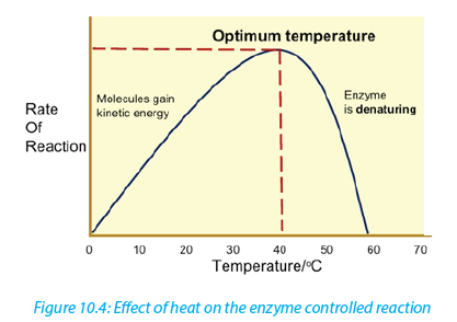
The rate doubles for each 10oC rise in temperature between 0oC and 40oC (figure 10- 5). The temperature coefficient Q10 is the number which indicates the effect of rising the temperature by 10oC on the enzyme-controlled reaction. The Q10 is defined as the increase in the rate of a reaction or a physiological process for a 10°C rise in temperature. It is calculated as the ratio between rate of reaction occurring at (X + l0) oC and the rate of reaction at X oC. The Q10 at a given temperature x can be
calculated from:
Worked out example
The rate of an enzyme-controlled reaction has been recorded at different temperatures as follows:
Solution

This means that the rate of the reaction doubles if the temperature is raised from 30°c to 40°c
Be aware that not all enzymes have an optimum temperature of 40°c. Some bacteria and algae living in hot springs (e.g. Amashyuza in Rusizi) are able to tolerate very high temperatures. Enzymes from such organisms are proving useful in various industrial applications because they do not denature up to 70oc
ii. The pH
Most enzymes are effective only within a narrow pH range. The optimum pH is the pH at which the maximum rate of reaction occurs. Below or above the optimum pH the H+ or OH- ions react with functional groups of amino acids in the enzyme which loses its tertiary structure and become natured.
Different enzymes have different pH optima (look in the table).
Table 10.1. Optimum pH of some digestive enzymes
iii. Enzyme concentration
The rate of an enzyme-catalyzed reaction is directly proportional to the concentration of the enzyme if substrates are present in excess concentration and no other factors are limiting.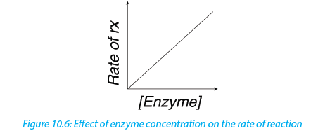
iv. Substrate concentration
At low substrate concentration, the rate of an enzyme reaction increases with increasing substrate concentration. The active site of an enzyme molecule can only bind with a certain number of substrate molecules at a given time. At high substrate concentration, there is saturation of active sites and the velocity of the reaction reaches the maximum rate.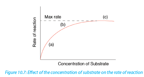
b. Inhibitors
The inhibitors are chemicals or substances that prevent the action of an enzyme. An inhibitor binds to an enzyme and then decreases or stops its activity. There are three types of inhibitors:i. Competitive inhibitors are molecules that have the similar shape as the substrate. At high concentration, they compete with the substrate for the active site of the enzyme e.g. O2 competes with CO2 in RuBP-carboxylase.
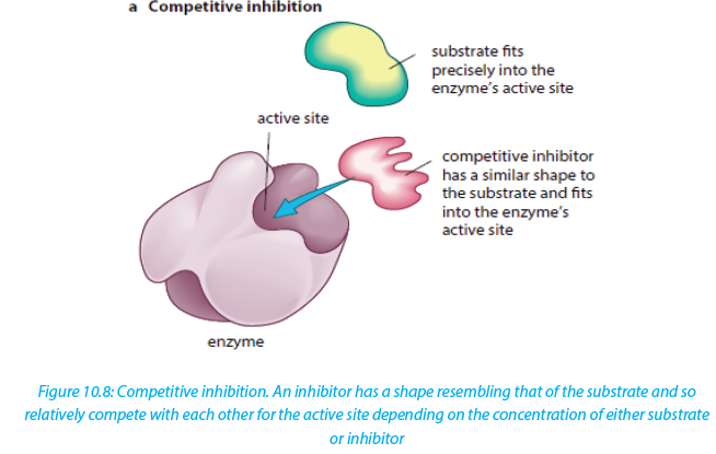
ii. Non-competitive inhibitors are molecules that can be fixed to the other part of enzyme (not to the active site) so that they change the shape of active site,due to this the substrate cannot bind to the active sit of the enzyme.

iii. End product inhibitor, Allosteric inhibitor or Allostery.
This is a chain enzymatic metabolic pathway where the final end product acts as an allosteric reversible inhibitor for the first, the second or the third step in the metabolic pathway. The shape of an allosteric enzyme is altered by the binding of the end product to an allosteric site. This decreases enzymatic activity. By acting as allosteric inhibitors of enzymes in an earlier metabolic pathway, the metabolites
can help to regulate metabolism according to the needs of organisms. This is an example of negative feedback.This often happens when few enzymes are working on a large number of substrate e.g. ATP is an end-product inhibitor of the enzyme PFK (Phosphofructokinase) in glycolysis during cell respiration. The end-product inhibitor leads to a negative feedback.

The products of enzyme-catalysed reactions are often involved in the feedback control of those enzymes. Glucose-1-phosphate is the product formed from this enzyme-catalysed reaction. As its concentration increases, it increasingly inhibits the enzyme.
Importance of reversible and irreversible inhibition
The nerve gas DIPF (DiIsopropyl Phospho Fluoridate) is an irreversible inhibitor. It binds permanently with enzyme acetylcholisterase, altering its shape. The enzyme cannot bind with and break down its substrate acetylcholine (neurotransmitter). Acetylcholine molecules accumulate in the synaptic cleft. Nerve impulses cannot be stopped causing continuous muscle contraction. This leads to convulsions, paralysis and eventually death.Many pesticides such as organophosphate pesticides act as irreversible enzyme inhibitors. Exposure to pesticides can produce harmful effects to the nervous and muscular systems of humans. Heavy metal ions such as Pb2+, Hg2+, Ag+, As+ and iodine-containing compounds which combine permanently with sulphydryl groups in the active site or other parts of the enzyme cause inactivation of enzyme. This usually disrupts disulphide bridges and cause denaturation of the enzyme.
Self-Assessment 10.4
1. What is Q10 of an enzyme controlled reaction?
2. You are provided with the table below of the rate of an enzyme controlled reaction.
Calculate the value of Q10 at:
a. 0° c
b. 10° c
c. 50° c
3. Explain why thermophile bacteria and algae are useful in some industrial processes
4. The diagram below represents a metabolic pathway controlled by enzymes.
–– V is a substrate
–– W, X and Y are intermediate compounds
–– Z is a product
–– e1, e2, e3, and e4 are enzymes
a. Name the type of control mechanism which regulates production of compound Z
b. Explain how an excess of compound Z will inhibit its further production.10.5 Importance of enzymes in living organisms
Activity10.5
Discuss and present your ideas about the need for different enzymes in living organisms.Without enzymes, most of the biochemical reactions in living cells at body temperature would occur very slowly or not at all. Enzyme can only catalyze reactions in which the substrate shape fits that of its active site
There are thousands of metabolic reactions that place in the body that require enzymes to speed up their rate of reaction, or will never happen. Enzymes are very specific, so nearly each of these chemical reactions has its own enzyme to increase its rate of reaction. In addition, the organism has several areas that differ from one another by the pH. Therefore, the acid medium requires enzymes that work at low
pH while other media are alkaline and require enzymes that work at high pH. In addition to digestion, enzymes are known to catalyze about 4,000 other chemical reactions in your body. For example, enzymes are needed to copy genetic material before your cells divide.Enzymes are also needed to generate energy molecules called ATP, move fluid and nutrients around the insides of cells and pump waste material out of cells. Most enzymes work best at normal body temperature about at 370 c -- and in an alkaline environment. As such, high feverand over-acidity reduce the effectiveness of most enzymes. Some enzymes need co-factors or co-enzymes to work properly.
Self-Assessment 10.5
1. Fill the blank with appropriate terms:
Enzymes are biological ____________________ produced by
___________________________ cells. Enzymes reduce the amount of
____________________ energy required for reactions to occur. They consist of
globular ____________________ with _______________________ structure.
2. Answer the following questions:
a. What is the main role of enzymes?
b. What would happen if there are no enzymes in the cell?10.6 Enzymes technology
Learning activity 10.6
Enzymes are needed in everyday life. At school you can use salivary amylase to hydrolyse starch. There is industrial technique used to get large amounts of enzyme amylase.Read through the notes below and answer the following questions below:
a. State the different processes in which enzyme technology is applied
b. What is the role of thermophilic bacteria in this process?
c. How is the effectiveness of an enzyme improved for used in industry?The market for enzymes is prosperous. The demand keeps on increasing as new applications of enzymes are discovered. Enzymes have been used in cheese-making, in leather industries, and making washing powders.
Microbial cells are still the most sources of industrial enzymes because microorganisms naturally produce enzymes inside their cells known as intracellular enzymes. When e microorganisms secrete their enzymes for an action outside their cells, the enzymes are called extracellular enzymes. Microorganisms may have specific genes introduced into their DNA by genetic engineering so that they produce enzymes naturally made by other organisms.
Once enzymes are produced by the microorganisms they are isolated by centrifugation in order to remove the large cell fragments. The enzyme is precipitated from solution by a salt such as (NH4)2SO4 or an alcohol such as CH3-CHOH-CH3. Thereafter the enzyme can be purified by the process known as electrophoresis or column chromatography. The enzyme stability is a key factor in the industrial use of
enzymes. The stability of an enzyme is its ability to retain its tertiary structure under a wide range of conditions.As many industrial processes require high temperatures and extreme pH, it is recommended to use bacteria such as Bacillus subtilis which withstand harsh conditions such as high temperature. Those thermophilic bacteria produce thermostable enzymes that do not denature at high temperature because their optimum temperature between 65 - 750c.
Some useful enzymes are not thermostable. Such enzymes should be improved by the technique called immobilization i.e. the enzyme is attached to or located within an unreactive support such as nylon that protects it from denaturation.
Self-Assessment 10.6
1. What is the role of alcohol or ammonium sulphate during the extraction of enzymes?
2. Why is thermostability of enzymes so important for many industrial processes?
End of unit assessment 10
1.
a. What is the meaning of the following terms related to enzyme activity?
i. Catalyst
ii. Activation energy
iii. Lock and key hypothesis
iv. Q10
b. Why are there hundreds of different enzymes in a cell?
c. How do enzymes reduce the activation energy of a reaction?
2. Enzyme activity is affected by a number of factors.
a. Explain why enzymes work faster at relatively high temperatures
b. Describe what happens to the enzyme structure if the temperature is raised
above the optimum temperature.
c. How are enzymes affected by pH?
d. Why do different enzymes have a different optimum pH?
e. What is the difference between a reversible and irreversible enzyme
inhibitor?
3. Some bacteria and algae can survive in boiling water of hot springs. Enzymes
from those organisms are used in industrial processes. Why are those enzymes useful?
4. The following set of data shows the effect of temperature on the completion time of an enzyme reaction.
a. Plot the data on a graph
b. What is the optimum temperature of this reaction?
c. Describe the shape of the graph between 10 and 40o c
d. Calculate the rate of increase between 20 and 30o c.5. The table below shows the rate of an enzyme reaction at a range of temperature:
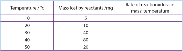
a. Fill that table with the values of the rate of reaction and plot a graph of rate at different temperatures (use x-axis for temperature).
b. Calculate Q10 at 30°c.
c. Explain what happen between 20 and 30°c, and between 40 and 50°c.
6. The graph below shows the activity of a commercial enzyme alcalase at different pH value. Alcalase is a protease enzyme.
a. What are the compounds digested by this enzyme?
b. Describe the change in enzyme activity with pH.
c. How does this curve compare to the pH curve of a human digestive
enzyme such as pepsin?
7. Outline how a specific enzyme can be produced from bacteria.UNIT 11: PRINCIPLES OF GAS EXCHANGE SYSTEMS
Key Unit Competence
Explain the principles of gaseous exchange systemsLearning objectives
At the end of this unit learners will be able to:
–– Explain the relationship between size and surface area to volume ratio.
–– Describe how different respiratory surfaces are modified to speed up the diffusion process.
–– State the characteristics of gaseous exchange surfaces.
–– Describe the effects of tar and carcinogens in tobacco smoke on gas exchange
system with reference to lung cancer and Chronic Obstructive Pulmonary
–– Describe the short-term effects of nicotine and carbon monoxide on the cardiovascular system.
–– Observe prepared slides of gaseous exchange surfaces and identify their
characteristics.
–– Dissect fish gills and observe the surface area for gas exchange.
–– Observe mammal’s lungs and state their adaptation for gaseous exchange.
–– Use internet to make research and deduce the findings
–– Appreciate the evolution of gaseous exchange surfaces from simple to complex.Introductory activity
Kalisa and Uwase wanted to rear tilapia at their home. They bought a nice transparent plastic box. They filled it with 1.5L of clean mineral water, put in some pieces of meat and plant leaves. They finally introduced a living tilapia in the box and covered. After two days they were happy to see their fish swimming. But on the third day, they become sad after finding it dead and yet the food was still in
water. What could have caused the death of the fish?11.1 Relationship between size and surface area to volume
Actirvaittyi o11.1
1. Use Manila paper, scissors, and graduate ruler to create three cubes: 3cm x 3cm,2cm x 2cm, 1cm x 1cm
a. Calculate the surface area, the volume, and the surface area to volume ratio of each cube. What do you conclude from these ratios?
b. Compare the surface area to the volume of a spherical alveolus having a radius of 0.001m and that of another animal with a radius of 0.000001m.
2. What do you understand by surface area to volume ratio?
The surface area to volume ratio is the relationship between the surface area and the volume of an object. Small or thin objects have a large surface area compared to the volume. For example, the surface area of a sphere is calculated by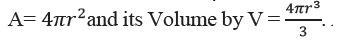
As the length or radius of the sphere increases, the increase in the surface area is squared (X2) and the increase in the volume is cubed (X3). The surface area to the volume ratio gets smaller as the cell or animal gets larger. Thus, if the cell grows beyond a certain limit, not enough material will be able to cross the membrane fast enough to accommodate the increased cellular volume.As a cell grows, its surface area to volume ratio decreases. At some point in its growth its surface area to volume ration becomes so small that its surface area is too small to supply its raw materials to its volume. The cell will reach a size at which substances cannot enter or leave the cell in sufficient time to sustain life. At this point the cell cannot get larger. The volume of the cell will also be so large that the diffusion
rate will be too low to distribute necessary substances throughout the cell within a reasonable time. This brings about the need of having a mechanism of ventilation that speeds up the rate of gaseous exchange.
The rate of oxygen consumption by an animal gives a relatively accurate indication of the rate of its metabolic activity. The need of oxygen varies with the activity, the size of the organism, and their health. In general, small mammals need more oxygen than large mammals because:
–– Small mammals have a big respiratory surface area to the volume ratio
–– Small mammals are too motile than large mammals. Therefore, they need to produce more energy through aerobic respiration
–– Small mammals reproduce more rapidly than large mammals.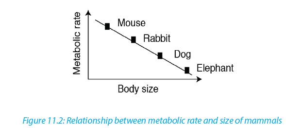
A running man needs a double volume of oxygen than a sleeping man and a pregnant woman needs more oxygen than a normal woman.
Self-Assessment 11.1
Determine the surface area to volume ratio of a sphere having a diameter of 4 mm11.2 Characteristics of gas exchange surfaces
The following are the characteristic features of gaseous exchange surfaces:Large surface area: they should have a large surface area to allow adequate and fast gaseous exchange in order to provide enough oxygen to cells and to get rid of the carbon dioxide that is released.
Rich supply of blood: in animals with a transport system, the respiratory surface areas found in the lungs and gills have rich supply in blood capillaries to quickly transport gases to and from the cells. Gases diffuse into the blood and are carried to and from the body cells.
Thin surface or thin wall: respiratory surfaces should have thin walls or thin surface area to maximize the diffusion. The alveoli in the lungs have thin squamous epithelium that enables gases to diffuse quickly between the alveoli and blood. According to Fick’s law, the rate of diffusion is proportional to:

Moist surfaces area: to enable gases to dissolve and pass through the solution. High diffusion deficit / concentration gradient: respiratory surface areas should have a high diffusion deficit / concentration gradient to ensure faster diffusion of respiratory gases.
Protection against injury and dry out: lungs and gills are protected by the bones and cartilage and mucus protects them from drying outSelf-Assessment 11.3
State the features common to all respiratory surfaces in living organisms
1. Explain how the following features of a respiratory surface helps gaseous exchange:
2. Short diffusion distance
a. Protection
b. A rich blood supply
c. Protection11.3 Modifications of gaseous exchange surfaces to speed up the rate of gaseous exchange in different organims
Activity 11.2
Use appropriate laboratory equipment to extract gills in fish to show the gill filaments. Draw and label to show the parts observed.a. Insects
The spiracles are openings of small tubes running into the insect’s trachea system that terminates into small fluid-filled tracheoles in which the gases are dissolved. The fluid is drawn into the muscle tissue during physical exercise, and this increases the surface area of air in contact with the cells.Ventilation movements of the body during exercise may help this diffusion. The spiracles can be closed by valves and may be surrounded by tiny hairs. The later help to keep humidity around the opening to ensure that there is a lower concentration gradient of water vapour, and so less is lost from the insect by evaporation.
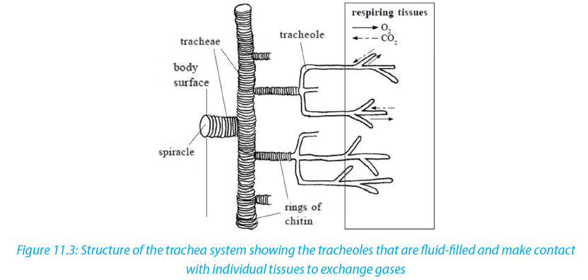
b. Fish and tadpoles
Fish and young amphibians (tadpoles) use gills for the gaseous exchange.
Gills have numerous folds that give them a very large surface area.
–– The rows of gill filaments have many protrusions called gill lamellae. These filaments help in the exchange of respiratory gases
–– They also have an efficient transport system within the lamellae which maintains the concentration gradient across the lamellae. The arrangement of water flowing passes the gills in the opposite direction to the blood (called counter-current flow) means that they can extract oxygen at 3 times the rate a human can.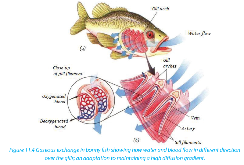
c. Amphibians, Reptiles, Birds and Mammals
These have alveoli in their lungs. Air reaches the alveoli via a system of tubes (trachea, splitting into two bronchi - one for each lung - and numerous bronchioles):
–– Numerous alveoli - air sacs, providing a massive surface area over which gases can diffuse
–– Have a short diffusion distance between the alveolus and the blood because the lining of the lung and the capillary as they are only one cell thick.
–– The blood supply is extensive, which means that oxygen is carried away to the cells as soon as it has diffused into the blood.
–– Ventilation movements and out of the lungs due to changes in volume and press- Ventilation movements also maintain the concentration gradients because a pressure

Activity11.3
You will need: Lungs of a sheep or pig, newspaper, plastic sheets, dissecting board, sharp scalpel, dissecting needles, scissors, dissecting tray, latex gloves, CPR mouth piece, soap to wash hands and surfaces.

Procedure
–– Place the dissecting board on the newspaper and lay the lungs on the board.
–– Use a scalpel to cut the lungs in half in longitudinal section.
–– Identify the trachea, right lung, left lung, cartilage rings, bronchus, larynx, alveoli, and bronchiole. You can use a magnifying hand lens to observe structures in the lungs.
–– Inflate the lungs by blowing through the CPR (cardio-Pulmonary Resuscitation) mouth piece to see how the lungs expand.
–– Feel the slippery inside of trachea, press the lung with your finger and look at cartilaginous rings.
–– Remember to wash your hand s with soap as you finish your experiment.
1. Explain what it feels like as you press the lungs with your fingers.2. Look at cartilaginous rings. What function do they serve?
3. (a) List four features of respiratory surfaces you can identify from the specimen.
(b) Examine the lung and explain how the lungs are suited for efficient
gaseous exchange.
Table 11.1: Parts of the human gas exchange system and their respective functions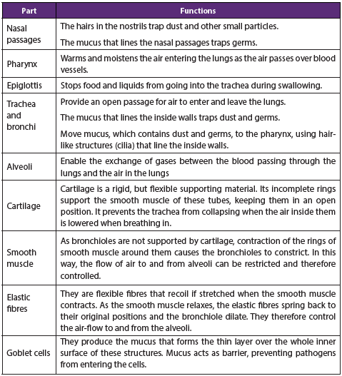
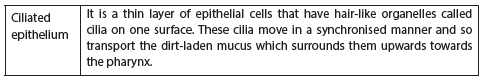
Self-Assessment 11.2
1. List the adaptations of the gills for gaseous exchange
2. List the structures through which air passes on its way from the nose to the alveoli.
3. Give two reasons why mammals need lungs, rather than exchanging gases
through the skin.11.4 Smoking and related risks
Activity 11.4
In groups,make research to find out main health risks related to smoking. Analyse the photographs below and answer questions that follow.
Between the lungs of individuals, A and B, which one is most likely that of the cigarette smoker?
Read the notes below to identify at least three risks related to smoking cigarette. Cigarette smoking harms nearly every organ of the body, causes many diseases, and reduces the health of smokers in general (Figure 11.6). Quitting smoking lowers the risk for smoking-related diseases and can increase the longevity. Inhaling cigarette smoke is called passive smoking and presents a health hazard to people nearby who inhale it. Of the thousands of chemicals in tobacco smoke three important ones are:
–– Carbon monoxide (CO), a poisonous gas form incomplete combustion carbon. CO in tobacco smoke combines easily, but irreversibly, with haemoglobin to form carboxy haemoglobin and therefore reduces oxygen carrying capacity of the blood. This can lead to hypotension and heart failure.–– Nicotine, a poisonous alkaloid drug that is addictive. Nicotine in tobacco smoke stimulates the production of the hormone adrenaline by adrenal gland, leading to an increase in the heart rate and raised blood pressure. Nicotine also makes the red blood cell stickier and this leads to high risk of thrombosis and hence of the strokes.
–– Tar- is a sticky and brown substance. It appears in tobacco spoke minute droplets.
Tar in tobacco smoke is a mixture of chemicals that enter the respiratory tract. It is an irritant and causes inflammation of the mucous membranes lining the trachea, bronchi and bronchioles, resulting in producing more mucus. Tar also thickens the epithelium and paralyses the cilia on its surface. As a result, cilia cannot remove the mucus secreted by epithelium lining.a. Short-term effects of smoking
–– Tar causes constriction of finer bronchioles by increasing resistance to the flow of air.
–– Tar paralyses the cilia which remove dirt and bacteria; the accumulation of
extra material in the air passage can restrict air flow.
–– Smoke acts as an irritant; this causes secretion of excess mucus from goblet
cells and excess fluid into the airways, making it more difficult for the air to pass through them.
–– Mucus accumulating in the alveoli limits the air that they can contain and lengthens the diffusion pathway.
–– Coughing of many smokers, way of trying to remove the build-up of mucus
from the lungs, can cause damage to the airways and alveoli; scar tissue builds
up which again reduces air movement and rates of diffusion
–– Infections arise because the cilia no longer remove mucus and pathogens
–– Allergens such as pollen also accumulate, leading to further inflammation of the airways, reduced air-flow in and out of the lungs, and possible asthma attacks.b. The long-term effects of smoking
–– Bronchitis: Bronchitis is inflammation of the lining of the air passages and may be acute or chronic.
–– Emphysema: One in five smokers develop the crippling lung disease called emphysema i.e. condition of gradual breakdown of the thin wall of the alveoli leading to sensation of breathlessness as the gaseous exchange reduces.
–– Lung cancer: Lung cancer usually starts in the epithelium of the bronchioles and then spreads throughout the lungs as dividing cells cease to respond to the normal signals around them and form unspecialized masses of cells called
tumours. The tar is carcinogen i.e. contains chemicals which cause cancer. The irritation causes thickening of the epithelium by extra cell division and this may trigger the cancer. Almost all people who die from lung cancer are smokers.Self-Assessment 11.4
Analyze the photograph and share ideas with your group members.
1. Between the baby and the parent who will suffer more the effects of tobacco? Give reasons
2. Discuss any side effects of smoking.
3. Design a sign post to advocate against smoking.End of unit assessment11
1. Match the terms in Column A with the correct definition in Column B.
2. Describe how the human lungs serve as good gaseous exchange organs. Emphysema is a disease of the lungs. People who smoke cigarettes are more likely to suffer from emphysema. The diagrams show lung tissue from a healthy person and lung tissue from a person with emphysema.

a. Identify on the figure above by using E (for emphysema) and we (without emphysema) and give a reason for your choice.
b. Explain how emphysema reduces the amount of oxygen which diffuses into the blood.
c. What are the features that make the gill of fish an efficient respiratory organ?
d. Compare respiratory system of fish and with that of a mammal.
e. Why do people who smoke have high chances of developing lung cancer?
f. Design a simple model that shows the structure and functioning of gas exchange system in mammals.UNIT 12: GAS EXCHANGE IN PLANTS
Describe structures of gaseous exchange organs in plants
Learning objectives
By the end of this unit I should be able to:
–– Describe the structure of the stoma.
–– Explain how stomata, lenticels and breathing roots are adapted to their function.
–– Explain the theories of opening and closure of stomata stating limitations of each.
–– Relate the differences between the structures of aquatic and terrestrial leaves to a habitat.
–– Draw and label a diagram of stoma as observed under alight microscope.
–– Compare gaseous exchange structures of aquatic and terrestrial plants
–– Relate the structure and function of aquatic and terrestrial plants
–– Defend the relationship between structure and function in aquatic and terrestrial plantsIntroductory activity
Suggest the different parts of a plant that are used in gaseous exchange12.1 Structure of stoma
Activity 12.1
Requirements
Light microscope, glass slide, cover slip, Commelina zebrine leaves, razor blade,
forceps, Pasteur dropper and iodine solution.
Procedure
–– Identify Commelina zebrina or commelina tradescantia plant nearby the school. You can also use any other monocotyledonous plant with succulent leaves.
–– Remove a leaf from a plant. Then peel off gently the lower epidermis. It must be thin enough to allow light to pass through–– Smear the epidermis on a slide containing one drop of dilute iodine solution.
–– Put on a cover slip and then observe under the lower and medium magnification.
–– Repeat the observation in morning hours and in the afternoon hours.Questions
1. Why should the sample used in the preparation must be transparent?
2. Draw and label structures observed under light microscope
Stomata (stoma in singular) are microscopic pores in epidermis of the leaves and stems of terrestrial plants. They function in gas exchange between plant and the atmosphere and in transpiration.
Each stoma is bordered by two saucer shaped cells called guard cells, which are specialized epidermis cells whose movements control the size of the aperture (pore). Unlike other epidermis cells, guard cells have kidney shape and have many chloroplasts. Their inner cell wall is thick and less elastic while the outer cell wall is thin and more elastic. Guard cells shrink when the plant has too little water. This
closes the stomata. When the plant has enough water, the guard cells swell up again. This opens the stomata. In this way, the guard cells enable gaseous exchange. Oxygen in the atmosphere diffuses through the stomata into the air spaces between the cells of the spongy mesophyll tissue while carbon dioxide diffuses through the stomata out to the atmosphere.Self – assessment 12.1
Analyze the diagram below and answer to the following questions
1. What title fits better to this diagram:
a. an open stoma
b. a closed stoma
c. of a guard cell and neighbouring cells
d. of a stoma and neighbouring cells
2. The part labelled C is:
a. Vacuole
b. Thick inner cell wall
c. Chloroplast
d. Thin outer cell wall
3. The part which better represents the neighbouring cell is:
a. Part A
b. Part B
c. Part C
d. Part D
4. If the guard cells become more turgid, what is more likely to happen?
a. The cells A will swell
b. The pore will increase its diameter
c. The number of structures C will decrease
d. The structure B will stretch in ward
5. Which of the following statements is false about that diagram?
a. The stoma is closed
b. The inner cell wall of guard is thicker than the outer cell wall
c. There are many chloroplasts in neighbouring cells
d. The guard cells have many chloroplasts.12.2 Theories used to explain the mechanism of opening and closure of stomata
Activity10.2
The diagram below shows a closed and an open stoma.
Carefully analyse the diagrams above and brainstorm your observation.
Illustrate how stomata open and close
Many theories have been proposed regarding opening and closing of stomata. The four important theories of stomatal movement are the following:
–– Theory of photosynthesis in guard cells
–– Theory of starch sugar inter-conversion
–– Theory of glycolate metabolism and
–– Theory of active potassium pump.
The combined outcome of the four theories shows that in general stomata open
during the day (light) and close during the night (dark). But how does this happen?In light, guard cells are stimulated. They absorb K+ ions from the neighbouring cells. K+ ions make the guard cells more permeable to CO2. As the guard cells perform photosynthesis, the concentration of CO2
falls and the pH rises. Elaborated starch therefore splits into malate. The high concentration of malate and the rise of pH in guard cells develop a decrease in water potential. Hence, the guard cells withdraw
water from the neighbouring cells and extend backward leaving an open pore in between whereby water is lost by evaporation.During the night, there is no light to stimulate neither the absorption of K+ ions nor the photosynthesis. Guard cells undergo cell respiration using photosynthetic products as source of energy (carbohydrates: malate, glucose). Therefore, the concentration of malate decreases making the guard cells hypotonic than neighbouring cells. As guard-cells lose their water content, they shrive and the pore in between closes. Stomatal transpiration ceases.

Plant physiologists are certain that stomatal aperture varies as a result of changes in the turgidity of the guard cells. But they are less certain about how these changes are brought about, though the following observations have been made:
–– Most stomata open during the day and close at night
–– Some stomata show a circadian (daily) rhythm of opening and closing even when kept in constant conditions
–– Stomata generally close when a plant suffers water stress, for example, when
transpiration exceeds water absorption
–– The stomata of some desert plants close during the day and open at night to reduce transpiration
Plants can therefore vary the stomatal aperture. This allows a compromise between the need to conserve water and the need to exchange gases for photosynthesis.The compensation point is the point when the rate of photosynthesis is equal to the rate of respiration. This means that the CO2 released from respiration is equivalent to that which is taken up during photosynthesis. The compensation point is reached as light intensity increases. If the light intensity is increased beyond the compensation point, the rate of photosynthesis increases proportionally until the point of light saturation is reached, beyond which the rate of photosynthesis is no longer affected
by light intensity.For a plant under water stress, its need to conserve water is greater than its need to obtain carbon dioxide for photosynthesis. Under these conditions a plant secretes abscisic acid (ABA). This is a chemical messenger which causes stomata to close. It is thought that ABA triggers a metabolic pump which actively secretes potassium ions out of guard cells, causing the cells to lose water and become flaccid.
Self-assessment 12.2
1. According to the ionic theory of opening and closing stoma, what is the role
of potassium ions in the guard cell?
2. What would happen to guard cells if the concentration of malate doubled?
3. What is meant by compensation point?12.3 Structural adaptations and function of stomata, lenticels and breathing roots.
Activity 12.3
Observe the adaptations of these plants for gas exchange.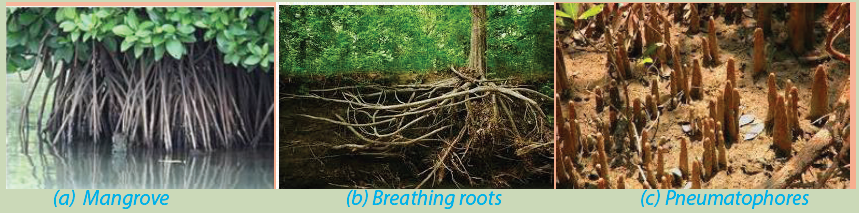
1. How is each of these plants adapted for gas exchange?
2. Read through the notes that follow and describe any two adaptations for gas exchange
The exchange of atmospheric gases is essential to photosynthesis and cell respiration. In plants, the gas exchange takes place through stomata, breathing roots, lenticels and cuticles. Most stomata are on the lower epidermis of the leaves on plants. Unlike other plant epidermal cells, the guard cells contain chlorophyll to carry out photosynthesis. This allows the cells to expand or contract to open or close the stomata.Guard cells swells, through the process of osmosis, to allow opening of the stomata for CO2 to enter and excess O2 and H2O to leave, and they shrink in order to force the stomata shut either partially or completely to prevent dehydration. The number of stomata on the epidermal surface depends on the ecology of plants. Usually, plants on wet climate have fast growth and a high concentration of stomata. Plants on dry weather have lower rates of photosynthesis, lower growth and lower concentrations of stomata.
Xerophytic plants or xerophytes are plants that inhabit arid regions (desert). They have the following adaptations:
–– Stomata sunken in grooves and reduced in number
–– Ability to fix CO2 at night, so the stomata are closed during the day.–– Epidermis infolded to reduce the surface area
–– Leaves reduced to scales or thorns to reduce the surface area for transpirationHydrophytes or water plants are plants that grow submerged or partially submerged in water. To thrive in this environment, hydrophytes have the following features
–– developed stomata on large upper surface of their leaves (rather than underside) making gas exchange more efficient.
–– large airspace to facilitate evaporation from the mesophyll.
–– little or no lignified supporting tissues on the submerged parts.
–– poorly developed transport tissue,
–– stems and leaves have little or no lower cuticle large continuous air spaces,
forming reservoir of oxygen and CO2 which also provides buoyancy to the plant tissues when submerged.
A halophyte is a plant that grows in water of high salinity and they comeinto contact with saline water through its roots or by salt spray, such as in saline semi-deserts, mangrove swamps, and marshes. Halophytes are adapted in the flowing ways;
–– store water in succulent tissues which have high concentration of salt. They can thus take up water from the sea water by osmosis.
–– extensive air spaces throughout the stem and roots making air available to all cells, and giving buoyancy to the stem and leaves at highest tides.
–– they develop breathing roots called pneumatophores which grow upward and protrude out of the ground. e.g. mangrove tress.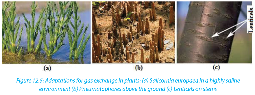
Self-assessment 12.3
What features are common to plants living in desert and saline soils?End of unit assessment 12
Section A: Objective questions
1. You are provided with the diagram below. Analyze it and then chose the correct answer. Transpiration in the leaf depends on the transport of potassium ions into:
a. Into O
b. Into P
c. From M to L
d. From M to Q
e. From P to L.
2. The theory that says than during the light time, potassium pumps open and this brings about diffusion of CO2 from the atmosphere to the guard cells for photosynthesis is called:
a. Theory of photosynthesis in guard cells
b. Theory of starch sugar inter-conversion
c. Theory of glycolate metabolism
d. Theory of active Potassium Pump.
3. What is the main difference between the guard cells and the other epidermal cells?
a. Guard cells have chloroplast while the remaining epidermal cells have no chloroplast
b. Guard cells have oval shape while other cells have cubic shape
c. Guard cells are beneath the spongy mesophyll
d. Guard cells are covered by a transparent cuticle
4. Water lily is:
a. Xerophytes
b. Halophyte
c. Hydrophyte
d. Heleophyte
5. Mangroves are plants adapted to estuaries or marine region with high salinity.
What statement does not describe the adaptations of mangroves?
a. The presence of lenticels that help in gas exchange and evaporation
b. Presence of large number of stomata on the upper side of the leaves
c. The presence of pneumatophores which are breathing roots
d. Presence of succulent tissues that have high concentration of saltSection B
6. Explain how gaseous exchange occurs in the leaf.
7. How does gaseous exchange occur in woody stems?
8. Describe how roots get oxygen.
9. a. Draw a labelled diagram of a stoma
b. Draw arrows on the diagram to show how gaseous exchange occurs.
10.The drawing shows a 24-hour cycle for the opening and closing of stomata from
the same plant.
a. Explain how this cycle of opening and closing of stomata is advantageous to the plant.
b. The diagram shows the potassium (K+) concentrations in the cells around open and closed stomata in commelina. The concentrations are in arbitrary units.
i.Explain how the movement of K+ ions accounts for the opening of stomata.
ii. Explain how K+ ions are moved against a concentration gradient.UNIT 13: GROWTH AND DEVELOPMENT IN PLANTS AND ANIMALS.
Key Unit Competence
Account for the processes of growth and development in plants and animalsLearning Objectives
By the end of this unit, I should be able to:
–– Describe dormancy as a state of inactivity to absolute minimum due to the morphological and physiological state of a plant structure
–– Explain how dormancy is maintained and broken
–– State the conditions required for germination
–– Outline the role of enzymes in the process of germination
–– State types of plant growth hormones and their functions
–– Identify the hypocotyl and coleoptile in a germinating seed
–– Describe the stages and types of germination
–– Recognize that a meristem is a growing point of the plant and the main meristematic regions of a tree
–– Describe current views about photoperiodic control of flowering
–– Observe structures of endospermic and non-endospermic seeds
–– Demonstrate how fruit and seed dispersal takes place
–– Demonstrate hypogeal and epigeal germination
–– Carry out an investigation to distinguish between primary and secondary growth
–– Appreciate the importance of fruit and seed dormancy and germination in the life cycle of plants
–– Carry out an experiment on the development of eggs at different temperatures
–– Describe the process of metamorphosis in arthropods and amphibians
–– Distinguish the various stages of development in frogs
–– Analyse complete and incomplete metamorphosis
–– Compare growth patterns in arthropods and vertebrates
–– Appreciate the demands of the terrestrial environment to the adaptation of amphibiansIntroductory activity
Suggest morphological differences by which different organisms develop and grow to maturity.
13.1 Fruit, seed and bud dormancy
Activity 13.1
–– Put dry bean seeds and maize grains in boiling water for 10 minutes.
–– Use nails to make a longitudinal section of the bean and maize.
–– Compare the two sections.
–– From your point of view, do you think that all plants’ organs are always active?
Justify your point of view.
–– Produce a picture showing how plant organ (seed and bud) behaves in
relation to oxygen, temperature and water.A seed is a plant organ that develops from the fertilized ovule. As the seed embryo develops from the zygote, the seed makes a food reserve of macronutrients including carbohydrates, proteins and lipids. The amount of reserved food type depends on the plant species. Legumes like peanuts and soybeans store proteins as well as fats while beans store more starch and proteins. A seed consists of a plant embryo surrounded by the food supply in cotyledon or endosperm and a protective coat called seed-coat or testa. The plant embryo is composed of the radicle or embryonic root, the hypocotyl which is the embryonic stem and the plumule, the embryonic leaves

Dormancy is a period of cells’ inactivity due to a very low metabolism to prevent growth when environmental conditions are unfavorable. The dormancy can be for fruit, bud or seed.
a. Bud dormancy
Bud dormancy is a suspension of most physiological activities and growth that can be reactivated. It may be a response to environmental conditions such as seasonality or extreme heat, drought, or cold. The exit from bud dormancy is marked by the resumed growth of the bud. Bud dormancy may proceed to dormancy of the whole plant.b. Seed dormancy
Seeds exhibit dormancy to avoid growth during unfavorable conditions. During the last stages of its maturation, the seed dehydrates until it gets dry. The embryo which is surrounded by a food supply (cotyledons, endosperm or both), enters dormancy.
Most seeds are enough durable that can last a year or two until conditions are favorable for germinating. However, the length of time a dormant seed remain viable and capable of germinating varies from a few days to years depending on the plant species and environmental conditions. This may justify the reason why after a bushfire or other environmental disruption, vegetation reappears as their seeds
have accumulated and stayed for longer in the soil. Many plants are easily grown from seeds. Although its embryo is alive, a dormant seed cannot germinate until it is exposed to certain environmental conditions to break the dormancy. Thus, it germinates and starts to grow.Conditions needed for seed germination
Germination is the process by which a seedling develops from a seed embryo. There are requirements for germination to be successful. Environmental conditions such as water, oxygen, and temperature trigger seed germination. For a seed to germinate, it must be found in place where all these conditions are available. If one condition is not available, germination becomes impossible. These conditions include:a. Water
For a seed to germinate, it requires water. Mature seeds are very dry and must absorb water by imbibition to germinate. Water softens the seed coat for embryo sprouting and provides a medium for reactions during hydrolysis. Enzymes breakdown macronutrients such as starch, proteins and fats stored in the cotyledons and the endosperm to simple sugars such as glucose, amino acids which provides energy for the embryo to grow.b. Oxygen
This is needed for needed for cellular respiration by oxidizing glucose to liberate ATP to provide energy for embryo development.c. Temperature
Seeds germinate only if exposed to their optimum temperature varying between 5 to 40 0c depending on the seed species. Enzymes are sensitive to temperature because enzymes need a favorable temperature to work efficiently. Burying seeds too deeply in the soil prevents them from germinating as they are cut off from temperature and air.
Self-assessment 13.1
1. Define the term dormancy?
2. Explain how dormancy is maintained and broken?
3. Suggest the advantage of dormancy in plants?
4. What are conditions needed for seed germination?
5. Explain the role of enzymes during the process of seed germination.
6. Draw and label endospermic and non-endospermic seeds13.2 Types and stages of germination.
Learning activity 13.2
Conduct the experiment below to compare the two types of germination. Requirements Two bean seeds or soybean seeds, two sorghum or maize grains, water and two plastic devices/containersProcedure
–– Fill each container with soil
–– Put two grains of maize in the container labeled A and two seeds of bean in the container labeled B
–– Pour some water in each container simply to moisture the soil.
–– Put both containers A and B in a secured place.
–– Record your observations from day five to day twenty one with emphasis to developmental changes.
In conducive environment, a seed can germinate. Germination involves three main
stages: imbibition, radicle sprouting and plumule sprouting.
The dormant seed contains very little water, when placed in moist conditions, it absorbs water by osmosis. The intake of water by a dry material is called imbibition.
As the seed absorbs water and oxygen,it swells,the ambryo grows and the seed-coat cracks, and the radicle also called primary root emerges downward. This is seed coat rupture and radicle sprouting. The primary root is the fi rst organ to appear during embryo development. The plumule cells develop and the fi rst leaf forms upwards in the process known as plumule sprouting.
A germinating seed shows the primary root from which secondary roots start to
develop and the primary leaf which is surrounded by a protective sheath called
coleoptile. The Hypocotyl is the stem below the plumule while the epicotyl is the
stem above the plumule.
13.2.1. Types of seed germination
There are two types of seed germination: hypogeal and epigeal seed germination.
a. Epigeal germination
Epigeal germination is typical to non-endospermic seeds like beans, soybeans and coff ee seeds.In this type of germination, the cotyledons come above the soil surface into air and light, due to rapid growth and elongation of hypocotyl. The cotyledons are green and make food to be used by stem during growing season. They fi nally dry off and seedlings become independent. The example of epigeal germination is like the one found in bean seeds.
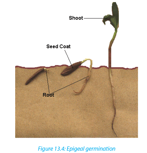
b. Hypogeal germination
In this type of germination, the cotyledons remain in soil or just above the surface.Here the epicotyl elongates, pushing plumule upwards. Cotyledons do not turngreen and gradually dry up and fall off . An example of this type of germination is found in pea, mango, and groundnut seeds.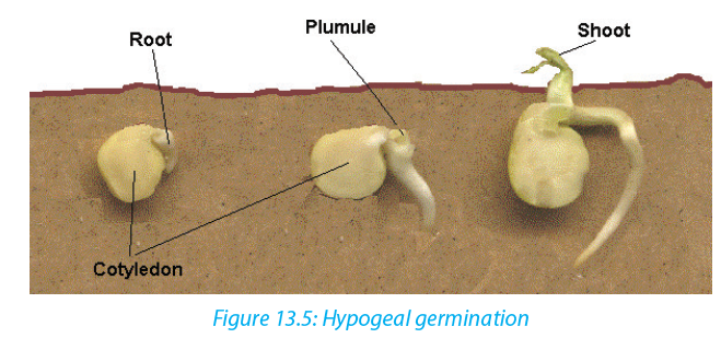
Self-assessment 13.2
1. Use diagrams to demonstrate and distinguish the types of seed germination.
2. Describe the three stages of seed germination.13.3 Primary and secondary growth
Learning activity 13.3
Move around the school garden or anywhere in your surroundings, then collect two plant species in which one is a monocotyledon and another is a dicotyledon.
1. By examining their physical characteristics, describe similarities and differences between collected plants.
2. Predict the reason why all plants grow in length.
3. From your observation, suggest/ predict the reasons justifying why monocotyledons do not grow in width while dicotyledons do.
Growth is a permanent increase in the size of an organism or of some parts of it. It is brought by cell division and the assimilation of new material within the cells which result from the division and the cell expansion which follows. Cell expansion is particularly noticeable in plants, where rapid enlargement can occur as a result of water taken up by osmosis. If the organism achieves its mature size, it starts
development, the formation of new structures or organs to perform specific functions. That is the production of reproductive organs and locomotive or protective organs. It is controlled by cell differentiation. Growth is either primaryor secondary.a. Primary growth
Primary growth consists of the increase in length and formation of primary plant organs including roots, stem, leaves, flowers, and fruits. It occurs in most herbaceous plants. The roots elongate to penetrate the soil, and shoots elongate to reach the sunlight. Primary growth is controlled by apical meristems. Meristematic cells divide by mitosis. Some daughter cells absorb water and nutrients. As the cell takes
in water the cell walls stretches, the cell elongates and slightly enlarges. After this growth by elongation, cells differentiate and specialize for specific functions. The cells formed from apical meristems do not expand laterally and this limits their size. The herbaceous plants exhibiting such growth tend to be short lived. They are called annual plants because most of them do not live for more than one growing season
(a year) after which they enter dormancy and survive as seeds.b. Secondary growth
It consists of getting wider or thicker, and occurs in roots and stems of perennial woody plants, all trees and shrubs. Woody plants grow taller than herbaceous plants and they live longer, more than two years some plants while some others may live for and over 30 years.
Secondary growth is controlled by lateral meristems: vascular cambium and cork cambium. Vascular cambium is located between primary phloem and primary xylem. Vascular cambium cells divide by mitosis. Some cells remain meristematic while other cells expand sideways and differentiate. They form the secondary phloem outwards and the secondary xylem inwards. As the secondary growth continues,the outermost phloem cells die. Their death causes no matter to the whole plant as dead cells are continually replaced by new ones.
The secondary xylem cells are strengthened by the accumulation of lignin and cellulose. They form a wood also called secondary xylem providing the mechanical support to very taller trees. The cork cambium situated between the epidermis and secondary phloem produces the cork cells by mitosis. The cork cells are pushed toward the epidermis and accumulate a waxy substance called suberin making
the cork waterproof. The cork gradually replaces the epidermis. Like the epidermis, the cork protects the plant from dehydration and infection. The expansion of the internal tissues results into the continual cork shedding. Because of meristematic cells that continuously divide, perennial plants have unlimited growth.Self-assessment 13.3
1. In the table list the differences between primary growth and secondary growth.
2. Describe briefly what the wood is and its major function.
3. What is the name given to a substance that makes the cork to resist plant dehydration?
4. Identify the importance of apical and lateral meristems in plant growth.13.4 Determination of growth
Activity 13.4
In the learning activity 13.1 you have grown two types of seeds (bean and maize). Based on that experiment do the following to investigate the primary growth in a seedling.
–– Use a centimeter ruler, measure the height of each plant once each five days.
–– Use a thread and a centimeter ruler to measure the width.
–– Record your measurement in a tabular form as shown below.
1. On the basis of the period of time indicated in the above table, count the number of leaves. What do you notice?
2. Among the two given plants, predict the one with an increased volume of protoplasm and dry mass.The growth rate of an organism is measured by recording the variation in length, in width and in mass through a period of time. The aspects that can allow the measurement of the growth of a plant are the following:
–– The increase in the dry mass
–– The increase in the volume of protoplasm
–– The increase in the length
–– The increase in the thickness
–– The ability to reproduce.
Experiments show that the growth rate is faster in young plants and starts to decrease as the plant gets older.Self-assessment
1. A sign is hammered into a tree 2 m from the tree’s base. If the tree is 10 m tall and elongates 1m each year, how high will the sign be after 10 years? A mark is hammered into a tree 2 m from the tree’s base. If the tree is 10 m tall and elongates 1m each year, how high will the mark be after 10 years
2. What features would enable you to conclude that this organism has grown?
3. Would you expect a tropical tree to have distinct growth rings? Why or why not?
4. If a complete ring of bark is removed around a tree trunk (a process called girdling), the tree usually dies. Explain why?hormone prevents the action of another they are antagonist.The plant hormones include five major groups:
a. Auxins
It is produced in growing regions of plant such as shoots, tips, and young leaves, and developing fruits. The most known auxin is Indol Acetic Acid (IAA). Artificially produced auxins are widely used to:
–– Promote the cell elongation in the region behind the apex of the stem
–– Promote root formation on stem and leaf cuttings
–– Increase number of fruit
–– Prevent dropping of fruit
–– Prevent sprouting of stored potatoes and onionsb. Gibberellin or gibberellic acid (GA)
Produced in all parts of plants, especially in immature seeds. Gibberellin has different functions:
–– It promotes the parthenocarpy (formation of fruits without fertilization).
–– It breaks down the bud dormancy
–– It promotes the seed germination.c. Cytokinins
They are produced in developing roots, fruits and seeds, cytokinin and have the following roles:
–– Promote cell division
–– Promote the growth of lateral buds
–– Promote the growth of fruits
–– Are used to delay aging and death (senescence).
–– Work with gibberellins to break down the bud dormancy and to promote the seed germinationd. Abscissic acid (ABA)
It is produced in leaves:
–– Promotes the abscission i.e. falling of some organs of the plants
–– Promotes the bud and seed dormancy
–– Inhibits the stem growth during the stress
–– Promotes stomatal closure
–– Inhibits other hormones blocking thus the growth.e. Ethene or ethylene (C2H4)
It is produced in fruits, flowers, leaves and roots and:
–– Promotes ripening of fruit
–– Promotes flowering in mangoes and pineapples
–– Promotes abscission (detachment of leaves).Commercial application of synthetic phytohormones
Artificial auxins are widely used. For example, 2,4-D or 2,4-dichlorophenoxyacetic acid and MCPA 4-chloro-2-methylphenoxyacetic acid are used as weeds killer (selective herbicides). Synthetic auxins used in right concentration, cause excessive growth and very rapid metabolism of broad-leaved dicotyledons herbaceous plants that are weeds in cereals. As weeds grow faster, they soon die increasing thus the cereals crop yield.Napthaleneacetic acid (NAA) or rooting powder is another kind of auxin sprayed on stem cuttings for stimulating the development of adventitious root. This is very important in vegetative propagation of plants of economic value. Synthetic gibberellin helps to increase fruit crop yield because when sprayed on non-fertilized flowers, they promote parthenocarpy (fruit formation without fertilization). It is
mostly used in production of seedless grapes such as seedless tomatoes or citrus. Synthetic cytokinin is sprayed on cut flowers, fruits and vegetables to keep them fresh and extend shelf-life.Self-assessment 13.5
1. What are the plants hormones?
2. Describe the role played by each plant hormone.
3. Explain why some plants develop lateral shoots when the apex is cutoff.13.6 Plant movements and photoperiodism
13.6.2. Plant movements
Activity 13.6.2
Take the container with the bean seedling in the classroom nearby the window and observe the changes within one week.
Like animals, plants move as response to changes in their environment (internal or external changes). Plant movements are grouped into two categories: tropisms and nastic movements.1. Tropism
A tropism is a movement of parts of a plant in response to external stimulus. The movement is always a growth movement. External stimuli cause changes in the direction of the plant’s growth, such as bending, turning or curving. Tropic responses are described as positive or negative depending on whether growth is towards or away from the source of the stimulus respectively. According to the type of stimulus,
tropisms are classified as follow:
a. Phototropism
Phototropism is a directional growth depending on the direction of the light source. Growth towards a light source is a positive phototropism, while growth away from light is called negative phototropism. It is believed that light destroys auxin where it strikes the stem, causing an imbalance in which the side of the stem that receives less light has more auxin. This causes the plant to have elongated cells on the farthest
side from the light. Because more auxin is present, the cells on the darker side are able to elongate more than the cells on the lighted side, causing the plant to bend toward the light.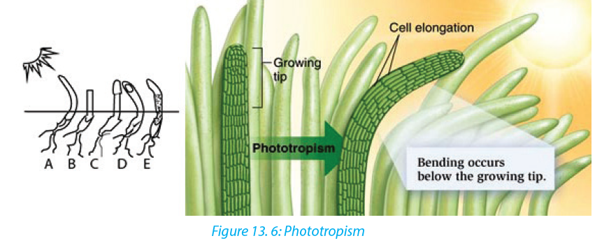 Phototropism is caused by unequal distribution of auxins in the plant stem. There is more auxin on the side of the stem away from the light. This results in cell elongation, but only on that side. As the cells grow, the stem bends toward light.b. Gravitropism
When a seed germinates, the young root turns downward regardless of the way in which the seed is planted. This bending, known as positive geotropism, enables a plant to anchor itself in the soil. The young stem, which turns upward away from the earth, is said to be negatively geotropic. The gravity causes auxins to concentrate on the down side than on the upper side. High concentration of auxins on the down side of the stem promotes elongation of its cells. Therefore, the stem grows and bends upward. But high concentration of auxins on the down side of the stem inhibits cell elongation; therefore, the root grows and bends downward, that is why the root grows shorter than the stem.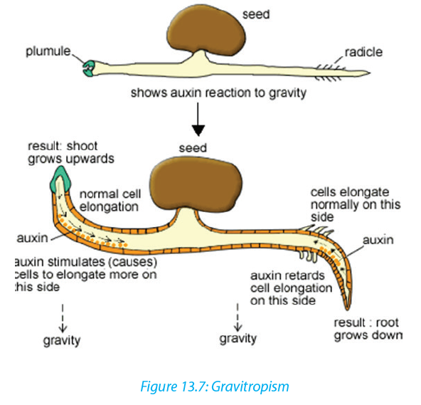 Use of clinostat in tropism
A clinostat is a piece of laboratory equipment with a turntable that allows a plant placed on it to be exposed to a stimulus such as light or gravity equally on all sides.
As the clinostat is turning, all parts of the plant receive the stimulus equally and therefore the plant will not bend but will grow straight away. In otherwise the clinostat is used to minimize the effect of the direction of a stimulus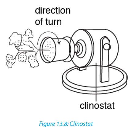 c. Chemotropism
Chemotropism is a growth of a plant or plants parts, navigated by chemical stimulus from outside of the organism. An example of chemotropic movement can be seen during the growth of the pollen tube, where growth is always towards the ovules.
Fertilization of flowers by pollen is achieved because the ovary releases chemicals that produce a positive chemotropic response from the developing pollen tube. It is different from chemotaxis. The major difference being that chemotropism is related to growth, while chemotaxis is related to locomotion. For example, the movement of antherozoids (sperm) in ferns, swim toward the chemicals produced by archegonia.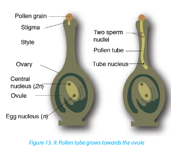 d. ThigmotropismThigmotropism is a movement in which an organism grows in response to touch or contact stimuli. Usually thigmotropism occurs when plants grow around a surface, such as a wall, pot, or trellis. Climbing plants, such as vines, develop tendrils that coil around supporting objects. Touched cells produce auxin and transport it to untouched cells. Some untouched cells will then elongate faster so cell growth bends around the object. Some seedlings also inhibit triple response, caused by pulses of ethylene which cause the stem to thicken (grow slower and stronger) and curve to start growing horizontally.
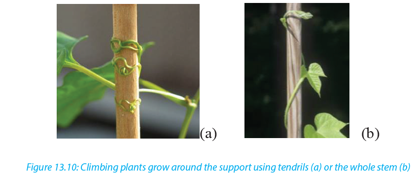 2. Nastic movement
Nastic movements are non-directional responses to stimuli such as temperature, humidity, light and irradiance. An example of such a response is the opening and closing of flowers known as photonastic response and the opening and closing of carnivorous plants known as thigmonastic response. The folding and unfolding of some sensitive plants like Mimosa pudica when touched, is a nastic response which protects them from insect damage or water loss during winds They are named with the suffix “-nasty” and have prefixes that depend on the stimuli:
–– Photonasty (response to light),
–– nyctinasty (movements at night or in the dark),
–– chemonasty (response to chemicals or nutrients),
–– hydronasty(response to water),
–– thermonasty (response to temperature).Self-assessment 13. 6
1. A bean seedling has been placed on horizontal position as shown by the diagram below.
a. Illustrate the expected shape of the shoot and that of the root after a week.
b. Suggest names (of what) for the expectations in the above experiment.
c. Based on the above experiment, draw a diagram illustrating the shape and size of cells of both upward and downward side of the root.
d. Suggest a technique that can be used to minimize the effect of the stimulus
in this experiment.
2. a. What is the difference between ‘antagonistic’ and ‘synergistic’ when referring to plant growth substances?
b. What are the two plant growth substances that act antagonistically and which act synergistically?
3. Copy and complete the following table
Activity 13.6
Most of plants grow toward the sunlight direction. The few which have been observed respond to external stimuli like touching and temperature.
1. From your experience, brainstorm what will happen to the plant when:
–– it is exposed to the direction of the sunlight–– its growing part is exposed to a physical material like a stone
–– a living organism touches on it (Mimosa pudica)
2. Use internet and textbooks to describe why some plants flourish during long daylight while others do not?
3. What names can be given to the above processes.13.6.1. Photoperiodism
The light provides energy that plants need to make its own food. The duration of daylight affects the plant growth and plant development.
Photoperiodism is a plant physiological response to relative lengths of daylight and darkness. Photoperiodism affects many plant processes, including the formation of storage organs, flowering and bud dormancy.
Plants monitor changes in day length with a bluish, light-sensitive pigment called phytochrome. The alternation of darkness and light triggers the phytochrome to change from one chemical form to another. By detecting the type and the amount of phytochrome present, plants determine the length of darkness and light each day.
One of the effects of photoperiodism is that plants produce fruits at different times and are classified into three categories:
–– Short-day plants (SDP),
–– Long-day plant (LDP)
–– Day–neutral plants (DNP).
–– Short-day plants (SDP): they only flower when the days are short and the night are longer than a certain length. Examples strawberry, blueberry, goldenrods, cocklebur and soybeans, tobacco are short day plants for flowering.
–– Long-day plant (LDP): they produce flower when the period of daylight exceeds a critical minimum length. Radishes, asters, apple trees, squash trees, and beets.
–– Day–neutral plants (DNP) are not dependent on day length for flowering. They produce flower regardless of the length of the daylight. Day neutral plants for flowering include tomatoes, roses, corn, cucumber, carrot, cotton and beans. They can produce fruit throughout the entire growing season.Self –assessment13.6.1
1. What role does phytochrome play in photoperiodism?
2. What is the difference between a short-day plant and a long-day plant?
3. What could happen if a short-day plant is grown in the long days of summer?13.7 Metamorphosis and growth patterns in insects and amphibians
Activity 13.7
From a pond or swamp, collect frog eggs together with water. Keep some eggs in warm conditions at room temperature between 2 to 30 days.
1. visit them regularly and note the observation.
2. Use the diagram below and relate your observation from question (1) to the one given in the diagram.
1. Metamorphosis and growth patterns in animals
Metamorphosis is a process consisting of changes in body form of a young organism before it reaches its adult size and becomes sexually mature, for example, the change from tadpole to frog or from caterpillar to butterfly.a. Metamorphosis and growth patterns in insects
All insects develop and grow by metamorphosis. Some insects show incomplete metamorphosis. In incomplete metamorphosis, an immature nymph is hatched from the egg that looks like the adult, but it is smaller, and its wings and reproductive organs are undeveloped. It molts several times with each molt, the wings become larger and more fully formed. The fi nal molt transforms the nymph into an adult
that can reproduce and fl y. Insects with incomplete metamorphosis are known as heterometaboles e.g. weevil, cockroach (Periplaneta americana), Grasshopper; mayfl ies; dragonfl ies and termites.
Complete metamorphosis
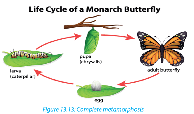
In complete metamorphosis eggs hatch into larvae which are morphologically, physiologically and behaviorally diff erent from adult (wormlike larva) or caterpillar. The caterpillar molts several times, when it reaches its full size, it prepares the pupa, or chrysalis, a hard, sometimes thorny and oval structure. Inside the pupa, the larval tissues break down and group of cells called imaginal disks develop into wings
and other tissues of the adult: imago stage, the pupa becomes sexually mature.Examples: Butterflies, moths, mosquitoes, beetles, bees, housefly.

Importance of metamorphosis
In life cycle based on complete metamorphosis, the larval and adult stages often fulfill different functions, live in different habitat and eat different foods. Example: mosquitoes.
Metamorphosis also enhances insect survival by helping insects survive harsh period. Example: butterflies (caterpillars feed on leaves, but adult butterflies feed on nectar from flowers).b. Metamorphosis and growth patterns in amphibians
Many amphibian species like the frog breed in water and their eggs are fertilized externally. The fertilized eggs hatch into swimming, tailed larvae called tadpoles. Tadpoles, which usually live in water, look somewhat like small fish. A tadpole has an oval body, gills for breathing, and a long, muscular tail with fins along the upper and lower edges for swimming.
Then the tadpole grows legs—the hind legs appear first—and resorbs its tail. It loses its gills and grows lungs, and the structure of the heart, digestive system, and skeleton changes. The horny beak and other mouthparts adapted for eating algae disappear and are replaced by the long, sticky, projectile tongue that helps adult frogs catch insects. Frogs reach reproductive age anywhere from several months to
several years after metamorphosing.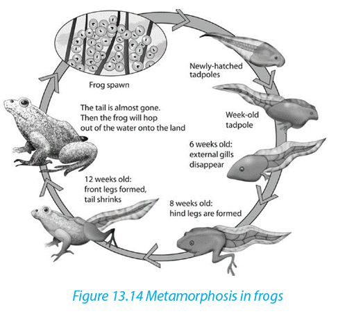
Self-assessment 13.7
1. What is metamorphosis?
2. Describe the changes that occur during metamorphosis in frog.
3. Discus reasons why complete metamorphosis may have greater adaptive value for an insect than incomplete metamorphosis.
4. Compare metamorphosis of a butterfly and that of a grasshopper.End of unit assessment 13
1. What do you understand by:
a. Dormancy
b. Gravitropism
c. Chemonasty
d. Phytochrome
e. Short-day plants
2. a. What factors can allow to measure the growth of a plant?
b. State any four external factors that can affect the growth of a plant.
3. a. State any three characteristics of the phytohormones.
b. What is phytohormone (s) responsible for:
i. The falling of some plant organs during the stress
ii. The fruit ripening.
iii. Development of the lateral buds
iv. Stem growth and parthenocarpy
c. What is meant by parthenocarpy? Give one example of a plant that shows
this phenomenon
4. A seedling has been grown in an opaque box receiving the light from a single
direction as shown by the diagram below
a. What will happen on the coleoptile (seedling) as it grows?
b. Suggest a name to the phenomenon investigated in this experiment.UNIT 14: SUPPORT AND LOCOMOTION
Key Unit Competence
Explain and demonstrate modes of locomotion in protists, insects, fish, amphibians, birds and mammalsLearning objectives
By the end of this unit, I should be able to:
–– Explain non-muscular movement in amoeba or paramecium.
–– Describe support and movement on land.
–– Describe skeletal modification in birds.
–– Explain how movements and support of fish are brought about in water.
–– Explain how support structures are related to the environment of the animal.
–– Observe locomotion of animals and identify reasons for their movement.
–– Demonstrate the arrangement of muscles in fish.
–– Dissect a fish to observe its swim bladder.
–– Observe and explain the relationship between muscles, joints and musculoskeletal attachments in fish, birds, amphibians and mammals.
–– Compare the flight of birds and insects.
–– Compare the jumping movement of grasshoppers and toads/frogs.
–– Appreciate the need for locomotion in animals.
–– Recognize that the types of locomotion of animals depends on their habitat.Introductory activity
Animals have muscles and different types of skeleton.
What might happen if a large animal such as a cow does not have a skeleton.
How that animal would look like? What will happen to animal without skeleton or muscles? Can you then think about the role of skeleton and muscles in living organisms?Activity 14.1
From your experience and knowledge from books and the internet:
1. Give details about the concept of locomotion
2. How do different animals move?
3. Explain why animals need to move from one place to another?14.2 Locomotion and its requirements
Living organisms particularly animals need to move from one place to another. This is known as locomotion which should not be confused with movement which occurs in plants. Movement is the displacement of part of an organism. Therefore,
movement is a characteristic of all living things.
Locomotion in animals is brought about by the action of muscles on a skeleton.A skeleton is a rigid framework that maintains shape and supports the internal organs and provides attachment for muscles, while a muscle is a soft tissue formed by muscle cells which found in most animals. Each muscle cell contains actin and myosin proteins that produce a contraction that changes both the length and the shape of the cell. Thus, muscles function to produce force and motion. In animals without muscles such as sea sponges, locomotion is brought by mesohyl or cells which act as actual muscles. Depending on the type of animals, three types of skeletons are distinguished.1. Hydrostatic skeleton
It is mostly seen in invertebrates and earthworm. These consist of fluid filled body cavity surrounded by antagonistic sets of muscles. Movement results from compressive contraction action of the contraction of these muscles on this fluid.2. Exoskeleton
It characterizes the arthropod insects. It is a hard cuticle made of chitin which lies outside the muscles. It sheds during molting when the organism outgrows it. It does not grow because it is a dead material.3. Endoskeleton
It is seen in vertebrates where the bones and cartilages are found within the interior of the body on which muscles are attached. It is a living tissue and it grows with the rest of the body.
For efficient locomotion,exoskeleton and endoskeleton provide a system of levers to which muscles are attached.14.3 Types of locomotion
The locomotion can be either terrestrial, aerial or aquatic (swimming). In most animals, the locomotion is by running, climbing, crawling, swimming, jumping, gliding, hoping, and flying with aid of limbs or appendages. For animals without limbs such as snake, its locomotion is by forming its body into zig - zag, gripping the ground with its undersides and pushing itself forward. For ducks, their movement in water is by floating. Some invertebrates like roundworms, flatworms, squids, octopus, and jellyfish without special organs of locomotion are propelled by the bmuscular contractions.Advantages of locomotion
Based on the types of locomotion mentioned above, an animal is capable to:
–– Escape danger such as fire or predator
–– Look for food, water and shelter–– Reproduce
–– Avoid competition with other animals of the same or different species
–– Avoid overcrowding which enables offspring to move to another place
–– Avoid unfavourable conditionSelf-assessment 14.1
1. What is locomotion?
2. What are the requirements for locomotion?
3. Given the following animals: Frog, dragonfly, squid, spider, antelope,
kangaroo, fish, grasshopper, bee, duck, worm, zebra, snake, and cow.
Identify those which fly, crawl, hop, and or run/walk.
4. Discuss why locomotion is very important in animals?14.2 Support and locomotion in non-muscular organisms
Learning activity 14.2
From a culture of paramecium:
1. Use a microscope to observe the locomotion in Amoeba and Paramecium
2. From what you have observed identify Amoeba, Paramecium, Euglena and in Trypanosoma moves in relation to their structures / diagrams below
3. Discuss how is locomation performed in those organisms.
Non-muscular locomotion is identified in animals that belong into protoctista kingdom. Depending to individual animal, locomotion is either amoeboid, ciliary, flagellated or euglenoid type.Amoeba moves by amoeboid locomotion i.e. by putting out pseudopodia. Locomotion is not maintained in any particular direction for long. Amoeba is constantly changing shapes as it changes direction. Amoeboid locomotion is brought about by cytoplasmic streaming and between a gel and sol state. These
cytoplasmic streaming requires Ca2+ ions and ATP. Amoeboid locomotion is common to all rhizopodes including Amoeba and white blood cells of the vertebratesa. Amoeba

b) Paramecium

c. Euglena

Paramecium moves by means of cilia and Euglena move by the use of flagella. Cilia and flagella have similar structure except that cilia are short and many. Both cilia and flagella consist of fine tubes composed of an extension of plasma membrane. Euglenas have an intricate cell membrane called a pellicle. The latter is folded into ribbon-like ridges and each ridge is supported by microtubules. The pellicle is tough and flexible, letting euglenas crawl through mud when there is not enough water
for them to swim.
During cilia or flagellum locomotion, tubules slide past each other in a movement similar to that of actin and myosin filaments in skeletal muscles. Hence Ca2+ ions and ATP are also required in the ciliary locomotion.Self-assessment 14.2
1. Describe the type of locomotion found in:
a. Amoeba
b. Paramecium caudatum
c. Trypanosoma gambiense
d. Trypanoma vaginalis
e. Giardia intestinalis
2. How do cilia differ from flagellum?
3. Produce picture showing the locomotion of amoeba14.3 Support and locomotion in fish
Activity 14.3
1. Observe the freshly collected fish or the figure, to label fins and lateral line.
2. Dissect a fresh fish or observe the above given diagram. Redraw and show the swim bladder and the arrangement of muscles
3. If you have a live fish, put it in water and observe its locomotion.
4. From what you have observed, draw and label the external and internal features that contribute to fish locomotion.
Fish like other aquatic animals are adapted to such habitat in terms of locomotion
due to its structural adaptive features particularly skeleton which gives shape as well
as muscles arrangement and swim-bladder.Adaptive features of fish for locomotion in water
The streamlined body shape of the fish reduces friction between water and fish.
The body of fish is mostly covered by scales which overlap one another and point backwards and lie close to the body. The scales are covered by mucus which reduces the drag.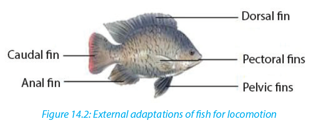
Tail or caudal fin has a large surface area, which increases the amount of water that is displaced as it provides much of the push during swimming. Paired pectoral and pelvic fins bring about downward and upward movement. With pectoral fins, the control of direction of a fish in water is possible whereas the pelvic fins bring about the balance, preventing diving and rolling. There are also unpaired dorsal and anal fins for stabilizing the fish and thus preventing it from rolling or yawing.
Fish is also adapted to locomotion in water by its strong tail muscles and highly flexible vertebrate column which enables the tail to move from side to side against water. In addition, inflexible head and neck maintain forward thrush.
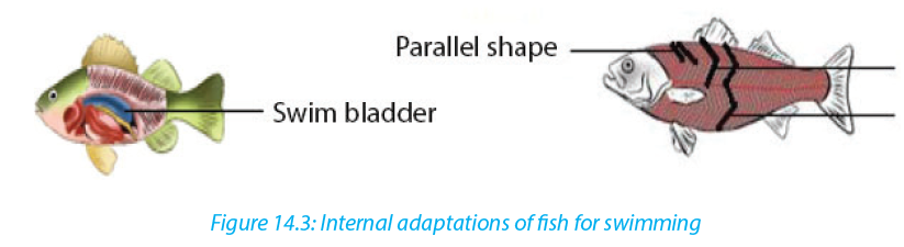
Internally, a fish is adapted to swimming by swim bladder and muscles. Air or gasfilled sac called swim-bladder, outgrowth of the pharynx, helps a fish to change its buoyancy as it alters the gas pressure in the bladder. So that, it floats at any depth in water without using its muscles. Swim-bladder also helps fishes to maintain a density that is equal to that of the surrounding water. Muscles or myotomes / myomeres (segments or sheets of muscles separated from its neighbor by a sheet of connective tissue) enable fishes to move in water owing the shapes of muscles that are located on either side of vertebral column.
Myotomes contribute to the mechanism of swimming by its arrangements. They may be parallel, V-shape, or W-shape arranged in bundles or blocks that are separated by myosepta.
Although there are such arrangements, the myoseptal organization and orientation of fibres is complex. In bony fish, myomeres are V-shaped with new myomeres added posteriorly. With those myotomes, a fish swim by passing a wave of contracting muscle from anterior to posterior. Muscles near the head of the fish contract first and contraction proceeds posteriorly down the length of the fish to the caudal fin. Thus, a fish moves forward from the contraction and relaxation (antagonistic) of myotome
on either side of the body.Undulatory swimming of the fish is also powered by the segmental body musculature of the myotomes. Myotome and myosepta orient more perpendicularly to midline to push aside. Therefore, the fish can bend laterally. With contraction muscle fibres shorten by half their length while maintaining volume. Without myosepta, but simply a series of interconnected muscle fibres, then the wave would be much dampened.
Self-assessment 14.3
1. How does swim bladder help the fish in locomotion?
2. Illustrate how the arrangements of fish myotomes contribute to fish locomotion.?
3. What does it make a fish to move in undulatory propulsion?
4. What are the anatomical structures that give rise to the direction of a fish and preventing diving and rolling?14.4 Support and locomotion in terrestrial animals
Activity 14.4
1. Through internet observe and think about how locomotion in dogs, chicken, frog and earthworm brought about.
2. Make a diagram showing how support and movement of different animals such as dogs, chicken, frog and worm is brought about on land.
3. Show by using diagrams the relationship between muscles, joints and musculo-skeletal attachment in mammals, birds, frog and earthworm.All animals living on land move due to the musculoskeletal system. The rigid nature of bone also gives a structure for muscles to pull, by their contraction, to create a movement as they act as levers. The synovial joints also allow certain movements.
The support and movement differ from specimen to another. Thus, animals can walk and run on land for moving from one place to another. This is possible by their endoskeleton and its muscles. By its muscles, flexor (a muscle whose contraction bends a limb or other part of the body) and extensor (a muscle whose contraction extends or straightens a limb or other part of the body or any or a muscle that
increases the angle between members of a limb, as by straightening the elbow or knee or bending the wrist or spine backward); contractions of those muscles cause the limbs act as levers for them which result to the foot being pressed downwards and backwards against the ground. For example, flexor and extensor work as illustrated below: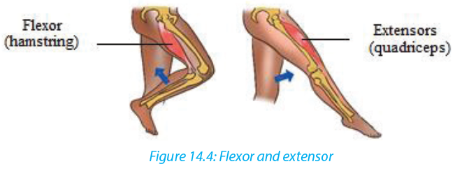
a. Locomotion in quadruped animals e.g. dog and frogs
When a dog walks, its vertebral column remains rigid, and the forward movement is achieved by the activity of the hind limbs. When its extensor muscle contracts, each hind limb, acting as a lever, extends and exerts a backward force against the ground, thrusting the animal forward and slightly upwards. When the flexor contracts, the limb is lifted clear of the ground and pulled forward. Only one limb is raised at any one time, the other three providing a tripod of support which balances the rest of the body.Beginning with the left forelimb in a stationary dog, the sequence of leg movement is as follows when it walks forward: left forelimb-right hind limb-right forelimb-left hind limb and so on. Such walking in quadrupedal animal is also identified in frogs when they can walk on land
b. Running of the dog
As a dog begins to run, it loses its quadrupedal movement which means, it develops a type of movement where the forelimbs move together, followed by the hind limbs.c. Walking in bipedal animals e.g. humans
Humans are bipedal, they walk on two legs. When standing upright, the weight is balanced over the two legs. When a stride is taken by the right leg, the heel is raised first by the contraction of the calf muscles. As this occurs, the weight of the body is brought over the left foot which is still in contact with the ground and acting as the prop for the rest of the body.When the right leg extends the heel is the first part of the foot to touch the ground. The weight off the body is gradually transferred from the left side to a position over the right heel and then the body continues to move forward, over the right toes, backward pressure against the ground generally being exerted through the right big toe. Like human does, a bird also can walk on ground through the movement of contractions of its leg muscles particularly flexor and extensor.
d. Crawling of earthworm
Earthworms are organisms having hydroskeleton with soft-bodied animals due to fluid secreted within the body and surrounded by the muscles of the body wall. They are capable to move by aid of their muscles. These muscles are not attached to any structures and thus can pull against each other. The combined effect of musclecontraction and fluid pressure serves to maintain the shape and form of the animal.
Generally, there are two muscle layers, longitudinal in which muscle fibres are arranged parallel to the long axis from one end of a segment to another and circular with muscle fibres arranged in concentric circles to the circumference of the worm. When those muscles act antagonistically against each other, locomotion is achieved.
The fluid which acts as presssurisable hydrostatic skeleton contained in body cavity or coelom presses against the muscles which in turn are able to contract against the fluid. Earthworm movement is also helped by bristles like setae or called chaetae (hair like structures on ventral surfaces) which anchor the worms to the substrate. Contraction of the circular muscles makes the worm thinner, but because liquid is
essentially
incompressible (and so maintains a constant volume) and the increase in pressure forces the liquid outwards, stretching the worm, so the worm becomes longer and thinner. Contraction of the longitudinal fibres shortens the worm, former the coelomic liquid out to the sides and making the worm fatter. If the body is segmented, then such pressure is localized and only certain segments will move or
change shape.Self-assessment 14.4
1. What are the main muscles that contribute to locomotion in mammals, amphibians and birds?
2. Draw an earthworm and illustrate the muscles that contribute to its locomotion.
3. What type of skeleton system found in mammals, birds, amphibians and annelids?
4. . Illustrate how flexor and extensor muscles contribute to lifting up a leg in human being.14.5 Flight through air by birds and insects
Learning activity 14.5
Make a research on internet as well as books and do the following:
1. Observe pictures below (Figure 14.6) related to birds and make a description of skeletal modification in birds. Illustrate the skeletal modification in birds
2. Draw a bird and show by using arrow the structures that enable a bird to fly
3. How will the external features of birds will behave when flying in high or low atmospheric pressure4. Make a table illustrating how does flight of birds and insects differ and similar
5. Observe and compare the flight of birds and insects
Bird can fly either by flapping their wings or gliding by spreading its wings. Like in animals moving on land, locomotion by flying in birds is brought about by the action of flexor and extensor muscles as well some other structures given diagram below like pectoralis major, pectoralis minor and keel of sternum.
Based on the above diagram, wings move down by the contraction of pectoralis major and then move up under the contraction of pectoralis minor.
Adaptive features of birds for movement in air
A number of features enable birds to aerial locomotion. Those features include body shape, modified limbs, and modification in internal organs particularly bones.
The body of bird is highly streamlined and covered with light feathers that overlap backwards thus reducing air resistance during flight. Those light features that increase the surface area of wings without increasing weight.Differently from quadrupedal animals, birds are adapted to flying by modification of fore limbs into wings. Such modification goes with a well-developed or large keel sternum that provides large surface area for the attachment of the flight muscles namely major and minor pectoral muscles which give the power to flap the wings in flight. Also, birds have hollowed bones making the body light and vertebrae of trunk
are fused. As flight requires much energy, birds are adapted to that by an efficient breathing system with air sacs attached to the lungs necessary to provide oxygen for respiration and to remove the resulting carbon dioxide.Other adaptations include a high metabolic rate for providing the high amount of energy required, an efficient circulatory system necessary for transporting both the nutrients and respiratory gases at speed related with the body needs, a high red blood cell count for efficient oxygen transport, and a keen eye sight to enable them to judge distances correctly especially on landing.
Self-assessment 14.5
1. What are the muscles that enable the flight in birds?
2. Describe how bird skeleton contributes to its flight?
3. Describe how birds are adapted to flying.14.6 Hopping locomotion in grasshoppers and toads
Activity 14.6
Use a collecting net to catch a grasshopper and toad from school compound. Put them down on cemented ground for observing them very carefully when they make a jump and then answer to the following:
1. Identify and describe anatomic structures that enable grasshoppers to jump
2. Illustrate how legs’ muscles behave when they are resting and or jumping
Skeletal muscles such as extensor and flexor that occur in pairs are often antagonistic.
With such antagonistic behaviour, when one contracts the other relaxes to produce controlled movement in the opposite directions.a. Locomotion of grasshopper
Insects have a skeleton which is on the outside of the body called an exoskeleton.
They can walk on the land but they are mostly adapted to hopping owing to their muscles which are inside the hard shell as well as skeleton system. The muscles which make them capable to move are flexors and extensors which are antagonists, attached to internal surface of exoskeleton and the rear or back legs of a grasshopper which are long and muscular, adapted for hopping. Additionally, there are two main muscles inside are the extensor tibiae muscle which contracts to extends the leg, and the flexor tibiae muscle which contracts to flex the leg as illustrated in figure below. Those muscles pull on tendons which are attached to the tibia on either side of the joint pivot.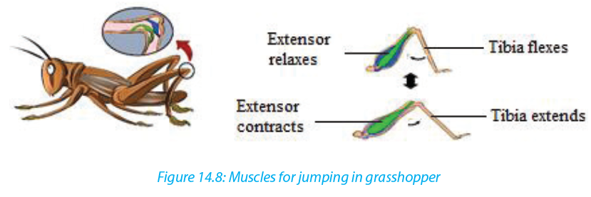
The back legs are much longer than the others for helping in hopping. With those long legs, grasshopper is capable to make high jumping distance. As illustrated above, flexor muscles bend a joint whereby extensor ones straighten it. The flexor muscle contracts and the lower leg is pulled towards the body. Thus, the hind leg is folded in a Z shape and ready for jumping. Being in resting or sitting position,
the extensor muscle contracts which enable then the legs jerk or move very quickly
backwards propelling the grasshopper.b. Locomotion in toads and frogs
On land, frogs and toads move by hopping (going from place to place).
–– When a frog is at rest, the hind legs are folded up in the shape of a letter Z.
–– When it hops, the legs are quickly straightened out, lifting the animal of the ground.
–– The fore-limbs are used as shock absorbers on landing and they also prop up (to give support) the front end of the body when the animal is at rest.
They also hope but do not travel as high as far as a frog does at each hop.Self-assessment 14.6
1. What are the muscles that contribute to high jumping in a grasshopper?
2. How do muscles (flexor and extensor) behave when toads and grasshopper are resting?
3. Draw a leg of grasshopper and the one of toad when are jumpingEnd of unit assessment 14
1. Describe ways of locomotion in Amoeba, Paramecium, Euglena and in Trypanosom
2. Produce a cartoon showing different adaptive features of fish for aquatic locomotion
3. Describe how different fish fins contribute to locomotion and balance
4. Describe how the movements and support of fish in water do they occur? are brought in water
5. Show by diagrams the relationship between muscles, joints and musculoskeletal attachment in mammals, birds, frog and earthworm.
6. Describe how flexor and extensor muscles work to enable the locomotion on land, water and in air
7. What are the features that enable aerial locomotion in animals?
8. Describe how a grasshopper and toad is adapted to jumpingUNIT 15 CLASSIFICATION AND PATTERNS OF DISEASE
Key Unit Competence
Describe the social factors that affect good health and apply knowledge gained in familiar and unfamiliar contexts.Learning objectives
–– By the end of this unit, I should be able to:
–– Explain what is meant by health and disease.
–– Identify different categories of disease and give an example of each.
–– Explain the theory of the disease and the causes, sources, transmission, symptoms and controls of the disease.
–– Discuss how global patterns of disease are studied.
–– Analyze and interpret records from a given hospital to identify diseases as
endemic, epidemic or pandemic.
–– Apply knowledge gained to classify common diseases.
–– Appreciate the importance of germ theory of disease by showing that the
death rate related to infections is greater than those caused by accidents.Introductory activity
a. Suggest measures to be taken for addressing issues related to eating without washing hands.
b. Discuss on different communicable diseases got from eating without washing hands.15.1 Germ theory of diseases
Activity 15.1
Discuss the following questions
a. What are the causes of death?
b. Why it is difficult to eradicate malaria in Rwanda?The germ theory states that many diseases are caused by the presence and actions of specific microorganisms within the body. In 1677, Antoni van Leeuwenhoek was the first to observe microorganisms in the droplets of water. but he did not make the connection with disease. Later, Spallanzani and Louis Pasteur observed germs in the blood of people suffering from disease. They suggested that the germs were an effect of the disease rather than the cause. The observations and actions of Ignaz Semmelweis, Joseph Lister and John Snow were a great contribution to the acceptance of germ theory. However, the laboratory works of Louis Pasteur in the 1860s and Robert Koch in the following decades, provided the scientific proof for germ theory. Their works opened the door to research related to the identification of disease-causing germs and potential life-saving treatments.
15.1.1 The work of Louis Pasteur and Semmelweiss
The work of Eduard Jenner and Ignas Semmelweiss showed that infectious diseases maybe caused by an infectious agent or germ. This was accepted as the germ theory of disease for a very long time. The work of Robert Koch and Louis Pasteur led to a wide acceptance of the germ theory
Pasteur made a number of important steps forward. He indicated that fermentation is the result of the action of microorganisms (Yeast) on sugar. Huge number of people at the time believed that living things could arise spontaneously from non-living things and this theory is known as spontaneous generation. To reject this theory, he showed that if broth is boiled in a sealed container, it would stay clear, but once he added material which had been exposed to the air, microorganisms grew in the broth. Finally, he designed a series of experiments using swan necked flasks which showed once and for all that any microorganisms which appear in boiled broth come from the air not arise spontaneously from nonliving organism such as broth.
In 1845-1846, Pasteur found a way of avoiding the disease of silkworms by observing the infected eggs under microscope and thereby saving the silk industry. This was the first clear evidence of microorganisms causing disease. Pasteur even developed vaccines against a number of these diseases.
15.1.2 Summary of the contributions of Louis Pasteur in microbiology and medicine
The contribution of Louis Pasteur in microbiology and medicine also include:
–– The fight against spontaneous generation theory
–– The technique of sterile culturing of microorganisms
–– The technique of fermentation and conservation of drinks. This technique is known as Pasteurization.
–– The technique of antiseptic surgery to prevent contamination of wounds during the surgical operations in hospitals.
Louis Pasteur developed the germ theory of disease which postulates that all contagious and infectious diseases must be caused by pathogenic microorganisms.15.1.3 The Germ Theory and Koch’s Postulates
Diseases can be spread by air, water, food, and human as well as animal vectors. In, an english physician called John Snow (1854) and a German microbiologist called Robert Koch (1884) found a relationship between polluted water and disease. Robert Koch, has isolated the bacterium Vibrio cholera, the cause of cholera from Elbe River water to provide the relationship.
Koch went on to formulate an established set of procedures to isolate and identify the causative agent of a particular microbial disease. The following four steps, which are still used today, are known as Koch’s Postulates:
Postulate 1: A specific organism must always be observed in association with the disease.
Postulate 2: The organism must be isolated from an infected host and grown in pure culture in the laboratory.
Postulate 3: When the organism from the pure culture is inoculated into a susceptible host organism, it must cause the disease.
Postulate 4: The infectious organism must be re-isolated from the diseased organism and grown in pure culture.Self-assessment 15.1
1. What are the Koch’s Postulates?
2. Explain the theory of spontaneous generation.15.2 Classification of diseases
Activity 15.2
Use the Knowledge gained to answer to the following questions:
1. Propose two infectious diseases and for each disease, give:
a. their causal agents
b. causal agents’ type
c. their symptoms
d. their methods of prevention
e. Treatment
2. Does being healthy means just the absence of the disease? Explain.
The following are the meaning of disease, signs and symptoms:
–– Disease is the disruption of normal body function.
–– Signs are indications of a disease that can be observed by examining the patient.
–– Symptoms are indications of disease perceive only by the patient.
The normal functioning of the body is disturbed, when the body is infected. Many types of diseases are broadly divided into two categories: Infectious diseases and non-infectious diseases15.2.1 Infectious diseases
Infectious diseases are caused by microorganisms known as pathogens which may include viruses, bacteria, fungi and protozoa. Those diseases are called communicable diseases. as they can be transmitted from one person to another. They include cholera, malaria, typhoid, HIV and AIDS…Malaria is one of the most dangerous infectious diseases, endemic in Latin America, Africa and South-East Asia. Some infectious diseases can also be from animals to humans.The following are some technical terms used when discussing about infectious disease.
–– Aetiology: The study of the cause of disease.
–– Epidemiology: The study of all the factors that contribute to the appearance of a particular disease
–– Causative agent: The organism which causes the disease
–– Vector: An organism which carries the causative agent of the disease from one person to another or from infected animal to human.
–– Incubation period: The period of time between the original infection and the appearance of signs and symptoms.
–– Infective period: The time during which a person is capable of passing the
disease on to another person.
–– Carrier: The person who has been infected but develop no signs and symptom, the carrier can pass the disease on to another person
–– Prevention: Measures taken to prevent diseases.
–– Treatment: Measures taken to cure diseases Antibody: Is a protein produced by the body’s immune system when it detects harmful substances called antigen.
–– Antigen: Is any substance that causes your immune system to produce antibodies against it.
–– Host: A host can be anything living organism ion which pathogens can survive
–– Hygiene: Practices that help to maintain health and prevent the spread of diseases
–– Immunity: Is the ability of the body to resist to infections.a. Important advances in the work against infectious disease
Table 15.1: The chronology of some of the most important advances in the work against infectious disease
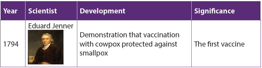
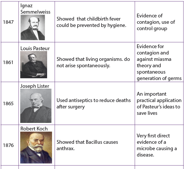


b. Some groups of communicable diseases
– Bacterial diseases: these are diseases caused by bacteria. They include cholera, typhoid, tetanus, tuberculosis, etc.
– Viral diseases: these are diseases caused by viruses. They include AIDS, polio, measles, Ebola, etc.
– Protozoan diseases: these are diseases caused by protozoa. They include malaria, sleeping sickness, trichomoniasis, etc.
– Fungal diseases: these are diseases caused by fungi. They include candidiasis, athlete’s foot, ring worms, etc.
– Worm diseases: these are diseases caused by worms. They include elephantiasis, bilharzias, etc.
– Sexually transmitted diseases: these are diseases transmitted through sexual contact. They include HIV-AIDS, syphilis, gonorrhea, etc.c. Transmission of infectious diseases
Pathogens can spread when you have direct contact with an infected person. For example, if you have contact with the person’s blood, body fl uids or open wounds. Pathogens can also be spread through contaminated food, water or air. Infected animals can spread pathogens to people.
The following conditions lead to the spread of an infectious disease:
–– A pathogen which causes the disease.
–– A source which is an infected organism.Mode of transmission a pathogen must be able to enter the body of the new host to cause an infection. Infectious diseases follow a pattern of development from the time of infection. The pattern of development has five distinct periods, as described in table below:

d. Epidemiology
The study of patterns of disease and of the various factors that affect the spread of disease is called epidemiology.
Epidemiologists try to discover the factors that cause a disease and develop methods to prevent its spread. The main clue they use come from data about the number of people in a particular area affected by specific diseases, and the number of death The data are commonly expressed as incidence or morbidity and mortality rates.
The incidence rate is the number of new cases of disease in a given population occurring during a specific period (a week, month or a year). It is calculated as:

Example: among 900 students at a given school, 50 suffer from malaria and 4 deaths were recorded. Calculate the morbidity or the incidence of malaria at that school.

To find how many cases of a disease are new, this calculation requires information about the prevalence rate. This is the total number of individuals infected in a population at any one time.
The mortality rate of a disease may be estimated for a whole population irrespective of whether they have the disease or not.

It may be calculated by using only those people who have disease.

To make fair comparisons between different populations with the same disease, epidemiological information is usually adjusted. For example, the mortality rate among those with a particular disease is usually expressed as a percentage or ratio per year. Hence, if in one year 7500 people in a given area die as a result of AIDS and the total number of population infected was 30 000, the mortality rate would be 25% for the rate.
Epidemiological studies are used to identify whether a disease is endemic, epidemic, or pandemic:
–– Endemic disease is a disease that is always present in a people e.g. malaria in
tropical Africa.
–– Epidemic disease is a disease that spreads rapidly, suddenly, and unexpectedly
to affect many people. e.g. cholera in
refugees’ camp. Pandemic disease is a disease that affects people over very
large area, such as a continent or even the whole world e.g. AIDS and TB are
pandemic at present15.2.2 Non-infectious diseases
These diseases are also called non-communicable diseases. They cannot be transmitted from one person to other examples: albinism, kwashiorkor, cancer, diabetes, etc

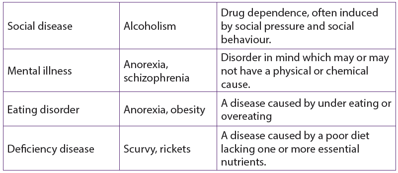
Lesson self-assessment 15.2
1. Answer by true or false
a. Epidemic disease is a disease that is always present in population.
b. The diseases that transmitted among people by pathogens are called transmissible diseases
c. The study of patterns of disease and of the various factors that affect the spread of disease is called epidemiology.
d. Cholera is infectious disease
e. Malaria is non-infectious disease whose vector is mosquito.
2. Distinguish between morbidity and mortality15.3 Common infectious diseases
Activity 15.3
Choose in the following list the infectious diseases and explain why:
Cholera, typhoid, Alcoholism, tetanus, tuberculosis, AIDS, Haemophilia, polio, measles, Ebola, malaria, Anorexia, obesity, sleeping sickness, trichomoniasis, sickle-cell anaemia, candidiasis, athlete’s foot, ring worms, elephantiasis, bilharzias, syphilis, gonorrhea, cystic fibrosis, Arthritis, Anorexia, schizophrenia.
15.3.1 Measles
Measles is a contagious acute viral disease with symptoms that include a bright red rash of small spots that spread to cover the whole body. Small white spots, known as Koplik’s spots, appear in the mouth on the inside of the cheeks a few days before the rash appears and can be used in diagnosis.
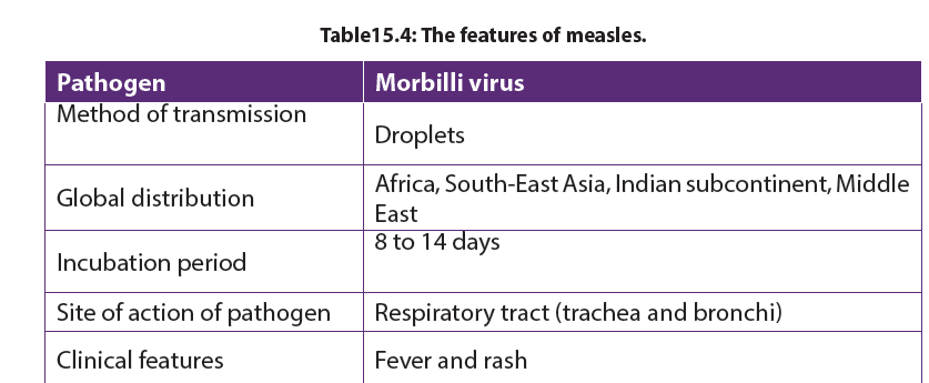
Failure to eradicate measles
–– Incubation period is short hence it is difficult to identify and isolate before they become infectious
–– It is transmitted through a carrier mother to healthy children hence it is hard to eradicate.
–– It targets young children who like playing together. This makes ease the spread of the disease.15.2.3 Typhoid
a. Causal agent of typhoid
Typhoid is waterborne disease caused by Salmonella typhus, a Gramnegative bacterium. The bacteria are derived from the feces of a patient. It has high infectivity as low dosage of organisms is only needed for typhoid to spread. Common sources of typhoid infection are contaminated water, milk and food.

b. Development of typhoid illness
The incubation period is of ten days. After this time, the germs enter the bloodstream and the patient develops the following symptoms: Headache, Muscular pains, Fever reaching its peak after about 1 week, faint rash may appear, diarrhea in the second week, mental confusion, etc. The third week shows the peak of the illness and the patient may die if not treated.
c. Treatment and prevention of typhoid
The disease had a 20% fatality rate before the use of antibiotics. Chloramphenicol and Ampicillin are effective and reduce fatality rate from 1 up 5%. Today, Ciprofloxin is used as another antibiotic. The two most important preventive measures are; proper treatment of sewage and disinfection of water supplies. Hygienic measures in the food trade and at home, and control of flies, which can transfer fecal material to food. Vaccine is formed by a polysaccharide from the capsule of the bacteria.
15.2.4 Cholera.
Cholera is a good example of a waterborne disease. It is endemic in parts of Asia, particularly India. The organism which causes cholera is a comma shaped motile bacterium called Vibrio cholerae.
a. Transmission and symptoms of cholera

The main source of infection is water contaminated by feces with Vibrios. It is estimated that only about one infected person in 50 develops the disease, the rest being carriers. Drinking contaminated water, or washing food or utensils in it, is the most common means of transmission. Direct contamination of food with feces as a result of poor hygiene is also possible, house flies being the main vector in this last case.
b. Signs and symptoms of cholera
Vibrio cholerae multiply in the intestine, releasing a powerful toxin which results in violent inflammation of the intestine and production of the watery diarrhea.
The main sign of the disease is severe diarrhea due to irritation of the bowel by toxins from the Vibrios. The liquid of the feces is so profuse and cloudy like “rice water”.
Abdominal pain and vomiting are also common. Dehydration is rapid and quickly results in death unless rehydration treatment is given. Fever is absent; in fact, the skin feels deathly cold and often damp.
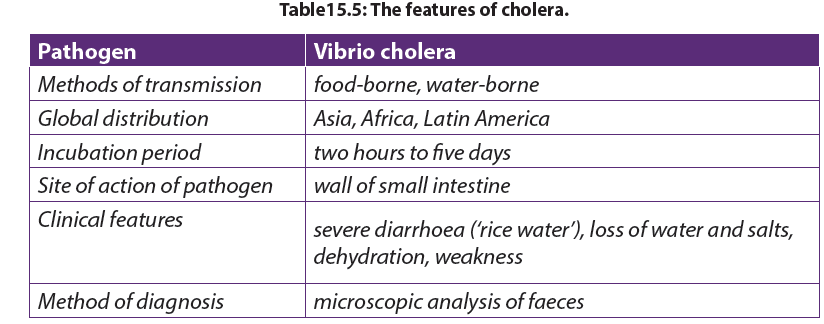
c. Treatment of cholera
The primary cause of death from cholera is dehydration i.e. loss of water with its minerals salts. For that, it is obligatory to rehydrate with oral serum which contain mineral salts and sugar.
The loss fluid may be replaced by administration of a drip food into a vein. Various antibiotics, such as tetracycline’s and chloramphenicol, are used to treat cholera. Chloramphenicol is effective against tetracycline-resistant Vibrios.
d. Prevention of cholera
–– Use clean drinking water,
–– Proper treatment of sewage and sanitation
–– High standards of public and personal hygiene, particularly in relation to food
(such as washing hands after defecation)
–– Health education
–– Vaccination is recommended for people visiting areas where cholera is
endemic and for those living in such areas. But the vaccine lasts few months.
–– Isolation of patients and hygienic disposal of feces and vomit from patients.e. Failure to eradicate cholera
–– Vaccination is not very effective
–– It is a waterborne disease i.e. transmitted through contaminated water
–– Poor sanitation condition in camps.15.2.5 Tuberculosis (T.B)
TB spreads when infected people with the active form of the illness cough or sneeze and the bacteria are carried in the air in tiny droplets of liquid.a. Causal agent of tuberculosisTuberculosis is caused by bacterium called Mycobacterium tuberculosis, first discovered by Robert Koch in 1882. It is sometimes referred to as the tubercle bacillus, bacilli being rod-shaped bacteria. The common form is pulmonary T.B which infects the lungs, although other organs may be affected.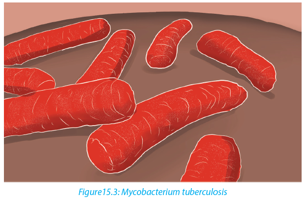 Two strains of the bacterium may cause the disease, the human and the bovine forms. The latter can be present in cattle and can enter the milk of cows. It is very resistant and can remain alive for long time in milk products as well as in dust .
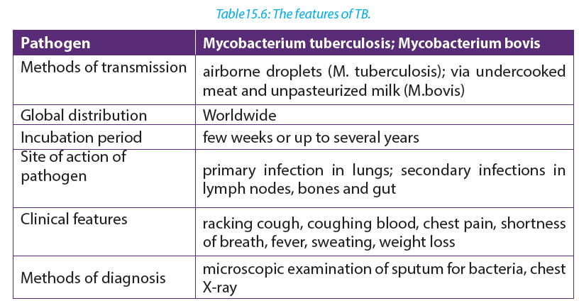 b. Transmission of tuberculosisTuberculosis is mainly airborne disease. The infection is done through the droplets from the patient. It is much less infectious as it requires prolonged contact between people, poor ventilation and overcrowded living conditions. In addition, TB is an opportunistic infection, striking many people with a depressed immunity.c. Signs and symptoms of tuberculosisThe disease is frequently characterized by vague symptoms such as: loss of appetite; loss of weight; excessive sweating; coughing, appearance of blood in the sputum, pains on the chest, shortness of breath (case of lung tuberculosis).d. Treatment and prevention of tuberculosisVaccine against the disease has been developed by Albert Calmette and Camileguérin). Antibiotics such as rifampicin, isoniazid and streptomycin are used to treat tuberculosis.e. Failure to eradicate tuberculosis–– Patients can carry pathogen and infection without showing symptoms.
Therefore, they are difficult to identify due to a long period of incubation
–– Germs of tuberculosis can survive longer in the house dust
–– The disease is related to poverty where many people share the same room and
have malnutrition.
–– The disease is associated with AIDS that reduced the body immunity
–– Long period of treatment (6-8 moths), hence patients give up when not yet
fully healed. The pathogens then form endospores that resists to medicines.
–– The disease is also spread through milk from infected animals. Tuberculosis is
an airborne disease i.e. spread in air15.2.6 Malaria.
a. Causal agentMalaria is caused by four species of plasmodium: Plasmodium falciparum, P. vivax, P. ovale, and P. Malariae.The parasite is transmitted by the bite of female mosquitoes (the vector) belonging to the genus Anopheles.b. SymptomsMalaria is characterized by severe chills, fever, sweating, fatigue and great thirst. Victims die of anemia, kidney failure or brain damage.c. Occurrence of malariaThe disease now occurs in tropical and subtropical regions of the world, and its distribution is limited by conditions of the development of the mosquito vector such as temperature and altitude.Malaria is endemic in tropics because:– Tropical climate provides the best breeding and living conditions for the Anopheles mosquito which transmits malaria–– The Anopheles cycle requires areas of stagnant water, common within tropics
–– In the tropical areas, there is presence of bushes or abundant vegetation which
makes suitable habitat for mosquitoes
–– Plasmodium needs temperature in excess of 20ᵒC for it to complete its cycle
within the mosquito. d. Eradication and prevention of malaria
–– Drainage of stagnant water: The larval stages of the mosquito live in stagnant
water, so drainage removes breeding sites.
–– Destruction of the adult mosquitoes by spraying insecticide.
–– Clean bushes nearest houses where mosquitoes lay eggs
–– Sleeping under mosquito net during the nighte. Failure to eradicate malaria
–– There is no effective vaccine against malaria
–– The pathogens are transmitted by mosquitoes which are difficult to eradicate.
–– The plasmodium has become resistant to different anti-malarial drugs
–– Ignorance of some people toward the disease and how it is spread.15.2.7 Smallpox
a. Cause of smallpox
Smallpox was a horrible viral disease caused by Variola virus (DNA virus), a pox virus.
It was a highly infectious disease transmitted by direct contact and it affects the
respiratory passage.b. Signs and Symptoms of smallpox The following are Signs and Symptoms of smallpox:– Obvious symptoms of the disease were red sports on the face, trunk, and extremities that change to pea-sized blisters and became filled with pus. High fever and generalized aching.c. Modes of transmissionThis disease spread through droplet infection (contagion possible via wounds in skin, clothing, bedding and dressing)d. Prevention and treatmentLarge populations were vaccinated through Ring vaccination and people with the disease were isolated.The eradication of Smallpox was successful because:–– The smallpox virus is stable. so the same vaccine could be used
–– The smallpox virus does not linger in the body after infection, nor does it infect
other animals, so it cannot remain hidden anywhere.
–– The vaccine used was highly effective and easy to administer by scratching
technique.
–– It was easy to identify people with the disease.
18.3.7 Tinea
Tinea is a skin infection due to a fungus. Often, there are several patches of ringworm on the skin at once. Tinea is also known as Ringworm.a. Cause of Tinea–– Tinea is caused by a tiny fungus known as dermatophyte. These tiny organisms
normally live on the superficial skin surface, and when the opportunity is right,
they can induce an infection.
–– The disease can also be acquired by person-to-person transfer usually via
direct skin contact with an infected individual. Animal-to-human transmission
is also common.
–– Ringworm commonly occurs on pets (dogs, cats) and the fungus can be
acquired while petting or grooming an animal.
–– Ringworm can also be acquired from other animals such as horses, pigs, ferrets
and cows.
–– The fungus can also be spread by touching inanimate objects like personal
care products, bed linen, combs, athletic gear, or hair brushes contaminated
by an affected person.Individuals at high risk of acquiring ringworm include those who:–– Sweat excessively, as sweat can produce a humid wet environment where
the pathogenic fungi can thrive.
–– Wear tight, constrictive clothing with poor aeration.
–– Have a weakened immune system
–– Live in crowded, humid conditions.
–– Participate in close contact sports like soccer, rugby.b. Sign and symptoms of Tinea The following are Sign and symptoms of Tinea:
–– Enlarging raised red rings with a central area of clearing (ringworm).
–– The edge of the rash appears elevated and is scaly to touch.
–– Sometimes the skin surrounding the rash may be dry and flaky.
–– There will be hair loss in areas of the infection.c. DiagnosisSuperficial scrapes of skin examined under a microscope may indicate the presence of a fungus. Diagnostic method called KOH Test is used, where the skin scrapings are placed on a slide and immersed on a drop of potassium hydroxide solution to dissolve the keratin on the skin scrapings thus leaving fungal elements such as hyphae, septate or yeast cells viewable. If the skin scrapings are negative and a fungus is still suspected, the scrapings are sent for culture. Because the fungus grows slowly, the culture results do take several days to become positive.d. Prevention
Basic prevention measures include:
–– Serious washing of hands after handling animals, soil, and plants.
–– Avoiding touching characteristic lesions on other people.
–– Put on loose-fitting clothes.
–– Promoting good hygiene when participating in sports that involve physical contact with other people.e. TreatmentApplication of topical antifungals creams to the skin. In extensive or difficult cases, systemic treatment with oral medication may be required. Among the available prescription drugs are tolnaftate, terbinafine, naftifine, itraconazole.15.3.8 Hookworm
Hookworm is a humans’ intestinal parasite. The adult worms and their larvae can cause intestinal disease in which they live.a. Cause of hookworm diseaseThe hookworm is caused by two main species of hookworm infecting humans which are: Ancylostoma duodenale and Necatora mericanusb. Method of transmissionIf an infected person defecates near bushes, in a garden, or field, or if the feces from an infected person are used as fertilizer, eggs are deposited on soil. They can then mature and hatch, releasing larvae (immature worms). The larvae mature into a form that can penetrate the skin of humans. Hookworm infection is transmitted primarily by walking barefoot on contaminated soil.c. Signs and symptoms of hookworm–– Itching and a localized rash are often the first signs of infection. These symptoms occur when the larvae penetrate the skin.
–– A person with a light infection may have no symptoms but a person with a heavy infection may experience abdominal pain, diarrhea, loss of appetite, weight loss, fatigue and anemia (pale skin etc.) and protein deficiency caused by blood loss, constipation, congestive heart failure, excessive coughing during larvae migration, stomach or chest pain, vomiting, weight loss.
–– The physical and cognitive growth of children can be affected. There is a decreased rate of growth and mental development in children (caused by protein and iron deficiency)d. Diagnostic methodTaking a stool sample and using a microscope to look for the presence of hookworm eggs.e. Prevention of hookwormAvoid walking barefoot in areas where hookworm is common and where there may be fecal contamination of the soil. Avoid skin-to-soil contact and ingesting such soil. Avoid defecating outdoors or using human feces as fertilizer, and by effective sewage disposal systems.f. Treatment for hookwormHookworm infections are generally treated for 1-3 days with medication prescribed by your health care provider. The drugs are effective and appear to have few side effects. Iron supplements may be prescribed if you have anemia.g. Preventive treatment–– In developing countries, groups at higher risk for soil-transmitted infections
are often treated without a prior stool examination. Treating in this way is
called preventive treatment.
–– School-age children are often treated through school-health programs and
preschool children and pregnant women at visits to health clinics.h. Life cycle of Hookworm
15.4 Health and community: criteria for good housing
Self-assessment 15.31. Which of the following diseases is transmitted by an insect vector?
a. Cholera
b. HIV/AIDS
c. Malaria
d. TB
2. What are the ways in which cholera is transmitted from person to person?
3. Explain why there is such a high risk of cholera following natural disasters such
as earthquakes, hurricanes, typhoons and floods.
4. Explain why there is a high death rate from TB in countries with a high proportion
of the population who are HIV-positive.
5. TB is an opportunistic infection. Why?
6. Describe how malaria is transmitted.15.4 Health and community: criteria for good housing
Activity 15.4Housing quality is associated with morbidity from different factors. State any three factorsHousing refers to houses or buildings, accommodation of people. It is an important determinant of health, and substandard housing is a major public health issue. The public health community is aware of the importance of social determinants of health (including housing) in recent years. Yet defining the role of public health practitioners in influencing housing conditions has been challenging. Responsibility for social determinants of health is seen as lying primarily outside the scope of public health. The quality and accessibility of housing is, however, a particularly appropriate area for public health involvement.An evolving body of scientific evidence demonstrates solid relations between housing and health. The public health community is developing, testing, and implementing effective interventions that yield health benefits through improved housing quality.Criteria for good housing
–– Good housing must be well equipped.
–– Good housing must be well localized
–– Water and electricity
–– Big size and ventilated
 An increasing body of evidence has associated housing quality with morbidity from infectious diseases, chronic illnesses, injuries, poor nutrition, and mental disorders.Self-assessment 15.4What measures are taken by Rwanda government to ensure high quality of housing conditions?
15.5 Public health services
Activity 15.51. Suppose that you are one of Rwandans who have food industry in our country, and you expect your production to be inspected.Outline the main requirements for good production to be inspected?2. Clean water is good for health. Discuss the ways you would use to obtain clean water at home.15.5.1 Food inspection requirement
a. Food inspection servicesFood inspections services help to let you have a complete check of your running production in factories or across the country. An inspector performs a random selection checking on quantity, packing, labeling, dimension, weight and visual aspects. Inspection allows to spot inconsistencies in your production lots before they leave the factory: you can react timely and avoid costly rework, sorting or recalls.15.5.2 Need for control of housing conditions
Living conditions affect people’s lives, be it at home or the workplace. Without good living conditions, people’s health and work will be affected. Nowadays, the quality of housing accompanied with good housing facilities is now improving.15.5.3 Need for control of clean water.
For being healthy, only clean water must be used. Different materials are used to clean water. They include “Sûr–eau”, heating or by using water purification etc. The steps of water purification are: storage reservoir, aeration, filtration, disinfection, reduction of chlorine concentration, covered service reservoir, distribution.15.5.4 Need for control of hygiene
Many diseases can be prevented from having a damaging effect on the body by the action of natural defenses, antibiotics or other medicines. However, there are many steps that can be taken by individuals, and by the community as a whole, to fight microbes even before they enter the body and cause disease. These steps concern personal hygiene (cleanliness) and sanitation (public cleanliness involving community efforts in disease prevention), both of which help to prevent disease.Note that: The World of Health Organization defines health as a state of complete physical, mental and social well-being and not merely the absence of disease and infirmity. The responsibility for good health lies in the hands of each individual in the community and the nation.a. Personal hygiene
–– Hands frequently touch many things which may carry pathogens. They must be always washed using a soap before preparing food, eating and after a visit to a toilet.
–– It is essential to bath frequently because sweat and oil secretions on the skin
enable bacteria and fungi to breed easily. This helps to prevent skin infections.
–– Bath towels and sponges should not be shared Combs and hairbrushes should not be shared.
–– Hair should be washed frequently to avoid lice and mites. These can spread
typhus fever.
–– Teeth should be cleaned at least twice a day, preferably after each meal
because the spaces between teeth where food particles are trapped provide
excellent breeding grounds for bacteria.
–– Clothes would be clean and changed frequently.
–– Shoes should be worn to prevent cuts and infection by hookworms.
–– Finger nails and toe nails must be always kept short and clean so that they do
not provide breeding places for germs.
Exercise, recreation and rest are other factors that are important in promoting health.i. Exercise
Exercise makes the muscles strong so that they can support the body better.
It helps to get rid of excretory materials and to improve digestion.
It quickens blood circulation and improves the action of the glands and nerves.ii. Recreation
–– Gardening, playing games and reading can remove any dullness and mental tiredness resulting from everyday work.iii. Sleeping is the best form of rest.
–– Adults need about eight hours of sleep a day. A great deal of repair of wornout tissues in the body and the building up of new ones takes place when body rests.
–– Other good personal habits include avoiding smoking, alcoholic drinks and other drugs.Lesson self-assessment 15.5
1. In which ways a personal cleanliness may be achieved.
2. How to promote a hygienic food preparation?
3. Discuss about good personal Hygiene.End of unit assessment 15
1. Answer to the following questions:
a. What does the germ theory of disease mean?
b. State any four causes of diseases in our life.
2. State any TWO diseases caused by:
a. Bacteria
b. Protozoa
c. Microscopic fungi
3. Match the following scientists with their scientifi c contributions: 4. List the reasons why smallpox is easier to eradicate than AIDS.
5. Suggest reason why Malaria is endemic disease in tropics.
6. Describe the biological factors that make malaria a diffi cult disease to
control.
7. Describe the precautions that people can take to avoid catching malaria.
8. Explain what is meant by ring vaccination.UNIT 16 ASEXUAL REPRODUCTION IN PLANTS
Key Unit Competence
Account for various methods of asexual reproduction as means of increasing crop yield.Learning ObjectivesBy the end of this unit, I should be able to:
–– Describe the various methods of asexual reproduction: fragmentation, budding, and spore formation.
–– Demonstrate asexual reproduction mechanisms in lower organisms.
–– Discuss the advantages and disadvantages of asexual reproduction.
–– Describe the characteristics of vegetative reproductive parts in a flowering plant.
–– Apply principles of artificial propagation in growing varieties of plants that are economically important.
–– Apply the knowledge acquired to produce economically important plants.
–– Appreciate the use of artificial propagation in increasing crop yields.
Introductory activity
The kingdom Plantae comprises about 260,000 known species including flowering
and non-flowering plants. All plants have a general organization which includes
vegetative and reproductive organs. Plants reproduce through different ways:
Use the books and other source of information to
1. Write on how lower organisms such unicellular plant and another like cassava, sugar cane and apple reproduce.
2. Describe the techniques used by people to grow Irish potatoes, cassava and bananas.
3. Describe each of the following methods of asexual reproduction:
fragmentation, budding and spore formation.Activity 16.116.1 Asexual reproduction
Using textbooks or internet and search on asexual reproduction.Asexual ReproductionThis is a type of reproduction without production of gametes. It usually results in the production of identical offspring; the genetic variation arises as a result of random mutation among the individuals.Self-assessment 16.1Write an essay on asexual reproduction.16.2 Types of asexual reproduction
Activity 16.2.1Discuss on asexual reproduction in lower organisms and write a report as an outof-class activity.There are five common modes of asexual reproduction: fission, budding, vegetative reproduction, spore formation and fragmentation.16.2.1 Fission
An important form of fission is binary fission. In binary fission, the parent organism is replaced by two daughter organisms, because it divides in two, i. e: Unicellular Fungi. Another type of fission is multiple fission that occurs in many algae. The nucleus of the parent cell divides several times by mitosis, producing several nuclei. The cytoplasm then separates, creating multiple daughter cells.16.2.2 Budding
Some cells split via budding resulting in a ‘mother’ and ‘daughter’ cell. The offspring organism is smaller than the parent. Budding is also known on a multicellular level.16.2.3 Vegetative reproduction
Vegetative reproduction is a type of asexual reproduction found in plants where new individuals are formed without the production of seeds or spores. Examples of vegetative reproduction include the formation of miniaturized plants called plantlets on specialized leaves (for example in kalanchoe) and some produce new plants out of rhizomes or stolon (for example in strawberry). Other plants reproduce by forming bulbs or tubers (for example tulip bulbs and dahlia tubers). Some plants produce adventitious shoots and suckers that form along their lateral roots. Vegetative reproduction may form a clonal colony where all the individuals are clones.Activity16.2.2
Demonstration of asexual reproduction by fragmentation in algaeRequirements:Glass beakers of 500ml, Scalpel, Forceps, Pins, Spatula, Weighing balance, Labels,
Artificial, fertilizers, Clear river water and Spirogyra (algae).Procedure1. Label five beakers of the same size as A, B, C, D and E. Pour water in each
beaker. Weigh several measures of artificial fertilizers of 1 g each.
2. Transfer 1g of fertilizer to beaker A, then 2g to beaker B, 3g to beaker C,
4g to beaker D and 5g of fertilizers to beaker E. Note the concentration of
fertilizers is increasing from A- E.
3. Using forceps pick spirogyra and put it on a tile. Add several drops of
water to avoid drying. Tease off a piece of spirogyra using a pin. Cut that
piece into 5 fragments of the same length and transfer each piece into the
beaker.
4. Stand the beakers in a place where they can receive adequate sunlight for
the seven days. On the next day, start to examine the fragments in each
beaker every day and record any observable changes such the increase in
size of the spirogyra.Draw a table as this shown here and record your observation.
16.2.5 Fragmentation
Fragmentation is a form of asexual reproduction where a new organism grows from a fragment of the parent. The fragment develops into a mature and fully grown individual. Fragmentation is seen in many organisms such as fungiplants and gemma in liverworts. Most lichens, which are a symbiotic union of a fungus and photosynthetic algae, reproduce through fragmentation to ensure that new individuals contain both symbionts. These fragments can take the form of soredia, dust-like particles consisting of fungal hyphen wrapped around photobiont cells.
16.3 Advantages and disadvantages of asexual reproduction
Self-assessment 16.21. Write a short essay on asexual reproduction.
2. What is itfragmentation? give one example of plant which reproduces by fragmentation16.3.1 Advantages of asexual reproduction.
Activity 16.3Discuss on asexual reproduction in lower organisms and higher plants, and outlining advantages and disadvantages.Since asexual reproduction does not involve the process of gamete formation. It can be completed much more quickly, allowing the individual to spread its genetic material in a shorter period of time. It is also an advantage for small populations. as there may not be an adequate number of plants to pollinate one another to reproduce. If the parent plants are successful, it is a good idea to make clones of the plant that can be successful as well.16.3.2 Disadvantages of asexual reproduction.
Asexual reproduction does not have genetic diversity. With sexual reproduction, mixing a gene pool can produce diversity. The organism becomes less adapted to certain environmental changes when there is less variation produced in offspring. The entire communities of the genetically identical organisms might not survive such changes. Asexual reproduction usually leads to struggle for existence as well as overcrowding. The main distinction of asexual reproduction from sexual reproduction is that there is no need for two parents as well as special cells to reproduce. It means that special the mechanisms of combining sex cells for fertilization is no longer required. This form of reproduction process is simply mitosis that copy a parent organism.Self-assessment 16.3Explain the advantages and disadvantages of asexual reproduction.16.4 Vegetative and artificial propagation in flowering plants
Activity 16.4Demonstration of asexual reproduction in plants by cuttingsRequirementsGrowth medium or moist soil, sweet potatoes vines, elephant grass, sugarcane or cassava stems, secateurs/sharp knife and rooting hormone.Procedure1. Collect clean and healthy stems from cassava, sugarcane or potato plants.
2. Using a secateurs/sharp knife, cut the stem of either cassava, sugarcane or
sweet potato stems into fragments of suitable sizes.
3. Place them in either suitable medium of growth with rooting hormone if
available or plant them in moist soil in the school garden.
4. Leave the set up for about 13 days, and then observe the development of
roots and leaves at nodes.
Draw and record what you will observe after 13 days on the development of roots
and leaves at nodes.Artificial vegetative propagation is the deliberate production of new plants from parts of old plants. This can be done by following three methods: Cutting, layering, and grafting.a. CuttingThis is a simple procedure in which part of the plant called a ‘cutting’is removed from the parent plant by cutting and placed in a suitable medium for grow. In this method, one-year-old stem of root is cut from a distance of 20 to 30 cm and buried in the moist soil in natural position. After sometime, roots develop from this cutting and it grows into a new plant. This method is commonly used in rose and sugar cane. Care is taken that nodes which were lower in parent plant (morphologically) are put in the soil, while the morphologically higher nodes are kept up. Adventitious roots are given off at the lower nodes. b. LayeringThis method of vegetative propagation is used in those plants with soft branches such as jasmine plant. In this method, a branch of the plant which is near to the ground is pulled towards the ground and covered with moist soil leaving the tip of this branch above the ground. After sometime, roots develop from that part of the branch which was buried in the soil. This branch is then cut of along with the roots from the parent plant and develops into a new plant. This method of asexual reproduction is also used in the production of plants such as Bougainvillea, guava, strawberries, lemon, China rose etc.
 c. GraftingIn this method of vegetative propagation, the stems of two diff erent plants are joined together so as to produce a new plant containing the characters of both plants. The plant of which the root system is taken is called ‘stock’, while the other plant of which the shoot is selected is known as ‘scion’ or ‘graft’. These two stems i.e. the stock and the scion are fi tted together by making slanting cuts into them and bound tightly with a piece of cloth and then covering it with a polythene sheet.The diameter of the stock and scion chosen for grafting should be equal. Scion getsthe mineral and water from the soil through the stock and develops branches and produce fruits. This method of propagation is used in mango, apple, banana, pear, grape, pineapple and peach.
 Vegetative reproductive parts in fl owering plantsThe reproductive part of the plant is a fl ower. The union of male and female gametes to form a zygote is called fertilization. The transfer of pollen grains from the anther to the stigma of the same fl ower or the diff erent fl ower is called pollination. Plants reproduce asexually in a variety of ways. The vegetative reproductive parts in fl owering plant are stem, branches, and leaves and they have the following characteristics:Characteristics of Stem:Stem develops from the plumule of embryo. It is generally the ascending part of the plant axis.It bears a terminal bud for growth in length and is diff er entiated into nodes and internodes. The stem nodes possess dissimilar appendages called leaves. The young stem is green and capable of performing photosynthesis. In the mature state, stem bears fl owers and fruits. Leaves and stem branches develop exog enously. Stem exposes leaves, fl owers and fruits to their most suitable position in the aerial environment for optimum function. Stems usually show positively phototropic, negatively geotropic and negatively hydrotropic.Characteristics of Leaf– It is dissimilar lateral fl attened outgrowth of the stem,
– The leaf is exogenous in origin
– It is borne on the stem in the region of a node,
– An axillary bud is often present in the axil of the leaf.
– Leaf has limited growth. An apical bud or a regular growing point is absent,
– The leaf base may possess two lateral outgrowths called stipules,–– A leaf is differentiated into three parts: leaf base, petiole and lamina.
–– The lamina possesses prominent vascular strands called veins,
–– It is green and specialized to perform photosynthesis,
–– Leaf bears abundant stomata for exchange of gases and it is the major seat of
transpiration.Characteristics of branchesA branch a woody structural member connected to but not part of the central trunk of a tree. Large branches are known as boughs and small branches are known as twigs. Due to a broad range of species of trees, branches and twigs can be found in many different shapes and sizes.Self-assessment 16.41. Discuss on the methods of artificial vegetative propagation.
2. Cassava produces flowers, fruits and seeds. Why people prefer to grow
cassava by cutting rather than using seed?
3. Describe the characteristics of vegetative reproductive parts in a flowering
plant16. 5 Application of artificial propagation in production of improved varieties of plants
Activity 16.5Artificial vegetative propagation is usually used in agriculture for the propagation (or reproduction) of those plants which produce either very few seeds or do not produce viable seeds. Such plants include: Banana, Pineapple, Orange, Grape, Rose, etc.Reasons for preferring vegetative rather than sexual means of reproduction vary, but commonly include greater ease and speed of propagation of certain plants. Another major attraction is that the resulting plants are clones of the parent plants.The methods of vegetative reproduction range from rooting cuttings to grafting and artificial propagation by laboratory tissue culture. In horticulture, a “cutting” is a piece that has been cut off from a mother plant and then caused to grow into a whole plant. A popular use of grafting is to produce fruit trees, sometimes with more than one variety of the same fruit species growing from the same stem. Rootstocks for fruit trees are either seedlings or propagated by layering. Self-assessment 16.5Explain the application of artificial propagation in production of improved varieties of plants.
End of unit assessment16
A. Multiple choice questions: choose the best answers.1. In vegetative propagation, cuttings are mainly taken from
a. Leaves of parent plant
b. Roots or stems of parent plant
c. Shoots of parent plant
d. Buds of parent plant
2. Artificial methods of vegetative propagation includes
a. Cloning
b. Grafting
c. Cuttings
d. Both b and c
3. Example of plant in which vegetative propagation is occurred by leaves is
called
a. Cannabis
b. Chrysanthemum
c. Bryophyllum
d. Brassica
4. Which of the following is NOT an advantage of asexual reproduction?
a. Rapid reproduction.
b. High genetic diversity.
c. No need for a mate.
d. Low resource investment in offspring.B.Questions with short and long answers
1. Name the plants which are grown by grafting method.
2. What do you understand by grafting?
3. Show how vegetative propagation takes place in potatoes?
4. Explain the method by which the sugarcane and rose are propagated.
5. Give the names of the different methods of artificial vegetative reproduction.
6. Explain the term vegetative reproduction and give one example of plant
which reproduces by using this type of asexual reproduction.UNIT 17 SEXUAL REPRODUCTION IN PLANTS
Key Unit Competence
Describe sexual reproduction in plants.
Learning objectives
By the end of this unit, I should be able to:
–– Explain the meaning of the term alternation of generations.
–– Describe the types and structure of flowers.
–– Describe pollination and fertilization in flowering plants.
–– Explaining the events that takes place in a flower after fertilization.
–– Describe the types and structure of seeds and fruits.
–– Discuss the modes of dispersal of fruits and seeds.
–– Observe and draw pollen grains.
–– Draw and interpret floral formulae and diagrams.
–– Relate the floral structures to the mode of pollination.
–– Draw and label structures of fruits and seeds.
–– Appreciate the role of pollinating agents in flowering plants.Introductory activity
1. Observe the following pictures and suggest what is going on.
2. How are the pictures below related to reproduction in flowering plants?
17.1 Alternation of generations in bryophytes and pteridophytes
Activity 17.1Using different resources to compare the lifecycles of mosses and ferns.The life cycle of an organism is the progressive sequence of changes which an organism goes through from the moment of fertilization to death. During its life cycle, the organism produces new generations of individuals which repeat continuously the process. New generations are produced by reproduction, which may be sexual or asexual. The life cycle involves the mitosis and meiosis. This unit concerns how meiosis can affect the life cycle of living organisms.The life cycle is seen in seaweeds, mosses ferns and their relatives. Their life cycles start with the sexually mature adult plants. Since they produce gametes, they are called the gametophytes (gamete plants). These are haploid, as they produce eggs and sperms. The egg and sperm fuse to produce a diploid zygote, but this does not develop directly into a new gametophyte. Instead it grows (by mitotic divisions) into another plant which is quite distinct from the gametophyte called sporophyte (spore plant). The function of sporophyte is to produce spores. In bryophytes, the sporophyte depends on gametophyte for nourishment. As spores are formed by meiosis, they are haploid. When the later are dispersed by wind on suitable soil, they germinate and grow by mitosis into gametophytes, which then repeat the sequence of life cycle. Ferns and mosses consist of two distinct plants: haploid gametophyte and diploid sporophyte, which alternate each other within the lifecycle. This phenomenon is known as alternation of generation. So,alternation of generation is a phenomenon in the plant life cycle in which a diploid stage a sporophyte alternates with a haploid stage of gametophyte.The importance of alternation of generation to organisms:–– Spores produced can survive hash conditions and only germinate when conditions are favorable.
–– It ensures rapid multiplication of the plant species as spores are usually produced in vast numbers.
–– Interdependence between the gametophyte and sporophyte generations ensures that both generations exist at any given time. This prevents extinction of the plant species The life cycle in mosses involves alternation of generations between the diploid (2n) sporophyte and haploid
gametophyte. In bryophytes, the gametophyte is a dominant stage of the life cycle, while the sporophyte is dependent on gametophyte for supplying water and nutrients. Gametes are formed in special reproductive organs at the tips of gametophytes. Sperms are produced in antheridia (singular: antheridium), and eggs in archegonia (singular archegonium). Some species produce both sperms and eggs on the same plant, whereas others produce sperms and eggs on separate plants.
During fertilization which requires water, the sperms released from antheridia fuse with the eggs and form a diploid (2n) zygote. The zygote grows into the sporophyte. Sporophyte is a long stalk ending in a capsule in which haploid Checkpoint: why bryophytes grow in habitats where water is available constantly:For fertilization to occur, the sperm of bryophyte must swim to an egg. Without water, this movement is impossible. Because of this dependence on water for reproduction, bryophytes must live in habitats where water is available at least part of the year
17.1.1 Alternation of generations in pteridophytes (ferns)
Pteridophytes also exhibit alternation of generations. Ferns are formed of true roots, stem and leaves (fronds) with vascular tissues. They have lignified tissues. The horizontal underground stem is called rhizome which bears adventitious roots. The leafy plant is a sporophyte. Mature leaves commonly called fronds bear yellow or orange masses of sporangia which are grouped into the structures called sori (singular: sorus) on their lower side. Ferns have a life cycle in which the diploid (2n) sporophyte is the dominant generation. Sporophyte is large (some fern trees can have 7 m of height), and differentiated into leaf, stem and roots with vascular tissues, while the haploid gametophyte (prothallus) is very simple with few millimeters. In ferns, the sporophyte produces haploid spores by meiosis. This is done on the underside of the leaf called frond in sporangia (singular: sporangium). Sporangia are grouped into sori (singular: sorus).
 When spores are mature enough, the sporangia burst and spores are released on ground. If conditions are favorable, spores germinate and grow into the haploid heart-shaped gametophytes
17.1.2 Differences between the mosses and ferns
 Self-Assessment 17.11. Explain the meaning of the term alternation of generation
2. Why is water essential in the life cycle of a bryophyte?
3. What are the archegonium and antheridium?
4. Why are these structures important in the life cycle of a moss plant?
5. What is the dominant stage of the fern life cycle?
6. Explain the relationship between gametophyte and sporophyte phases of the fern.
7. Compare gametophyte and sporophyte stages of the plant cycle. Which is haploid? Which is diploid?
8. How do bryophytes reproduce asexually?17.2 Types and structures of flowers
Activities 17.2Collect different forms of flowers from the school compound or around the school, such as hibiscus, morning glory, sweet potato, or maize flower:
1. Observe and describe the structures of collected flowers.
2. How do collected flowers differ externally?
3. Cut one of the flowers into two halves, draw and label one half of the flower.17.2.1 Structure of a typical complete flower
A flower is a reproductive organ of a plant, which produces fruits and seeds. A typical hermaphrodite or bisexual flower contains the following parts:–– Pedicel: it is the stalk which attaches the flower on the main floral axis.
–– Receptacle: it is the swollen part at the end of the stalk where other parts of the flower are attached.
–– The calyx: it is the set of sepals, generally having green colour. They protect the internal parts of the flower. In some plants, the sepals are coloured and are called petaloids.
–– The corolla: it is the set of petals, with different colours and nectar glands that produce sugary substances which participate in attraction of pollinating agents. In some plants, the petals are green and are called sepaloids. Both calyx and corolla are collectively called perianth. They form a floral envelope or accessory organs as they do not participate directly in reproduction, or in formation of fruits and seeds, they all insure the protection of internal parts of the flower.
–– Androecium: is the male reproductive organs of the flower. It consists of many stamens. A stamen consists of: the filament which supports anther, and anther which contains the pollen grains or male gametes.
–– Gynoecium/pistil: is the female reproductive organ. It consists of many carpels, and each carpel is made of: stigma (plural: stigmata), style and ovary with ovules.a. The stigma: receive pollen grains from anther during pollination.
b. Style: maintains the stigma in a good position to receive pollen grains.
c. Ovary: a sac where ovules are produced. Ovules become seeds after fertilisation.17.2.2 Types of flowers
1. According to absence of some reproductive parts of the flower, we can distinguish:a. Unisexual flower: is a flower that consists of one type of reproductive organ. This can be: staminate: unisexual male (with androecium only), or
carpellate: unisexual female (with gynoecium only). E.g. flower of papaya.
b. Bisexual or hermaphrodite flower: a flower with both male and female reproductive organs (androecium and gynoecium). E.g. flowers of beans.
Dioecious plants are plants that have male flowers and female flowers on separate plants (e.g. papaya/pawpaw) while monoecious plants are plants that have both male and female flowers on the same plant (e.g. maize).2. According to the position of ovary in the point of insertion of calyx, corolla and stamen, we can distinguish:a. A flower with inferior ovary: it is when the ovary is located below the point of insertion of calyx, corolla and stamens.
b. A flower with superior ovary: it is when the ovary is located over the point of insertion of calyx, corolla and stamens.
c. The semi-infer or semi-super flower: when ovary is neither inferior norsuperior but in the middle of receptacle which is hollowed.
–– When sepals are joined together, the flowers are called gamosepal, and where
are not joined together, the flower is called dialysepal.
–– When petals are joined together, the flowers are called gamopetal, and when
are not joined together, the flower is called dialypetal. When they are absent,
the flower is called apetal.3. According to the shape and symmetry of the flower, we can distinguish:
i. Zygomorphic or irregular flower: a flower with a bilateral symmetry.
The flower cannot be divided into two similar halves. E.g. flowers of beans, cassia.ii. Actinomorphic or regular flower: a flower with a radial symmetry. The flower can be divided into two or more planes to produce similar halves.
E.g. flowers of coffee, orange.
Dichogany: it is when male and female organs of the flower mature at different
times. We can distinguish:
1. Protandry: when stamens mature before pistil.
2. Protogyny: when pistil matures before stamen.
Inflorescence is when two or more flowers are borne on a common stalk.17.2.3 Representation of the number and characteristics of a flower
There are two ways by which we can present the number and characteristics of different parts of the flower. These ways include: floral diagram and floral formula.Floral formulaThe floral formula indicates the number and characteristics of different floral organs. It varies from one flower to another. By convention, there are standard symbols that are used to represent different parts of the flower and their characteristics:K: for calyx, K5: calyx with five free sepals, K (5): calyx with five fused sepals.C: for corolla, C5: corolla with five free petals, C (5): corolla with five fused petals.P: for perianth, P4: four free tepals, P (4): four fused tepals, P2+2: four tepals in two whorls of free each.A: for Androecium or stamen, A5: androecium with five free stamens, A (5): five fused stamens, A5+5: ten stamens in two whorls of five each, A0: stamens absent, A ∞: stamens indefinite in number, A (9) +1: androecium of 10 stamens nine fused together and one free.G: for pistil or gynoecium, G2: two free carpels, G (2): two fused carpels, G0: carpels absent, G (2): Bicarpellary, syncarpous semi-inferior ovary.Representation of the symmetry of flower: zygomorphic or irregularflower: Actinomorphic or regular flower.Representation of sex of the flower: ♂: staminate flower, ♀: pistillate flower, ♀: bisexual flowerThe floral formula is specific to each species of plant. Examples:Write a floral formula of coffee having–– The bilateral symmetry
–– Hermaphrodite flower
–– 1 calyx with 5 fused sepals
–– 1 corolla with 5 petals
–– 5 fused stamens
–– 1 pistil with 2 carpels each one with infer ovary of 2 chambers
–– (5S) + (5P) +(5A) + 2C-2 or K (5) + C (5) + A (5) + G-2(2)Write a floral formula of Irish potato having–– 5 free sepals
–– 5 free petals
–– 5 free stamens
–– 2 fused carpels with 2 chambers having many ovules
–– 1 infer ovary
–– Bisexual flower
–– Radial symmetry.
–– K (5) + C (5) + A5 + G-2(2) ∞Self-assessment 17.21. What are the male and female structures of a flower?
2. What is the advantage for a plant to have many flowers together in a single structure?
3. Where does the female gametophyte develop?
4. Describe the flower and how it is involved in reproduction.17.3 Pollination and double fertilization in flowering plants
Activity 17.3Use various resources to identify different pollinating agents and describe the process of double fertilization in flowering plants.Pollination is transfer of pollen grains from anther to the stigma.Types of pollination: there are two types of pollination such as: self-pollination and cross-pollinationi. Self-pollination: it is the transfer of pollen grains from anther to the stigma of the same flower, or of different flowers but of the same plants. It involves one plant. E.g. flowers of maize and beans.
ii. Cross-pollination: it is the transfer of pollen grains from anther to the stigma of the flower of another plant. It involves two plants. E.g. flowers of pawpaw.
17.3.1 Main Pollinating agents
Flower structure is closely related with the way they are pollinated. This means that flowers are adapted to specific agents or mode of pollination. The common agents of pollination are: insects (entomophily), wind (anemophily), water (hydrophily), humans (anthropophily), and birds (ornithophily).Characteristics of insect-pollinated flowers: (entomophilous flowers):–– Flowers produce the nectar to attract pollinators.
–– Flowers have a large brightly coloured corolla to attract pollinators.
–– Production of scents to attract pollinators.
–– The surface of the stigma should be sticky to hold pollen grains.
–– Pollen grains are sticky and rough enough to remain on the surface of stigma.Characteristics of wind-pollinated flowers: (anemophilous flowers)–– The flowers have large stigma to hold pollen grains.
–– The surface of the stigma should be sticky to hold pollen grains.
–– Pollen grains are rough enough to remain on the surface of stigma.
–– The flowers are or are not brightly-colored.
–– They have or do not have scent.
–– They do or do not secrete nectar.
–– They produce large quantities of pollen grains, as much of them never reach the stigmas.17.3.2 Double fertilization and events after fertilization in flowering plants
Double fertilization is a complex fertilization mechanism of flowering plants (angiosperms). This process involves the joining of a female gametophyte (megagametophyte, also called the embryo sac) with two male gametes (sperms).Development of pollen grains and plant ovules.The pollen grains are produced in the anthers while the ovules are produced in the ovary.Pollen grainsEach anther has four pollen sacs which contain many diploid microspore mother cells that undergo meiosis to form four microspores each. At first, the four microspores remain together as tetrads. The nucleus of each microspore then divides by mitosis, forming a generative nucleus and a tube or vegetative nucleus. At this point, the content of the pollen grain may be considered as the male gametophyte. A twolayered wall forms around each pollen grain. The outer wall, the exine is thick and sculptured. The inner wall, the intine is thin and smooth. There are many pores or apertures in the wall through which a pollen tube may emerge. Plant ovuleEach ovule is attached to the ovary wall by a short stalk called funicle.The main tissue in the ovule is the nucellus which is enclosed and protected by the integuments. At one end of the ovule, there is a small pore called micropyle. A single diploid megaspore mother cell in the nucellus undergoes meiosis, producing four megaspores. Three of the four megaspores degenerate, while the remaining cell, called the embryo sac, grows to many times its original size. The nucleus of the embryo sac divides mitotically three times, resulting in eight haploid nuclei which are arranged in groups of four nuclei at the two poles. At this point, the contents of the embryo sac may be regarded at the female gametophyte.One nucleus from each pole migrates to the centre of the embryo sac. These two nuclei are called polar nuclei, and they fuse to form a single diploid nucleus. Meanwhile, cell walls form around the remaining six nuclei and they form the synergids, antipodals and the egg (ovum). Only the egg functions as the female gamete.
 In summary, the pollen grain: contains two haploid nuclei: one called generative nucleus, and the other the tube nucleus.
 On the other hand, the ovule or embryonic sac contains eight nuclei:
–– Three antipodal nuclei/cells at one end
–– Two polar nuclei/cells in the middle of ovule
–– Two synergids (non-functional nuclei)
–– One big egg cell. The process of double fertilization: It begins when a pollen grain adheres to the stigma of the carpel, the female reproductive structure of a flower. The pollen grain then takes in moisture and begins to germinate, forming a pollen tube that extends down toward the ovary through the style.The growth of the pollen tube is controlled by the pollen tube nucleus. In the pollen tube, the generative nucleus divides mitotically into two haploid nuclei which are the male gamete nuclei. These follow behind the tube nucleus as the pollen tube grows down the style towards the ovule. The tip of the pollen tube then enters the ovary and penetrates through the micropyle opening, releasing the two sperms in the megagametophyte or ovule.The tube nucleus degenerates, leaving a clear passage for the entry of male nuclei. One nucleus fertilizes the egg cell to form a diploid zygote (2N), which will grow into a new plant embryo; the other fuses with polar nuclei to form a triploid nucleus (3N), which will grow into a food-rich tissue known as endosperm, which nourishes the seedling as it grows.
 This process is described as double fertilisation and is typical of angiosperms. If there is more than one ovule in the ovary, each must be fertilised by separate pollen grain and hence the fruit will have many seeds genetically different from each other.a. Events in a flower after fertilizationAfter fertilisation, the calyx, corolla, stamens and style may wither gradually and fall off, but in some flowers the calyx may persist. The ovule forms the seed, the two integuments of the ovule will form the seed coat, and the ovary will develop into fruit, with the ovary wall forming the pericarp (fruit wall). The diploid zygote undergoes cell division to form the embryo, the triploid primary endosperm nucleus develops into endosperm, a store used by the developing embryo. This persists in endospermic seeds of monocotyledons. The micropyle persists as a small hole in the seed coat through which water is absorbed during germination.
 Self-assessment 17.3
1. Are angiosperms typically wind or animal pollinated? How does this process occur?
2. What is meant by the term endosperm?
3. What is the importance of brightly coloured petals to the plant?
4. What is double fertilization?
5. What happens to the antipodal cells and synergids cells after fertilization?17.4 Structures and types of fruits and seeds
Activity 17.4Observe slides containing micrographs of different fruits and seeds. According to
their characteristics:
a. Differentiate fruits.
b. Draw and show a structure of seed as seen on microscopeBelow are some examples of fruits: A fruit is a structure formed from the ovary of a flower, usually after the ovules have been fertilized. It is normally produced only after fertilization of ovules has taken place, but in many plants, largely cultivated varieties such as seedless citrus fruits, grapes, bananas, and cucumbers, fruit matures without fertilization, a process known as parthenocarpy. Ovules within fertilized ovaries develop to produce seeds. In unfertilized varieties, seeds fail to develop, and the ovules remain with their original size.A fruit consists of two main parts; pericarp (fruit wall) and the seed. The pericarp has three layers: epicarp or exocarp (outermost), mesocarpe (middle) and endocarp (inner).
 The fruit can have a dry pericarp or fleshy pericarp. The fruits with fleshy pericarp include: berry and drupe. Drupe is a fleshy fruit with only one seed, E. g. avocado.
 Berry is a fleshy fruit having many seeds inside of it. E.g. tomatoes, orange, and pawpaw.
 The fruits with dry pericarp include indehiscent fruit or dehiscent fruit. Indehiscent fruits do not open. Seeds remain inside of the fruits. E.g. fruits of coconuts. Dehiscent fruits open and release seeds. They include: dehiscent fruits with one carpel, and those with many carpels. Dehiscent fruits with one carpel include; those which open along one side, e.g. follicle; and those which open along both sides, e.g. legume (beans). Fruits of eucalyptus are examples of dehiscent fruits with many carpels.
 The major function of a fruit is the protection of developing seeds. In many plants, the fruit also aids in seed distribution (dispersal).Food valueFruits are eaten raw or cooked, dried, canned, or preserved. Carbohydrates, including starches and sugars constitute the principal nutritional material of fruits. Citrus fruits, tomatoes, and strawberries are primary sources of vitamin C, and most fruits contain considerable quantities of vitamin A and vitamin B. In general, fruits contain little protein or fat. Exceptions are avocados, nuts, and olives, which contain large quantities of fat, and grains and legumes, which contain considerable protein.A seed is an embryonicplant enclosed in a protective outer covering. The formation of the seed is part of the process of reproduction in seed plants, the spermatophytes, including the gymnosperm and angiosperm plants. Seeds are the product of the ripened ovule, after fertilization by pollen and some growth within the mother plant. The embryo is developed from the zygote and the seed coat from the integuments of the ovule.
 The main components of the embryo are: seed made up of a seed coat (testa), one or two cotyledons and an embryonic axis. The embryonic axis is made up of a plumule, an epicotyl, a hypocotyl and a radical. A seed which has one seed-leaf is described as monocotyledonous and one which has two, as dicotyledonous. Maize is monocotyledonous seed while bean is a dicotyledonous seed.– The cotyledons: the seed leaves attached to the embryonic axis. There may be one (Monocotyledons), or two (Dicotyledons). The cotyledons are also the source of nutrients in the non-endospermic dicotyledons, in this case, they replace the endosperm, and they are thick and leathery. In endospermic seeds, the cotyledons are thin and papery.– The epicotyl: the embryonic axis above the point of attachment of the cotyledon(s)– The plumule: It is located at the tip of the epicotyl and has a feathery appearance due to the presence of young leaf primordial at the apex. It will become the shoot upon germination.– The hypocotyl: the embryonic axis below the point of attachment of the cotyledon(s) connecting the epicotyl and the radicle, being the stem-root transition zone.– The radicle: the basal tip of the hypocotyl and it grows into the primary root.Monocotyledonous plants have two additional structures in the form of sheaths. The plumule is covered with a coleoptile that forms the first leaf while the radicle is covered with a coleorhiza that connects to the primary root and adventitious roots form from the sides. Here the hypocotyl is a rudimentary axis between radicle and plumule.
 Self-Assessment 17.4
1. Describe the structure of a drupe
2. Differentiate between a drupe and a berry
3. What would happen to the fruit if ovules in the flower did not develop?
4. Compare the typical structure of seeds that are dispersed by animals to those dispersed by wind and water.17.5 Fruits and seeds dispersal with their adaptations
Activity 17.5Use books and internet and to answer to the following questions:
1. Suggest the ways of fruits and seeds dispersal.
2. Explain adaptation of fruits dispersed by animals.Dispersal of fruits and seeds is the scattering of fruits and seeds from their mother plants. They are four methods of seeds and fruits dispersal such as: (1) Dispersal by Wind (2) Dispersal by Water (3) Dispersal by Animals and (4) Mechanical Dispersal. Seeds dispersed by wind or water are typically lightweight, allowing them to be carried in air or to float on the surface of water. The wind carries also small seeds that have wing-like structure. Seeds dispersed by animals are typically contained in sweet and nutritious flesh fruits. They can be carried externally on their feet, fur, feathers, or beaks. Those seeds with hooks or sticky substances rely on the chance that they will attach themselves to a passing animal. Other seeds are eaten by animals and passed out in the faeces. These seeds will germinate where the faeces will be deposited.With mechanical dispersal: all dehiscent fruits scatter the seeds when they burst. This dehiscence is accompanied by the expression of great force in many fruits so that seeds are jerked at a considerable distance away from the mother plant. Such fruits are called explosive fruits.The dispersal of seeds is important for the survival of the plant species because:–– It minimises overcrowding of plants growing around the parent plant that
could then result in too much competition for nutrients and light;
–– It allows the plant species to colonise new habitats which can offer suitable
conditions.Self-assessment 17.5
1. Why is it adaptive for some seeds to remain dormant before they germinate?
2. The seeds of a bishop pine germinate only after they have undergone a forest fire. Evaluate the significance of this structural adaptation.
3. Evaluate the importance of seed dispersal.End of unit assessment 17
1. Answer by true or false
a. Mosses have life cycle that depends on water for reproduction.
b. In ferns, the gametophyte depends on sporophyte.
c. In mosses, the sporophyte dominates over the gametophyte.
d. Seeds that are dispersed by animals are not contained in a flesh-sweet
tissue.
e. During pollination, pollen grains move from stigma to anthers.
2. Chose the letter that best answers the question or complete the statement.
a. Which of the following is not part of a flower?
i. Stamens
ii. Petals
iii. Carpels
iv. Stem
v. Sepals
b. Which is the structure of a flower that includes all parts listed below?
i. Stigma
ii. Carpel
iii. Ovary
iv. Style
v. Ovule
c. The thickened ovary wall of a plant may join with other parts of the flower to
become the
i. Cotyledon
ii. Fruit
iii. Endosperm
iv. Seed
d. In angiosperms, the structures that produce the male gametophyte are called
the
i. Pollen tubes
ii. Stigma
iii. Anthers
iv. Sepals
e. The small and multicellular structures by which liverworts reproduce
asexually are
i. Archegonia
ii. Gemmae
iii. Protonema
iv. Rhizoids
f. In angiosperms, the mature seed is surrounded by a
i. Flower
ii. Fruit
iii. Cotyledon
iv. Cone
g. The leaves of ferns are called
i. Spores
ii. Fronds
iii. Sori
iv. Rhizomes
h. The most recognizable stage of a moss is the
i. Gametophyte
ii. Archegonium
iii. Protonema
iv. Sporophyte
3. Which are more likely to be dispersed by animals- the seeds of angiosperms or the spores of a fern? Explain your answer.
4. Pollination is a process that occurs only in seed plants. What is the process in
seedless plants is that is equivalent to pollination?
5. What is the dominant stage of the ferns life cycle? Explain the relationship
between gametophyte and sporophyte phases of the fern
6. Why is water essential in the life cycle of a bryophyte?
7. What is the characteristic responsible for the small size of bryophytes? Explain.
8. Briefly explain why a seed may remain dormant even when the environmental
conditions are favorable for germination.
9. Describe the relationship between the gametophyte and sporophyte in
mosses.
10. During the lifecycle of a moss, what are the environmental conditions
necessary for fertilization to occur?
11. Describe the dominant stage in the lifecycle of a fern.
12. Propose a hypothesis to explain why angiosperms have become the dominant
type of plant on the earth.
13. Moss plants are small. Ferns can grow as tall as small tree. Explain why. How
does your answer illustrate a major characteristic of the plant kingdom?
14. Study the structure of the seed bellow a. Name the parts A, B and C
b. What is the importance of the part C for a growing seedling?
15. Many flowers have bright patterns of coloration that directly surround the
reproductive structures. Evaluate the importance of those bright-colored
patterns to plants.
16. What is the function of endosperm?
17. Some plants form flowers that produce stamens but no carpels. Could fruit
form on one of these flowers? Explain your answer.
18. Distinguish between pollination and fertilization.
19. Give names corresponding to the following to the letters: from A to J. Explain
the function of the parts represented by: B, G, and E. 20. Explain why the relationship between bees and flowers is described as
mutually beneficial.
21. What is the main advantage of cross-pollination?
22. Why are the stamens of wind-pollinated plants and insect-pollinated plants
different?
23. Differentiate wind-pollinated flowers from insect-pollinated flowers.
24. Give one example of a plant that uses each of the following dispersal
mechanism:
a. An explosive device which works by being inflated with water.
b. A winged seed lifted by air currents
c. A buoyant seed carried by sea currents
d. A gluey substance which sticks the seed to an animal.UNIT 18: MICROBIOLOGY
Key Unit Competence
Describe the structure and characteristics of viruses, bacteria, and fungal and nonfungal moulds.Learning objectives
By the end of this unit, I will be able to:
–– Describe the basic structure of viruses.
–– Explain how a retrovirus reproduces.
–– Identify the effects of viruses (e.g. AIDS, influenza, measles, feline leukemia, some human cancers) and prokaryotes (e.g. tuberculosis, bubonic plague, cholera) on organisms.
–– Describe how plant viruses can be transmitted.
–– Explain how and why archaebacteria are thought to have been the first forms of life.
–– Describe the structure and life cycles of Escherichia coli
–– Relate the structures and functions of Prokaryotes
–– Describe the structure of fungal and non-fungal moulds and explain how they reproduce
–– Appreciate the importance of microorganisms in life.Introductory activity
A student left fresh milk in acupexposed to the air. After 6 hours, he/she found that milk changed its state from fresh milk to stale milk. Why do you think this happened?Mukamukiza prepared food for dinner. Some of the food was immediately put in tightly covered flask while the remaining food was left in the saucepan covered with banana leaves. In the evening, food in the flask was warm and safe while food in the saucepan has deteriorated. What is the cause of the food spoilage in the saucepan?
18.1 Introduction to microbiology.
Activity 18.1.1
Discuss on the term microbiology and on the groups of microorganisms.
The term “microbiology” comes Greek words: ‘micros’ which means small, ‘bios’ which means life and ‘logos’ which means science. Microbiology is the study of microorganisms which are too small organisms to be seen with the unaided eye and require a microscope to be seen. They are also referred to as microbes. They include bacteria, fungi, algae, protozoa and viruses,they are useful to humans and they play a vital role in decay and recycling of nutrients in the environment. Some of them cause diseasesMicro-organisms are everywhere: in the air, water soil, on plants, on rock surfaces in very hot and cold places (ice). Before the invention of the microscope, microbes were unknown and thousands of people died in devastating epidemics because, vaccines and antibiotics were not available to fight against infectious diseases.
Nowadays, microorganisms can be grown in the laboratory and studied.
a. The Prokaryotes
Prokaryotes can be categorized by their mode of nutrition and how they obtain energy and the carbon used to build the organic molecules that make up cells.
Organisms that obtain energy from light are called phototrophs and those that obtain energy from chemicals are called chemotrophs. Organisms that need only inorganic compounds such as CO2 as a carbon source are called autotrophs. Heterotrophs require at least one organic nutrient such as glucose to make other organic compounds. Prokaryotes usually range in size from 1 to 5 micrometers making them much smaller than most eukaryotic cells.
b. Classification of prokaryotes
Traditionally, bacteria have been classified based on their structure, physiology, molecular composition rather than on their evolutionary relationships. The bacteria that we generally refer to as germs are classified in the domain Eubacteria. More frequently, members of this kingdom are simply called bacteria. The other type of bacteria is known as archaebacteria. These bacteria, which are more ancient than the Eubacteria, are classified in the domain Archaebacteria. Taxonomists used to classify all prokaryotes in kingdom Monera, yet they slightly differ in characteristics.
18.1.2 Archaebacteria and Eubacteria
Activity 18.1.2Discuss on the characteristics of given examples of both archaebacteria and Eubacteria.a. ArchaebacteriaTaxonomists treat archaebacteria as a separate kingdom because they are so different from other bacteria. Archaebacteria have unusual lipids in their cell membranes. Their cell wall is characterized by the absence of peptidoglycans, a protein carbohydrate compound found in the cell walls of Eubacteria. Archaebacteria were first discovered in extreme environmental conditions such as swamps, salt lakes, hot springs. Examples include:1. Methanogens
–– They have unique method of harvesting energy by converting H2 and CO2 in
methane.
–– Methanogens can live only in anaerobic condition, such as the bottom of a
swamp, and in sewage where they are the source of marsh gas, because
oxygen is a poison to them.2. Extreme homophiles
–– These are salt-loving archaebacteria living in environment with very high salt
concentration such as the Dead Sea. High salt concentration would kill most
bacteria.
–– These organisms use salt to generate ATP.3. Thermoacidophiles
–– This third group of archaebacteria lives in extremely acidic environments that have extremely high temperature such as hot springs. Thermoacidophiles live at 110ºC and at a pH of 2.
–– Thermoacidophiles live near volcanic vents on land or near hydrothermal vents.How and why Archaebacteria are thought to have been the first forms of life?The Archaebacteria comprise a group of single-celled microorganisms that, like bacteria, are prokaryotes that have no cell nucleus or any other organelles within their cells. They are known to have an independent evolutionary history and have numerous differences in their biochemistry compared to other forms of life.Archaebacteria are now classified as in separate domain in the three-domain system by Carl Woese who introduced three main branches of evolutionary descent currently known as the Archaea, Eukarya and Bacteria. Classifying Archaea remains difficult, since many of them have never been studied in the laboratory and have only been detected by analysis of their nucleic acids.b. EubacteriaThey occur in many shapes and sizes and have distinct biochemical and genetic characteristics. Eubacteria that are rod-shaped are called bacilli, sphere-shaped are called cocci (sing. Coccus) and spiral-shaped are called spirilla (sing. Spirillum).1. The bacilli: bacteria with rod-shape. Ex: Clostridium tetani, Bacillus subtilis
2. Vibrios: comma-shaped with a single flagellum. eg: Vibrio cholera
3. The cocci: group of bacteria with spherical shape such as Streptococci.
Cocci that occur in chains are Staphylococci which are grapelike clusters of
cocci and Diplococci which is sphere shaped that are grouped two by two.
4. The spirilla: bacteria with spiral shape. e.g.: Spirillum volutans.
18.1.3 Gram stain
Bacteria have a peptidoglycan or murein cell wall that maintains cell shape, provides protection and prevents the cell from lysis. Based on the composition of the cell wall, bacteria can be classified as Gram-positive and Gram-negative. During the process ofGram staining t, some bacteria without a lipid layer along with their peptidoglycan cell wall take the gram stain and appear violet (purple) and are therefore called gram positive. Example streptococcus and staphylococcus. Bacteria having a lipid layer along with their peptidoglycan cell wall do not take up the gram stain and are therefore called gram negative.Example: Escherichia coli, Azotobacter, Salmonella.Self-assessment 18.1
1. Describe the characteristics of the two domains of prokaryotes.
2. What factors can be used to identify prokaryotes?
3. How do bacteria maintain equilibrium in the environment?
4. Identify the parts of a prokaryote.
5. Describe briefly how some prokaryotes obtain their energy.18.2 The structure and life cycle of Escherichia coli
Activity 18.2.1Using text books, videos or computer aided materials to describe the cycle life of E. coli.E. coli reproduce asexually by undergoing binary fission. This type of reproduction begins with the replication of DNA molecule. Then, the copies of the genetic material attach themselves to the cell membrane. When the bacterium’s size has doubled from its original size, the cell membrane starts pinching inward and a cell wall is produced between the two DNA molecules. Finally, the cell wall divides the cell into two daughter cells.E. coli can also go through another process of reproduction known as conjugation. Conjugation is a reproduction process which involves the transfer of genetic material by the sex pili between two bacteria. This is not a sexual reproduction because there is no combination of gametes. The process of conjugation starts once the E. coli, called a donor,has finished to replicate its genetic material in form of a plasmid.The enzyme of the donor can now send signals to show that it is ready to mate. Once a mate is found, the donor attaches itself to the sex pilus of its mate. By doing so, the donor transfers the plasmid.
18.2. E. coli and food poisoning
Activity 18.2.2Using textbooks to brainstorm the process of food poisoning, evolution of harmful strain of E. coli and food preservationE. coli is a rod-shaped bacterium measuring about 2.5µm by 0.5µm. I t is mainly found in guts of vertebrates. It is chemoheterotrophic, capable of thriving on a variety of the organic molecules. Its presence in water indicates contamination by faeces. E. coli reproduces asexually by binary fission. It can also take part in a primitive form of sexual activity called conjugation where genetic material is passed in one direction from bacterium to another through a pilus. Although conjugation does not in itself produce new offspring, after the process has finished, the bacteria reproduce asexually, passing on their new genetic make-up to their offspring.18.2.1 Evolution of harmful strain of bacteriaE. coli was thought to be a relatively harmless resident of the human gut which might linked to the occasional upset stomach and mild diarrhoea. When massive colonies of mutualistic bacteria are present in the gut, including most strains of E. coli, they help to keep harmful bacteria away from starving them of food. They also help make vitamin K. But in 1982, it became clear that a new strain of E. coli had evolved into a much more troublesome organism. The strain had acquired a gene that enabled it to produce a powerful toxin which damages the intestinal wall, causing severe diarrhoea and internal bleeding.This may lead to internal serious dehydration in young children and elderly people, and may result into death. In majority of the cases, infections of pathogenic strain of E. coli are not fatal and the disease clears without treatment.18.2.2 Sources of infectionTouching a source of contamination and not washing hands before handling food may be sufficient to cause the infection.In 1996, there was an outbreak which led to 20 deaths in Scotland due to contaminated meat. In the same period, another one was traced due to apple juice poisoning. Contaminated person can pass the bacteria on vegetables, and other foods.We must practice good habits of dealing and handling food to minimise cases of contamination. It is therefore, important to practice good hygiene. It is also essential to store and package food. It might be vital to pasteurise all fresh fruit juices just as milk is required to be pasteurised.18.2.3 Food storage and packagingThe optimum storage conditions differ; raw meat and poultry are kept at around 00c, meat products at 1oc - 40oc. Canned foods and many vegetables in dry conditions at 10oc - 150oc, and dried foods such as flour are stored, in air tight containers at10oc – 150oc. For long term storage, meat and fish are vacuum-sealed or can be vacuum packed in laminated plastic containers. For pasteurisation, food and drinks such as milk are heated to a temperature that kills disease causing microorganisms. Example: Mycobacterium tuberculosis.Self-assessment 18.2
1. Suggest the process by which E. coli reproduces.
2. What is the probable source of the gene that transforms harmless E. coli into pathogenic E. coli?
3. At what temperature is E. coli in meat killed?
4. How is food poisoned?
5. How can you minimise food and drink poisoning?18.3 The structure and life cycle of viruses
Activity18.3.1Using textbooks, chart or videos to describe the structure, life cycle and effects of virusesThe term “virus” was first used in the 1890s to describe agents smaller than bacteria that cause diseases. The existence of viruses was established in 1892, when, Russian scientist, Dmitry Ivanovsky discovered later microscopic particles known as the tobacco mosaic virusThere are at least 3,600 types of virus. Hundreds of which are known to cause diseases in animals, bacteria, and plants. Viruses consist of an inner core of either ribonucleic acid (RNA) or deoxyribonucleic acid (DNA) plus a protein protective coat called capsid made of protein or of protein combined with lipid or carbohydrate components. An entire virus particle is called vibrios?The core confers infectivity, and the capsid provides specificity to the virus. In some virions, the capsid is further enveloped by a fatty membrane. The later may cause virion inactivation by exposure to fat solvents such as ether and chloroform. 18.3.1 Characteristics of viruses
–– Viruses are complex biochemical molecules having the following characteristics:
–– Viruses are not visible under light microscope because they are very small than bacteria.
–– They possess a single type of nucleic acid either DNA or RNA enclosed in a protein coat.
–– They can reproduce and grow inside the host cell.
–– They have no cell and no cell organelles.
–– They are obligate parasite i.e. cannot survive outside a host cell.
–– They do not feed, respire and excrete.18.3.2 Virus typesDNA and RNA viruses differ in the way they use the host cell’s mechanisms to produce new viruses.For example, a DNA virus may act in one of the two ways:The virus may directly produce RNA that is used to make more viral proteins or it may join with the host cell’s DNA to direct the synthesis of new virusesRNA viruses replicate differently from DNA viruses. Upon entering the host cell, a viral RNA is released into the host cell’s cytoplasm. There, it uses the host cell’s ribosomes.Some RNA viruses known as retroviruses contain an enzyme called reverse transcriptase in addition to RNA. Reverse transcriptase uses RNA as a template to make DNA. The DNA then makes an RNA transcript of itself. This RNA is then translated into proteins that become part of new viruses. Reverse transcriptase is so named because it reverses the normal process of transcription, in which DNA serves as a template for producing RNA.18.3.3 Viral replicationBecause viruses are not cells, they can replicate only by invading a host cell and using the enzymes and organelles of the host cell to make more viruses. Because they depend on host cells for replication, viruses are called obligate intracellular parasites. Outside the host cell, a virus is a lifeless particle with no control over its movements. It is spread by wind, water, in food, or via blood or other body secretions.18.3.4 Life cycle of BacteriophageBacteriophage is a virus that infects bacteria. Bacteriophage is composed of an icosahedral head that contains a nucleic acid. Beneath the head is a contractile tail that includes a collar and a sheath.The contractile tail helps to inject the nucleic acid into the host cell. The tail rests on a base plate from which tail fibers emerge. These fibers assist the virus to attach to a host cell.Viruses replicate by using either the lytic cycle or the lysogenic cycle:a. The lytic cycleActivity 18.3.2Describe the sequence of events that occur during a lytic infection. During the lytic cycle, a virus invades a host cell, produces new viruses, destroys the host cell, and releases newly formed viruses. Viruses that undergo the lytic cycle are called virulent because they cause disease. The lytic cycle consists of five phases:– The Bacteriophage first attaches to susceptible bacterium by attaching its tail fibers to a receptor site. Receptor sites are specific sites that viruses recognize and attach to on the host cell’s surface. If the Bacteriophage does not find a receptor site, it cannot infect the cell.– Next the Bacteriophage releases an enzyme that weakens a spot in the cell wall of the host. Then the phage presses its sheath against the cell and injects its DNA into the host cell through the weak spot in the cell wall. The Bacteriophage leaves its capsid outside.– The virus then takes control of the host’s protein synthesizing mechanisms, transcribing mRNA from the viral DNA. The resulting Bacteriophage mRNA is translated on ribosomes and proteins that are synthesized form B a capsid. So the viral DNA is also replicated during this phase.– Every replicated viral DNA is enclosed in the newly created viral capsid. The assembly of new virus particles usually occurs in the cytoplasm.– During the last phase of the lytic cycle, one of the enzymes that are produced by the Bacteriophage genome causes the host cell to disintegrate, releasing new Bacteriophage. The cell disintegration is called lysis. In case of the enveloped viruses, the newly formed viruses move to the cell surface and force their way through the cell membrane. The first step in the replication of the phage in its host cell is called adsorption or binding. The Bacteriophage adheres to the receptor site by means of its tail fibres. Following adsorption, the phage injects its DNA into the bacterial cell.The tail sheath contracts and the nucleic acid or the core is driven through the wall to the membrane. This process is called penetration and it may be both mechanical and enzymatic.Immediately after injection of the viral DNA there is transcription and translation of a section of the phage DNA to make a set of proteins that are needed to replicate the phage DNA and proteins that make up the capsid and the various components of the tail.After making all viral parts, the assembly process follows. While the viruses are assembling, produced lysozymes are used to break down the cell wall peptidoglycans of the host bacteria. This is known as lysis and then mature viruses are released and spread to nearby cells for new infection.b. The lysogenic cycle.Activity 18.3.3Using textbooks to describe what happens to the host cell infected by a temperate virus.Some viruses can infect a cell without causing its immediate destruction. Such viruses stay in their host cell for an extended period of time: days, months or years in a lysogenic cycle. A virus that replicates through lysogenic cycle and does not kill the host cell immediately is called a temperate virus.
 Retroviruses, such as HIV, have RNA that is transcribed into DNA by the viral enzyme Reverse transcriptase upon entry into the cell. (The ability of retroviruses to copy RNA into DNA earned them their name because this process is the reverse of the usual transfer of genetic information, from DNA to RNA). The DNA form of the retrovirus genome is then integrated into the cellular DNA and is referred to as the provirus. The viral genome is replicated every time the host cell replicates its DNA and is thus passed on to daughter cells.18.3.5 Some common viral diseases
 18.3.6 Virus as living or non-livingActivity 18.3.4
“Viruses are said to be on the border line of living organisms and non-living things”. Discuss on this statement. Viruses do not belong to any of the five kingdoms into which life is classified. It is difficult to say whether they are living or non-living.a. Features that make viruses to look like living things:
–– They have the genetic material composed of either DNA or RNA They cause
diseases to other living things: All viruses are infectious.
–– They evolve as a result of mutation and natural selection.
–– They reproduce /multiply only in other living things: they are obligate
intracellular parasitesb. Features that make viruses non-living things:
–– They cannot metabolize
–– They crystallize in isolation.
–– They cannot reproduce outside of host.
–– They are not made of cells. This means that they have a relatively simple noncellular
organisation.
–– They cannot respond to stimuli
–– They have one type of nucleic acid, either DNA or RNA. But living cells contain both DNA and RNA.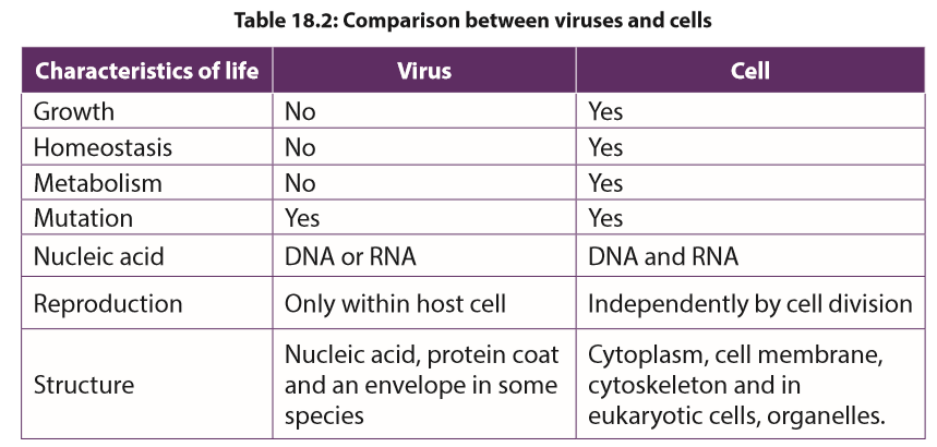 Self-assessment 18.3
1. What are the parts of a virus?
2. Describe the two ways by which viruses cause infection.
3. Distinguish between Bacteriophage and a prophage.
4. What is meant by retrovirus?
5. What are the strengths and weaknesses of the tobacco mosaic virus hypothesis?
6. Which characteristic feature is common to all viruses?
7. How is a capsid protein important to the functioning of a virus?
8. What is the best way to protect humans against most viral diseases?18.4 Moulds
Activity 18.4
Using text books or computer aided materials to describe the life cycle of bread mould.Moulds pervade our world, living wherever moisture is present. Some are of great benefit to humans, providing antibiotics, acting as decomposers so that nutrients can be recycled, or taking part industrial processes. Other moulds cause diseases which lead to serious damage.Moulds have cells arranged in long thread-like filaments, the hyphae, that form a mass called Mycelium. Moulds are usually considered as fungi, but mould may also be formed by filamentous bacteria, slime moulds, and water moulds. Therefore, there are two main types of moulds: fungal moulds and non-fungal moulds.18.4.1 Fungal mouldsAll fungi that produce mycelia can be called moulds, but the term is usually used for an organism in which the mycelium forms the main body of the fungus. In the black bread mould Rhizopus and the pin mould Mucor, the mycelium consists of a tangled mass of hyphae with many nuclei. These hyphae are called coenocytic because the fungal tissue is not separated by cell walls.Fungal hyphae have an outer cell wall made of chitin and inner lumen which contains the cytoplasm and organelles. A cell surface membrane surrounds the cytoplasm and sticks tightly to the cell wall.Rhizopus and Mucor are Saprotrophic, obtaining their nutrients from dead organic material. Rhizopus nigricans and Mucor mucedo can live on bread but some species of Rhizopus feed on living plants, and Mucor commonly grows on rotting fruits and vegetables, in the soil or on dung.Rhizopus and Mucor secrete hydrolytic enzymes onto their food source and digest the food outside the organism and then absorb the soluble digestion products and assimilate them.a. Life cycle of Rhizopus and Mucor.Rhizopus and Mucor belong to the fungal phylum Zygomycota. The phylum got its name because its members produce two kinds of spores: Sexual zygospores as well asexual sporangiospores.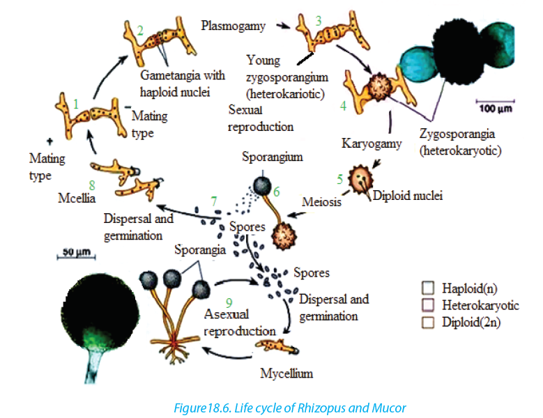 The asexual sporangiospores formed by mitosis, develop insporangium at the tip of hyphae. When sporangium busts, the spores are released.In most species of Mucor, the sporangium dissolves then water enters the spore mass, and the spores are dispersed by the raindrop or are transported by the insects. In most Rhizopus species, the sporangium wall fractures and dry spores are released by the wind.The sexual reproduction involves conjugation. Usually the hyphae from mycelia of different mating types meet and interconnect via outgrowths. The interconnecting walls break down and their cytoplasm containing haploid nuclei mix, then the diploid zygote formed by the fusion of two nuclei develops a thick, rough, black coat and becomes a dormant zygospores. Meiosis probably occurs at the time of germination; the zygospore cracks open to liberate several haploids spores which can give rise to asexual sporangia and mycelia of either mating strain.b. Use of moulds–– They are used to make the human foods. For example, Mucor is used with soya
beans to make a cheese called sufu, in eastern Asia.In Indonesia, R. oligosporus
and R. oryzae are used to produce a food called tempeh from boiled skinless
soya beans.
–– The fungal moulds belonging to the Zygomycota are used to make anaesthetics,
birth control pills, meat tenderisers, and the yellow colouring agents used in
margarines and butter substitutes.