UNIT 9 NERVOUS COORDINATION
UNIT 9: NERVOUS COORDINATION
Key Unit Competence
Describe the structure of neurons and explain the mechanisms of impulsetransmission.
Learning Objectives
By the end of this unit, I should be able to:
– Describe the arrangement of neurons in a reflex arc.
– Describe the structure neurones.
– Explain how a resting potential is maintained.
– Explain how an action potential is generated.
– Explain how a nerve impulse is propagated along a neurone.
– Explain the factors affecting the speed of impulse transmission.
– Describe the properties of a nerve impulse limited to: saltatory conduction, all
or nothing law, and refractory period.
– Describe the functions of neurones in a reflex arc.
– Explain how information passes across a synapse from one neurone to another
or from a neurone to its effector.
– Outline the roles of synapses.
– Describe the roles of neuromuscular junctions, transverse system tubules and
sarcoplasmic reticulum in stimulating contraction in striated muscle.
– Relate the structure of a cholinergic synapse to its functions.
– Interpret graphs for all or nothing law and refractory period.
– Investigate the nature of a nerve impulse in a nerve tissue of a frog
– Appreciate the importance of a coordinated behaviour in organisms.– Show concern about the need to have reflexes as rapid responses
9.1 Overview of control and co-ordination in mammals
Activity 9.1
– Use charts showing the parts of human brain and watch the movies on you
tube showing the different parts of human brain.
– Use the school library and search additional information on the internet.
Read the information related to human brain, and take short notes on
human brain.
1. Illustrate with diagram the main parts of human brain
2. Write down the relative functions of each identified part of the humanbrain
Coordination:It is the process in which body coordinate, ordinate and control
different activities.
The nervous system plays the main functions such as: (i) Sensory input: Sensory
receptors present in skin and organs respond to external and internal stimuli by
generating nerve impulses that travel to the brain and spinal cord, (ii) Integration:
The brain and spinal cord sum up the data received from all over the body and send
out nerve impulses (iii) Motor output: The nerve impulses from the brain and spinalcord go to the effectors, which are muscles and glands.
The Nervous system is divided into two main divisions: The central
nervous system (CNS) and the peripheral nervous system (PNS) The central nervous
system (CNS) consist of the brain and spinal cord, which are located in the midline
of the body. The peripheral nervous system (PNS), which is further divided into the
somatic division and the au- tonomic division, includes all the cranial and spinal nerves.
9.1.1 Some key word definitions
–Irritability or sensitivity. This is the ability of living organisms to respond to a stimulus
–A stimulus: This is any change in the external or internal environment which provokes a response
–Receptors: These are specialized cells that detect a stimulus
–Neurons: These are cells which transmit nerve impulses
–Effectors: are organs that respond to the stimuli and bring about a response.
–A nervous system: This is a system which involved in the detection of stimuli (sensory inputs) integration and response (motor output)
–The response may be to both the external and internal environments.
–Neurone or nerve cell: It is the basic functional unit of the nervous system. Neurones are cells specialized to generate and transmit nerve impulses (action potentials) are cells which transmit nerve impulses (action potentials).
9.1.2 The division of nervous system
The nervous system of a mammal comprises of the central nervous system (CNS)
consisting of the brain and the spinal cord, and the peripheral nervous system (PNS)
consisting of the cranial nerves from the brain, the spinal nerves from the spinalcord and the sensory organs (Figure 9.1).
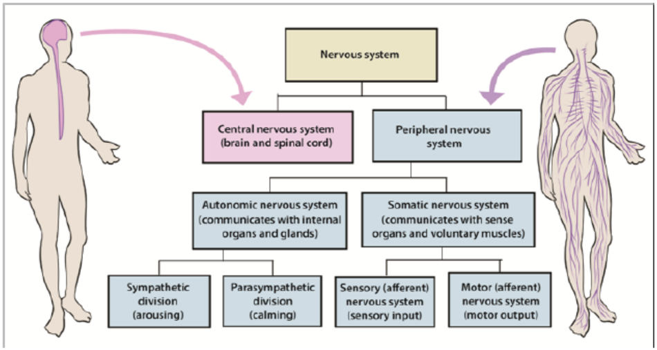
Figure 9.1: Organization of the human nervous system
1. The human brain
The brain is the enlarged end of the spinal cord. It is enclosed in the skull and is
divided into three main parts namely: the fore brain, the mid brain, the hind brain.
a. The fore brain
This consist of: cerebrum, thalamus, hypothalamus and pituitary gland
• The cerebrum:
This is the largest part of the brain made up of two hemispheres called the right and
the left cerebral hemispheres. The left cerebral hemisphere controls those activities
of the right side of the body while the right cerebral hemisphere controls those of
the left side of the body.
The functions of the cerebral hemisphere
– It is the centre of the judgment, memory, reasoning and imagination.
– It receives the impulses from the sensory organs: sight, taste, sound and touch.
– It controls all the body’s voluntary activities, e.g. walking, eating, singing,
• The thalamus:
This is a relay centre. It relays sensory information towards higher centre. It is thecentre for the perception of pain and pleasure.
• The hypothalamus
It performs many functions such as; regulates and monitors the temperature of
blood, monitors and regulates the water content of blood, a co-ordinating centre
for activities of the internal organs, e.g. rate of heart beat, blood pressure. It is a
centre of for feelings such as; hunger, thirst, sex drive, satisfaction, sleep, speech, etc.
As an endocrine gland, it produces hormones i.e. anti-diuretic hormone (ADH) andoxytocin.
• The pituitary gland:
It produces hormones such as: Follicle-stimulating hormone (FSH), Thyroidstimulating
hormone (TSH), Adreno-cortico trophic hormone (ACTH), Prolactin
hormone and Luteinizing hormone (LH)
b. The mid brain
This acts as an association centre between the fore and the hind brain. It is a relay
centre for audio and visual information. It is also responsible for movement of the
head and the trunk.
c. The hind brain
This receives the impulse from the ear, the skin and the semi-circular canals. It
consists of: The cerebellum and the medulla oblongata
The cerebellum: It lies behind the optic lobes. It receives impulses simultaneously
from the eyes and the ears. It regulates and co-ordinate muscular movement,
especially those concerned with maintaining body equilibrium and controls all the
unconscious activities of the body.
The medulla oblongata: This control all the involuntary movements of the body
especially those concerned with respiration, digestion, heartbeat, breathing rateand sneezing.
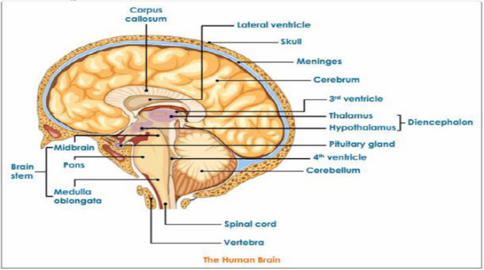
Figure 9.2: Main parts of the brain
2. The spinal cord
The spinal cord is a dorso -ventrally flattened cylinder of nervous tissue running
from the base of the brain down the lumbar region. It is protected by the vertebrateof the backbone and the meninges.
Functions of the spinal cord include;
– It is a coordinating centre for simple reflex such as the knee-jerk response and
the autonomic reflexes such as contraction of the bladder.
– Providing a means of communication between peripheral nerves and the brain.– It sends messages to the effectors
Figure 9.3: Position and external structure of spinal cord
A transverse section of the spiral cord shows an H-shaped central core of grey matter.
Grey matter is composed of nerve cell bodies, dendrites and synapses surrounding
a central canal which contains cerebrospinal fluid. White matter: around the grey
matter, is an outer layer containing nerve fibres whose fatty myelin sheaths give itits characteristics colour.
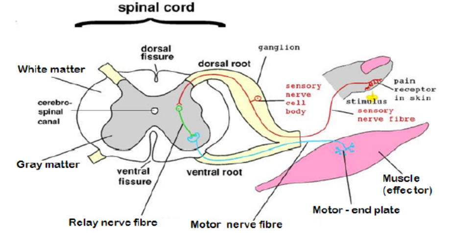
Figure 9.4: The transverse section of the spinal cord
The spinal cord acts as a coordinating centre for simple reflex such as knee jerk
response and autonomic reflexes. The spinal cord acts as means of communication
between spinal nerves and the brain. It sends impulses to the brain through sensory
neurons from the body and returns the motor impulses to the effectors which aremuscles and glands.
Application 9.1Describe the form in which the information is conveyed in the nervous system
9.2 Structure, types and functions of neurons
Activity 9.2
– Use charts describing the neuron and watch the movies showing the types
of neuron.
– Using textbooks or searching additional information on the internet, read
the information related to the structure, types and functions of neurone.
a. Draw and label the structure of a neuroneb. Make a table compering different types of neurons
Aneuron also called nerve cell is the basic functional unit of the nervous system.
Neurons are cells specialized to generate and transmit nerve impulses (actionpotentials) are cells which transmit nerve impulses (action potentials).
9.2.1 Types of neurons
Nerve cells may be grouped according to the number of processes they possess so
that their types include:
– Unipolar neurons: those with one process only, found mainly in invertebrates.
– Bipolar neurons: those with two separate processes such as neurons in the
retina of the vertebrate eye.
– Multipolar neurons: those with more than two processes such as most of thevertebrate neurons.
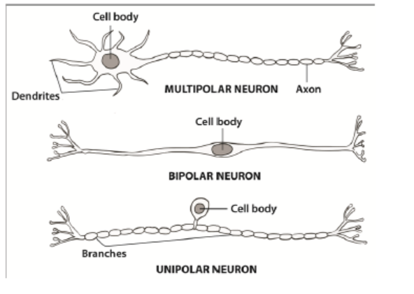
Figure 9.5: Multipolar, bipolar, unipolarneurons
9.2.2 Classification of neurons by their functions
In vertebrates, it is also common to group neurons according to their functions. They
include:
– Sensory or afferent neurons: transmit impulses from the receptors to the
central nervous system. In addition to sensory or afferent neurons.
– Motor or efferent neurons: that transmits impulses from the central nervous
system to effectors motor organs such as muscles or glands that carry out the
response. Most motor neurones are stimulated by impulses conducted by
interneurons. However, there are some others that are stimulated directly by
sensory neurons.
– Interneurons also known as intermediate or association, or relay or interneuron
connect the pathways of sensory and motor impulses, and are found mainly inthe central nervous system.
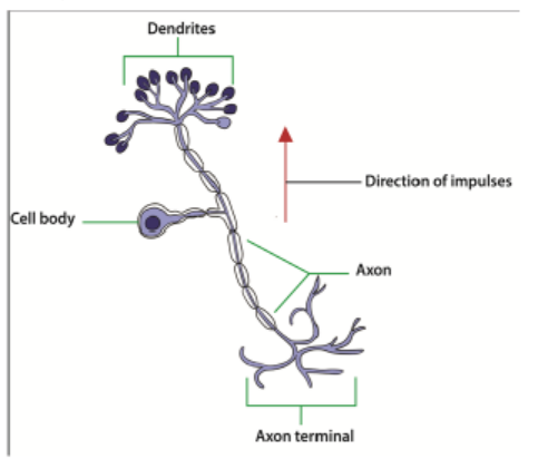
Figure 9.6: Sensory neuron
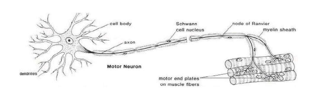
Figure 9.7: Motor neuron (image from google)
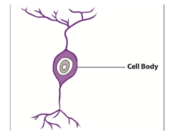
Figure 9.8: Intermediate neuron
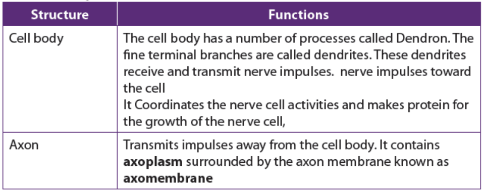
9.2.3 Parts of a neuron and their functions
Each motor neuron possesses a cell body and cytoplasm with many mitochondria,
endoplasmic reticulum, golgi apparatus and ribosomes. The Nissl granules which
consist of endoplasmic reticulum and ribosomes function in protein synthesis. Thetable below (Table 9.1) shows all parts of neuron and their functions.
Table 9.1: The parts of a neuron and their functions
Application 9.2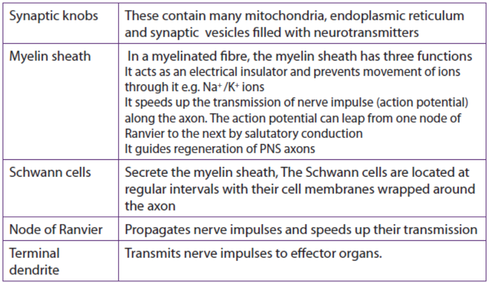
Explain what would happen when a neuron is damaged
9.3 Nature and generation of a nerve impulse
Activity 9.3
– Watch the movies showing the generation of a nerve impulse.
– Use the school library and search additional information on the internet.
– Read the information related to the generation of the nerve impulse and
take short notes on generation of the nerve impulse.
– Answer the following questions:
a. Draw, label and interpret the graph showing the action potentialb. What do you understand by action potential?
All cells in animal body tissues are electrically polarized—in other words, they
maintain a voltage difference across the cell’s plasma membrane, known as the
membrane potential. This electrical polarization results from a complex interplay
between protein structures embedded in the membrane called ion pumps and ion
channels. Each excitable patch of membrane has two important levels of membrane
potential: the resting potential, which is the value the membrane potential maintains
as long as nothing passes along the cell, and a higher value called the thresholdpotential.
9.3.1. Resting potential in a neuron
A neuron is said to be in the resting state when it is not conducting an impulse. The
membrane potential of an unstimulated excitable cell is called the resting potential.
A resting potential is the difference in charge (electrical potential difference) which
exists between the inside and the outside of the cell membrane. In excitable cells, the
resting potential is about -70 millivolts (mV) and the threshold potential is around
-55 mV. The negative sign indicates the interior of the cell is negative with respect tothe exterior environment.
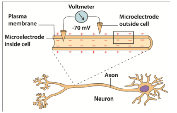
Figure 9.9: Resting potential in a neuron
The resting potential difference across the neuron membrane is maintained by:
– The sodium –potassium pump (Na+ /K+). This is always working. Three sodium
ions (Na+) are actively transported out of the cell for every two potassium ions
(K+) pump into the cell. Energy supplied by ATP is used for the transport of ions
against their electrochemical gradients.
– The axon membrane: It is more permeable to potassium ions than the sodium
ions. This is due to the presence of more potassium ion non-gated, voltageindependent
channels and few sodium ion non-gated channels. More K+ ions
can diffuse out back again faster than Na+ ions which can diffuse back in.
The resting membrane potential is mainly determined by sodium-potassium
pump, facilitated diffusion and electrochemical gradient of K+ ions across themembrane.
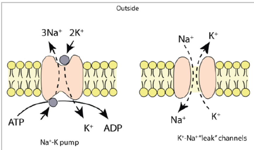
Figure 9.10: Sodium- potassium pump
9.3.2. Action potential
Action potential is the technical term for impulse. An action potential is rapid
temporary reversal in the electrical potential difference of an excitable cell
e.g. a neuron or a muscle cell. It is caused by changes in the permeability of the
membrane following the application of a threshold stimulus. The action potential
has a depolarization phase and a repolarization phase. There may be a short
hyperpolarized phase after the repolarization phase. The time taken for an actionpotential is 2 to 3 milliseconds.
9.3.3. Depolarization
When a stimulus such as electric current reaches a resting neuron, some sodium
voltage gated channels in the stimulated region of the axon membrane open. Sodium
ions (Na+) move into the axon by facilitated diffusion down an electrochemical
gradient. The initial influx of sodium ions is slow. The axon membrane becomes
slightly depolarized and the sodium voltage gates are sensitive to voltage changes.
More gates open allowing more Na+ ions to diffuse into the cell leading to further
depolarization.
When the potential difference across the membrane reaches a threshold value
(-50mV), many more sodium voltage gated channels open. This is an example of
positive feedback. The rapid diffusion of Na+ ions leads to a sudden increase in the
cell’s potential difference which becomes positive (+ 40mV). This reversal in thepotential difference is known as depolarization and lasts for about 1 millisecond
9.3.4. Repolarization
The reversal in polarity to + 40 mV causes the voltage gated sodium channel to
close. At the same time the voltage gated potassium channels open. The potassium
ions K+ diffuse out of the cell down their electrochemical gradient to the tissue fluid
outside. The axon membrane is repolarized. The action potential alters from + 40 mVto -70mV.
9.3.5. Hyperpolarization
The potassium voltage-gated channels are slow to close. An excess of K+ ions leave the
axon. The inside of the membrane becomes more negative. The voltage falls slightly
below -70mV and causes hyperpolarization. However, within a few milliseconds,
the potassium voltage-gated channels close. The resting potential of -70mV is re-
established by the Na+ /K+ pump and different rates of facilitated diffusion of K+ andNa+ ions through the non-gated ion channels.
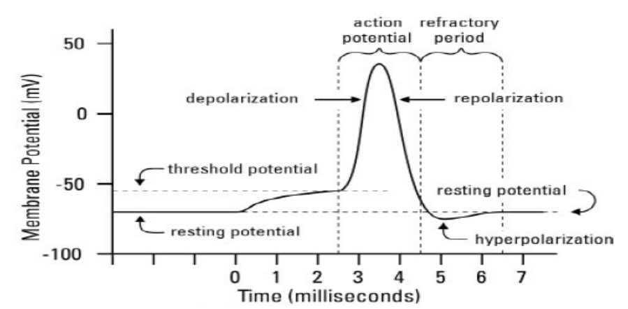
Figure 9.12: The action potential
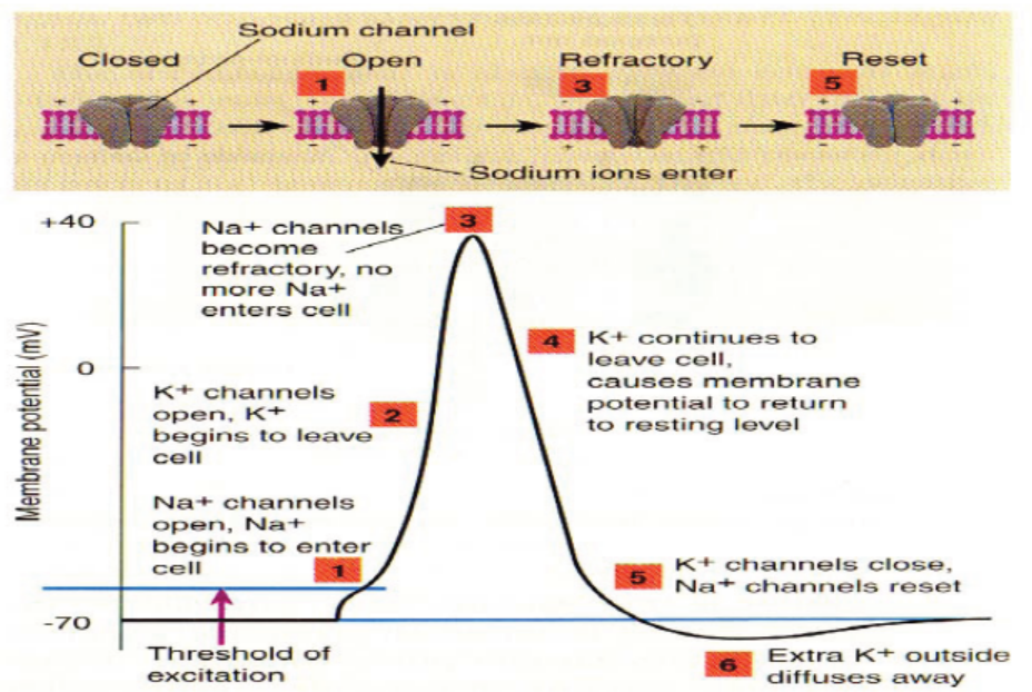
Figure 9.10: The sodium –potassium pump (Na+ /K+) and action potential
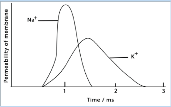
9.3.6. Frequency of action potentials
Information in axons is coded in the frequency of the action potentials. A weak
stimulus above threshold produces fewer action potentials. A stronger stimulus
produces a greater frequency of action potentials. As the intensity of stimulation
increases, more action occurs.
Application 9.3
The graphs below show the changes that occur during an action potential in
a membrane potential and the relative membrane permeability to sodium andpotassium ions in a neurone. Observe well to answer the following questions:
a. Describe the movement of ions during an action potential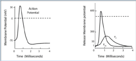
b. Explain what is the effect of an action potential generation if there is alowering of sodium ions in the extracellular fluid
9.4 Transmission of nerve impulses
Activity 9.4
The dissection of a frog sciatic nerve
Materials required
Laptop computer, projector, nerve chamber, cable and nerve chamber leads (red
and black), glass hooks, Stimulator cable, grounding adapter or cable, forceps,
scalpel, frog Ringer’s solution at two temperatures.
Procedure
– To begin dissection, retrieve a frog from your teacher and place it in a dissecting
tray.
– Remove the skin from the legs by making an incision through the skin and
around the entire lower abdomen.
– Cut the connections between the skin and the body especially around the base
of the pelvic girdle.
– Use stout forceps to pull the skin off the frog in one piece (like a pair of pants).
– Place the frog with its dorsal side up.
– Moisten the exposed tissue (legs) with Ringer’s solution and place a wet paper
towel (saturated with Ringers solution) over one of the legs of the frog so that
it is completely covered and wet.
– Use forceps to separate the muscles of the thigh (the leg not covered with the
paper towel).
– Pin the muscles apart so that more underlying muscle is visible.
– This should also expose the cream-colored Sciatic nerve lying deeply between
the muscles.
– Use a glass hook to separate the nerve from the fascia and the vessels. If
possible, avoid cutting the blood vessels. If bleeding does occur, rinse away
the blood with lots of Ringer’s solution. Free the nerve from the knee joint to
the pelvis.
– Use the glass hook to place a suture thread under the nerve. Move the thread
as close to the knee joint as possible.
– Ligate (tie off) the nerve; you may observe calf muscle fibrillation or foot
movement as the knot is tied off.
– Be sure the knot is tied tightly. Cut the nerve between the knot and the knee
joint. Keep the exposed nerve moist at all times with Ringer’s solution.
– Carefully separate the muscles of the pelvis to expose the sciatic nerve.
Remember to rinse any blood away with Ringer’s solution.
– Carefully expose the remainder of the nerve through an opening along the
lateral side of the urostyle. To avoid cutting the nerve, lift the end of the
urostyle with forceps as you cut the muscle away from the urostyle with blunt
scissors.
– Cut along the urostyle from its tip to the vertebral column.
– Deflect the muscle away from the urostyle to expose the Sciatic nerve.
– Use a glass hook to separate connective tissue from the nerve and to place
a piece of suture thread under the nerve.
– Move the thread as high as possible on the nerve to obtain as large a
section as possible.
– Ligate (tie off) the nerve; the leg may jump again as the knot is tied tightly.
– Cut the nerve between the knot and the vertebral column and keep the
exposed nerve moist at all times.
– Use forceps to grasp the suture thread at the proximal end (end closest to
head) and lift the nerve out of the body cavity.
– Do not pinch or stretch the nerve.
– Remove any connective tissues, blood vessels, or nerve branches that may
still keep the nerve attached to the frog.
– Continue to grasp the suture to lift the nerve until it is clear of the abdomen,
the pelvis, and the thigh.
– Grasp the suture at either end to remove the nerve from the body entirely.
– Place the nerve across the gold-coloured electrode pins in the nerve bath.
– Add a small quantity of Cold Frog Ringers to the bottom of the chamber.
– The Frog ringers should not touch the gold-plated electrode pins.– Cover the chamber with a glass slide.
Questions
1. Draw a picture of the laboratory setup used for this exercise.2. Find and dissect the frog sciatic nerve for placement in a nerve chamber.
9.4.1 Mechanism of transmission of nerve impulses along an axon
– The neurons, like other cells, are positively charged outside and negatively
charged inside. The membrane of the axon is said to be polarized. The potential
difference (voltage) across their membranes is of – 70mV and is called resting
membrane potential (RMP).
– A stimulus (heat, pain, bite, sound …) creates an action potential (AP) or an
impulse that is transmitted along an axon by electro-chemical change.
– During an action potential, the membrane potential falls until the inside
becomes positively charged with respect to the exterior. The membrane at this
point is said to be depolarized. It takes few milliseconds to happen. In fact, the
potential changes from – 75 mV to + 40 mV at the point of stimulation. That is
an electrical change that runs along the axon.
– As the impulse is transmitted along the axon, the Na+/K+ pumps of the axolemma
are re-established. Sodium channels open first, allowing a large number of Na+ions to flow in.
– The axoplasm becomes progressively more positive with respect to the outside
of the axolemma. Then, almost instantly, the permeability of the membrane to
Na+ ions ceases, and the net flow of Na+ ions stop. At the same time K+ ion
channels start to open and K+ ions flow out from axoplasm where they are in
high concentration. The counter-flow is of 3Na+ ions against 2K+ ions.
– The axoplasm now starts to become less positive again. This begins the process
of re-establishing the resting potential difference of the membrane. That is anelectro-chemical change.
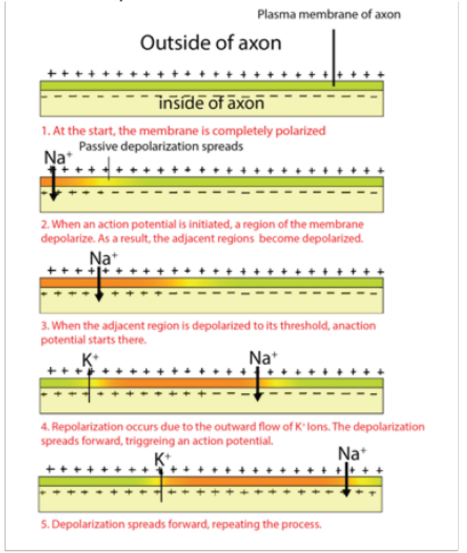
Figure 9.13: The nerve impulse transmission along axon
a. Factors that affect the transmission of nerve impulses along the axon
membrane
Along the axon membrane, the transmissions of nerve impulses are affected as
follows:
– The diameter of the axon: the greater the diameter the faster the speed of
transmission of nerve impulses.
– The myelin sheath: myelinated neurones conduct impulses faster than nonmyelinated
neurones.
– The presence of nodes of Ranvier: speeds up the movement of impulses in
myelinated neurones.
b. Structure of a synapse
Information from one neuron flows to another neuron across a synapse. The synapse
is a small gap separating two adjacent neurons. The synapse consists of:
– A presynaptic ending that contains neurotransmitters, mitochondria and other
cell organelles,
– A postsynaptic ending that contains receptor sites for neurotransmitters and,
– A synaptic cleft or space between the presynaptic and postsynaptic endings. It
is about 20nm wide.
– The swollen tip of the axon of the presynaptic neuron, called synaptic knob
or synaptic bulb contains many membrane – bounded synaptic vesicles,
mitochondria and microfilaments.
– The synaptic vesicles contains neuro transmitter molecules such as acetylcholineor noradrenaline
c. Neurotransmitter
A neurotransmitter is a relatively small chemical found in the synaptic vesicle. It
helps to transmit an impulse across a synapse or neuromuscular junction. There
are about 50 different types of neurotransmitters in the human body. Examples
are acetylcholine released by cholinergic neurons, noradrenaline (norepinephrine)
released by adrenergic neurons, dopamine and serotonin including amino acidsglutamate and glycine.
9.4.2 Mechanism of nerve impulse transmission across a synapse
– The arrival of an impulse on the synaptic knob causes the opening of Ca+2 ion
channels on the presynaptic membrane, and Ca+2 ions flow in the presynaptic
region from the synaptic cleft.
– The Ca+2 ions induce a few presynaptic vesicles to fuse with presynaptic
membrane and to secrete their neurotransmitters (e.g. acetylcholine) by
exocytosis into the synaptic cleft
– The neurotransmitter then binds with the receptor protein on the postsynaptic
membrane. This causes the opening of Na+ channels on the postsynaptic
neuron which in turn becomes depolarized.
– This causes a depolarization of the post-synaptic cell membrane, which may
initiate an action potential, if the threshold is reached
– The action of the neurotransmitter does not persist because an enzyme
cholinesterase catalyses the hydrolysis of acetylcholine into choline and acetate.
The breakdown products (choline) are absorbed by the pre-synaptic neuron
by endocytosis and used to re-synthesize more neurotransmitter, using energyfrom the mitochondria.
9.4.3 Properties of a nerve impulse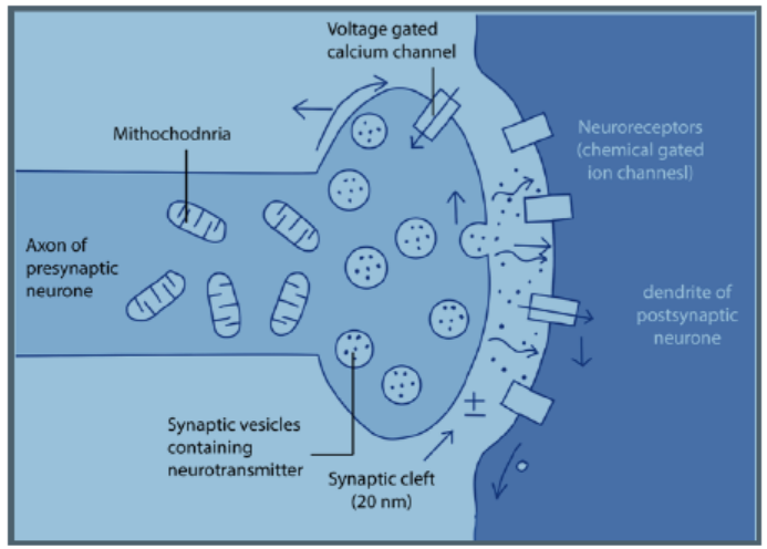
a. All or nothing law
An action potential can only be generated after the threshold value is exceeded.
After the threshold is reached, the size of the action potential produced remains
constant and is independent of the intensity of the stimulus. This is the all or nothing
response. All action potentials are of the same amplitude.
b. Refractory period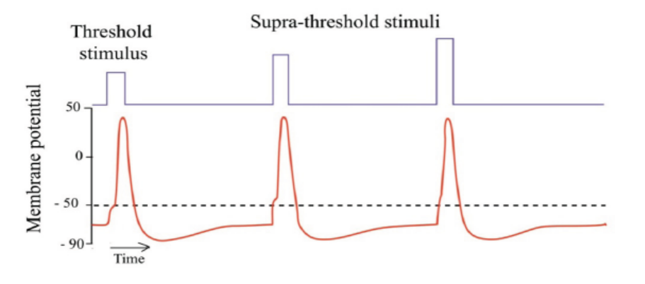
This is a brief period when an axon is unable to transmit an impulse following
transmission of the same. It lasts about 5-10 milliseconds. It is divided into two;absolute and relative periods. During the absolute refractory period which lasts
about 1ms, the axon membrane is unable to respond to another stimulus, no matter
how strong it is. An action potential cannot be produced. This is because there is
conformational change in voltage-gated sodium channels which are still in a closed,
inactive state. This also prevents the action potential from moving backwards.
Following the absolute refractory period, there is a relative refractory period which
lasts around 5ms. During this period, the resting potential is gradually restored by
Na+ /K+ pump and the relative permeability of membrane to facilitated diffusion of
ions is also restored. A new action potential can then be produced if the stimulus is
greater than the usual one. The refractory period therefore allows impulses to move
only in one direction and limits the frequency at which successive impulses can passalong axon.
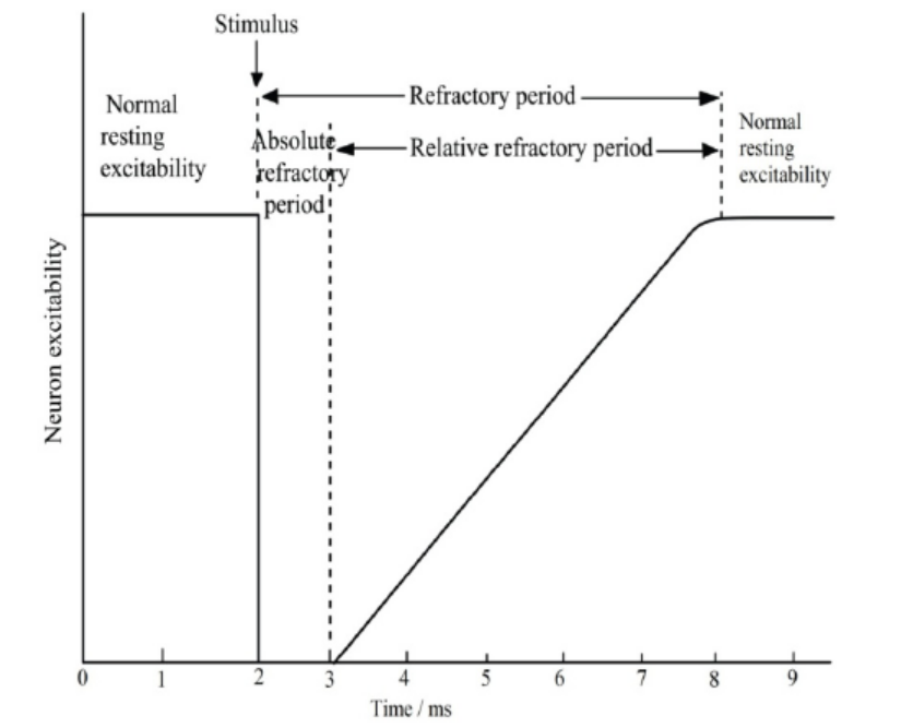
Figure 9.16: Neuron excitability before and after a nerve impulse
c. Salutatory conduction
It is movement or jump of nerve impulses from one node of Ranvier to anotheralong the axon membrane of neurone.
Application 9.4
1. Suppose a cell’s membrane potential shifts from -70 mV to -50 mV. What
changes in the cell’s permeability to K+ or Na+ could cause such a shift?
2. The diagram below shows the changes in potential difference across an axonmembrane as a nerve impulse passes
a. Explain what happens at M, N, O, P, Q and R as shown in the graph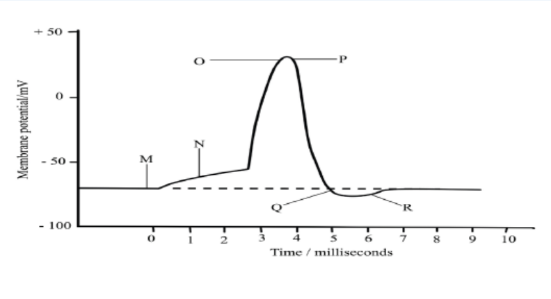
b. Name two factors that can determine the speed of transmission of a nerve
impulse and how each affects the speed
c. Explain why the initiation of an action potential is considered a positivefeedback mechanism
9.5 Structure and function of a cholinergic synapse.Activity 9.5
Use textbooks from school library and other additional information using
internet, read the information related to the cholinergic synapse and take shortnotes on cholinergic synapse.
a. Draw and label a diagram showing a cholinergic synapseb. Make a table of different functions of a cholinergic synapse
The cholinergic synapse is a synapse which uses acetylcholine (Ach) as
neurotransmitter. Calcium and vesicles are involved in the release of neurotransmitter
across the synaptic cleft in the mechanism of synaptic transmission to generate anexcitatory post-synaptic potential.
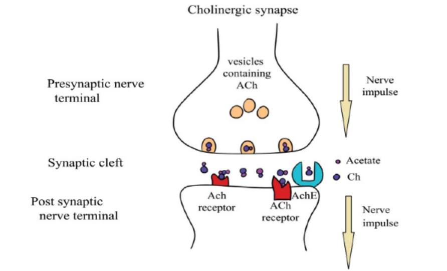
Figure 9.17: The cholinergic synapse
9.5.1. Functions of synapses
Synapses have a number of functions which include:
a. Transmit information between neurones
The main function of synapses is to convey information between neurons. It is from
this basic function that the others arise.
b. Pass impulses in one direction only
As the neurotransmitter substance can only be released from one side of a synapse,
it ensures that nerve impulses only pass in one direction along a given pathway
c. Act as junctions
Neurons may converge at synapse. In this way a number of impulses passing along
different neurons may release sufficient neurotransmitter to generate a new action
potential in a single postsynaptic neuron whereas individually they would not. This
is known as spatial summation. In this way responses to a single stimulus may be
coordinated.
d. Filter out low level stimuli
Background stimuli at a constantly low level, e.g. the drone of machinery, produce
a low frequency of impulses and so cause the release of only small amounts of
neurotransmitter at the synapse. This is insufficient to create a new impulse in the
postsynaptic neuron and so these impulses are carried no further than the synapse.
Such low level stimuli are of little importance and the absence of a response to them
is rarely, if ever harmful. Any change in the level stimulus will be responded to in theusual way.
e. Allow adaptation to intense stimulation:
In response to a powerful stimulus, the high frequency of impulses in the presynaptic
neuron causes considerable release of neurotransmitter into the synaptic cleft.
Continued high-level stimulation may result in the rate of release of neurotransmitter
exceeding the rate at which it can be formed. In these circumstances the release of
neurotransmitter ceases and hence also any response to the stimulus. The synapseis said to be fatigued.
9.5.2. Effects of drugs on synapses
Several types of chemicals such as drugs interfere at synapses, either amplifying or
inhibiting the transmission o of impulses. For example,
– Caffeine and nicotine amplify the transmission of impulses by mimicking the
action of natural neurotransmitters.
– Insecticides that prolong the effect of neurotransmitters by blocking the
enzymatic breakdown of transmitters. Other drugs such as
– Anaesthesia including atropines inhibit the transmission of impulses across
the synaptic membranes. Atropine acts to prevent an action potential being
generated by acetylcholine when it attaches to its receptor protein on thepostsynaptic membrane
9.5.3. The neuromuscular junction
A special kind of synapse is the nerve-muscle known as neuromuscular junction,
the point where the terminal dendrite of a motor nerve cell makes contact with a
muscle fibre. The region of the sarcolemma (cell surface membrane) of muscle fibre
that lies directly under the terminal portion of the motor neuron is known as the
motor end plate. At the nerve-muscle junction the membrane of the muscle fibre ismodified to form an end-plate to which the dendrite is attached.
When an impulse arrives at the nerve-muscle junction, acetylcholine is discharged
from synaptic vesicles into the synaptic cleft. The acetylcholine diffuses across the
gap and depolarizes the muscle end plate. End-plate potentials can be recorded and
it has been shown that if these build up sufficiently an action potential is fired off inthe muscle fibre.
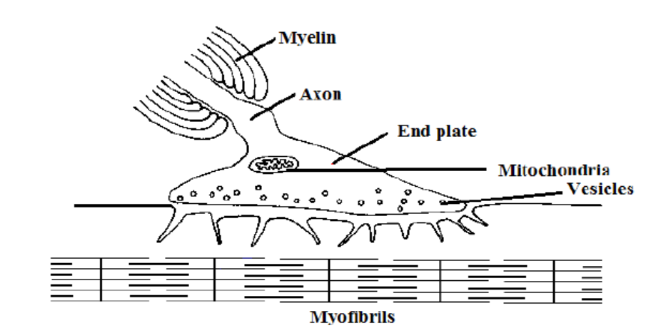
Figure 9.18: The neuromuscular junction
Application 9.5The diagram shows the structure of a nerve synapse
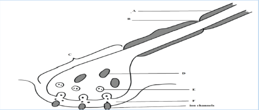
1. Label structures A to F
2. Draw an arrow on the diagram to show the direction of a nerve impulse
(an action potential) in the presynaptic neurone.
3. Name a common neurotransmitter presents in the synaptic vesicle
4. Name an ion that
a. Moves into the postsynaptic neurone in an excitatory synapse
b. Moves out of the postsynaptic neurone in an inhibitory synapse
4. Suggest one reason why structure C contains large numbers of organelle
D
5. What is the major chemical component of structure A?6. State the functions of structure A and structure B
9.6 Functions of sensory, relay and motor neurons
in a reflex arc
Activity 9.6
Aim: Description of a reflex arc
The diagram below shows the sequences of events when one is hit on the patella(bone situated in front of the knee joint) by a small hammer.
1. Observe carefully and discuss what is happening according to the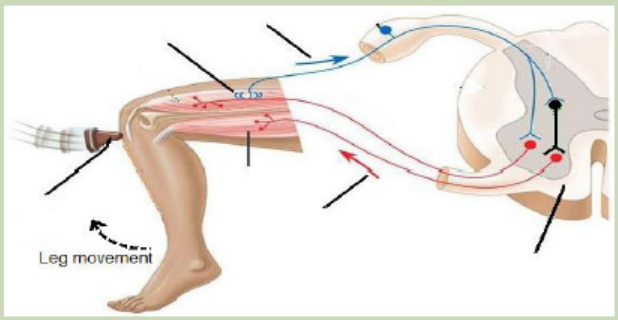
direction indicated by the arrows.
2. Identify the organ/part where the information starts and the organ
responsible for the response.
3. Label the figure by using the following words: stimulus, sensory receptor,sensory transmission, motor transmission, effectors, and spinal cord.
9.6.1. Reflex actions
A reflex action is a quick and involuntary response of the central nervous system to
a stimulus. Example: The quick withdrawal of the hand from a hot object. When the
spinal cord alone is involved, the reflex action is called spinal reflex and when thebrain alone is involved, it is a cranial reflex e.g. blinking of eyes.
Reflex actions are described as involuntary actions and the same stimuli producethe same responses every time. Reflexes are useful because they make autonomic
involuntary adjustments to changes in the external environment, such as the irispupil
reflex and the balance during locomotion. They also control the internal
environment, such as breathing rate and blood pressure, and prevent damage to
body as in cuts and burns. These help to maintain constant conditions, in other word
they are involved in homeostasis.The sequences of changes that occur during a spinal reflex are:
– A sensory receptor receives a stimulus and impulse is generated in it
– The impulse is transmitted along a sensory neuron towards the spinal cord via
the dorsal root
– Once the impulse reaches the grey matter inside the spinal cord, it is passed on
to the relay neuron across a synapse
– The relay neuron then transfers the impulse to a motor neuron across another
synapse.
– The motor neuron conveys the impulse to an effector such as a muscle where a
response takes place.
The pathway that is followed by an impulse along the sensory neurons relay andmotor neurone, during a reflex action is called reflex arc.
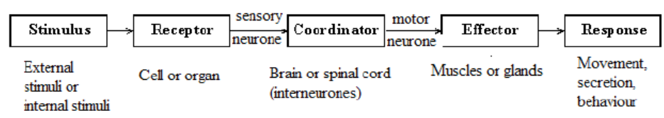
Figure 9.19: Sequence of change in a spinal reflex
The components of reflex arc are:
– Stimulus
– Receptors
– The sensory receptor that detects the stimulus
– The sensory (or afferent) neurone along which the sensory impulse is
transmitted;
– The relay neurone in the central nervous system to which the sensory impulse
is passed on.
– The motor (or efferent) neurone along which the motor impulse is transmitted;
and
– The effector (Muscle or gland) which the motor impulse triggers to bring about
an appropriate response.– CNS (Brain or spinal cord)
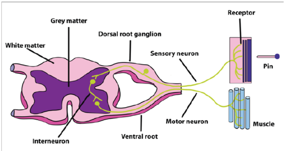
Figure 9.20: The diagram showing reflex arc
9.6.2 Conditioned reflex actions
This type of reflex involves the brain but it is also as fast as the simple reflex. Salivation
on smelling one’s favourite food is an example of conditional reflex. The individual
recognizes and based on the previous experience, the response (salivation) occurs.
The recognition of the previous experience involves the association centres of thebrain.
A series of experiments were conducted by Ivan PAVLOV, a Russian biologist who
demonstrated conditioned reflex. He found that when a bell rung every time a dog
was given food, the dog showed salivation only at the sound of the bell. The ringing
of the bell is called stimulus. The dog had, thus, learnt to associate the sound of thebell to the food and this made it salivate at the sound of the bell.
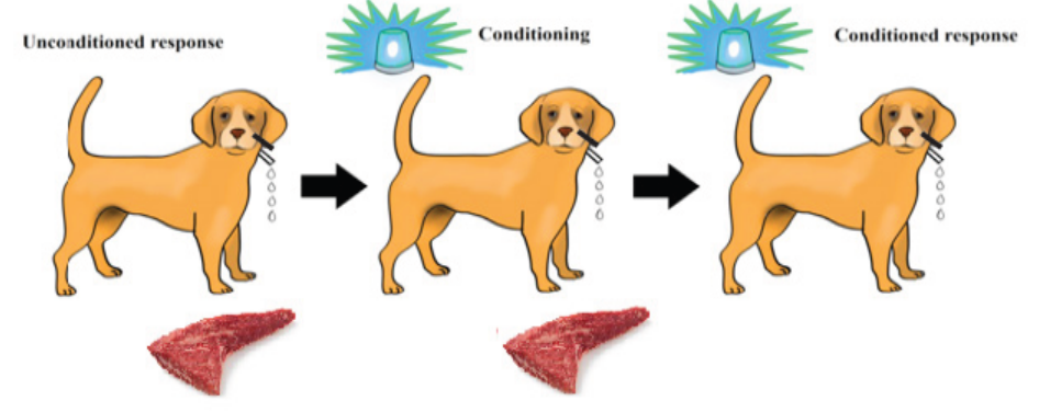
Figure 9.21: The experiment representing the conditioned reflex
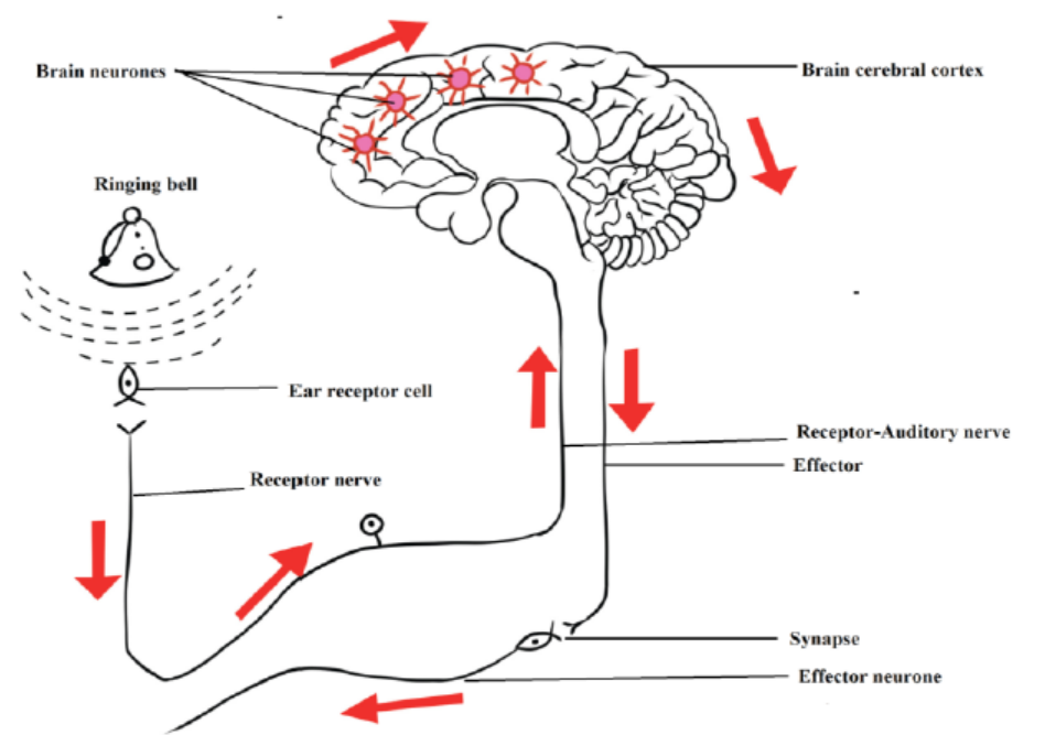
Figure 9.22: The pathway events of conditioned reflex
Application 9.6Describe the functions of sensory, relay and motor neurones in a reflex arc
End of unit assessment 9
A. Multiple choice questions: Choose the best answer
1. What happens when a neuron’s membrane depolarizes?
a. There is a net diffusion of Na+ out of the cel1.
b. The equilibrium potential of K+ becomes more positive.
c. The neuron’s membrane voltage becomes more positive.
d. The neuron becomes less likely to generate an actionpotentia1.
e. The inside of the cell becomes more negative relative to the outside.
2. Why action potentials are usually conducted in only one direction along an
axon?
a. The nodes of Ranvier can conduct potentials in only one direction.
b. The brief refractory period prevents reopening of voltage gated Na+
channels.
c. The axon hillock has a higher membrane potential than the terminals
of the axon.
d. Ions can flow along the axon in only one direction.
e. Voltage-gated channels for both Na+ and K+ open in only one
direction.
3. A common feature of action potentials is that they
a. Cause the membrane to hyperpolarize and then depolarize.
b. Can undergo temporal and spatial summation.
c. Are triggered by a depolarization that reaches the threshold.
d. Move at the same speed along all axons.
e. Result from the diffusion of Na+ and K+ through ligand gated
channels.
4. Where are neurotransmitter receptors located?
a. On the nuclear membrane
b. At nodes of Ranvier
c. On the postsynaptic membrane
d. On the membranes of synaptic vesicles
e. In the myelin sheath
5. During the repolarisation phase of an action potential, the permeability of
the axon membrane to:
a. Na+ increases
b. K+ increases
c. Ca+ increasesd. Organic anions increases
6. The graph shows the changes in the permeability of an axon to Na+ and K+ions during an action potential
Which of the following shows the correct movement of these ions in the axon?
a. Na+ ions enter the axon, K+ ions leave the axon
b. Na+ ions leave the axon, K+ ions enter the axon
c. Both Na+ and K+ ions enter the axon
d. Both Na+ and K+ leave the axon
7. The graph shows the potential difference across an axon membrane. Whichpart of the graph shows the action potential?
a. 3, 4 and 5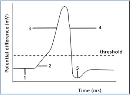
b. 2,3, 4 and 5
c. 1,2, 3 and 4d. 1,2,3, 4 and 5
B. Questions with structured answers
8. The diagram below shows a human brain seen from the right side
a. Name the parts labelled A, B and C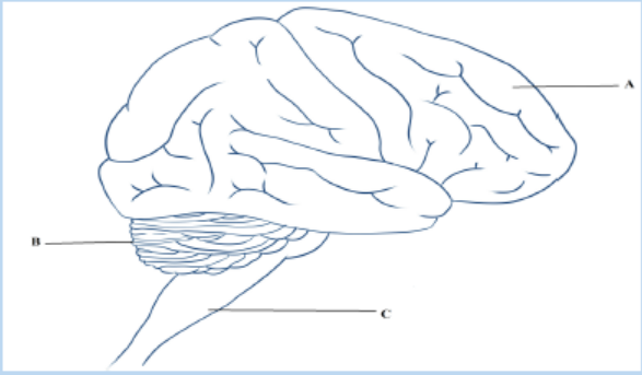
b. Give two functions of the part labelled B.
9. The list describes the main stages in the process by which information is
transmitted across cholinergic synapses.
– An action potential arrives at synaptic knob of presynaptic neurone.
This causes…. the ions to enter the synaptic knob.
– Vesicles move to the………………. membrane.
– A neurotransmitter called……………….is released into the synaptic
cleft
– This moves across the cleft by a process known as…………. the
neurotransmitter combines with a………………. on the postsynaptic
membrane.
– Influx of……………. ions cause local depolarisation and an action
potential is set up in the postsynaptic neurone
a. Copy the list. Using the correct scientific terms, add the words that
have been omitted.
b. Explain what happens to the neurotransmitter after it has passed
information across a cholinergic synapse
c. Some nerves, especially those of the sympathetic nervous system,
produce noradrenaline in their synaptic vesicles. Name this type ofsynapse
10. The graph shows the changes in permeability of the cell surface membraneof an axon to sodium and potassium ions during an action potential.
a. Explain how the events which take place between X and Y on the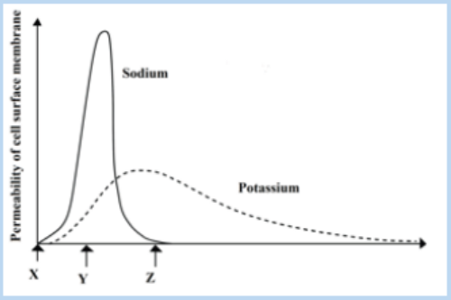
graph can lead to a change in the potential differences across the
membrane
b. What happens to the potential difference across the membrane
between times Y and Z?
c. Explain why a nerve impulse travels faster in myelinated neurone
than in a non-myelinated one.11. The diagram below shows a nerve cell or neuron
a. Name the type of neurone shown.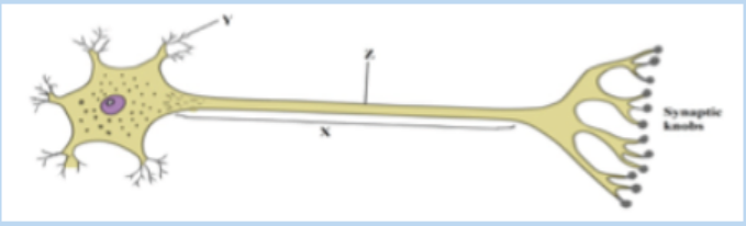
b. Name the structures labelled X and Y
c. A nerve impulse can be initiated by stimulation with a microelectrode.
What would be the effect of stimulation at point Z?
d. The synaptic knobs release a chemical transmitter, acetylcholine.
Nerve gases prevent the breakdown of this chemical. From this
information suggest
I. One early symptom of nerve gas poisoningII. One reason for this observed symptom
12. Complete the following table by stating which region of the brain controlseach of the functions listed.
13. The diagram below represents the structures visible at a synapse with the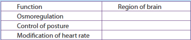
aid of electron microscopy.a. Identify the structures labelled A and B
b. Name the chemical found in the numerous vesicles that occur in the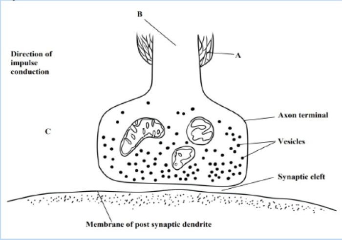
synaptic knob
c. Identify the structure labelled C and suggests a reason for its
presence in the synaptic knob
d. A powerful hydrolytic enzyme is found in the synaptic cleft. What isits function in normal synaptic transmission?
C. Essay question
14. Describe what happens when an action potential arrives at a synaptic knobof an excitatory synapse