Topic outline
UNIT 1 POPULATION AND NATURAL RESOURCES
UNIT 1: POPULATION AND NATURAL RESOURCES
Key Unit Competence
Describe the factors affecting population size and the importance of natural
resourcesLearning Objectives
By the end of this unit, I should be able to:
– State and define population characteristics.
– Explain factors that affect population density.
– Explain population growth patterns.
– Explain the terms renewable and non-renewable resources.
– Explain how environmental resistance affects the balance of nature.
– Explain the importance of natural resources in growth of the Rwandan economy
and methods of conservation.
– Demonstrate methods used in estimating populations by using quadrats and
line transects.
– Research how the human population has grown over the past 250 years.
– Compare statistics on the population age-sex structure of developing and
developed countries.
– Analyse the costs and benefits of managing renewable and non-renewable
resources.
– Support that human population explosion impacts negatively on the
environment.
– Recognize that some resources are renewable and others are non-renewable
and that effective use of these resources is of great value.
– Justify the practice of family planning as a tool for reducing populationexplosion.
Introductory activity
Analyse pictures below and answer the questions that follow.
1. Specify the appropriate ecological terms to describe picture A and picture B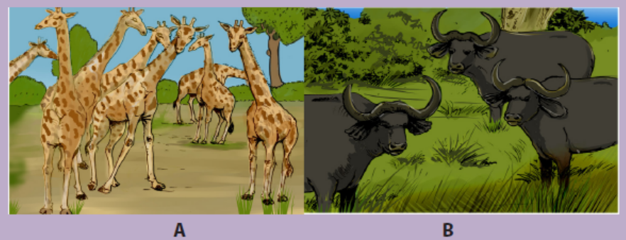
2. Describe what would happen if the number of animals in pictures A and B
were highly increased3. Using search engine categorize the natural resources.
Pictures A and B represent ecological populations. In biology, an ecological
population is a group of organisms of the same species that live in the same area at a
certain period of time. The population is the unit of natural selection and evolution.
How large population is and how fast it is growing are often used as measures of itshealth.
1.1 Population characteristics
Activity 1.1
Using search engine, define the following terms:
1. Density
2. Age structure
3. Growth pattern
4. Birth rate5. Death rate
A given population is characterized by its density, age structure, growth patterns,birth and death rate.
1.1.1 Population density
Population density is the number of individuals of the same species per unit area or
volume. For example, the number of Acacia tree species per square kilometer in the
Akagera National park in Rwanda or the number of Escherichia coli per millilitre in a
test tube express the density of these individuals per square kilometre in a natural
forest and per millilitre in a test tube.
1.1.2 Population age structure
Age structure is the number or proportion of individuals in each age group within
a population. The figure 1.1 below provides the distribution of the populationaccording to age.
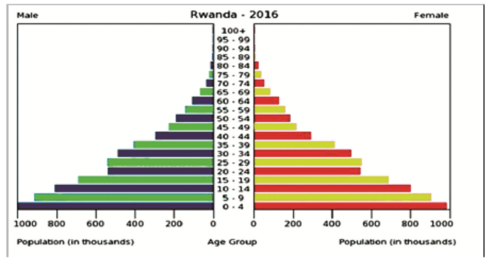
Figure 1.1: Age structure in Rwanda
Information is included by sex and age group as follows: 0-14 years (children), 15-
24 years (early working age), 25-54 years (prime working age), 55-64 years (mature
working age), 65 years and over (elder age). The age structure of a population affects
a nation’s key socioeconomic issues. For example, countries with young populations
(high percentage under age 15) need to invest more in schools while countries with
older populations (high percentage ages 65 and over) need to invest more in thehealth sector.
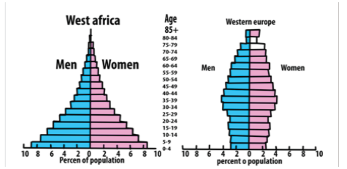
Figure 1.2: Age- sex structure pyramids of developing and developed countries
The shapes of the age-sex structure pyramids shown above show the age sexstructure
of a developing and developed country. The main characteristics of
developing countries including some of the African countries in terms of population
growth include high death rate; high birth rate and low life expectancy, while the
main characteristics of developed countries such as most European countries
in terms of population growth are low death rate, low birth rate and longer life
expectancy
1.1.3 Population explosion
Population explosion is the rapid increase in number of individuals of a particular
species. For example, the world’s human population increase since the end of World
War II is attributed to; an accelerating birth-rate, a decrease in infant mortality and
an increase in life expectancy. Such human population increase impacts negatively
the environment. For instance, human population explosion contributes to pollution
leading to; ozone depletion, eutrophication, acid rain, global deforestation, soilerosion and desertification.
Figure 1.3: World population growth from 1950 to 2050.Source: U.S. Census Bureau, International Data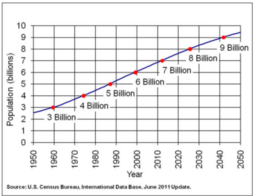
Base, June 2011 Update.
As the figure 1.3 above indicates, the World population is exponentially growing.
This is the reason why most countries, including Rwanda, are practicing the familyplanning. Family planning is the practice of controlling the number of children in a
family and the intervals between their births. If a married couple is sexually active,
they have to adopt at least one family planning technique such as contraception
and timing of reproduction. Other techniques commonly used include; sexuality
education, prevention and management of sexually transmitted infections, preconception
counselling and management, and infertility management.
1.1.4 Population growth patterns
Population growth patterns are graphs (population growth curves) in which increases
in size are plotted per unit time. When a population size increases, the growth rate
also increases. The larger the population becomes, the faster it grows. The factors
that contribute to the population growth are immigration of new species as well as
the birth rate. Population growth is also influenced negatively by emigration and
the death rate.
1.1.5 Birth and death rates
Birth rate is the ratio of live births in a specified area to the adults in population of
that area. It is usually expressed per one thousand individuals per year. It is estimatedfrom this calculation:
Death rate is the ratio of deaths to the adults in population of a particular area during
a particular period of time. It is usually calculated as the number of deaths per onethousand individuals per year and it is estimated from this calculation:
Application 1.1
1. Distinguish between population density and age structure.
2. There are 100 adult elephants in a population of an area. Each year, 10
elephants are produced while 2 elephants die.
a. Calculate the birth rate of this population.
b. Calculate the death rate of that population.
3. Explain the impact of population explosion on the environment.4. Describe the family planning techniques.
1.2 Population density: Dependent and independent factorsaffecting population density
Activity 1.2
The list of factors including; space, nutrients/food, shelter, natural disasters,
competition, predation, disease, sunlight, parasitism, temperature, water,
human activities, physical characteristics of the environment, and behaviour
of organisms in an environment have the relationship with figures belowindicating the relationship between animals and their environment.
1. Categorize the listed factors into density-dependent and densityindependent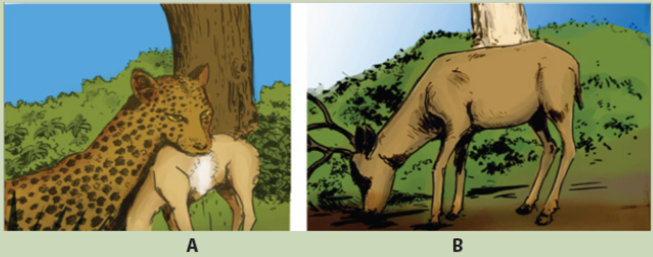
factors.
2. Among the 2 categories of factors given above, suggest the factors illustratedby the figures A and B.
Populations are differently distributed. The distribution and the density are controlled
by environmental factors, which can either increase or decrease the population size
by affecting birth rate, death rate, immigration and emigration (Table 1.1 below).
These factors are grouped into two major categories: Density -dependent factorsand Density- independent factors.
1.2.1 Density-dependent factors
Density dependent factors are factors whose effects on the size or growth of the
population vary with the population density. The types of density dependentfactors include: availability of food, predation, disease and migration. However, food
1.2.2 Density-independent factors
Density independent factors can affect the population without being necessary
based on the density. They include; natural disasters (droughts, floods, hurricanes
and fires), temperature, sunlight, seasonal cycle, human activities, and levels of
acidity, cited among many others.
Table 1.1: Environmental factors that affect the population densityavailability is considered as the main factor.
Application 1.2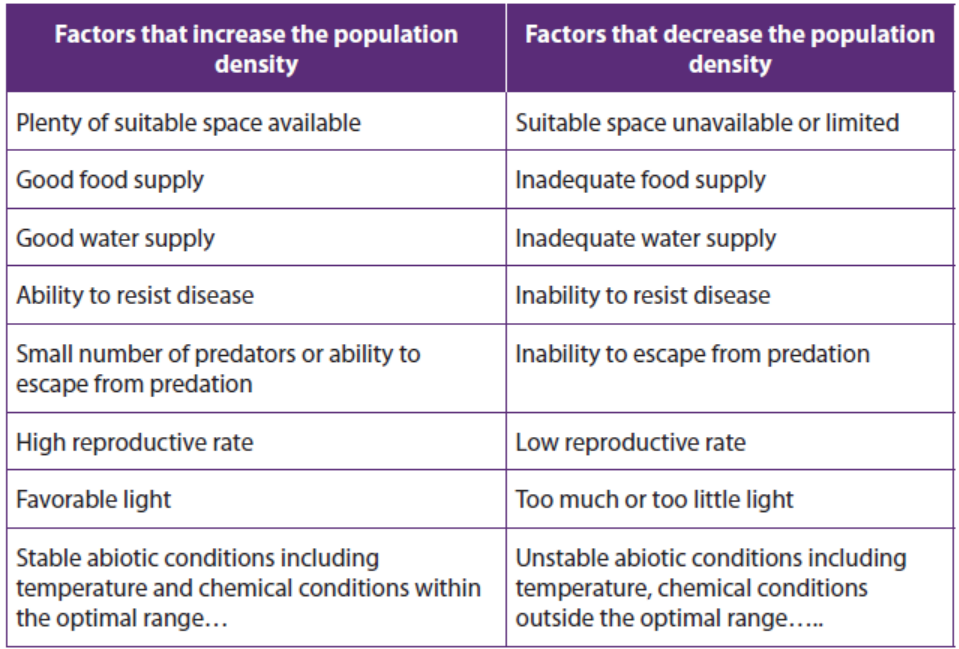
Discuss the ways by which natural disasters (droughts, floods, hurricanes andfires) affect the population growth.
1.3 Methods or techniques of measuring population density
Activity 1.3.1
Using pegs, strings/ropes, meter-ruler, and quadrats in your school ground,
carry out the following field work.
Move in the school ground and make a line transect of 15 meters by the use
of a decametre. Use pegs and strings/ropes to collect all plants and animal
species found at each five meters across transect.
In the ground, make five different quadrats of one square meter separated
by 3 meters and use pegs and strings/ropes to collect different plants and
animal species within each quadrat.
Record your samples in the following table with respect to each quadrat:
Calculate the population density and species frequency for each studiedquadrat.
Calculate the population density and species frequency for each studied
quadrat.
1.3.1 Quadrat method
A quadrat is a square frame that marks off an area of ground, or water, where you can
identify different species present and/or take a measurement of their abundance.
Before any experiment, the decision on a suitable size for the quadrat and the
number of samples to use is taken. Samples must be selected randomly to avoid
any bias, such as choosing to take all of samples from the place with fewest species
simply because it is the easiest to do. This would not represent the whole area youare surveying.
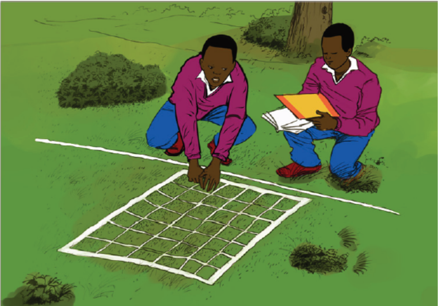
Figure 1.5: Sampling using a quadrat method
A quadrat method enables the calculations of 3 aspects of species distribution
including; species frequency, species density and species percentage cover. Theresults can be used to calculate species frequency and species density.
1.3.2 Species frequency
Species frequency is a measure of the chance (probability) of a particular species
being found within any one of the quadrat, and it is found simply by recording
whether the species was present in each analysed quadrat. For example, if a quadratis placed 50 times, and a given plant was identified in 22 samples, then the species
frequency for this plants equals:
1.3.3 Species density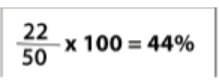
Species density is a quantity of how many individuals there are per unit area, for
example, per square meter. To achieve this, one takes the total number of counted
individuals and then divide it by the number of quadrats done. An example is:Total number of individuals = 200
Total area of quadrats = 480m2
1.3.4 Species cover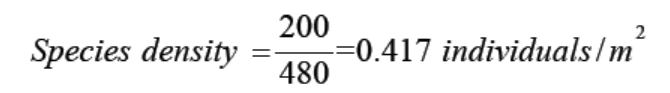
Species cover is a measure of the proportion of ground occupied by the species and
gives an estimate of the area covered by the species as the percentage of the total
area. For example, if there are 100 small squares in one quadrat, then the squares
in which the plant species is present are counted. If plants are found in 25 squares
within that quadrat, the conclusion is that the plant covers 25% of the area.
1.3.5 Line transect method
Line transect is a tape or string laid along the ground in a straight line between
two poles as a guide to a sampling method used to measure the distribution of
organisms. For example, the investigation on change at the edge of a field where it
becomes very marshy is done by randomly selecting a starting point in the field and
lay out a measuring tape in a straight line to the marshy area, and then sample the
organisms that are present along the line, which is called a transect. The simplest
way to do this is to record the identity of the organisms that touch the line at setdistances – for example, every two meters.
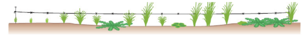
Figure 1.6: Line transect
1.3.6 Capture-recapture method
Activity 1.3.2
Mukamana is a fish farmer in Bugesera district. She wanted to know the total
population in her fish pond. She netted 240 fishes and tagged (marked) their
opercula with aluminium discs. She released those fishes into the pond. After
one week, she netted again 250 fishes among which 15 had the aluminiumdiscs. Calculate the estimated population from marked individuals.
Capture-recapture method involves capturing the organism, marking it without
any harm, and release it in the same area so that it can resume a normal role in
the population. For example, fish can be netted and their opercula is netted with
aluminium discs, birds can be netted and rings can be attached to their legs, small
animals may be tagged by dyes, or by clipping the fur in distinctive pattern, while
arthropods can be marked with paint. In all cases, some form of coding may be
adopted so that individual organisms are identified. Having trapped, counted andmarked a representative sample of the population.
Application 1.3
1. Kalisa and Mutoni conducted an experiment within a quadrat of 0.5m2 andfound the following statistics for a couch grass by quadrat:
a. Calculate the species frequency, and the species density of couch grass
from the results of this survey.
b. Suggest when it might be more appropriate to use species frequency
rather than species density to record the abundance of a species.
c. Given that the total surface area of the school ground is 200 m2 and
couch grasses were found on 50 m2. Calculate the percentage cover
occupied by couch grasses.
4. A population of 820 insects occupies a surface area of 1.2 km2. These insects
gather nectar from a population of 560 flowering plants which occupy asurface area of 0.2km2. Which population has greater density
At a later stage, the population is trapped again and counted, and the populationsize is estimated using the Lincoln index as follows:
Where: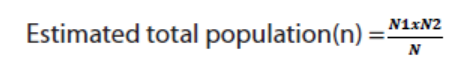
N1: the number of organisms in initial sample,
N2: the number of organism in a second sample,N: the number of marked organisms recaptured.
1.4 Population growth patterns and environmental resistance1.4.1. Population growth patterns
Activity 1.4
1. You are provided with the following statistics on population size of insects inthe following table:
a. Plot a graph of the population size against the generations.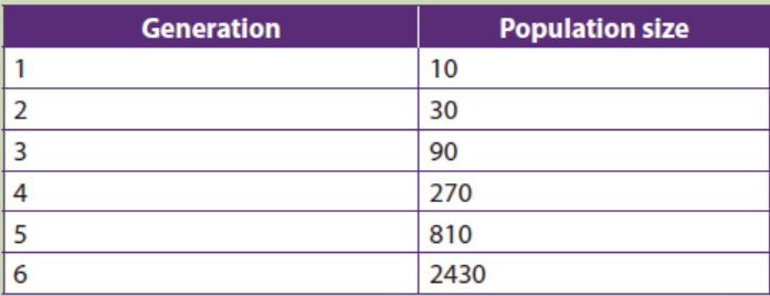
b. Compare the plotted graph in (a) above with the one given bellow andnote any similarities and differences.
2. Explain how environmental resistance affects the balance of nature.
1.4.1. Population growth patterns
Population growth patterns are graphs also called population growth curves in
which the increases in size are plotted per unit time. Two types of population growth
patterns may occur depending on specific environmental conditions:
a. Exponential growth pattern/J-shaped curve/J-shaped curve
Exponential growth is a pattern of population growth in which a population starts out
growing slowly but grows faster as population size increases. An exponential growth
pattern also called J- shapes curve occurs in an ideal, and unlimited environmental
resources. In such an environment there will be no competition. Initially populationgrowth is slow as there is a shortage of reproducing individuals that may be widely
dispersed. As population numbers increase, the rate of growth similarly increases,
resulting in an exponential J-shaped curve. Exponential population growth can be
seen in populations that are very small or in regions that are newly colonized by aspecies.
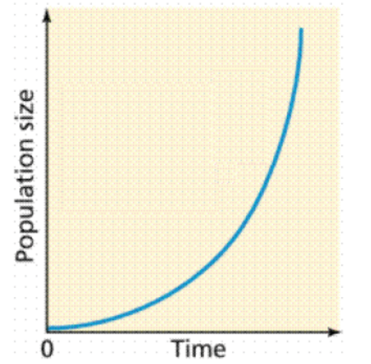
Figure 1.7: Exponential (unrestricted) growth curve
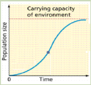
b. Logistic growth pattern / sigmoid growth curve
Logistic growth is a pattern of population growth in which growth slows and
population size levels off as the population approaches the carrying capacity. A
logistic growth pattern also called S-shaped curve occurs when environmentalfactors slow the rate of growth.
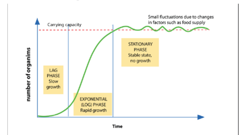
Figure 1.8: Logistic growth curve
The sigmoid or S- shaped curve represented by the figure 1.8 shows three main
stages in population growth: The lag phase where there is a slow growth, the log
phase or exponential growth phase, also called logarithmic phase, in which the
number of individuals increases at a faster rate and the plateau phase or stationary
phase, in which the number of individuals are stabilized.
Causes of the exponential phase are various and include the plentiful of resources
such as; food, space or light, little or no competition from other organisms, and
favourable abiotic factors such as; temperature or oxygen and reduced of lack of
predation or diseases. The stationary phase, however is caused by a balanced
number of; births plus the number of immigrations and the number of deaths plus
the number of emigration. Other causes may include; the increase of mortality
caused by predators and diseases, excess of wastes and competition for available
resources such as food, space, shelter and minerals. Some of these causes may
include the carrying capacity explained as is the maximum number of individualsthat a particular habitat can support.
1.4.2 Environmental resistance
Environmental resistance is the total sum of limiting factors, both biotic and abiotic,
which act together to prevent the maximum reproductive potential also called
biotic potential from being realized. It includes external factors such as predation,
food supply, heat, light and space, and internal regulatory mechanisms such asintraspecific competition and behavioural adaptations.
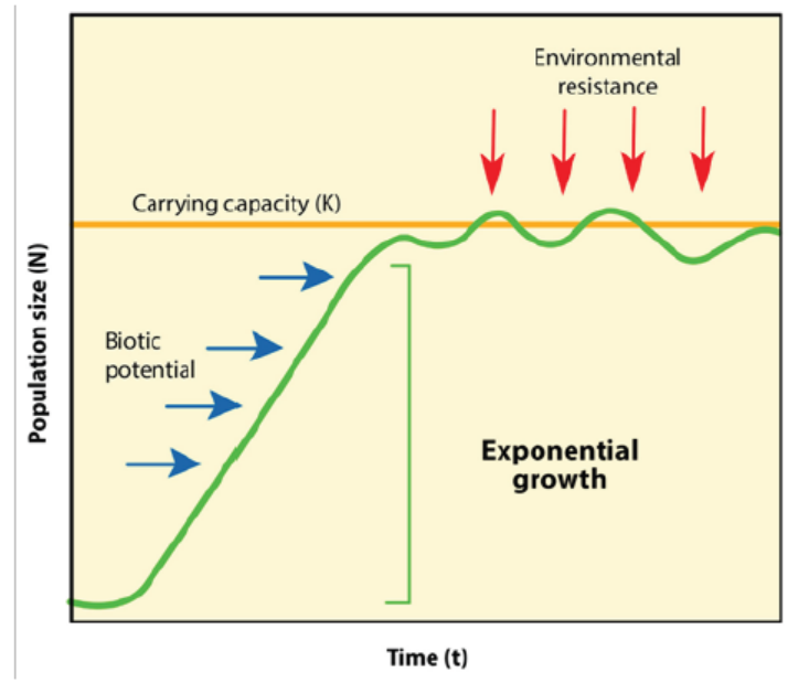
Figure 1.9: Effect of environmental resistance to population growth population growth..
1.4.3 Environmental balance
A balance of nature is the stable state in which natural communities of animals and
plants exist, and are maintained by competition, adaptation and other interactions
between members of the communities and their non-living environment. Every
biotic factor affects or causes a change in the natural environment. For example,
when a living organism interacts with the environment, this causes a change in the
environment. The following are some of the examples of biotic factors and theireffects on balance of nature:
– Respiration: when animals are respiring, they take in oxygen and give out
carbon dioxide (CO2) from respiration. The CO2 can be taken in by plant leaves
and be used in the process of photosynthesis to make food and give out oxygen.
– Predation: when animals, for example, predate on other animals, this reduces
the numbers of prey, which in turn affects the ecosystem.
– Parasitism: cause diseases that may slow down the growth rate of a population
and/or reduces the number of organisms.
– Competitors: when organisms compete over nutritional resources, this couldreduce the growth of a population.
Application 1.4
1. Explain any 3 biotic factors that affect the balance of nature.
2. Distinguish between carrying capacity and biotic potential.3. Explain how environmental resistance affects the population growth.
1.5 Natural resources and their importance
Activity 1.5
The pictures below illustrate some natural resources of Rwanda. Study themand respond to the following questions.
1. Categorize the natural resources mentioned in the above figures into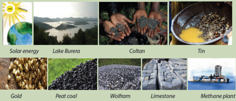
renewable and non-renewable resources.
2. Explain how those natural resources contribute to the economic growth ofRwanda.
1.5.1. Natural resources
Natural resources refer to materials or substances occurring in environment and
which can be exploited for economic gain. Natural resources such as; solar energy,
wind, air, water, soil and plants are renewable natural resources while others including
fossil fuels, oil, coal natural gas cited among many others are non-renewable natural
resources. A renewable resource can or will be replenished naturally in the course of
time, while a non-renewable resource is a resource of economic value that cannotbe readily replaced by natural means on a level equal to its consumption.
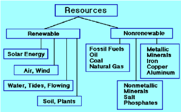
Figure 1.10: Renewable and non-renewable natural resources
1.5.2. Importance of natural resources in economic growth of Rwanda
– Water is used for; irrigation, domestic activities, industrial use, and mining.
– Lakes and rivers are source of food (fish) for humans and contribute for recreation
(tourism).
– Land serves as the storehouse of water, minerals, livestock, and home for wild
anima ls which generate an income in different ways.
– Minerals including gravel, coal, metals, oil, clay, sand, stones…are used for
construction and for income generation.
– Soil contributes to agricultural crop production, and supports forest and food
crops.
– Trees are the major sources of timber, construction materials and firewood
and contribute to fight against erosion, water and air purification and wind
protection.
– Some plants are source of food and money for humans and other animals
– Some animals including; mountain gorillas in Volconoes National Park, lions in
Akagera National Park and many other wild animals contribute to economicdevelopment of the country through tourism.
Application 1.5
1. Karekezi, Karake and Uwimana extract and sell legally the minerals from the
soil of Rwanda.
a. Describe the impact of their job on the economy development of
Rwanda.
b. Advise Karekezi, Karake and Uwimana on what they have to do at
the mine sites after the extraction of minerals.
2. Explain the reasons why we have to conserve and wisely use water in ourdaily activities.
1.6 Methods of conserving natural resources
Activity 1.6Look at the following pictures and respond to the following questions.
1. Identify the methods of conservation of natural resources mentioned in the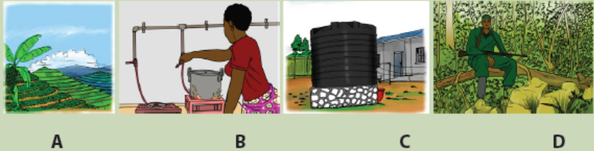
above figures.
2. Suggest all other possible methods of conservation of natural resources.
3. Discuss the measures established by Government of Rwanda for environment,biodiversity and natural resources conservation
They are various and different methods used for conservation of natural resources
and they include:
– Use of alternative sources of power such as; solar and wind energy:
These alternative sources of energy are bio friendly particulars because they
do not produce harmful gases that damage the ozone layer, compared to the
burning of fossils fuels such as; coal and charcoal. They are also; cheap to use,
not easily depleted, and are renewable.
– Tree planting to prevent soil erosion: This entails planting trees and other
vegetation to control soil erosion caused by wind and water. Trees and
vegetation are essential in the maintenance of the ecosystem. They also act as
home for most insects, birds and some symbiotic plants. This creates a habitat
for wildlife therefore conserving wildlife altogether.
– Practicing of judicious ways to conserve water in our homes: This entails
simple practices like ensuring that taps are closed when they are not in use.
Taking less time in the shower aids to conserve lots of water per month.
– Use pipelines to transport oil: During oil transportation on ships, spills can
happen which will negatively affect both plant and animal life. Therefore, use of
pipelines is more recommended.
– Growing vegetation in catchment areas: Catchment areas act as a source of
water that flows in; streams, rivers and oceans. Vegetation in the catchment
areas allows sufficient infiltration of water into deeper soil layers thus leadingto formation of ground water.
– Prior treatment of human sewage and Industrial wastes: Water flowing from
industries comes with many toxic wastes that must be treated before getting to
the natural water bodies. This reduces harm inform of pollutants e.g. chemical
and thermal forms.
– Harvesting rain water: This is done through usage of water tanks that collect
water during the rainy season and maintain use during dry periods. This reduces
tension on water reservoirs (e.g. lakes).
– Practice of in-situ or on-site conservation of wildlife: This involves
conservation of fauna and flora in their natural habitats. This entails setting up
measures that protect areas such as national parks and game reserves.
– Practice Ex-situ or offsite conservation of wildlife: It involves the conservation
of animals and plants outside the natural habitats. These include areas such as;
pollen banks, DNA banks, zoos, seed banks, botanical gardens, tissue culture
banks among others.
– Formulation of policies and regulations to curb poaching: Poachers continue
to kill many animals such as; elephants, rhinos, leopards for their tusks and
skins which are sold off in the black market. Poachers are a major threat to our
biodiversity as they are slowly making some species extinct. These regulations
will ensure that poaching is done away with.
– Practice judicious ways of conservation energy: Such practices
include switching off the lights when not in use, unplugging electrical
appliances when not in use. Plugged-in appliances continue to use electricity
even when not in use. Other practices include spending less time when taking
hot showers.
– Use of biogas in our homes: Around the World, Liquefied Petroleum Gas
(LPG) is the most rampant source of fuel in our homes today. Continued LPG
use results into the depletion of oil reserves, biogas is therefore an alternative.
Biogas is mainly produced from cattle dung, biogas plants are a source of both
biogas and manure.
– Use of bio-fuels: For more than a century, fossil fuels have been a major source
of energy. However, they are depleting rapidly, this calls for alternative sources
of fuel such as bio-fuels which are mainly from plant species. Bio-fuels are
known to be bio friendly and they reduce the occurrence of air pollution.
– Ensure the recycling of wastes: These wastes include; plastics, paper bags
that have resulted to tones of garbage. Recycling entails re-manufacturing of
already used materials. This reduces the amount of waste available reducing
soil and water pollution.
– Make use of electronic mails: Electronic mails are paperless and present a
good way to minimize the usage of paper. Technology has made this possible
reducing the usage of paper and envelops. This has reduced the production of
paper and also minimized cutting down of trees.
– Purchase hybrid cars instead of the conventional cars: Hybrid cars use a
combination of electricity and minimal amounts of gas to run them. This is a
break from the use of petroleum consuming cars that are now in large numbers.
– Water the lawns and farms in the evening: Watering the farm when it is dry
and hot results to increased water evaporation and a lot of water is used for the
same. During the evening, the weather is much cooler reducing evaporation
thus conserving water.
– Reuse old furniture: It is common to dispose of old furniture and opt for new
furniture. The old furniture should be sold off for use or donated to charity where
they can be reused. The old furniture can also be re-sculptured and redecorated
to save wood. This will reduce deforestation.
– Practice crop rotation: Planting the same crops for a long period of time
reduces soil fertility. The practice of crop rotation will restore and maintain soil
fertility thus conserving the soil.
– Translocation of wild animals: The growing population has led to human
encroaching on the wildlife habitat. This has resulted to human-animal
conflict where the wildlife are killed by humans as a way of protecting themselves
from them. Translocation involves moving wild animals to adjacent areas and
fencing to curb the conflict.
– Establish special schemes to preserve endangered plant and animal
species: This includes; botanical gardens, sanctuaries that may be established
to protect the endangered species so that they can be available for future
generations.
– Constructions of reservoirs: This will regulate the amount of water that
is used daily. The dams also act as a source of hydro-electric power which is
another alternative source of energy.
– Formulate regulations to stop overfishing: Overfishing interrupts aquatic
life and depletes the fish available in our water bodies. In some cases, it poses
a threat to the endangered aquatic species. Regulations to avoid over fishing
should be put in place.
– Construction of terraces in sloping land: This will prevent soil erosion aswater tends to run downhill.
Application 1.6
1. Distinguish between in-situ and ex-situ wildlife conservation.
2. Describe the energy sources that you can advise Rwandans to use forprotecting the environment.
End of unit assessment 1
Instruction: From question 1 to 5, choose the letter corresponding to the
best answer.
1. Which is the best definition of a population?
a. The unit of natural selection and evolution.
b. All the species that live in the same area.
c. A group of species that live in the same area.
d. A group of organisms of the same species that live in the same area.
2. Which of the following would be an example of population density?
a. 100 caterpillars
b. 100 caterpillars per maple tree
c. 100 caterpillars clumped into 5 specific areas
3. Exponential growth:
a. Is a characteristic of most species under ideal conditions?
b. Is a fast growth rate with a large population?
c. Begins with a slowly growing population.
d. All of the above.
4. Which of the following is a characteristic of developing countries?
a. A fast population growth due to a high death rate but higher birth rate.
b. A fast population growth due to a high birth rate but falling death rate.
c. A slow population growth due to a low birth rate and falling death rate.
d. A slow population growth due to a low birth rate and low death rate.
5. Fill in the blank with the term that best completes the sentence.
a. The ____________ is the largest population size that can be supported
in an area without harming the environment.
b. Populations gain individuals through births and ____________.
c. Under ideal conditions, populations can grow at ____________ rates.
6. Circle the letter of the correct choice.
i. Non-renewable resources include
a. Wind and sunlight.
b. Metals and other minerals.
c. All of the above.
ii. Renewable resources include
a. Wind and sunlight.
b. Fossil fuels.c. All of the above.
7. Observe the pictures below and respond to the following questions.
a. Identify the human activities shown above and that harm the natural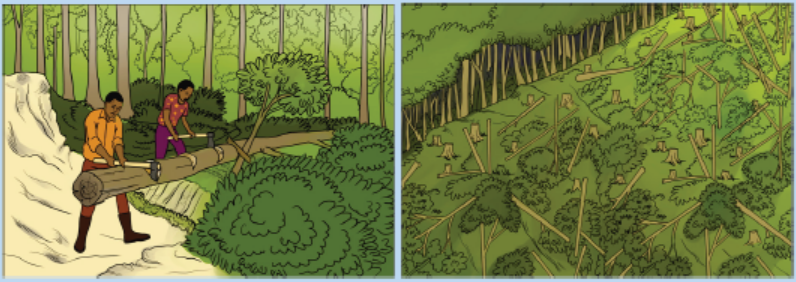
resources.
b. Describe all effects of the identified activities on the environment.
c. Suggest the possible measures to solve the above problems.
8. Students made a survey of blackjack (Bidens pilosa) growing on two
different gardens in the school environment. Ten quadrats of 1.0 m2 were
placed randomly in each garden, and the number of blackjack plants in eachquadrat was counted. The results are summarized in the following table:
Calculate:
a. The species frequency in each of the two gardens.
b. The species density of blackjack plants in each of the two areas.
c. Compare the species frequency and density for both gardens.
d. Explain why it is important to use randomly placed quadrats.
9. Explain how age structure of human population affect its growth rate?
10. Describe how has the growth of Earth’s human population has changed in
the 2 recent centuries? Give your answer in terms of growth rate and thenumber of people added each year.
11. A group of students investigated the size of quadrat that they should use to
assess the abundance of plant species in an old field of forest plantations.
They used quadrats of side 10, 25, 50, 75 and 100 cm and recorded the
number of plant species were encountered in each quadrat. They repeated
their investigation five times and calculated the mean numbers of speciesper quadrat. Their results are plotted as follows:
a. Calculate the area of each quadrat used for this study.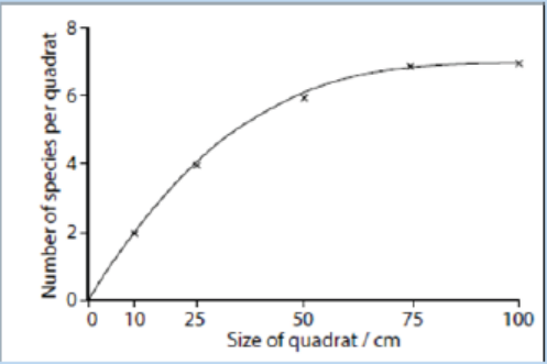
b. Explain why these students repeated the experiment five times for
each quadrat.
c. Based on their results, the students decided to use the 50 cm quadrat
to study the old field. Why did they choose the 50 cm quadrat instead
of others?
d. Explain how they would use the 50 cm quadrat to estimate theabundance of different plant species in the field.
12. A sample of 39 ground beetles was captured from an area of waste ground
measuring 100 x 25 meters. Each animal was marked and then released. A
second sample of 35 was captured the following day and 20 individuals of
them were marked.
a. Estimate the number of ground beetles in the population.
b. State three assumptions that must be made in order to make this
estimation.
c. Describe a method that could be used to verify that the mark–
release–recapture method gives a valid estimate of the groundbeetle population in the area of waste ground.
UNIT 2 CONCEPT OF ECOSYSTEM
UNIT 2: CONCEPT OF ECOSYSTEM
Key Unit Competence
Describe the different components of an ecosystem, biogeochemical cycles andhow energy flows in an ecosystem.
Learning objectivesBy the end of this unit, I should be able to:
– Describe an ecosystem
– State the types and properties of an ecosystem
– Describe the main components of an ecosystem
– Explain the ecological factors influencing the life of organisms in an ecosystem
– Define the terms: population, community, ecosystem, biome, niche and
biosphere
– Distinguish among; individuals, populations, communities, niche, habitat,
ecosystems, biomes, biosphere
– Describe feeding relationships in an ecosystem
– Describe a food chain and a food web
– Explain the relative merits of pyramids of numbers
– Analyse the relation between organisms (example: producers, consumers,
decomposers) and their trophic levels.
– Distinguish between abiotic and biotic factors
– Interpret energy flow diagrams
– Compare; gross primary, net primary production and secondary succession in
biotic communities
– Explain what is meant by trophic efficiency
– Explain energy flow and the recycling of nutrients in an ecosystem
– Describe biogeochemical cycles
– Identify processes, components, and roles of organisms in the hydrologic,
carbon and nitrogen cycles
– Distinguish between primary and secondary succession in biotic communities
– Appreciate the existence of different components of an ecosystem and their
roles in the life of organisms
– Beware of the effect of bioaccumulations at different trophic levels.– Recognise the source and transfer of energy in an ecosystem
Introductory activity
The following pictures indicate different types of ecosystems. Observecarefully the pictures A, B and C and answer the questions that follow.
1. What do you understand by the terms: ecosystem, biotic and abiotic factors?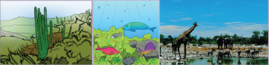
2. Suggest the types of ecosystems illustrated by pictures A, B, and C.
3. Distinguish between abiotic and biotic factors illustrated on picture A, B and
C.
4. Describe how energy flows through ecosystem B and ecosystem C.
5. Explain how feeding relationships are expressed in food chains on picture B
and C.
6. Identify trophic levels in food chains and food webs on the picture B and
picture C.
7. What would happen if plant species are removed from an ecosystem ofpicture C?
Ecology is the study of how living things interact with each other and with their
environment. It is one of the major branches of biology with different areas that
overlap with geography, geology, climatology, mathematics, and chemistry cited
among other sciences. This lesson introduces fundamental concepts in ecology
with a particular focus on organisms and their environment. Organisms are
individual living things. Despite their tremendous diversity, all organisms have
the same basic needs such as energy and matter, obtained from the environment.
Therefore, organisms are not closed systems. They depend on and are influenced
by the environmental factors including abiotic (non-living factors such as water,
temperature, humidity…) and biotic (living factors such as animals, plants…). The
unit of nature consisting of all the biotic and abiotic factors in an area and theirinteractions is called an ecosystem.
2.1 Ecosystem
Activity 2.1Observe carefully the diagram below, and answer the questions that follow
1. Define an ecosystem and give its different types.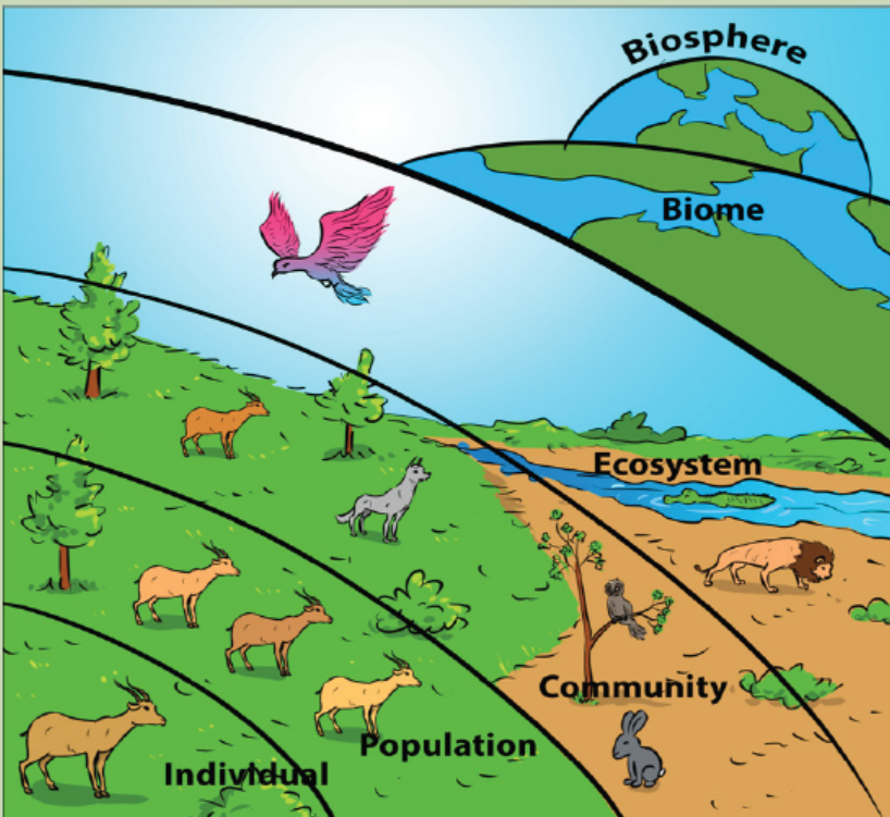
2. Distinguish among; individuals, populations, communities, niche,habitat, ecosystems, biomes and the biosphere.
Different concepts define levels in ecology. From the low to high level, the conceptsinclude:
a. Species
Species such as bees in figure 2.1 is defined as a group of organisms that can breedto produce fully fertile offspring.
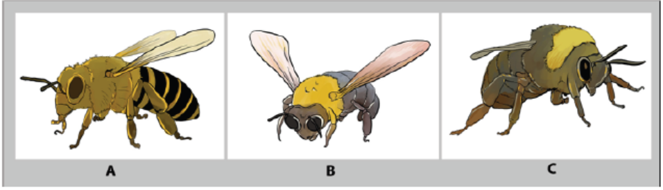
Figure 2.1: Species of bees
b. Population
A population is defined as a group of organism of the same species which live in the
same habitat at the same time where they can freely interbreed. Elephants such asthose indicated in figure 2.2 constitute a population.
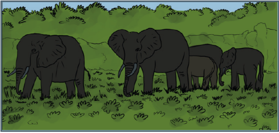
Figure 2.2: Population of elephants
c. Community
In ecology, a community consists of all populations of different species living and
interacting at a certain level in the same ecosystem. Animals indicated in the figure2.3 interact and share the same ecosystem
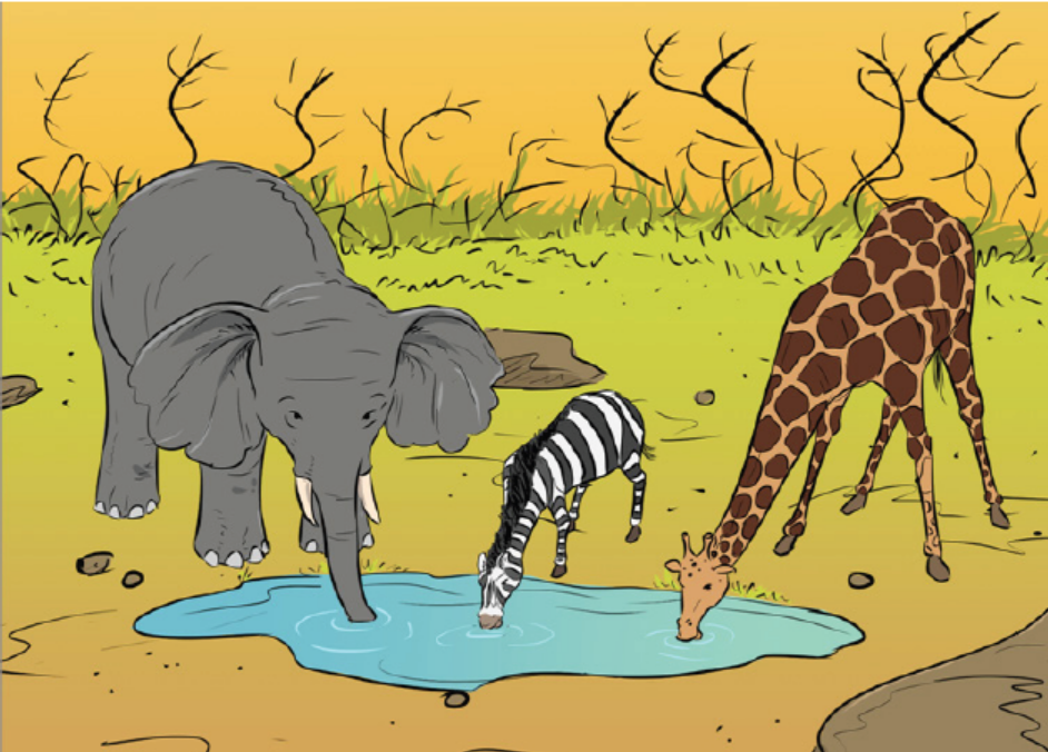
Figure 2.3: Ecological community
d. Niche
A niche refers to the role played by a species in its ecosystem. It includes all the ways
that the species interacts with the biotic and abiotic factors of the environment.
Two important aspects of a species’ niche are the food it eats and how the food is
obtained. Birds on the figure 2.4 live in the same ecosystem, but they have different
adaptations for food. For example, the longest slender beak of the nectarivore allows
it to sip the nectar from flowers, the short study beak of the granivore allows it tocrush hard and tough grains.
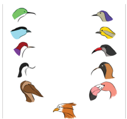
Figure 2.4: Adaptations of birds’ beak for food in an ecosystem
Another aspect of a species’ niche is its habitat. The habitat is the physical environment
in which a species lives and to which it is adapted. A habitat’s features are mainly
determined by abiotic factors such as temperature and rainfall, which in turn have
an influence on the traits of the organisms that live in that habitat. A habitat is also
influenced by biotic factors as it may contain many different species. However, in the
same habitat, two different species cannot occupy the same niche in the same place
for very long. This is known as the competitive exclusion principle. If two species
were to occupy the same niche, they would compete with one another for the same
food and other environmental resources leading to the extinction of the weakerspecies.
e. Ecosystem
An ecosystem consists of a natural unit consisting of all the living organisms in an
area functioning together with all the non-living physical factors of the environment.
The concept of an ecosystem can apply to units of different sizes. For example, a
large body of fresh water could be considered an ecosystem, and so could a small
piece of dead wood. Both contain a community of species that interact with oneanother and with the abiotic components of their environment.
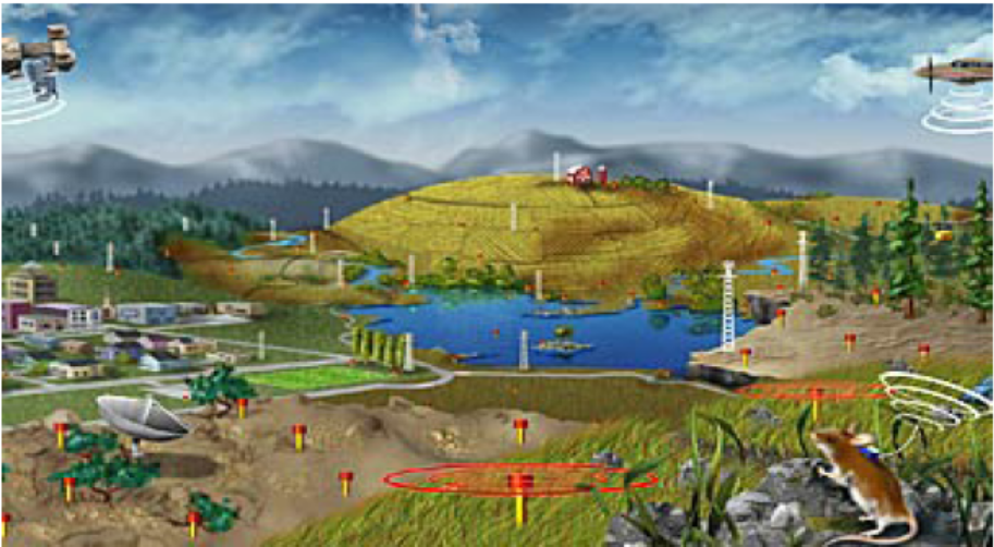
Figure 2.5: Example of ecosystems
They are two major classification of ecosystems: natural ecosystem and artificial
ecosystem. Natural ecosystems are those ecosystems that are capable of operating
and maintaining themselves without any major interference by man. Natural
ecosystems are furthermore classified into terrestrial ecosystems including; forest,
grassland and desert, and in Aquatic ecosystems including fresh water ecosystem
such as; ponds, lakes, rivers and marine ecosystems such as ocean, sea or estuary.
Artificial Ecosystem are those ecosystems maintained by the intervention of humans.
They are manipulated by man for different purposes including; croplands, artificiallakes and reservoirs, townships and cities.
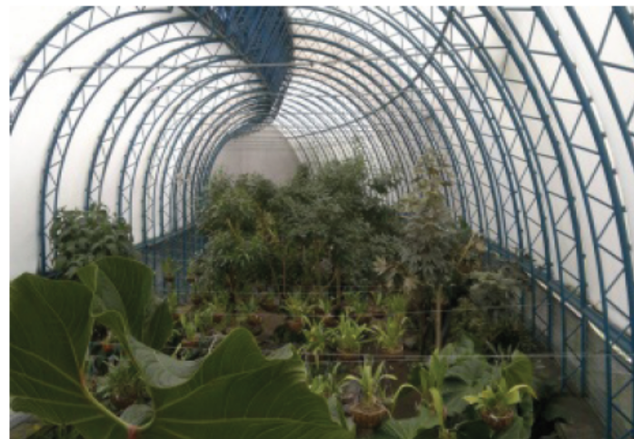
Figure 2.6: Artificial ecosystem
f. Biomes
A biome is a broad regional type of an ecosystem characterized by distinctive climate
and soil conditions and a distinctive kind of biological community adapted to those
conditions. Biomes are of various types including terrestrial and aquatic biomes.
Terrestrial biomes consist of all the land areas on Earth where organisms live. The
distinguishing features of terrestrial biomes are determined mainly by climate.
The dominant terrestrial biomes include; tundra, temperate forests, grasslands,temperate, tropical deserts, tropical forests and grasslands (Figure 2.7).
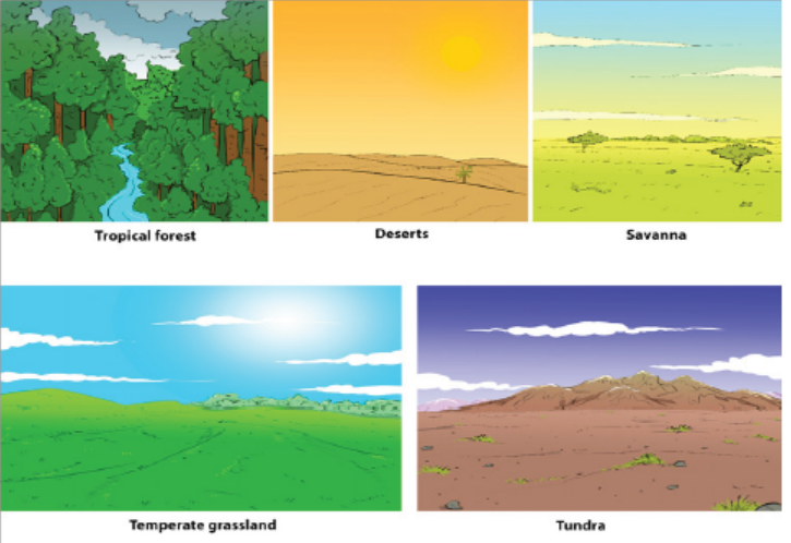
Figure 2.7: Different types of biomes
Aquatic biomes occupy the largest part of biosphere. These are divided into two,
i.e. marine and freshwater. The marine biomes e.g. oceans which is the biggest
of the two (Figure 2.8 below) have a very high salt concentration and have fauna
adapted to them. The fresh water biomes such as lakes and rivers have a low saltconcentration of less than 1%.

Figure 2.8: An example of aquatic biome
g. Biosphere
The biosphere is the portion of Earth inhabited by life and which represents the sumof all communities and ecosystems.
Application 2.1
1. Distinguish among; individuals, populations, communities, ecosystems,
biomes and biosphere.
2. Give an example of any three aquatic and three terrestrial ecosystems
found in Rwanda
3. Use the examples above and make a brief description of an ecosystem4. Discuss the competitive exclusion principle.
2.2 Properties of an ecosystem and ecological factorsinfluencing the life of organisms
Activity 2.2
1. Go to your school garden and collect 3 living things and 3 non living
things
2. Discuss differences and similarities between collected living and nonliving
things
3. Analyze carefully the diagram below and answer the questions that
follow: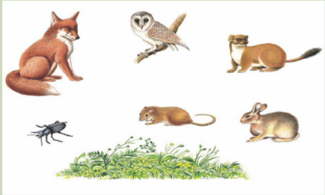
Make a classification of living things by the letters A, B, C, D, E, F and G based
on the principle of being eaten by
2.2.1 Relationships in an ecosystem
In an ecosystem, living things have feeding relationships. In terms of sources of food,
organisms are classified as; producers, consumers, or decomposers.
– Producers are organisms that can manufacture their own food. They include;
green algae , green plants and other autotrophs that are able to make their own
food through photosynthesis or chemosynthesis
– Consumers are organisms that obtain food from other organisms because they
cannot make their own food. Based on their level of feeding, consumers are
classified as primary consumers when they feed directly on plants. Primary
consumers include herbivorous or omnivorous animals. Consumers are
also classified as secondary consumers, when they feed directly on primary
consumers. Secondary consumers include carnivorous animals. Tertiary
consumers are consumers that feed directly on secondary consumers and are
top carnivorous or omnivorous animals.
– Decomposers are organisms that break down the tissues of dead organisms
into simpler substances, for example bacteria and fungi that break down dead
plants and animals into compounds of carbon and nitrogen. These compounds
are released into the soil to be used by plants and animals for growth.
In a food chain, producers such as plants produce their own energy without
consuming other life forms. They gain their energy from conducting photosynthesis
via sunlight. Consumers exist on the next level of the food chain and they are three
main types of consumers namely herbivores, carnivores and omnivores. Consumersget the energy by feeding on plants or by eating other carnivores or herbivores.
2.2.2 The ecological factors influencing the life of organisms in an
ecosystem
In an ecosystem, life is influenced by biotic and abiotic factors.a. Abiotic factors
Light: Light plays an important role in the species composition and development
of vegetation. Light is abundantly received on the surface of the earth from solar
energy and it is used by primary producers to do photosynthesis. Light intensity
shows special variations due to the factors like atmospheric water layer, particles
dispersed in the air, etc. Furthermore, the vegetation of an area may also affect the
light intensity. In deep shade under trees, or under water, light becomes limitingfactor below which photosynthesis is not sufficient for effective growth.
Temperature: Temperature is a measurement of the degree of heat. Like light,
heat is a form of energy. The radiant energy received from the sun is converted into
heat energy. Heat is measured in calories. The temperature at which physiologicalprocesses are at their maximum efficiency is called optimum temperature.
The minimum, optimum and maximum temperatures are called cardinal
temperatures. The cardinal temperature varies from species to species and in the
same individual from part to part. The distributions of plants, animals are alsoinfluenced by temperature.
Water: Water is an indispensable part of land contributing to soil productivity, and
the well beings of organisms. All physiological processes take place in the medium
of water. For example, cellular protoplasm is made up mostly of water contributingto the maintenance of cells and hence the entire living organism survives.
Rainfall: The rainfall provides water to plants and animals, and determines the
types of vegetation in a given region. For example, the evergreen forests are found
in tropical regions. Changes in rainfall influence the vegetation types in different
parts of the earth, and in turn, vegetation causes changes in the types of forests,
animals and birds. The quantity of water that a soil holds or that infiltrates into the
soil depends upon the properties of soil and type and density of vegetation covering
it. In a bare area, the rain drops beat the compact surface of the soil and loosen thesoil particles which are washed away.
Wind: Air in motion is called wind. It modifies the water relation and light conditions
of a particular region, and brings about a number of physical, anatomical and
physiological changes of plants. Such changes are breakage and uprooting of
plants, deformation, erosion and deposition of different organic particles. The wind
accelerates transpiration, removes solid moisture and at high velocities causes soil
erosion, which contributes to the removal of the surface soil, rich in organic matterand fine mineral particles.
Humidity: Humidity is greatly influenced by intensity of solar radiation, temperature,
altitude, wind, and water status of soil. Low temperature causes higher relative
humidity by decreasing the capacity of air for moisture. Processes as transpiration,absorption of water are influenced by atmospheric humidity.
Atmospheric Gases: Some principal gases like nitrogen, oxygen, carbon-dioxide,
helium, hydrogen, methane, and ozone are found in atmosphere. In addition to
these gases, there are water vapor. Industrial gases, dust, smoke particles, microorganisms
are present in the atmosphere. These gases have different influences onthe environment and hence on the living things.
Biotic Factors
The biotic factors constitute the living organisms of the environment and their
direct or indirect interactions. The population occurring together in an area interacts
with each other in several ways including predation, competition for mating and for
different natural resources including; food, water and oxygen.
b. Edaphic Factors
Edaphic factors deal with different aspects of soil, such as the structure and
composition of soil, its physical and chemical features. A galaxy of complex factor
constitutes the soil. Soil is usually defined as any part of earth’s crust in which plants
root. The soil is constituted as a result of long-term process of complex interaction
leading to the production of a mineral matrix in close contact with interstitial
organic matter both living and dead organisms. Soil is composed of; mineral matter,
soil organic matter or humus, soil water and soil solutions, and biological systemsincluding bacteria, fungi, algae, protozoans and arthropods.
Application 2.2
1. Discuss the ecological factors driving the biodiversity of Akagera National
Park.
2. Discuss the relationship between plant diversity and soil composition.2.3. Energy flow in an ecosystem
Activity 2.3Observe carefully the diagram below and answer the questions that follow.
1. Discuss how the energy flows in the above food chain of living things.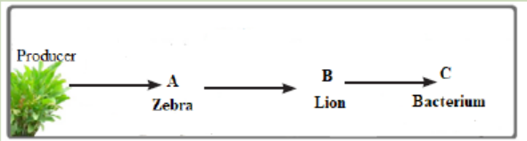
2. Indicate which living organisms above are consumers, decomposers in
the figure.
3. Discuss the role played by organism represented by the letter C.4. What would happen if A is removed from the food chain?
Energy enters in an ecosystem in the form of sunlight or chemical compounds. Some
organisms including plants and green algae use sunlight energy to make their own
food. Other organisms get energy through food by eating producers or consumersor by decomposing producers and consumers.
2.3.1 Food chains and food webs
Food chains and food webs are diagrams that represent feeding relationships. They
show who eats who. In this way, they model how energy and matter move through
ecosystems.
a. Food chains
A food chain represents a single pathway through which energy and matter flow
through an ecosystem. Food chains are generally simpler than what really happensin nature. Most organisms consume and are consumed by more than one species.
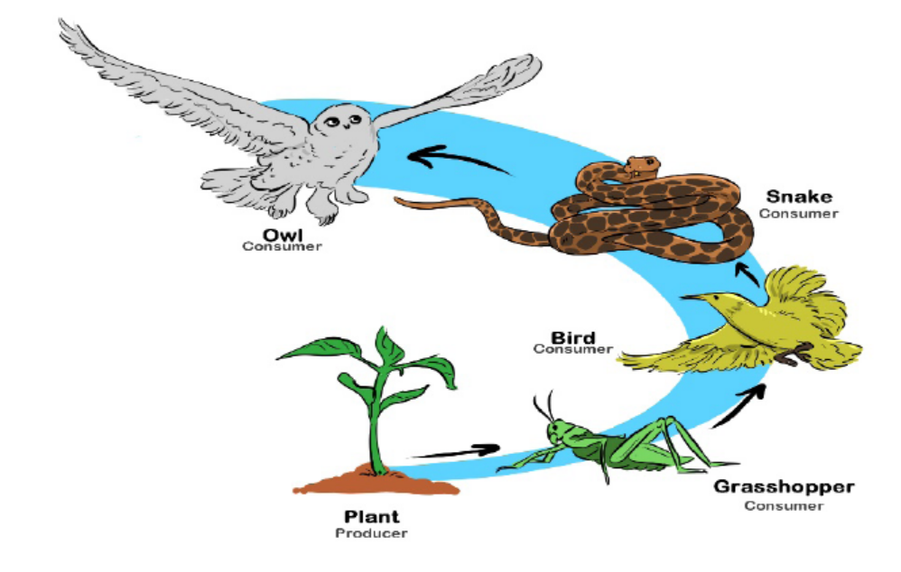
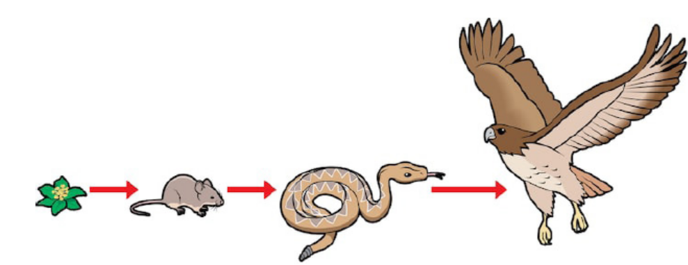
Figure 2.9: Illustration of a food chain (Source shutterstock.com)
b. Food Webs
A food web represents multiple pathways through which energy and matter flow
through an ecosystem. It includes many intersecting food chains. It demonstratesthat most organisms eat, and are eaten, by more than one species.
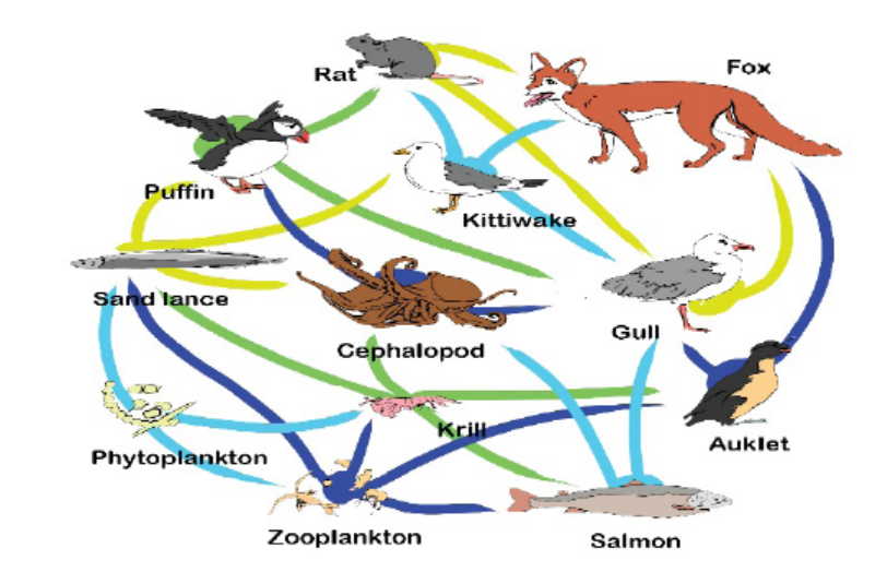
Figure 2.10: Illustration of the Food Web
c. Trophic levels
The feeding positions in a food chain or web are called trophic levels. The different
trophic levels are defined in the table below (Table 2.1). All food chains and food
webs have at least two or three trophic levels, the maximum being of four trophic
levels. Many consumers feed at more than one trophic levels. Humans, for example,
are primary consumers when they eat plants, secondary consumers when they eat
meat from primary consumers, and are tertiary consumers when they eat meat of
secondary consumers.
Table: 2.1. Description of producers, primary, secondary and tertiary trophiclevels
2.3.2 Ecological pyramids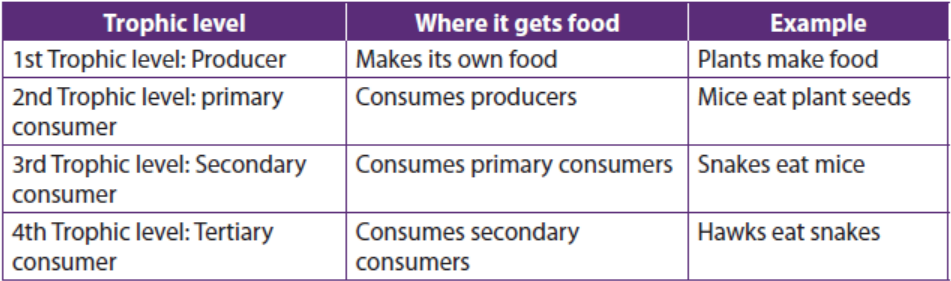
Ecological pyramid is a graphical representation in the form of a pyramid showing
the feeding relationships of groups of organisms. It is often represented in a waythat the producers are at the bottom level and then proceeds through the various
trophic levels in which the highest is on top. There are 3 types of ecological
pyramids: pyramid of numbers, pyramid of biomass and pyramid of energy.Pyramid of numbers
Pyramid of numbers is a graph representing the total number of individuals present
at each trophic level. This type of pyramid can have two different forms depending
on the number of organisms: upright and inverted. In an upright pyramid of numbers,
the number of organisms generally decreases from the bottom to top. This generally
occurs in grassland and pond ecosystems where plants occupy the base of the
pyramid. An inverted pyramid of numbers, on the other hand, is just the opposite
of the upright one. It is usually observed in tree ecosystems with the trees as theproducers and the insects as consumers.
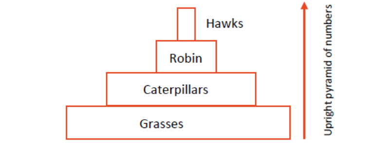
Figure 2.11: Figure 2.11: illustration of the upright pyramid of numbers
d. Pyramid of biomass
Biomass is defined as the amount of biomass per unit area product of the living
material present in an organism and the total number of organisms present in a
specific trophic level. In less complicated terms, it refers to the food available for
the succeeding trophic level. A pyramid of biomass is a depiction of the amount of
food available and how much energy is being passed on at each trophic level. Most
the biomass that animals consume is used to provide the energy, converted to newtissues, or just remain undigested.
Most of the time, pyramids of biomass are in a true pyramidal shape with biomass
in the lower trophic levels are greater than the trophic levels above them. Like the
pyramid of numbers, the pyramid of biomass can either have two forms: upright and
inverted. Usually, terrestrial ecosystems are characterized by an upright pyramid of
biomass having larger base for primary producers with the smaller trophic levels for
consumers located at the top (figure 2.17). Aquatic ecosystems are the complete
opposite as they will assume the inverted structure of the pyramid. This is because
the phytoplankton producers with generally smaller biomass are located at the base
while the consumers having larger biomass are located at the top of the pyramid(figure 2.18)
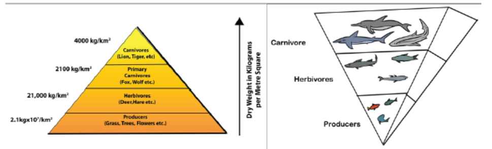
Figure 2.12: Illustration of upright pyramid of biomass(left) and the inverted pyramid of biomass(right).
In other words, the phytoplankton has a short turnover time, which means they have
a small standing crop compared to their production. The turnover time is calculatedby the following formula:
2.3.3 Pyramid of energy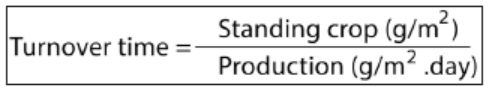
The pyramid of energy shows the overall energy in the ecosystem and how much
energy is required by organisms as it flows up the higher trophic levels. This pyramid
shows that energy is transferred from lower trophic levels with more amount of energy
(producers) to higher ones (consumers) and converted in the biomass. Therefore, it
can be concluded that organisms found at the highest trophic levels of shorter food
chains bear greater amount of energy than the ones found in longer ones. Unlike
the first two ecological pyramids, the pyramid of energy is always illustrated in an
upright position, with the largest energy carriers at the base. The pyramid shows thetotal energy stored in organisms at each trophic level in an ecosystem.
Starting with primary consumers, each trophic level in the food chain has only 10
percent of the energy of the level below it (Figure 2.18). The energy available at agiven trophic level is measured in Kilojoules per square metre per year (kJm-2Y-1).
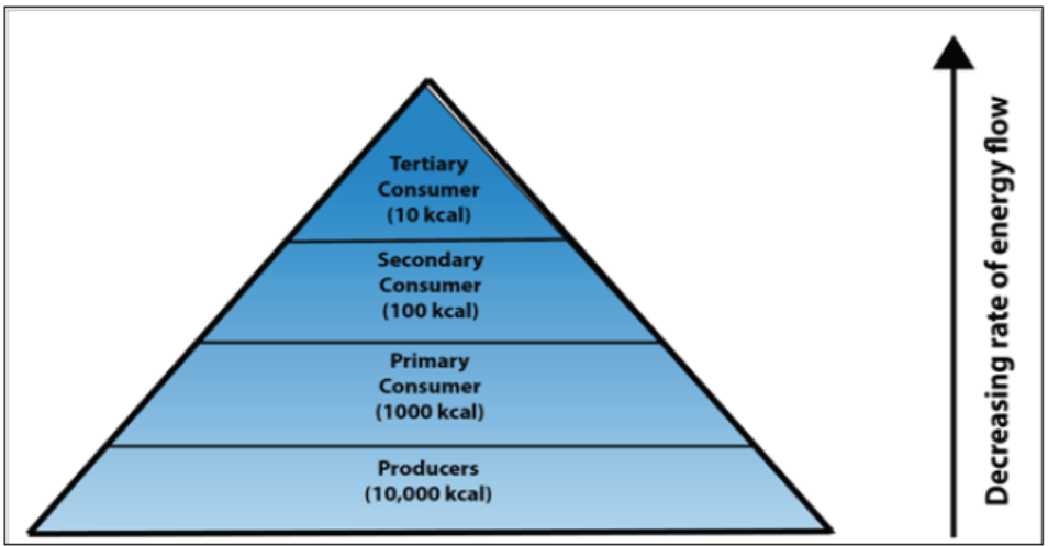
Figure 2.13: Illustration of the Pyramid of energy
2.3.4 Limitations of ecological pyramids
While the three ecological pyramids are highly specific to the aspect of ecosystem
they want to describe, all of them still tend to overlook important aspects. Some of
these limitations are the following:
– These types of pyramids only are applicable in simple food chains and not for
the food webs and they also do not consider the possible presence of the same
species at different trophic levels.
– None of the three ecological pyramids provide any idea related to variations in
seasons and climates.
– Other organisms like microorganisms and fungi are not given specific role inthe pyramids despite their vital roles in ecosystems.
Application 2.3
1. All scientists agree that the activities of living organisms play an important
role in driving biogeochemical cycles, and that organism shape their
environment to a considerable extent.
a. Explain how, herbivores affect their grassland environment.
b. What would happen if herbivores were removed from Akagera National
Park?
c. What would happen to Akagera National Park if overgrazing occurs?
2. Explain why is only small portion of the solar energy that strikes Earth’s
atmosphere stored by primary producers.
3. The diagrams A, B, C and D indicate different cases of pyramid of numbers.Using your knowledge on pyramids, analyses and interpret each diagram
4. Discuss the reasons why the transfer of energy in an ecosystem is referred to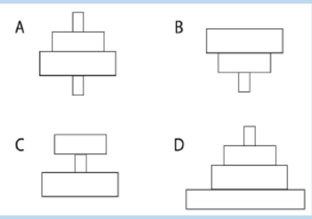
as energy flow, not as energy cycling.
2.4 Ecological succession
Activity 2.4
In pair discuss the following:
1. What happen to a but a month after bush fire?
2. What would happen to your school basketball playground after 1, 5, 50,500 years if it was completely abandoned?
Communities are not usually static, and the numbers and types of species that live in
them generally change through time. This is called ecological succession. Important
cases of succession are primary and secondary succession.
a. Primary succession
Primary succession occurs in an area that has never been colonized such as bare
rock. This type of environment may come about when lava flows from a volcano and
hardens into rock, a glacier retreats and leaves behind bare rock or when a landslide
uncovers an area of bare rock.
The first species to colonize a disturbed area are called pioneer species including
bacteria and lichens that can live on bare rock. These species change the environment
and make the way for other species to come into the area. Along with wind and
water, they help weather the rock and form soil. Once soil begins to form, plants can
move in from pioneer species to intermediate stages and to climax communities
(Figure 2.14). At first, the plants include herbs, grasses and other species that can
grow in thin, poor soil. As more plants grow and die, organic matter is added to the
soil. Soil is improved and get the capacity to hold water. The improved soil allowsshrubs and trees to move into the area.
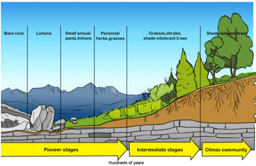
Figure 2.14: Primary succession
b. Secondary succession
Secondary succession occurs in a formerly inhabited area that was disturbed. The
disturbance could be a fire, flood, or human action such as farming. This type of
succession is faster because the soil is already in place. In this case, the pioneer
species are plants such as grasses, birch trees, and fireweed. Organic matter fromthe pioneer species improves the soil and lets other plants move into the area.
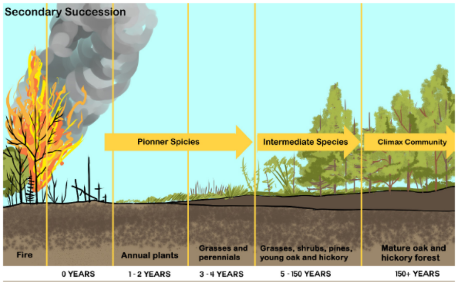
Figure 2.15: Secondary succession
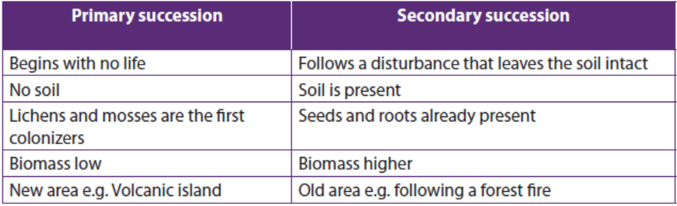
Similarities and differences between primary and secondary succession are
summarized in the following table:Table: 2.2 Comparison between primary succession and secondary succession
Application 2.4
Differentiate between primary and secondary succession
2.5 Bioaccumulation and Bio magnification
Activity 2.5
Use the school library and search additional information on the internet.Discuss between bioaccumulation and bio magnifications
2.5.1 Bioaccumulation
Bioaccumulation refers to the accumulation of toxic chemical substances such as
pesticides, or other chemicals in the tissue of a particular organism. Bioaccumulation
occurs when an organism absorbs a substance at a rate faster than that at which thesubstance is lost by catabolism and excretion
2.5.2 Bio magnification
Bio magnification is a process by which chemical substances become more
concentrated at each trophic level. Bioaccumulors of toxic substances such as heavy
metals and polychlorinated biphenyls that slowly increases up in concentration in
living organisms including bacteria, algae, fungi, and plants.Bioaccumulants enter
a body through contaminated air, water, and/or food, and keep on accumulating
because they are very either slowly metabolized, not all metabolized, or are excretedvery slowly
2.5.3 Example of the causes of bio magnification
Some toxic chemicals were deliberately put in the environment to kill insect pests.
One of these pesticides is Dichloro Diphenyl Trichloroethane (DDT), which was
used to control mosquitoes and other insect pests. It was commonly sprayed on
plants and eventually entered water supplies. There it was absorbed by microscopic
organisms, which in turn were eaten by small fish and the small fish eaten by larger
fish from where it could have transferred to other animals, where it accumulates in
the fat tissue of animals at the top of the food chain. This food chain shows typicalconcentrations of DDT found in a food chain (in parts per million, ppm):
Another biological magnification of Polychlorinated Biphenols (PCBs) was found in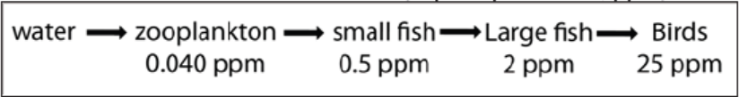
the food web of great lakes, where the concentration of PCBs in herring gull eggs, at
the top of the food web, is nearly 5,000 times that in phytoplankton at the base ofthe food web.
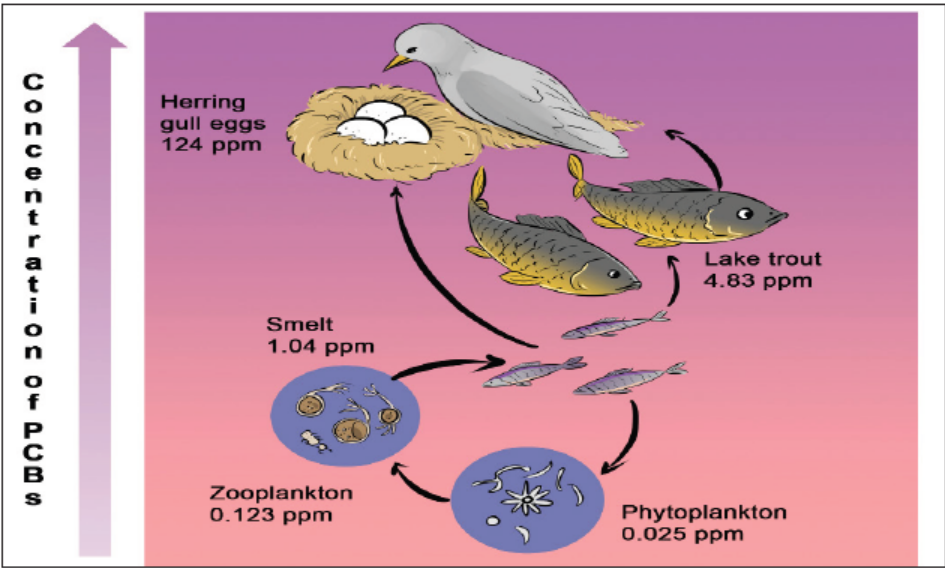
Figure 2.16: Biological magnification of PCBs in a Great Lakes food web.
2.5.4 Consequences of bio magnification
The first sign of the problem was a decline in the number of predator birds. Studies
showed that the eggs of these birds were easily cracked. In fact, the weight of the
mother sitting on the eggs cracked them. It was finally discovered that DDT was
building up in the tissue of the birds and interfering with the calcium needed for theshell to be hard.
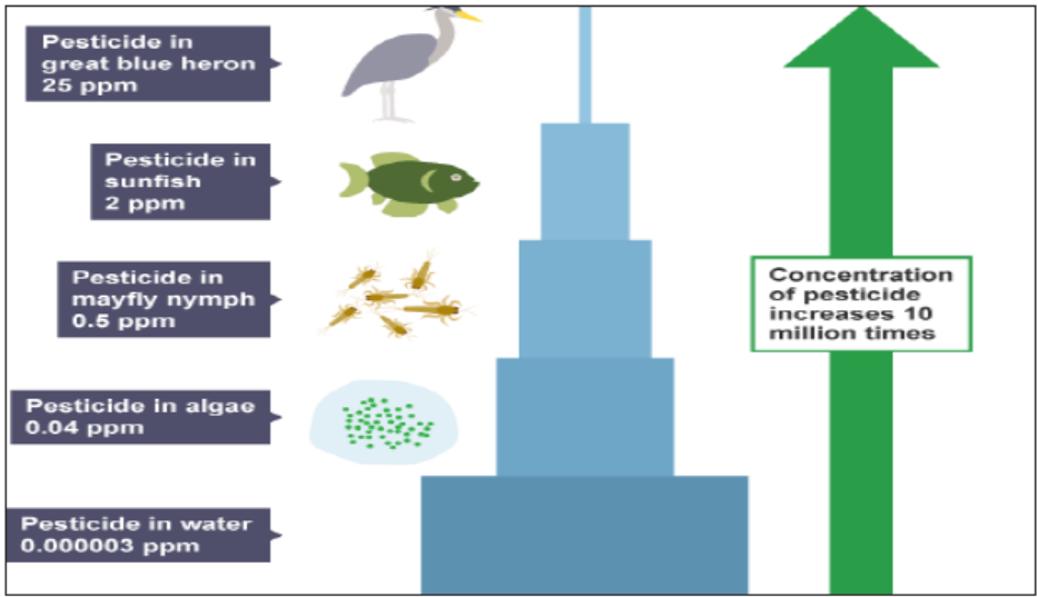
Figure 2.17: Biomagnification of pesticides in food chain
2.5.5 Relationship between bioaccumulation and bio magnification
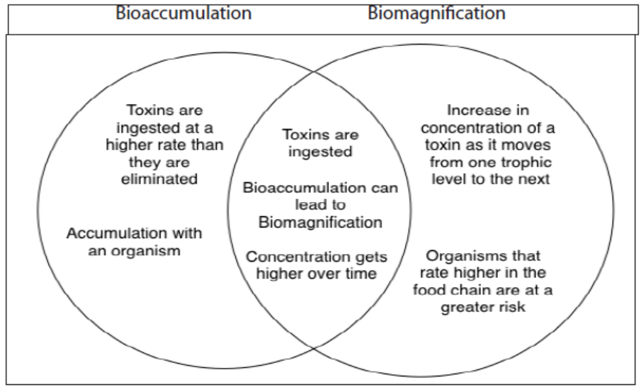
Figure 2.18: Differences and similarities between bioaccumulation and bio magnification
2.5.6 Prevention and reduction of bioaccumulation of toxic substances
The following are some of the ways to prevent and to reduce bioaccumulation of
toxic substances:
– Do not put harmful substances into water system or storm drains.
– Reduce the use of toxic chemical pesticides.
– Eat certified organic foods when possible.– Avoid fishing or spending time in contaminated areas.
Application 2.5
1. Discuss how the addition of excess nutrients to a lake threatens the
population of fishes.
2. In the face of biological magnification of toxins such as DDT, discuss thelevels of food chains where it is healthier to feed on
2.6 Efficiency of ecological production
Activity 2.6
Use the books from the school library and search further information from
the internet. Discuss the roles of efficiency of ecological production and
make a brief description of the ecosystem primary production, total primaryproduction, and net primary production.
2.6.1 Efficiency of primary production
The amount of light energy converted to chemical energy in the form of organic
compounds by autotrophs during a given period of time is called ecosystem
primary production (R). Most primary producers use light energy to synthesize
energy rich-organic molecules, which are subsequently broken down to generate
adenosine triphosphate (ATP). The total primary production in an ecosystem’s gross
production (GPP) is the amount of light energy that is converted to chemical energy
by photosynthesis per unit time.
Note that not all of this production is stored as organic material in the primary
producers because they use some of the molecules as fuel in their own cellular
respiration. The net primary production (NPP) equals the gross primary production
minus the energy used by the primary producers for respiration(R), as it is summarizedin the following formula, i.e
NPP = GPP – R.
In many ecosystems, NPP is about one-half of GPP.
To an ecologist, net primary production is the key measurement because it representsthe storage of chemical energy that will be available to consumers in the ecosyste
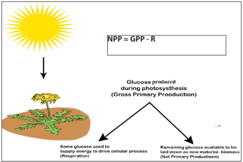
Figure 2.19: Illustration of the net primary productivity
2.6.2 Efficiency of secondary production
The amount of chemical energy in consumer’s food that is converted to their own
biomass during a given period of time is called the secondary production of the
ecosystem. Consider the transfer of organic matter from primary producers to
herbivores, the primary consumers. In most ecosystems, herbivores eat only a small
fraction materials produced by plants. Moreover, they cannot digest all the eaten
plant materials. Thus, much of primary production is not used for consumers. In thiscase, the secondary production is calculated by:
Net Secondary Production (NSP) = Gross Secondary Production (GSP) – Respiration(R)
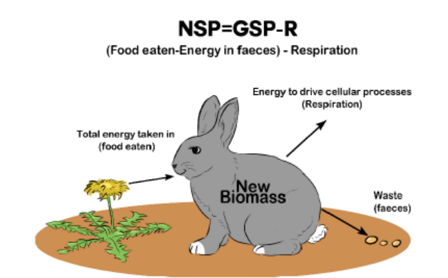
Figure 2.20: Net secondary production
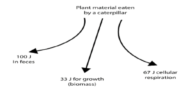
2.6.3 Ecological production efficiency
Production efficiency is the percentage of energy stored in assimilated food that isnot used for respiration. It is calculated as follows:
Production efficiency is expressed in percentage (%)
As an example, when a caterpillar feeds on a plant leaf, only about 33 J of out 200 J, or one-sixth
of the energy in the leaf is used for secondary production or growth. The caterpillar uses some of
the remaining energy for cellular respiration and passes the rest in faeces. The energy contained in
faeces remains in the ecosystem temporarily, but most of it is lost as heat after the faeces are
consumed by detritivores. The energy used for caterpillar’s respiration is also lost from theecosystem as heat.

Application 2.6
1. As part of a new reality show on television, a group of overweight people are
trying to safely lose in one month as much weight as possible. In addition to
eating less, what could they do to decrease their production efficiency forthe food they eat?
2. Tobacco leaves contain nicotine, a poisonous compound that is energetically
expensive for the plant to make. What advantage might the plant gain by
using some of its resources to produce nicotine?
3. If an insect eats plant seeds containing 100J of energy, energy from which 30
J is used for respiration while 50J remains in faeces.
4. a. Calculate the net secondary production.b. Estimate the production efficiency.
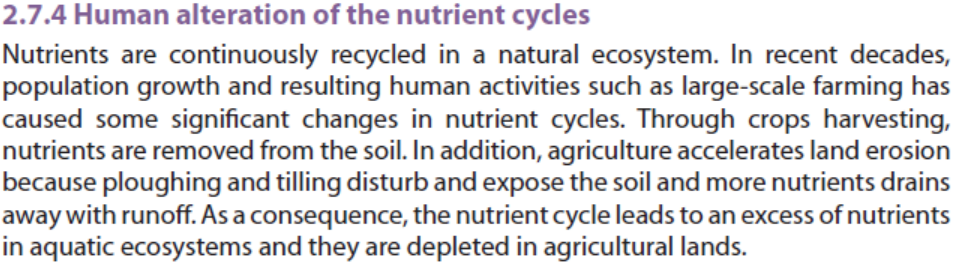
2.7 Biogeochemical Cycles
Activity 2.7Observe carefully the diagrams below and answer the questions that follow.
1. Name the biogeochemical cycles represented by X, Y and Z.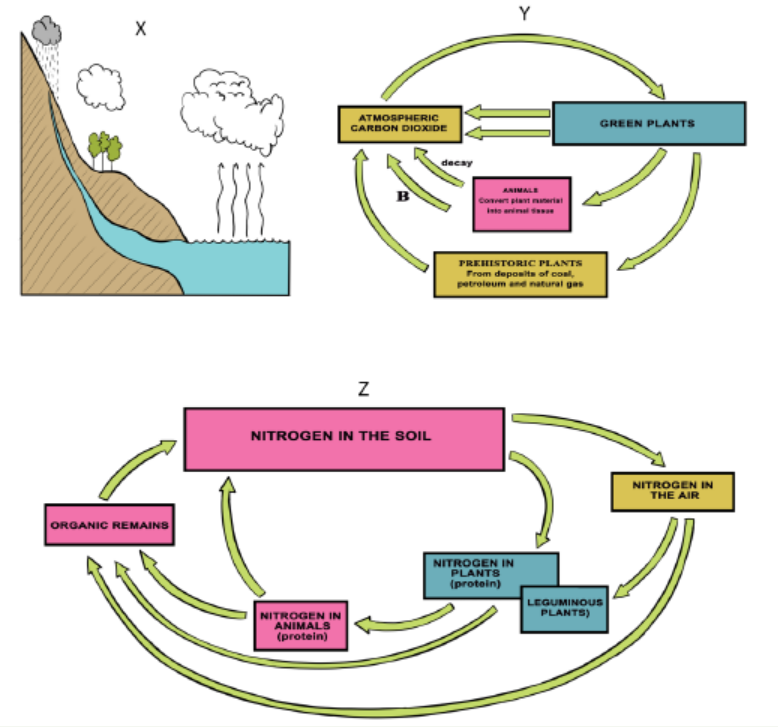
2. For the biogeochemical cycles denoted X, Y and Z, make a description of
steps represented by the letters A, B and C.
3. What do you understand by the term biogeochemical cycle?4. Discuss the importance of biogeochemical cycles to living things e.g. man.
A biogeochemical cycle is a closed loop through which a chemical element or
water moves through ecosystems. In the term biogeochemical, bio- refers to
biotic components and geo- to geological and other abiotic components. During
biogeochemical cycle, chemicals cycle through both biotic and abiotic components
of ecosystems. For example, an element might move from the atmosphere to the
water of the ocean, goes to ocean organisms, and then back to the atmosphere torepeat the cycle.
Elements or water may be held for various lengths of time by different components
of a biogeochemical cycle. Components that hold elements or water for a relatively
short period of time are called exchange pools. For example, the atmosphere is
an exchange pool for water. It holds water for several days. This is a very short time
compared with the thousands of years the deep ocean can hold water. The ocean
is an example of a reservoir for water. A reservoir is a component of a geochemicalcycle that hold elements or water for a relatively longer period of time.
2.7.1 Water Cycle
Earth’s water is constantly in motion. Although the water on Earth is billions of years
old, individual water molecules are always moving through the water cycle. The
water cycle describes the continuous movement of water molecules on above and
below Earth’s surface. Like other biogeochemical cycles, there is no beginning or
end to the water cycle. It just keeps repeating. During the cycle, water occurs in its
three different states: gas (water vapour), liquid (water), and solid (ice). Processes
involved in changes of state in the water cycle include; evaporation, sublimation,and transpiration.
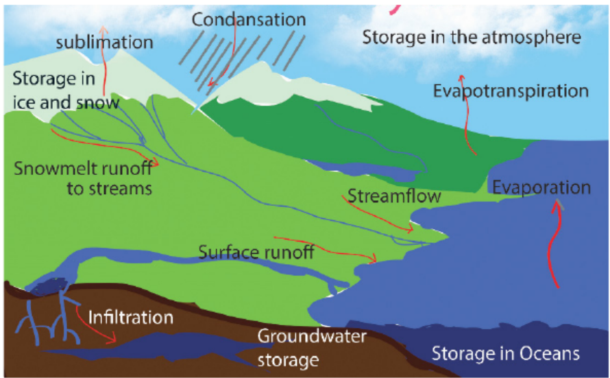
Figure 2.21: Illustration of the water cycle
2.7.2 Carbon Cycle
Carbon is essential to all life as it is the main constituent of living organisms. It serves
as the backbone component for all organic polymers, including; carbohydrates,
proteins, and lipids. Carbon compounds such as carbon dioxide (CO2) and methane(CH4) circulate in the atmosphere and influence global climates. Carbon circulates
between living and non-living components of the ecosystem primarily through
the processes of photosynthesis and respiration. Plants and other photosynthetic
organisms obtain CO2 from their environment and use it to build biological
materials. Plants, animals, and decomposers (bacteria and fungi) return CO2 to the
atmosphere through respiration. CO2 trapped in rock or fossil fuels can be returned
to the atmosphere via volcanic eruptions, or fossil fuel combustion. The movement
of carbon through biotic components of the environment is known as the fast
carbon cycle.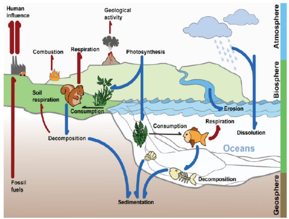
Figure 2.22: The carbon cycle
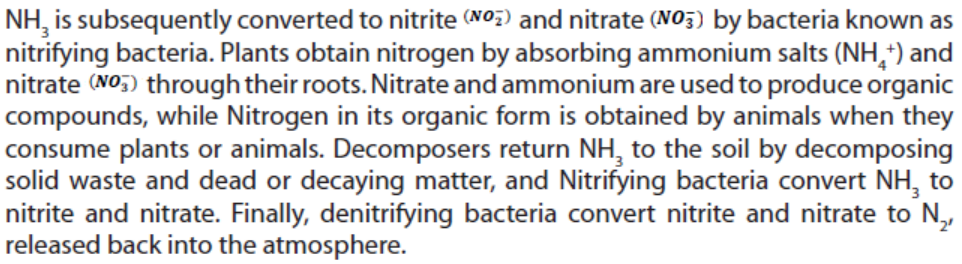
2.7.3 Nitrogen Cycle
The atmosphere is the largest reservoir of nitrogen on Earth. It consists of 78%
nitrogen gas (N2). Similar to carbon, nitrogen is a necessary component of biological
molecules. Atmospheric nitrogen (N2) is converted to ammonia (NH3) by nitrogenfixing
bacteria in aquatic and soil environments. These organisms use nitrogen to
synthesize the biological molecules they need to survive. Some nitrogen-fixing
bacteria live in soil, others live in the root nodules of legumes such as; peas andbeans. In aquatic ecosystems, some cyanobacteria are nitrogen fixing.
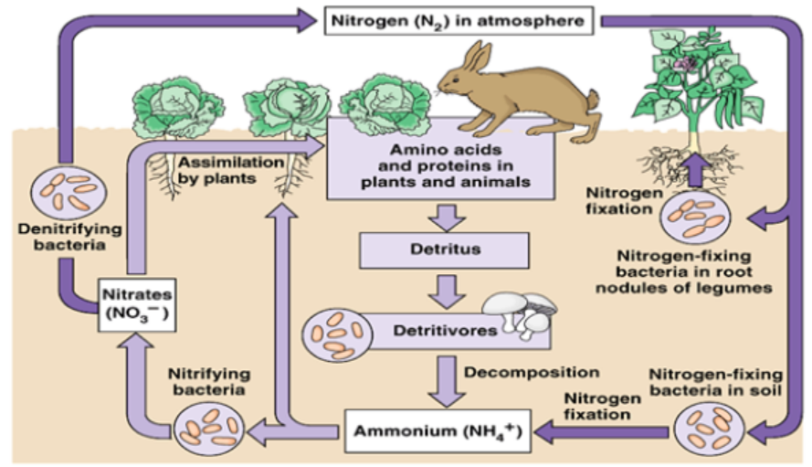
Figure 2.23: Illustration of the nitrogen cycle (Adapted from Pearson Education, 2003)
2.7.5 The Greenhouse Effect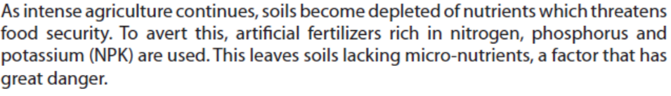
The greenhouse effect is a natural process that warms the Earth’s surface. When the
sun’s energy reaches the Earth’s atmosphere, some of it is reflected back to space
and the rest is absorbed and re-radiated by greenhouse gases. Greenhouse gases
include water vapor, carbon dioxide, methane, nitrous oxide, ozone and some
artificial chemicals such as chlorofluorocarbons (CFCs). The absorbed energy warms
the atmosphere and the surface of the Earth. This process maintains the Earth’s
temperature at around 330C warmer than it would otherwise be, allowing life on
Earth to exist. The problem we now face is that human activities particularly burning
fossil fuels (coal, oil and natural gas), agriculture and land clearing are increasing the
concentrations of greenhouse gases. This is the enhanced greenhouse effect, whichis contributing to the global warming.
Application 2.7The diagram below shows the carbon cycle.
Identify processes labelled ①, ② and ③.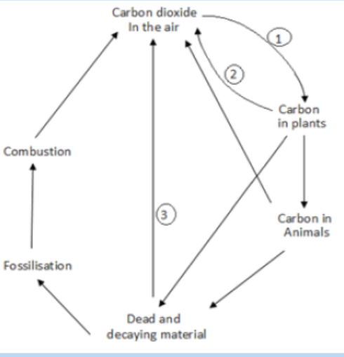
b. Describe two ways by which carbon can be removed from the cycle for
long period of time.
c. Describe two activities of humans that are disrupting the naturalcarbon cycle.
End of unit assessment 2
Section A: Multiple choice questions
Choose the letter that best answers the question or completes the statement
1. All of life on Earth exists in a region known as
a. Ecosystem
b. Biome
c. Biosphere
d. Ecology
2. Groups of different species that live together in a defined area make up
a. Population
b. Community
c. Ecosystem
d. Biosphere
3. The series of steps in which a large fish eats a small fish that has eaten algae
is a) Food web b) Food chain c) Pyramid of numbers d) Biomass pyramid
4. The total mass of living tissue at each trophic level can be shown in
a. Energy pyramid
b. Pyramid of numbers
c. Biomass pyramid
d. Biogeochemical cycle
5. An ecosystem is not considered to be self-sustaining if
a. There is interaction between biotic and abiotic factors
b. Some of its living organisms incorporate energy into organic compounds
c. Cycling of materials occurs between organisms and their environment
d. It lacks a constant supply of energy
Section B: Questions with short answers
6. What is the meaning of the term ecology?
7. Name the different levels of organization within the biosphere, from smallest
to largest
8. How is sunlight important to most ecosystems?
9. By what process do:
a. Decomposers convert organic matter into ammonia
b. Bacteria convert gaseous nitrogen into ammonia
c. Nitrosomonas convert ammonia into nitritesd. Pseudomonas convert nitrates into gaseous nitrogen
10. Why is the transfer of energy and matter in a food chain only about 10
percent efficient?
Section C: Essay questions
11. Describe the three different types of ecological pyramids.
12. Why do the rectangles in a pyramid of energy get smaller at each higher
trophic level?
13. Discuss the reasons why the secondary succession is usually much faster
than primary succession?
14. The diagram below shows part of the nitrogen cycle
a. Name a genus of bacteria which is responsible for each of the reactions
A, B, C and D.
b. Describe the conditions in which the bacteria responsible for reaction
D will thrive.
15. The table below shows mean values for primary productivity for four
ecosystems: temperate deciduous forest, tropical forest, temperate grassland,and intensively cultivated land in a temperate region
a. Suggest two reasons to account for the higher primary productivity of
a tropical forest compared with a temperate forest.
b. Suggest explanations for the difference in primary productivity
between temperate grassland and intensively cultivated land.
c. Describe how you would estimate the fresh biomass of the producersin a grassland ecosystem.
16. The diagram shows a number of stages in an ecological succession in a lake.
a. Use information from this diagram above and explain what is meant by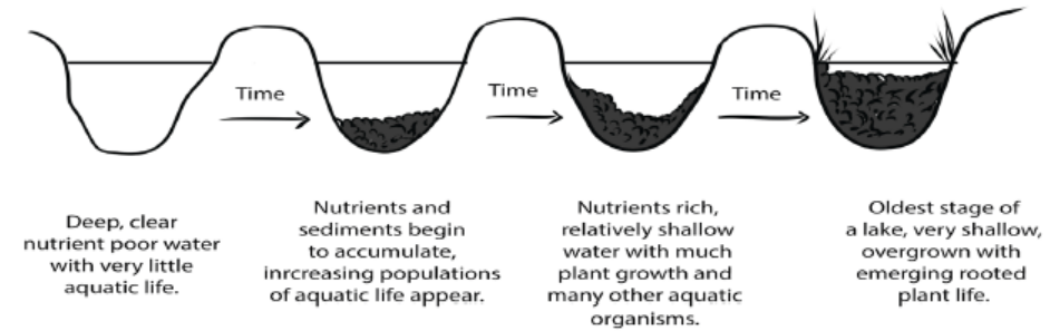
an ecological succession.
b. Give two general features this succession has in common with other
ecological successions.
c. A number of small rivers normally flow into the lake. These rivers flow
through forested areas. Explain how deforestation may affect the process
of succession in the lake.
17. Use the skills learnt in classroom and give answers to the following questions:
a. What is an ecosystem?
b. What is the required information to fully describe the make-up of an
ecosystem?
c. Discuss the flow of energy through ecosystems and make a description of
the various ways in which human activity can influence the energy flow at
all levels in terrestrial ecosystems
18. As part of a science project, Abingondo Diane is trying to estimate total primary
production of plants in a prairie ecosystem for a period of one year. Once per
quarter, Abingondo cuts a plot of grass with a lawnmower, do a collection and
weighs the cuttings with the main purpose to estimate plant production. Whatis missing for Abingondo to estimate the total primary production?
UNIT 3 EFFECT OF HUMAN ACTIVITIES ON ECOSYSTEM
UNIT 3: EFFECT OF HUMAN ACTIVITIES ONECOSYSTEM
Key Unit Competence
Evaluate the effects of human population size, resource use, and technology on
environmental quality.Learning objectives
– Explain how modern agricultural technology has resulted in increased food
production
– Explain the negative impacts to an ecosystem of large scale monoculture of
crop plants
– Explain the reasons for habitat destruction (agriculture and extraction of natural
resources)
– Explain the undesirable effects of habitat destruction
– Explain the sources and effects of the pollution of air, water and land
– Explain the causes and effects of acid rain, eutrophication and non
biodegradable plastics
– Explain the main methods of the conservation of resources
– Describe an example of conservation in action
– Assess the negative impacts to an ecosystem of intensive livestock production
– Conduct shows and dramas on wildlife conservation
– Research the effects of the excessive use of fertilisers on the environment
– Assess the different methods of the conservation of nature
– Carry out a research project on recycling sewage
– Carry out research on the African species endangered by human activity
– Evaluate the reasons for conserving wildlife
– Demonstrate ways of reducing pollution and protecting the environment
– Organise clubs focused on environmental and wildlife protection
– Suggest ways in which one could take positive action to help conserve biological
resources
– Appreciate the balance between society, environment and the economy
– Recognise that extinction is a natural part of the evolution of life on earth but
has taken place in an unprecedented rate, mainly as a result of human activities
– Support the Rwandan government policy of protecting the environment– Adapt regulations designed to prevent overfishing into action
Introductory activityThe illustrations below show two inhabited areas by human population.
1. Based on your observations, discuss what would be the impact of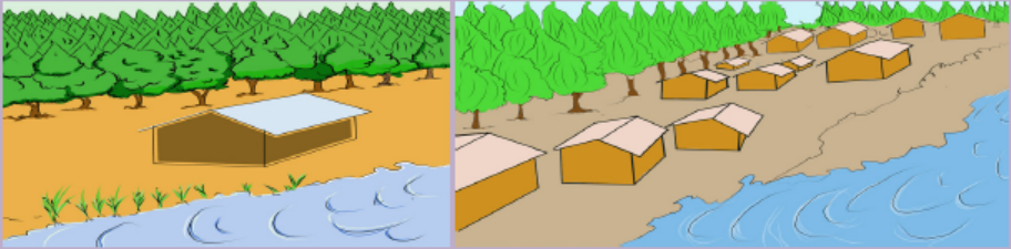
humans on natural ecosystems.2. Suggest what can be done to sustainably conserve natural ecosystems.
The increase in human population size causes changes in natural ecosystems.
Intentionally and unknowingly, human activities on Earth have negative impacts for
all kind of life form of an ecosystem. This unit intends to describe different human
activities on the Earth’s natural ecosystems and their effects. It also informs different
biodiversity conservation methods and indicates how disturbed ecosystems can be
restored. It raises the awareness towards the restoration of degraded environmentsas well as biological conservation.
3.1 Modern agricultural technologies for food production
Activity 3.1
1. Based on everyday experience, identify the modern agricultural
technologies.
2. Discuss how modern technologies increase food production in terms
of; agricultural machinery, chemical fertilisers, insecticides, herbicides,and selective breeding.
The increased population size brought changes in different sectors of any country
including agriculture, one of the most human activities that is practiced on ecosystem
for increasing food supply. Currently, agriculture is practiced with advancement
in science and technology where contemporary farming methods were invented
and adopted mostly in monoculture and intensive farming. Modern agriculture is
named mechanized agriculture or mechanized faming. It uses different equipment
including; tractors, trucks, sprayers, harvesters, aeroplanes and helicopters
depending to their manufactured purposes. It even uses computers in junction with
satellite imagery among others for easy and effective management and monitoringof land and crops.
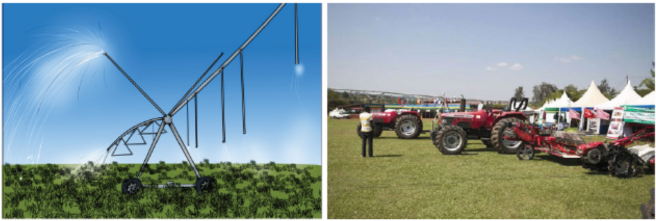
Figure 3.1: Tools and techniques in modern agriculture
The agricultural equipments are used in all process of farming starting from preparing
the land to crop storage. Beside efficient production, mechanisation encourages
large-scale production and sometimes it can improve the quality of the land. Despite
their role in increasing food production, mechanized farming intentionally or due tounskilled farm labour and awareness harms the soil, biodiversity, water and air.
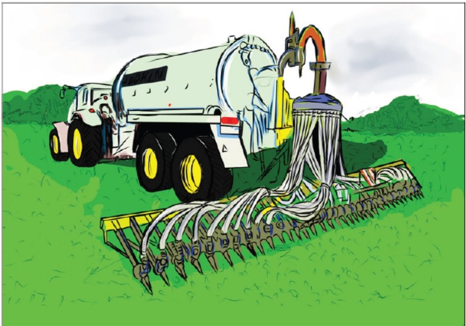
Figure 3.2: Nitrogen spreading machine
Apart from farming machineries, there are chemical fertilisers that are used on
farm land to boost levels of soil nutrients needed by plants for growing faster andproducing more food. Some of the most used fertilisers are nitrogen, phosphorus,
and potassium or a combination of them. Use of fertilizers is expensive and improper
use can harm the environment. If too little is added, crops will not produce as much
as they should. If too much is added, or applied at the wrong time, excess nutrients
will run off the fields and pollute streams and groundwater. These are the reasons
why the right amount chemical fertilizers have to be used at the right time, to avoidpotential negative effects to the environment.
Modern agriculture uses also pesticides which are organic compounds or substances.
They include; insecticides, herbicides and fungicides, used with the purpose of
killing unwanted plants, insects or fungi which might harm the plants. Utilization
of pesticides escalate food production in case of their effective use. However, some
of the pesticides present negative effects on the environment. Examples include
the 3, 5, 6-Trichloro-2-pyridinyloxyacetic acid which inhibits soil bacteria that
transform ammonia into nitrite, Glyphosate (C3H8NO5P) which reduces the growth
and activity of free-living nitrogen-fixing bacteria in soil, and oryzalin and trifluralin
which inhibits the growth of certain species of mycorrhizal fungi. Insecticides can
contaminate non-targeted organisms including; insects, fish, or plants through thespray onto eroding soil or when heavy rain falls right after an application.
The last but not the least among the modern agricultural technologies is the selective
breeding also called artificial selection. It is used in order to produce varieties of
plants or animals having phenotypic traits suitable to a particular area and of high
productivity. It allows natural evolutionary process, eliminates diseases, influencing
the production of food coming from plants in a positive way, giving to plants the
ability to grow on lands that are previously not suitable for farming, sustainability of
food chain, creation of higher-quality products, and contributes to the availability ofanimals and plants that produce higher yields.
Application 3.1
1. Explain your understanding of the term modern technology.
2. Research and discuss how the use of selected breeds is beneficial infood production
3.2 Impacts of human activities on ecosystem
Activity 3.2
Use the books from school library and search further information from the
internet. Use the searched information and do the following questions.
1. Explain the negative impacts of large scale monoculture on ecosystems.
2. Assess the negative impacts of monoculture and intensive livestock on
ecosystems.
3. Discuss the contribution of deforestation on flooding and desertification.
4. Explain how fishing and deforestation can impact aquatic ecosystems.5. Discus how mining and industrialization impact different ecosystems.
3.2.1. Negative impacts of large scale monoculture on ecosystem
Intensive cropping practices and its impacts on ecosystem
A key component of agricultural intensification is monoculture, the cultivation of
a single crop species in a given area. Unlike traditional polyculture (which mix crop
varieties or intersperse crops with trees or domesticated animals), monoculture
allows farmers to specialize in crops that have similar growing and maintenance
requirements. Monoculture is increasingly adopted by farmers to achieve higher
yields through economies of scale. However, monoculture may negatively impact
biodiversity, soil, water and air.
a. Impacts on Biodiversity
By reducing natural plant biodiversity to include only one crop, monoculture affects
the composition and abundance of associated biodiversity. For example, the balance
of plant pests and their natural enemies that may exist in polyculture fields can be
disrupted in monoculture systems, which provide habitat for a narrower range of
insects. Populations of; bees, flies, moths, bats, and birds, which provide important
pollinating and pest pressure services to crops, also tend to be lower in monoculturesthan in fields containing diverse forage and nesting sites.
b. Impact on soils
Continuous cropping impacts soils properties whereby soil fertility declines as
consecutive crop cycles reduce the amount of nutrients from soils. As plants grow,
they absorb nutrients from the soil such as nitrogen, phosphorous, potassium,
and calcium. Harvesting crops is another mechanism contributing to the removal
of these nutrients from the soil. In addition, when monoculture is continuously
applied in the same area, it affects soil organisms due to soil pesticides. Natural soil
properties including aeration and water infiltration might be affected due to theloss of soil organisms that increase these soil properties and hence soil fertility.
In addition, due to population pressure and land scarcity, farmers in some areas are
increasingly adopting intensive cultivation methods on hillside areas characterised
by steep slopes with the soils often inherently poor quality. As rainfall hits loose or
unprotected soil on cultivated sloping land, soils erode and carry away sediments
and nutrients. The resulting redistribution of nutrients may leave upward sloping
soils less fertile than lower areas, and fertilizers or other chemical particles in run-off
may negatively impact aquatic ecosystems and water quality.
c. Greenhouse effects
Tillage as one of the practice in continuous cropping, impacts on greenhouse emissions
whereby increases carbon dioxide (CO2) emissions by causing decomposition of soil
organic matter (SOM) and soil erosion. Intensive tillage practices also emit CO2, a
greenhouse gas that contributes to climate change. Mechanical tillage release CO2
and stimulates CO2 emissions by enhancing decomposition of soil organic matter.
The tendency for tillage to increase erosion also contributes to CO2 emissions. A
large percentage of soil carbon particles carried by erosion are emitted into the
atmosphere as CO2 rather than buried and sequestered in deposit sites.
Intensive livestock farming and its impacts on ecosystem
Livestock play an important role in agricultural systems. Cattle, sheep, and goats
can provide manure for soil fertilization and a diversified source of food and income
generation. Traditional livestock management involves mixing animals and crops
on the same farm or grazing livestock on grasslands. Intensive livestock systems
exacerbate the impacts that livestock activities have on the environment, including
effects on soil conditions, biodiversity, water quality and quantity, and greenhousegas emissions.
a. Impacts on Soils
Increased animal stocking rates puts pressure on grazing lands, leading in some
cases to soil compaction, erosion, grasslands degradation, and desertification in
semi-arid areas. Concentrated “hoof action” compacts wet soils, making them less
able to absorb water and more prone or more likely to run-off and erosion. Livestock
grazing between land and streams can destabilize stream banks and release large
amounts of sediment into fragile aquatic ecosystems. Additionally, high rates of
nitrogen contained in bovines’ manures can lead to topsoil acidification.
b. Impacts on Biodiversity
Intensive grazing impacts biodiversity in several ways. Populations of birds, rodents,
and other wildlife that depend on grasslands for food and habitat may decline as
livestock densities increase. In addition, intensive grazing often involves reseeding
natural meadows, resulting in a loss of native grassland plants. Higher rates of
organic or inorganic fertilizer application typically accompany reseeding, which
may degrade water quality through nitrogen or phosphorous leaching. Nutrient
contamination in water bodies reduces oxygen levels and harms fish and plantpopulations.
Leaching of nitrogen and other fertilizer nutrients into fresh and saltwater
environments can lead to a state of eutrophication (overabundant nutrient
concentrations), resulting in increased algae blooms and oxygen depletion.
Thus, dead zones may develop in these areas, whereby decreased oxygen levels
dramatically reduce fish populations and species diversity.
c. Impacts on water quality and quantity
Untreated livestock waste causes high nutrient concentrations in water bodies,
also known as eutrophication. Untreated livestock waste can significantly impact
water quality. Livestock manure contains high amounts of nitrogen, phosphorous,
and potassium and may enter water directly when livestock graze near streams
or indirectly through run-off or percolation into groundwater. Confined livestock
systems present high risks of water pollution due to difficulties containing and
treating large quantities of manure. Degraded water quality may also pose health
risks to humans who rely on water for drinking and household uses.
d. Impacts on greenhouse gas emissions
Enteric fermentation and livestock manure are significant sources of methane (CH4)
and nitrous oxide (N2O) greenhouse gases emissions. Ruminant livestock such
as cattle and sheep release CH4 during enteric fermentation and the microbial
digestion of fibrous plants. Animal manure emits N2O and CH4 during storage and
after application to croplands or grazing areas. Additional activities related to raising
livestock are responsible for emissions such as releases of CO2 in producing fertilizer
for grazing lands and animal feed, N2O emissions from applying fertilizer, and CO2emissions from overgrazing and land degradation.
e. Impacts on air quality
Nitric gas contributes to smog, ozone, and acid rain. During the microbial processes
of nitrification and denitrification that take place in fertilized soils, nitric gas is
released. Nitric emissions impact local and regional air quality by contributing to
the formation of smog, ozone, and acid rain.
Fishing and their impacts on the ecosystem
Techniques for catching fish include hand gathering, spearing, and netting, angling
and trapping. It is normally done in fish farms including ponds, rivers, lakes, seas,
oceans where fish are raised commercially. With the advancement in technology,
rearing of aquatic animals is known as “aquaculture” aiming at producing more
aquatic food due to the drastic increase of the population. Despite the significance
of fish farming and harvesting technologies, fisheries are in danger of collapsing,
due to overfishing and pollution.
Fishing nets called ghost nets used by fishermen are sometimes left or lost in
oceans whereby they can entangle fish, dolphins, sea turtles, sharks, dugong,
crocodiles, seabirds, crabs, and others. These living things are restricted from
movement which led to laceration (cut in skin), infection, starvation and suffocation
sometimes causing the death. Other effects include overfishing which is a form of
overexploitation where fish stocks are reduced to below accepted levels. It can result
in resource depletion, reduced biological growth rates and low biomass levels. Since
organisms ecologically depend each other, overfishing of one species decreases the
presence of other species and favour the invasive species. For example, with the
shark population reduced, in some sea places almost totally, the rays have been free
to dine on scallops to the point of greatly decreasing their numbers. Since then, avariety of sharks have fed on rays, which are the main predator of scallops.
Deforestation and its effects on ecosystem
Deforestation is the permanent clearing or removal of trees and undergrowth.
Deforestation happened in the past and continues extensively today particularly
in tropical area. The forests are cut mostly for mainly searching agricultural land.
In Rwanda like elsewhere, deforestation was driven by the need for food, charcoal,
and timber, especially for commercial products. Worldwide agriculture continues to
be the main cause of the loss of natural forests. Other reasons include supplying
firewood as fuel, constructing houses, industrial buildings, roads, and dams, removal
of trees for pulp and paper, cutting trees for timber used in the construction industry,
replacement of native trees with fast growing species such as conifers, eucalyptus,
and rubber trees.
a. Effects of deforestation on biodiversity
Deforestation has the dramatic effect on biodiversity particularly in tropical
rainforests. Complete replacement of native plantations with introduced species or
keeping only a few native species, leads to a reduction in biodiversity. Organisms are
being driven to extinction by the loss of their suitable habitat. In tropical rainforest,
attention should be paid to species with great human value including medicines,where forest plant products are used as anticoagulants, tranquillisers, and antibiotics.
b. Effect of deforestation on nutrients cycles
Deforestation is contributing to an increase in carbon dioxide due to the removal
of forests which actually use this gas for photosynthesis. Forests burning release
huge amounts of carbon dioxide directly and very quickly into the atmosphere and
is probably a major contributor to rising carbon dioxide levels. Burning trees was
also found to significantly reduce the nitrogen held in the ecosystem. In addition,
tree roots bind soil particles together, and tree canopy prevents rain beating down
on the soil. Deforestation therefore causes nutrients to be lost through leaching and
runoff.
c. Desertification
Deforestation is also one of the process speeded by deforestation even though some
scientists believed that it was caused mainly by climatic changes. Deforestation
disrupts water cycle and soil structure. Reduction in tree cover means reduced
transpiration, few clouds, and less rain fall in the area. Removing trees increases the
risk of flooding following heavy rains. Agricultural land becomes heavily populated,
it is likely to be over cultivated or overgrazed, and the soil will be less fertile and
more easily eroded during periods of droughts.
Mining and industrialization
a. Effects of Mining on the Ecosystem
Mining as one of economic activity applied on natural ecosystem plays an important
role to humans. It is at the same time affecting environmental ecosystem through
soil compaction, lowering overall soil fertility, erosion, soil pollution and minimizing
the availability of nitrogen and phosphorus. Soil compaction is one of the most
severe effects mining has on ecosystems and it is often the result of large machines.
As the soil is compacted, there are fewer pore spaces for oxygen and water to move
through the soil profile, minimizing the potential for plant establishment. Mining
operations often contaminate the soil with toxic heavy metals and acids, preventing
plants and soil micro-organisms from thriving
b. Effects of industrialization on ecosystem
Industrialization contributes for the nation economic development and prosperity
by providing employment opportunities and generating wealth. It is also one of the
human activity that negatively deteriorates ecosystems. The major negative effects
of industrialisation include depletion or reduction of natural resources, air, water and
soil pollution, global warming and climatic changes. Industrialization expose living
organisms to acid rain and it is among the major causes of land degradation. Thus
poor land quality, and issues generated by hazardous waste lead to some diseases
including silicosis and pneumoconiosis, tuberculosis, skin diseases and deafness.
By metallic contaminant like Cd, Zn, Hg, radioactive industrial pollutant bacteria and
beneficial micro-organisms in the soil are exposed to death. There is also a number
of undesirable effects caused by toxins from industrial wastes that enter in the food
chain. Moreover, industrial effluent damages the natural biological purificationmechanism of sewage treatment causing several soil and water borne diseases.
Application 3.2
1. Can modern agriculture and extraction of natural resources, cause the
habitat destruction. Explain why and how.
2. Explain the undesirable effects of habitat destruction.
3. Research the effect of the excessive use of fertilizers and pesticides.
4. Explain how intensive monoculture and livestock impacts on soil as
well as water ecosystems.5. Suggest what can be done for effective farming on hillsides.
3.3 Pollution
Activity 3.3
1. Produce a picture of a polluted area.
a. Explain the sources and effects of the pollution of; air, water and land.
b. Explain the causes and effects of acid rain, eutrophication
c. Demonstrate ways of reducing pollution and protecting theenvironment.
Pollution refers to the introduction of substances or energies into the natural
environment that cause adverse change. A pollutant may be physical (for example;
noise, heat, and other form of radiation), chemical (such as heavy metals in industrial
wastes), or biological (sewage for example). A pollutant may be a substance of
natural origin present in excess (such as a volcanic dust or particles of sea salt)), but
the term is more used often used to describe changes brought about by human
activities such as the emission of industrial pollutants, or the discharge of domesticwastes. The pollutant can be in any part of the biosphere: in air, land, or water.
Air pollution and its effect on the ecosystem
Air pollutant can be in form of gases (such as carbon monoxide from car exhausts),
or aerosols (soil or liquid particles suspended in the atmosphere). Pollutants have
many and different effects on the health of humans and other organisms, as well
as on the natural and built environments. Oxides of nitrogen and sulphur emitted
as industrial gases can form acid precipitation. Some pollutants can cause the
greenhouse effect as well as ozone depletion.
a. Greenhouse effect
Solar energy reaches the Earth in the form of short-wave radiation. When the
radiation strikes a surface, much of its energy is converted into heat, a form of
radiation which has a long wavelength. CO2, H2O vapour, and other gases present in
the atmosphere absorb and retain long wave radiation or reflect it back toward the
surface of the earth. These gases therefore act like panes of glass in a greenhouse,
letting light in, but retaining some of the heat before it escapes into space, hencethe term greenhouse effect.
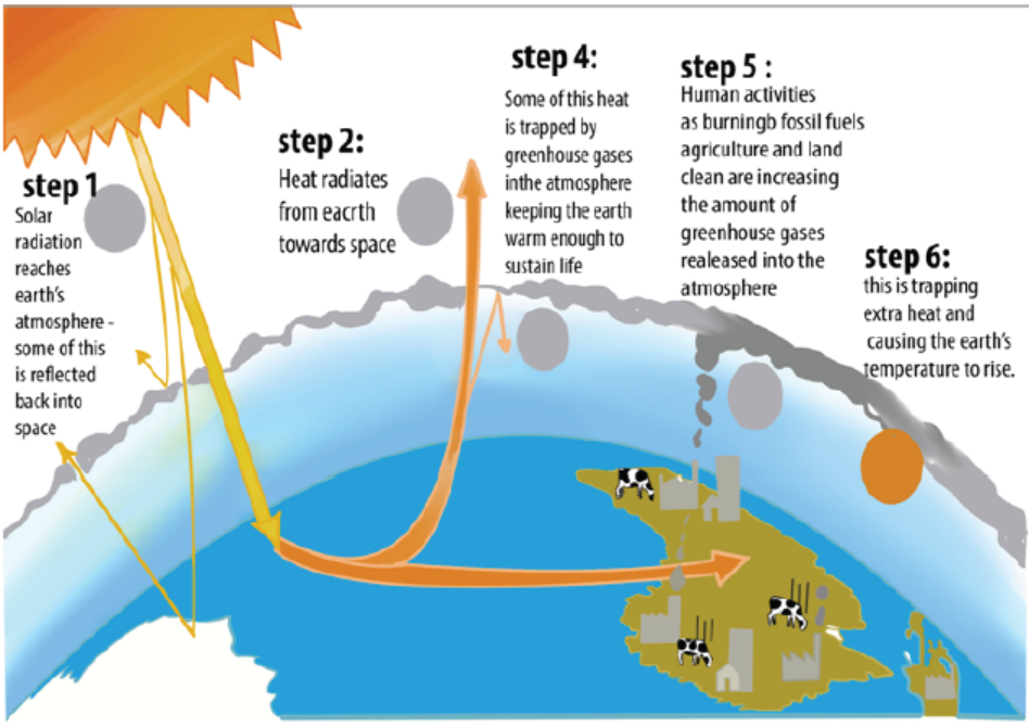
Figure 3.4: Illustration of the greenhouse effect
The retention of heat by the greenhouse effect is a natural process, essential for
the evolution of life on the earth. It has been calculated that without it, average
surface temperatures would be between -17 and -230C; the actual average surface
temperature being +150C. However, the greenhouse effect appears to be increased
by emission of certain industrial gases, called greenhouse gases, the most importantbeing carbon dioxide, water vapours, chlorofluorocarbons, methane, and ozone.
b. Global warming
The increase in the concentration of greenhouse gases in the atmosphere cause a
rise in global temperatures, and hence could bring about changes in climate. The
global warming was detected to rise the sea levels, increase melting of ice, causechanges in vegetation, and contributes to unusual weather patterns.
c. Acid precipitation
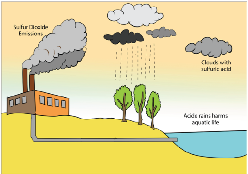
Figure 3.5: Illustration of the formation of acid rain
The burning of wood and fossil fuels, including coal and oil, releases oxides of sulphur
and nitrogen that react with water in the atmosphere, forming sulphuric and nitric
acid, respectively and forming acid precipitation or rain, snow, sleet, or fog that has
a pH less than 5.2. Acid precipitation lowers the pH of streams and lakes and affectssoil chemistry and nutrient availability.
d. Depletion of Atmospheric Ozone
Life on Earth is protected from the damaging effects of ultraviolet (UV) radiation by
a layer of atmospheric ozone (O3) layer located in the stratosphere, around 17–25
km above Earth’s surface. Like carbon dioxide and other greenhouse gases, ozone
has also changed in concentration because of human activities. The destruction of
atmospheric ozone results primarily from the accumulation of chlorofluorocarbons
(CFCs) widely used in refrigeration and manufacturing. In the stratosphere, chlorine
atoms released from CFCs react with ozone, reducing it to molecular O2. Subsequent
chemical reactions liberate the chlorine, allowing it to react with other ozonemolecules in a catalytic chain reaction.
The decrease of ozone thickness in the stratosphere increase the intensity of
ultraviolet (UV) rays reaching Earth’s surface. The consequences of ozone depletion for
life on Earth may be severe for plants, animals, and microorganisms. Some scientists
expect increases in both lethal and nonlethal forms of skin cancer and in cataracts
among humans, as well as unpredictable effects on crops and natural communities,
especially the phytoplankton that are responsible for a large proportion of Earth’s
primary production. The most severe consequence of ozone depletion is DNA
damage which could occur if ozone layer is continually destroyed or when filters to
decrease or block the UV radiation in sunlight are not used as ecologists reportedbased on their experiments using filters.
Water is also polluted by industrial sewage from abattoirs, factories, hospitals and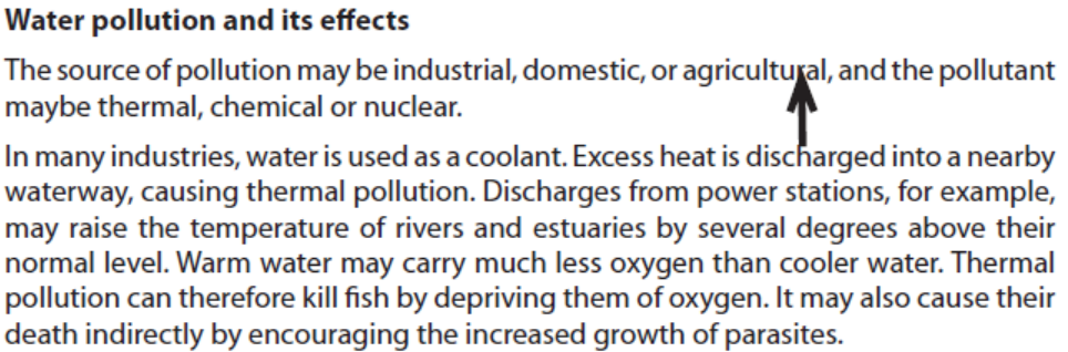
or domestic waste such as human faeces, urine and detergents. Adding organic
material to water stimulates the growth of microorganisms which feed on the
material. As the density of microorganisms increases, their demand for oxygen also
rises. Water that is very heavily polluted with raw sewage become deoxygenated
and this can lead to the death of aerobic aquatic organisms such as fish.
Eutrophication occurs when organic material or inorganic nutrients, especially
nitrates or phosphates, enter a freshwater habitat, either naturally or as a result ofpollution by sewage or agricultural runoff containing fertiliser.
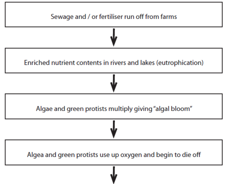
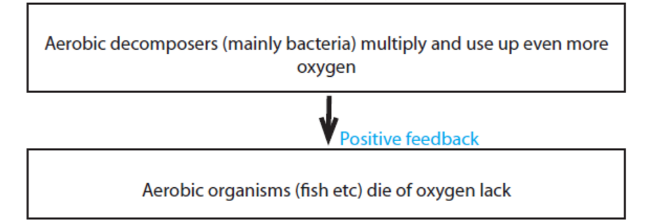
Fig 3.6: Flowchart showing the sequence of events which may result from eutrophication
Oxygen depletion and eutrophication are not only caused by sewage pollution,
they may be caused by any pollutant containing high concentrations of organic or
inorganic nutrient, such as fertilisers (inorganic or organic), slurry (animal faeces and
urine), or silage (a fermented grass product used to feed cattle in winter) effluent
which can leach off farmland and pollute water. Marine water like fresh water is
contaminated by agricultural fertilisers which have negative effects on aquaticlivings.
Soil pollution and its effects
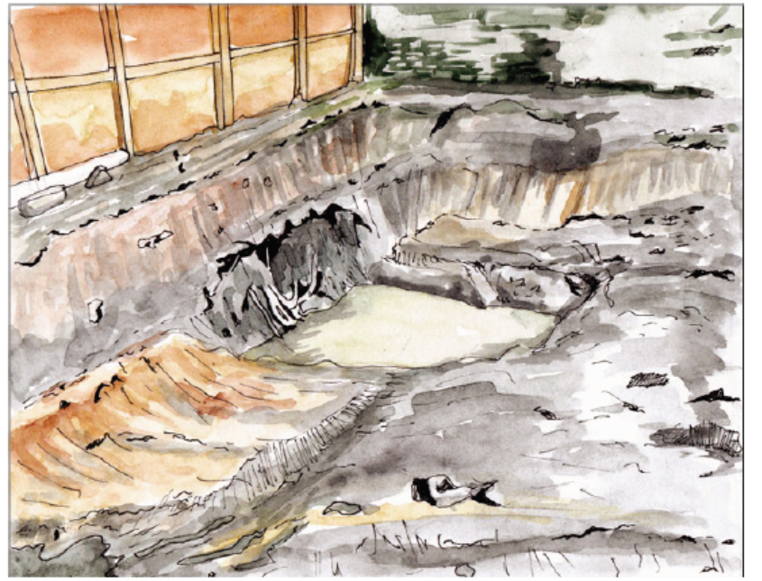
Figure 3.7. Water polluted by home garbage “Ikimoteri”
Soil is polluted as a result of human activities. It is polluted by both inorganic and
organic pollutants. These two main soil pollutants are human-made chemicals or
other alteration in the natural soil environment. It is typically caused by industrial
activity, agricultural chemicals, or improper disposal of waste such as plastics bottles
and bags. Contamination is correlated with the degree of industrialization andintensity of chemical usage.
Application 3.3
1. Question is so irrelevant and does not cater for all schools
2. Explain how plastic bags and polythene bags are dangerous for farmers
and other soil dwelling animals.
3. Suggest ways of mitigating water, soil, and air pollution. How can youimplement the suggested strategies in your school area?
3.4 Biological conservation and restoration
Learning activities 3.4
1. Carry out research and list African species threatened by human
activities
2. Explain some main methods of the conservation of resources.3. Discuss the consequences due to the loss of biodiversity.
To date, scientists described and formally named about 1.8 million species of
organisms. About 10 million more species are not yet identified. A greatest portion
of species is found in tropics particularly in the tropical forests. Additionally, over
half of all accessible surface water is used for different purposes. Throughout the
biosphere, human activities altered trophic structures, energy flow, chemical cycling,
and natural ecosystem processes. Considering the above background, it is now timeto rethink about and seek how to preserve life on the Earth.
a. Biological conservation
Biological conservation integrates; ecology, physiology, molecular biology, genetics,
and evolutionary biology to conserve biological diversity at all levels. It is aimed to
maintain the quality of natural environments and their biological resources. Unlike
preservation which tries to prevent human interference, conservation involves
actively managing biotic and abiotic components to ensure the survival of the
maximum number of species and genetic diversity. Common reasons for conservingwildlife are:
Utilitarian reasons: Species are conserved due to their benefits to humans in terms
of food, medicines including quinine and codeine among plants, and snake venom
used as anticoagulants and anaesthetics, aspirin to antibiotics are made from natural
resources, and alkaloids that inhibit cancer cell growth), industrial use (timber, fuel,
gums, dyes, and oils), natural genetic resistance to pests, and whether they providenew variety.
Aesthetic reasons: Wild animals and plants biodiversity are conserved for thepleasure they provide human well-being.
Ecological reasons: Biodiversity is conserved due to the complex ecosystem
goods and services they provide including network of relationships which maintainbiogeochemical cycles in the biosphere and the energy flow in an ecosystem.
Ethical reasons: Most of the people conserve biodiversity due to the moral duty
to look after the environment and that all species have right to live. It is thereforemorally wrong to destroy ecosystems or to allow species to become extinct.
b. Conservation methods
Zoned reserves or protected areas approach
A zoned reserve is an extensive region that includes areas relatively undisturbed
by humans surrounded by areas that have been changed by human activity and
are used for economic gain. In Rwanda, there are now four national parks namely,
Akagera National Park, Nyungwe National Park, Volcano National Park, and Mukura-
Gishwati National Park which are the reserves for natural wildlife. The key challenge
of the zoned reserve approach is to develop a social and economic climate in the
surrounding lands that is compatible with the long-term viability of the protected
core. These surrounding areas called buffer zones continue to be used to support
the human population, but with regulations that prevent the types of extensive
alterations likely to impact the protected area. As a result, the surrounding habitats
serve as buffer zones against further intrusion into the undisturbed area. The
neighbouring communities should be involved in ecotourism activities as one wayof benefiting from ecosystem services.
Eco-farming approaches
Eco-farming is a modern method for conserving natural ecosystems. It combines
science and innovation with respect for nature and biodiversity. It ensures healthy
farming and healthy food. It protects soil, water and the climate from pollutants.
It does not contaminate the environment with chemical inputs or use genetically
engineered crops. And it places people and farmers, consumers and producers at its
very heart rather than the corporations who control the food now. It is envisioned
for sustainability and food sovereignty in which food is grown with health and safety
first and where control over food and farming rests with local communities, ratherthan transnational corporations. The methods have seven principles which are:
– Food sovereignty in which producers and consumers, not corporations, should
control the food chain and determine how food is produced.
– Rewarding rural livelihoods for ensuring food security and fighting poverty
in rural development.
– Smarter food production and yields which aimed at creating higher yields to
help feed the world.
– Biodiversity for promoting diversity in crops, instead of monocultures like corn
and soy, essentially to protecting ecosystem.
– Sustainable soil fertility is improved using eco-farming methods and refraining
from chemical fertilizers and inputs.
– Ecological pest protection where farmers can control pest damage and weeds
effectively through natural means instead of chemical pesticides.
– Food Resilience where diverse and resilient agriculture, not monoculture
crops, is the best way to protect communities from shocks from climate andfood prices.
Other conservation practices
In additional to the conservation methods, there are other practices that can be
applied for biological restoration since the above methods may be difficult andexpensive for some countries. They include:
– Restricting urban and industrial development and reclaiming abandoned sites
or other areas.
– Legally protecting endangered species and prohibiting the release or
introduction of non-native animals and plants into an area.
– Controlling pollution in sensitive environments in which species are at risk of
extinction.
– Recycling materials such as paper, glass bottles, clothes, and limiting the
exploitation of renewable resources to sustainable yields.
– Restricting trade of endangered species and providing breeding programs for
endangered species for example in zoos and botanic garden.
– Avoiding poaching and forest fires and voids habitat loss.
– Not introducing new species or exotic species and avoid overharvesting and or
overfishing.– Preventing global change through practices like afforestation.
Biodiversity conservation improves the quality of life for local people and leads to
a sustainable development. Many nations, scientific societies, international and
local NGOs embraced the concept of sustainable and economic development that
meets the needs of people today without limiting the ability of future generations
to meet their needs. In Rwanda, the Rwanda Environmental Management Agency
(REMA) and Rwanda Development Board (RDB) aims at protecting and conserving
ecosystems. The main conservation initiatives include forbidding people to use
swamps, not cultivating near the streams, rivers, and lakes, reforestation, ecotourism,
buffer zones, polythene or plastic bags not allowed to be used and enter inthe country are highlighted.
Application 3.4
1. How does coppicing contribute to the restoration of species?
2. Discuss the ways in which one could take action to help conserve
biological resources.
3. a) What could happen to Nyabarongo river ecosystem if there are
continuous soil erosions from the hillsides?b) How could the effects due to erosions be resolved?
End of unit assessment 3
1. Discuss how could human activities have negative impacts on
ecosystem.
2. Explain the major causes of deforestation in tropical area?
3. What are the advantages and disadvantages of agricultural practices?
applying nitrogenous fertilizers to crops, burning agricultural wastes,
growing crop plants with genetically engineering resistance to
herbicide e.g. glyphosate)?
4. Zoologists and conservationists fear that many if not all species of
amphibians would be extinct due to global pollution and climate
change. Explain how global pollution and climate change contribute to
the extinction of amphibians.
5. How can the addition of excess nutrients to a lake threaten its fish
population?
6. Based on biological magnification of toxins, is it healthier to feed at a
lower or higher trophic level? Explain.
7. Describe how the newly introduced species may damage natural
ecosystem.
8. Appreciate how modern agricultural technologies are a challenge aswell as a solution.
UNIT 4 THE CIRCULATORY SYSTEM
UNIT 4: THE CIRCULATORY SYSTEM
Key Unit Competence
Relate the structures of the circulatory and lymphatic systems to their functions.
Learning objectives
– Explain the need for a transport system in animals.
– Explain the advantages and disadvantages of different types of circulatory
systems.
– Describe the external and internal structure of a mammalian heart.
– Explain how a heartbeat is initiated.
– Describe the main events of the cardiac cycle.
– Explain the relationship between the structure and function of blood vessels.
– Explain how blood circulation is controlled.
– Describe the effects of exercise on respiration and on circulation.
– Describe the process of blood clotting.
– Recall the structure of haemoglobin and explain how haemoglobin transports
oxygen.
– Explain how tissue fluid and lymph are formed.
– Describe the risk factors associated with cardiovascular diseases.
– Carry out an investigation on the effects of exercise on the pulse rate and blood
pressure.
– Distinguish between open and closed, single and double circulation with
reference to insects, earthworm, fish and mammals.
– Recognize blood vessels from their structures using a light microscope.
– Relate the structure of blood vessels to their functions.
– Differentiate between blood, tissue fluid, and lymph.
– Relate blood as a tissue to its functions.
– Interpret oxygen dissociation curves for haemoglobin and other respiratory
pigments.
– Appreciate the importance of the need for transport systems when animals
become larger, more complex and more active, to supply nutrients to, and
remove waste from, individual cells.
– Recognize possible risk factors as diet, stress, smoking, genetic predisposition,age and gender in relation to cardio vascular diseases.
Introductory activity
Mass sports in Rwanda has been encouraged, where people of all ages
participate in sports.Discuss the advantages of doing sports to a human health?
Physical activities can make people including students to be stronger and healthier.
They contribute also to lowering obesity rate. All individuals who practice physical
activities tend to; have lower body mass indexes, benefit from developing muscles
and burning calories. Physical activities help in lowering the rates of diabetes and
high blood pressure. Doing physical exercises regularly contribute to better heart
and lung function.4.1 Blood circulatory system in animals
Activity 4.1Observe the illustrations of animals below and answer the following questions
1. Do the above animals have the same circulatory system? Justify your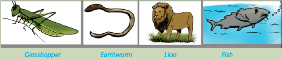
reasoning by distinguishing the type of circulatory system(s) found ineach animals?
All, except the smallest and tiniest animals need a system to transport substances
from cell to cell within themselves. The primary tasks of the system are to import,
distribute/deliver nutrients and oxygen to every cell and then to remove waste
products including carbon dioxide. The design of the transport system depends
upon the size of the organism and on how active it is.
In animals, there are two types of circulatory systems i.e.
i. Open circulatory systemii. Closed circulatory systems:
4.1.1 Open circulatory system and closed circulatory system
In animals, circulatory system is either open or closed. Table 4.1 below, showsdifferences between open and closed circulatory systems:
Table 4.1. A comparison between open and closed circulatory systems
a. Open circulatory system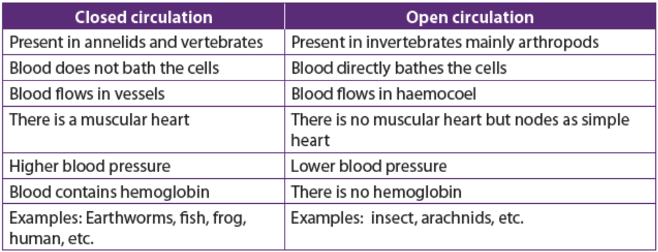
The open circulatory system is common to molluscs and arthropods. In this system,
blood is pumped into a hemocoel where it comes into direct contact with body cells
and there after goes back to the tubular ‘heart’ via openings called ostia/pores.
Insects and other arthropods have a heart which is an elongated tube located
dorsally. The internal organs are suspended in a network of blood-filled sinuses
which collectively form the haemocoel. Blood from the heart mixes with the
interstitial fluid in the haemocoel to form haemolymph. The advantage this has, isthe direct exchange of materials between body cells and haemolymph.
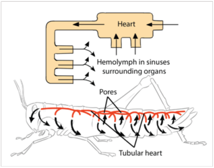
Figure 4.1: Open circulation in insect Adapted from Campbell Biology 11th Edition
b. Closed circulatory system
Vertebrates, and a few invertebrates like earthworms, have a closed circulatory
system. Closed circulatory systems have the blood closed at all times within vessels
of different sizes and wall thickness. In this type of system, blood is pumped by theheart through vessels, and does not fill body cavities.
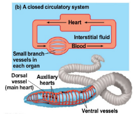
Figure 4.2: Closed circulation in annelids (adapted from Campbell Biology 11th edition
The earthworm possesses a closed circulation system whereby the blood is confined
to a series of blood vessels and not permitted to mix with the body tissues. Blood is
pumped around the system by muscular longitudinal and ventral vessels and five
pairs of lateral pseudo-hearts in segments 7 to 11. Backflow of blood is prevented
by valves. The blood itself contains haemoglobin dissolved in the plasma and some
phagocyte cells. It is advantageous for an organism to have closed circulatory system
because:
– It helps in control of distribution of blood to different parts of the body.
– Muscular walls of vessels can constrict and dilate to vary the amount of flow
through specific vessels
– Blood pressures are fairly high and the circulation can be vigorous
– It is more efficient hence the blood can reach further distances
– Allows for more control over oxygen delivery
All vertebrates including; fish, amphibians, reptiles, birds and mammals possess
a prominent muscular heart which pumps blood around the body. The closedcirculatory system can be single, partial and double.
1. Single circulation in fish
Fish have a two-chambered heart made of one atrium and one ventricle.
Deoxygenated blood from the body is pumped by the heart to the gills. Here blood
is oxygenated before passing around the body and ultimately returning to the
heart. Blood has to pass through two capillary systems, the capillaries of the gills
and then those of the body before returning to the heart. The two system capillaries
offer considerable resistance to the flow of blood. This means that in fish there is a
marked drop in blood pressure before the blood completes a circuit. In this type of
circulation, it is an advantage that the blood circulating in the body cells has alreadybeen oxygenated in the gills.
2. Partial double circulation in amphibians
All amphibians and most of the reptiles possess a heart with two atria and one
ventricle. Blood from the body enters the right atrium and is pumped to the lungs by
the common ventricle. It returns to the heart and enters the left atrium before being
pumped around the body. It is called partial because the only one ventricle receivedoxygenated and non-oxygenated blood which can be mixed as illustrated below:
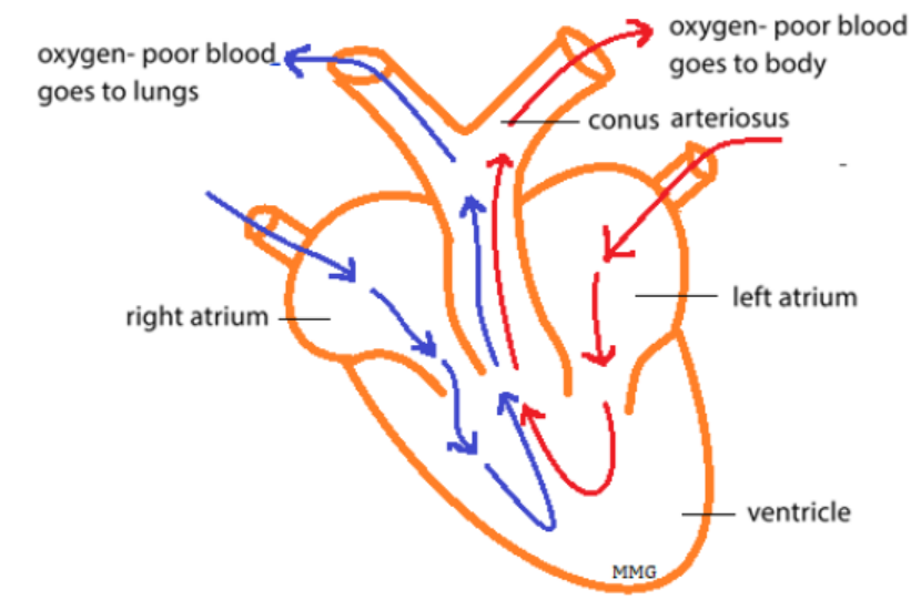
Figure 4.3: Illustration of partial double circulation in amphibians
A spiral valve called conus arteriosus helps to keep deoxygenated and oxygenated
blood separate to some extent. The figures below distinguish how closed circulationoccurs in fishes and in amphibians.
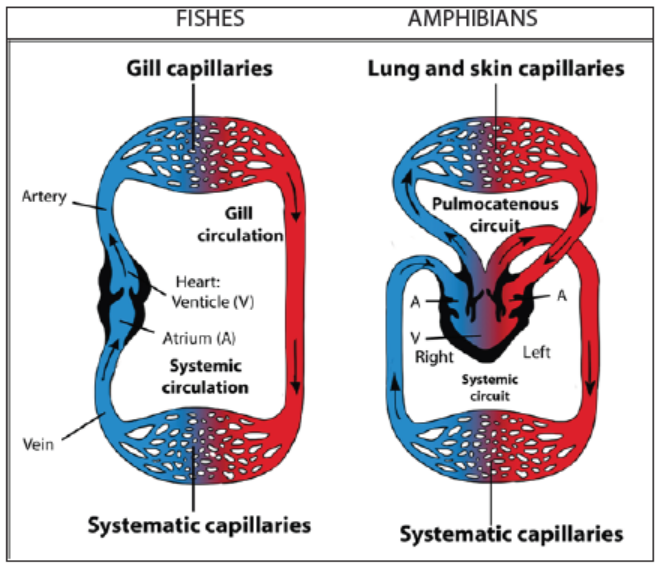
Figure 4.4: Closed circulation in fish and amphibian
3. Complete double circulation in mammals
This circulation is called double circulation because blood must pass twice in the
heart for one complete circuit. The right side of the heart delivers oxygen poor
blood to the capillary beds of the gas exchange tissue in lungs, where there is a
net movement of O2 into the blood and of CO2 out of the blood. This part of the
circulation is called a pulmonary circuit or pulmonary circulation.
After the oxygen enriched blood leaves the gas exchange tissues (the lungs), it
enters the left side of the heart. Contraction of the left part of the heart propels this
blood to the capillary beds in organs and tissues throughout of the body. Following
the exchange of O2 and CO2 as well as nutrient and waste products, then the oxygen
poor blood returns to the right part of the heart, completing the systemic circuit orthe systemic circulation.
Mammals and birds have a four-chambered heart and a complete double circulation.
The following are some of the advantages of double circulation:
– The heart can increase the pressure of the blood after it has passed through the
lungs, so the blood flows more quickly to the body tissues.
– There is no mixing of oxygenated blood with deoxygenated blood.
– Blood is pumped exactly where it is needed
– The blood pressure must not be too high in the pulmonary circulation, otherwiseit may damage the delicate capillaries in the lungs
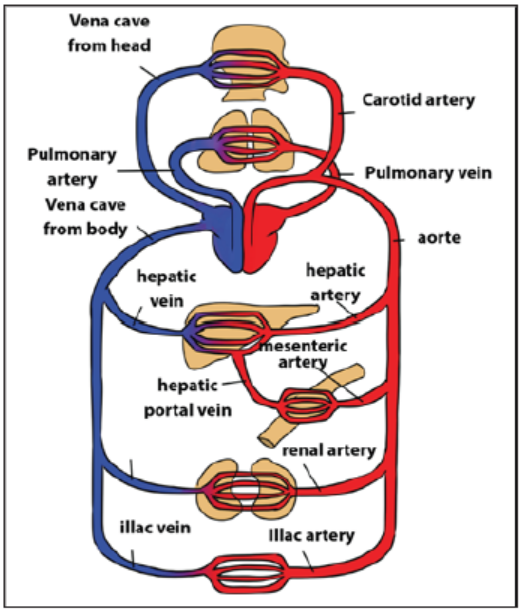
Figure 4.5: Closed double circulation in mammals and birds
The following table 4.2 indicates the comparison between single and doublecirculation
Table 4.2: Comparison between single and double circulation.
Application 4.1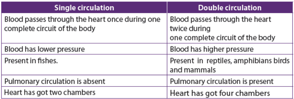
1. Briefly explain why animals need a transport system.
2. Explain how open and closed circulatory systems differ.
3. Describe the differences between single and double circulation.4. Describe how circulation take place in humans.
4.2 Structure of the human heart
Activity 4.2
– Obtain an intact heart of sheep or goat from a butcher’s shop or slaughter
house
– Rinse it under a tap to remove excess blood
– Observe the surface of the heart and identify the main visible features
– The blood vessels may have been cut off, but it is possible to identify where
these would have been attached later in the dissection
– Gently squeeze the ventricles. They can be distinguished because the wall
of the right ventricle is much thinner than that of the left ventricle
– Using a pair of sharp scissors or a scalpel, make an incision from the base of
the left ventricle, up to the left atrium and then from the base of the right
ventricle up to the right atrium
– Using a pair of forceps, remove any blood clots lying in the exposed chambers
– Identify the main components of internal structure of the heart– Compare the thickness of the left ventricle wall to that of the right ventricle
The human heart is made up of a cardiac muscle which contracts in order to propel
blood throughout the body. It is located between the two lungs, behind the
sternum in the thorax. The heart is surrounded by a tough sac called pericardium. A
pericardial fluid is secreted between the membranes allowing them to move easily
over each other. The pericardium protects the heart from overexpansion caused by
elastic recoil when it is beating very fast. The heart (Figure 4.6) is divided into a leftand a right side separated by the septum.
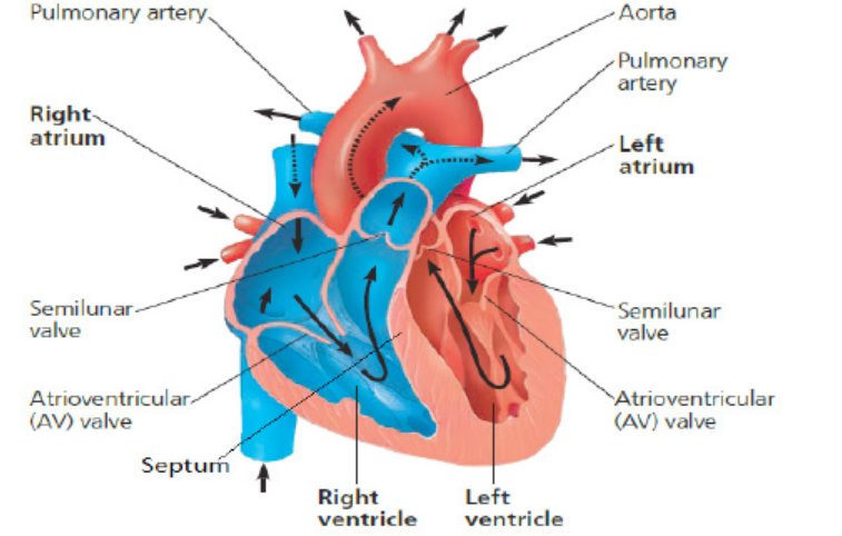
Figure 4.6: Structure of human heart (From Campbell 11th Edition)
The heart of mammals and birds is composed of 4 chambers including 2 upper atria
and 2 lower ventricles. The right side deals with deoxygenated blood and the left
side with oxygenated blood. The muscular wall of the left ventricle is thicker than
that of the right ventricle because the left ventricle has to pump blood to the whole
body with much higher pressure.
The left atrium is separated from the left ventricle by a bicuspid or mitral valve, whilst
a tricuspid valve separates the right atrium from the right ventricle. Jointly, these
two valves are known as atrioventricular valves. Atrioventricular valves are pushed
open when atria contract but, when ventricles contract they close and produce the
first sound of the cardiac cycle, the second being that of the closing semilunar valves(aortic and pulmonary valves).
Application 4.2
1. Suggest a reason for each of the following:
a. The right atrium is larger than the left atrium.
b. The left ventricle has a thicker muscular wall than the right ventricle.
2. Discuss the functions of pericardium and pericardial fluid that surroundthe heart.
4.3 Heart beat and mammalian cardiac cycle
Activity 4.3
Work to find out the number of pulses of each other using a thumb above the
vein ahead of your wrest or a sphygmomanometer.
a) Record in a table the number of pulses for the class
b) Who has the highest number? The lowest number?c) Explain significance of such a technique.\
4.3.1. Initiation of a heartbeat
Heart beat is a rhythmic sequence of contractions of the heart. It is coordinated by
two small groups of cardiac muscle cells called the sinoatrial (SA) and atrioventricular
(AV) nodes. The sinoatrial node (SAN), often known as the cardiac pacemaker,
is found in the upper wall of the right atrium and is responsible for the wave of
electrical stimulation that starts atrial contraction by creating an action potential.
The action potential causes the cardiac cells to contract. This wave of contraction
spreads across the cells of the atria, reaching the atrioventricular node (AV node/
AVN) which is found in the lower right atrium.The atrioventricular node/AVN conducts the electrical impulses that come from the
SA node/SAN through the atria to the ventricles. The impulse is delayed there before
being conducted through special bundles of heart muscle cells called the bundle of
His. This delay allows for the ventricles to be filled with blood before they contract
There is a collection of heart muscle cells (fibres) specialized for electrical conduction
that transmits the electrical impulses from the AVN, through the Purkinje fibres,which leads to a contraction of the ventricles.
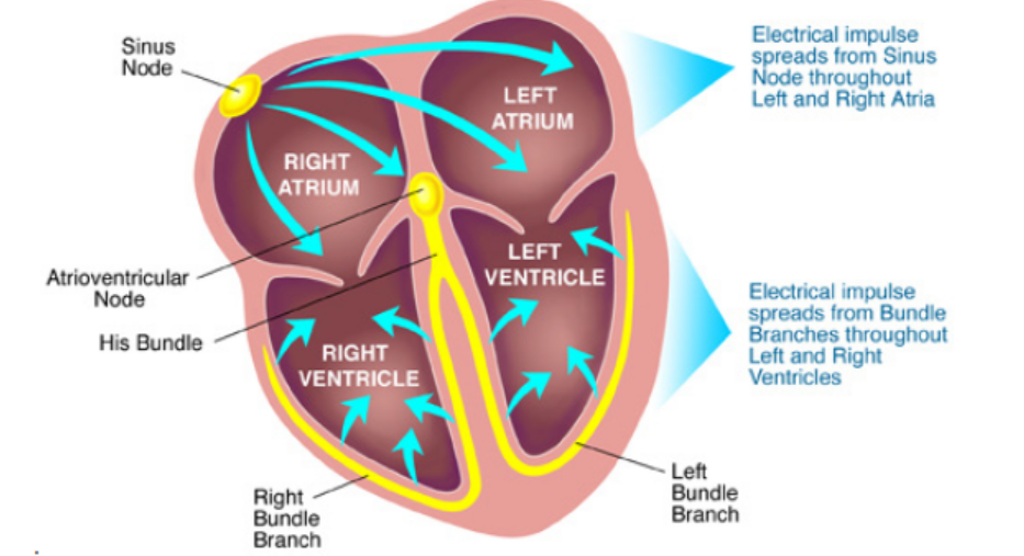
Figure 4.7: The initiation (origin) of heart beat.
4.3.2. Mammalian cardiac cycle and cardiac sounds
The cardiac cycle refers to the sequence of events which take place during the
completion of one heartbeat. It involves repeated contraction (systole) and relaxation
(diastole) of the heart muscle. The three steps in cardiac cycle are the followings:
1. Atrial systole and ventricular diastole
In this brief period of 0.1 seconds that follows atrioventricular diastole, blood from
the vena cava and pulmonary vein enter the both atria and they get filled with blood.
The walls of the atria undergo contraction (systole) forcing blood into the ventricles
via bicuspid and tricuspid valves. During this time, the ventricles are relaxed and
semilunar valves remain closed.
Ventricular systole and atrial disatole
During this stage, the ventriles undergo contraction (systole) hence forcing blood
out of the heart via the semilunar valves into the aorta and pulmonary artery. At
this time, the atria relax and expand waiting to be filled with blood. The contraction
of ventricles causes the atrioventricular valves to close simultaneously in order to
prevent back flow of blood. The closure of the valves produces the first heart soundtermed as ‘lub’.
2. Atrioventricular diastole
Upon expelling of blood, ventricles relax and their pressure lowers compared to
aorta and pulmonary artery pressures. This would cause back flow of blood to the
heart but it is prevented by sudden closure of the semilunar valves. The closure of
the semilunar valves causes a second heart sound called ‘dub’.
Note: The two sounds ‘lub’ and ‘dub’ are so close and often describes as ‘lub –dub’
and they form a single heartbeat.
The atrioventricular diastole ends the cardiac cycles and is followed by the atrial
systole. Hence the cycle restarts. When the heart rate is 75/min, which means 75heartbeats per minute, the period of one cardiac cycle is 0.8 sec.
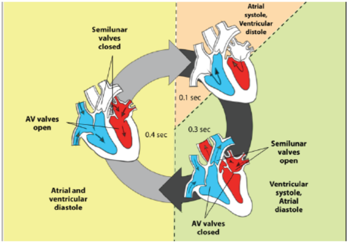
Figure 4.8: The cardiac cycle
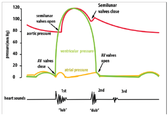
Figure 4.9: The relationship between heart sounds and key events in cardiac cycle
The electrical activity of the heart can be monitored using an Electrocardiogram
(ECG) as shown in figure 4.10. This involves attaching of sensors to the skin. Some
of the electrical activity generated by the heart spreads through the tissue next to
the heart and onwards to the skin. The sensors on the skin pick up the electrical
excitation created by the heart and convert this into a trace. The trace of a health
person has particular shape. it consists of a series of waves that are labelled P, Q, R, S
and T. Wave P shows the excitation of the atria, while QRS indicates the excitation of
the ventricles and T shows diastole.
The shape of the ECG trace can sometimes indicates the parts of the heart muscles
which are not healthy. It can show if the heart is being beating irregularly, fibrillation
(the heart beat is not coordinated), or if it is suffering the heart attack (myocardial
infarction). It can also show if the heart has enlarged or if the Purkinje fibre is notconducting electrical activity properly.
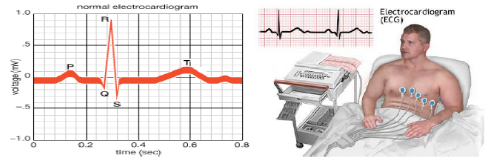
Figure 4.10: Electrocardiogram normal wave and electrocardiogram machine.
Application 4.3
1. Briefly describe the main events of cardiac cycle.
2. During the mass sports the medical doctor made a check-up and found
the following data from three participants A, B and C.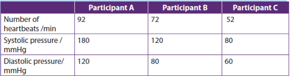
a. Among the three participants, who shows more signs of
cardiovascular problem? Why?b. Differentiate between systolic and diastolic ventricular pressures.
3. Observe the illustration below and answer to the following questions:
a. Describe the shape of the electrocardiogram trace above.
b. Explain why the QRS complex has a larger peak than the P wave.
4.4 Control of the heart rate.
Activity 4.4
a. Place your middle finger on the artery found near the opening of the ear
then count the number of pulses and write it down. Repeat this 3 times,
then calculate the average of the heart beat per minute.
b. Do some warm up exercises within 2 minutes, again place your thumb
finger on the artery found at the back of the wrist then count the number
of pulses after the exercise. Repeat this 3 times then calculate the average
of the heartbeat per minute. Use the stop clock or a watch to count the
number of pulse (beatings) within one minute.
i. How does your heart rate immediately after a warm up exercises differ
from that while at rest?ii. How would you explain the differences?
4.4.1. Nervous and hormonal control of heart rate
In the nervous control of the heartbeat, there is a cardiovascular center located in
the medulla oblongata of the hindbrain which controls the activities of the SAN. Thecenter has two nerves from the autonomic nervous system i.e. sympathetic nerve
whose stimuli accelerates activity of the SAN (increases heartbeat) and vagus nerve
whose stimuli slows down the activity of SAN (decreases heartbeat).
With regard to the hormonal control, the adrenal glands under influence of
hypothalamus secrete the hormone adrenaline into blood. Upon reaching the heart,
adrenaline will speed up the activity of the SAN thus increasing heartbeat. The
reduction comes about when the levels of adrenaline reduce through a negativefeedback mechanism.
4.4.2. Other factors controlling heart rate
Other factors affecting heart rate include; the levels of carbon dioxide, temperature,pH and mineral ions.
a. Carbon dioxide
Chemically, high CO2 levels stimulate the vasomotor Centre (VMC) to vasoconstrict
arterioles. The resulting high blood pressure transports CO2 more rapidly to the
lungs for expulsion and exchange with O2. Where tissues suddenly become active,
they produce more CO2. This causes vasodilation of local blood vessels, thus
increasing their blood supply and allowing more oxygen and glucose to reach them
for respiratory purposes.
b. Body temperature
When the body temperature changes, so does the heart rate. This is one of the
thermoregulatory changes that occur to prevent the body’s core temperature of
370C from increasing or decreasing. Heart rate increases when heat is gained by the
body such as in hot climates and during physical exercise in order to transfer more
heat away from the body. When the body loses heat such as in cold weather or a cold
shower, heart rate decreases to preserve core temperature.
c. pH and mineral ions
The importance of plasma electrolytes and pH levels in determining heart rate is
not yet well grounded. A significant heart rate increase was obtained after a decrease
of potassium and calcium and an increase in pH levels and with no significant
variations in indices of autonomic activity. The analysis revealed that changes
in physiological range of; potassium, calcium, and pH could cause large heart rate
variations from 60 to 90 bpm. It was concluded that electrolyte and pH changes in
physiological range have an important complex impact on the pacemaking rhythm
independently of autonomic outflow.
Effect of drugs, and physical activity on cardiac frequency
a. Physical exercise
The heart rate and blood pressure both rise during physical exercise. Over time,
regular physical exercise can help lower the resting blood pressure and heart rate.
This is because physical exercise training improves the health of the heart and blood
vessels, allowing the cardiovascular system to function more efficiently. This enables
increased blood flow to muscles without putting excess pressure on blood vessel
walls. While blood pressure rises during exercise, it is too much smaller degree than
the increase in heart rate. Like the heart rate, blood pressure returns to resting level
a few minutes after the end of physical exercise.
b. Caffeine and Other Drugs
Caffeine found in coffee, tea and soda is a stimulant drug that influences the nervous
system to increase heart rate. It mimics the effect of adrenaline, a natural hormone
in the body responsible for elevating heart rate. Other stimulants such as cocaine
and ephedrine work in a similar manner.
On the other hand, there are specific drugs used in lowering heart rate such as betaand
calcium channel blockers. Beta-blockers work by interfering with the receptors
that adrenaline binds to, subsequently decreasing hormonal influence on heart
rate. Calcium channel blockers reduce the amount of calcium that enters the heart
muscle. Because calcium is needed for muscle to contract, the heart beats at a slowerrate when this drug is taken.
Application 4.4
1. Discuss how both nervous and hormonal systems are involved in
regulation of heart beat rate.2. Discuss how some drugs like caffeine affect the heart beat rate.
4.5 Blood vessels
Activity 4.5
1. Use a microscope to observe prepared slides of blood vessels.
2. Draw and label the observed blood vessels.
3. Compare those blood vessels.4. Explain the relationship between each blood vessel and its function.
Blood vessels include; arteries, capillaries and veins. Illustrations, structure of
walls, lumen, valves, branching, and functions of arteries, capillaries and veins aresummarized in the figure 4.11
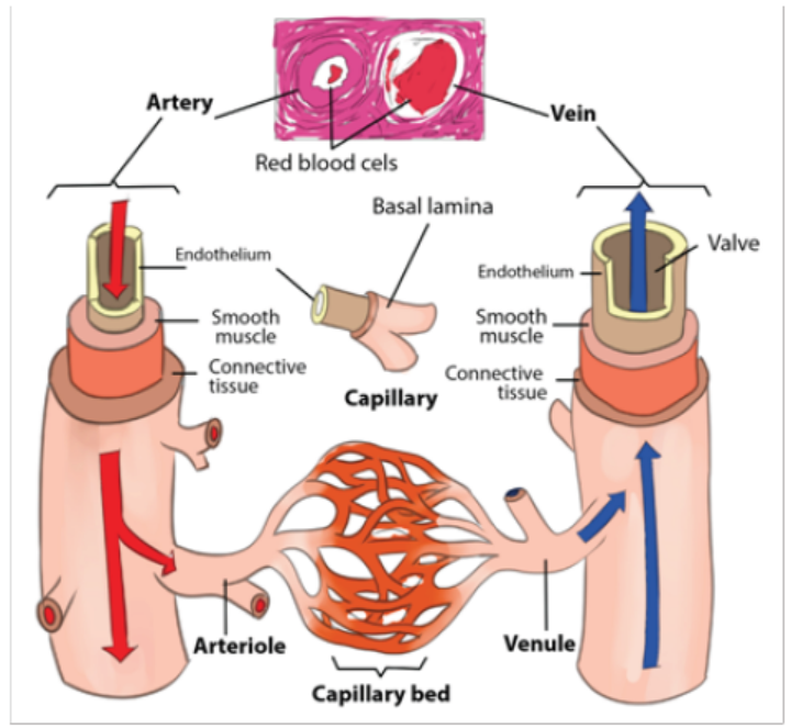
Figure 4.11: Illustration of blood vessels.
Table 4.3. A comparison between arteries, capillaries and veins.
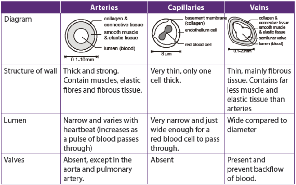
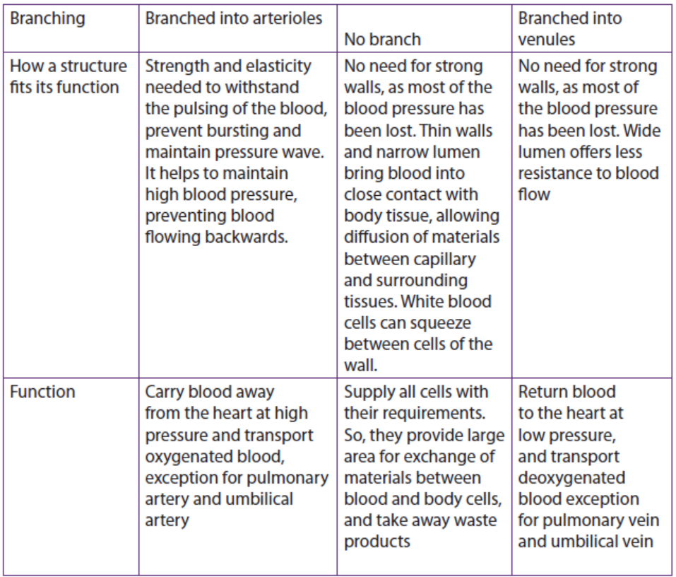
Application 4.5
1. Associate the following vessels with their functions
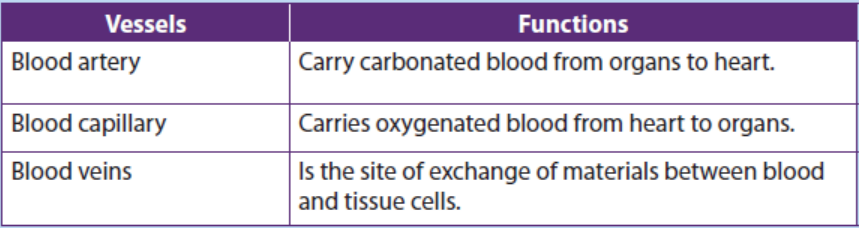
2. Explain how each blood vessel is adapted to its function.
4.6 Body fluids, composition and functions
Activity 4.6
1. List the main body fluids.2. Look at the figure below and answer the questions that follow.
a. Identify the blood components represented by the letters A, B, C, D, E, F, G,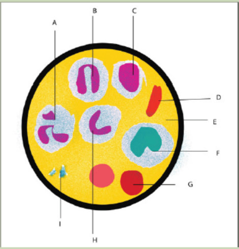
H, I.
b. Suggest the functions of each of those blood components.c. State the origin of each blood component.
4.6.1. Main types of body fluids and their compositions
Body fluids are liquids originating from inside the body of living humans. The main
body fluids are; blood, plasma, serum, tissue fluid and lymph which are describedbelow in the table 4.4.
Table 4.4. Body fluids and their composition
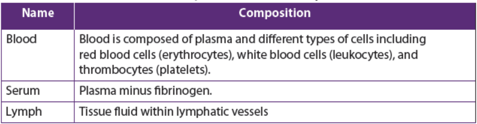
4.6.2. Composition and functions of blood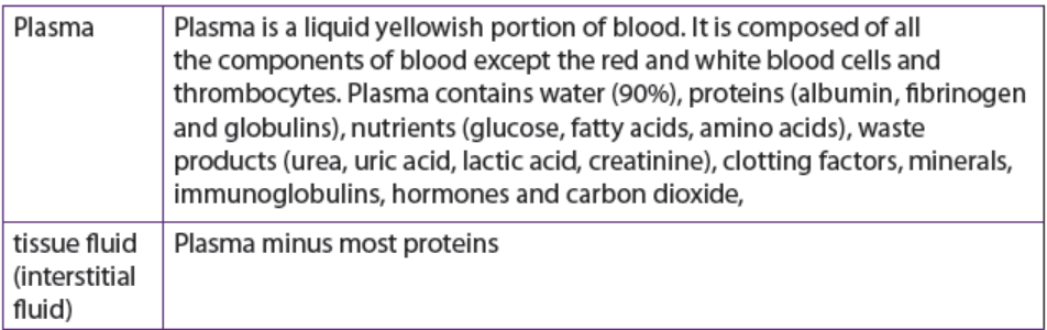
The main blood components are formed elements and plasma. Formed elements
are erythrocytes (red blood cells), leukocytes (white blood cells) and thrombocytes(platelets).
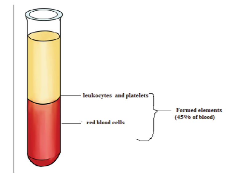
Figure 4.12: Blood sample in a test tube.
a. Erythrocytes
Erythrocytes also called red blood cells, their core function is to carry oxygen from
the respiratory organs to tissues and their structure are well modified accordingly to
perform the purpose. There are five million per cubic millimetre each having about
8 μm in diameter and 3 μm thick in widest part. The cell has red pigment calledHaemoglobin a complex protein c
ontaining four iron haem groups.
b. Leukocytes
Leukocytes (white blood cells) are involved in immune system that fights against
infections. . white blood cells are responsible for destroying infectious agents and
infected cells, and secrete protective substances such as antibodies, which fight
infections. Leukocytes are divided into:
– Granulocytes or polymorph nuclear cells. They are neutrophils, basophils
eosinophils. They take the name from the possession of numerous granules in
their cytoplasm.
– Agranulocytes or monomorphonuclear cells: They are lymphocytes andmonocytes. They lack granules in the cytoplasm.
Thrombocytes
Thrombocytes are also called platelets, are small cell fragments with 2-3 mm in
diameter. They are formed from cytoplasm of large cells (mega karyotypes. Normal
quantitative value is between 250,000 and 450,000 platelets per mm³. They help inblood clotting. A comparison between formed elements is summarized in the table
4.5 below.
Table 4.5: Blood composition
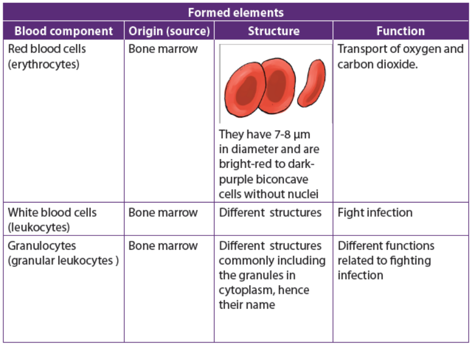
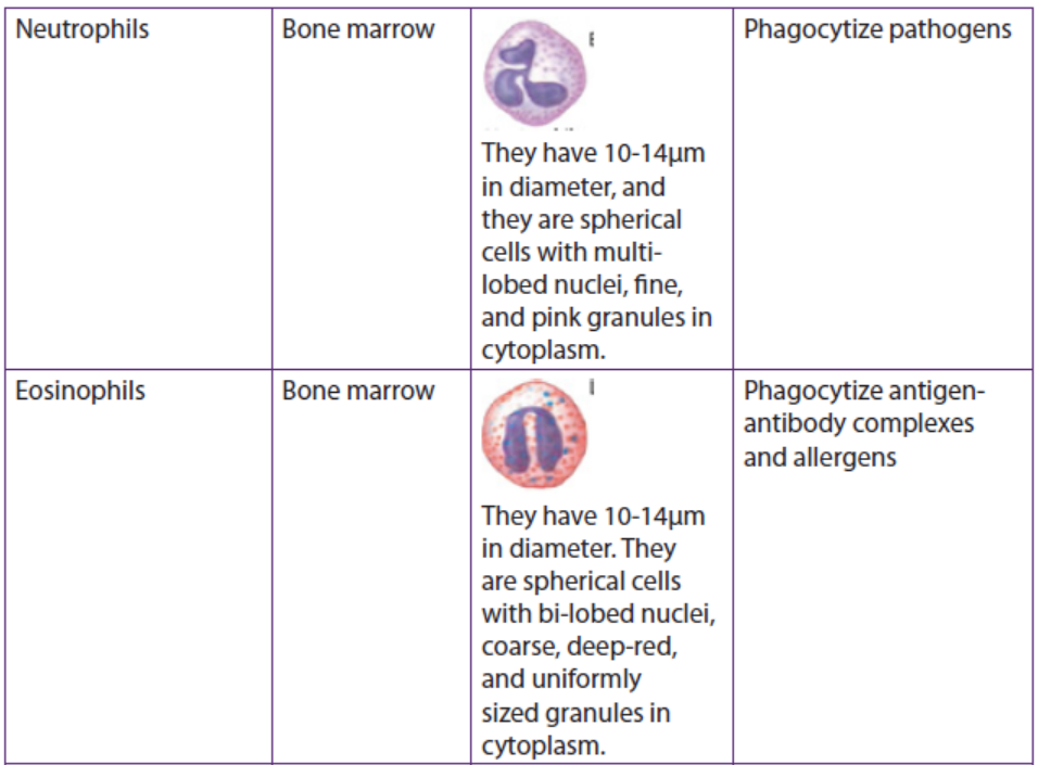
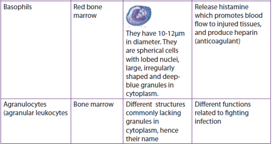
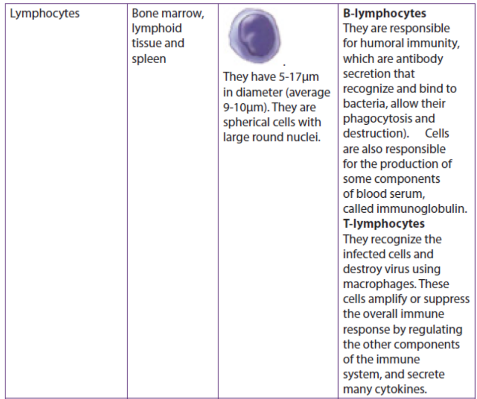
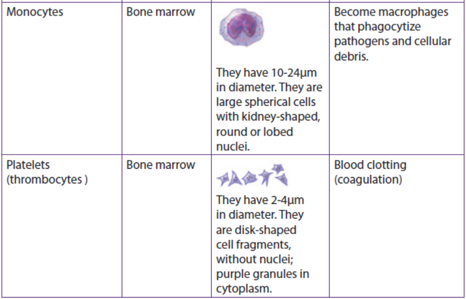
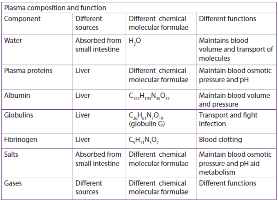
Application 4.6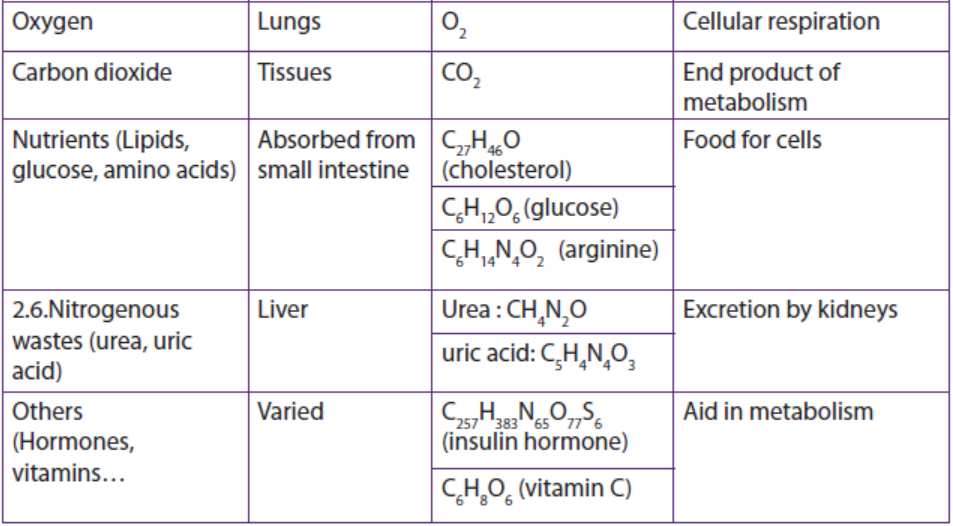
1. Discuss the functions of:
a. Macrophage.
b. T-lymphocytes.
c. Erythrocytes2. Explain the relationship between blood and tissue fluid.
4.7 Transport of respiratory gases
Activity 4.7
Refer to unit 8 in S4 to answer the following questions:
1. What protein is responsible for the transport of oxygen in human blood?
Describe it.
2. Explain how that protein behaves when blood reaches the alveoli in thelungs and when blood reaches active muscle cells.
a. Structure of haemoglobin of red blood cells.
Haemoglobin is a red protein responsible for transporting oxygen in the blood of
vertebrates. It is also involved in the transport of carbon dioxide. Haemoglobin is
composed of haem and globin (polypeptide chains). Haem is an iron porphyrin
compound. Iron occupies the centre of the porphyrin ring and establishes linkages
with all the four nitrogen of all the pyrrole rings.
Globin part is made of four polypeptide chains, two identical α-chains and two
identical β-chains in normal adult haemoglobin. Each chain contains a “haem” in the
so called ‘haem pocket’ and one haemoglobin molecule possess four haem units.
Haem pockets of α-subunits are of just adequate size to give entry to an O2 molecule.Entry of O2 into haem pockets of β-subunits is blocked by a valine residue.
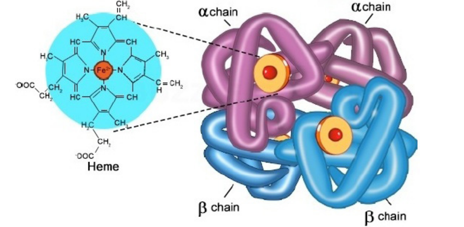
Figure 4.13: Structure of haemoglobin.
b. Transport of carbon dioxide (CO2)
At systemic capillaries in the body cells, CO2 enters red blood cells. Some CO2combines with Hb to form HbCO2 (Carbaminohaemoglobin):
I.e. Hb + CO2 →HbCO2 (Carbaminohaemoglobin)
Most CO2 is converted to HCO3- (bicarbonate ion), which is carried in the plasma.
Haemoglobin is in relation with chloride shift. It is a process which occurs in
a cardiovascular system and refers to the exchange of bicarbonate (HCO3−)
and chloride (Cl−) across the membrane of red blood cells (RBCs). The chloride shiftoccurs in this way:
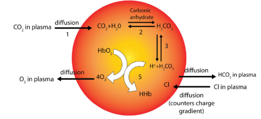
Figure 4.14: Chloride shift and transport of carbon dioxide by haemoglobin erythrocyte.
: H + Hb is reduced haemoglobin which is haemoglobin combined with hydrogenion (H+).
c. Transport of oxygen
Haemoglobin gets oxygen in lungs from external environment to form a compound
called oxyhaemoglobin (HbO8). , In this form, oxygen is transported to the body cellsto sites where it is needed for aerobic respiration.
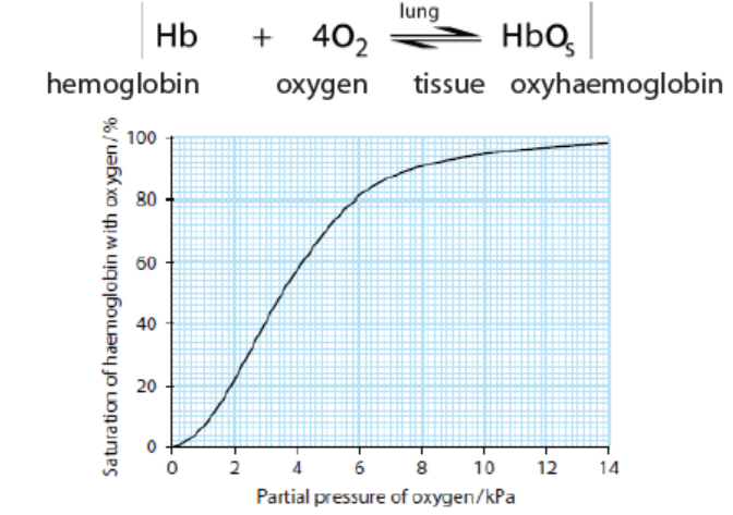
Figure 4.15: Oxygen dissociation curve
The curve above in figure 4.15 shows the oxygen dissociation curve by haemoglobin.
Oxygen dissociation curves determined by plotting the partial pressure of
oxygen in blood against the percentage of haemoglobin combined with
oxygen in the form of ox haemoglobin. The S-shape of the oxygen dissociation
curve can be explained by the behaviour of a haemoglobin molecule as it combines
with or loses oxygen molecules. When an oxygen molecule combines with one haem
group, the whole haemoglobin molecule is slightly distorted. The distortion makes
it easier for a second and third oxygen molecules to combine the haem groups. It is
then still easier for the fourth and final oxygen molecule to combine.
If all the oxygen binding sites contain oxygen, then the oxygen saturation is
100%. Oxygen saturation is defined as the ratio of oxyhaemoglobin to the total
concentration of haemoglobin present in the blood The Bohr Effect is a physiological
phenomenon in which a raise of carbon dioxide in the blood and a decrease in pH
results in a reduction of the affinity of haemoglobin for oxygen. This causes the
oxygen dissociation curve for haemoglobin to shift to the right. The Bohr Effectoccurs in this way:
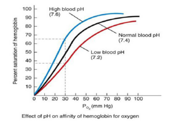
Figure 4.16: Bohr effect curve (Adapted from brainscape.com)
Application 4.7
1. Explain the importance of hemoglobin to a human being.
2. In a healthy adult human, the amount of haemoglobin in 1 dm3 of
blood is about 150 g. Given that 1 g of pure haemoglobin can combine
with 1.3 cm3 of oxygen at body temperature, how much oxygen can becarried in 1 dm3 of blood?
4.8 Blood clotting and common cardiovascular diseases
Activity 4.8
Warning to medical staff!
Doctor NINA called upon all medical staff and warned them about three major
causes of death in the theater. She said into the: “Please, pay attention to
hemophilic people though they are rare in Rwanda. Embolus and thrombosis
are now reported from time to time. Beware!”
1. Differentiate between:
a. a hemophilic and non-hemophilic person
b. embolus and thrombosis
2. why Dr Nina warns medical practitioners about above cases:3. Explain the mechanism of blood clotting.
a. Blood clotting
Blood clotting also known as blood coagulation is the process by which blood becomes
thick and stops flowing, forming a solid cover over any place where the skin has
been cut or broken. Blood that has been converted from a liquid to a solid state
is called blood clot. A blood clot called thrombus is stationary within a vessel or
the heart. If a blood clot moves from that location through the bloodstream, it isreferred to as an embolus.
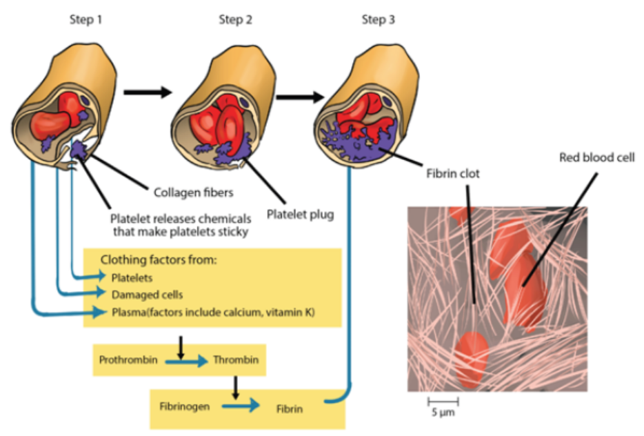
Figure 4.17: Illustration of blood clotting process
Blood clotting is a series of different processes:
Step 1: The blood coagulation process begins when the endothelium of a vessel is
damaged, exposing the connective in the vessel wall to blood. Platelets adhere to
collagen fibres in the connective tissue and release a substance that makes nearby
platelets sticky.
Step 2: The thrombocytes form a plug that provides emergency protection against
blood loss.
Step 3: This seal is reinforced by a clot of fibrin when vessel damage is severe.
Fibrin is formed via a multistep process where clotting factors released from the
clumped platelets or damaged cells mix with clotting factors in the plasma, forming
an activation that converts a plasma protein called prothrombin to its active form,
called thrombin. This is facilitated by calcium ions and vitamin K. Thrombin itself is
an enzyme that catalyses the final step of the clotting process. This final step is the
conversion of fibrinogen to fibrin. The threads of fibrin become interwoven into a
patch. And the blood clot is formed. These threads trap red blood cells and otherblood components, preventing the continuous bleeding.
b. Common cardiovascular diseases
1. Stroke
Stroke is a cardiovascular disease due to the lack of oxygen to the brain which may
lead to reversible or irreversible paralysis. The damage to a group of nerve cells in
the brain is often due to interrupted blood flow, caused by a blood clot or blood
vessel bursting. Since atherosclerosis is a body wide process, similar events can also
occur in the arteries to other parts of the body, including the brain. A stroke is a loss
of brain function due to a stoppage of the blood supply to the brain. It can be caused
by a stationary blood clot known as thrombus, a free-floating clot moving blood
clot or embolus that gets caught in a blood vessel, or by bleeding (haemorrhage).
Hypertension or high blood pressure promotes atherosclerosis and increases the
risk of heart attack and stroke.
2. Atherosclerosis
Atherosclerosis is a cardiovascular disease characterized by the progressive narrowing
and hardening of the arteries over time. Atherosclerosis normally begins in later
childhood, and is usually found in most major arteries. It does not usually have any early
symptoms. Causes of atherosclerosis include a high-fat diet, high cholesterol, smoking,
obesity, and diabetes. Atherosclerosis becomes a threat to health when the plaque
build-up interferes with the blood circulation in the heart known as coronary circulation
or the brain known as cerebral circulation. A blockage in the coronary circulation, canlead to a heart attack, and blockage of the cerebral circulation can lead to a stroke.
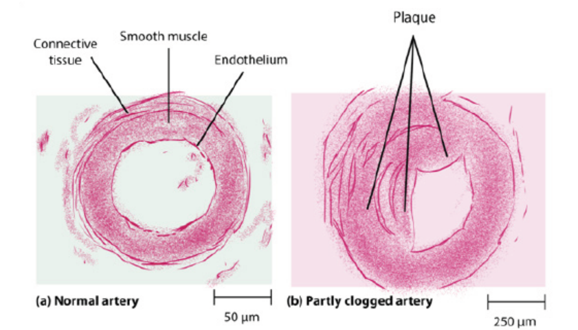
Figure 4.18: Plaque formation in blood vessels
3. Coronary heart disease
Coronary heart disease (CHD) is a disease in which a waxy substance called plaque
builds up inside the coronary arteries. Cardiac muscle cells are fed by the coronary
arteries. Blocked flow in a coronary artery can result in oxygen starvation and death
of heart muscle. Most individuals with coronary heart disease have no symptoms for
many years until the first sign, often a heart attack, happens.
c. Risk factors associated with cardiovascular diseases
There are several risk factors for heart disease. Some of those factors are controllable
and others are uncontrolled. Uncontrollable factors include the gender (males are
at greater risk), age (old people have higher risk), and family history in relation to
heart diseases as well post-menopausal stages for females. Making some changes in
lifestyle can reduce chance of having heart disease. Controllable risk factors includesmoking, high blood pressure, physical inactivity, obesity, diabetes, stress and anger
Application 4.8
1. State the role of fibrinogen, calcium and thrombin in blood clotting.
2. Explain the cause and effects of stroke.
3. Describe the impact of smoking on the cardiovascular system.
4. Discuss the effects of high consumption of lipids such as fats and oilson the body.
4.9 Lymphatic system
Activity 4.9
1. Define the following terms:
a. Lymph
b. Lymph nodes
c. Lymphatic vessels
2. Describe the function of lymphatic system.
3. Explain how the tissue fluid and lymph are formed.
4. Suggest any 2 similarities and 2 differences between a circulatorysystem and a lymphatic system.
4.9.1 Structure of a lymphatic system
A lymphatic system is a system composed of tissues and organs, including; bone
marrow, spleen, thymus, and lymph nodes that produce and store cells that fight
infection and disease. The channels that carry lymph are also part of this system.So, the lymphatic system is part of the circulatory system and an important part of
the immune system.
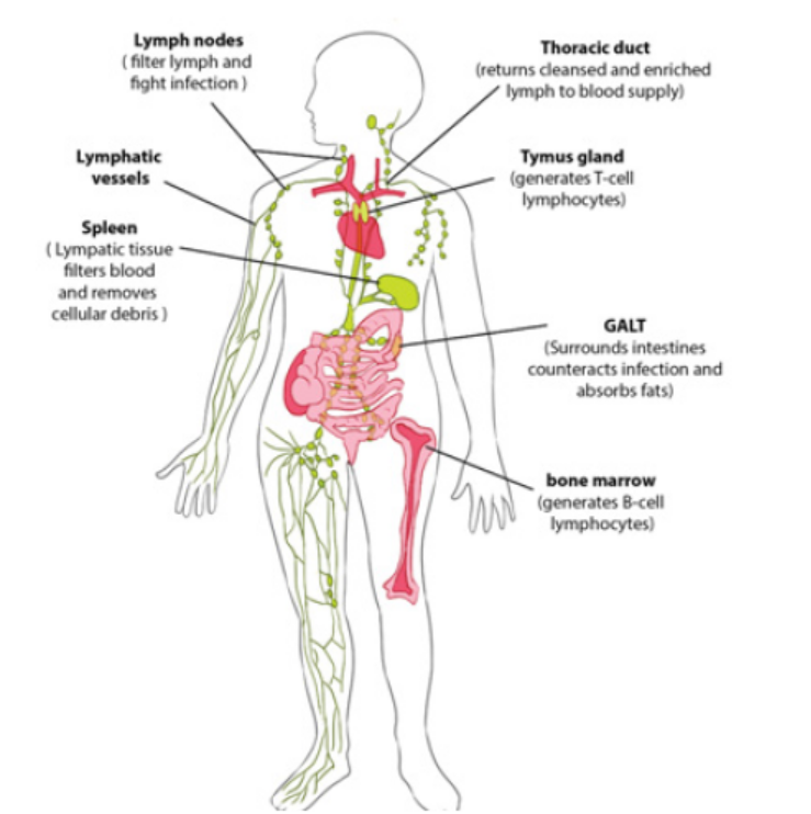
Figure 4.19: Structure of human lymphatic system.
4.9.2 Functions of a lymphatic system
– Drainage of fluid from blood stream into the tissues: The circulating blood
through narrow vessels leads to leakage of fluid or plasma into the tissues
carrying oxygen and nutrients to the tissues and taking waste materials from
the tissues into the lymph channels. The leaked fluid drains into the lymph
vessels.
– Filtration of the lymph at the lymph nodes: The nodes contain white blood
cells that can attack any bacteria or viruses they find in the lymph as it flows
through the lymph nodes.
– Filtering blood: This is done by the spleen which filters out bacteria, viruses
and other foreign particles.
– Raise an immune reaction and fight infections: The lymphatic system
especially the lymph nodes are over active in case of an infection the lymph
nodes or glands often swell up in case of a local infection in so doing, thelymphocytes fight the foreign bodies trapped in the lymph nodes.
4.9.3 Formation of tissue (interstitial) fluid
Fluids and some soluble proteins leak from the blood capillaries into the interstitial
fluid that bathes the cells of tissues. This occurs due to the arterial end of capillary,
where the blood pressure is greater than osmotic pressure so that fluid flows out
of capillary into the interstitial fluid. This process is called pressure filtration or
ultrafiltration
4.9.4 Formation of lymph
The lymph is the tissue fluid that moves within the lymphatic vessels. The lymphatic
vessels recover some leaked fluid and proteins, and carry them to large veins at thebase of the neck (figure 4.20).
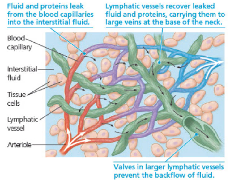
Figure 4.20: The close association of lymphatic vessels and blood capillaries.
4.9.5 Comparison between lymphatic and circulatory systems
Both the cardiovascular and lymphatic systems are vascular networks carrying bodyfluids. Differences and similarities are summarized in the table 4.6.
Table 4.6. Differences between lymphatic and circulatory system
Application 4.9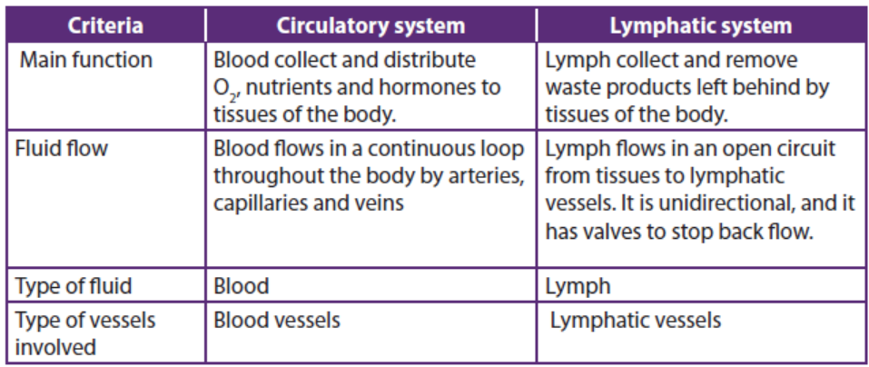
Observe the figure below and respond to the following questions.
a. Identify the organs W, X, Y, Z shown on this figure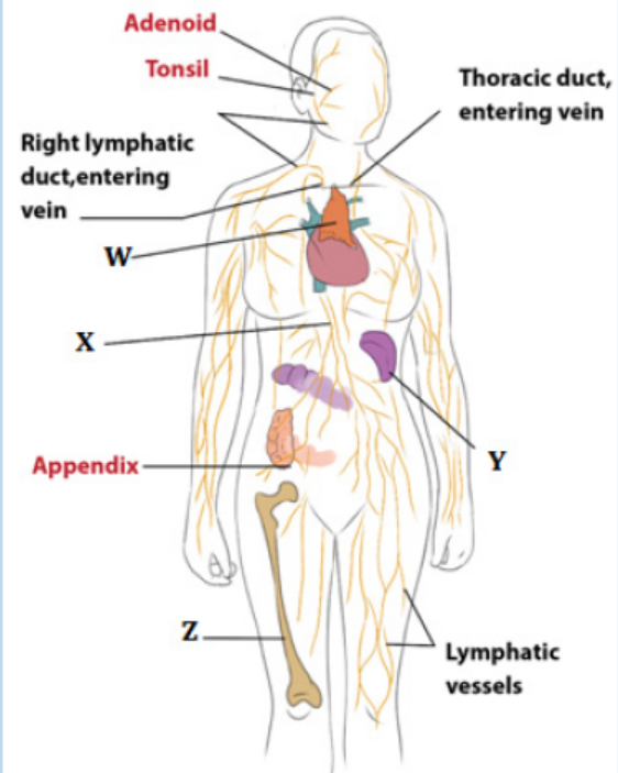
b. Describe the functions of the organs W, X, Y, Z.
End of unit assessment 4
1. Blood returning to the mammalian heart in a pulmonary vein drains first into
the:
a. Vena cava
b. Left ventricle
c. Right ventricle
d. Left atrium
2. Pulse is a direct measure of:
a. Blood pressure.
b. Breathing rate.
c. Cardiac output
d. Heart rate.
e. Stroke volume
3. Complete the following paragraph by filling in the blank spaces.
Blood is ………………in the lungs. The red pigment ………………has a high
affinity for oxygen. The pumping action of the……………creates pressure
which pushes the blood around the body. In the tissues the partial pressure
of…………….is low. This causes the ………………of the oxyhaemoglobin. In
the tissues, the oxygen is used in the process of……………………. Most of the
carbon dioxide produced in this process enters the……………. cells. Here it is
converted to carbonic acid by the action of the enzyme carbonic anhydrase.
The carbon dioxide is transported as ………………. back to the lungs
4. How many oxygen molecules can each haemoglobin molecule transport?
5. Explain the function of fibrinogen.
6. Distinguish between plasma and serum.
7. a) Explain why haemoglobin is called conjugated protein.
b) Describe the effect of high carbon dioxide concentrations on the
oxygen dissociation curve of haemoglobin.
8. a) By which process does fluid leave the blood and enter the tissue fluid?b) Which component of the blood does not enter the tissue fluid?
9. The figure below shows a cross section through the human heart
a. Label the structure A-E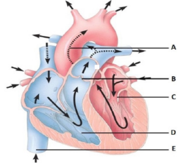
b. What are the functions of the structures A and B
10. Why is it important that the AV node delay the electrical impulse moving from
the SA node and the atria to the ventricles?
11. Draw a pair of simple diagrams comparing the essential features of single and
double circulation.
12. The figure below shows pressure changes to the left side of the heart and theaorta during the cardiac cycle.
a. State what is happening at point A-D on the graph. Explain your answer.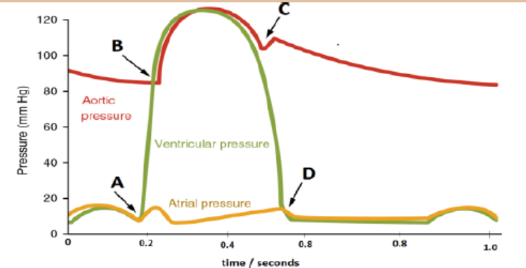
b. If the time taken for one complete cardiac cycle is 0.8 seconds, how
many cardiac cycles are there in one minute?
13. Explain any two advantages of closed double circulatory system and two
disadvantages of open circulatory system
14. a) Where is the radial pulse taken?
b) Suggest what will happen to the heart rate if the vagus nerve is cut off.
15. The diagram shows a vertical section through a human heart. The arrows
represent the direction of movement of the electrical activity which starts
muscle contraction Carefully, observe the following and answer the questionsthat follow.
a. Name the structure denoted by the letter A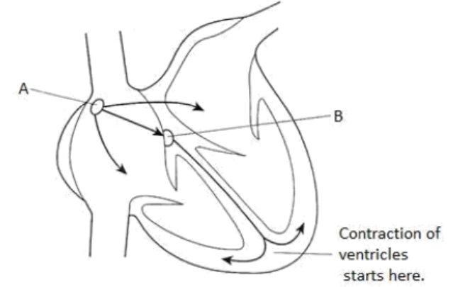
b. Explain why each of the following is important in then pumping of blood
through the heart.
i. There is a slight delay in the passage of electrical activity that takes place
at the point A
ii. The contraction of the ventricles starts at the base
c. Describe how stimulation of the cardiovascular centre in the medulla mayresult in an increase in heart rate
16. Read the following passage and answer the questions that follow
The human heart is a double pump adapted to forcing blood, at the same rate but
at different pressures, along the two systems of double circulation. High pressure
in the systemic circulation has evolved with lower pressure in the pulmonary
circulation and low pressure lymphatic circulation. Each heart beat is controlled
by a wave of electrical excitation. In turn, the cardiac output of the heart adapts
to meet the body needs and is influenced by nervous and hormonal control.
a. Based on the statement: “The human heart is a double pump adapted to
forcing blood, at the same rate but at different pressures, along the two
systems of double circulation”. Explain how the mechanism that controls
each heartbeat, and the structure of the heart, enable it to do this.
b. Describe the role played by hormones and the nervous system in
controlling heart rate.c. Describe the formation of lymph fluid.
UNIT 5 ENERGY FROM RESPIRATION
UNIT 5: ENERGY FROM RESPIRATION
Key Unity Competence
Describe the structure and importance of ATP, outline the roles of the coenzymes
NAD, FAD and coenzyme A during cellular respiration.
Learning objectives
– Discuss the need for energy in living organisms as illustrated by anabolic
reactions, active transport, and the movement and maintenance of body
temperature.
– Describe the structure of ATP as a phosphorylated nucleotide formed by
condensation reaction.
– Explain that ATP is synthesized in substrate-linked reactions in glycolysis and in
Krebs (tri-carboxylic acid [TCA] cycle.
– Explain the relative energy value of carbohydrate, lipid and protein as respiratory
substrate and explain why lipids are particularly energy-rich.
– Define the term Respiratory Quotient (RQ) as the ratio of the volume of CO2,
evolved to the volume of O2 uptake during aerobic respiration.
– Design simple experiments using respirometers to determine the RQ of
germinating seeds or small invertebrates. Example: woodlice.
– Calculate RQ values from the equations of respiration of different substrates.– Interpret graphs for varying RQ values during seed germination.
Introductory activity
From your daily experience, brainstorm the following questions.
1. What do you understand about energy used by living organisms?
2. Where is that energy obtained from?3. How is that energy obtained from the source you have mentioned?
This unit deals with the energy from respiration. It focuses on the description of the
structure and importance of adenosine triphosphate (ATP), and outline the roles of
the coenzymes including nicotinamide adenine dinucleotide (NAD), flavin adenine
dinucleotide (FAD) and coenzyme A CoA during cellular respiration. Specifically,
this unit contributes to a better understanding of the reasons why organisms need
energy, the structure of adenosine triphosphate (ATP), synthesis and breakdown of
ATP, respiratory substrates and their relative energy values, and measurement ofrespiration and respiratory quotient.
5.1 Need for energy by organisms
Activity 5.1
Use books from the school library and search further information about
metabolism reactions on the internet. Read the information and discuss thereasons why all living organisms need energy.
Chemical energy is the most important type of energy potential for life, where energy
is either released out or consumed through metabolism reactions. Metabolism
reactions constitute the sum of all chemical reactions taking place in a living cell.
The biological process by which metabolic pathways breakdown molecules into
smaller units that are either oxidized to release energy is called catabolism, while
the biological process by which a set of metabolic pathways construct molecules
from smaller units through reactions consuming energy is called anabolism. During
catabolism reactions, energy is released to the surrounding environments. These
are exergonic reactions. During anabolism reactions, energy is absorbed from thesurrounding environment. These are endergonic reactions.
All living organisms need energy to grow and reproduce, maintain their structures,
and respond to their environments. Metabolism reactions are the set of life-sustaining
chemical processes that enables organisms to transform the chemical energy stored
in molecules into energy that can be used for cellular processes. Animals consume
food to replenish energy. Their metabolism breaks down the carbohydrates, lipids,
proteins, and nucleic acids to provide chemical energy for these processes. Plants
convert light energy from the sun into chemical energy stored in molecules during
the process of photosynthesis.
Active transport of solutes such as sodium (Na+), potassium (K+) magnesium (Mg+),
calcium (Ca+) and chloride (Cl-)across the plasma membrane cannot be possible
without the use of energy. The transport proteins that move solutes against their
concentration gradients are all carrier proteins rather than channel proteins.
Active transport enables a cell to maintain internal concentrations of small solutes
that differ from concentrations in its environment. Some transport proteins act
as pumps, moving substances across a membrane against their concentration or
electrochemical gradients. Energy is usually supplied by adenosine triphosphate(ATP) hydrolysis (Figure 1).
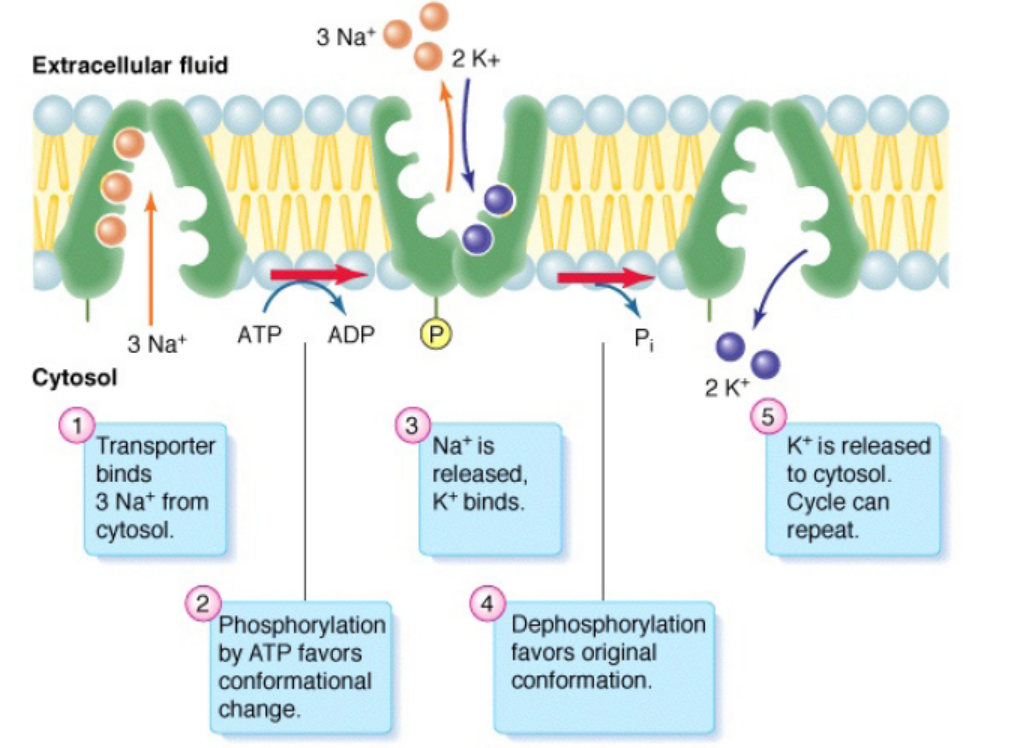
Figure 5.1: Active transport of chemical ions/anions across the cell membrane
Application 5.1
1. What is energy?
2. What is it used for?
3. What is the major source of energy for organisms?
4. What would happen to all living organisms if sunlight energy is not
available?
5. Discuss the reasons why living things need to always take food?6. Is photosynthesis an anabolic or catabolic process? Explain your answer
5.2 Structure of Adenosine Triphosphate and its importance
Activity 5.2
ATP can be describes as a nucleotide made of Ribose as pentose sugar, Adenine
as nitrogenous base and 3 phosphate groups linked by phosphodiesteric
bond.Find out the structure of ATP.
The special carrier of energy is the molecule of adenosine triphosphate (ATP). The
building blocks of ATP are carbon, nitrogen, hydrogen, oxygen, and phosphorus,
contained in the ribose sugar, a nitrogen base called adenine and a chain ofphosphate group (Figure 5.2).
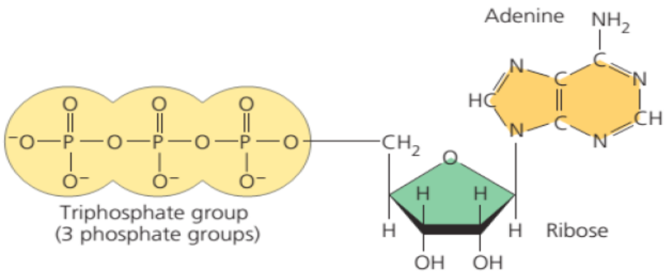
Figure 5.2: Structure of Adenosine Triphosphate (ATP
ATP has the following biological functions in the cell:
a. Active transport
ATP plays a critical role in the transport of macromolecules such as proteins and
lipids into and out of the cell membrane. It provides the required energy for active
transport mechanisms to carry such molecules against a concentration gradient.
b. Cell signaling
ATP has key functions of both intracellular and extracellular signaling. In nervous
system, adenosine triphosphate modulates the neural development, the control of
immune systems, and of neuron signaling.
c. Structural maintenance
ATP plays a very important role in preserving the structure of the cell by helping the
assembly of the cytoskeletal elements. It also supplies energy to the flagella andchromosomes to maintain their appropriate functioning.
d. Muscle contraction
ATP is critical for the contraction of muscles. It binds to myosin to provide energy
and facilitate its binding to actin to form a cross-bridge. Adenosine diphosphate
(ADP) and phosphate group (Pi) are then released and a new ATP molecule binds to
myosin. This breaks the cross-bridge between myosin and actin filaments, thereby
releasing myosin for the next contraction.
e. Synthesis of DNA and RNA
The adenosine from ATP is a building block of RNA and is directly added to RNA
molecules during RNA synthesis by RNA polymerases. The removal of pyrophosphateprovides the energy required for this reaction. It is also a component of DNA.
Application 5.2
1. Energy is contained within ATP. Explain to someone who doesn’t
have any knowledge about ATP how this biochemical compound is
important to all living organisms.2. Observe the figure and answer the following questions:
a. What does it represent?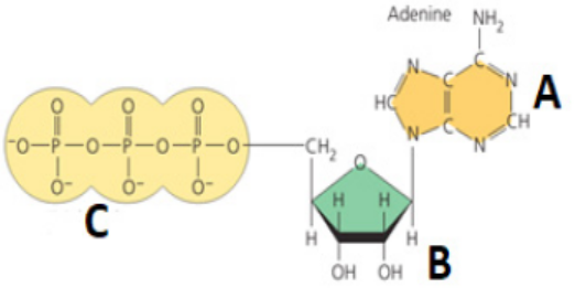
b. Give the names of the parts denoted by the letters A, B and C.
c. What might happen to a living organism if the above molecules are not
present?5.3 Synthesis and breakdown of ATP
Activity 5.3
Use books from the school library and search further information on the
internet about ATP. Read the information and discuss the synthesis andbreakdown of ATP.
Adenosine triphosphate (ATP) is the energy currency for cellular processes. It provides
the energy for both energy-consuming endergonic reactions and energy-releasing
exergonic reactions. When the chemical bonds within the phosphate group of ATP
are broken, energy is released and can be harnessed for cellular work.
a. Synthesis and hydrolysis of ATP
ATP is hydrolysed into Adenosine Diphosphate (ADP) and inorganic phosphate (Pi)in the following reaction:
ATP+H2O→ADP+Pi+free energy,
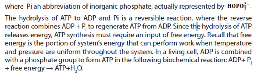
Figure 5.3: The hydrolysis of ATP: The reaction of ATP and water yields ADP and inorganic phosphate Pi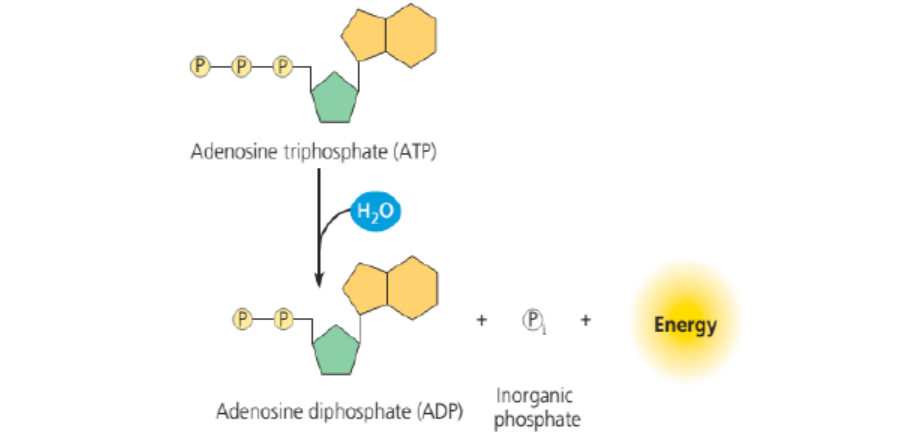
and release energy.
b. ATP and energy coupling
Now that the synthesis and breakdown of ATP is understood, the remaining
interesting question is to know exactly how much free energy denoted ΔG is
released with the hydrolysis of one mole of ATP, and how is that free energy used
to do cellular work. The calculated ΔG for the hydrolysis of one mole of ATP into
ADP and Pi is estimated at −7.3 kcal/mole equivalent to −30.5 kJ/mol. However, thisis only true under standard conditions, and the ΔG for the hydrolysis of one mole
of ATP in a living cell is almost double the value at standard conditions and equals
-14 kcal/mol or −57 kJ/mol. ATP is a highly unstable molecule. Unless quickly used
to perform work, ATP spontaneously dissociates into ADP + Pi, and the free energy
released during this process is lost as heat. To harness the energy within the bondsof ATP, cells use a strategy called energy coupling.
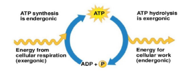
Figure 5.4: Summative processes between synthesis and hydrolysis of ATP
Application 5.3
1. Based on chemical equations explain the synthesis and the hydrolysis
of ATP in a living cell.
2. The hydrolysis and synthesis of ATP are reversible reactions. Estimate
the amount of energy for each process.
3. Calculate the amount of energy produced by 5 moles of ATP
a. Under standard conditions
b. In a living cell
4. Explain what might happen if the reaction of hydrolysis of ATP is not
reversible.5.4 Respiratory substrates and their relative energy values
Activity 5.4
Use books from the school library and search further information on respiration.
Read the information and discuss the respiratory substrates and their relative
energy values.
1. What do you understand by a respiratory substrate?
2. Give any 2 examples of respiratory substrate.
3. What is the relationship between respiratory substrate and energy
values?
A respiratory substrate refers to the substance required for cellular respiration to
derive energy through oxidation. They include carbohydrates, lipids and proteins.
Carbohydrates include any of the group of organic compounds consisting of
carbon, hydrogen and oxygen, usually in the ratio 1:2:1. Hence the general formula
of carbohydrates is . The examples of carbohydrates include sugars, starch and
cellulose. Carbohydrates are the most abundant of all classes of biomolecules,
and glucose whose chemical formula is C6H12O6 is the most known and the most
abundant. Its breakdown produces energy in the following way:
C6H12O6 +6 O2→6 CO2 +6 H2O+Energy (ATP + heat).
This breakdown is exergonic metabolic reaction, having a free-energy change of
-686 kcal (2,870 kJ) per mole of glucose decomposed.
Lipids include diverse group of compounds which are insoluble in water but
dissolved readily in other lipids and in organic solvents such as ethanol (alcohol).
Lipids mainly fats and oils contain carbon, hydrogen and oxygen, though the
proportion of oxygen is lower than in carbohydrates. Fats and oils have a higher
proportion of hydrogen than either carbohydrates or proteins. This property makes
them a more concentrated source of energy, where each gram of fat or oil yields
about 38kJ (38 kJ/g) more than twice the energy yield of a gram of carbohydrate.
Proteins are other respiratory substrate. They are large and complex biological
molecules which play many and diverse roles during respiration. They mainly work
as enzymes. Enzyme is a biological catalyst that controls biochemical reactions in
Back to glucose when it is broken down during the process called glycolysis, the
dehydrogenases enzymes transfer electrons from substrates, here glucose, to
NAD+ which in turn forms NADH. At this stage the electron transport chain accepts
electrons from NADH and passes these electrons from one molecule to another in
electron chain transfer leading to a controlled release of energy for the synthesis of
ATP. At the end of the chain, the electrons are combined with molecular oxygen and
hydrogen ions (H+) to form one molecule of water. (Figure 5). When NAD is oxidized,
its oxidized form NAD+ is converted into its reduced from NADH, and two moleculesof ATP are produced.
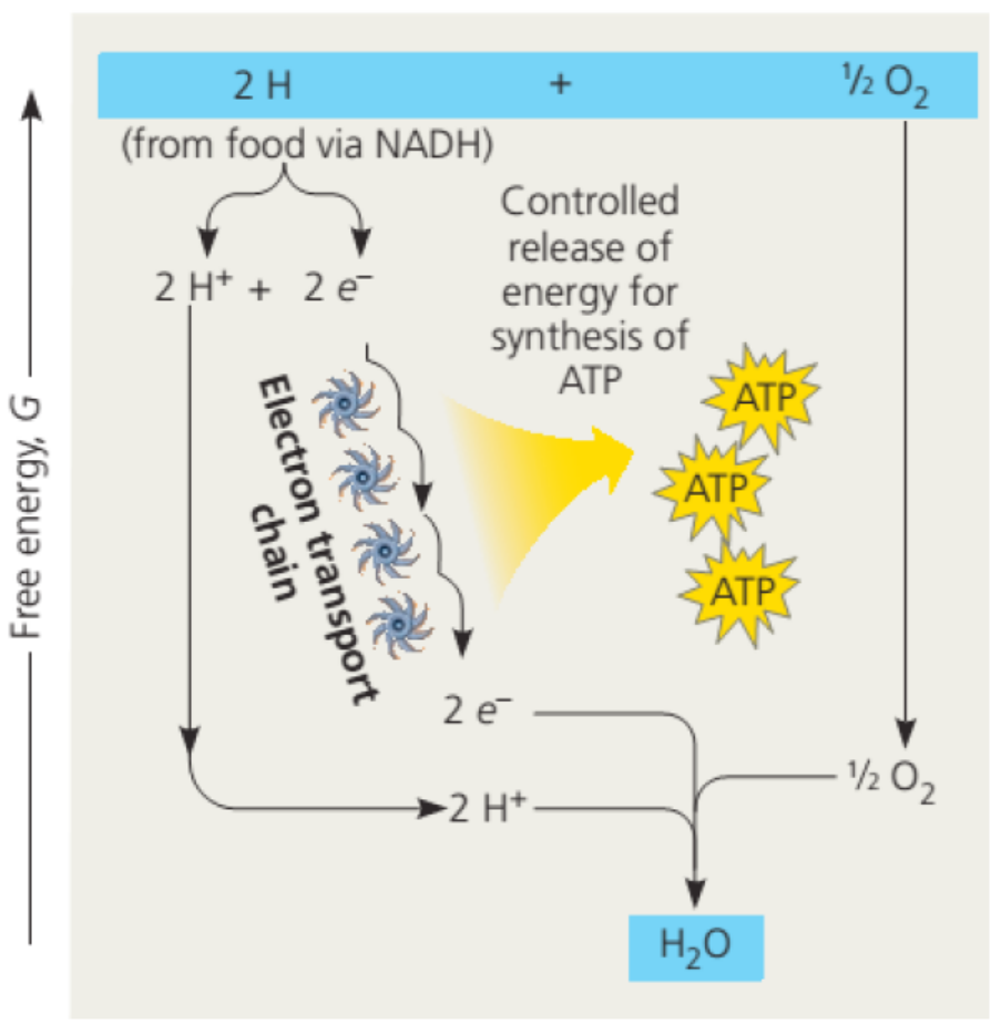
Figure 5.5: Electron transport chain from food to the formation of water
The transformation of succinate to fumarate, the sub-products of the breakdown of
glucose during glycolysis process, two hydrogens are transferred to flavin adenine
dinucleotide (FAD), forming FADH2. The reduced coenzymes NADH and FADH2
transfer higher energy electrons to the electron transport chain. Finally, another
coenzyme called coenzyme A sometimes abbreviated by CoA, a sulfur-containing
compound is attached via its sulfur atom to the two-carbon intermediate, forming
acetyl CoA. The Acetyl CoA has a high potential energy, which is used to transfer the
acetyl group to a molecule in the citric acid cycle, a reaction that is therefore highlyexergonic producing great number of energy in the form of ATP.
Application 5.4
1. What is the oxidizing agent in the following reaction?
Pyruvate + NADH + H+→ Lactate+NAD+ oxygen
a. NADH
b. Lactate
c. pyruvate
2. When electrons flow along the electron transport chains of
mitochondria, which of the following changes occurs?
a. The pH of the matrix increases.
b. ATP synthase pumps protons by active transport.
c. The electrons gain free energy.
d. NAD+ is oxidized.
3. Most CO2 from catabolism is released during which stage?
a. Glycolysis.
b. Electron transport.
4. Give the chemical equation summarizing the decomposition of glucose
and specify the amount of energy produced in kJ.
5. Calculate the amount of energy produced by moles of glucose in kcal
and kJ if one mole of glucose produce -686 kcal and 2,870 kJ per mole
of glucose.
6. Differentiate between NAD+ and NADH2? . How are they related to FAD
and FDH2?
7. Specify the number of ATP produced by glycolysis during respiration
process.5.5 Measurement of respiration and respiratory quotient
Activity 5.5
Use books from the school library and search further information on
respiration. Read the information and discuss the measurement of respiration
and respiratory quotient.
1. What do you understand by respiratory quotient?
2. Draw a well labelled figure indicating the structure of a respirometer
and specify its role in biological studies.
3. Explain how the respiratory coefficient can be calculated fromconsumed oxygen and released carbon dioxide during respiration.
The rate of respiration is measured by the use of respirometer device, typically by
measuring oxygen consumed and the carbon dioxide given out. It can also be used
to measure the depth and frequency of breathing, and allows the investigation on
how factors such as; age, or chemicals can affect the rate of respiration. Currently,
the computer technology is also used to automatically measure the volume of gases
exchanged and drawing off small samples to analyse the proportions of oxygen andcarbon dioxide in the gases.
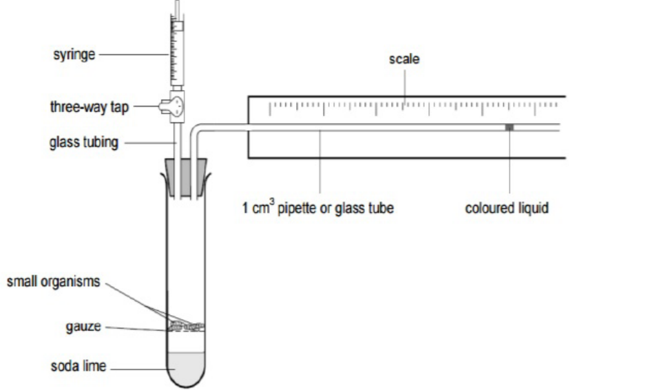
Figure 5.6: Respirometer
The respiratory quotient (RQ) is the ratio of the volume of carbon dioxide produced
to the volume of oxygen used in respiration during the same period of time. The
RQ is often assumed to equal the ratio of carbon dioxide expired: oxygen inspiredduring a given time as it is summarized in the following formula:
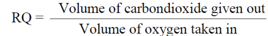
The RQ is important as it can indicate whether the respiration is aerobic or anaerobic.
C6H12O6 +6 O2→6 CO2 +6 H2O+ Energy (ATP + heat).
As each molecule of gas occupies the same volume, this would give RQ = 1.0, and this
is common for all carbohydrates. Further studies indicated the respiratory quotient
to be 0.9 for proteins and 0.7 for fats, and concluded that an, RQ greater than 1.0
indicates anaerobic respiration, while RQ equals or less than 1.0 indicates aerobicrespiration.
Note that respiration during germination, especially in early stages was also studied.
Results indicated that it is difficult for oxygen to penetrate the seed coat, so that at
this stage, the RQ is about 3 to 4. Later when the seed coat is shed, it becomes easier
for oxygen to reach respiration tissues and the levels of RQ falls. Results indicated
that eventually seeds with large carbohydrate stores have an RQ around 1.0 and
those with large lipid stores have RQs of 0.7 to 0.8.
This graph suggests that the seed begins with carbohydrate as a metabolite, changesto fat/oil then returns to mainly using carbohydrate
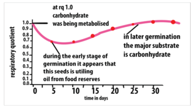
Figure 5.7: The graph showing the RQ values during seed germination
a. Measuring and obtaining the RQ values during seed germination process
During seed germination, CO2 is released. To test its presence, chemicals including
Sodium hydroxide or Potassium hydroxide are used due to their ability to absorb
CO2. As the germinating seeds use oxygen, pressure reduces in tube A so the
manometer level nearest to the seeds rises (figure 5.8). The syringe is used to return
the manometer fluid levels to normal. The volume of oxygen used is calculated by
measuring the volume of gas needed from the syringe to return the levels to the
original values. If water replaces the sodium hydroxide, then the carbon dioxideevolved can be measured.
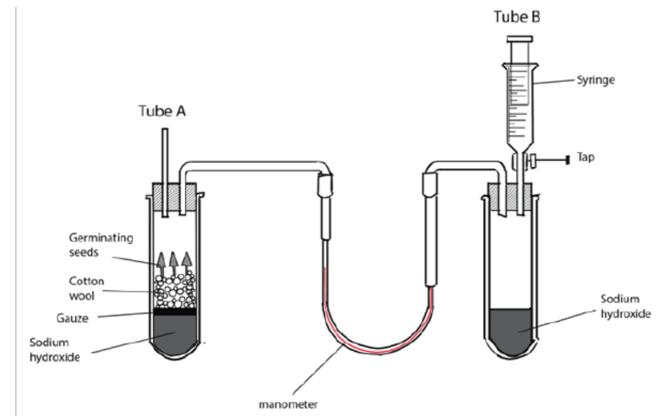
Figure 5.8: Simple experiment using respirometer to determine the RQ in germinating seeds
Measuring and obtaining the RQ values in invertebrate (e.g. woodlice)
In this particular respirometer, woodlice have been placed in a boiling tube which
is connected to a U-tube. The U-tube acts as a manometer (a device for measuring
pressure changes). The other end of the U-tube is connected to a control tube which
is treated in exactly the same way as the first tube, except that it has no woodlice but
instead glass beads which take up the same volume as the woodlice. The two boiling
tubes (but not the manometer) are kept in water bath at constant temperature. The
U-tube contains a coloured liquid which moves according to the pressure exerted
on it by the gases in the two boiling tubes. Both tubes contain potassium hydroxide
solution which absorbs any carbon dioxide produced.
When the woodlice respire aerobically, they consume oxygen, which causes
the liquid to move in the U- tube in the direction of arrows. The rate of oxygen
consumption can be estimated by timing how long it takes for the liquid to rise
through a certain height. The experiment can be repeated by replacing the potassium
hydroxide solution with water. Comparing the changes in manometer liquid level
with and without potassium hydroxide solution gives an estimate of carbon dioxide
production can be used to measure the respiratory quotient.
If the internal radius of the manometer tube is known, the volumes of gases can becalculated using the equation:
Volume of gases = π r2 h,
where π is equal to 3.14, r is the internal radius of the tube and h is the distancemoved by the liquid.
Application 5.5
1. Using the following equation of oleic acid (a fatty acid found in oliveoil):
2C18H34O2 + 51O2 →36CO2 + 34H2O.
a. Calculate the RQ for the complete aerobic respiration.
b. Based on your findings, state which substrate is being respired.
2. Suggest an explanation when RQ equals 1 for germinating maize grains.
3. Based on the values of RQ, when can you conclude that the respirationprocess is:
a. Aerobic.
b. Anaerobic.
4. Calculate the volume of gases in a manometer tube having a radius of1.7 cm, knowing that the gas was displaced about 3cm distance.
End of unit assessment 5
1. Explain the reasons why chemical energy is the most important type of
energy for living organisms.
2. Why do all organisms need energy and where does this energy come from?
3. Give the structure of ATP and specify its importance to living organisms?
4. The equation C57H104O6 + 80O2 → 57CO2 + 52H2O + Energy represents
oxidation of lipids. Calculate RQ for this equation.
5. Calculate the total amount of energy produced for:
a. 3 moles of hydrolysed ATP
b. moles of synthesized ATP
c. 5 moles of decomposed glucose
6. Active mitochondria can be isolated from liver cells. If these mitochondria are
then incubated in a buffer solution containing a substrate, such as succinate,
dissolved oxygen will be used by mitochondria. The concentration of
dissolved oxygen in the buffer solution can be measured using an electrode.
When this experiment was done, the concentration of dissolved oxygen was
measured every minute for five minutes. Sodium azide which combines with
cytochromes and prevents electron transport was added thereafter. Theresults are shown in the graph below.
a. Suggest what effect the addition of sodium azide will have on the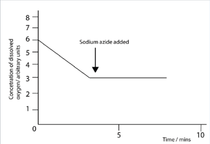
production of ATP and give an explanation for your answer.
b. Explain why the concentration of oxygen decreased during the first
five minutes.
c. Suggest what effect the addition of sodium azide will have on theproduction of ATP and give an explanation for your answer.
7. Analyse the following figure:
The graph shows the pH difference across the inner mitochondrial membrane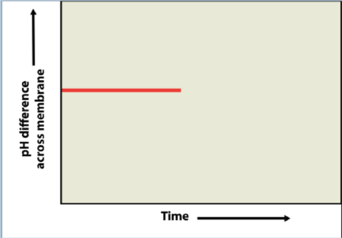
over time in an actively respiring cell. At the time indicated by the vertical
arrow, a metabolic poison is added that specifically and completely inhibits
all function of mitochondrial ATP synthase. Draw what you would expect to
see for the rest of the graphed line, and explain your graph.
8. During an experiment, the mouse was inside the bell jar. The air pipe from
the bell jar was connected to the first beaker containing lime water and filter
pump. The glass wool containing soda lime covered by a piece of paper was
connected to the second beaker by air pipe. Another air pipe was connected
from the second beaker containing lime water to the belly jar in the first step.The set of the experiment looked like the following:
a. Name the gas trapped in beaker B?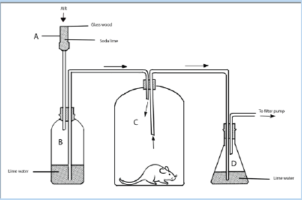
b. Why does the mouse still live since it is covered in a bell jar?
c. Why does lime water turn milky?
d. Is this experiment related to respiration and energy production or to the
respiration and energy consumption? Explain.
9. The following figure indicates the variations of RQ in function of time. Analyseit and make its interpretation
a. Observe the graph and make its interpretation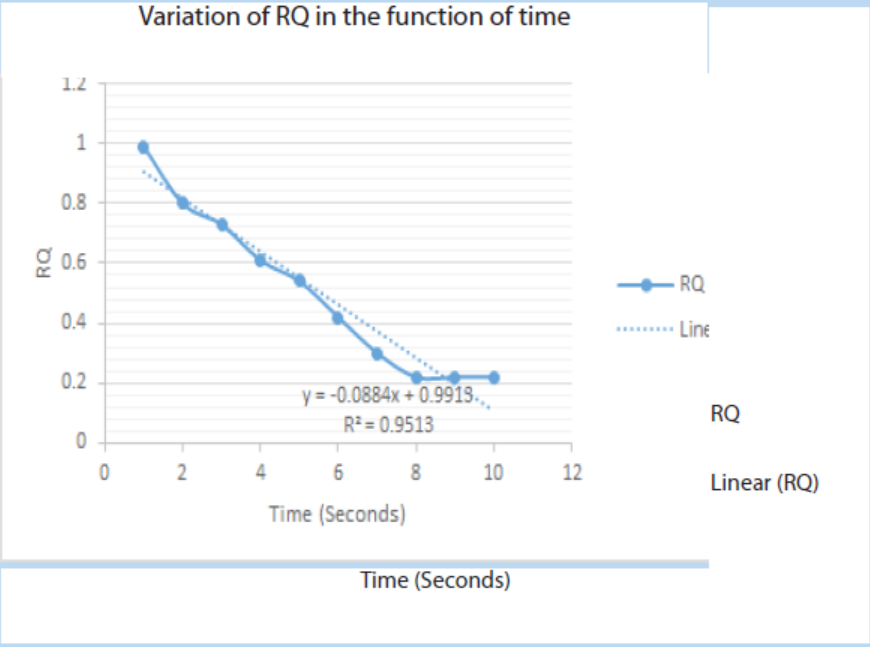
10. The following data were collected for RQ of an insect during one minute:
a. Plot the graph of RQ in function against time
b. Explain the reasons why there is no change in RQ for the last threeseconds
UNIT 6 CELLULAR RESPIRATION
UNIT 6: CELLULAR RESPIRATION
Key Unit Competence
To be able to describe the process of cellular respiration
Learning objectives
By the end of this unit, I should be able to:
– Outline the four stages in aerobic respiration (glycolysis, link reaction, TCA cycle
and oxidative phosphorylation) and state where each occurs in the eukaryotic
cells.
– Explain that when oxygen is available, pyruvate is converted into acetyl
coenzyme A, which then combines with oxaloacetate (4C) to form citrate (6C).
– Explain that reactions in the TCA cycle involve decarboxylation and
dehydrogenation and the reduction of NAD and FAD.
– Outline the process of oxidative phosphorylation including the role of oxygen
(details of the carriers are not required).
– Describe the relationship between the structure and function of the
mitochondrion.
– Explain the production of a small yield of ATP from anaerobic respiration in
yeast and mammalian muscle tissue, including the concept of oxygen debt.– Explain how other substrates are involved in glycolysis and the TCA cycle.
Introductory activity
Use of books from your library and search further information on the internet
and answer the following questions. The person in the picture below is usingenergy.
1. Where is the energy used by the person in the picture coming from?
2. All living organisms need a continuous supply of energy. Explain why.
3. Identify the processes exhibited by the person on the picture that
consume too much energy if compared with another one who is at rest.4. How is the energy produced in our body?
6.1 Overview of respiration process
6.1.1 Respiration
Activity 6.1.1
With the help of textbooks and simulations of the process of respiration,
answer the questions that follow:
1. Differentiate between glucose and pyruvate.2. What is the role of glycolysis?
Cellular respiration is the complex process in which cells make adenosine
triphosphate (ATP) by breaking down organic molecules. The energy stored in
ATP can then be used to drive processes requiring energy, including biosynthesis,
locomotion or transportation of molecules across cell membranes. The main fuel
for most cells is carbohydrate, usually glucose which is used by most of the cells as
respiratory substrate. Some other cells are able to break down fatty acids, glycerol
and amino acids.
Glucose breakdown can be divided into four stages: glycolysis, the link reaction, theKrebs cycle and oxidative phosphorylation.
6.1.2 Glycolysis
Activity 6.1.2
With the help of textbooks and simulations from internet / YouTube observe
the process of respiration, answer the questions that follow:
1. Observe and note the stages of the process of respiration.2. Draw the structure of a glucose molecule.
Glycolysis is the splitting or lysis of a glucose molecule. It is a multi-step process
in which a glucose molecule with six carbon atoms is eventually split into two
molecules of pyruvate, each with three carbon atoms. Energy from ATP is needed in
the first steps, and it is released in the later steps to synthesize ATP. There is a net gain
of two ATP molecules per molecule of glucose broken down.
Glycolysis takes place in the cytoplasm of a cell. Glucose enters the cell and is
phosphorylated by the enzyme called hexokinase, which transfers a phosphate
group from ATP to the sugar. The ATP used in this process has 2 advantages: the
charge of the phosphate group traps the sugar in the cell because the plasma
membrane is impermeable to large ions. Phosphorylation also makes glucose morechemically reactive. Even though glycolysis consumes two ATP molecules,
It produces a gross of four ATP molecules (4 ATP), and a net gain of two ATP (2 ATP)
molecules for each glucose molecule that is oxidized. Glycolysis results in a net gainof two ATP, two NADH and two pyruvate molecules.
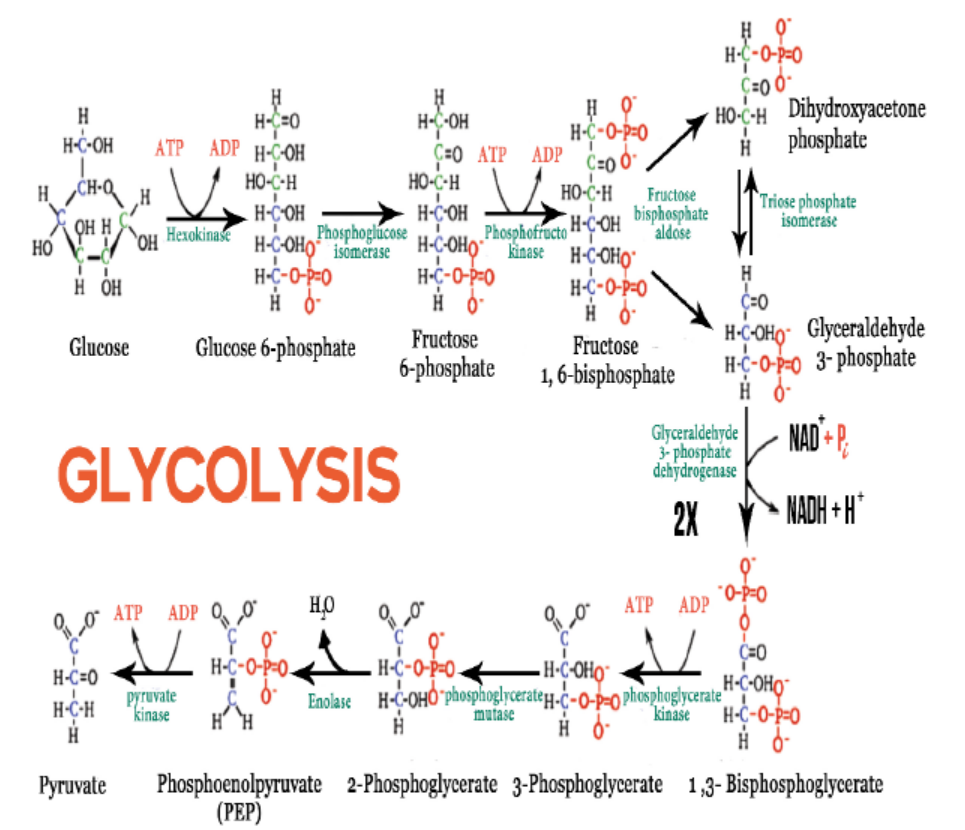
Figure 6.1: Reactions of glycolysis
Applicatioin 6.1
1. Why is ATP needed for glycolysis?
2. How many gross ATP molecules are produced during glycolysis of one
glucose molecule?3. How many NADH are made during glycolysis?
6.2 Link reaction and the Krebs cycle
Activity 6.2
Use the books from the school library and search further information on the
internet. Then:
1. Observe and write the number of carbon atoms in an acetyl-coA molecule.
2. Use the chemical equation to show the conversion of pyruvate into acetyl-
coA.
3. Observe and note the main products of the Krebs cycle from one glucosemolecule
6.2.1 Link reaction
Pyruvate, the end product of glycolysis is oxidized to Acetyl-CoA by enzymes located
in the mitochondrion of eukaryotic cells as well as in the cytoplasm of prokaryotes.
In the conversion of pyruvate to Acetyl-CoA, one molecule of NADH and one
molecule of CO2 are formed (Figure 6.2). This step is also known as the link reactionor transition step, as it links glycolysis to the Krebs cycle.
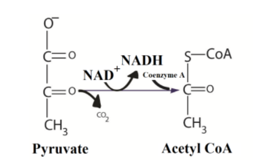
Figure 6.2: Link reaction between glycolysis and Krebs cycle
6.2.2 The Krebs cycle (Citric acid cycle)
The coenzyme has a sulphur atom, which attaches the acetyl fragment by an
unstable bond. This activates the acetyl group for the first reaction of the Krebs cycle
also called citric acid cycle or Tricarboxylic Acid Cycle (TCA). It is also known as the
citric acid cycle, because the first molecule formed when an acetyl group joins the
cycle. When oxygen is present, the mitochondria will undergo aerobic respirationwhich leads to the Krebs cycle.
In the presence of oxygen, when acetyl-CoA is produced, the molecule then enters
the citric acid cycle inside the mitochondrial matrix, and gets oxidized to CO2 while
at the same time reducing NAD+ to NADH. NADH can then be used by the electron
transport chain to create more ATP as part of oxidative phosphorylation. For the
complete oxidation of one glucose molecule, two Acetyl-CoA must be metabolized
by the Krebs cycle. Two waste products namely H2O and CO2, are released duringthis cycle.
The citric acid cycle is an 8-step process involving different enzymes and co-enzymes.
Throughout the entire cycle, Acetyl-CoA (2 carbons) combines with oxaloacetate (4
carbons) to produce citrate. Citrate (6 carbons) is rearranged to a more reactive form
called iso citrate (6 carbons). Iso citrate (6 carbons) is modified to α-Ketoglutarate (5
carbons), Succinyl-CoA, Succinate, Fumarate, Malate, and finally to Oxaloacetate. The
net energy gain from one cycle is 3 NADH, 1 FADH2, and 1 Guanosine Triphosphate
(GTP). The GTP may subsequently be used to produce ATP. Thus, the total energy
yield from one whole glucose molecule (2 pyruvate molecules) is 6 NADH, 2 FADH2,
and 2 ATP. 2 molecules of carbon dioxide are also produced in one cycle (for a totalof 4 molecules of carbon dioxide from one glucose molecule).
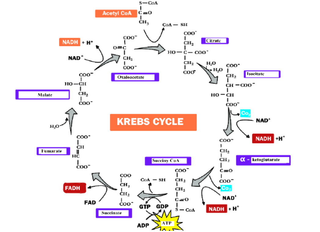
Figure 6.3: The Krebs cycle
Application 6.2
1. In which part of the cell does the Krebs cycle take place?
2. How many ATP molecules are generated by each revolution of the Krebs
cycle?3. Which six carbon sugar is formed in the first reaction of the Krebs cycle?
6.3 Oxidative phosphorylation and electron transport chain
Activity 6.3
Download and watch a movie of the electron transport chain from internet /
you tube. Make a simulation of it in the following way.
– In a line, move warm stones from one area to another.
– Take the first stone and passes it to the second up to the last one.
– The last one will have a bucket where the last stone is thrown.
– Compare what we’re doing to what you watched in the movie (carriers of
electrons)
Write short notes and share information on how the electron transport chaintakes place.
In the final stage of aerobic respiration known as the oxidative phosphorylation,
the energy for the phosphorylation of ADP to ATP comes from the activity of the
electron transport chain. Oxidative Phosphorylation is the production of ATP using
energy derived from the redox reactions of an electron transport chain.
In eukaryotes, oxidative phosphorylation occurs in the mitochondrial cristae. It
comprises the electron transport chain that establishes a proton gradient across
the inner membrane by oxidizing the NADH produced from the Krebs cycle. ATP is
synthesized by the ATP synthase enzyme when the chemiosmotic gradient is used to
drive the phosphorylation of ADP. Chemiosmosis is the production of ATP from ADP
using the energy of hydrogen ion gradients. The electrons are finally transferred to
oxygen and, with the addition of two protons, water is formed. The average ATP yield
per NADH is probably 3 and for FADH2 of this electron carrier is worth a maximum ofonly two molecules of ATP.
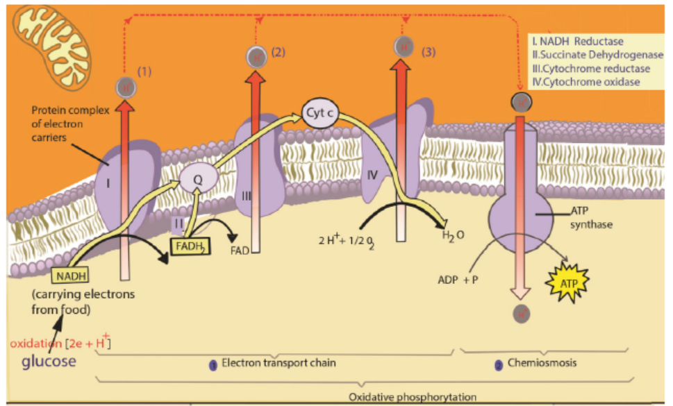
Figure 6.4: The electron transport chain
The role of oxygen in chemiosmosis
ATP can be synthesized by chemiosmosis only if electrons continue to move from
molecule to molecule in the electron transport chain. Oxygen serves as the final
acceptor of electrons. By accepting electrons from the last molecule in the electron
transport chain, and allows additional electrons to pass along the chain. As a result,
ATP can continue to be synthesized. Oxygen also accepts the protons that were once
part of the hydrogen atoms supplied by NADH and FAD2. By combining with bothelectrons and protons, oxygen forms water as shown in the following equation:
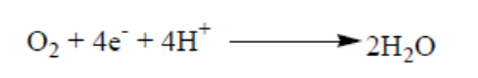
Overview of cellular respiration
A considearable number of ATP is produced during oxidative phosphorylmation
and it is estimated between 32 and 34 ATPs. These are added to 2 ATP produced
during glycolysis and 2 ATP produced during citric cycle. The total number of ATP
produced during a complete respiration process for one molecule of glucose is thenestimated between 36 and 38 ATPs.
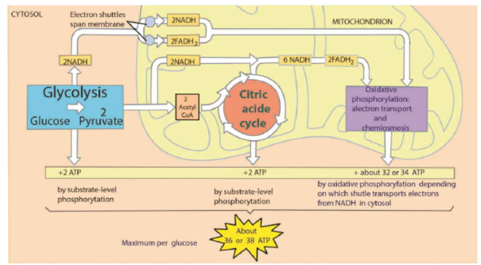
Figure 6.5: Overview of cellular respiration
Note that the amount of ATP produced from glucose is usually less than 38 ATP for
the following reasons: some ATP is used to transport pyruvate from the cytoplasm
into the mitochondria and some energy is used to transport NADH produced inglycolysis from the cytoplasm into the cristae of mitochondria.
Application 6.3
1. What is the importance of NADH and FADH?
2. How many ATP are formed from 1 NADH?
3. How many ATP are formed from 1 FADH?
4. How many ATP are formed after a complete oxidation of one glucosemolecule?
6.4 Efficiency of aerobic and anaerobic respiration
Activity 6.4
Visit a nearby bakery and observe how bread is made and answer to the
following questions. Use also books, internet and prior knowledge from
chemistry.
1. On a sheet of paper write down the ingredients used to manufacture
bread
2. Which ingredients make the bread rise?
3. What do you understand by anaerobic respiration?
4. State the examples of the applications of anaerobic respiration in
everyday life?
5. Give a table comparing aerobic to anaerobic respiration
6. How can the efficiency of anaerobic and aerobic respiration be
calculated from one glucose molecule?
7. Between aerobic and anaerobic respiration, which one do you think ismore efficient? and why?
Without oxygen, pyruvate (pyruvic acid) is not metabolized by cellular respiration
but undergoes a process of fermentation. The pyruvate is not transported into
the mitochondrion, but remains in the cytoplasm, where it is converted to waste
products that may be removed from the cell. This serves the purpose of oxidizing the
electron carriers so that they can perform glycolysis again and removing the excess
pyruvate. Fermentation oxidizes NADH to NAD+ so it can be re-used in glycolysis.
In the absence of oxygen, fermentation prevents the build-up of NADH in the
cytoplasm and provides NAD+ for glycolysis. This waste product varies depending
on the organism. In skeletal muscles, the waste product is lactic acid. This type
of fermentation is called lactic acid fermentation. In yeast and plants, the waste
products are ethanol and carbon dioxide. This type of fermentation is known as
alcoholic or ethanol fermentation. The ATP generated in this process is made bysubstrate-level phosphorylation, which does not require oxygen.
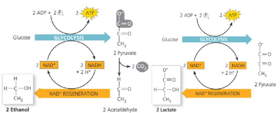
Figure 6.6: Alcoholic and lactic fermentation
Fermentation is less efficient at using the energy from glucose since only 2 ATP are
produced per glucose, compared to the 38 ATP per glucose produced by aerobic
respiration. This is because the waste products of fermentation still contain plentyof energy. Glycolytic ATP, however, is created more quickly.
a. Applications of anaerobic respiration
Some food products and drinks are produced by using anaerobic microorganisms:
– Production of beer
– Production of wine
– Production of yoghurt
– Production of cheese– Production of bread
b. Efficiency of aerobic and anaerobic respiration
The complete oxidation of glucose produces the energy estimated at 686 Kcal.
Under the condition that exists inside most of the cells, the production of a
standard amount of ATP from ADP absorbs about 7.3 Kcal. Glucose molecule can
generate up to 38 ATP molecules in aerobic respiration. The efficiency of aerobicrespiration (EAER) is calculated as follows:
This result indicates that the efficiency of aerobic respiration equals 40%. The remain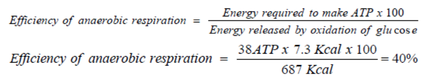
of the energy (around 60%) is lost from the cell as heat.
Due to the fact that anaerobic respiration produces only 2 ATP, the efficiency ofanaerobic respiration is less than that of aerobic respiration. It is calculated as follows:
c. Oxygen debt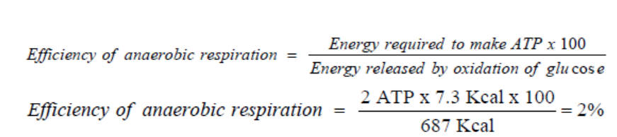
Standing still, the person absorbs oxygen at the resting rate of 0.2 dm3 min−1. (This
is a measure of the person’s metabolic rate.) When exercise begins, more oxygen is
needed to support aerobic respiration in the person’s muscles, increasing the overall
demand to 2.5 dm3 min−1. However, it takes four minutes for the heart and lungs to
meet this demand, and during this time lactic fermentation occurs in the muscles.
Thus the person b-uilds up an oxygen deficit. For the next three minutes, enough
oxygen is supplied. When exercise stops, the person continues to breathe deeply
and absorb oxygen at a higher rate than when at rest. This post-exercise uptake of
extra oxygen, which is ‘paying back’ the oxygen deficit, is called the oxygen debt.
The oxygen is needed for:
– Conversion of lactate to glycogen in the liver
– Re oxygenation of haemoglobin in the blood
– A high metabolic rate, as many organs are operating at above resting levels.
The presence of the lactic acid is sometimes described as an ‘ oxygen debt’. This is
because significant quantities of lactic acid can only be removed reasonably quickly
by combining with oxygen. However, the lactic acid was only formed due to lack
of sufficient oxygen to release the required energy to the muscle tissue via aerobic
respiration. Lactic acid can accumulate in muscle tissue that continues to be overworked.
Eventually, so much lactic acid can build-up that the muscle ceases working
until the oxygen supply that it needs has been replenished.
To repay such an oxygen debt, the body must take in more oxygen in order to get ridof the additional unwanted waste product lactic acid.
d. Muscle cramps
A muscle cramp is an involuntarily and forcibly contracted muscle that does not
relax. Muscle cramps can occur in any muscle; cramps of the leg muscles and feet
are particularly common.
Almost everyone experiences a muscle cramp at some time in their life. There are
a variety of types and causes of muscle cramps. Muscle cramps may occur during
exercise, at rest, or at night, depending upon the exact cause.
Overuse of a muscle, dehydration, muscle strain or simply holding a position for a
prolonged period can cause a muscle cramp. In many cases, however, the cause isn’t
known.
Although most muscle cramps are harmless, some may be related to an underlying
medical condition, such as:
– Inadequate blood supply. Narrowing of the arteries that deliver blood to your
legs (arteriosclerosis of the extremities) can produce cramp-like pain in your
legs and feet while you’re exercising. These cramps usually go away soon after
you stop exercising.
– Nerve compression. Compression of nerves in your spine (lumbar stenosis) also
can produce cramp-like pain in your legs. The pain usually worsens the longer
you walk. Walking in a slightly flexed position such as you would use when
pushing a shopping cart ahead of you may improve or delay the onset of your
symptoms.
– Mineral depletion. Too little potassium, calcium or magnesium in your diet can
contribute to leg cramps. Diuretics or medications often prescribed for highblood pressure also can deplete these minerals.
Application 6.4
1. What is the product of anaerobic respiration in animal cells?
2. Under which conditions can anaerobic respiration take place in animal
cells?
3. Calculate the efficiency of anaerobic and aerobic respiration, when
a complete oxidation of glucose produce the energy estimated at
500Kcal under a production of a standard amount of ATP from ADPabsorbed is about 7.3 Kcal.
6.5 Factors affecting the rate of respiration
Activity 6.5
Observe carefully the pictures below and answer the questions that follow;
1. Make a short report on the respiration rate of the person on the picture A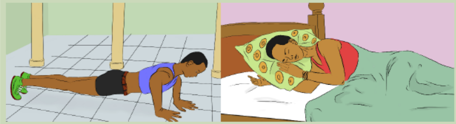
and that of the person on the picture B.
2. Which one between person A and that of person B has a high respiration
rate?
3. What are factors could show that the respiration rate has increased in theperson on the picture A above?
Cellular respiration is the process of conversion of chemical energy stored in the food
to ATP or higher energy compounds. The factors that affect the cellular respirationare:
a. Amount of nutrients
If the amount of nutrients is high, then the energy is high in the cellular respiration.
The nutrients which can go through cellular respiration and transform into energy
are fat, proteins and carbohydrates. The amount of nutrients available to transform
into energy depend upon the diet of the person.
b. Temperature
The rate of the cellular respiration increases if the body temperature is warmer. The
lower the temperature, the slower the rate of cellular respiration. The reason for
this is enzymes which are present in cellular respiration process. Enzyme reactions
require optimum temperatures.
c. State of the cell
Metabolically active cells such as neurons, root of human hair have higher
respiration rate than the dormant cells such as skin cells and bone cells. This is
because metabolically active cells can store energy in the body because of the many
metabolic reactions that take place in them.
d. Water
It is the medium where the reaction happens. When a cell is dehydrated the
respiration and other metabolism decreases.
e. Cellular activity
Some cells need more energy than others. For example, growing cells or very active
cells such as neurons need a lot of energy.
f. O2 /CO2 content
Higher O2 and lower CO2 make higher respiration rates.
g. ATP/ADP range
When there is more ATP than ADP, respiration rate slows down to avoid excess ofATP
Application 6.5
1. Which cells in the human body have a high respiration rate?2. Explain how the temperature affects the rate of respiration.
6.6 Use of other substrates in respiration
Activity 6.6
When one has eaten carbohydrates such as cassava and sweet potatoes you
do not feel hungry in the same time as another one who has consumed milk
or cheese.
1. Can you suggest the reason for this?2. Which one can take a short time for digestion and why?
Carbohydrates are the first nutrients that most organisms can catabolise for energy.
In some cases, living things must be able to metabolize other energy-rich nutrients
to obtain energy in times of starvation. Most organisms possess metabolic pathways
that, when necessary, metabolize proteins, lipids. In each case, the larger molecules
are first digested into their component parts, which the cell may reassemble into
macromolecules for its own use. Otherwise, they may be metabolized for energy byfeeding into various parts of glycolysis or the Krebs cycle.
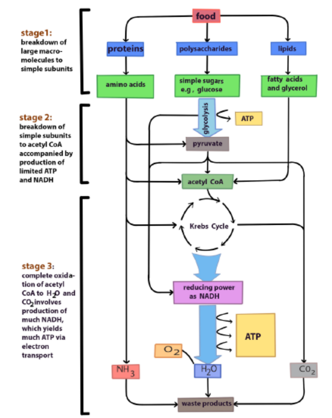
Figure 6.8: Oxidation of different organic substrates
Carbohydrates, fats and proteins can all be used for cellular respiration. Monomers
of these foods enter glycolysis or the Krebs cycle at various points. Glycolysis and the
Krebs cycle are catabolic pathways through which all kinds of food molecules arechannelled to oxygen as their final acceptor of electrons.
Application 6.6
1. Explain how proteins and lipids are metabolized for energy during
respiration
2. Explain why the body does not use fats to produce energy ascarbohydrates given that they produce much energy than carbohydrates.
End of unit assessment 6
Multiple choice questions: from question 1 to 7, choose the letter corresponding
to the best answer.
1. Before the Krebs cycle can proceed, pyruvic acid must be converted into
a. Citric acid
b. Glucose
c. Acetyl-CoA
d. Glucose
e. NADH
2. The net number of ATP made directly by glycolysis is
a. 2
b. 4
c. 32
d. 38
3. Cellular respiration is similar to photosynthesis in that they both
a. Produce ATP
b. Involve chemiosmosis
c. Make phosphoglyceraldehyde (PGAL)
d. All of the above
4. By accepting electrons and protons, the oxygen used in aerobic respiration
turns into
a. CO2
b. H2O
c. C6H12O6
d. ATP
5. The Krebs cycle occurs in the
a. Cytosol
b. Outer mitochondrial membrane
c. Mitochondrial matrix
d. Space between the inner and outer mitochondrial membrane
6. During each turn of the Krebs cycle,
a. Two CO2 molecules are produced
b. Two ATP molecules are consumed
c. Pyruvic acid combines with oxaloacetic acid
d. Glucose combines with a four-carbon molecule.
7. Most of the ATP synthesized in aerobic respiration is made
a. During glycolysis
b. Through fermentation
c. In the cytosold. Through chemiosmosis
Structured answer questions
8. What are the major differences between cellular respiration and
photosynthesis?
9. Compare aerobic respiration with anaerobic respiration or fermentation.
10. A student set up an experiment using germinating seeds and boiled seedsas shown in the diagram below:
a. State the objective of this experiment and the observation made after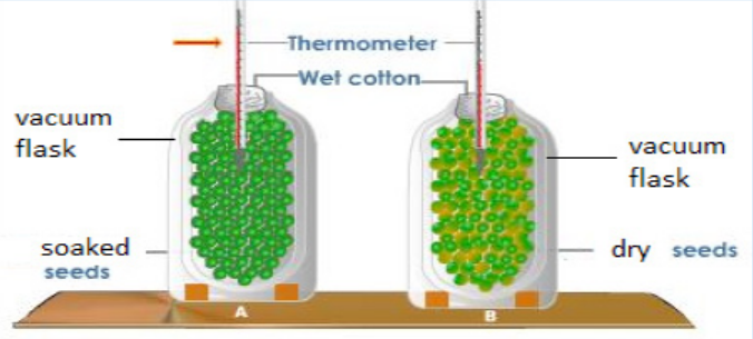
24 hours?
b. Account for the observation made in (a) above?
c. Suggest why vacuum flasks were used in the experiment?d. What was the purpose of the set-up B?
UNIT 7 EXCRETION AND OSMOREGULATION
UNIT 7: EXCRETION AND OSMOREGULATION
Key Unit CompetenceExplain the principles of excretion and osmoregulation
Learning Objectives
By the end of this unit, the student should be able to:
– Describe the structure and role of excretory organs in mammals.
– Dissect, display, draw and label the urinary system of a toad, rat/rabbit etc.
– Describe the detailed structure of the nephron with its associated blood vessels.
– Describe and outline the ornithine cycle and its role in the conversion of
ammonia to urea.
– Describe how the process of ultrafiltration and selective reabsorption are
involved in the formation of urine in the nephron.
– Describe the use of dialysis in kidney machines.
– Describe how kidney transplants are performed.
– Describe the role of hypothalamus, posterior pituitary, ADH and collecting
ducts in osmoregulation.
– Explain the principles of osmoregulation in organisms living in marine,
freshwater and terrestrial habitats.
– Explain dialysis in terms of salt balance, the maintenance of glucose
concentration and the removal of urea.
– Explain why plants do not have specialised excretory organs.
– State the excretory products of plants and how they are eliminated.
– Dissect, display, draw and label the urinary system of a toad, rat/rabbit etc.
– Interpret the ornithine cycle diagram with reference to urine formation.
– Relate adaptations of different organisms to their habitat in terms of
osmoregulation.
– Compare the advantages and disadvantages of kidney transplants with dialysis
machines.
– Support the use of dialysis machine or kidney transplants in solving problems
associated with kidney failure.
– Appreciate the adaptation of organisms to different habitats in relation toosmoregulation.
Introductory activity
Metabolic reactions generate different kinds of wastes. These metabolic
wastes are removed by different organs of our body.
a. Identify any three metabolic waste products of our body.
b. Where are those metabolic wastes produced?
c. What is the name given to the process by which metabolic wastesproducts are removed from the body?
7.1 Structure and functions of excretory organs in mammals
Activity 7.1
Dissection of the rabbit to study the urinary system
Materials required: A mature rabbit, dissecting tray, and dissecting kit,
chloroform.
Safety: Gloves, safety goggles, and apron must be worn at all times. Anyone
not wearing these items will Not dissect. Be sure to follow all lab safety rules.
Procedure
– Place the rabbit in the dissecting tray, ventral side up.
– Tie the legs securely to the corners of the tray by passing a string or rubber
bands (2 bands together) under the tray from front leg to front leg and hind
leg to hind leg.
– Be sure that the specimen is held firmly before you begin dissecting.
– Find the lower edge of the sternum (breastbone) and make an incision
through the skin from that point to the pelvis. This will expose the layers of
the abdominal muscles.
– Strip the skin well back to the sides and examine the muscle layer.
– Using the scissors or the scalpel, make another incision through the muscle
layer. This will expose a thin membrane, the peritoneum, which lines the
abdominal cavity.
– Cut through the peritoneum to expose the abdominal organs.
– Open the abdominal cavity wide by making several lateral cuts and pulling
the skin and muscle layer well to the side.
– Use pins to pin back the cut sections of skin and muscle.
– Discard the digestive organs and examine the kidneys.
– Cut under each kidney and remove it along with the ureter tube.
– Cut a kidney laterally and examine its internal structure.
– You should find a spongy cortex on the other curved side and a hollow pelvis
on the inner concave side. See if you can find the renal blood vessels which
lead to and from the kidneys. Discard the kidneys.Identify the functions of each part of the urinary system.
Excretion the removal of toxic waste products of metabolism from the body. The
term is generally taken to mean nitrogenous wastes such as; urea, ammonia and uric
acid but other materials like carbon dioxide and the bile pigments are also waste
products of metabolism, and their removal is as much a part of excretion as theelimination of urea.
Excretion is an essential process in all forms of life. When cells metabolize or break
down nutrients, waste products are produced. For example, when cells metabolize
amino acids, nitrogen wastes such as ammonia are produced. Ammonia is a toxic
substance and must be removed from the blood and excreted from the body.
Although the kidneys are the main organs of excretion of wastes from the blood,
several other organs are also involved in the excretion, including the; liver, skin, andlungs.
– The large intestine eliminates waste products from the bile synthesis.
– The liver breaks down excess amino acids in the blood to form ammonia, and
then converts the ammonia to urea, a less toxic substance. The liver also breaks
down other toxic substances in the blood, including alcohol and drugs.
– The skin eliminates water and salts in sweat.– The lungs exhale water vapour and carbon dioxide.
The importance of excreting wastes
i. To maintain life processes, the body must eliminate waste products, many of
these which can be harmful. The lungs eliminate carbon dioxide, one of the
products of cellular respiration. The large intestine removes toxic wastes from
the digestive system.
ii. The liver transforms ingested toxins, such as alcohol and heavy metals, into
soluble compounds that can be eliminated by the kidneys.Table 7.1: Metabolic wastes products and their organs of excretion
Kidneys and Excretion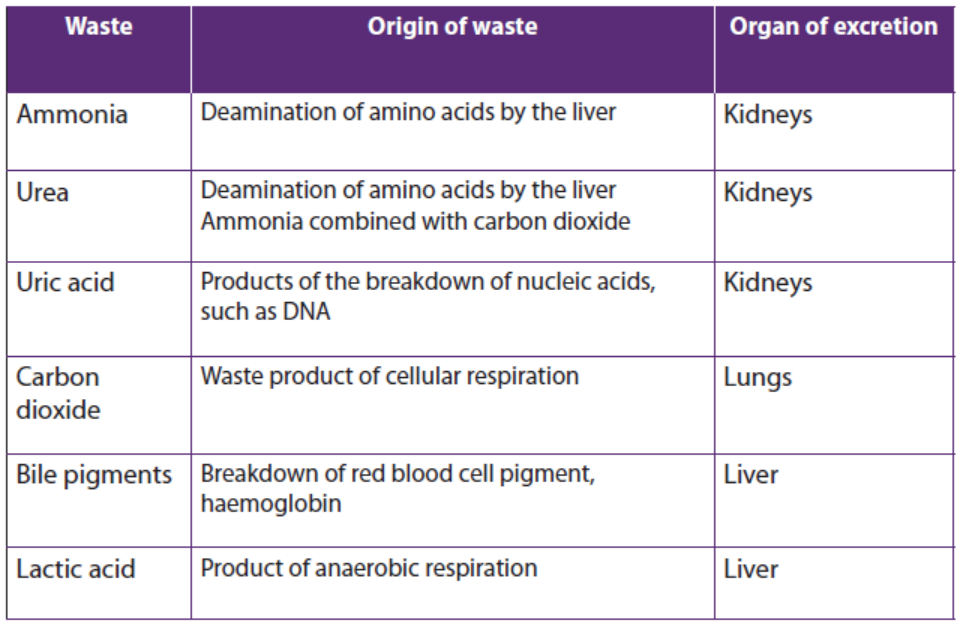
The kidneys are part of the urinary system (Figure 7.1). The kidneys work togetherwith other urinary system organs in the function of excretion
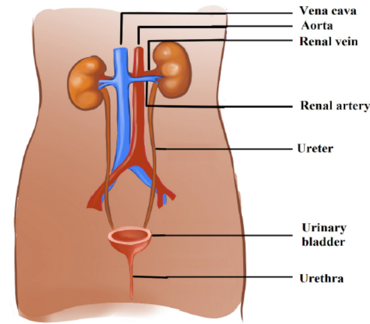
Figure 7.1: The human urinary system
a. The Urinary System
In addition to the kidneys, the urinary system includes the; ureters, bladder, and
urethra. The main functions of the urinary system are to; filter waste products and
excess water from the blood and remove them from the body.
From the kidneys, urine enters the ureters. Each ureter is a muscular tube about
25 centimetres long. Peristaltic movements of the muscles of the ureter send urine
to the bladder in small amount. Ureters carry urine to the bladder. The bladder is a
hollow organ that stores urine. It can stretch to hold up to 500 millilitres. When the
bladder is about half full, the stretching of the bladder sends a nerve impulse to the
sphincter that controls the opening to the urethra. In response to the impulse, the
sphincter relaxes and lets urine flow into the urethra.
The urethra is a muscular tube that carries urine out of the body. Urine leaves the
body through another sphincter in the process of urination. This sphincter and theprocess of urination are normally under conscious control/voluntary system.
b. Kidneys
The kidneys are a pair of bean-shaped, reddish brown organs about the size of a fist.
They are located just above the waist at the back of the abdominal cavity, on either
side of the spine. The kidneys are protected by the ribcage. They are also protected
by a covering of tough connective tissues and two layers of fat, which help cushion
them. Located on top of each kidney is an adrenal gland. The two adrenal glands
secrete several hormones. Hormones are chemical messengers in the body that
regulate many body functions. The adrenal hormone aldosterone helps regulatekidney functions. The functional unit of a kidney is a nephron.
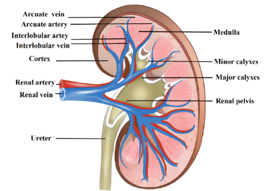
Figure 7.2: Human kidney
Application 7.1
1. What are the functional units of the kidney?
2. What are the main parts of a kidney?
3. Which blood vessel carries filtered blood away from the kidney?4. Which blood vessel brings oxygenated blood to the kidney?
7.2 Structure and the functions of the nephron.
Activity 7.2
1. Download from internet /YouTube and watch a simulation showing the
working of the nephron. 2. Study the diagram below then answer thequestions that follow.
a. Name the structures marked Q, R, S, T, U and W.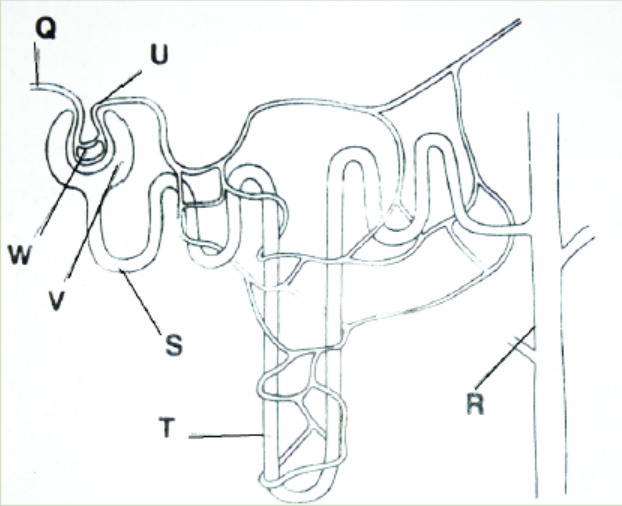
b. When some pressure is applied at W, a fluid appears at V. Name the fluid andstates its contents.
Nephrons are the structural and functional units of the kidneys. A single kidney may
have more than a million nephrons. An individual nephron (Figure 7.3) includes aglomerulus, Bowman’s capsule, and renal tubule.
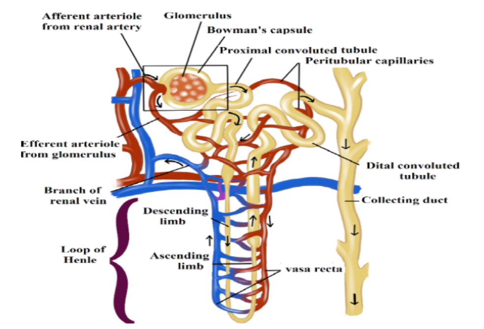
Figure 7.3: The structure of a nephron
a. Parts of the nephron and their functions
– The glomerulus is a cluster of arteries that filters substances out of the blood.
– Bowman’s capsule is a cup-shaped structure around the glomerulus that
collects the filtered substances.
– The renal tubule is a long, narrow tube surrounded by capillaries that reabsorbs
many of the filtered substances and secretes other substances.
b. Ultra-filtration, selective reabsorption and tubular secretion
The renal arteries, which carry blood into the kidneys, branch into the capillaries
of the glomerulus of each nephron. The pressure of blood moving through these
capillaries forces some of the water and dissolved substances in the blood through
the capillary walls and into Bowman’s capsule. Bowman’s capsule is composed of
layers. The space between the layers, called Bowman’s space, fills with the filtered
substances.
The process of filtering substances from blood under pressure in the glomerulus
is called ultra-filtration, while the fluid that collects in Bowman’s space is called
glomerular filtrate. The filtrate is mainly composed of; water, salts, glucose, amino
acids, hormones and urea. Larger structures in the blood including; the protein
molecules, blood cells, and platelets do not pass into Bowman’s space. Instead, theyremain in the main circulation.
From Bowman’s space, the filtrate passes into the renal tubule whose main function
is reabsorption. Reabsorption is the return of needed substances in the glomerular
filtrate back to the bloodstream. It is necessary because some of the substances
removed from the blood by filtration including; water, salts, glucose, and amino
acids which are useful and needed by the body. About 75 % of these substances arereabsorbed in the renal tubule.
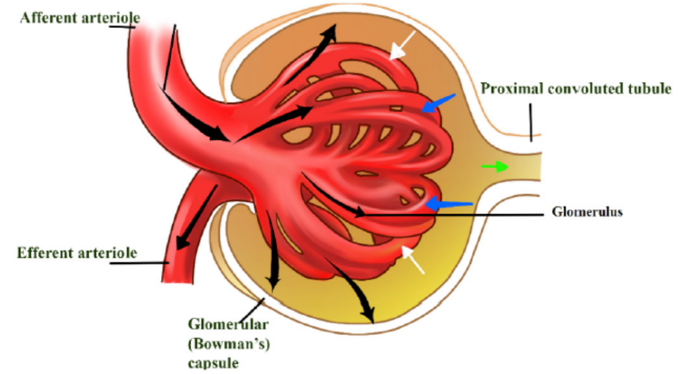
Figure 7.4: The glomerulus
Under conditions in which the kidney conserves as much water as possible, urine can
reach an osmolality of about 1200 milliosmoles (mOsm/L), considerably hypertonic
to blood (about 300 mosm/L). Osmolarity is the solute concentration expressed as
molarity. This ability to excrete nitrogenous wastes with a minimal loss of water is
a key terrestrial adaptation of mammals. The loop of Henle is known as a countercurrent
multiplier. The term counter-current refers to the fact that the fluid flows in
opposite directions in the two sides of the loop, down one side and up in the other.
The multiplier effect is seen by comparing the osmolality of the fluid in the cortexand that in the renal medulla at the hairpin end of the loop.
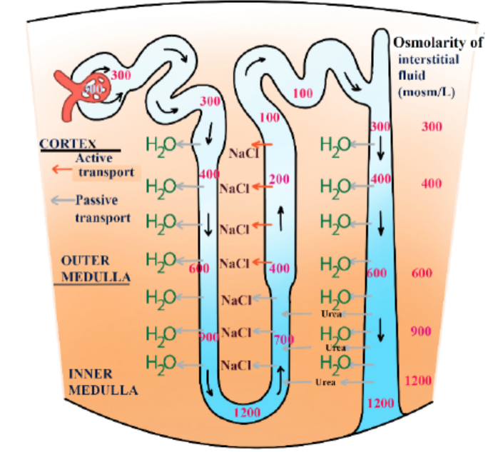
Figure 7.5: Transport of substances across the loop of Henle
The remaining fluid enters the distal tubule. The distal tubule carries the fluid, now
called tubular fluid, from the loop of Henle to a collecting duct. As it transports
the fluid, the distal tubule also reabsorbs or secretes substances such as calcium
and sodium following the influence of hormones (e.g. aldosterone). The process ofsecreting substances into the tubular fluid is called secretion.
Application 7.2
1. What are the main parts of a nephron?
2. In which part of the nephron does each of the following processes
takes place?
a. Ultrafiltration
b. Reabsorption
c. Secretion3. What is the function of the loop of Henle?
7.3 Formation of urine and purification of blood
7.3.1 Urine formation
Activity 7.3
Download from internet/YouTube and watch a movie about the formation of
urine and after answer the questions that follow:
1. Make a list of processes which are involved in the formation of urine.2. What are the main components of urine?
Urine formation depends on three processes including ultrafiltration, selective
reabsorption and secretion/tubular secretion.
a. Ultra-filtration
Each nephron of the kidney has an independent blood supply, which moves through
the afferent arteriole into the glomerulus, a high-pressure filter. Normally, pressure
in a capillary bed is about 25 mm Hg. The pressure in the glomerulus is about 65 mm
Hg. Dissolved solutes pass through the walls of the glomerulus into the Bowman’s
capsule. Although materials move from areas of high pressure to areas of low
pressure, not all materials enter the capsule.
b. Selective reabsorption
The importance of reabsorption is emphasized by examining changes in the
concentrations of fluids as they move through the kidneys. On average, about 600
mL of fluid flows through the kidneys every minute. Approximately 20% of the fluid,
or about 120 mL, is filtered into the nephrons. This means that if none of the filtrate
were reabsorbed the quantity of around 120 mL of urine each minute would be
formed and the amount of at least 1 L of fluids would be consumed every 10 minutes
to maintain homeostasis.
Fortunately, only 1 mL of urine is formed for every 120 mL of fluids filtered into
the nephron. The remaining 119 mL of fluids and solutes is reabsorbed. Selective
reabsorption occurs by both active and passive transport. Carrier molecules move
Na+ ions across the cell membranes of the cells that line the nephron. Negative ions,
such as Cl-and HCO3- follow the positive Na+ ions by charge attraction. Numerous
mitochondria supply the energy necessary for active transport. Reabsorption
occurs until the threshold level of a substance is reached. Excess NaCl remains in the
nephron and is excreted with the urine.
Other molecules are actively transported from the proximal tubule. Glucose and
amino acids attach to specific carrier molecules, which shuttle them out of the
nephron and into the blood. However, the amount of solute that can be reabsorbedis limited. For example; excess glucose will not be shuttled out of the nephron by
the carrier molecules. The solutes that are actively transported out of the nephron
create an osmotic gradient that draws water from the nephron. A second osmotic
force, created by the proteins not filtered into the nephron, also helps reabsorption.
The proteins remain in the bloodstream and draw water from the interstitial fluid
into the blood. As water is reabsorbed from the nephron, the remaining solutes
become more concentrated. Molecules such as urea and uric acid will diffuse from
the nephron back into the blood, although less is reabsorbed than was originallyfiltered.
c. Secretion
Secretion is the movement of wastes from the blood back into the nephron. Nitrogencontaining
wastes, excess H+ ions, and minerals such as K+ ions are examples of
substances secreted.
Even drugs such as penicillin can be secreted. Cells loaded with mitochondria line
the distal tubule. Like reabsorption, tubular secretion occurs by active transport,but, unlike reabsorption, molecules are shuttled from the blood into the nephron.
7.3.2 Formation of urea
The body is unable to store proteins or amino acids, and any surplus is destroyed in
the liver. Excess amino acids which are brought to the liver by the hepatic portal vein,
are deaminated by the liver cells. In this process the amino (NH2) group is removed
from the amino acid, with the formation of ammonia. The amino acid residue is
then fed into carbohydrate metabolism and oxidized with the release of energy.
Meanwhile the ammonia must not be allowed to accumulate because it is highly
toxic even in small quantities. Under the influence of specific enzymes in the liver
cells, the ammonia enters a cyclical series of reactions called the ornithine cycle, in
which it reacts with carbon dioxide to form the less toxic nitrogenous compound
urea. The urea is then shed from the liver into the bloodstream, and taken to thekidney which eliminates it from the body.
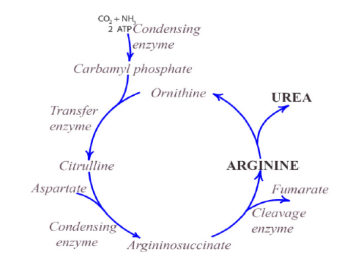
Figure 7.6: The ornithine cycle
Application 7.3
1. The following is a random list of processes that occur in the formation
and excretion of urine once the blood has entered the kidney. Place these
subsequent processes in the correct order:
a. Urine is stored in the bladder
b. Blood enters the afferent arteriole
c. Fluids pass from the glomerulus into the Bowman’s capsule
d. Urine is excreted by the urethra
e. Na+ ions, glucose, and amino acids are actively transported from the
nephronf. Urine passes from the kidneys into the ureters
3. The table below shows the percentage of various components in the blood
plasma in the part labelled A, the fluid in the part labelled B and in the urineof a human.
a. Give a reason why there is no protein in urine.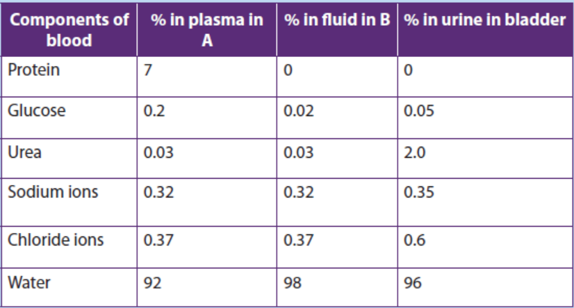
b. Which component of urine shows the greatest percentage increase
in concentration compared to the fluid in B?
c. Give a reason why the component you have named in (ii) above has
the greatest increase in concentration in urine.
d. Suggest with a reason the health condition of the person fromwhom the figure was obtained.
7.4 Role of hypothalamus, pituitary gland, adrenal gland andnephron in varying the blood osmotic pressure
Activity 7.4
Read the following text and answer to the questions that follow: “Water in
essential for all living organisms. People living around lakes and rivers can
drink safely the water without problems but people living around oceans
cannot drink sea water”.
1. Provide an explanation for the possible reason for this.
2. Write on paper the possible endocrine glands involved in this regulation.
3. Make a list showing hormones involved in this regulation.
The body adjusts for increased water intake by increasing urine output. Conversely,
it adjusts for increased exercise or decreased water intake by reducing urine output.
These adjustments involve nervous system and the endocrine system.
7.4.1 Regulation by antidiuretic hormone (ADH)
A hormone called antidiuretic hormone (ADH) helps to regulate the osmotic
pressure of body fluids by causing the kidneys to increase water reabsorption. When
ADH is released, more concentrated urine is produced, thereby conserving body
water. ADH is produced by specialized nerve cells in the hypothalamus, and it moves
along specialized fibres from the hypothalamus to the pituitary gland, which stores
and releases ADH into the blood. Specialized nerve receptors, called osmoreceptors,
located in the hypothalamus detect changes in osmotic pressure when there is a
decrease in water intake or increase in water loss by sweating, causing blood solutes
to become more concentrated. This increases the blood’s osmotic pressure.
Consequently, water moves into the bloodstream, causing the cells of the
hypothalamus to shrink. When this happens, a nerve message is sent to the pituitary,
signalling the release of ADH, which is carried by the bloodstream to the kidneys. By
reabsorbing more water, the kidneys produce more concentrated urine, preventingthe osmotic pressure of the body fluids from increasing any further.
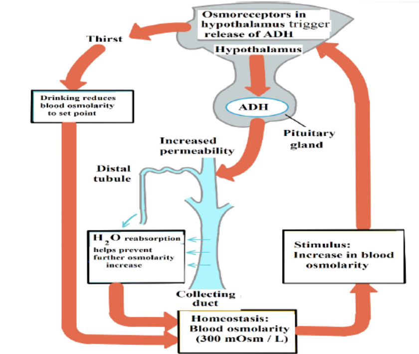
Figure 7.7: Water balance by ADH
7.4.2 Kidneys and Blood Pressure
The kidneys play a role in the regulation of blood pressure by adjusting for blood
volumes. A hormone called aldosterone acts on the nephrons to increase Na+
reabsorption. The hormone is produced in the cortex of the adrenal glands which
lies above the kidneys. Not surprisingly, as NaCl reabsorption increases, the osmotic
gradient increases and more water move out of the nephron by osmosis.
Aldosterone is secreted by the adrenal cortex in response to a high blood potassium
levels, to a low blood sodium levels, or to a decreased blood pressure. When
aldosterone stimulates the reabsorption of Na+ ions, water follows from the filtrate
back to the blood. This helps maintain normal blood volume and blood pressure. In
the kidneys, aldosterone increases reabsorption of Na+ and water so that less is lost
in the urine. Aldosterone also stimulates the kidneys to increase secretion of K+ and
H+ into the urine. With increased water reabsorption by the kidneys, blood volumeincreases.
Application 7.4
1. Describe the mechanism that regulates the release of ADH.
2. Where is the thirst centre located?3. Write ADH in full where is it produced and stored?
7.5 Kidney transplants and dialysis machines
Activity 7.5
Nowadays kidneys diseases are well known and some people with kidney
failure are being treated in different hospitals in our country and abroad.
1. Write the types of treatments you know for the person with kidney
failure.2. Discuss the advantages and disadvantages of such treatments.
Dialysis is a medical procedure in which blood is filtered with the help of a machine.
Blood from the patient’s vein enters the dialysis machine through a tube. Inside the
machine, excess water, wastes, and other unneeded substances are filtered from the
blood. The filtered blood is then returned to the patient’s vein through another tube.
A dialysis treatment usually lasts three to four hours and must be repeated three
times a week. Dialysis is generally performed on patients who have kidney failure.Dialysis helps them stay alive, but does not cure their failing kidneys.
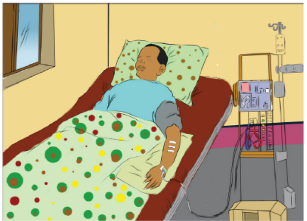
Figure 7.8: Patient under dialysis treatment
Kidney transplants are sometimes performed on people who suffer from severe
renal failure. Usually, the donor has suffered an accidental death and had granted
permission to have his or her kidneys used for transplantation. An attempt is made
to match the immune characteristics of the donor and recipient to reduce the
tendency for the recipient’s immune system to reject the transplanted kidney. Even
with careful matching, however, recipients have to take medication for the rest of
their lives to suppress their immune systems so that rejection is less likely. The major
cause of kidney transplant failure is rejection by the recipient’s immune system.
In most cases, the transplanted kidney functions well, and the tendency for the
recipient’s immune system to reject the transplanted kidney can be controlled. Theadvantages and disadvantages of kidney transplants, compared with dialysis.
Advantages
– The patient can return to a normal lifestyle – dialysis may require a lengthy
session in hospital, three times a week, leaving the patient very tired after each
session.
– The dialysis machine will be available for other patients to use.
Disadvantages
– Transplants require a suitable donor – with a good tissue match. The donor may
be from a dead person, or from a close living relative who is prepared to donate
a healthy kidney (we can survive with one kidney).
– The operation is very expensive.
– There is a risk of rejection of the donated kidney; immunosuppressive drugs
have to be used.– Transplants are not accepted by some religions.
Application 7.5
1. What is the most difficult challenge to overcome in achieving successful
kidney transplants? Provide a reason.
2. Why do you think it is beneficial to humans to have two kidneys ratherthan one? Explain your answer.
7.6 Principles of osmoregulation in marine, freshwater andterrestrial organisms.
Activity 7.6.
Aim: To demonstrate the process of osmoregulation in earthworms and
amphibians
Materials required: earthworms, amphibians (toads or frogs), beaker, salt
solution and tap water.
Procedure:
– Put three earthworms in beaker A containing water from the tap and note
the observations after 20 minutes.
– Put other three earthworms in a beaker B containing concentrated salt
solution and note the observations after 20 minutes.
– Put one amphibian in beaker C containing water from the tap and note the
observations after 20 minutes.
– Put the other amphibian in a beaker D containing concentrated salt solution
and note the observations after 20 minutes.– Prepare a report that explains the above observations.
Organisms in aquatic and terrestrial environments must maintain the right
concentration of solutes and amount of water in their body fluids. This involvesexcretion through the skin and the kidneys.
a. Marine animals
Marine bony fishes, such as the salmon, constantly lose water by osmosis. Such fishes
balance the water loss by drinking large amounts of seawater. They then make use of
both their gills and kidneys to rid themselves of salts. In the gills, specialized chloride
cells actively transport chloride ions (Cl-) out, and sodium ions (Na+) follow passively.
In the kidneys, excess calcium, magnesium, and sulphate ions are excreted with theloss of only small amounts of water.
b. Freshwater animals
The body fluids of fresh water animals must be hypertonic because animal cells
cannot tolerate salt concentrations as low as those of lake or river water. Having
internal fluids with an osmolality higher than that of their surroundings, freshwater
animals face the problem of gaining water by osmosis and losing salts by diffusion
through their gills. Many freshwater animals, including fishes, solve the problem
of water balance by drinking almost no water and excreting large amounts of very
dilute urine. At the same time, salts lost by diffusion and in the urine are replaced bythose found in the food they eat.
c. Land animals
The threat of dehydration is a major regulatory problem for terrestrial plants and
animals. Humans, for example, die if they lose as little as 12% of their body water.
Adaptations that reduce water loss are key to survival on land. Much as a waxy cuticle
contributes to the success of land plants, the body coverings of most terrestrialanimals help prevent dehydration.
Examples are the waxy layers of insect exoskeletons, the shells of land snails, and the
layers of dead, keratinized skin cells covering most terrestrial vertebrates, including
humans. Despite these and other adaptations, most terrestrial animals lose water
through many routes: in urine and faeces, across their skin, and from moist surfaces
in gas exchange organs. Land animals maintain water balance by drinking and eating
moist foods and by producing water metabolically through cellular respiration. A
number of desert animals, including many insect-eating birds and other reptiles,
are well enough adapted for minimizing water loss that they can survive without
drinking water. A noteworthy example is the kangaroo rat loses so little water that
90% replaced by water generated metabolically; the remaining 10% comes from the
small amount of water in its diet of seeds.
Application 7.6
Explain why organisms in aquatic and terrestrial environments need to
maintain the right concentration of solutes and amount of water in their body
fluids?
7.7 Excretion and osmoregulation in protoctista, insects, fish,amphibians and birds.
Activity 7.7
Aim: To demonstrate the process of osmoregulation in a fish.
Materials required: A living fish, bucket, salt solution and tap water.
Procedure:
– Put a fish into a bucket containing water from the tap and note your
observations after 10 minutes.
– Take a fish into a bucket containing concentrated salt solution and note
your observations after 10 minutes.
– Explain your observations.
a. Osmoregulation in protists such as Amoeba
Amoeba makes use of contractile vacuoles to collect excretory wastes, such as
ammonia, from the intracellular fluid by diffusion and active transport. As osmotic
action pushes water from the environment into the cytoplasm, the vacuole moves
to the surface and disposes the contents into the environment.
b. Excretion in insects
Insects and other terrestrial arthropods have organs called Malpighian tubules that
remove nitrogenous wastes and also function in water balance. The Malpighian
tubules extend from dead-end tips immersed in haemolymph (circulatory fluid) to
openings into the digestive tract. The filtration steps which are common to other
excretory systems are absent. Instead, the transport epithelium that lines the tubules
secretes certain solutes, including nitrogenous wastes, from the haemolymph into
the lumen of the tubule.
Water follows the solutes into the tubule by osmosis, and the fluid then passes into
the rectum. There, most solutes are pumped back into the haemolymph and water
reabsorption by osmosis follows. The nitrogenous wastes mainly insoluble uric acid,
are eliminated as nearly dry matter along with the faeces. Capable of conserving
water very effectively, the insect excretory system is a key adaptation contributingto their success on land.
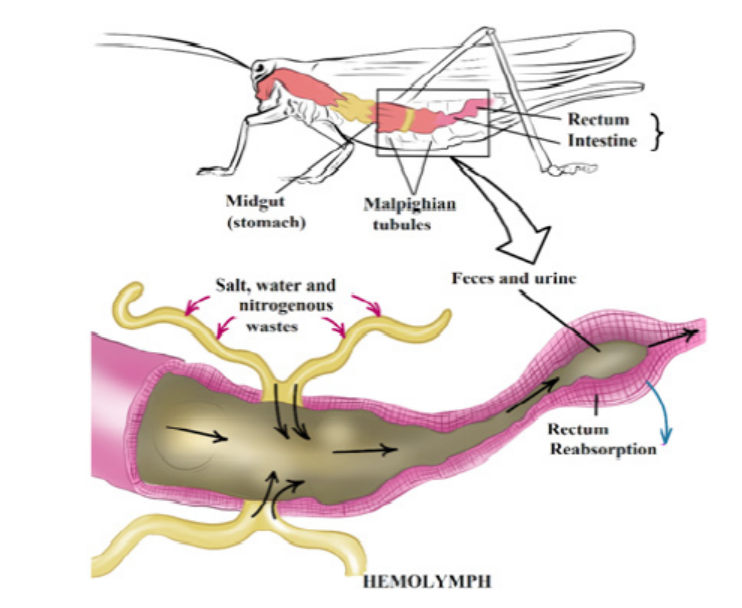
Figure 7.9: Malpighian tubules of insects
c. Excretion in Birds and Reptiles
Most birds live in environments that are dehydrated. Like mammals, birds have
kidneys with juxtamedullary nephrons that specialize in conserving water.
However, the nephrons of birds have loops of Henle that extend less far into the
medulla than those of mammals. Thus, bird kidneys cannot concentrate urine to
the high osmolarities achieved by mammalian kidneys. Although birds can produce
hyperosmotic urine, their main water conservation adaptation is having uric acid as
the nitrogen waste molecule. Since uric acid can be excreted as a paste, it reduces
urine volume.
The kidneys of reptiles having only cortical nephrons, produce urine that is osmotic
or hypo-osmotic to body fluids. However, the epithelium of the chamber called the
cloaca helps conserve fluid by reabsorbing some of the water present in urine and
faeces. Also like birds, most reptiles excrete their nitrogenous wastes as uric acid.
Freshwater fishes and amphibians
Freshwater fishes are hyperosmotic to their surroundings, so they must excrete excess
water continuously. In contrast to mammals and birds, freshwater fishes produce
large volumes of very dilute urine. Their kidneys, which contain many nephrons,
produce filtrate at a high rate. Freshwater fishes conserve salts by reabsorbing ions
from the filtrate in their distal tubules, leaving water behind.
Amphibian kidneys function much like those of freshwater fishes. When in freshwater, the kidneys of frogs excrete dilute urine while the skin accumulates certain
salts from the water by active transport. On land, where dehydration is the most
pressing problem of osmoregulation, frogs conserve body fluid by reabsorbingwater across the epithelium of the urinary bladder.
Marine bony fishes
The tissues of marine bony fishes gain excess salts from their surroundings and
lose water. These environmental challenges are opposite to those faced by their
freshwater relatives. Compared with freshwater fishes, marine fishes have fewer and
smaller nephrons, and their nephrons lack a distal tubule. In addition, their kidneys
have small glomeruli, and some lack glomeruli entirely. In keeping with thesefeatures, filtration rates are low and very little urine is excreted.
Application 7.7
1. What is the importance for birds and reptiles to excrete their nitrogenous
wastes in the form of uric acid?2. Explain how osmoregulation occurs in protozoa such as amoeba.
7.8 Excretion in plants
Activity 7.8
All living organisms carry out the process of excretion. Plants as other living
organisms need to remove the metabolic wastes products outside of their
bodies. Yet plants do not have kidneys and other excretory organs as seen
in animals. Use books from your school library and use internet for further
research to answer the questions that follow:
– Identify and write the structures that are involved in the excretion in plants.
– List the differences between the excretory system of a plant and that of a
human.
– Explain why plants do not have complex organs systems as animals?
Compared to animals, plants do not have a well-developed excretory system to
throw out nitrogenous waste materials. This is because of the differences in theirphysiology. Therefore, plants use different strategies for excretion.
The gaseous waste materials produced during respiration (carbon dioxide) and
photosynthesis (oxygen) diffuse out through stomata in the leaves and through
lenticels in other parts of the plant. Excess water evaporates mostly from stomata
and also from the outer surface of the stem, fruits, etc., throughout the day. This
process of getting rid of excess water is called transpiration. The waste products, like
oxygen, carbon dioxide and water, are the raw materials for other cellular reactions
such as photosynthesis and cellular respiration. The excess of carbon dioxide and
water are used up in this way. The only major gaseous excretory product of plants isoxygen.
Many plants store organic waste products in their permanent tissues that have
dead cells, for example in heartwood. Plants also store wastes within their leaves
or barks, and these wastes are periodically removed as the leaves and barks fall off.
Some of the waste products are stored in special cells or cellular vacuoles. Organic
acids, which might prove harmful to plants, often combine with excess cations and
precipitate out as insoluble crystals that can be safely stored in plant cells. Calciumoxalate crystals accumulate in some tubers like yam.
Aquatic plants lose most of their metabolic wastes by direct diffusion into the water
surrounding them. Terrestrial plants excrete some wastes into the soil around them.
Plants do not have complex excretory systems. This is because of the followingreasons:
– There is very little accumulation of toxic wastes. Often the plant wastes are
utilized by the plant. For example, carbon dioxide is used for photosynthesis
and oxygen for respiration.
– The extra gaseous waste is removed from the plant by simple diffusion through
the stomata and the lenticels.
– Most of the waste substances formed in plants are not harmful and can be
stored in the plant tissues.
– Some plants store other waste such as resins in their tissues in a non-toxic form.
These tissues or organs later fall off the plant.
– Excess water and dissolved gases are removed by the process of transpiration
through the stomata.
– Some plants remove waste products by exudation, for example gums, resins,
latex and rubber.
– In some plants water with dissolved salts oozes out through hydathodes. This
is called guttation.
Note that hydathodes are specialized structures and they are mainly responsible
for secreting water in liquid form. They are generally restricted to the apex or theserrated edges of the margins of leaves.
Application 7.8
1. What are the excretory products produced by plants? State any four.
2. Identify three ways by which plants excrete their waste products.3. What are hydathodes? What are their functions in excretion?
End of unit assessment 7
Multiple choice questions: choose the letter corresponding to the best answer.
1. Glucose is small enough to be filtered from the blood in glomeruli in the
kidney, but is not normally found in the urine. This is because glucose is:
a. Reabsorbed in distal convoluted tubules
b. Reabsorbed in proximal convoluted tubules
c. Reabsorbed along the whole length of the nephrons
d. Respired by cells in the kidney
2. Which of these does not contribute to the process of filtration in the kidney?
a. High hydrostatic blood pressure in glomerular capillaries.
b. Large surface area for filtration.
c. Permeability of glomerular capillaries.
d. Active transport by epithelial cells lining renal tubules.
3. The most important function of the kidney is:
a. Removal of water from the body.
b. Regulating blood composition.
c. Storage of salts in the body.d. Elimination of urea from the blood.
Structured answer questions4. The following diagram shows the nephron.
a. From the diagram above write the number that represents the: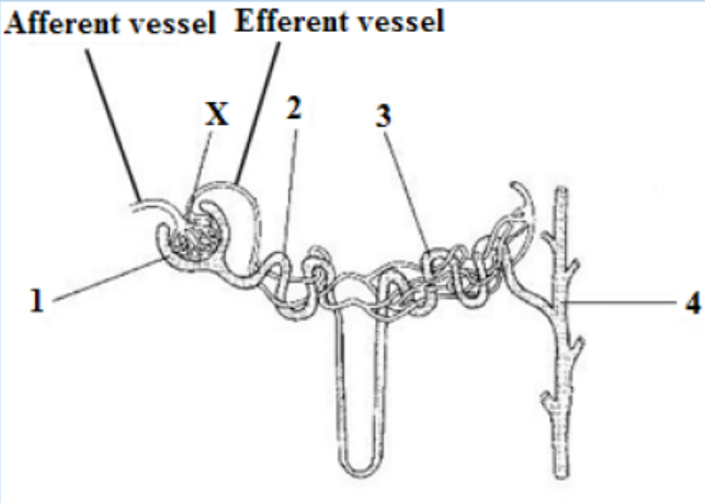
i. Collecting duct
ii. Bowman’s Capsule
b. On the diagram above label the loop of Henle.
c. Name structure X.
d. Compare the blood pressure in the afferent and efferent arterioles
and explain the cause of this difference.
e. Proteins are not present in the glomerular filtrate but amino acids
are. Explain.
f. Compare the urea concentration in the renal artery with that in the
renal vein.g. Name TWO organs that excrete urea.
5. Observe the diagram below and identify the following structures:
a. The structure that filters blood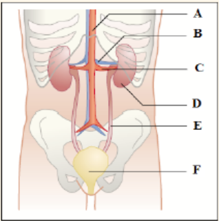
b. The structure that carries urine from the kidney
c. The structure that carries blood containing urea into the kidney
d. The structure that stores urine6. Use the figure below to answer the following:
a. Identify which letters indicate the afferent and efferent arterioles.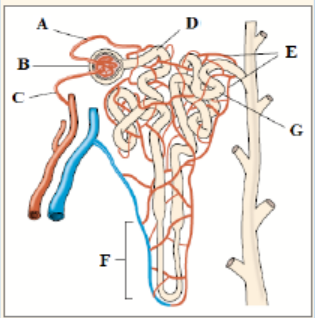
b. Explain how an increase in blood pressure in area (B) would affect
the functioning of the kidney.
c. Explain why proteins and blood cells are found in area (B) but not in
area (D).
d. In which area of the nephron would you expect to find the greatestconcentration of glucose?
UNIT 8 GENERAL PRINCIPLES OF RECEPTION AND RESPONSE IN ANIMALS
UNIT 8: GENERAL PRINCIPLES OF RECEPTION ANDRESPONSE IN ANIMALS.
Key Unit Competence
xplain the general principles of reception and response in animals.
Learning Objectives
By the end of this unit, I should be able to:
– Explain the necessity of responding to internal and external changes in the
environment.
– Describe the main types of sensory receptors.
– Discuss the main functions of a sensory system.
– Explain the significance of sensory adaptation.
– Describe the structure of the human eye.
– Describe the structure of the retina.
– Explain how rods transduce light energy into nerve impulses.
– Explain how retinal convergence improves sensitivity.
– Explain how the cones achieve visual acuity.
– Explain how cone cells produce colour vision.
– Discuss the significance of binocular vision.
– Describe the structure of the human ear and the functions of its main parts.
– Describe the process of hearing and balance.
– Locate the taste buds on the tongue and sensory cells in the skin.
– Observe the structure of the skin, retina, cochlea and vestibular apparatus from
prepared slides or micrographs and relate them to their functions.
– Interpret graphs on sensory adaptation in response to a constant stimulus.
– Relate the number of retinal cells to sensitivity and visual acuity
– Recognise the role of sense organs in the perception of different stimuli.
– Appreciate the role of sensory adaptation in protecting the sense organs fromoverload with unnecessary or irrelevant information.
Introductory activity
This scenario is involving bat and moth, snail and a cultivating human. Imagine
the situation in which a moth is flying in the darkness. At the same time there
is a bat flying in the same zone. There is also another situation in which a snail
is moving on the land as usual nearby its crawling area, there is a human who
is cultivating in the land where the above snail is moving. The two scenariosare illustrated below
1. What do you think would happen to a moth during the darkness when
it is in area where the bat is living?2. What would be the reaction of the snail to the human digging?
Animals realize different activities including searching for food, select a mate, and
escape from predators. They also have the ability to feel changes in environmental
factors and keep their internal environment within tolerable limits. These and
other activities depend on the animal’s ability to gather information about what
is happening inside and outside the body. The survival of animals depends upon
the ability to respond in an appropriate way to environmental changes through the
ability of detecting stimuli. Some other animals have become highly specialized to
detect a particular form of energy by the use of specialized receptor cells which
are able to perceive whichever form of energy and elaborate adequate response
respond to nervous impulse.8.1 Types of sensory receptors and stimuli
Activity 8.1
Use the school library and search additional information on the internet, read
the information related to different types of sensory receptors, while taking
short notes on each type of sensory receptors. What are the main sensoryreceptors?
The physical and chemical conditions in an animal’s internal and external
environments are continually changing. A change that can be detected is calleda stimulus. To some extent, all animal cells are sensitive to stimuli, and some cells
called receptors have become especially sensitive to particular stimuli. There are
a huge number of environmental variables that an animal could sense. However,
each species has evolved receptors only to environmental variables that have an
appreciable effect on its chances of survival. For example, humans can sense all thecolors of the rainbow but can sense neither infrared nor ultraviolet light.
Classification of receptors
Receptors are commonly classified according to the type of stimulus energy they
detect. The main types are:
– Mechanoreceptors which detect changes in mechanical energy, such as
movements, pressures, tensions, gravity, and sound waves.
– Chemoreceptors which detect chemical stimuli, for example, through taste and
smell.
– Thermoreceptors which detect temperature changes.
– Electroreceptors which detect electrical fields.– Photoreceptors which detect light and other forms of electromagnetic radiation.
Receptors can also be classified according to their structure. Simple receptors, known
as primary receptors, consist of a single neurone, one end of which is sensitive to
a particular type of stimulus. A primary receptor gathers sensory information and
transmits it to another neurone or an effector. For example, Pacinian corpuscles
are mechanoreceptors located in the skin, tendons, joints and muscles. Their ends
consist of concentric rings of connective tissue. Application of pressure against the
connective tissue deforms stretch-mediated sodium ion channels in the cell surfacemembrane, causing an influx of sodium ions which leads to a generator potential.
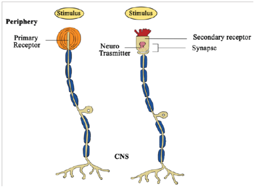
Figure 8.1: Primary receptor and secondary receptor (CNS: Central Nervous System)
A secondary receptor is more complex. It consists of a modified epithelial cell which
is sensitive to a particular type of stimulus. The cell senses changes and passes this
information on to a neurone which transmits it as nervous impulse. Sense organs are
complex stimulus – gathering structures consisting of grouped sensory receptors.
In many sense organs, several receptors make synaptic connections with a single
receptor neuron.
A third classification of receptors is based on the source of stimulation and includes
exteroceptors responding to stimuli outside the body, interceptors responding to
stimuli inside the body, and proprioceptors respond to changes of joint angle andamount of tension in muscles.
Application 8.1
1. Describe the main types of sensory receptors
2. Distinguish between a primary receptor and a secondary receptor
3. Which type of receptor detects changes in the internal environment of the
body?
4. Which one of the five categories of sensory receptors is primarily dedicatedto external stimuli?
8.2 Components of the sensory system: transduction, trans-
mission and processing
Activity 8.2
Use the school library and search additional information on the internet, read
the information related to the sensory system while taking a short summary
on sensory system, make a table showing the component and the functions
of the sensory system. What do you think about those components andfunctions?
8.2.1 Sensory systems
Receptors are the first component of a sensory system, which has three main
functions:
– Transduction: Receptor cells gather sensory information and then convert it
into a form of information that can be used by the animal (nerve impulses)
– Transmission: Sensory neurones transmit nerve impulses from the receptors
to the central nervous system
– Processing: the central nervous system processes the information so that
appropriate responses can be made to environmental changes.
A receptor converts the energy from the stimulus into an electrical potential that
is proportional to the stimulus intensity. This graded electrical potential is known
as the receptor potential or generator potential. If the stimulus is sufficiently high
(above a critical threshold level) the graded potential is high enough to fire an actionpotential. If the stimulus is beneath the threshold, no action potential takes place.
8.2.2 Sensory adaptation
Receptors are adapted to detect potentially harmful or beneficial changes in the
environment. When given an unchanging stimulus, most receptors stop responding
so that the sensory system does not become overloaded with unnecessary or
irrelevant information. Loss of responsive is brought about by a process called sensory
adaptation. An unchanging stimulus results in a decline in the generator potentials
produced by sensory receptors. Consequently, the nerve impulses transmitted in
sensory neurones become less frequent and may eventually stop. The mechanism
of sensory adaptation involves changes in the membranes of receptor cells and
explains why, for example, a person becomes insensitive to the touch of clothing onskin. Even a hair shirt becomes tolerable after wearing it for a long period of time.
8.2.2. Transferring information
After gathering and transducing the stimuli, the sensory system transmits
information about the stimulus to the central nervous system and effectors. The
frequency of nerve impulses propagated along a sensory neurone usually gives
information about stimulus strength. The transfer of information is rarely direct. In
mammals, much of the sensory information goes to sensory projection areas in thebrain where information processing takes place.
Application 8.2
1. Distinguish between an action potential and a generator potential
2. Explain the significance of sensory adaptation
3. Distinguish between transduction, transmission and perception
4. If you stimulated a sensory neuron electrically, how would thatstimulation be perceived?
8.3 Structure and functioning of the eye
Activity 8.3
Dissection of a mammalian eye
Materials needed:
Diagram of a dissected eye, scissors (optional), wax paper, plastic garbage bag,
a cutting board or other surface, on which you can cut, a sheet of newspaper,
soap, water, and paper towels for cleaning up, one cow’s eye for every sixlearners, and one single-edged razor blade or scalpel for every team
Procedure
– Examine the outside of the eye and see how many parts you can identify.
– Cut away the fat and the muscle.
– Use scalpel to make an incision in the cornea.
– Cut until the clear liquid in the cornea is released.
– Use the scalpel to make an incision through the sclera in the middle of the
eye.
– Cut around the middle of the eye until you get two halves.
– Remove the front part and place it on the board.
– Cut the front part with scalpel or razor
– During cutting of the front part, listen and explain what happens.
– Pull out the iris between the cornea and the lens.
– Observe in the centre of the iris after pulling out the iris.
– Remove the lens and mention its texture.
– Hold the lens in front of you and observe. What do you observe?
– Empty the vitreous humor out of the eyeball.
– Remove the retina and mention whether the spot is attached to the back of
the eye.
– Find the optic nerve and pinch the nerve with your fingers or with a pair ofscissors. What do you see there?
Questions
1. Draw and label the internal structure of the mammalian eye.2. Write in your own words the functions of each part of a mammalian eye
The eye is a complex light – sensitive organ that enables us to distinguish minute
variations of shape, color, brightness, and distance. The function of eye is to
transduce light (visible frequencies of electromagnetic radiation) into patterns of
nerve impulses. These are transmitted to the brain, where the actual process ofseeing is performed.
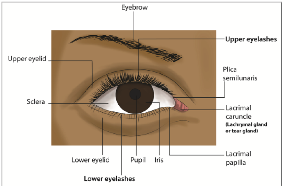
Figure 8.2: External structure of human eye
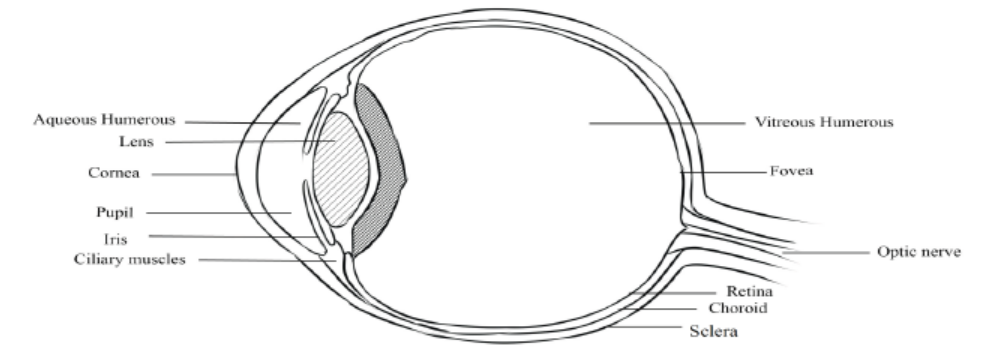
Figure 8.3: Internal structure of human eye
8.3.1. Functions of parts of eye
– The lens: Refracts light and focuses it on retina. Made up of elastic material that
adjusts when the eye focuses on far or near object.
– The ciliary body: Made up of muscle fibres which contract or relax to change
the shape or curvature of the lens. It produces aqueous humour.
– The suspensory ligament: The suspensory ligament is a tissue that attaches
the edge of the lens to the ciliary body.
– The iris: It is coloured part of the eye, it has radial and circular muscles which
control the size of the pupil; it has melanin pigment that absorbs strong light to
prevent blurred vision.
– Pupil: It is a hole at the centre of the iris through which light pass into the eye.
– Aqueous humour: Has fluids to maintain the shape of eye ball and to refract
light rays. It contains oxygen and nutrient for cornea and lens. It is a transparent
and allow light to pass through
– Vitreous humour: It is the space behind the lens and it is filled with fluids, a
transparent, jelly-like substance. Vitreous humour keeps the eyeball firm and
helps to refract light onto the retina.
– Cornea: Is transparent part of the eye and allows the passage of light. It refracts
light ray. It is made up of tough tissues to strength the eye.
– Choroid: The choroid is the middle layer of the eyeball that lies between the
sclera and retina. It has two functions, one being able to prevent internal
reflection of light as it is pigmented black. Secondly, it contains blood vessels
that bring oxygen and nutrients to the eyeball and remove metabolic waste
product.
– Retina: The retina is the innermost layer of the eyeball. It is the light sensitive
layer on which images are formed. It contains light sensitive cells called
photoreceptors. Photoreceptors consist of rods and cones. Cones enable us to
see colours in bright light while rods enable us to see in black and dim light. The
photoreceptors are connected to the nerve endings from the optic nerve.
– Blind spot: The blind spot is the region where the optic nerve leaves the eye. It
does not contain any rods or cones. Therefore, it is not sensitive to light.
– Optic nerve: It is a nerve that transmits nerve impulses to the brain for
interpretation when the photoreceptors in the retina are stimulated.
– Fovea or yellow spot: It is a small yellow depression in the retina. It is situated
directly behind the lens. This is where images are normally focused. The fovea
contains the greatest concentration of cones, but has no rods. The fovea enables
a person to have detailed colour vision in bright light.
– Conjunctiva: Thin and transparent to allow light to pass through.
– Sclera: It is a tough, white outer covering of the eyeball, which is continuous
with the cornea. It protects the eyeball from mechanical damage.
– The eye brows: Prevent sweat and dust from entering the eye.
– The eye lashes: Prevent dust particles from entering the eye.
– The tears glands: Secrete tears that wash away dust particles in the eye andkeep the eye moist.
8.3.2. Accommodation of the eye
The ability of the eye to see far and near objects on the retina is possible because the
eye is able to adjust the size of the lens and its power to bend light. Adjustment of
the size of the lens is done by the ciliary muscles inside the ciliary body which exert
a force on the suspensory ligament and then onto the lens. Changes that occur inthe eye during accommodation include:
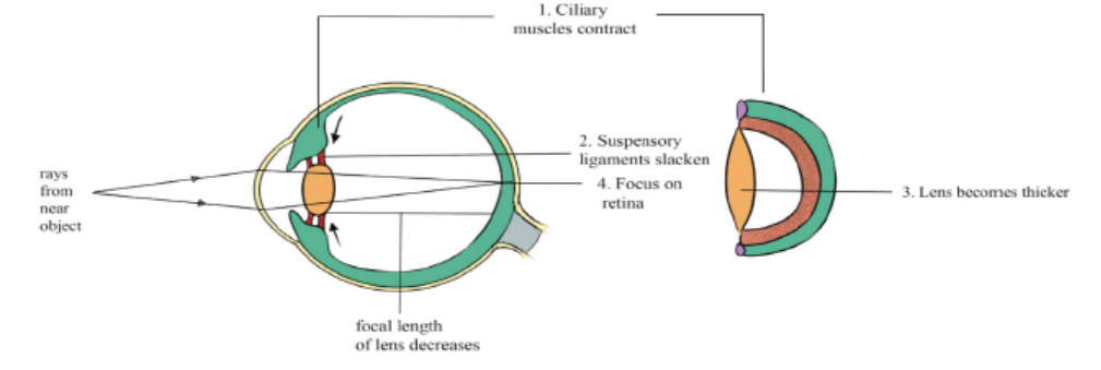
Figure 8.4: Illustration of seeing a near object
When the eye focuses on a near object, several changes occur:
– The ciliary muscles contract, relaxing their pull on the suspensory ligaments.
– The suspensory ligaments slacken, also relaxing their pull on the lens.
– The lens, being elastic, becomes thicker and more convex, decreasing its focal
length.
– Light rays from the near object are sharply focused on the retina.
– Photoreceptors are stimulated.
– The nerve impulses produced are transmitted by the optic nerve to the brain.
The brain interprets the impulses and the person sees the near object.
b. Focusing on a distant object: When a person is looking at a distant object,
the light rays reflecting off the object are almost parallel to each other
when they reach the eye. These ‘parallel’ light rays are then refractedthrough the cornea and the aqueous humour into the pupil
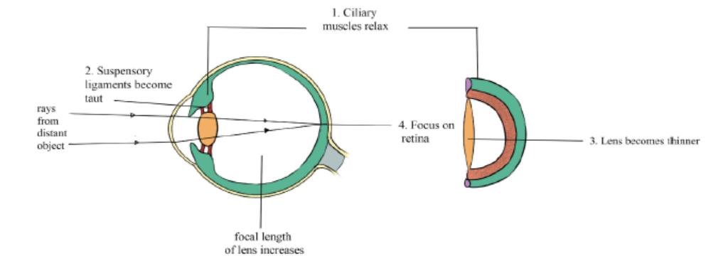
Figure 8.5: Illustration of seeing a distant object
When the eye focuses on a distant object, several changes occur.
– The ciliary muscles relax, pulling on the suspensory ligaments.
– The suspensory ligaments then become taut, pulling the edge of the lens.
– The lens become thinner and less convex, the focal length is increased. The
focal length is the distance between the middle of the lens and the point of
focus on the retina.
– Light rays from the distant objects are sharply focused on the retina and
photoreceptors are stimulated.
– The nerve impulses produced are transmitted by the optic nerve to the brain.
The brain interprets the impulses and the person sees the distant object
Table 8.1. Summary of changes that occur in the eye during accommodation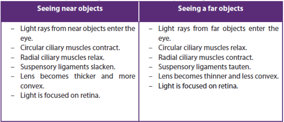
8.3.4. Some changes that occur in eye when you see in bright and dim
light
In bright light
– Circular iris muscle contracts.
– The radial iris muscles relax.
– The iris elongates in wards each other.
– The pupil is reduced (narrowed).– Small amount of light rays enters the eye.
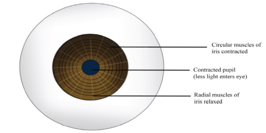
Figure 8.6: Illustration of changes that occur in eye when you see in bright light
Table 8.2: Illustration of changes that occur in eye during bright and dim light
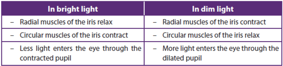
8.3.5. The retina of the eye
The retina possesses the photoreceptor cells. These are of two types, cones and rods.
Both converts light energy into the electrical energy or nerve impulses. Both rods
and cones are embedded in the pigment epithelial cells of the choroid layer. In cats
and some other nocturnal mammals. They have reflecting layer called the tapetumwhich reflects light back into the eye and so allow the rod cells to absorb it.
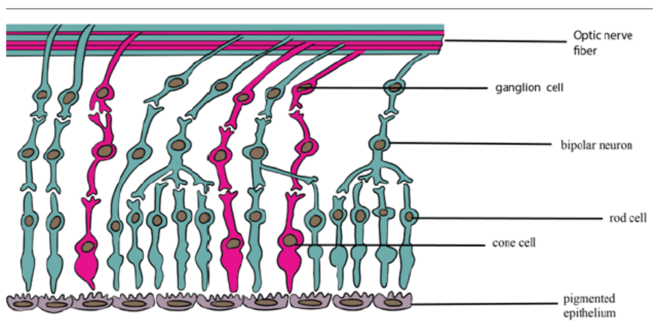
Figure 8.8: Structure of the retina
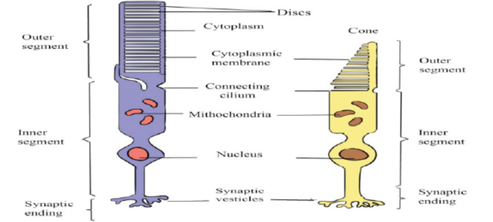
Figure 8.9: Structure of photosensitive cells
8.3.6. Adaptations of photosensitive cells.
– They have numerous mitochondria to provide energy in form of ATP.
– They have photosensitive pigment i.e. rhodopsin in rods and iodopsin in cones
to absorb light rays.
– They have lamellae (vesicles) to increase the surface area for holding the
pigment molecules.
– Many rods cells share a single bipolar neurone such that a single stimulation
builds up a big generator potential.
8.3.7. Changes which occur on rod cells when light strikes the retina
Each rod cell has in its outer segment up to 1000 vesicles, each containing aphotosensitive pigment called rhodopsin. Rhodopsin is made up of the protein
opsin and retinal, a derivative of vitamin A. Light causes retinal to change shape
from its normal cis-isomeric form to trans-isomeric form. As result, retinal and opsin
break apart. This process is called bleaching. This triggers a series of events which
alters the permeability of rod’s cell surface membrane.
If light stimulation exceeds the threshold level, an action poetical is set up in a
bipolar neurone, and then passes along a neurone in the optic nerve. The pattern of
nerve impulses transmitted along different neurones is interpreted in the brain as
patterns of light and dark. Before the rod cell can be activated again, the opsin and
retinal must first be resynthesized into rhodopsin.
This re-synthesis is carried out by the mitochondria found in the inner segment
of rod cell, which provide ATP for the process. Re-synthesis takes longer time than
splitting of rhodopsin but is more rapid in lower light intensity. Rhodopsin of rods
spits into opsin protein and retinal (derivative of vitamin A). About 3 minutes are
required to reform again. That is why our eyes need some minutes to adapt to darkwhen we come from bright light.

The splitting of iodopsins of cone cells also produces an action potential (impulse)
but they quickly re-form. There are three types of iodopsins and each responds to
the wavelength of a particular colour: red – green – blue.
The impulses are then transmitted along the optic nerve to the visual area of the
brain. There, the image is interpreted. Note that the image that is cast on the retina
is virtual I to mean not real, small, inverted upside down and laterally, and reversedfor example from right to left.
8.3.8. Changes which occur on cones when light strikes the retina
When light of high intensity strikes the cones, the iodopsin pigment decomposes
into iodide ions and opsin, this process is called bleaching. On the contrary, when
enough iodopsin is decomposed, the membrane develops an action potential
when it reaches threshold level. An impulse is fired via bipolar neurone to the optic
nerve to the brain for interpretation. A comparison between cone and rod cells issummarized in the table 8.3.
Table 8.3: Differences between rods and cones
8.3.9. The process of vision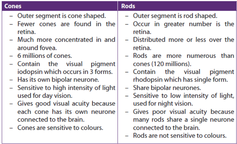
When light enters the eye, it is refracted by the curved surface of the cornea, the
lens, the aqueous and vitreous humour. The refraction of light causes the image to
be formed upside down on fovea centralis. When cones and rods are stimulated by
light, they send impulses through the optic nerves to the brain where the correct
impression of the object is formed
Colour vision in organism is explained by the trichromatic theory which states that,
there are three forms of iodospin each responding to light of different wave length
that is each responds on one of the three primary colours which are, blue, green
and red. When these colours are mixed in appropriate intensities they can give rise
to any other colour for example equal stimulation of red and green cones gives
yellow perception. Alternative theory of colour vision known as the retinex theory,
suggests that the brain cortex as well as retina is involved in colour perception. This
would explain why we usually perceive a particular object as being the same colourunder different types of illumination.
a. Stereoscopic vision: combining two images
Having two eyes (binocular vision) is better than having one because it gives a larger
field of vision, a defect in one eye does not result in blindness. In animals with two
forward facing eyes, it provides the potential for stereoscopic vision which depends
on each eye being able to look at the same object from slightly different perspective.
The visual centre in the brain combines the two views to make a three dimensional
image. Stereoscopic vision provides information about the sizes and shapes of object
and enables distance to be judged accurately. However, because the eyes have to
be relatively close together for stereoscopic vision, the field of vision is relatively
small. Mammalian predators tend to have well developed stereoscopic vision, while
herbivores tend to have eyes wide apart, sacrificing stereoscopic vision for a widefield of view
b. Nocturnal animals
Nocturnal animals have a lot of rods in their retinas, but no cones. The levels of light
at night are very low, so even if the animals have lot of cones, they would not be able
to see in colour because the level of light is too low to stimulate the cone cells. At
night, animals need to be able to detect shape and movement and the very sensitiverod cells are ideal of this because they are stimulated by very low levels of light.
Application 8.3
1. What is meant by the term adaptation of the eye?
2. Describe the adjustments which occur in the eye in bright and dim
light.
3. If you perceive an object floating across your field of view, how can you
determine whether the image represents a real object or a disturbance
in your eye or a neural circuit of your brain?
4. Distinguish between visual acuity, adaptation and photoreception of
the eye
5. Describe the shape of the lens when the eye is focused on a near object?
6. Study the section of the human eye and then complete the table, byfilling in the letter and the name of the correct part
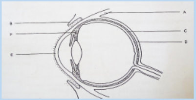

7. Which type of photoreceptors occur in the fovea
8.4 Structure and functioning of the ear
Activity 8.4
Use textbooks and other additional sources (e.g. internet), read the information
related to the human ear and make notes about it.
1. Draw and label a diagram of human ear2. Give the functions of each part of the ear
The human ear is a complex sensory organ that enables us to hear sounds, detect
body movements, and maintain balance. The ear has three main parts: an air-filled(outer ear), an air-filled middle ear, and a fluid- filled inner ear
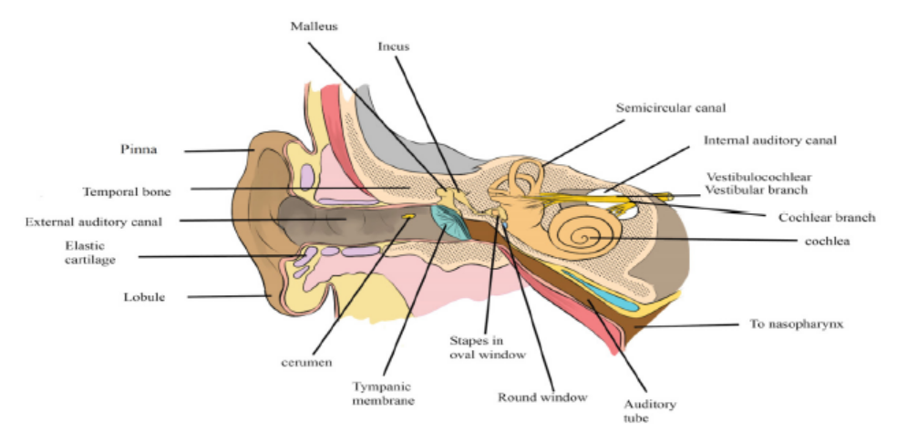
Figure8.10: Illustration of external and internal structures of human ear
Each part of the ear has specifc feature and function as it is indicated in the table 8.4.Table 8.4: The functions of the parts the ear
8.4.1. Sound perception in the ear (Hearing)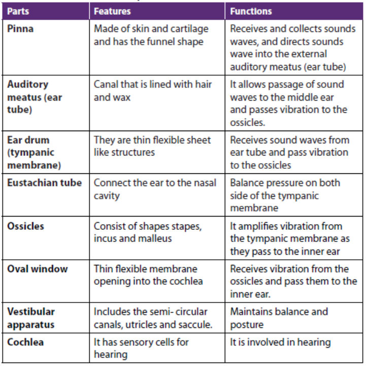
The most function of the ear is hearing. The hearing process include the following
processes:
– Sound waves are collected by the pinna and directed to the auditory canal,
which then strike the ear drum (tympanic membrane)
– The sound waves cause the tympanic membrane to vibrate and the vibrations
are sent to the ossicles.
– The ossicles amplify the vibration and amplified vibration are received by
the oval window that setting up vibration in the perilymph of tympanic and
vestibular canal.
– Vibration in perilymph cause movement of Reissner’s membrane which in
turn displaced relative to the tectorial membrane, the sensory hair cell located
between the basilar membrane and tectorial become distorted.
– This distortion set up an action potential, which is transmitted along theauditory nerve to the brain which interprets the impulses as sound.
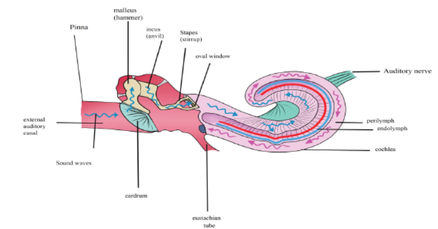
Figure8.11: The diagram showing the process of hearing
8.4.2. The cochlea and the organ of corti
The cochlea is coiled around above and their internal region is crossed by two
membranes, i.e. upper Reissner’s membrane and lower basilar membrane. In
between there is a membrane which is short called tectorial membrane. From the
basilar membrane are sensitive sensory hair cells whose hair tips are close to the
tectorial membrane. These cells have fibres which take impulses to the brain along
the auditory nerve for interpretation. The upper and lower chambers of the cochlea
are filled with perilymph while the middle chamber is filled with endolymph. The
basilar membrane, tectorial membrane, Reissner’s membrane and sensitive hair cellsare collectively known as the organ of corti and are directly concerned with hearing.
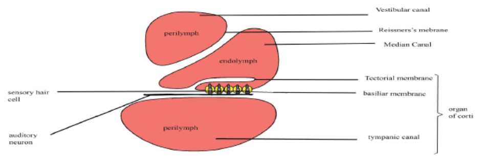
Figure 8.12: Structure of cochlea and organ of corti
8.4.3. The vestibular apparatus and sense of balance
Our sense of balance and information about position and movement come from
the vestibular apparatus in the inner ear. The vestibular apparatus consists of the
semicircular canals, containing organs called cristae sacs including the saccule and
utricle. The utricle and saccule are receptors containing sense organs called maculae
that give information on the position of head in space in relation to gravity (static
equilibrium).
These receptors consist of sensory hair cells which are embedded in fine granules
of calcium carbonate called otoliths. According to the position of the head, the pull
of gravity on the otolith will vary and otolith will be titled accordingly. The different
distortions of the sensory cells that result from impulses discharge in the vestibular
nerve fibres and this is interpreted by the brain, which sends impulses to the relevant
organs which then restore the balance of the body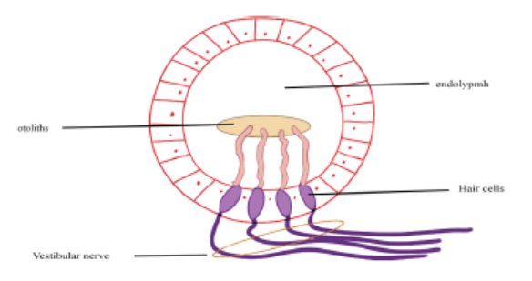
Figure 8.13: The diagram illustrating the macula
8.4.4. The role of semicircular canals in the maintenance of balance
Semicircular canals are responsible for maintaining the balance of the body during
motion (dynamic equilibrium). These are fluid – filled canals, three in number and
arranged in three mutually perpendicular planes: vertical canals detect movement
in the upward direction, horizontal canals detect back ward and forward motion
while lateral canals detect sideways movement of the head.
A swelling, the ampulla in the canal contains the receptor. This consists of sensory
hair cells supported by hairs embedded in a dome – shaped of a gelatinous structure
called cupula. Movements of head in any of the planes causes the fluid in the relevant
canal to move and therefore displacing the cupula. Due to inertia, the cupula is
deflected in direction opposite to that of head. This put strain on the sensory cells
and causes them to fire impulses in the different nerve fibres to the brain.
The pattern of impulses sent to the brain varies depending on the canal stimulated.
The brain interprets impulses and detects the speed and direction of movement
of head. Then impulses from brain are sent to the relevant organs which thenmaintained the balance of the body.
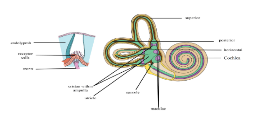
Figure 8.14: Diagram of semi-circular canals
Figure 8.15: Internal structure of semicircular canal
8.4.5 Ear as a balance organ
The vestibular apparatus is concerned mainly with detecting changes in the head
position and body posture. When the head moves quickly, the cupula, knob in the
ampulla, moves in the opposite direction. Sensory hairs below the cupula detect theimpulse that is brought by a vestibular nerve to the brain.
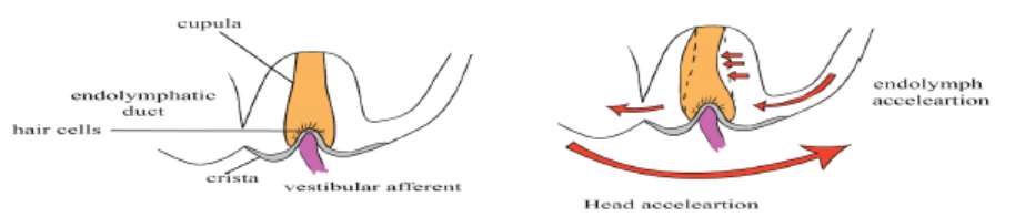
Figure 8.16: Illustration of the ear as a balance organ
Likewise, as the head moves by changing its posture, some crystals of CaCO3 knownas otoliths also move. The membrane of the otoliths also moves pulling on the
sensitive hairs and making them bend. The sense cells are stimulated to varying
degrees, causing an action potential to be sent to the cerebellum (hindbrain) that
actually controls the muscles in maintenance of body balance. The cerebellum sends
out impulses to the muscles of the body which contract or relax or maintain bodybalance.
Application 8.4
1. In which part of the ear are the organs of balance?
2. What is the role of ossicles during transmission of sound waves?
3. Which structure equalizes the pressure on either side of the eardrum?
4. Distinguish between pitch and intensity of sound
5. Suppose a series of pressure waves in your cochlea causes a vibration of
the basilar membrane that moves gradually from the apex toward the
base. How would your brain interpret this stimulus?
6. If the stapes became fused to the other middle ear bones or to the ovalwindow, how would this condition affect hearing? Explain
8.5 Structure and functioning of the tongue
Activity 8.5
Use the school library and search additional information on the internet,
read the information related to the tongue while taking a short summary on
tongue, list all taste buds on the tongue and answer the following questions:
1. Which taste buds are found at the tip of the tongue?2. Which taste buds are found on sides of the tongue?
The tongue is the receptor organ for taste. Taste is due to chemicals taken into themouth and for this reason the tongue is called chemoreceptor.
The tongue is able to distinguish between four different kinds of taste including
sweet, sour, salt and bitter which are also called primary taste. This is possible
with the help of group of sensory cells found in taste buds located on the surface
of the tongue in specific taste areas through four types of taste buds in which they
are located in overlap as shown on the Figure 8.18, the detection of sour and bitter
substances is important for they can be easily rejected if harmful. For a chemical
to be tasted it must be dissolved in the moisture of the buccal cavity where it canstimulate the sensory cells grouped in taste buds.
Different types of taste and their sites on the tongue
In human, there are four kinds of taste including sweet, salty, sour and bitter.
Different taste buds are sensitive to different chemicals: Those which are sensitive to
sugary and salty fluids are usually found at the tip of the tongue while those at the
sides of the tongue are sensitive to acidic substances and thus give the sensation ofsourness while those at the back are responsible for the sensation of the bitterness.
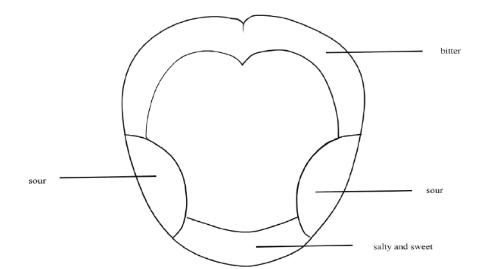
Figure 8.18: Location of different papillae
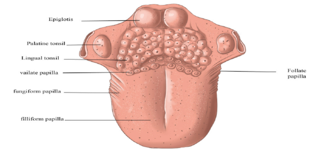
Figure 8.18: Location of different papillae
Application 8.5
1. Explain why some taste receptor cells and all olfactory receptor cells use
G protein-coupled receptors, yet only olfactory receptor cells produce
action potentials
2. If you discovered a mutation in mice that disrupted the ability to taste
sweet, bitter, but not sour or salty, what might you predict about theidentity of the signalling pathway used by the sour receptor?
8.6 Structure and functioning of the skin
Activity 8.6
Use the school library or the internet, make a research about the human skin
and make a short summary on it with all the sensory cells in it
1. Draw and label a diagram of human skin
2. How many types of sensory cells found in human skin?3. Write in your own words the functions of each part of human skin.
The human skin is the largest organ of the body. Being a vast organ, it has
many functions including protection from microbes, regulation of the body
temperature, and permits the sensations of touch, heat, and cold. This is possible
thanks to the presence of different glands. The skin consists of three main layers: The
epidermis, the outermost layer of skin that provides a waterproof barrier and creates
our skin tone, the dermis, beneath the epidermis that contains tough connective
tissue, hair follicles, and sweat glands and the deeper subcutaneous tissue called
hypodermis that is made of fat and connective tissue.The epidermis consists of three regions:
– The Cornfield layer also known as keratinized layer. This is the thin outermost
layer made up of dead cells. It is resistant to bacterial infections and damage,
and reduces water loss from the body. It is very thick on the soles of the feet and
the palm and is also modified as nails.
– The Granular layer that contains living cells which give way to the cornfield
layer.
– Malpighian layer that is the continuous layer of living cells and they
continuously divide to produce new cells. This layer has melanin pigment
granules that determine the skin colour and act as screen against ultraviolet
light.
The dermis consists of the thick connective tissue. It consists of blood capillaries,
receptors (sensory organs), lymphatic, sweat glands, sebaceous glands and hair
follicles with different functions:
– Capillaries supply food and oxygen, remove excretory waste products and
help in temperature regulation.
– Sweat glands are coiled tubes consisting of secretory cells with duct that
passes sweat to the skin surface.
– Hair follicles are deep pit (hole) of cells which divide and build the hair inside
the follicle. They are richly supplied with sensory nerve endings which are
stimulated by the hair movements.
– Sebaceous gland opens into the hair and secretes oil which makes the hair
waterproof.
– Sensory nerve endings include sensory receptors for temperature, touch,
pressure and pain.
Subcutaneous layer attaches dermis to underlying structures, composed of adipose
and connective tissue. It serves as shock absorbers for vital organs, it stores energy. Itvaries in thickness according to age, sex, general health of individual.
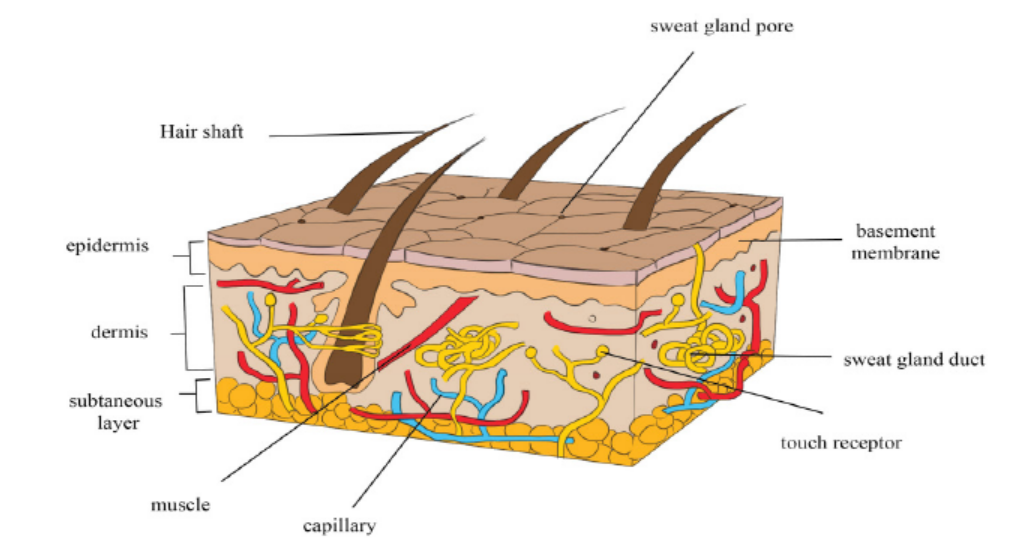
Figure 8.19: Human skin structure
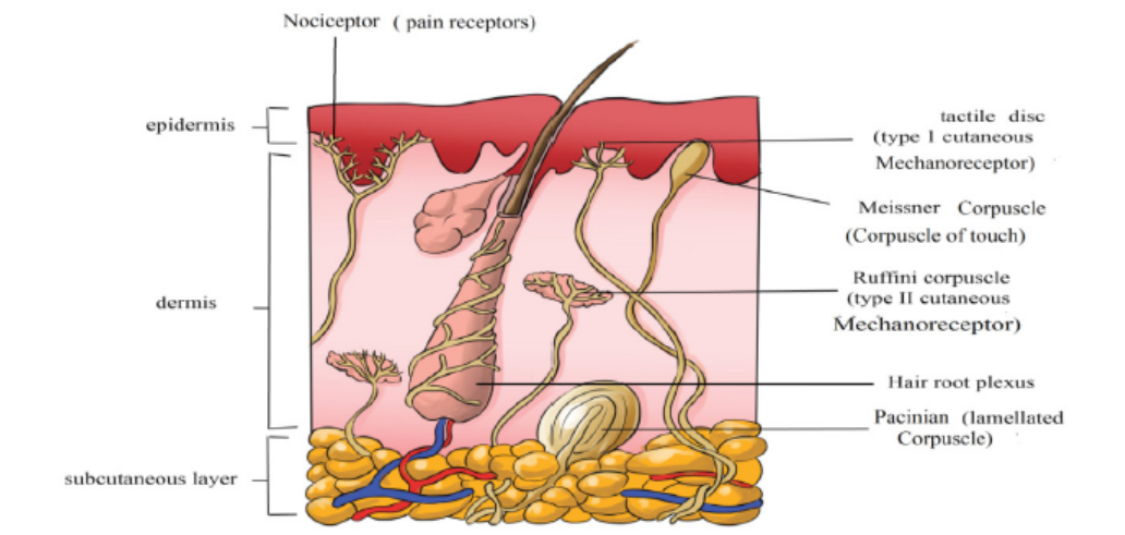
Figure 8.20: Figure the location of human skin receptors
A comparative study of sense organs
Sense organs have different biological functions beneficial to the living organisms.
A brief summary is given in the table 8.5.Table 8.5: The functions of sense organs
Application 8.6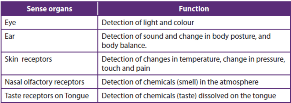
1. Describe how the skin contributes to the regulation of body temperature,
storage of blood, protection, sensation, excretion and absorption, and
synthesis of vitamin D.
2. Why do eating food containing hot peppers sometimes cause you to sweat?
3. If you stimulated a sensory neuron electrically, how would that stimulationbe perceived?
End of unit assessment 8
A. Multiple choice questions: choose the best answer
1. Human receptors are classified into:
a. sensory and motor receptors
b. Photoreceptors, mechanoreceptors, chemoreceptors,
thermoreceptors
c. Pacinian, Meissner, and Ruffini receptors
d. Central, peripheral and sympathetic receptors
e. Mechanical, electrical and gravitational
2. The eye contains:
a. Mechanoreceptors
b. Photoreceptors
c. Chemoreceptors
d. Proprioceptors
3. The small bones located in the middle ear, collectively as ossicles,
include:
a. Tympanum, oval and round windows.
b. Pinna, vestibule and Eustachian.
c. Malleus, incus, and stapes.d. Ossicles I, II and III.
B. Answer by True or False
4. Pain receptors are a type of mechanoreceptor.
5. Receptors for a particular sensation, such as touch, are spread evenly
throughout the skin surface.6. The image formed on the retina is inverted.
C. Essay questions
7. Describe what would happen to rhodopsin when it absorbs light
8. According to the trichromatic theory of colour vision, discuss which
colours of light are the three different types of cone sensitive to.
9. The diagram represents enlarged section of part of the retina andchoroid of a human eye.
a. Draw an arrow on a sketch of the diagram to show the direction in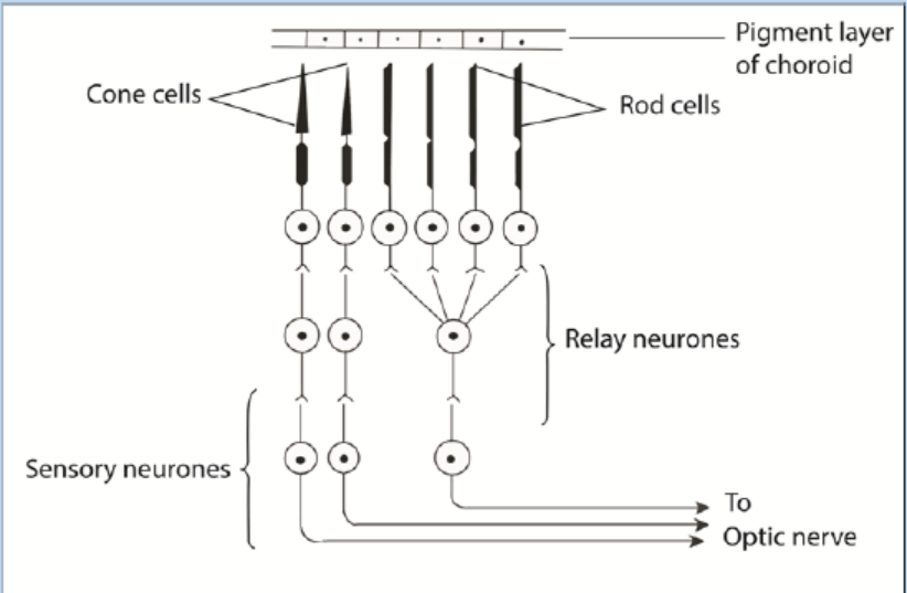
which light passes through the retina
b. Suggest a function of the black pigment which occurs in the choroid
layer of the eye
c. Use information in the diagram to explain how a person is able to:
i. see light of low intensity
ii. see in great detail in bright light
10. Describe the significance of three semi-circular canals being in differentplanes?
UNIT 9 NERVOUS COORDINATION
UNIT 9: NERVOUS COORDINATION
Key Unit Competence
Describe the structure of neurons and explain the mechanisms of impulsetransmission.
Learning Objectives
By the end of this unit, I should be able to:
– Describe the arrangement of neurons in a reflex arc.
– Describe the structure neurones.
– Explain how a resting potential is maintained.
– Explain how an action potential is generated.
– Explain how a nerve impulse is propagated along a neurone.
– Explain the factors affecting the speed of impulse transmission.
– Describe the properties of a nerve impulse limited to: saltatory conduction, all
or nothing law, and refractory period.
– Describe the functions of neurones in a reflex arc.
– Explain how information passes across a synapse from one neurone to another
or from a neurone to its effector.
– Outline the roles of synapses.
– Describe the roles of neuromuscular junctions, transverse system tubules and
sarcoplasmic reticulum in stimulating contraction in striated muscle.
– Relate the structure of a cholinergic synapse to its functions.
– Interpret graphs for all or nothing law and refractory period.
– Investigate the nature of a nerve impulse in a nerve tissue of a frog
– Appreciate the importance of a coordinated behaviour in organisms.– Show concern about the need to have reflexes as rapid responses
9.1 Overview of control and co-ordination in mammals
Activity 9.1
– Use charts showing the parts of human brain and watch the movies on you
tube showing the different parts of human brain.
– Use the school library and search additional information on the internet.
Read the information related to human brain, and take short notes on
human brain.
1. Illustrate with diagram the main parts of human brain
2. Write down the relative functions of each identified part of the humanbrain
Coordination:It is the process in which body coordinate, ordinate and control
different activities.
The nervous system plays the main functions such as: (i) Sensory input: Sensory
receptors present in skin and organs respond to external and internal stimuli by
generating nerve impulses that travel to the brain and spinal cord, (ii) Integration:
The brain and spinal cord sum up the data received from all over the body and send
out nerve impulses (iii) Motor output: The nerve impulses from the brain and spinalcord go to the effectors, which are muscles and glands.
The Nervous system is divided into two main divisions: The central
nervous system (CNS) and the peripheral nervous system (PNS) The central nervous
system (CNS) consist of the brain and spinal cord, which are located in the midline
of the body. The peripheral nervous system (PNS), which is further divided into the
somatic division and the au- tonomic division, includes all the cranial and spinal nerves.
9.1.1 Some key word definitions
–Irritability or sensitivity. This is the ability of living organisms to respond to a stimulus
–A stimulus: This is any change in the external or internal environment which provokes a response
–Receptors: These are specialized cells that detect a stimulus
–Neurons: These are cells which transmit nerve impulses
–Effectors: are organs that respond to the stimuli and bring about a response.
–A nervous system: This is a system which involved in the detection of stimuli (sensory inputs) integration and response (motor output)
–The response may be to both the external and internal environments.
–Neurone or nerve cell: It is the basic functional unit of the nervous system. Neurones are cells specialized to generate and transmit nerve impulses (action potentials) are cells which transmit nerve impulses (action potentials).
9.1.2 The division of nervous system
The nervous system of a mammal comprises of the central nervous system (CNS)
consisting of the brain and the spinal cord, and the peripheral nervous system (PNS)
consisting of the cranial nerves from the brain, the spinal nerves from the spinalcord and the sensory organs (Figure 9.1).
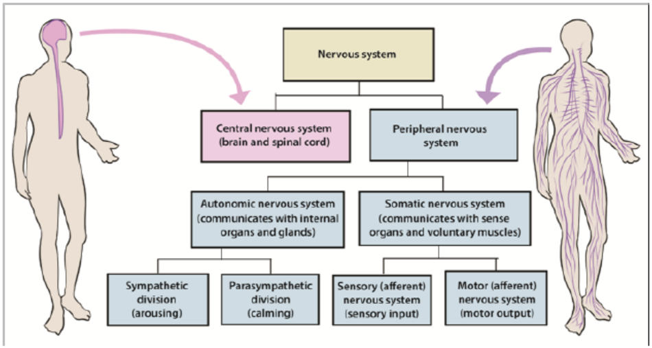
Figure 9.1: Organization of the human nervous system
1. The human brain
The brain is the enlarged end of the spinal cord. It is enclosed in the skull and is
divided into three main parts namely: the fore brain, the mid brain, the hind brain.
a. The fore brain
This consist of: cerebrum, thalamus, hypothalamus and pituitary gland
• The cerebrum:
This is the largest part of the brain made up of two hemispheres called the right and
the left cerebral hemispheres. The left cerebral hemisphere controls those activities
of the right side of the body while the right cerebral hemisphere controls those of
the left side of the body.
The functions of the cerebral hemisphere
– It is the centre of the judgment, memory, reasoning and imagination.
– It receives the impulses from the sensory organs: sight, taste, sound and touch.
– It controls all the body’s voluntary activities, e.g. walking, eating, singing,
• The thalamus:
This is a relay centre. It relays sensory information towards higher centre. It is thecentre for the perception of pain and pleasure.
• The hypothalamus
It performs many functions such as; regulates and monitors the temperature of
blood, monitors and regulates the water content of blood, a co-ordinating centre
for activities of the internal organs, e.g. rate of heart beat, blood pressure. It is a
centre of for feelings such as; hunger, thirst, sex drive, satisfaction, sleep, speech, etc.
As an endocrine gland, it produces hormones i.e. anti-diuretic hormone (ADH) andoxytocin.
• The pituitary gland:
It produces hormones such as: Follicle-stimulating hormone (FSH), Thyroidstimulating
hormone (TSH), Adreno-cortico trophic hormone (ACTH), Prolactin
hormone and Luteinizing hormone (LH)
b. The mid brain
This acts as an association centre between the fore and the hind brain. It is a relay
centre for audio and visual information. It is also responsible for movement of the
head and the trunk.
c. The hind brain
This receives the impulse from the ear, the skin and the semi-circular canals. It
consists of: The cerebellum and the medulla oblongata
The cerebellum: It lies behind the optic lobes. It receives impulses simultaneously
from the eyes and the ears. It regulates and co-ordinate muscular movement,
especially those concerned with maintaining body equilibrium and controls all the
unconscious activities of the body.
The medulla oblongata: This control all the involuntary movements of the body
especially those concerned with respiration, digestion, heartbeat, breathing rateand sneezing.
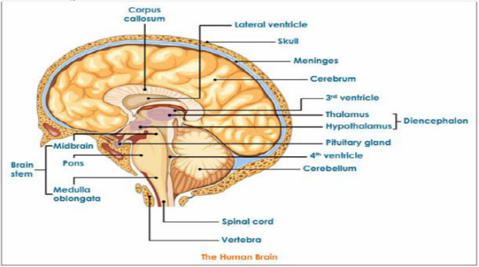
Figure 9.2: Main parts of the brain
2. The spinal cord
The spinal cord is a dorso -ventrally flattened cylinder of nervous tissue running
from the base of the brain down the lumbar region. It is protected by the vertebrateof the backbone and the meninges.
Functions of the spinal cord include;
– It is a coordinating centre for simple reflex such as the knee-jerk response and
the autonomic reflexes such as contraction of the bladder.
– Providing a means of communication between peripheral nerves and the brain.– It sends messages to the effectors
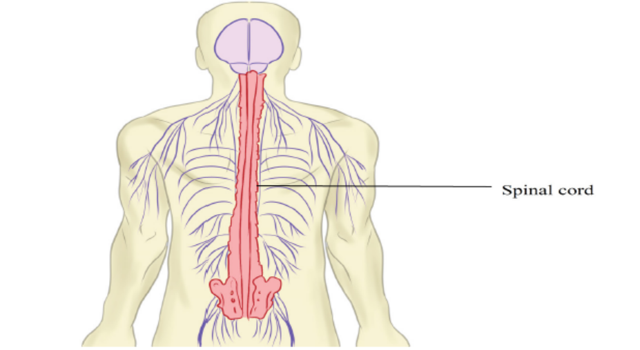
Figure 9.3: Position and external structure of spinal cord
A transverse section of the spiral cord shows an H-shaped central core of grey matter.
Grey matter is composed of nerve cell bodies, dendrites and synapses surrounding
a central canal which contains cerebrospinal fluid. White matter: around the grey
matter, is an outer layer containing nerve fibres whose fatty myelin sheaths give itits characteristics colour.
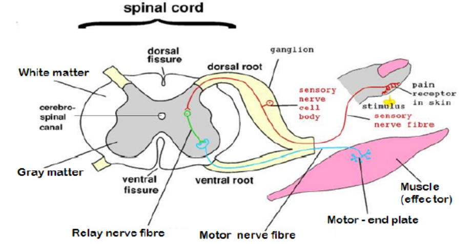
Figure 9.4: The transverse section of the spinal cord
The spinal cord acts as a coordinating centre for simple reflex such as knee jerk
response and autonomic reflexes. The spinal cord acts as means of communication
between spinal nerves and the brain. It sends impulses to the brain through sensory
neurons from the body and returns the motor impulses to the effectors which aremuscles and glands.
Application 9.1Describe the form in which the information is conveyed in the nervous system
9.2 Structure, types and functions of neurons
Activity 9.2
– Use charts describing the neuron and watch the movies showing the types
of neuron.
– Using textbooks or searching additional information on the internet, read
the information related to the structure, types and functions of neurone.
a. Draw and label the structure of a neuroneb. Make a table compering different types of neurons
Aneuron also called nerve cell is the basic functional unit of the nervous system.
Neurons are cells specialized to generate and transmit nerve impulses (actionpotentials) are cells which transmit nerve impulses (action potentials).
9.2.1 Types of neurons
Nerve cells may be grouped according to the number of processes they possess so
that their types include:
– Unipolar neurons: those with one process only, found mainly in invertebrates.
– Bipolar neurons: those with two separate processes such as neurons in the
retina of the vertebrate eye.
– Multipolar neurons: those with more than two processes such as most of thevertebrate neurons.
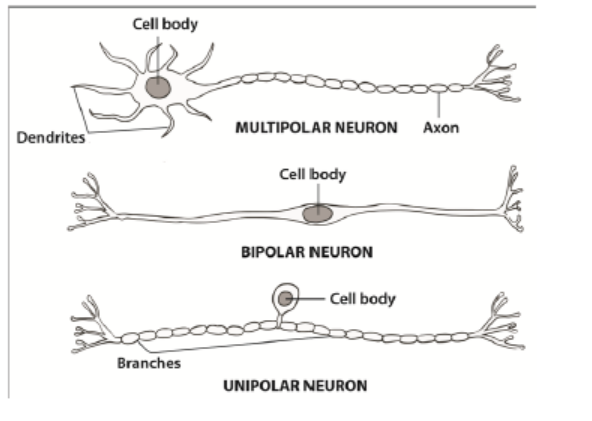
Figure 9.5: Multipolar, bipolar, unipolarneurons
9.2.2 Classification of neurons by their functions
In vertebrates, it is also common to group neurons according to their functions. They
include:
– Sensory or afferent neurons: transmit impulses from the receptors to the
central nervous system. In addition to sensory or afferent neurons.
– Motor or efferent neurons: that transmits impulses from the central nervous
system to effectors motor organs such as muscles or glands that carry out the
response. Most motor neurones are stimulated by impulses conducted by
interneurons. However, there are some others that are stimulated directly by
sensory neurons.
– Interneurons also known as intermediate or association, or relay or interneuron
connect the pathways of sensory and motor impulses, and are found mainly inthe central nervous system.
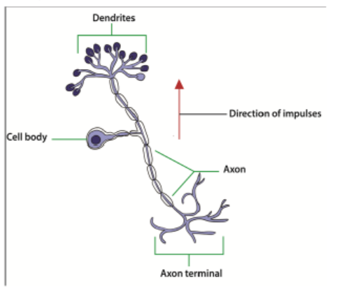
Figure 9.6: Sensory neuron
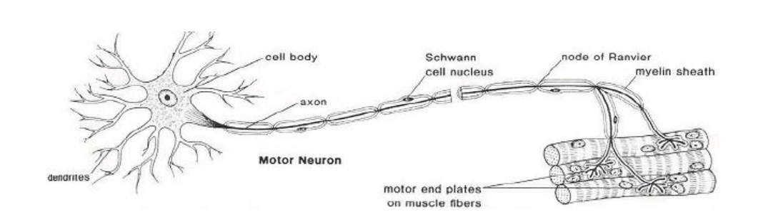
Figure 9.7: Motor neuron (image from google)
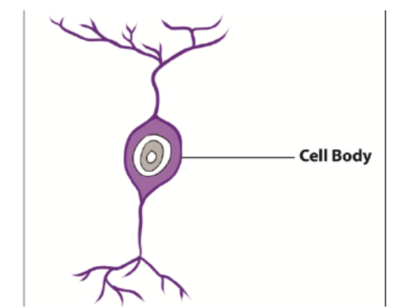
Figure 9.8: Intermediate neuron
9.2.3 Parts of a neuron and their functions
Each motor neuron possesses a cell body and cytoplasm with many mitochondria,
endoplasmic reticulum, golgi apparatus and ribosomes. The Nissl granules which
consist of endoplasmic reticulum and ribosomes function in protein synthesis. Thetable below (Table 9.1) shows all parts of neuron and their functions.
Table 9.1: The parts of a neuron and their functions
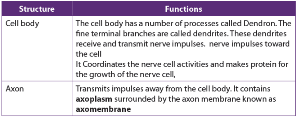
Application 9.2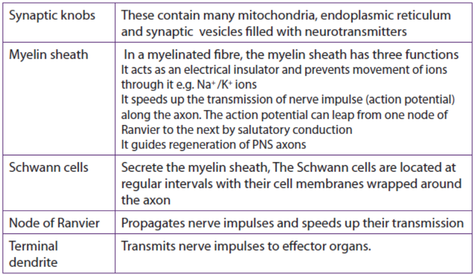
Explain what would happen when a neuron is damaged
9.3 Nature and generation of a nerve impulse
Activity 9.3
– Watch the movies showing the generation of a nerve impulse.
– Use the school library and search additional information on the internet.
– Read the information related to the generation of the nerve impulse and
take short notes on generation of the nerve impulse.
– Answer the following questions:
a. Draw, label and interpret the graph showing the action potentialb. What do you understand by action potential?
All cells in animal body tissues are electrically polarized—in other words, they
maintain a voltage difference across the cell’s plasma membrane, known as the
membrane potential. This electrical polarization results from a complex interplay
between protein structures embedded in the membrane called ion pumps and ion
channels. Each excitable patch of membrane has two important levels of membrane
potential: the resting potential, which is the value the membrane potential maintains
as long as nothing passes along the cell, and a higher value called the thresholdpotential.
9.3.1. Resting potential in a neuron
A neuron is said to be in the resting state when it is not conducting an impulse. The
membrane potential of an unstimulated excitable cell is called the resting potential.
A resting potential is the difference in charge (electrical potential difference) which
exists between the inside and the outside of the cell membrane. In excitable cells, the
resting potential is about -70 millivolts (mV) and the threshold potential is around
-55 mV. The negative sign indicates the interior of the cell is negative with respect tothe exterior environment.
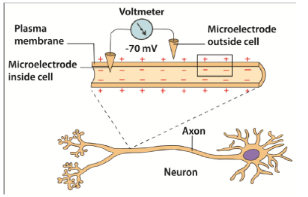
Figure 9.9: Resting potential in a neuron
The resting potential difference across the neuron membrane is maintained by:
– The sodium –potassium pump (Na+ /K+). This is always working. Three sodium
ions (Na+) are actively transported out of the cell for every two potassium ions
(K+) pump into the cell. Energy supplied by ATP is used for the transport of ions
against their electrochemical gradients.
– The axon membrane: It is more permeable to potassium ions than the sodium
ions. This is due to the presence of more potassium ion non-gated, voltageindependent
channels and few sodium ion non-gated channels. More K+ ions
can diffuse out back again faster than Na+ ions which can diffuse back in.
The resting membrane potential is mainly determined by sodium-potassium
pump, facilitated diffusion and electrochemical gradient of K+ ions across themembrane.
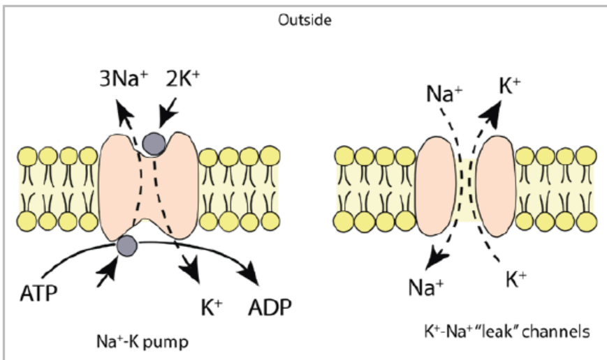
Figure 9.10: Sodium- potassium pump
9.3.2. Action potential
Action potential is the technical term for impulse. An action potential is rapid
temporary reversal in the electrical potential difference of an excitable cell
e.g. a neuron or a muscle cell. It is caused by changes in the permeability of the
membrane following the application of a threshold stimulus. The action potential
has a depolarization phase and a repolarization phase. There may be a short
hyperpolarized phase after the repolarization phase. The time taken for an actionpotential is 2 to 3 milliseconds.
9.3.3. Depolarization
When a stimulus such as electric current reaches a resting neuron, some sodium
voltage gated channels in the stimulated region of the axon membrane open. Sodium
ions (Na+) move into the axon by facilitated diffusion down an electrochemical
gradient. The initial influx of sodium ions is slow. The axon membrane becomes
slightly depolarized and the sodium voltage gates are sensitive to voltage changes.
More gates open allowing more Na+ ions to diffuse into the cell leading to further
depolarization.
When the potential difference across the membrane reaches a threshold value
(-50mV), many more sodium voltage gated channels open. This is an example of
positive feedback. The rapid diffusion of Na+ ions leads to a sudden increase in the
cell’s potential difference which becomes positive (+ 40mV). This reversal in thepotential difference is known as depolarization and lasts for about 1 millisecond
9.3.4. Repolarization
The reversal in polarity to + 40 mV causes the voltage gated sodium channel to
close. At the same time the voltage gated potassium channels open. The potassium
ions K+ diffuse out of the cell down their electrochemical gradient to the tissue fluid
outside. The axon membrane is repolarized. The action potential alters from + 40 mVto -70mV.
9.3.5. Hyperpolarization
The potassium voltage-gated channels are slow to close. An excess of K+ ions leave the
axon. The inside of the membrane becomes more negative. The voltage falls slightly
below -70mV and causes hyperpolarization. However, within a few milliseconds,
the potassium voltage-gated channels close. The resting potential of -70mV is re-
established by the Na+ /K+ pump and different rates of facilitated diffusion of K+ andNa+ ions through the non-gated ion channels.
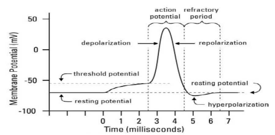
Figure 9.12: The action potential
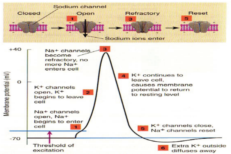
Figure 9.10: The sodium –potassium pump (Na+ /K+) and action potential
9.3.6. Frequency of action potentials
Information in axons is coded in the frequency of the action potentials. A weak
stimulus above threshold produces fewer action potentials. A stronger stimulus
produces a greater frequency of action potentials. As the intensity of stimulation
increases, more action occurs.
Application 9.3
The graphs below show the changes that occur during an action potential in
a membrane potential and the relative membrane permeability to sodium andpotassium ions in a neurone. Observe well to answer the following questions:
a. Describe the movement of ions during an action potential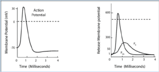
b. Explain what is the effect of an action potential generation if there is alowering of sodium ions in the extracellular fluid
9.4 Transmission of nerve impulses
Activity 9.4
The dissection of a frog sciatic nerve
Materials required
Laptop computer, projector, nerve chamber, cable and nerve chamber leads (red
and black), glass hooks, Stimulator cable, grounding adapter or cable, forceps,
scalpel, frog Ringer’s solution at two temperatures.
Procedure
– To begin dissection, retrieve a frog from your teacher and place it in a dissecting
tray.
– Remove the skin from the legs by making an incision through the skin and
around the entire lower abdomen.
– Cut the connections between the skin and the body especially around the base
of the pelvic girdle.
– Use stout forceps to pull the skin off the frog in one piece (like a pair of pants).
– Place the frog with its dorsal side up.
– Moisten the exposed tissue (legs) with Ringer’s solution and place a wet paper
towel (saturated with Ringers solution) over one of the legs of the frog so that
it is completely covered and wet.
– Use forceps to separate the muscles of the thigh (the leg not covered with the
paper towel).
– Pin the muscles apart so that more underlying muscle is visible.
– This should also expose the cream-colored Sciatic nerve lying deeply between
the muscles.
– Use a glass hook to separate the nerve from the fascia and the vessels. If
possible, avoid cutting the blood vessels. If bleeding does occur, rinse away
the blood with lots of Ringer’s solution. Free the nerve from the knee joint to
the pelvis.
– Use the glass hook to place a suture thread under the nerve. Move the thread
as close to the knee joint as possible.
– Ligate (tie off) the nerve; you may observe calf muscle fibrillation or foot
movement as the knot is tied off.
– Be sure the knot is tied tightly. Cut the nerve between the knot and the knee
joint. Keep the exposed nerve moist at all times with Ringer’s solution.
– Carefully separate the muscles of the pelvis to expose the sciatic nerve.
Remember to rinse any blood away with Ringer’s solution.
– Carefully expose the remainder of the nerve through an opening along the
lateral side of the urostyle. To avoid cutting the nerve, lift the end of the
urostyle with forceps as you cut the muscle away from the urostyle with blunt
scissors.
– Cut along the urostyle from its tip to the vertebral column.
– Deflect the muscle away from the urostyle to expose the Sciatic nerve.
– Use a glass hook to separate connective tissue from the nerve and to place
a piece of suture thread under the nerve.
– Move the thread as high as possible on the nerve to obtain as large a
section as possible.
– Ligate (tie off) the nerve; the leg may jump again as the knot is tied tightly.
– Cut the nerve between the knot and the vertebral column and keep the
exposed nerve moist at all times.
– Use forceps to grasp the suture thread at the proximal end (end closest to
head) and lift the nerve out of the body cavity.
– Do not pinch or stretch the nerve.
– Remove any connective tissues, blood vessels, or nerve branches that may
still keep the nerve attached to the frog.
– Continue to grasp the suture to lift the nerve until it is clear of the abdomen,
the pelvis, and the thigh.
– Grasp the suture at either end to remove the nerve from the body entirely.
– Place the nerve across the gold-coloured electrode pins in the nerve bath.
– Add a small quantity of Cold Frog Ringers to the bottom of the chamber.
– The Frog ringers should not touch the gold-plated electrode pins.– Cover the chamber with a glass slide.
Questions
1. Draw a picture of the laboratory setup used for this exercise.2. Find and dissect the frog sciatic nerve for placement in a nerve chamber.
9.4.1 Mechanism of transmission of nerve impulses along an axon
– The neurons, like other cells, are positively charged outside and negatively
charged inside. The membrane of the axon is said to be polarized. The potential
difference (voltage) across their membranes is of – 70mV and is called resting
membrane potential (RMP).
– A stimulus (heat, pain, bite, sound …) creates an action potential (AP) or an
impulse that is transmitted along an axon by electro-chemical change.
– During an action potential, the membrane potential falls until the inside
becomes positively charged with respect to the exterior. The membrane at this
point is said to be depolarized. It takes few milliseconds to happen. In fact, the
potential changes from – 75 mV to + 40 mV at the point of stimulation. That is
an electrical change that runs along the axon.
– As the impulse is transmitted along the axon, the Na+/K+ pumps of the axolemma
are re-established. Sodium channels open first, allowing a large number of Na+ions to flow in.
– The axoplasm becomes progressively more positive with respect to the outside
of the axolemma. Then, almost instantly, the permeability of the membrane to
Na+ ions ceases, and the net flow of Na+ ions stop. At the same time K+ ion
channels start to open and K+ ions flow out from axoplasm where they are in
high concentration. The counter-flow is of 3Na+ ions against 2K+ ions.
– The axoplasm now starts to become less positive again. This begins the process
of re-establishing the resting potential difference of the membrane. That is anelectro-chemical change.
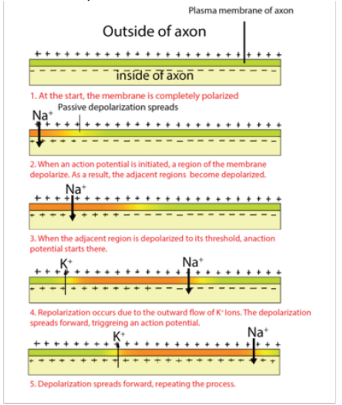
Figure 9.13: The nerve impulse transmission along axon
a. Factors that affect the transmission of nerve impulses along the axon
membrane
Along the axon membrane, the transmissions of nerve impulses are affected as
follows:
– The diameter of the axon: the greater the diameter the faster the speed of
transmission of nerve impulses.
– The myelin sheath: myelinated neurones conduct impulses faster than nonmyelinated
neurones.
– The presence of nodes of Ranvier: speeds up the movement of impulses in
myelinated neurones.
b. Structure of a synapse
Information from one neuron flows to another neuron across a synapse. The synapse
is a small gap separating two adjacent neurons. The synapse consists of:
– A presynaptic ending that contains neurotransmitters, mitochondria and other
cell organelles,
– A postsynaptic ending that contains receptor sites for neurotransmitters and,
– A synaptic cleft or space between the presynaptic and postsynaptic endings. It
is about 20nm wide.
– The swollen tip of the axon of the presynaptic neuron, called synaptic knob
or synaptic bulb contains many membrane – bounded synaptic vesicles,
mitochondria and microfilaments.
– The synaptic vesicles contains neuro transmitter molecules such as acetylcholineor noradrenaline
c. Neurotransmitter
A neurotransmitter is a relatively small chemical found in the synaptic vesicle. It
helps to transmit an impulse across a synapse or neuromuscular junction. There
are about 50 different types of neurotransmitters in the human body. Examples
are acetylcholine released by cholinergic neurons, noradrenaline (norepinephrine)
released by adrenergic neurons, dopamine and serotonin including amino acidsglutamate and glycine.
9.4.2 Mechanism of nerve impulse transmission across a synapse
– The arrival of an impulse on the synaptic knob causes the opening of Ca+2 ion
channels on the presynaptic membrane, and Ca+2 ions flow in the presynaptic
region from the synaptic cleft.
– The Ca+2 ions induce a few presynaptic vesicles to fuse with presynaptic
membrane and to secrete their neurotransmitters (e.g. acetylcholine) by
exocytosis into the synaptic cleft
– The neurotransmitter then binds with the receptor protein on the postsynaptic
membrane. This causes the opening of Na+ channels on the postsynaptic
neuron which in turn becomes depolarized.
– This causes a depolarization of the post-synaptic cell membrane, which may
initiate an action potential, if the threshold is reached
– The action of the neurotransmitter does not persist because an enzyme
cholinesterase catalyses the hydrolysis of acetylcholine into choline and acetate.
The breakdown products (choline) are absorbed by the pre-synaptic neuron
by endocytosis and used to re-synthesize more neurotransmitter, using energyfrom the mitochondria.
9.4.3 Properties of a nerve impulse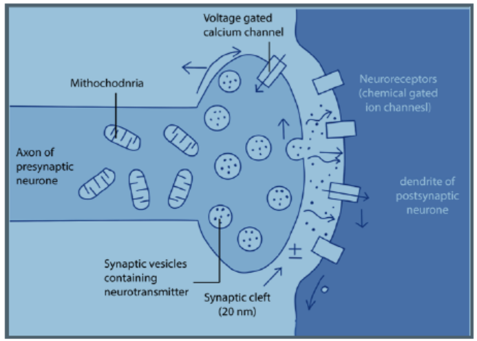
a. All or nothing law
An action potential can only be generated after the threshold value is exceeded.
After the threshold is reached, the size of the action potential produced remains
constant and is independent of the intensity of the stimulus. This is the all or nothing
response. All action potentials are of the same amplitude.
b. Refractory period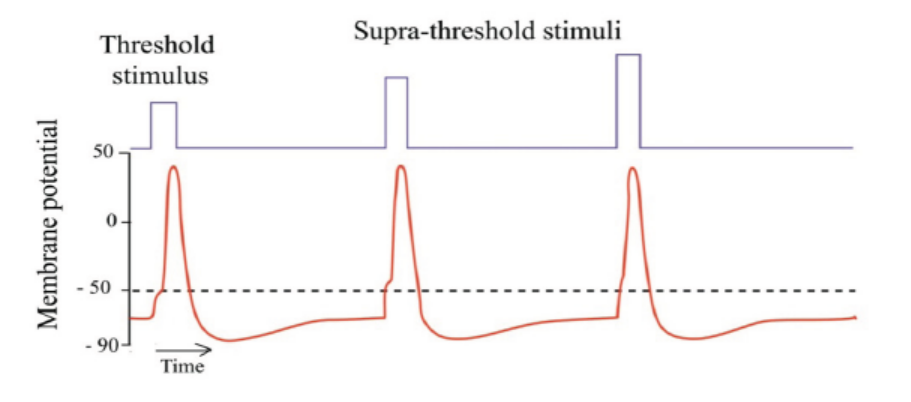
This is a brief period when an axon is unable to transmit an impulse following
transmission of the same. It lasts about 5-10 milliseconds. It is divided into two;absolute and relative periods. During the absolute refractory period which lasts
about 1ms, the axon membrane is unable to respond to another stimulus, no matter
how strong it is. An action potential cannot be produced. This is because there is
conformational change in voltage-gated sodium channels which are still in a closed,
inactive state. This also prevents the action potential from moving backwards.
Following the absolute refractory period, there is a relative refractory period which
lasts around 5ms. During this period, the resting potential is gradually restored by
Na+ /K+ pump and the relative permeability of membrane to facilitated diffusion of
ions is also restored. A new action potential can then be produced if the stimulus is
greater than the usual one. The refractory period therefore allows impulses to move
only in one direction and limits the frequency at which successive impulses can passalong axon.
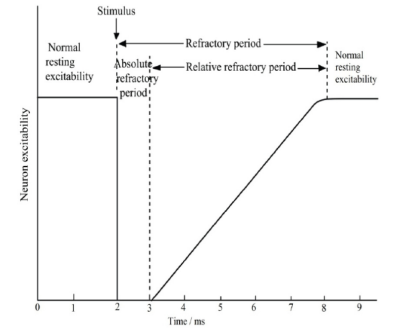
Figure 9.16: Neuron excitability before and after a nerve impulse
c. Salutatory conduction
It is movement or jump of nerve impulses from one node of Ranvier to anotheralong the axon membrane of neurone.
Application 9.4
1. Suppose a cell’s membrane potential shifts from -70 mV to -50 mV. What
changes in the cell’s permeability to K+ or Na+ could cause such a shift?
2. The diagram below shows the changes in potential difference across an axonmembrane as a nerve impulse passes
a. Explain what happens at M, N, O, P, Q and R as shown in the graph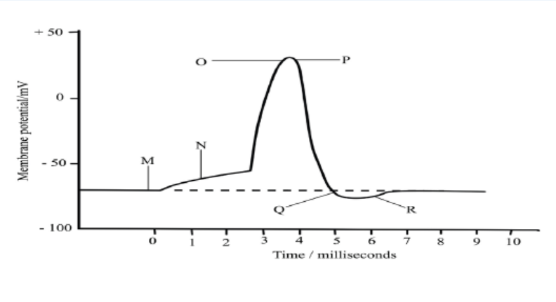
b. Name two factors that can determine the speed of transmission of a nerve
impulse and how each affects the speed
c. Explain why the initiation of an action potential is considered a positivefeedback mechanism
9.5 Structure and function of a cholinergic synapse.Activity 9.5
Use textbooks from school library and other additional information using
internet, read the information related to the cholinergic synapse and take shortnotes on cholinergic synapse.
a. Draw and label a diagram showing a cholinergic synapseb. Make a table of different functions of a cholinergic synapse
The cholinergic synapse is a synapse which uses acetylcholine (Ach) as
neurotransmitter. Calcium and vesicles are involved in the release of neurotransmitter
across the synaptic cleft in the mechanism of synaptic transmission to generate anexcitatory post-synaptic potential.
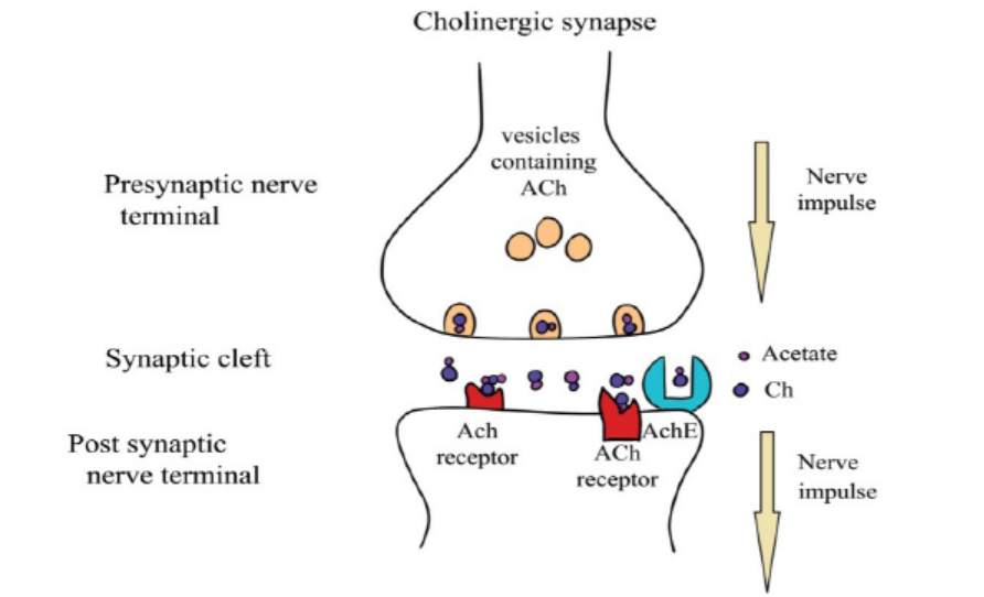
Figure 9.17: The cholinergic synapse
9.5.1. Functions of synapses
Synapses have a number of functions which include:
a. Transmit information between neurones
The main function of synapses is to convey information between neurons. It is from
this basic function that the others arise.
b. Pass impulses in one direction only
As the neurotransmitter substance can only be released from one side of a synapse,
it ensures that nerve impulses only pass in one direction along a given pathway
c. Act as junctions
Neurons may converge at synapse. In this way a number of impulses passing along
different neurons may release sufficient neurotransmitter to generate a new action
potential in a single postsynaptic neuron whereas individually they would not. This
is known as spatial summation. In this way responses to a single stimulus may be
coordinated.
d. Filter out low level stimuli
Background stimuli at a constantly low level, e.g. the drone of machinery, produce
a low frequency of impulses and so cause the release of only small amounts of
neurotransmitter at the synapse. This is insufficient to create a new impulse in the
postsynaptic neuron and so these impulses are carried no further than the synapse.
Such low level stimuli are of little importance and the absence of a response to them
is rarely, if ever harmful. Any change in the level stimulus will be responded to in theusual way.
e. Allow adaptation to intense stimulation:
In response to a powerful stimulus, the high frequency of impulses in the presynaptic
neuron causes considerable release of neurotransmitter into the synaptic cleft.
Continued high-level stimulation may result in the rate of release of neurotransmitter
exceeding the rate at which it can be formed. In these circumstances the release of
neurotransmitter ceases and hence also any response to the stimulus. The synapseis said to be fatigued.
9.5.2. Effects of drugs on synapses
Several types of chemicals such as drugs interfere at synapses, either amplifying or
inhibiting the transmission o of impulses. For example,
– Caffeine and nicotine amplify the transmission of impulses by mimicking the
action of natural neurotransmitters.
– Insecticides that prolong the effect of neurotransmitters by blocking the
enzymatic breakdown of transmitters. Other drugs such as
– Anaesthesia including atropines inhibit the transmission of impulses across
the synaptic membranes. Atropine acts to prevent an action potential being
generated by acetylcholine when it attaches to its receptor protein on thepostsynaptic membrane
9.5.3. The neuromuscular junction
A special kind of synapse is the nerve-muscle known as neuromuscular junction,
the point where the terminal dendrite of a motor nerve cell makes contact with a
muscle fibre. The region of the sarcolemma (cell surface membrane) of muscle fibre
that lies directly under the terminal portion of the motor neuron is known as the
motor end plate. At the nerve-muscle junction the membrane of the muscle fibre ismodified to form an end-plate to which the dendrite is attached.
When an impulse arrives at the nerve-muscle junction, acetylcholine is discharged
from synaptic vesicles into the synaptic cleft. The acetylcholine diffuses across the
gap and depolarizes the muscle end plate. End-plate potentials can be recorded and
it has been shown that if these build up sufficiently an action potential is fired off inthe muscle fibre.
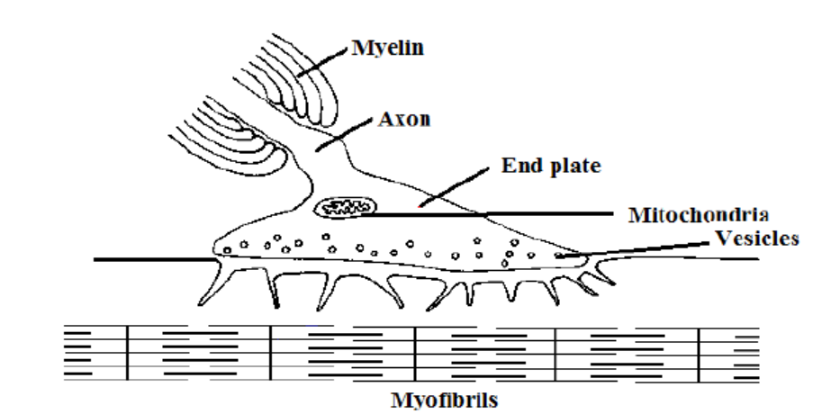
Figure 9.18: The neuromuscular junction
Application 9.5The diagram shows the structure of a nerve synapse
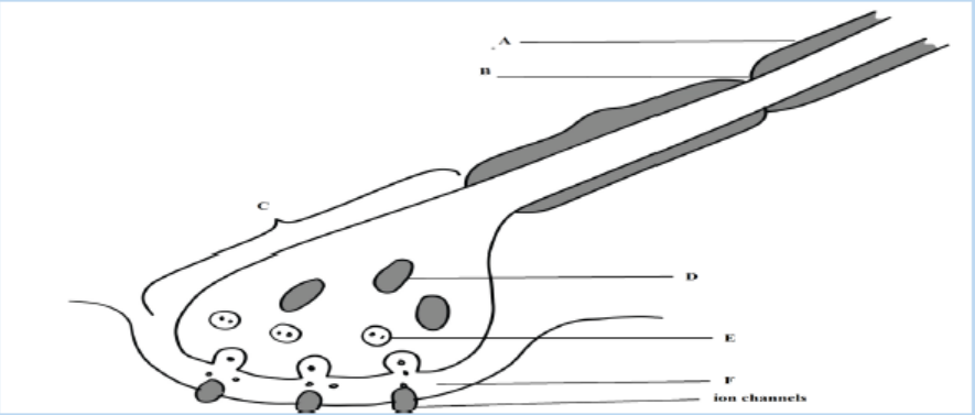
1. Label structures A to F
2. Draw an arrow on the diagram to show the direction of a nerve impulse
(an action potential) in the presynaptic neurone.
3. Name a common neurotransmitter presents in the synaptic vesicle
4. Name an ion that
a. Moves into the postsynaptic neurone in an excitatory synapse
b. Moves out of the postsynaptic neurone in an inhibitory synapse
4. Suggest one reason why structure C contains large numbers of organelle
D
5. What is the major chemical component of structure A?6. State the functions of structure A and structure B
9.6 Functions of sensory, relay and motor neurons
in a reflex arc
Activity 9.6
Aim: Description of a reflex arc
The diagram below shows the sequences of events when one is hit on the patella(bone situated in front of the knee joint) by a small hammer.
1. Observe carefully and discuss what is happening according to the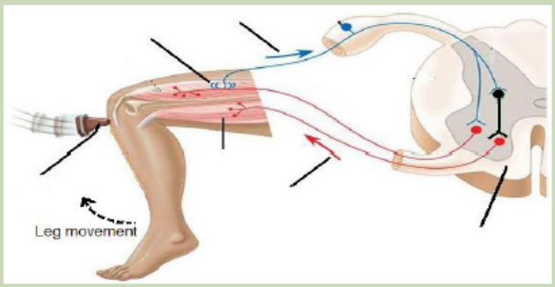
direction indicated by the arrows.
2. Identify the organ/part where the information starts and the organ
responsible for the response.
3. Label the figure by using the following words: stimulus, sensory receptor,sensory transmission, motor transmission, effectors, and spinal cord.
9.6.1. Reflex actions
A reflex action is a quick and involuntary response of the central nervous system to
a stimulus. Example: The quick withdrawal of the hand from a hot object. When the
spinal cord alone is involved, the reflex action is called spinal reflex and when thebrain alone is involved, it is a cranial reflex e.g. blinking of eyes.
Reflex actions are described as involuntary actions and the same stimuli producethe same responses every time. Reflexes are useful because they make autonomic
involuntary adjustments to changes in the external environment, such as the irispupil
reflex and the balance during locomotion. They also control the internal
environment, such as breathing rate and blood pressure, and prevent damage to
body as in cuts and burns. These help to maintain constant conditions, in other word
they are involved in homeostasis.The sequences of changes that occur during a spinal reflex are:
– A sensory receptor receives a stimulus and impulse is generated in it
– The impulse is transmitted along a sensory neuron towards the spinal cord via
the dorsal root
– Once the impulse reaches the grey matter inside the spinal cord, it is passed on
to the relay neuron across a synapse
– The relay neuron then transfers the impulse to a motor neuron across another
synapse.
– The motor neuron conveys the impulse to an effector such as a muscle where a
response takes place.
The pathway that is followed by an impulse along the sensory neurons relay andmotor neurone, during a reflex action is called reflex arc.
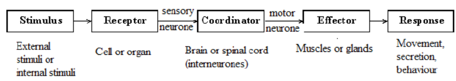
Figure 9.19: Sequence of change in a spinal reflex
The components of reflex arc are:
– Stimulus
– Receptors
– The sensory receptor that detects the stimulus
– The sensory (or afferent) neurone along which the sensory impulse is
transmitted;
– The relay neurone in the central nervous system to which the sensory impulse
is passed on.
– The motor (or efferent) neurone along which the motor impulse is transmitted;
and
– The effector (Muscle or gland) which the motor impulse triggers to bring about
an appropriate response.– CNS (Brain or spinal cord)
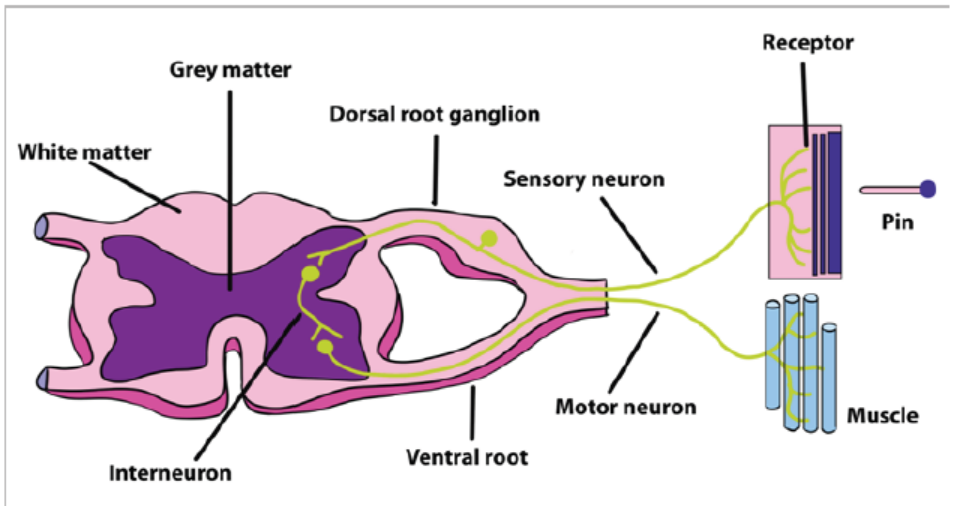
Figure 9.20: The diagram showing reflex arc
9.6.2 Conditioned reflex actions
This type of reflex involves the brain but it is also as fast as the simple reflex. Salivation
on smelling one’s favourite food is an example of conditional reflex. The individual
recognizes and based on the previous experience, the response (salivation) occurs.
The recognition of the previous experience involves the association centres of thebrain.
A series of experiments were conducted by Ivan PAVLOV, a Russian biologist who
demonstrated conditioned reflex. He found that when a bell rung every time a dog
was given food, the dog showed salivation only at the sound of the bell. The ringing
of the bell is called stimulus. The dog had, thus, learnt to associate the sound of thebell to the food and this made it salivate at the sound of the bell.
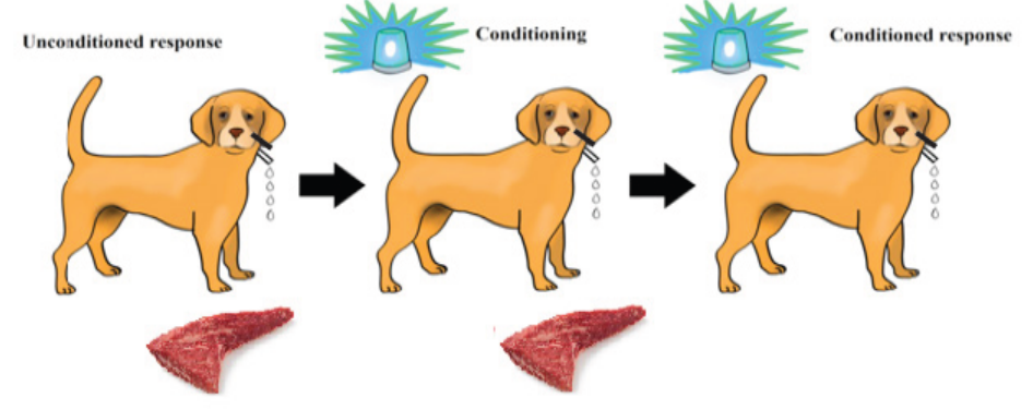
Figure 9.21: The experiment representing the conditioned reflex
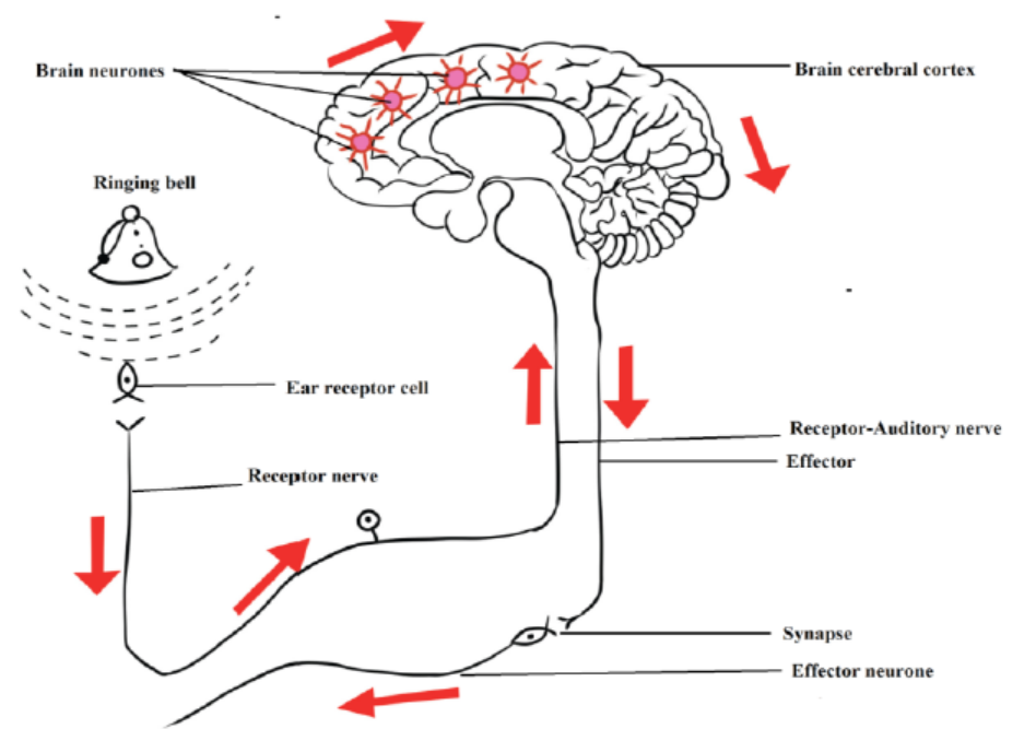
Figure 9.22: The pathway events of conditioned reflex
Application 9.6Describe the functions of sensory, relay and motor neurones in a reflex arc
End of unit assessment 9
A. Multiple choice questions: Choose the best answer
1. What happens when a neuron’s membrane depolarizes?
a. There is a net diffusion of Na+ out of the cel1.
b. The equilibrium potential of K+ becomes more positive.
c. The neuron’s membrane voltage becomes more positive.
d. The neuron becomes less likely to generate an actionpotentia1.
e. The inside of the cell becomes more negative relative to the outside.
2. Why action potentials are usually conducted in only one direction along an
axon?
a. The nodes of Ranvier can conduct potentials in only one direction.
b. The brief refractory period prevents reopening of voltage gated Na+
channels.
c. The axon hillock has a higher membrane potential than the terminals
of the axon.
d. Ions can flow along the axon in only one direction.
e. Voltage-gated channels for both Na+ and K+ open in only one
direction.
3. A common feature of action potentials is that they
a. Cause the membrane to hyperpolarize and then depolarize.
b. Can undergo temporal and spatial summation.
c. Are triggered by a depolarization that reaches the threshold.
d. Move at the same speed along all axons.
e. Result from the diffusion of Na+ and K+ through ligand gated
channels.
4. Where are neurotransmitter receptors located?
a. On the nuclear membrane
b. At nodes of Ranvier
c. On the postsynaptic membrane
d. On the membranes of synaptic vesicles
e. In the myelin sheath
5. During the repolarisation phase of an action potential, the permeability of
the axon membrane to:
a. Na+ increases
b. K+ increases
c. Ca+ increasesd. Organic anions increases
6. The graph shows the changes in the permeability of an axon to Na+ and K+ions during an action potential
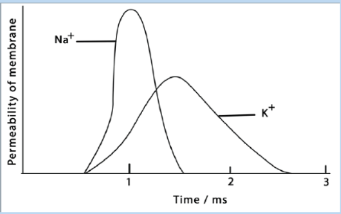
Which of the following shows the correct movement of these ions in the axon?
a. Na+ ions enter the axon, K+ ions leave the axon
b. Na+ ions leave the axon, K+ ions enter the axon
c. Both Na+ and K+ ions enter the axon
d. Both Na+ and K+ leave the axon
7. The graph shows the potential difference across an axon membrane. Whichpart of the graph shows the action potential?
a. 3, 4 and 5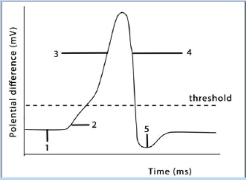
b. 2,3, 4 and 5
c. 1,2, 3 and 4d. 1,2,3, 4 and 5
B. Questions with structured answers
8. The diagram below shows a human brain seen from the right side
a. Name the parts labelled A, B and C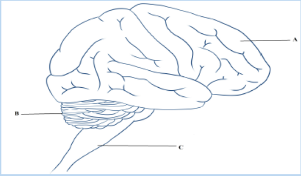
b. Give two functions of the part labelled B.
9. The list describes the main stages in the process by which information is
transmitted across cholinergic synapses.
– An action potential arrives at synaptic knob of presynaptic neurone.
This causes…. the ions to enter the synaptic knob.
– Vesicles move to the………………. membrane.
– A neurotransmitter called……………….is released into the synaptic
cleft
– This moves across the cleft by a process known as…………. the
neurotransmitter combines with a………………. on the postsynaptic
membrane.
– Influx of……………. ions cause local depolarisation and an action
potential is set up in the postsynaptic neurone
a. Copy the list. Using the correct scientific terms, add the words that
have been omitted.
b. Explain what happens to the neurotransmitter after it has passed
information across a cholinergic synapse
c. Some nerves, especially those of the sympathetic nervous system,
produce noradrenaline in their synaptic vesicles. Name this type ofsynapse
10. The graph shows the changes in permeability of the cell surface membraneof an axon to sodium and potassium ions during an action potential.
a. Explain how the events which take place between X and Y on the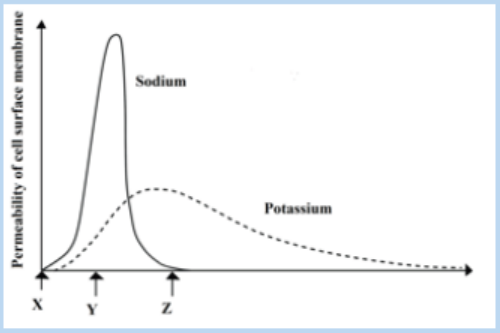
graph can lead to a change in the potential differences across the
membrane
b. What happens to the potential difference across the membrane
between times Y and Z?
c. Explain why a nerve impulse travels faster in myelinated neurone
than in a non-myelinated one.11. The diagram below shows a nerve cell or neuron
a. Name the type of neurone shown.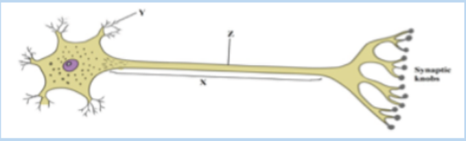
b. Name the structures labelled X and Y
c. A nerve impulse can be initiated by stimulation with a microelectrode.
What would be the effect of stimulation at point Z?
d. The synaptic knobs release a chemical transmitter, acetylcholine.
Nerve gases prevent the breakdown of this chemical. From this
information suggest
I. One early symptom of nerve gas poisoningII. One reason for this observed symptom
12. Complete the following table by stating which region of the brain controlseach of the functions listed.
13. The diagram below represents the structures visible at a synapse with the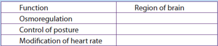
aid of electron microscopy.a. Identify the structures labelled A and B
b. Name the chemical found in the numerous vesicles that occur in the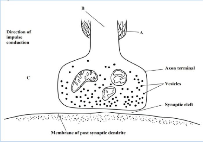
synaptic knob
c. Identify the structure labelled C and suggests a reason for its
presence in the synaptic knob
d. A powerful hydrolytic enzyme is found in the synaptic cleft. What isits function in normal synaptic transmission?
C. Essay question
14. Describe what happens when an action potential arrives at a synaptic knobof an excitatory synapse
UNIT 10 HORMONAL COORDINATION IN ANIMALS
UNIT 10: HORMONAL COORDINATION IN ANIMALS
Key Unit Competence
To be able to identify the location and function of endocrine glands in the body.
Learning objectives
By the end of this unit, I should be able to:
– Define hormones.
– Explain why hormonal balance is necessary for coordinating the functions
in the body.
– Describe the principle of the negative feedback mechanism by which
hormones produce their effects on target cells.
– Describe the structure and function of the endocrine system.
– Explain the effects of hormonal imbalances.
– Compare and contrast the actions of the endocrine and nervous systems.
– Draw and interpret the flow chart of negative feedback mechanisms.
– Appreciate the role of hormones in the growth and development oforganisms.
Introductory activity
At a given time, there are certain changes which occur in the body especially
during puberty. As girls and boys enter the period of puberty, they start to
develop remarkable differences in physical appearance and in their behaviour.
1. What do you think to be the causes of such changes?
2. What are some changes which can be observed in boys and not in girls
and vice versa?
3. Which the organs do you think are responsible for producing such
changes?
4. What will be the causes if some of those changes do not appear in a boyor in a girl?
10.1 Structure and function of the endocrine system in humans
Activity 10.1
By using different books from the school library and a chart showing different
endocrine glands, discuss the following:
1. What are endocrine glands?
2. Draw and locate the following endocrine glands
a. The adrenal glands
b. The pancreas
3. What are the hormones produced by the pancreas and their functions?4. Why the pituitary gland was once described as a master gland?
A hormone is an organic substance which is produced in minute quantity by an
endocrine gland, transported by blood to other parts or organs of the body where it
exerts maximum effects. Such parts of the body or organs are called target organs.
The word endocrine means internal secretion and endocrine glands are therefore
glands of internal secretion. Since they shed their secretion into the bloodstream,
they have no ducts and are hence known as ductless glands.
Hormones are released into the blood stream as a result of:
1. Stimulation of the endocrine gland directly by the nervous system e.g. the
sympathetic nervous system causes secretion of adrenaline by the adrenal
medulla.
2. The levels of particular metabolites in the blood e.g. glucose levels trigger
the release of insulin.
3. Presence of other hormones called releasing hormones mostly produced in
anterior pituitary e.g. TSH stimulates the release of thyroxin by the thyroid
gland.
4. Environmental changes such as high or low temperatures effects activities
of the pituitary gland.5. Animals’ general mental state does affect the activity of the pituitary.
Once in the bloodstream, the hormones are carried around the body, bringing
about responses in various places. Structures that respond to them are called targetorgans. A hormone is a chemical messenger having the following properties:
– It travels in the blood
– It has its effect at a site different from the site where it is produced. The site
where it has effect is called the target, while itself is called messenger
– It fits precisely into receptor molecules in the target like a key in a lock. It is
therefore specific for a particular target;– It is a small soluble molecule;
It is effective in low concentrations.
Hormones fulfill many functions. These include:
1. Regulation of growth and development.
2. Controls homeostasis e.g.in osmoregulation/thermo regulation etc
3. Regulation of metabolism e.g. digestion storage and utilization of food
substances.
4. Development of the skin coloration.
5. Enabling the body to withstand shock, tension wounding etc. and to
recover from it.
6. Together with the nervous system it provides for effective responses to all
kinds of stimuli both internal and external.
The endocrine glands include; the pituitary, thyroid, parathyroid, adrenal, and pineal
glands (Figure 10.1 below). Taken together, all endocrine glands and hormone-
secreting cells constitute the endocrine system.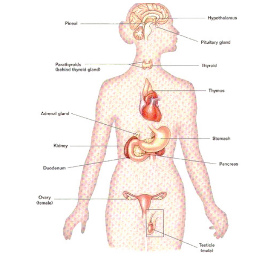
Figure 10.1: Major endocrine glands
a. The pituitary gland
The pituitary gland which was formerly called the master gland hangs from the
base of the brain by a stalk and is enclosed by bone. As shown in figure 10.2, the
pituitary gland consists of a hormone-producing glandular portion called anterior
pituitary and a neural portion called posterior pituitary, which is an extension of
the hypothalamus. The hypothalamus now called the master gland, regulates the
hormonal output of the anterior pituitary and synthesizes two hormones that it
exports to the posterior pituitary for storage and later release. Most anterior pituitary
hormones exhibit a diurnal rhythm of release, which is subject to modification bystimuli influencing the hypothalamus.
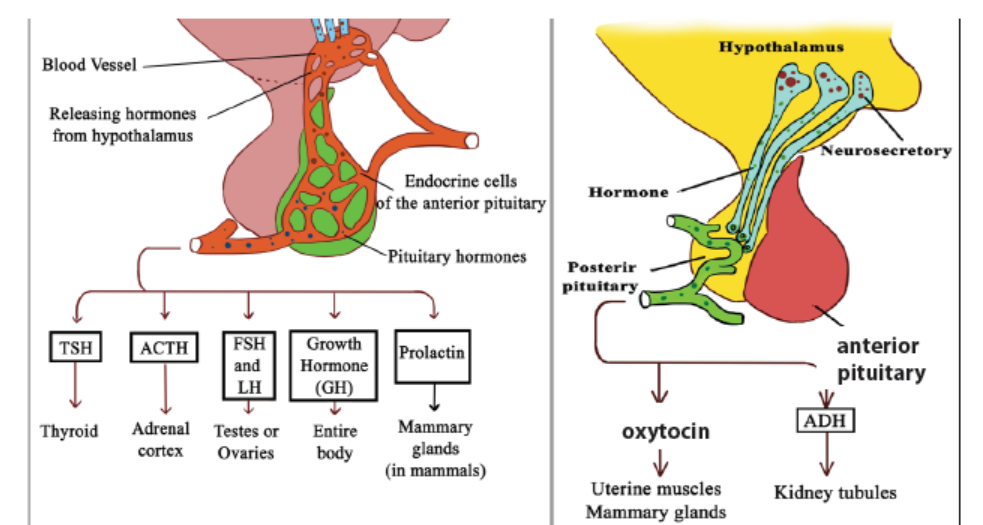
Figure 10.2: Pituitary and hypothalamic secretions
The following are the hormones produced by the anterior pituitary gland;
– Growth hormone (GH) or Somatotropic hormone: is a hormone that
stimulates growth of all body tissues especially skeletal muscle and bone.
GH; mobilizes the use of fats, stimulates protein synthesis, and promotes
glucose uptake and metabolism.
– Thyroid-stimulating hormone (TSH) This hormone causes thyroid glands
to secrete thyroxin. The secretion of TSH is controlled by levels of thyroxin
in blood. TSH also stimulates growth of thyroid gland.
– Adrenocorticotropic hormone (ACTH) stimulates the adrenal cortex to
release its hormones. ACTH release is triggered by corticotropin-releasing
hormone (CRH) and inhibited by rising glucocorticoid levels.
– The gonadotropins: follicle-stimulating hormone (FSH) and luteinizing
hormone (LH) regulate the functions of the gonads in both sexes.
– Prolactin (PRL) promotes the production of milk in human’s females. Its
secretion is triggered by prolactin-releasing hormone (PRH) and inhibited
by prolactin-inhibiting hormone (PIH).
The following are the two hormones released from the posterior pituitary gland:
– Oxytocin: It stimulates powerful contractions of the uterus, which trigger
labour and delivery of an infant, and milk ejection in nursing women. Its
release is mediated reflexively by the hypothalamus and represents a
positive feedback mechanism.
– Antidiuretic hormone (ADH) stimulates the kidney tubules to reabsorb
and conserve water, resulting in small volumes of highly concentrated urine
and decreased plasma osmolality.
b. The hypothalamus
The hypothalamus plays an important role in integrating the endocrine and
nervous systems. The region of the lower brain receives information from nerves
throughout the body and from other parts of the brain thus initiates endocrine
signals appropriate to environmental conditions. The reason it is called a master
gland is that a set of neurosecretory cells in the hypothalamus exerts control over
the anterior pituitary by secreting two kinds of hormones into the blood: Releasing
hormones which make the anterior pituitary to secrete its hormones and inhibiting
hormones that make the anterior pituitary stop secreting hormones. Every anterior
pituitary hormone is controlled at least by one releasing hormone and some have
both a releasing and an inhibiting hormone.
The posterior pituitary remains attached to the hypothalamus. It stores and releases
two hormones that are made by a set of neurosecretory cells in the hypothalamus.
c. Thyroid gland
The thyroid gland is located in the anterior throat. Thyroid follicles store colloid
containing thyroglobulin, a glycoprotein from which thyroid hormone is derived.
Thyroid hormone (TH) includes thyroxine (T4) and triiodothyronine (T3), whichperform the following tasks;
– Control the basal metabolic rate.
– Increase the rate of cellular metabolism. Consequently, oxygen use and
heat production rise.
Calcitonin, is another hormone produced by the thyroid gland in response to rising
blood calcium levels. Its role is to decrease blood calcium levels by inhibiting bonematrix reabsorption and enhancing calcium deposit in bone.
d. Parathyroid glands
The parathyroid glands are located on the dorsal aspect of the thyroid gland and secrete
parathyroid hormone (PTH), which causes an increase in blood calcium levels by;
– Increasing the rate of calcium reabsorption by the kidney at the expense of
phosphate ions.
– Increasing the rate of calcium absorption from the gut.– Causing the release of calcium reserves from the bones.
PTH is antagonistic calcitonin. PTH release is triggered by decreasing blood calciumlevels and is inhibited by increasing blood calcium levels.
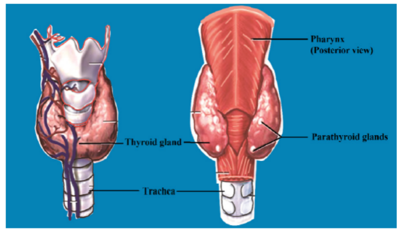
Figure 10.3: The location of the thyroid and the parathyroid glands
e. Pancreas
The pancreas is an organ located in the abdomen close to the stomach and is both
an exocrine and an endocrine gland. The endocrine portion (islets of Langerhans)
releases insulin and glucagon hormones. Glucagon is released by alpha (α) cells
when glucose levels in blood are low. Glucagon stimulates the liver to convert stored
glycogen to glucose thus increasing glucose levels. Insulin is released by beta (β)
cells of the islets of Langerhans when blood levels of glucose are rising. It increases
the rate of glucose uptake and causes the conversion of glucose to glycogen.
f. Gonads
The ovaries of the female which are located in the pelvic cavity, release two main
hormones. Secretion of oestrogen by the ovarian follicles begins at puberty under
the influence of FSH. Oestrogen stimulates maturation of the female reproductive
system and development of the secondary sex characteristics. Progesterone
is released in response to high blood levels of LH. It works with oestrogen in
establishing the human menstrual cycle. The testes of the male begin to produce
testosterone at puberty in response to LH. Testosterone stimulates the maturation
of the male reproductive organs, development of secondary sex characteristics, and
the production of sperm by the testes.
g. Adrenal Glands (Suprarenal Glands)
A fresh adrenal gland section shows a bright yellow cortex, making up about 80% of
the organ, and a more reddish-grey medulla. The endocrine activities of the adrenalcortex and the adrenal medulla differ both in development and function.
• Adrenal cortex
Adrenal cortex makes mineralocorticoids (such as aldosterone and cortisol). Cortisol
raises blood glucose level whereas aldosterone stimulates the reabsorption of Na+
and excretion of K+ in kidney.
• Adrenal Medulla
The adrenal medulla makes two hormones epinephrine (adrenaline) (80 %) and
norepinephrine (noradrenaline) (20 %). Epinephrine and norepinephrine are released
into the bloodstream during stress and they act on the whole organism by preparing
it for increased energy use. Both hormones, for instance, activate the liberation of
fatty acids from fat depots and liberate glucose from glycogen storage in the liver
(producing a rise in the blood sugar level). They raise the blood pressure and stroke
volume of the heart and may lead to vasoconstriction in certain defined areas.
h. Other hormone-producing structures
Many body organs not normally considered endocrine organs contain isolated cell
clusters that secrete different hormones. Examples include the; gastrointestinal tract
organs (gastrin, secretin, and others), the placenta (hormones of pregnancy such as
oestrogen, progesterone, and others) and the kidneys (erythropoietin and renin).
Table 10.1: Major human endocrine glands, their functions and the control oftheir secretions
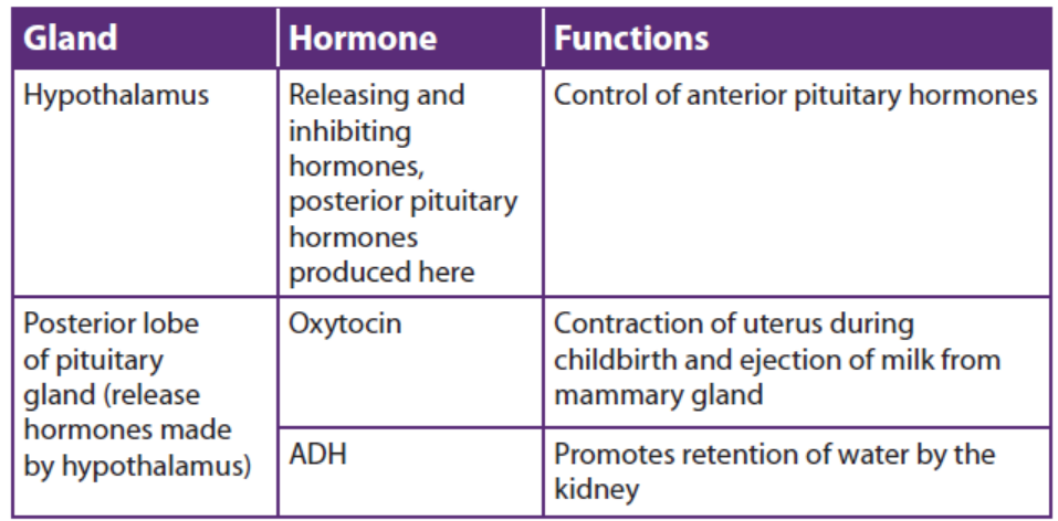
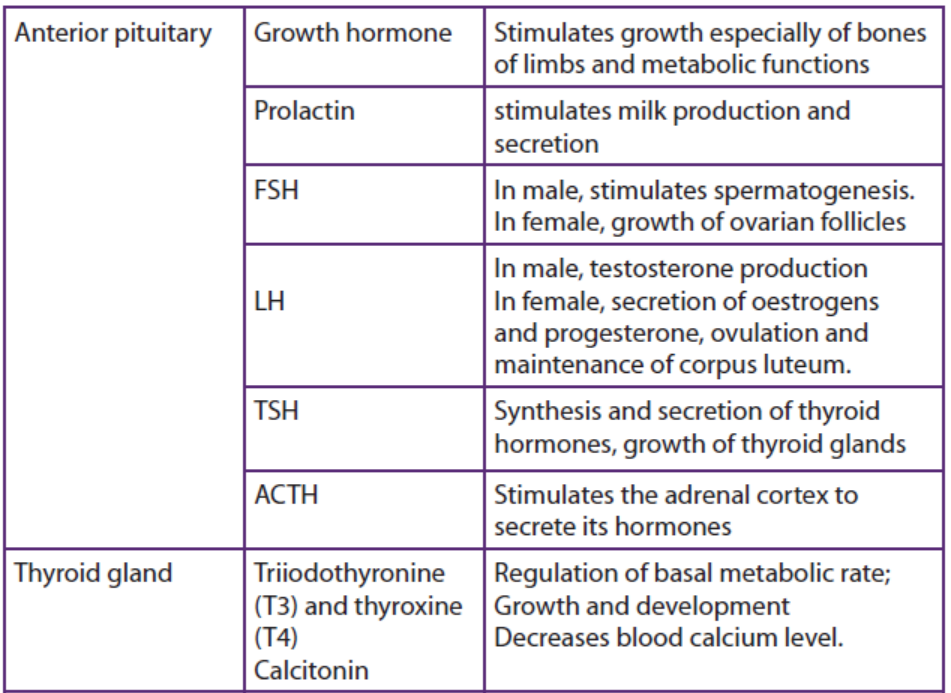
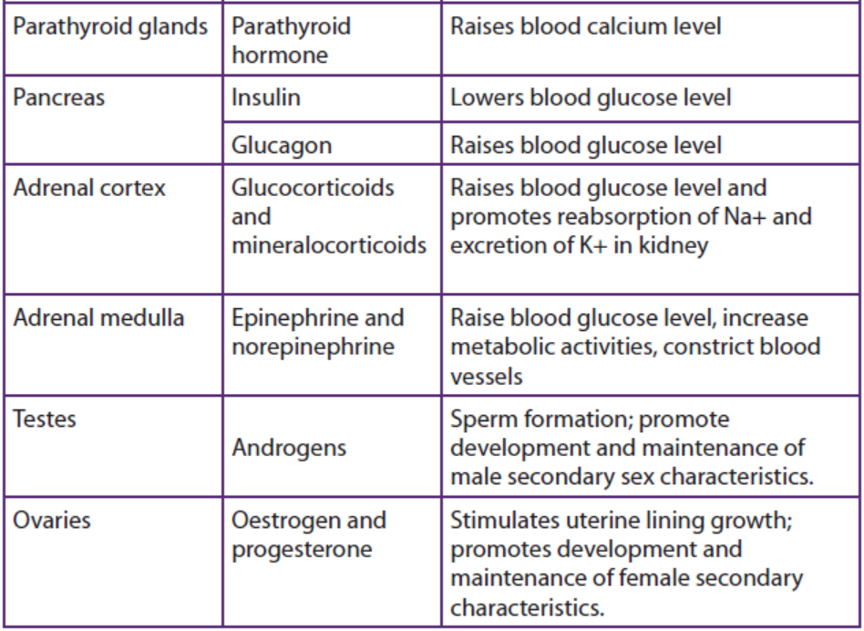
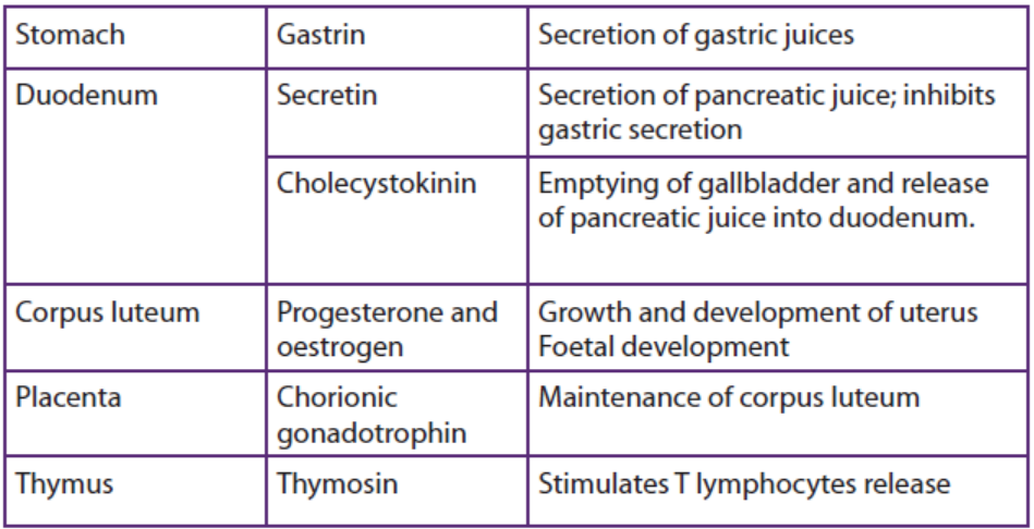
Application 10.1
1. What are the hormones produced by the thyroid glands?
2. What are the functions of the hormones stored and released by the
posterior pituitary gland?3. By which means are hormones transported in our body?
10.2 Principles of the negative feedback mechanism of hormonalaction.
Activity 10.2
1. The amount of urine produced varies according to the amount of water
consumed. Make a list of events that may occur in the following cases for the
human body:
a. Two days without drinking water
b. When you have drunk 1 litre of water per day
c. How can you explain the above observations?
2. Make short notes on what happens to your body if the level of sugars decreasesin the blood?
Feedback mechanisms are necessary in the maintenance of homeostatic mechanisms.
All homeostatic control mechanisms have at least three interdependent componentsfor the variable being regulated that work together i.e.
a. The receptor is the sensing component that monitors and responds to
changes in the environment. When the receptor senses a stimulus, it sends
information to a control center, the component that sets the range at which
a variable is maintained.
b. The control center determines an appropriate response to the stimulus. In
most homeostatic mechanisms, the control center is the brain.
c. An effector, which can be muscles, organs or other structures that receive
signals from the control center. After receiving the signal, a change occurs
to correct the deviation by either enhancing it with positive feedback ordepressing it with negative feedback.
The homeostatic mechanisms in mammals require information to be transferred
between different parts of the body. There are two coordination systems in mammals
that control this: the nervous system and the endocrine system.
– In the nervous system, information in the form of electrical impulses is
transmitted along nerve cells (neurons).
– The endocrine system uses chemical messengers called hormones thattravel in the blood, in a form of long-distance cell signalling.
10.2.1 Positive feedback mechanisms
These mechanisms are designed to accelerate or enhance the output created by
a stimulus that has already been activated. The positive feedback mechanisms are
designed to push levels out of normal levels. To achieve this purpose, a series of
events initiates a cascading process that builds to increase the effect of the stimulus.
This process can be beneficial but is rarely used by the body due to risks of the
acceleration’s becoming uncontrollable.
Examples include; the accumulation blood platelets which in turn causes blood
clotting in response to a break or tear in the lining of blood vessels. The release of
oxytocin to intensify the contractions of the uterus that take place during childbirth.
Another example of a positive feedback mechanism is the production of milk by
a mother for her baby. As the baby suckles, nerve messages from the mammary
glands cause the mother’s pituitary gland to secrete a hormone called prolactin.
The more the baby suckles, the more prolactin is released, which stimulates further
milk production by the mother’s mammary glands. In this case, a negative feedback
mechanism would not be helpful because the more the baby nursed, the less milkwould be produced.
12.2.2 Negative feedback mechanisms
These are mechanisms concerned with keeping changes in the factor within narrow
limits. Here, an increase in a factor (input) e.g. hormone levels results in somethinghappening that makes the factor decrease (output).
An example of negative feedback mechanism is regulation of thyroxine levels i.e. The
shedding of thyroxine into blood stream is triggered by thyrotropin releasing factor
(TRF) produced by the hypothalamus of the brain. TRF passes to the pituitary gland
along the blood vessels stimulating the anterior pituitary gland to produce Thyroid
stimulating hormones (TSH). TSH then stimulates the thyroid gland to produce
thyroxine into blood. A slight excess of thyroxine in blood detected by hypothalamus,
inhibits the anterior lobe of the pituitary gland which responds by secreting less
TSH. This in turn reduces the activity of the thyroid gland, leading to decrease in
the amount of the thyroxine produced. This removes the inhibitory influence on thepituitary so that more thyroid stimulating hormone will be produced again.
Table 10.2: Negative and positive feedback compared
Application 10.2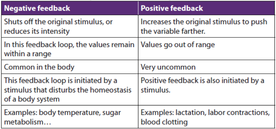
1. State any two examples of positive feedback.2. Why is the positive feedback not useful in many homeostatic mechanisms?
10.3 Effects of hormonal imbalances
Activity 10.3
1. You may know people suffering from diabetes mellitus or you may have heard
about the disease from the radio or from a newspaper.
a. Collect information about the cause of this disease.
b. Predict the ways this disease can be treated.
2. Observe carefully figure 10.4 below and suggest the type of disorders thefollowing people may be suffering from:
The disorders of the endocrine system often involve either the hypo-secretion (hypo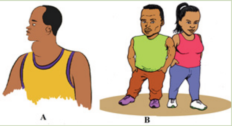
means too little or under), inadequate release of a hormone, or the hyper-secretion
(hyper means too much or above), excessive release of a hormone. In other cases,
the problem is faulty hormone receptors, an inadequate number of receptors, or
defects in second-messenger systems. Because hormones are distributed in the
blood to target tissues throughout the body, problems associated with endocrinedysfunction may also be widespread.
10.3.1. Pituitary Gland Disorders
a. Pituitary dwarfism, gigantism, and acromegaly
Several disorders of the anterior pituitary involve human growth hormone.
Hyposecretion of human growth hormone during the growth years slows bone
growth, and the epiphyseal plates close before normal height is reached. This
condition is called pituitary dwarfism. Other organs of the body also fail to grow,
and the body proportions are childlike. Treatment requires administration of humangrowth hormone during childhood, before the epiphyseal plates close.
Hypersecretion of human growth hormone during childhood causes gigantism, an
abnormal increase in the length of long bones. The person grows to be very tall,
but body proportions are about normal. Hypersecretion of human growth hormoneduring adulthood is called acromegaly.
b. Diabetes insipidus
The most common abnormality associated with dysfunction of the posterior pituitary
is diabetes insipidus. This disorder is due to defects in antidiuretic hormone (ADH)
receptors or an inability of the pituitary gland to secrete ADH. A common symptoms
of diabetes insipidus are: excretion of large volumes of urine resulting in dehydration
and thirst. Bed-wetting is common in afflicted children. Because so much water is
lost in the urine, a person with diabetes insipidus may die of dehydration if deprived
of water for only one day. Treatment of diabetes insipidus involves the injection ofADH into the body.
10.3.2. Thyroid gland disorders
Thyroid gland disorders affect all major body systems and are among the most
common endocrine disorders. Congenital hypothyroidism or the hyposecretion
of thyroid hormones that is present at birth has devastating consequences if not
treated quickly. Previously termed cretinism, it causes severe mental retardation and
stunted bone growth. At birth, the baby typically is normal because lipid-soluble
maternal thyroid hormones crossed the placenta during pregnancy and allowednormal development.
Hypothyroidism during the adult years produces a disorder called myxoedema. An
indication of this disorder is oedema (accumulation of interstitial fluid) that causes
the facial tissues to swell and look puffy. A person with myxoedema has a slow
heart rate, low body temperature, sensitivity to cold, dry hair and skin, muscular
weakness, general lethargy, and a tendency to gain weight easily. Because the brain
has already reached maturity, mental retardation does not occur, but the person
may be less alert.
The most common form of hyperthyroidism is Graves’ disease which is an autoimmune
disorder in which the person produces antibodies that mimic the action of thyroidstimulating
hormone (TSH). The antibodies continually stimulate the thyroid gland
to grow and produce thyroid hormones. A primary sign is an enlarged thyroid,
which may be two to three times its normal size. Graves’ patients often have a
peculiar oedema behind the eyes, called exophthalmos, which causes the eyes to
protrude. Treatment may include surgical removal of part or all of the thyroid gland
(thyroidectomy), the use of radioactive iodine to selectively destroy thyroid tissue,
and the use of anti-thyroid drugs to block synthesis of thyroid hormones. A goitre
is simply an enlarged thyroid gland. It may be associated with hyperthyroidism,hypothyroidism or by the lack of iodine.
10.3.3. Parathyroid gland disorders
Parathyroid gland disorders cause the hypoparathyroidism due to the too little
parathyroid hormone leading to a deficiency of blood Ca2+, causing neurons and
muscle fibres to depolarize and produce action potentials spontaneously. This leads
to twitches, spasms, and tetany (maintained contraction) of skeletal muscle. The
main cause of hypoparathyroidism is accidental damage to the parathyroid glands
or to their blood supply during thyroidectomy surgery.
Hyperparathyroidism or an elevated level of parathyroid hormone, most often
is due to a tumour of one of the parathyroid glands. An elevated level of PTH
causes excessive resorption of bone matrix, raising the blood levels of calcium and
phosphate ions and causing bones to become soft and easily fractured. High blood
calcium level promotes formation of kidney stones. Fatigue, personality changes,
and lethargy are also seen in patients with high levels of parathyroid hormone.10.3.4. Adrenal gland disorders
a. Cushing’s syndrome
Hypersecretion of cortisol by the adrenal cortex causes an endocrine disorder
known as Cushing’s syndrome. The condition is characterized by breakdown
of muscle proteins and redistribution of body fat, resulting in thin arms and legs
accompanied by a rounded moon face and buffalo hump on the back. Facial skin is
flushed, and the skin covering the abdomen develops stretch marks. The person also
bruises easily, and wound healing is very slow. The elevated level of cortisol causes
hyperglycaemia, osteoporosis, weakness, hypertension, increased susceptibility to
infection, decreased resistance to stress, and mood swings.
b. Addison’s disease
Hyposecretion of glucocorticoids and aldosterone causes Addison’s disease (chronic
adrenocortical insufficiency). The majority of cases are autoimmune disorders in
which antibodies cause adrenal cortex destruction or block binding of ACTH to
its receptors. Pathogens, such as the bacterium that causes tuberculosis, also may
trigger adrenal cortex destruction. Symptoms, which typically do not appear until
90% of the adrenal cortex has been destroyed, include mental lethargy, anorexia,
nausea and vomiting, weight loss, hypoglycemia, and muscular weakness. Loss of
aldosterone leads to the elevated potassium and decreased sodium in the blood,low blood pressure, dehydration, decreased cardiac output and even cardiac arrest.
10.3.5. Pancreas disorders
The most common endocrine disorder is diabetes mellitus caused by an inability
to produce or use insulin. According to the diabetes atlas of 2018, the prevalence
of diabetes in Rwanda is about 3.16% of the population with 1,918 diabetes
related deaths per year. Its complications can lead to heart attack, stroke, blindness,kidney failure and lower limb amputation.
Because insulin is unavailable to aid transport of glucose into body cells, blood glucose
level is high and glucose is found in the urine, the process known as glycosuria.
The cardinal signs of diabetes mellitus are; polyuria (excessive urine production due
to an inability of the kidneys to reabsorb water), polydipsia (excessive thirst) andpolyphagia (excessive eating).
Application 10.3
1. Which disorders are caused by:
a. Hypersecretion of GH in children?
b. Hyposecretion of insulin?
c. Hypersecretion of thyroid hormones?
2. What are some symptoms of?
a. Diabetes mellitus?b. Grave’s disease?
10.4 Comparison of hormonal and nervous systems
Activity 10.4
Use your knowledge of the nervous system and the endocrine system to
answer the questions that follow
1. Discuss the similarities between the structure and functioning of nervous
and hormonal systems.
2. Discuss the differences between the structure and functioning of nervousand hormonal systems.
The nervous and endocrine systems act together to coordinate functions of all our
body systems.
A basic similarity between the endocrine system and the nervous system is that both
provide means of communication within the body of an organism. Both involve
transmission of a message which is triggered by a stimulus and produces a response.
Several chemicals function as both neurotransmitters and hormones including
norepinephrine. Some hormones such as oxytocin are secreted by neuroendocrine
cells; neurons that release their secretions into the blood. The target organs of a
hormone are equivalent to nerve’s effectors.
Similarities
1. Both provide a means of communication and coordination in the body.
2. In both the information transmitted is triggered by a stimulus and produces
a response.
3. Both involve chemical transmission.4. Both are controlled by the brain.
The main differences between the two systems concern the nature of the message.
In the endocrine system, the message takes the form of a chemical substance
transmitted through the blood stream. In the nervous system it is a discrete-all or
none action potential transmitted along a nerve fibre. All other differences arise
from this fundamental one. They can be listed as follows:
– Because of the comparatively high speed at which impulses are transmitted
along nerves, nerves responses are generally transmitted more rapidly than
hormonal ones.
– Since it is conveyed by the bloodstream, there is nothing to stop a hormone
being carried to every part of the body. Nervous impulses however are
transmitted by particular neurons to specific destinations.
– As a result, hormones are often widespread, sometimes involving the
participation of numerous target organs. In contrast, nervous responses
may be much localized, involving perhaps the contractions of only one
muscle.
– Hormonal responses frequently continue over a long period of time. Obviousexamples of such long-term responses are growth and metabolism.
A comparison between nervous and endocrine system is summarized in the table
10.3Table 10.3: Comparison between nervous and endocrine system
Application 10.4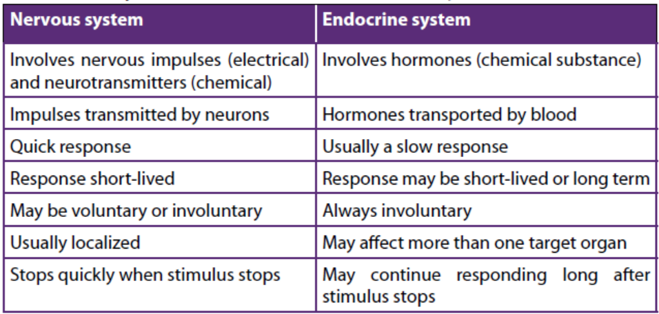
Describe a short term effect of the endocrine system and a long term effect ofthe nervous system.
End of unit assessment 10
Multiple choice questions: from question 1 to 5, choose the letter
corresponding to the best answer
1. What are the chemical messengers of the endocrine system called?
a. Neurons
b. Hormones
c. Blood cells
d. Carbohydrates
2. Endocrine glands
a. Function only after puberty
b. Function only before puberty
c. Release products through ducts
d. Release products into bloodstream
3. X and Y are hormones. X stimulates the secretion of Y, which exerts negative
feedback on the cells that secrete X. Suppose the level of Y decreases. What
should happen immediately afterwards?
a. Less X is secreted
b. More X is secreted
c. Secretion of Y stops
d. Secretion of X stops
4. Which one of the following hormones is secreted by the neurosecretory cells in
mammals?
a. Adrenaline
b. Antidiuretic hormone
c. Insulin
d. Thyroxin
5. Select the hormone INCORRECTLY paired with its target.
a. TSH - thyroid gland
b. LH - ovary or testis
c. ACTH - anterior pituitaryd. ADH - kidney
6. A number of metabolic diseases in mammals arise as a result of abnormalendocrine function. Complete the table below concerned with this:
7. During the control of blood sugar in a mammal two antagonistic hormones are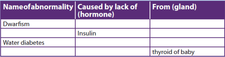
employed. Fill in the table about them
8. Name the hormone involved in the functions described below and the name of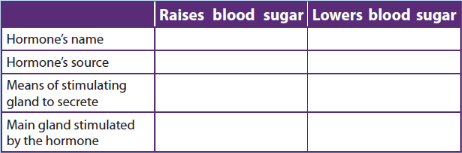
the gland which produces it:
a. Controls reabsorption of Na+ in the kidney.
b. Increases the permeability of convoluted distal tubule and collecting
duct.
c. Increases heart rate.
d. Increases blood glucose level.
e. Decreases blood glucose level.
f. Repair and growth of the endometrium.
g. Stimulates the anterior pituitary gland to release FSH.
h. Stimulates contraction of the uterus.
i. Stimulates the mammary glands to secrete milk.
9. What is the difference between diabetes mellitus and diabetes insipidus? What
are the characteristic signs of diabetes insipidus?
10. Use the following to describe a negative feedback mechanism: TSH, TRH,decreased metabolic rate, thyroxine and T3.
UNIT 11 SKELETONS, MUSCLES AND MOVEMENT
UNIT 11: SKELETONS, MUSCLES AND MOVEMENT
Key unit competence
Explain the structure of muscles in relation to movement.
Learning objectivesBy the end of this unit, I should be able to:
– Describe the three main types of animal skeletons.
– Discuss the functions of skeletons.
– State and discuss the advantages and disadvantages of exoskeletons.
– Describe the features of a synovial joint
– Appreciate the role of joints and muscles in bringing about movement.
– Describe the main types of mammalian muscles.
– Compare the structure of cardiac, smooth and skeletal muscle.
– Distinguish between slow twitch and fast twitch fibers.
– Demonstrate the structure and function of the sarcomere.
– Demonstrate the laws of muscle contraction.
– Distinguish between temporal summation and muscle fibre recruitment.
– Explain the role of antagonistic muscles in a joint.
– Adopt the practice of playing sport to develop healthy muscles and bones.
– Appreciate the role of joints and muscles in bringing about movement.
– Describe the ultrastructure of striated muscles with particular reference to the
sarcomere structure.
– Interpret the ultrastructure of striated muscle with particular reference to the
sarcomere structure
– Explain the sliding filament model of muscle contraction, including the roles
of troponin, tropomyosin, calcium ions and ATP.
– Explain the function of a motor unit/ neuromuscular junction/motor end plate.– Illustrate the sliding filament model of muscular contraction.
Introductory activity
Let do the physical exercise of push-up. Do as many as you can. Explain whatallows you to push-up.
11.1 Types of animal skeletons: hydrostatic, exoskeletonand endoskeleton
Activity 11.1.
You are provided with an earthworm and a grasshopper.AW: Put diagrams of both earthworm and grasshopper but without labels.
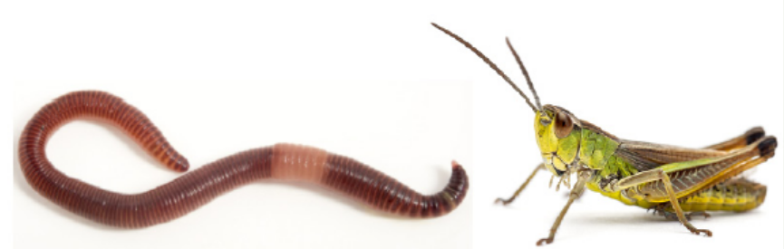
Observe locomotion and then differentiate their skeletons.
Figure 11.1: Structure of earthworm and insect
A support system is made up of those materials that bear the weight of the body,
strengthen its parts and endure all stresses that the body or its parts may be subjected
to during movement. Consequently, the strength of the supporting materials in
an organism is directly related to its size and weight. The strength of a supporting
material itself depends among other things on its length, shape, thickness andstructure.
11.1.1. Types of skeleton
There are three types of skeleton namely hydrostatic skeleton, endoskeleton andexoskeleton.
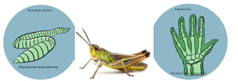
Figure 11.2: Types of skeletons: hydrostatic, exoskeleton and endoskeleton
a. Hydrostatic skeleton
Annelids (earthworm), nematodes (round worms), Echinoderms (starfish and the
sea urchin), cnidarians (Jellyfish), and some other organisms use the hydrostatic
skeleton for movement. This skeleton is found in soft-bodied and cold-blooded
animals having a coelom. This coelom is fluid-filled cavity surrounded by muscles
and the rigidity caused by the fluid. The muscles serve as a supporting structure for
organisms. Hydrostatic skeleton is basically composed of a fluid filled body cavity
surrounded by sets of antagonistic muscles. The hydrostatic skeleton operates on
the principle that water is incompressible and therefore can provide a rigid medium
against which muscles can contact. The hydrostatic skeleton is segmented and
therefore can be used for movement and locomotion. It is also flexible and therefore
allows expansion to allow growth.
However, hydrostatic skeleton presents some disadvantages as it provides relatively
little support and therefore neither supports the animal upright nor their body
weight off the ground. It does not provide strong levels on which powerful muscles
can operate fast locomotion. This coupled with the fact that the body weight is
dragged on the ground makes it unsuitable for fast locomotion. Consequently,
animals depending on hydrostatic skeleton are slow moving. The thin flexible cuticle
associated with it does not properly protect the animal against water loss, because ifthe cuticle was thick and inflexible, it would not allow free movement.
b. Exoskeleton
The exoskeletons also known as cuticles are found in all arthropods It is found
outside the body and forms a protective covering for the animals. It supports as well
as protects the animals. All crustaceans have exoskeleton. Crabs, spiders, lobsters,
insects are all arthropods. Animals with exoskeleton are usually small. This is because
large animals could not be supported by exoskeleton and need bones to support
them. Animals with exoskeleton have a head and abdomen and in some cases, a
thorax. The exoskeleton is soft and thin at the joints where it has to bend. The large
exoskeletons are called shells. Tortoise is one vertebrate animal that has a shell andendoskeleton.
Advantages of the exoskeletons (in movement and protection)
– It is joined and allows muscle attachment which makes it useful in locomotion.
It also forms locomotors devices like legs and wings.
– It maintains the shape of the insect. The shape is an important determinant of
how well movement can take place.
– It prevents water loss by having wax. This has helped insects to adapt to dry
environments.
– It is hard and offers protection of internal organs from mechanical injury,
friction and microbial attack.
– It is usually colored and offers protection from predators through camouflage
and mimicry.
– It is used to form various mouthparts. Mouth parts are adapted for the various
feeding methods in insects and also for various forms of protection especially
biting the enemy.
Disadvantages of the exoskeletons
– Its components are heavy compared to those of similar size in other skeletons.
This affects the locomotion of insects especially those which are big and
explains why large insects cannot fly for long without resting.
– It does not allow continuous growth because of its rigidity; growth has to beintermittent following moulting.
c. Endoskeleton
Mainly made of bones, the endoskeleton is a rigid internal skeleton of vertebrates.
It forms the frame work for the animal. The tissues and muscles are formed around
the skeletal system and the muscular forces are transmitted to this skeleton. It is
composed of mineralized tissues. In phylum Chordata, Porifera and Echinodermata
endoskeleton is present. The animals that come under Phylum Chordata are allvertebrates including human beings.
Advantages of the endoskeletons (in movement and protection)
– It does not restrict growth like the exoskeleton.
– It is relatively light and allows faster locomotion both on land and in the air.
– It is jointed and allows flexibility and movement.
– It maintains the shape and form of the body which allows it to move fast.
– Its skeletal elements (the bones) are metabolically active and synthesize blood
cells some of which offer protection against disease (white blood cells).
– It offers maximum protection to some delicate internal organs e.g. the brain
from mechanical injury.
– It is a stronger skeleton and therefore supports most of the body weight aboveground which allows faster locomotion.
Disadvantages of the endoskeletons in movement and protection.
– It does not completely enclose internal organs and therefore offers less
protection to them from mechanical shock.– It does not protect the animal from water loss.
Table 11.1: A comparative table of animal skeletons: hydrostatic, exoskeletonand endoskeleton
11.1.2. Functions of Bones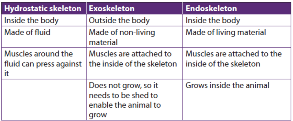
The skeletal system is important for the proper functioning of animal’s body.
In addition to giving shape and form to the body, bones have many importantfunctions as follow:
– Structural support of the body: The skeleton supports the body against the
pull of gravity. The large bones of the lower limbs support the trunk when
standing.
– Protection of internal organs: The skeleton provides a rigid frame work that
supports and protects the soft organs of the body. The fused bones of the
cranium surround the brain to make it less vulnerable to injury. Vertebrae
surround and protect the spinal cord and bones of the rib cage help protect
the heart and lungs.
– Attachment of the muscles: The skeleton provides attachment surfaces for
muscles and tendons which together enable movement of the body.
– Movement of the body: Bones work together with muscles as simple
mechanical lever systems to produce body movement.
– Production of blood cells: The formation of blood cells takes place mostly in
the interior (marrow) of certain types of bones.
– Storage of minerals: Bones contain more calcium than any other organ in the
form of calcium salts such as calcium phosphate. Calcium is released by the
bones when blood levels of calcium drop too low. Phosphorus is also storedin bones.
11.1.3. The human skeleton
Humans are vertebrates, which are animals that have a vertebral column, or
backbone. The study of internal framework of bones and cartilage that is found inside
vertebrates, including humans, is called an endoskeleton. The adult human skeleton
consists of approximately 206 bones. Cartilage is a type of fibrous connective tissue
that is made of tough protein fibers. The function of cartilage in the adult skeleton
is to provide smooth surfaces for the movement of bones at a joint. A ligament is a
band of tough, fibrous tissue that connects bones together. Ligaments are not very
elastic and some even prevent the movement of certain bones. The skeletons of
babies and children have many more bones and more cartilage than adults have.
As a child grows, the extra bones, such as the bones of the skull (cranium), and the
sacrum (tailbone) fuse together, and cartilage gradually hardens to become bontissue.
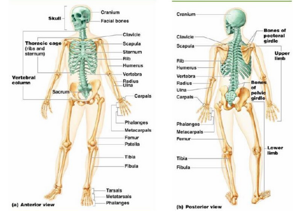
Figure 11.3: The human skeleton
The bones of the skeleton can be grouped in two divisions: the axial skeleton and
appendicular skeleton.
The axial skeleton includes; the bones of the; head, vertebral column, ribs andsternum. There are 80 bones in the axial skeleton.
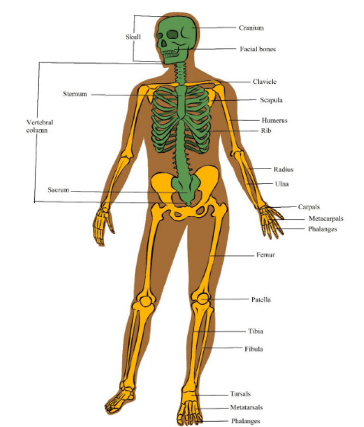
Figure 11.4: Divisions of the human skeleton
a. The axial skeleton
The axial skeleton forms the central axis of the body. It consists of the skull, the
vertebral column, the ribs and the sternum or breastbone. There are 80 bones inaxial skeleton.
i) The Skull
The skull consists of 28 different bones including the ossicles of the ear. The bones
of the skull can be divided into two main groups: the cranium which encloses and
protects the brain and the facial bones. The cranium is a rigid structure with an
opening, the foramen magnum (literally large hole) where the spinal cord enters.
ii) The Vertebral column
The vertebral column forms the central part of the skeleton. It supports the skull
and protects the spinal cord. It also serves as attachment for the ribs, the pectoral
and pelvic girdles. The vertebral column consists of separate bones, the vertebrae.
Because the separate vertebrae are attached to each other by means of fibrous
cartilaginous discs they form a flexible column. Each vertebra has articular surfaces
above and below, which allow articulation movement between them.
The vertebral column of 33 vertebrae is divided into five regions according to their
position and structure. The five regions consist of: seven cervical (neck) vertebrae,
twelve thoracic (chest) vertebrae, five lumbar vertebrae (vertebrae of the lower
back), five fused sacral vertebrae (vertebrae of the pelvic region), and four fused
vertebrae of the coccyx. The first two cervical vertebrae are known as the atlas and
axis. They are specially adapted to support the skull and to enable it to move. Theydiffer from the structure of the typical vertebra in certain respects species.
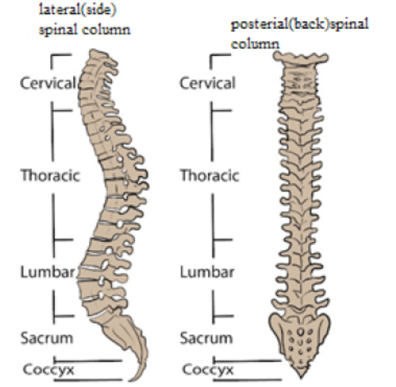
Figure 11.5: The Vertebral Column
A typical vertebra consists of the centrum (or body), a neural arch, a neural spine,two transverse processes and four articular processes with articulating surfaces. The
centrum is the front part (anterior) and consists of a solid piece of spongy bone
encircled by a layer of compact bone. The upper and lower surfaces are flat and
rough and provide attachment for the cartilaginous discs. These surfaces allow alimited degree of movement. The posterior (back) part is called the neural arch.
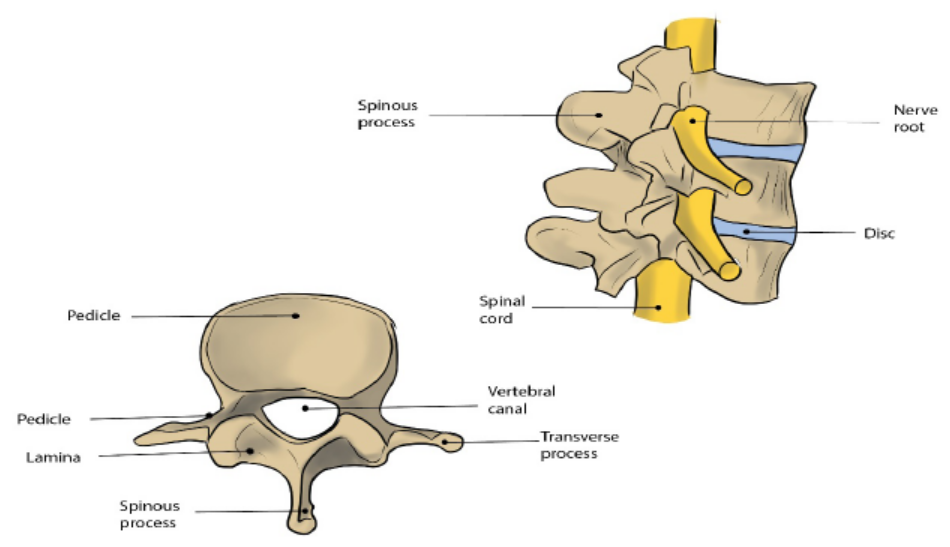
Figure 11.6: The structure of a vertebra
iii) The sacrum and the coccyx
The sacrum is roughly triangular in shape and consists of 5 fused vertebrae. It lies
between the hip bones with which it articulates. Horizontal ridges indicate the
divisions between the fused vertebrae. At the ends of these ridges are openings
which allow nerves and blood vessels to pass through. The coccyx consists of 4
fused tail vertebrae which are small and have a relatively simple structure. They do
not resemble the structure of a typical vertebra and the muscles of the buttocks areattached to the coccyx.
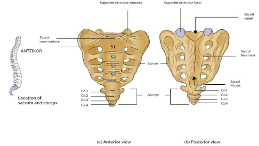
Figure 11.7: The Sacrum and Coccyx
iv) The Ribs
Twelve pairs of ribs articulate with the 12 vertebrae of the thoracic region. The ribs
are flat and narrow bones with a distinctive bow-shaped curve. Each rib consists of
a head or capitulum, a small tubercle (which is a short distance back from the head)
and the shaft. The tubercle fits into and articulates with the articulating facets on the
transverse process. All ribs articulate with thoracic vertebrae. True ribs (first seven
pairs) articulate directly with sternum by means of costal cartilages. Ribs 8 to 10
attach to the costal cartilage of rib 7, and ribs 11 and 12 do not attach to anything at
the distal end but are embedded in thoracic muscle. Ribs 8 to 12 are therefore called
false ribs, and ribs 11 and 12 are also called floating ribs for lack of any connectionto the sternum.
Figure 11.8: Diagram to illustrate the attachment of the ribs to the thoracic vertebrae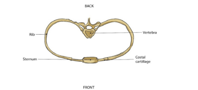
and sternum
b. The appendicular skeleton
The appendicular skeleton consists of the girdles (clavicle, scapula and pelvis) and
the skeleton of the limbs (arms and legs) There are approximately 126 bones in the
appendicular skeleton. Limbs are connected to the rest of the skeleton by girdles.
The upper (anterior) limbs are attached to the pectoral (shoulder) girdle and the
lower (posterior) limbs are attached to the pelvic (hip) girdle. The pectoral girdle
consists of the clavicle (collar bone) and scapula (shoulder blade). The pelvic girdle
consists of two pelvic bones (hipbones) that form the pelvic girdle. The vertebral
column attaches to the top of the pelvis; the femur of each leg attaches to the
bottom. The humerus is joined to the pectoral girdle at a joint and is held in place bymuscles and ligaments.
i) The pectoral (shoulder) girdle
The Pectoral girdle consists of two shoulder blades (scapulae) and two collar bones
(clavicles). These bones articulate with one another, allowing some degree ofmovement.
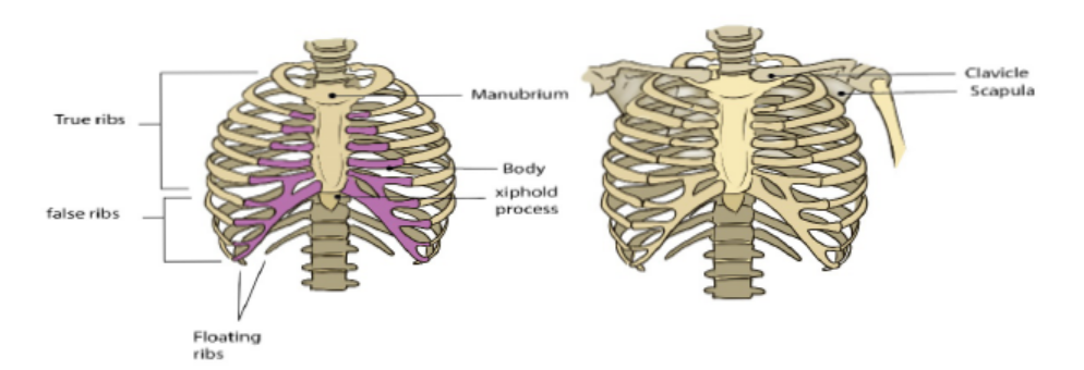
Figure 11.9: The thoracic cage and the pectoral girdle
ii) The pelvic (hip) girdle
The pelvic girdle consists of two large and sturdy hip bones. Each hip bone consists
of three fused bones namely the ilium, ischium and the pubis. The ilium is the largest
of the three and forms the upper part of the hip bones. The sacrum fits like a wedge
posteriorly between the two hip bones. The sacrum has a large, flat articular surface
on each side for articulation with the ilia. The ischium forms the inferior part of the
hip bone and the pubis at the central in front. The two pubic bones are attached in
the middle, on the front side by a symphysis which consists of fibrocartilage and
ligaments, the pubic symphysis. The two hip bones and the sacrum form a complete
bony ring, the pelvis. On the outer side of the point where the fused bones meet,
there is a deep hip socket into which the head of the femur fits. This is called theacetabulum.
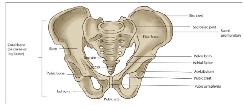
Figure 11.10: The pelvic girdle
The pelvic girdle forms a strong support for the attachment of the limbs. Strong
muscles of the back, the legs and the buttocks are attached to it. It protects some of
the internal organs. In females it forms a strong basin-like structure for supportingand protecting the developing foetus during child-bearing.
Application 11.1
1. What are the three main types of animal skeletons?
2. What are advantages and disadvantages of exoskeletons?
3. What is the difference between hydrostatic, exoskeleton and endoskeleton
skeletons?
4. What is importance of skeletal system human body apart from giving shapeand form to the body?
11.2 Types of joints
A joint is the junction between two or more bones. There are three major types ofjoints:
Activity 11.2Observe the joints below and answer questions that follow:
1. Why the joints of the skull are basically described as immovable joints?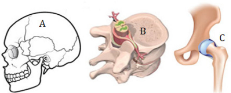
2. Why the joint in B is described as semi-movable?3. Describe the types of joints in B and C.
11.2.1 Immovable or Fused joints or sutures
These joints include the skull, sacrum, pelvis, and coccyx. As the name suggests,
these joints are points where joints fuse or grow together. The place where they growtogether is called the suture. These joints provide strength, support, and protection.
Figure 11.12: The fused joint
11.2.2 Slightly moveable joints
These joints are located between the vertebrae of the upper spine. There is cartilage
within the joints. They help pad and protect the bones. The bones are held together
by ligaments. The ligaments are tightly bound and limit the movement of the bones.This protects the spinal cord.
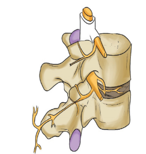
Figure 11.13: The slightly moveable joint
11.2.3 Freely moveable or synovial joints
At these joints the ends of the bones are covered with cartilage and there is a cavity
that separates the bones. The bones are held in place by ligaments which stop
the bones from moving too much. In addition to the ligaments the two bones are
joined together by sleeve-like capsule. The capsule encloses the synovial cavity. The
outer layer of the capsule is composed of ligaments. The inner layer of the capsule
is the synovial membrane. The synovial membrane secretes the lubricating synovial
fluid. Lubrication is essential to prevent frictional wear and tear. The cartilage at the
contact ends of the bones also reduces friction. The cartilage pads also act as shockabsorbers against mechanical damage.
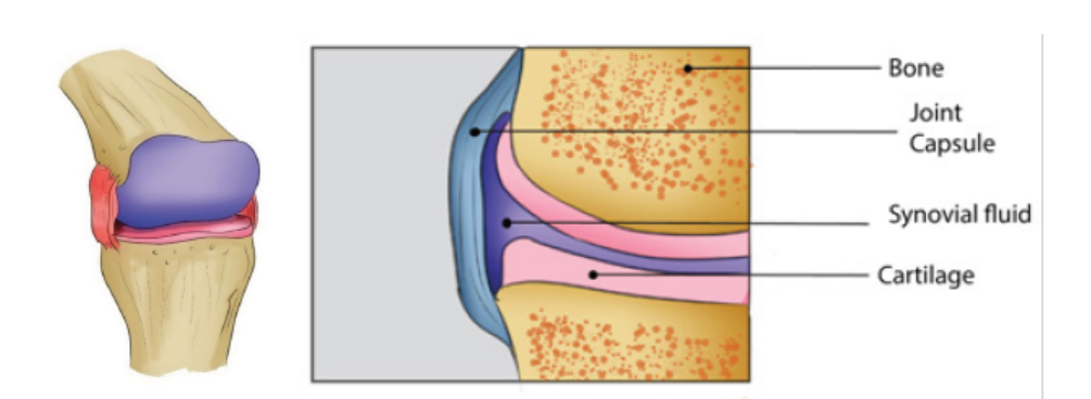
Figure 11.14: The synovial joint
11.2.4 There are four classes of synovial joints:
i) Gliding: The bones of these joints move across each other, back-and-forth and
side-to-side. Examples are between the carpals of the wrist and tarsals of the
ankle.
ii) Pivot: These joints allow a turning movement. Examples are between the first
and second vertebras when turning the head, between the ulna and the radius
of the lower arm when turning the palm of the hand up or down.
iii) Hinge: These joints allow movement in one plane during flexion and extension.
They act, as the name implies, like the hinge of a door. Examples are bending the
elbow or knee.
iv) Ball and Socket: This type of joint permits movement in three planes, i.e., in alldirections. Examples are the shoulder and hip joints.
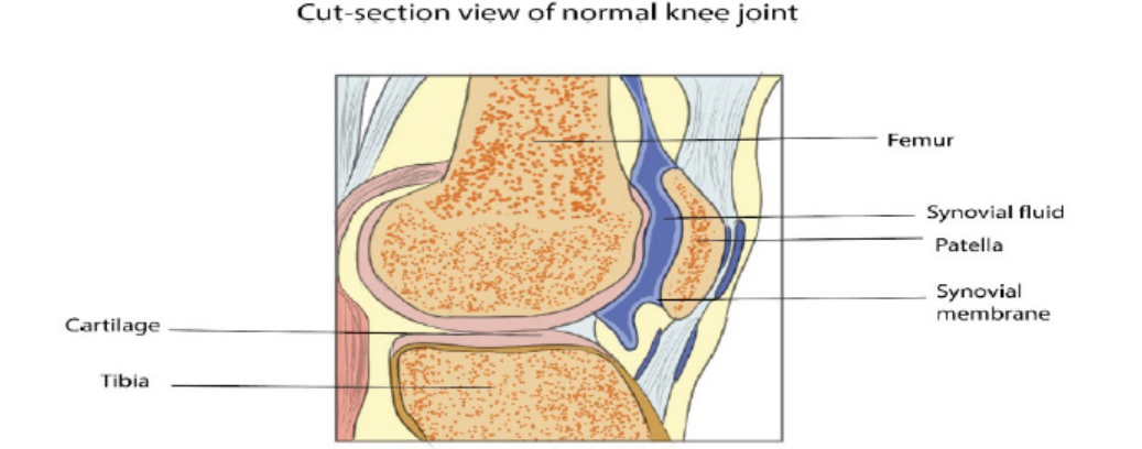
Figure 11.15: Cut-section view of normal knee joint.
Figure 11.16: Ball and Socket joint.
Table 11.2: Summary of the types of joints
Application 11.2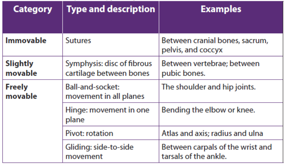
1. What is a joint?
2. Distinguish between fused joints and slightly moveable joint.3. What are the differences between the types of Synovial Joints?
11.3 Types of muscles: cardiac, smooth and skeletal muscle
Activity 11.3
Dissection of a frog / toad heart and observation of myogenic contraction.
Materials required
Dissection pan with 4 needles, 20 ml of physiological liquid (Ringer’s solution),
plastic eye-droppers, suture needle with thread attached, razor blade,
magnifying hand lens, pins, chloroform, cotton wool, frog or toad, bell jar,forceps, glass beaker, gloves, and water
Procedure
– Collect a living frog or toad from the nearest swamp
– Prepare 20ml of Ringer’s liquid in a glass beaker
– Put the cotton wool imbibed of 10 ml of chloroform in the bell jar
– Put your frog in the bell jar for 5 minutes, then remove it
– Lay your frog dorsally and fix its four limbs with pins on the dissection dish
– Carry out the longitudinal section from the abdomen to the chest usingsurgical blade (razor blade) or scissor.
There are 3 types of muscle: skeletal, smooth, and cardiac.
a. Skeletal Muscle
Skeletal muscle, as its name implies, is the muscle attached to the skeleton. It is also
called striated muscle. The contraction of skeletal muscle is under voluntary control.
These muscles are mainly responsible for movement of the body. Other purposes
are posture maintenance, support of the joints, and heat production. While its
contraction is fast and strong, skeletal muscle tires easily.
b. Smooth Muscle
Smooth muscle is found in the walls of all the hollow organs of the body (except the
heart). Its contraction reduces the size of these structures. Thus it regulates the flow
of blood in the arteries, moves your breakfast along through your gastrointestinal
tract, expels urine from your urinary bladder, sends babies out into the world from
the uterus, and regulates the flow of air through the lungs. The contraction of smooth
muscle is not under voluntary control. It is called involuntary muscle. It contracts
slowly and is slow to tire.
c. Cardiac Muscle
Your heart is made of cardiac muscle. This type of muscle only exists in your heart.
Unlike other types of muscle, cardiac muscle never gets tired. It works automatically
and constantly without ever pausing to rest. Cardiac muscle contracts to squeezeblood out of your heart, and relaxes to fill your heart with blood.
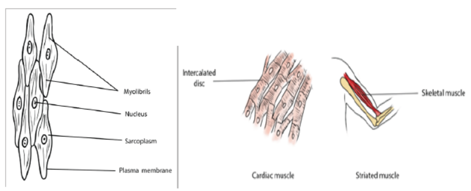
Figure 11.18: structure of three types of muscles
Application 11.3
Use prepared slides or charts of the three types of muscles and compare theircharacteristics.
11.4 Universal characteristics of muscles
Activity 11.4
Have you ever been injected medicine by intramuscular pathway? Or have
you been tested a rapid test of Covid-19?
In both cases the doctor or nurse advises in advance to keep calm so that
the muscle can relax and allow smooth flow of drugs through the muscle or
smooth flow of the strip through the nasal cavity.Why everybody including those without fear tend react on the same way?
The functions of muscle tissue are: movement, stability, control of body openings
and passages and heat production. To carry out those functions, all muscle tissuehas the following characteristics:
a. Responsiveness or excitability
Responsiveness is a property of all living cells, but muscle and nerve cells have
developed this property to the highest degree. When stimulated by chemical signals
(neurotransmitters), stretch, and other stimuli, muscle cells respond with electrical
changes across the plasma membrane.
b. Conductivity
Stimulation of a muscle fiber produces more than a local effect. The local electric
change triggers a wave of excitation that travels rapidly along the muscle fiber and
initiates processes leading to muscle contraction.
c. Contractility
Muscle fibers are unique in their ability to shorten substantially when stimulated.
This enables them to pull on bones and other tissues and create movement of the
body and its parts.
d. Elasticity
When a muscle cell is stretched and the tension is then released, it recoils to its
original resting length. Elasticity refers to the tendency of a muscle cell (or other
structures) to return to the original length when tension is released.11.4.1 Muscle contraction
Activity 11. 4.1
Using internet search simulations demonstrating the structure and functioning
of the sarcomere during muscle contraction with reference to sliding filamenttheory.
The excitability or the power of responding to an adequate stimulus is an innate
property of the muscle. When a brief stimulus is given, the muscle contracts and thisis followed by a wave of relaxation. This phenomenon is called a muscle twitch.
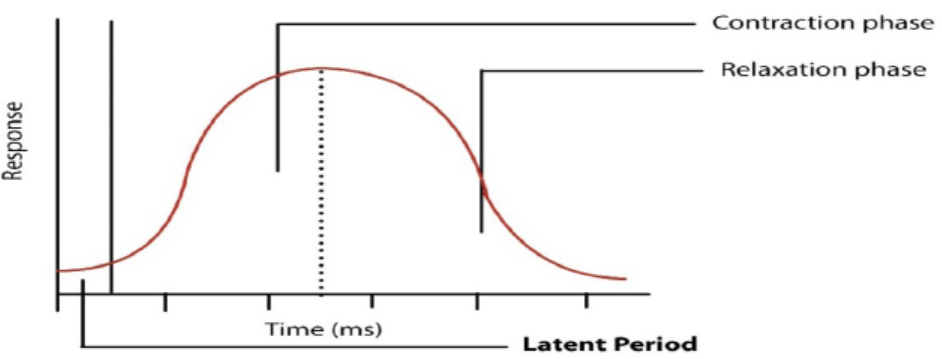
Figure 11.19: A muscle twitch
The Figure 11.19 shows a typical muscle curve of a skeletal muscle in response to
single stimulation. The muscle curve can be recorded with the help of a kymograph.
The curve indicates three phases: the latent phase, the contraction phase and the
relaxation phase. The period between the stimulus and beginning of contraction
is called the latent phase which lasts for about 0.01 second. During this period
chemical changes take place as a result of the stimulus. Latent period is required
for traversing the excitation along the nerve and the neuromuscular junctions. The
duration of the latent period varies with the species and depends on the type of
muscle, temperature and condition of the muscle.
The contraction phase during which the muscle actually contracts lasts for about
0.04 second in case of frog muscle. Shortening of the muscle takes place due to
chemical events which will be described in some details later. The third phase or
the relaxation phase lasts for about 0.05 sec. The total time taken by a single muscle
contraction is about 0.1 sec which varies with the temperature. At low temperature
contractions are prolonged, whereas with rising temperature the duration ofcontractions becomes shorter.
a. Muscle twitch, summation, and tetanus
A single action potential to the muscle fiber of a motor unit produces a muscle
twitch, a rapid and unstained contraction. If the impulses are applied to a muscle in
rapid succession through several motor units, one twitch will not have completely
ended before the next begins. Since the muscle is already in a partially contracted
state when the second twitch begins, the degree of muscle shortening in the second
contraction will be slightly greater than the shortening that occurs with a single
twitch. There are two types of twitch which are slow-twitch muscles and fast-twitchfibers.
– Slow-twitch are slower-contracting fibers but they are very efficient at using
oxygen to create energy without lactic acid build-up. These fibers are used for
high-endurance events like marathons.
– Fast-twitch fibers are white fibers, that contract very quickly making them
very strong and explosive but they also tire out very easily. The additional
shortening due to the rapid succession of two or more action potentials is
termed summation. At high stimulation frequencies, the overlapping twitchessum to one strong, steady contraction called tetanus.
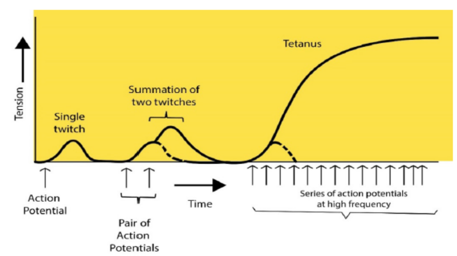
Figure 11.20: Patterns of muscle twitch, summation and tetanus
The graph 11.19 compares the tension developed in a muscle fiber in response to a
single action potential in a motor neuron, a pair of action potentials, and a series of
action potentials. The dashed lines show the tension that would have developed ifonly the first action potential had occurred.
b. Tetanic contractions
During normal activity such as locomotion, muscular contractions are not merely
twitches lasting for a second or a fraction of it. They are sustained for a longer period
during continued activity and exhibit compound or tetanic contractions. This can
be experimentally demonstrated by applying a number of stimuli to a muscle-nerve
preparation in rapid succession with little interval between successive stimuli, the
resulting contractions tend to fuse to give a maximum contraction. This sustained
contraction is called complete tetanus which, however, varies with the kind of
muscle and its condition. If repetitive stimuli are applied to muscle with long periods
of interval, the individual contractions can be seen because of little relaxation. Thiscondition is known as incomplete tetanus.
More interesting information is available about the tetanus. When a muscle is in tetany,
a musical note is produced by it which can be heard with the help of a stethoscope.
The pitch of the note is indicative of the vibrations that are produced at a rate
corresponding to the rate of application of stimuli. Most of the voluntary contractions
are of tetanus types which are produced by a series of nerve impulses arrivingin the muscle from the central nervous system.
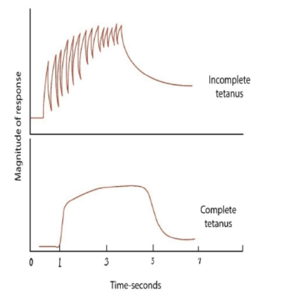
Figure 11.21: Diagram showing the condition of tetanus
a) The neuromuscular junction
This is a special kind of synapse where a motor nerve and muscle tissue meet. The
membrane of the muscle fiber, the sarcolemma is very folded in this region and
forms a structure known as an end plate. Electron microscopy shows us that the
structure of the neuromuscular junction is remarkably similar to that of any other
synapses. The end of the motor nerve is full of mitochondria and synaptic vesicles
which contain acetylcholine/neurotransmitter substances.
It appears that when an impulse arrives at the end of the motor neuron, it increases
permeability of the pre-synaptic membrane to calcium ions in the synaptic cleft. The
electrical impulse gets changed into a chemical message and gets stored into the
synaptic vesicles. The calcium ions then push the vesicles to fuse with the presynaptic
membrane thus discharging their neurotransmitter substances by exocytosis. The
neurotransmitter then diffuses through the synaptic cleft and get attached onto
receptor sites on the sarcolemma. This causes the sodium gated channels to open
thus causing a generator potential to be setup in the sarcolemma. If it reaches thethreshold, an impulse is fired into the muscle fiber.
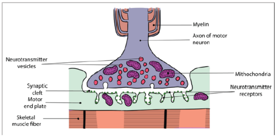
Figure 11.22: The neuromuscular junction
11.4.2 Laws of muscle contraction
A muscle contraction occurs when a muscle fiber generates tension through the
movement of actin and myosin. The sarcomere is the functional unit of muscle
contraction; it reaches from one Z-line to the next. In a relaxed muscle, the actin (thin
filament) and myosin (thick filament) overlap. In a muscle contraction, the filaments
slide past each other, shortening the sarcomere. This model of contraction is calledthe sliding filament mechanism.
Activity 11.4.2
Use of computer aided simulations to demonstrate the laws of musclecontraction (all or nothing, temporal summation and muscle fibre recruitment)
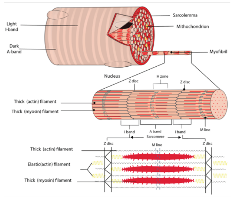
Figure 11.23: The sarcomere
Each muscle fiber contains cellular proteins and hundreds or thousands of myofibrils.
Each myofibril is a long, cylindrical organelle that is made up of two types of
protein filaments: actin and myosin. The actin filament is thin and threadlike; the
thin actin filaments are anchored to structures called Z lines. The region from one Z
line to the next makes up one sarcomere and the myosin filament is thicker. Myosin
has a head region that uses energy from ATP to walk along the actin thin filament.
The overlapping arrangement of actin and myosin filaments gives skeletal muscle
its striated appearance. When each end of the myosin thick filament moves along
the actin filament, the two actin filaments at opposite sides of the sarcomere are
drawn closer together and the sarcomere shortens. When a muscle fiber contracts,all sarcomeres contract at the same time, which pulls on the fiber ends.
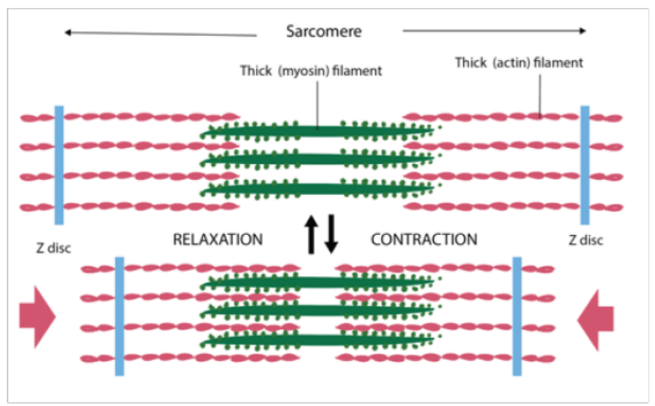
Figure 11.24: Muscle contraction
When each end of the myosin thick filament moves along the actin filament, the two
actin filaments at opposite sides of the sarcomere are drawn closer together and
the sarcomere shortens. In the contacted sarcomere, the A bands do not change in
length, but the I bands shorten and the H zone disappears. This behaviour can be
explained by the sliding filament model of muscle contraction.
How motor unit summation develops muscle tension
A skeletal muscle is an organ composed of multiple muscle cells or fibers, just like
any organ is made up of a whole bunch of cells. These fibers are arranged in motor
units, each of which is composed of a single motor neuron and all the muscle fibers
that that motor neuron innervates. Each motor unit contracts in an all-or-none fashion.
In other words, if the motor neuron is excited, it will stimulate all of the musclefibers to contract - that is, all of the muscle fibers within that particular motor unit.
11.4.3 Antagonistic skeletal muscles
Activity 11.4.3
Observe the following biceps and triceps muscles through the books and
internet and note down your observations (shortening and thickening of theantagonistic muscles).
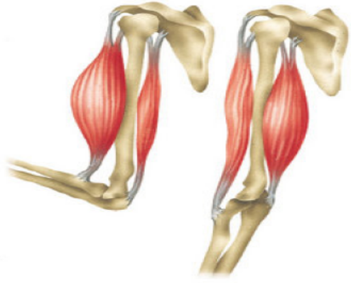
Antagonistic muscles are pairs of muscles. The action of one member is opposite to
that of the other member. Muscles can contract but they do not have the ability to
lengthen (stretch) themselves. They are arranged in pairs such that after one muscle
or muscle group contracts, a skeleton transfers the movement to stretch another
muscle or muscle group. The pairs of muscles that stretch each other are said to beantagonistic.
The biceps and triceps muscles of the arm are an example of an antagonistic pair.
Contraction of the biceps moves the arm toward the body and stretches the triceps.
Contraction of the triceps extends the arm and stretches the biceps. In this example
the bicep is said to be the flexor while the triceps is the extensor. Extensors are notas strong as flexors.
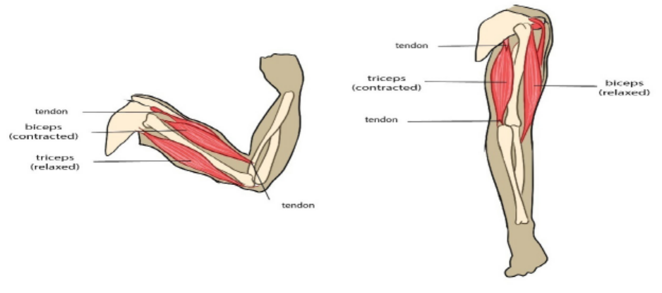
Figure 11.25: The antagonistic skeletal muscles
11.4.4 Movement in animals
Locomotion refers to the movement that causes a progression from one place to
another. There are several different types of locomotion exhibited by the animal
kingdom. It could either be active or passive. Sessile are animals that spend most
of their adult life in one place. Animals that move around are called motile. Corals,
sponges are examples of sessile organisms.
The act of flying is called aerial locomotion. Many organisms including; birds, insects,
bats, flying squirrels, many aquatic species and some amphibians including frog
have learnt to fly or glide.
Arboreal locomotion refers to species that live in and move through trees. Leopards
are good climbers that can climb up the tree along with their hunted prey to
keep them safe from other predators. The challenges of arboreal locomotion
include walking on narrow branches, moving up and down the inclines, balancing,
swinging with arms from one handhold to another and crossing gaps. Cats, parrots,
chameleons, goats, lizards and tree snakes are few examples of arboreal animals.
The movement on water is called aquatic locomotion. This involves swimming or
walking on the bottom surface of sea or ocean. Fish, ducks, bacteria, turtles, flat
worms, inchworms, leeches are organisms that can move through a liquid medium.
Most terrestrial animals move about using cursorial locomotion. Running
adaptation of different animals is referred to as cursorial locomotion. Forelimbs and
hind limbs play different roles in cursorial four-footed animals. These animals are
accustomed to long distance running at high speeds rather than high acceleration
over short distances. Cheetahs, wolves, ostriches are known for their cursorial
locomotion. Movement of animals that dig and live underground possess is called
fossorial locomotion. Such animals penetrate soil, wood or stone. Many soft bodiedinvertebrates, moles, earthworms and sea cucumbers are examples of organisms
with fossorial locomotion. Animals using hopping or jumping to move possess
saltatorial locomotion. Kangaroos, rabbits and few rodents exhibit saltatorialmotion.
Application 11.4
The diagram below is a drawing of the forelimb skeleton and part of theshoulder girdle of a mammal (rabbit).
Complete the diagram to show the following muscles and their attachments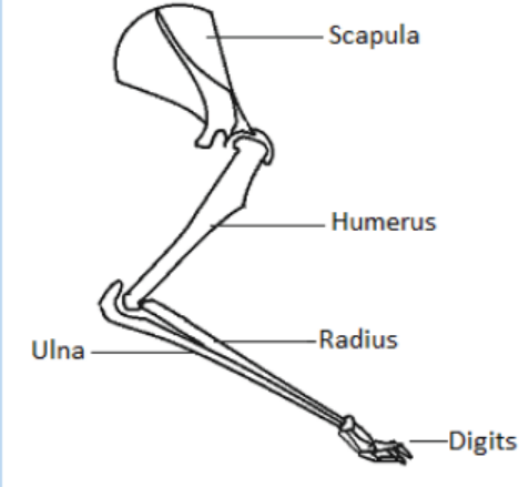
to the skeleton.
1. A muscle which contracts in preparation for landing after a leap. Label
this muscle X.
2. A muscle which will extend the digits. Label this muscle Y.
3. A muscle which will contract in response to a painful stimulus applied
to the forefoot. Label this muscle Z.11.5 Ultrastructure and functioning of striated muscle
Activity 11.5
Use the books from the school library and search further information from theinternet. Discuss Ultrastructure and functioning of striated muscle.
a. Ultrastructural appearance of skeletal muscle
The striated appearance of skeletal muscle fibres arises due to the organization
of two contractile proteins or myofilaments. The functional unit of contraction in a
skeletal muscle fibre is the sarcomere, which runs from Z line to Z line. A sarcomereis broken down into a number of sections:
– Z line – Where the actin filaments are anchored.
– M line – Where the myosin filaments are anchored.
– I band – Contains only actin filaments.
– A band – The length of a myosin filament, may contain overlapping actin
filaments.
– H zone – Contains only myosin filaments.
A useful acronym is MHAZI – the M line is inside the H zone which is inside the Aband, whilst the Z line is inside the I band.
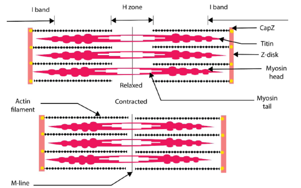
Figure 11.26: The sarcomere in contraction and relaxation
a. Function of striated muscles
Based on their fibrous and dense tissues, their main function is movement through
continuous contraction and relaxation. These muscles also help in; maintainingposture, stabilizing skeletal joints and producing body heat.
Application 11.5
1. Write on your own word the ultrastructure of muscle.
2. How many contractile proteins or myofilaments which constitute the
skeletal muscle fibres?3. What is the function of striated muscle?
11.6 Sliding filament theory of muscle contraction
Activity 11.6
Use the books from the school library and search further information from the
internet. Read and make summary about the sliding filament theory of musclecontraction.
The widely accepted theory of how muscles contract is called the sliding-filament
model also known as the sliding filament theory. According to this model, neither the
thin filaments nor the thick filaments change in length when the muscle contracts.
At rest, there is a low concentration of Ca2+ ions in the sarcomere, and the tropomyosin
blocks the actin sites to which myosin can bind. Upon arrival of an impulse, the
synaptic vesicles release their neurotransmitter substance (e.g. acetylcholine, Ach)
into the synaptic cleft. When Ach attaches on specific receptor sites, it causes the
release of Ca2+ ions from the triad vesicles into the sarcoplasm. Ca2+ ions bind to
Troponin-Complex which is protein that is integral to muscle contraction in skeletal
muscle and cardiac muscle, but not smooth muscle.
Once activated, the myosin head moves out and binds to actin, forming an action
myosin cross-bridge. The hydrolytic breakdown of ATP accompanies cross-bridge
formation and energy released causes the myosin head to pull the actin filament
towards the centre of the sarcomere. This leads to the shortening of the sarcomere
length and the overall contraction of the skeletal muscle. Cross-bridge formation
and breakage is repeated many times and on each occasion a new bridge is formed
between myosin head and another actin subunit further along the myofibril. After
stimulation, an active cation pump returns the Ca2+ ions to the triad vesicles; the
reduction in the level of Ca2+ ions in the sarcoplasm occurs and relaxation of thesarcomere begins.
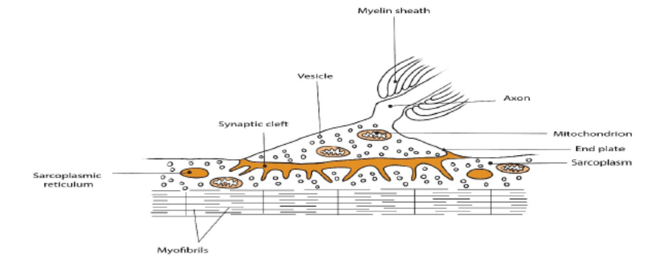
Figure 11.27: Neuromuscular junction or end plate
– When a muscle contracts, all the ATP present is rapidly used up. Replenishment
of ATP occurs when ADP and Pi are converted to ATP by phosphocreatine
breakdown. Later, after contraction has ceased, phosphocreatine is
reconstituted by ATP regeneration by energy from oxidation of fatty acids and
glycogen.
– In presence of adequate stimulus, the fibre contracts maximally. No further
increase in strength of stimulus will produce a stronger contraction this is
called all-or nothing response. A latent period of 0.05 seconds elapses prior to
muscle contraction.
– Contraction last for 0.1 second and is followed by a 0.2 second period of
relaxation. During this time, an absolute refractory is allowed by a relative
refractory period.
– When another stimulus is applied while the muscle is still responding to the
first stimulus, mechanical summation occurs whereby a second contraction
of greater force is caused. A rapid series of stimuli provokes a continued
contraction called tetanus. Tetanus ends when the muscle fatigues.
– If a muscle becomes very active, the respiratory and blood systems are unable
to supply sufficient oxygen for the muscle’s need. Consequently, pyruvic acid
is converted to lactic acid by the addition of H+ ions and the muscle builds up
an oxygen debt. Removal of lactic acid occurs when activity slows down or
ceases.
– The refractory period is the time after receiving a stimulus during which anerve or muscle cell cannot respond to further stimuli.
Application 11.6
1. Explain the sliding filament model of muscle contraction, including the
roles of troponin, tropomyosin, calcium ions and ATP.
2. Describe a neuromuscular junction?
3. What is the function of motor neurons?
4. Draw a well labelled diagram of sliding filament model of muscularcontraction.
End of unit assessment 11
1. What is the basic reason for the fact that animals show locomotion
whereas plants do not?
2. Briefly explain the role of each of the following in a mammalian locomotion:
3. Bones
a. Joints
b. muscles
4. What is meant by endoskeleton?
5. Outline the main functions of the endoskeleton.
6. Explain the various types synovial of joints.
7. In relation to antagonistic muscles, explain how it is possible to lift and
lower an object with your hands.
8. Outline the functions of fused joints and give an example.
9. What are the functions of muscle tissue?
10. What is the meaning of MHAZI in skeletal muscle fibres?
11. Explain what happened in refractory period in the sliding filament theory
of muscle contraction.
12. Explain what happened when mortar impulse reaches the end plate, the
vesicles release acetylcholine into the synaptic cleft of the end plate.
13. Draw a well labelled diagram of human skeleton.14. How does the structure of a muscle cell type relate to its function?
UNIT 12 HUMAN REPRODUCTION
UNIT 12: HUMAN REPRODUCTION
Key Unit Competence
Explain the role of hormones in human reproduction, stages of pregnancy and foetal
development.
Learning objectives
By the end of the lesson, I should be able to:
– Define menstrual cycle
– Describe main events of menstrual cycle
– Describe the hormonal changes involved in menstrual cycle.
– Distinguish oestrous and menstrual cycle
– Describe how mammals mate
– Explain how a sperm enters and fertilizes an ovum and how only one sperm
fertilizes an ovum.
– Outline the technique of in vitro fertilization (IVF).
– Explain the physiological changes in females during pregnancy.
– Explain how placenta forms and discuss its functions.
– Explain the gestation period birth.
– Describe the main stages of birth.
– Discuss the significance of parental care in mammals
– Explain how twins and multiple birth arise.
– Describe the main types of birth control techniques.
– Discuss advantages and disadvantages of different birth control methods.– State the causes and the ways of prevention of STIS and HIV.
Introductory activity
Human beings grow and develop from childhood to adulthood, during such
period of growth and development, there are changes in some parts of body
which may occur physiologically, physically and even psychologically. These
changes prepare individual adulthood to reproduce. Different researches
indicated these changes to be coordinated by different types of hormones.
1. Describe the hormones involved during such period of changes in
body parts?
2. Discuss the significance of these hormones you have mentioned above
during such period of changes.
3. Describe the role of hormones involved during menstrual cycle andbirth.
12.1 Menstrual cycle
Activity 12.1
Using flow-charts, diagrams and information collected in advance from the
library or internet, illustrate the action of hormones in the maintenance of themenstrual cycle.
This refers to the periodical changes in the reproductive behaviour of a female which
tend to occur in a sequence of events one after the other in the periodical circle. At
the onset of puberty, the cycle begins and repeats after 28 days unless interrupted
by pregnancy. The changes are stimulated by the gonadotrophic hormone such as;
follicle stimulating hormone (FSH) and luteinizing hormone (LH). These hormones
stimulate ovaries to secrete; oestrogen (steroid) and progesterone hormones.
These four hormones are involved in menstrual cycle. Two of them including; FSH
and LH are produced by pituitary gland and the other two are released by ovaries
respectively. The most obvious sign of the cycle is the monthly discharge of blood
a process called menstruation. The first day of menstruation is regarded as the first
day of the cycle. Figure 12.2 and 12.3 show the stages of menstrual cycle. Menstrualcycle is divided into three phases or events:
a. Follicular phase
Menstrual cycle usually begins when blood is first discharged from the uterus
during the first to fifth day (1-5 days). Following the reduction of progesterone, the
hypothalamus releases gonadotropin releasing hormone (GnRH) which stimulates
anterior pituitary gland to secrete follicle stimulating hormone (FSH). FSH bringsabout the following effects;
– Stimulates the development of a primary follicle
– Contributes to the shedding of uterine wall
– Causes production of oestrogen by uterine cells. The oestrogen produced
promotes healing, repair and growth of uterine lining, inhibits further secretion
of FSH. Oestrogen levels keep on raising until day 13 where they stimulate
secretion of luteinizing hormone (LH) by anterior pituitary gland.
b. Ovulatory phase
Around the 14th day, the high levels of oestrogen cause release of luteinizing hormone
(LH) the release of LH brings about ovulation (release of mature egg from the ovary).
Immediately after and slightly before ovulation, a woman is fertile and can conceivea baby if she has sexual intercourse or if sperm is present in her oviduct.
c. Luteal phase
After ovulation, the remains of ovarian follicle form corpus luteum also known as
Yellow body, which secrete large amounts of progesterone hormone and smaller
oestrogen. These two hormones; stimulate further development of mammary
glands, inhibit release of FSH and thickening wall of uterus in anticipation of
pregnancy. If oocyte (ovum) is not fertilized with in about 36 hours of being shed
into oviduct, it dies and corpus luteum gets smaller. Thus levels of progesterone
and oestrogen keep on reducing until day 28 days i.e. 14 days after ovulation. Low
levels of progesterone remove the inhibitory effect on FSH, causing its release thusmenstruation and the cycle starts again.
– At menopause there are no more fertile follicle so follicular development and
ovulation is ceased.
– The menstrual cycle is controlled by hormones from both brain and the ovary.
– The natural cycle repeats until there is either a pregnancy or the womanreaches menopause, the end of the reproductive phase of a woman’s life.
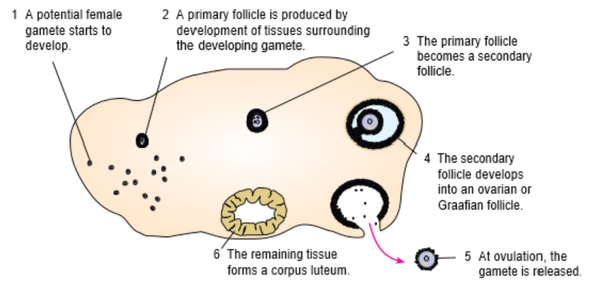
Figure 12.2: The growth of ovarian follicle.
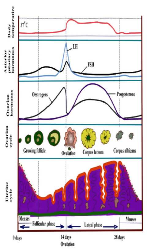
Figure 12.3: Hormonal and Menstrual cycle growth curve.
The uterine cycle also has three phases (events):
Proliferative phase: It stimulates the thickening of endometrium of the uterus. This
thickness of endometrium is stimulated by oestrogen from follicles before ovulation. This
results the development of ovary. It acts like follicular phase.
Secretory phase: it occurs after ovulation for describes further thickening of endometrium
(endometrium tissue become more complex) in preparation for implantation. This is
stimulated by progesterone which is secreted by corpus luteum and this occurs when
corpus luteum is functioning. It acts like lacteal phase.
Menstrual phase: when endometrium tissue is discharged and vaginal bleeding occurs at
the end of ovulatory cycle if pregnancy has not occurred. It is called menstruation.it describes
the shedding of endometrium when implantation does not occur. When pregnancy does not
occur the level of progesterone falls and this results shedding of endometrium. Menstrual
bleeding lasts between 3 and 5 days. The first day of the period is the first day of the cycle.
Oestrous cycle
The word oestrus is derived from the Latin language oestrus meaning sexual desire. It
describes the phase when the female animal is sexually receptive to a male. Females
of most species of mammals except human come into ‘heat’ known as oestrus in
regular cycles at particular times of year. Oestrus is the time when females are both
fertile and sexually receptive. Oestrus cycle is controlled by the same hormones as
the human menstrual cycle. FSH and oestrogen control the process until ripe ova are
released when LH and progesterone take over.
Application 12.1
1. What is the main difference between menstrual and oestrus cycle?
2. What are significant events which happen between day 13 and day 15
of menstrual cycle?
3. Asses the main events of menstrual cycle.
4. (a) Explain the meaning of oestrus cycle in mammals(b) State the difference between oestrous and menstrual cycle.
12.2 Copulation, fertilization and embryo development.
Activity 12.1
Watch a simulation from internet; illustrate the stages that bring aboutfertilization and development of an embryo.
12.2.1 Copulation
It is act of mating where sperms from male are transferred into the female tract.
Male mammals have an intromittent organ called penis which becomes erect at a
moment of mating for insertion into female’s vagina. The erection of penis is brought
by hydraulic action (penis becomes gorged with blood). This occurs as a result of
sexual arousal which brings about by ejaculation (release of sperm). The semen’s
are secreted from accessory glands into vas deferens and bladder sphincter closes
preventing urine from entering urethra. Sperms are expelled from epididymis into
vas deferens and out of the body by a series of muscle contraction of penis.
In a female, sexual arousal results in the swelling of clitoris and stimulates thesecretion of mucus which lubricates vagina during sexual intercourse.
12.2.2 Fertilisation
Fertilisation is the fusion of male and female nuclei to form zygote. Copulation
results in the ejection of spermatozoa into vagina. The spermatozoa swim in the
watery mucus of vagina and uterus up into the oviduct where the fertilisation takes
place in the upper part of the oviduct. From the vagina or uterus spermatozoa
propel using energy from mitochondria. If ovulation has already taken place, the
egg and sperm meet in the upper part of oviduct and once they come into contact,
acrosome raptures and release lytic enzyme which dissolve corona radiata of the
egg and soften zona pellucida and vetelline membrane. The following processestake place:
a. Capacitation
This is a stage where by sperm undergoes essential changes while passing through
female genital trackand this takes about 7 hours. These changes include the
removal of a layer of glycoprotein from outer surface of sperm, by enzyme in uterus.
Cholesterol also is removed to weaken the membrane.
b. Acrosome reaction
This involves the releasing of enzyme found in acrosome such as hyaluronidases
and protease. These enzymes digest corona radiata (narrow path in the follicle
cells) and the zona pellucida (a protective glycoprotein surrounding the plasma
membrane of the egg).
c. Fusion
In this stage the head of sperm will fuse with the microvilli surrounding the secondary
oocyte and penetrate its cytoplasm.
d. Cortical reaction
This stage involves the releasing of enzymes by lysosomes in cortical granules (outer
region of the secondary oocytes); the enzymes cause the zona pellucida to thicken
and harden forming a fertilization membrane. This cortical reaction prevents the
entry of other sperm inside ovum (polyspermy).
e. Zygote formation
The secondary oocyte is stimulated to complete meiosis II, during this time of
stimulation the nucleus of sperm and secondary oocyte are called pro-nuclei andthen the two nuclei fuse to form the zygote (2n).
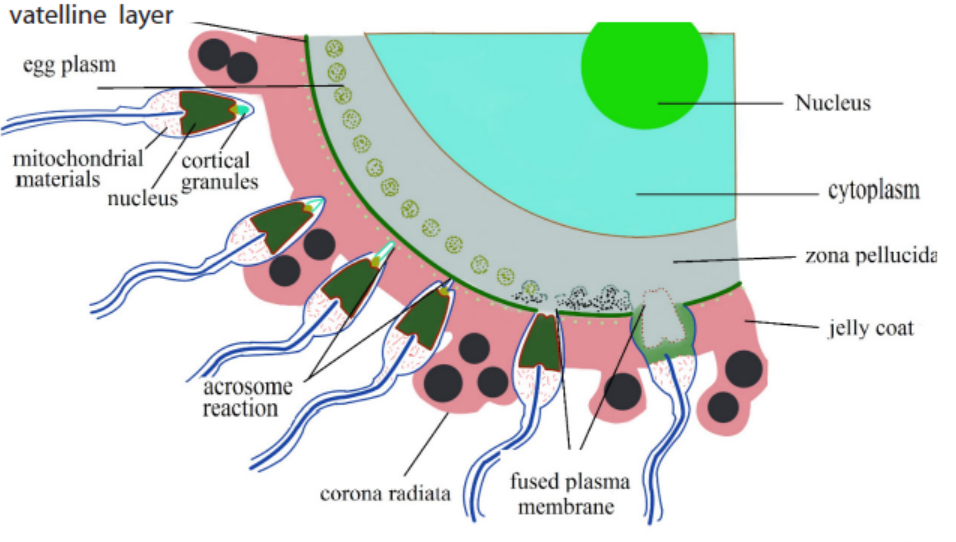
Fig 12.4: Process of fertilization
The movement of sperm in the female reproductive system;
Once sperm arrives the female reproductive tract, they moved largely by female
reproductive system:
– Around the time of ovulation, the vaginal mucus changes in PH in response
to changing levels of sex hormones. It is normally so acidic which can tend to
kill sperm. At the fertile time it becomes more alkaline to prevent sperm from
damage.
– The mucus which blocks the cervix, preventing the entry of pathogens and
become less viscous, allowing sperm to move through it more easily.
– Prostaglandin (local hormone) in semen and oxytocin hormone released by
posterior pituitary gland during sexual intercourse. Initiate the contraction inuterus, helps sperms to move towards fallopian tube.
12.2.3 Embryonic development
The zygote spends the next few days travelling down the oviduct (Fallopian tube) by
peristaltic contraction and by beatings of the cilia in wall of the oviduct toward the
uterus. As it travels, it divides by mitosis several times to form a ball of cells called a
morula. The cell divisions, which are called cleavage, increase the number of cells
but not their overall size. More cell divisions occur, and soon a fluid-filled cavityforms inside the ball of cells. At this stage, the ball of cells is called a blastocyst.
The blastocyst reaches the uterus and becomes embedded in the endometrium at
roughly the 5th – 10th day. Once in the uterus the blastocyst burrows into the uterine
wall a process called implantation. After implantation, the blastocyst becomes
embryo. It grows through multiplication and differentiation of its cells forming
tissues and organs. The heart and blood vessels are the first organs formed andembryo now called foetus.
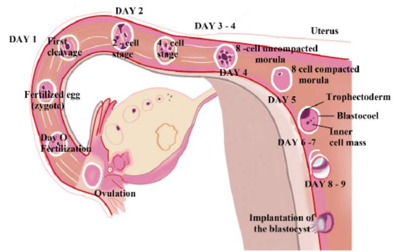
Figure 12.5: Embryo development during the first nine days
a. Stages of embryo development:
There are three major stages of embryo development;
i) Cleavage
The cleavage consists of the division of zygote without increase in mass into a ball of
consisting of many daughter cells.
ii) Gastrulation
It is the development of different layers of cells in the embryo. It generally occurs
during the second week after fertilization. During gastrulation, cells of the embryo
migrate to form three distinct cell layers: the ectoderm, mesoderm, and endoderm.
Each layer will eventually develop into certain types of tissues and cells in the body
of vertebrates.
– Ectoderm—it forms tissues that cover the outer body; develops into cells such
as nerves skin, hair, and nails.
– Mesoderm—it forms tissues that provide movement and support; develops
into cells such as muscles, bones, teeth, and blood.
– Endoderm—it forms tissues involved in digestion and breathing; develop intoorgans such as lungs, liver, pancreas, and gall bladder.
iii) Organogenesis and Differentiation
Differentiation of cells leads to the development of specific organs and tissues within
the three cell layers. This is called organogenesis. All the major organs begin to form
during the remaining weeks of embryonic development.
b. Extra-embryonic membranes
These membranes are part of placenta. The outer cells of the blastocyst, the
trophoblast grow and develop into an outer layer or membrane called the chorion.
This plays a major role in nourishing and removing waste products from the
developing embryo.
The amnion is a thin membrane covering the embryo like an umbrella and has a
protective function. Between the embryo and the amnion is the amniotic fluid. The
amniotic fluid supports the embryo and protects it from mechanical shocks.
The yolk sac has no significant function in humans but is important in reptiles and
birds, where it absorbs food from the separate yolk and transfers food to the gut of
the developing embryo.
Note:
The first trimester of the development or the embryo is critical. There is
high risk of spontaneous abortion or miscarriage due to alcohol, infection,
radiations (X-rays), nutritional deficiencies, genetic mistakes or abnormalities
in the developing embryo. From the 8th week until birth (around 38 weeks), the
developing organism is called a foetus. The foetus is not as sensitive to damage
from environmental exposures as the embryo, and toxic exposures often cause
physiological abnormalities or minor congenital malformation. All major structuresare already formed in the foetus, but they continue to grow and develop.
Application 12:2
1. Explain how sperms enter and later contribute to fertilisation of an
ovum?
2. Explain why only a single spermatozoon fertilises an ovum?3. What is implantation?
12.3 Role of Placenta in the development of embryo
Activity 12.3
Using a diagram of the placenta, discuss how its structure is related to itsfunctions
The placenta is a temporary organ in which nutrients and wastes are exchanged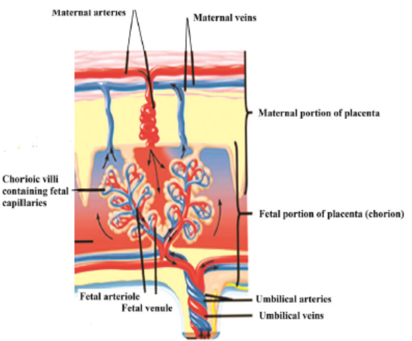
between the mother and the embryo or foetus.
The foetal part of the placenta consists of the allantoides and chorion. The chorion
forms many large projections called chorionic villi which contain a dense network of
foetal capillaries which in turn are connected to two umbilical arteries and umbilical
vein in the umbilical cord. The umbilical arteries carry blood from the foetus to the
placenta, while the umbilical vein carries blood in the opposite direction. Although
maternal blood in the endometrium is in close proximity with the foetal blood in the
umbilical capillaries, they do not mix because they separated by membranes of thevilli and capillary.
12.3.1 Functions of the placenta:
– Allows diffusion of nutrients such as water, glucose, amino acids, simple
proteins and mineral salts from maternal blood.
– It is a site of gaseous exchange: haemoglobin of the foetus has high affinity to
oxygen compared to adult haemoglobin.
– It offers passive natural immunity on the foetus. Certain maternal antibodies
can cross the placental barrier.
– It protects foetal circulation from the high pressure in the maternal circulation
– Prevents mixing of maternal and foetal blood which would cause agglutination
(clotting) if the two blood types are incompatible.
– It produces and secretes hormones such as the HCG (human chorionicgonadotrophin), progesterone, oestrogen, and relaxin.
Note that:
– The action of HCG is similar to that of LH. HCG stimulates the corpus luteum
to secrete progesterone and oestrogen throughout the first trimester. HCG
is produced in such large quantities that some of it is excreted in the urine
of a pregnant woman (positive test of pregnancy). Secretion of HCG declines
around tenth week and the corpus luteum reduces.
– The placenta does not give complete protection to the foetus. Certain
pathogens, toxins, and drugs can enter the foetal circulation and causedamage. Examples are; HIV, rubella toxins, alcohol, nicotine and heroin.
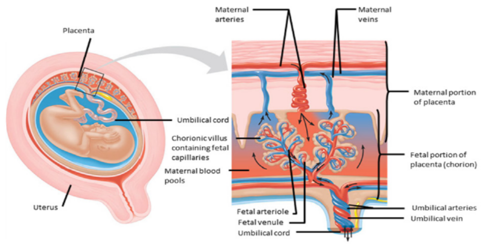
Figure 12.6: The structure of the placenta
12.3.2 How the placenta works?
Blood from the mother enters the maternal blood vessels of the placenta under
pressure, forcing the blood into the empty spaces. When the mother’s blood contacts
the foetal blood vessels, gases are exchanged. Oxygen from the mother’s blood is
exchanged with carbon dioxide from the foetus’s blood. A release of pressure brings
the mother’s blood back from the placenta and into her veins.
– The substances that are moved from the mother to the foetus include:
– Water
– Glucose by passive diffusion
– Hormones
– Amino acids by active transport
– Lipids by membrane lipid diffusion
– Oxygen is released by the maternal haemoglobin. The haemoglobin of the
foetus has a higher affinity for the oxygen.
– Alcohol, many drugs, nicotine (if taken by mother during pregnancy)– Vitamins, minerals.
The substances that are moved from the foetus to the mother include:
Carbon dioxide is taken up by the maternal plasma and transported to the lungs of
the mother for excretion
– Urea passes into the maternal blood and passes to her kidneys for excretion.
The exchange between the mother and the foetus is possible because of specific
structures in the placenta:
– The plasma surface membranes of the cells in the walls of the chorionic villi have
microvilli, which increase their surface area for the exchange of substances by
diffusion, facilitated transport and pinocytosis.
– Numerous mitochondria are found in these cells. They provide the energy for
the active transport and pinocytosis.
– The cell surface membranes contain carrier molecules (protein) used in the
uptake of materials into the villi by active transport.
– Numerous small vesicles are found inside the cells of the villi as a result ofmaterials being taken up from the blood by pinocytosis.
12.3.3 Pregnancy rapid test
Pregnancy tests look for a special hormone – human chorionic gonadotropin (HCG)
– that only develops in a woman’s body during pregnancy.
After the fertilized egg implants, the growing placenta starts releasing HCG into
your blood. Some HCG also gets passed in your urine. HCG can be found in the blood
before the first missed menstrual period. This can be as early as 6 days after the egg
implants.
These tests can use either your urine or blood to look for HCG.
At-home pregnancy tests are inexpensive and commonly used urine tests. There are
a few things to keep in mind when you take a home pregnancy test:
– Use your first morning urine when possible. This is the time of day when your
HCG levels will be the most concentrated and easily detected. If you do it at
another time of day, try and make sure your urine has been in your bladder forat least four hours.
– Don’t drink excessive amounts of fluids before you take a pregnancy test. Many
people think this will increase the volume of urine, but it can also dilute (thin
out) your HCG levels.
– Read the directions that come with the test thoroughly before starting the test
and following every step precisely.
– Collect your urine in a cup and use an eyedropper to move a small amount of
fluid into a special container
– Place the testing stick/strip into the area of your expected urine stream so that
it will catch your urine midstream.
– Wait for the recommended amount of time to view the results which can
include: a change in color – a line – a symbol, such as plus or minus, or – thewords “pregnant” or “not pregnant”
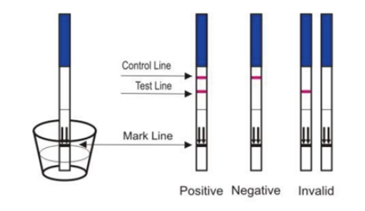
Figure 12.7 Home pregnancy test
Application 12.3
1. What do you understand by the term implantation?
2. Describe the composition of foetal blood entering the placenta and foetal
blood leaving the placenta.
3. Explain the urine of pregnant woman give a positive test while that of non –pregnant one give the negative test.
12.4 Physiological changes during pregnancy and parentalcare
Activity 12.4
Using models that show stages, discuss physiological, physical, and behaviouralchanges that occur during pregnancy.
Pregnancy refers to the development that take place between fertilisation of the
ovum to birth of the foetus. When fertilised egg becomes implanted in uterine wall,
pregnancy results. And a number of important events take place during this period.
The period from fertilisation to birth is called gestation period. In human it is aboutnine months.
12.4.1 Changes during pregnancy
A pregnant woman’s body undergoes various; physiological, physical and behaviouralchanges.
a. Some physiological changes during pregnancy:
– Respiration rate rises for increased maternal oxygen consumption which is
needed for demand of placenta, uterus and foetus.
– More blood vessels grow and pressure of expanding uterus on large veins
causes blood to slow in its return to the heart.
– Rise up and out of pelvic cavity this action displaces the stomach and intestine.
– Blood volume increase greatly.
– Placenta produces large amount of progesterone and oestrogen by 10 to 12
week of pregnancy to control uterine activity.
– Increased requirement of calcium due to increase of parathyroid gland.
– Experiences warm (hot flashes) caused by basal metabolic rate and increased
hormonal level.
– Stretching of abdomen wall and ligaments that support uterus.– Kidney work extra hard to excrete waste products of both mother and foetus.
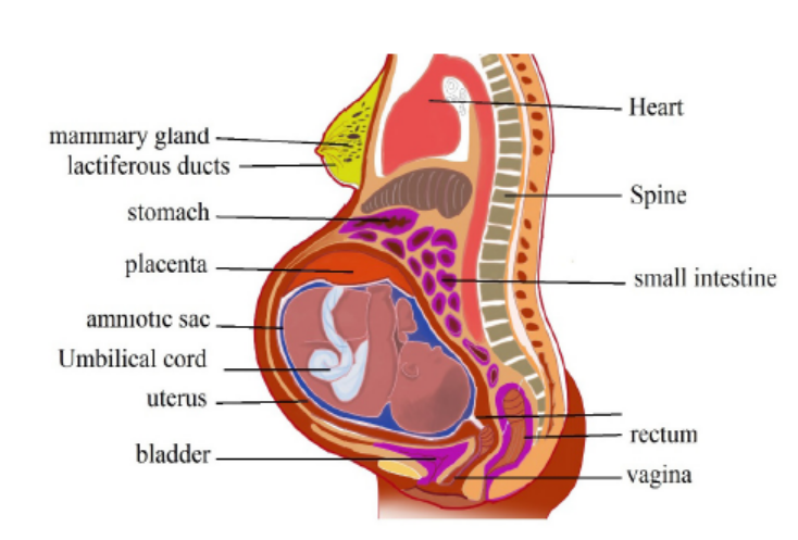
Figure 12.8: Changes during pregnancy.
b. Some physical changes during pregnancy
– Breast may become large and more tender because of increased level of
oestrogen hormone progesterone thus breast gets even bigger to prepare for
breast feeding.
– Nipples may stick out more.
– By the end of third trimester, a yellow, watery, pre-milk may leak from nipples.
– Changes in hair and nail growth and texture due to hormone changes.
– Leg cramp caused by fatigue from carrying pregnant weight.
– Feet and ankles may swell because of extra fluid in the body during pregnancy.
c. Some behavioural changes during pregnancy:
– Physical discomfort such as urinary frequency can be frustrating.
– Fear and anxiety lessen especially foetal movement are felt.
– Self-introspection
– Nesting behaviour begins. Some woman exhibit mood swings and emotional
liability.
12.4.2. Delivery process
By the end of pregnancy, near the time of birth, the amniotic sac raptures (breaks)
and amniotic fluid drains through birth canal and labour usually begins which
involves the contractions of muscular walls of the uterus.
Initiation of birth: Uterine contractions starts when the foetal pituitary gland secretes
adrenocorticotrophic hormone (ACTH) which stimulates foetal adrenal gland to
secrete corticosteroids. These hormones pass into blood sinuses in placenta to cause
maternal cells to secrete prostaglandins (local hormone) and cause uterine wall to
contract. This contraction pushes the foetal head against the cervix to stimulating
stretcher receptor to send information to mother’s brain and causes release of
oxytocin hormone. The prostaglandin and oxytocin hormone together result intense
contraction of uterine walls called labour which stimulates more release of oxytocinhormone and as positive feedback mechanism.
The delivery process can be summarized into three main stages:
– Dilation stage: During this stage, water sac filled with amniotic fluid forms
and precedes the head, widening soft tissue of birth canal, cervix, and vagina
for canal of constant diameter. The amnion raptures and amniotic fluid drains
through vagina.
– The expulsion stage: During this stage, cervix is fully dilated while abdominal
muscle bear down in supporting rhythmic contraction of uterus shorten the
uterine wall and baby is pushed into and through the birth canal. The head
and shoulder align themselves first.
– Placenta stage: This stage begins with complete expulsion of baby and ends
with expulsion of foetal membrane. The cord is clamped and cut when delivery
of baby is complete. This leads carbon dioxide enrichment into baby’s blood
which activates respiratory centre and baby begins to breath with the first cryat the same time foetal circulation changes to baby’s own systemic and
12.4.3 Parental care
The degree of maturity in mammalian new-borns varies from one species to another.
New-born in pigs can move around and eat solid food while new-born in humans,
dogs and rat are quite helpless and require a lot of parental care to survive. All
mammals feed their young ones by milk which contain all the nutrients required
by new born for the first few days. Parents also protect new born from predators
and from unfavourable weather. Some species make nest just before delivering the
new born. Some parents also become aggressive when they have young one. As the
young one grow older the parent start gathering food for them. Once the new born
get old enough to gather food for themselves can leave on their own. In humans’parental care extends for very long time up over 18 years.
In humans breastfeeding is associated with many benefits:
– It makes earlier a closer contact between the mother and her infant
– Breastfed babies do not get too fat
– The infant has a better control over its own milk intake, this prevents over
eating in late life
– Fats and irons from breast milk are better absorbed than those in cow’s milk
and milk is easily digested.
– Breast feeding provides important antibodies that help to prevent respiratory
infections and meningitis,
– Breastfeeding helps the mother’s reproduction organ return to a normal state
more rapidly
– Breast feeding promotes the secretion of LH (and prolactin) and this makes a
delay in follicle development and ovulation,
– The act of sucking on the breasts, promotes the development of the jaw, facial
muscles and teeth (sucking from a bottle requires less effort).
– Pulmonary circulation. After delivery, uterus contract so that placenta separates
from– Uterine wall expelled out as the sign of birth end.
Application 12.4
1. How can you assess physical changes that occur during pregnancy?
2. Discuss the significance of parental care in mammals3. Describe the different stages of birth?
12.5 Twins and multiple births
Activity 12.5
Watch a movie simulation from internets to illustrate the types of twins andexplain how multiple birth arise.
Twins are individuals born to the same mother at the same time. Twins include;
– Fraternal twins or non-identical twins or dizygotic twins: These are twins
which develop from two separate egg cells fertilised by two different sperms.
Fraternal twins are genetically different since they develop from different
gametes.
– Identical twins or monozygotic twins: these are twins which develop from the
same fertilised egg. Identical twins are genetically similar since they develop
from the same sperm and the same egg.
– Siamese twins: are conjoint identical twins i.e. they have not completely
separated during the embryo development. As consequence, they share same
organs. Conjoint identical twins develop without separating completely andare born attached to one another. Such twins may be separated surgically.
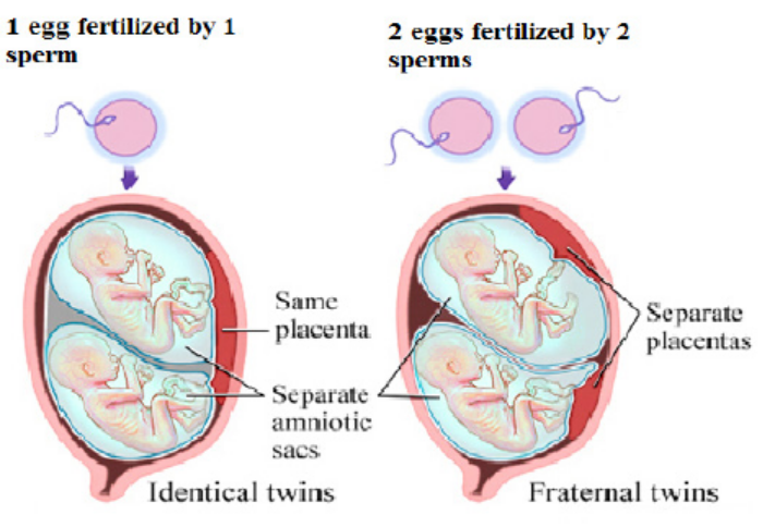
Figure 12.9: Identical and fraternal twins
Multiple births arise when several eggs are released at the ovulation and are
fertilised or when a zygote splits into several zygotes. It is commonly occurring in
mammals such as; pigs, dogs and cats.
Application 12.5
Explain how twins and multiple birth arise?
12.6 Infertility or barrenness
Activity 12.6
1. Discuss the social and economic consequences of barrenness
(infertility), producing many children by a couple and suggest methods
to cope with these issues.
2. Using the internet or library, research about in-vitro fertilization and
discuss the ethical implications.
12.6.1 Infertility
Infertility is the failure to achieve pregnancy when no contraceptive method is used.
In females, infertility may be due to:
– Failure to ovulate due to the lack of some hormones
– Damage of the Fallopian tubes / oviducts, for example the tubes may be
completely blocked by nature or after an infection,
– Damage on the uterus; for example, the endometrium can be destroyed
– Damage on the cervix, for example the cervix may be narrow or too wide or
may stop producing cervical mucus needed for the sperm to reach uterus
– Antibodies against sperms, for example, the cervix, the uterus or the oviduct
of a woman can produce antibodies against her husband’s sperms.
Some causes of infertility/barrenness in males include:
– Absence of sperms in the semen (Azoospermia).
– Low sperm count e.g. when ones ejaculate less than 1cm3 of semen.
– Abnormal sperm e.g. sperms with 2 tails, or without tail, or without acrosomes,
– Auto-immunity e.g. antibodies attack one’s sperms
– Premature ejaculation: the man has orgasm before copulation
– Impotence i.e. inability to achieve or maintain an erection of the penis.
a. Some social consequences include:
– Isolation including exclusion from ceremonies and social gathering.
– Rejection being an outcast and physical abuse perpetrated by community
members.
– Stigmatization or recognizable marginalization.
– Status loss that is no respect and social fail.
– Ridicule including insults and verbal abuse.
Some economic consequences include:
– Cost of infertility by either modern biomedical or traditional treatments.
– A feeling of rejection.
– Having few relations, receiving few gifts and less land.
– Marital instability including fear of husband taking second wife.
– Divorcing childless woman
– Violence perpetrated by partner.
Note:
While infertility may result into conflicts between couples and families, producing
many children also brings about some economic challenges. Many children affect
families’ financial wellbeing and some parents admit that children are expensive.
Consequences of many children per one family include:
– High rate of maternal depression.
– Low rate of immunization and parental care.
– Baby taxing both physical and emotional especially off work after birth.
– I come tend to go up when new members of the family arrive. Men see the
boost in their earnings after birth of child.
– There is economic wellbeing decline in time around birth.
b. Increasing fertility
Increasing fertility can be done in various techniques such as:
– Fertility drugs: a synthetic chemical which stimulates ovulation by either
proving gonadotrophins such as FSH which stimulates growth of follicles. Or
proving chemical which inhibits natural production of oestrogen.
– Artificial insemination: sperm from donor is inserted artificially through cervix
of mother to be.
– Using in-vitro fertilisation
12.6.2 In-vitro-fertilisation
In-vitro fertilisation is the process of fertilisation where an egg is fertilised by sperm
outside the body. It involves the fertilisation of egg cell outside the body which
are then artificially implanted in the uterus to produce test tube baby. The process
involves monitoring and stimulating of woman’s ovulatory process removing ovum
(egg) from woman’s ovaries and letting sperm to fertilise them in liquid laboratory.
The fertilised egg (zygote) undergoes embryo cultured for 2 to 6 days and then
transferred to the same or another uterus for successful pregnancy. The embryo is
implanted in woman’s uterus.
Advantages of in vitro-fertilization techniques include:
– Simplicity: living organisms are extremely complex functional system with
protein molecules, RNA molecules and genes. Therefore, the work of Vitro
simplifies system under study to focus on small number of components.
– Species specificity.in human cells in-vitro method can be studied without
extrapolation from experimental animal’s cellular response.
– Automation and convenience: In-vitro method can be automated, high
yielding throughout screening methods for testing molecule in pharmacology.
– In vitro- fertilisation can be used to achieve successful pregnancy but the
process usually produces more embryos which some scientists wish for
research design to improve our knowledge about disease.
Application 12.6
1. Define in-vitro-fertilisation
2. Outline the techniques of in-vitro-fertilisation.
12.7 Family planning: birth control and contraception
Activity 12.7
Using the internet or library, research about birth control methods and write a
summary of what you have learned.
– Birth control includes contraception, but is broader in meaning because it also
includes any measures taken after fertilization which are designed to prevent
birth. Contraceptionis preventing the fusion of the male gamete and female
gamete. Both natural and artificial methods exist.
Artificial methods:
– Oral Contraceptive pills: a chemical method of contraception. One version
uses a combination of progesterone and oestrogen that inhibits ovulation.
Others are single hormones that require very careful management when taken.
– Intrauterine device (IUD) the coil is placed inside the uterus an exact
understanding how this works is unclear. A possible explanation is that it
‘irritates’ the endometrium such that rejects implantation of embryos. The
device is made from plastic or copper and inserted by a doctor. Nevertheless,
this device is very effective.
– Condom is another mechanical method of contraception that prevents the
sperm from reaching the egg. Composed of a thin barrier of latex this is placed
over the erect penis and captures semen on ejaculation. This is also a good
barrier to prevent the transmission of sexual diseases.
– Cap (diaphragm) is another barrier method again made from latex. The cap is
placed over the cervix to prevent the entry of sperm in semen. This technique
requires that the cap is put in position in advance of sexual intercourse and
that it is used in combination with a spermicidal cream. When used correctly
this is an effective contraceptive however this is not a barrier against the
transmission of sexual diseases.
– Sterilisation is a surgical and near permanent solution for contraception such
as: Vasectomy. In men this involves cutting the vas deferens and prevents
sperm entering the semen. In this state, man still ejaculates normally and
releases semen however this does not contain sperm.
– Tubal ligation. Involves the cutting of fallopian tube so that eggs cannot reach
the uterus. In women the surgery cuts or ties the oviducts thus preventing
sperm from reaching the egg in fertilisation.
– Natural method:
– Natural birth control methods include specific actions that people can donaturally to help prevent an unintended pregnancy.
– Abstinence: the individual makes the choice to delay sexual intercourse until
the decision to conceive a child is made.
– Withdrawal is a behavioural action where a man pulls his penis out of the
vagina before he ejaculates. The withdrawal method also relies on complete
self-control. You must have an exact sense of timing to withdraw your penis in
time.
– Fertility awareness methods: This require a woman to monitor her body to
determine when she is most fertile. You then avoid having unprotected sex
around the time of ovulation.
– This natural birth control method involves paying attention to different body
changes (such as basal body temperature or cervical mucus) and recording
them to predict when you will ovulate. To be successful, you need to be willing
to record and chart your fertility signs.
– Then, you (and your partner) must agree to not have sex (or to use backup
birth control) for 7 days before and 2 days after you ovulate.
– Fertility awareness methods include the Billings Method, the Symptothermal
Method, and the Standard Days method.
– Continuous (Lactational Amenorrhea Method) can postpone ovulation for
up to 6 months after giving birth. This natural birth control method works
because the hormone required to stimulate milk production prevents therelease of the hormone that triggers ovulation.
Advantages and disadvantages of birth control
Some advantages of birth control/contraceptives
– Gives great protection against unplanned pregnancy if one follows instructions.
– Condoms to some extent protect against pregnancy and STDS.
– Combinations of pills reduce/prevent cysts in breasts and ovaries.
– Improved family wellbeing.
– Improved maternal and infant health.
Some disadvantages of birth control/contraceptives
– Necessity of taking medication continually.
– High cost of medication.
– Hormonal contraceptive does not protect against STDS.
– Eggs may fail to mature in the ovary for a woman who uses hormonal
contraceptives.
– Woman must remember to take them regularly.
– Woman must begin using hormonal contraceptive in advance before they
become effective.
– Some women experience several; headaches, breast tenderness, chest pain,discharge from vagina, leg cramps and swelling or pain.
Application 12.7
1. Describe the main types of birth control techniques.2. Discuss the advantages and disadvantages of birth control methods.
12.8 Causes and prevention of STIs and HIV
Activity 12.8
Make research from the internet or library on the causes and prevention ofSTIs and HIV.
Sexual transmitted infections include:
1. Acquired Immune Deficiency Syndrome (AIDS)
It is a serious disease which suppresses body defence. It is characterised by
suppression of immune system leading to development of a number of rare
infectious diseases. It is caused by virus known as Human Immunodeficiency Virus
(HIV). This virus can be transmitted from sick/infected person to healthy one in a
number of ways:
– None protected sexual intercourse either homosexually or heterosexually. It
passes from infected semen or vagina fluid to blood of health person through
damaged tissue in the vagina, penis or rectum.
– From sick mother to her baby during birth or through breast milk during
suckling.
– Through transfusion blood by contaminated needles.
– Through sharing contaminated sharp instruments.
HIV attach white blood cells (helper T cells) which is essential component of the
body’s immune system. HIV is retrovirus invades its genetic materials into the host’s
body and therefore its DNA remains dormant in host cells and being replicated
leading host cells to divide. When HIV uses host cells to manufacture new viruses.
New viruses burst out of host cells and eventually kill it and new host cells to infect
to supress immune system thus HIV develop into AIDS and show number of diseases
such as: tuberculosis, skin cancer, pneumonia and thrush and a person may show
some symptoms such as: swelling of lymph glands, fever, sweating and fatigue,
coughing, diarrhoea and unexplained loss of weight. The death may result as there
is no known cure for AIDS but drugs reduce its progress but cannot stop it. Other
symptoms include:
– Headache
– Vomiting, and upset stomach
– Mouth, genital, or anal sores
– Rash or flaky skin– Short-term memory loss
Treatment:
No specific treatment for AIDS but some drugs may be used to treat various infections
that come about as result of AIDS.
HIV infection is not easy to treat. Some reasons why HIV is difficult to treat are as
follow:
– HIV remains inactive in host cells for years and it cannot be targeted and
destroyed.
– Since its symptoms are not easily evident, the infected person may continue
spreading the virus knowingly or unknowingly.
– HIV is extraordinary variable therefore cells of immune system identify infective
agents by shapes of antigen on their protein coats means that HIV cannot be
detected easily by changing shape of its antigens.
– HIV destroys helper T cells which help in body defence thus difficult to control
it.
2. Syphilis:
– It is serious sexually transmitted disease caused by bacteria Treponema
pallidum. The symptoms of syphilis occurred in three stages if not cured.
– Stage I: it appears between 10 days to 3 months after the time between
contact and appearance of first symptom (incubation period). The disease
begins with painless sore which appear on sex organs and it heals itself.
– Stage II: it appears between 2 to 6 months after contact with disease such as:
headache, fever, pain in bones and joints and sore throat.
– Stage III: it appears about 10 years after contact with disease such as: nervous
system, heart and aorta therefore the result is serious damage to affected
organs.
Ways of transmission: Syphilis can be transmitted through sexual intercourse.Treatment: Syphilis can be cured completely by antibiotics such as penicillin.
3. Gonorrhoea
It is a common sexually transmitted disease caused by bacteria Neisseria
gonorrhoea. It can also have transmitted from mother to baby during birth. The
first symptoms appear from 3 to 5 days after sexual contact with infected individual
and discharges from genital thus burning sensation during urination but in female
there is no symptoms:
– Pain or burning when urinating
– Yellowish and sometimes bloody vaginal discharge
– Bleeding between periods
– Pain during sex
Ways of transmission: Gonorrhoea is transmitted through sexual intercourse. It can
also have transmitted through from mother to baby during birth thus affect newborn’s
eyes.
Ways of treatment: It can be cured by antibiotics but if untreated it may lead sterility,
heart disease and blindness.
4. Genital herpes (simplex).
It is a sexually transmitted disease caused by herpes simplex virus. Symptoms
include: small red bumps, blisters, or open sores where the virus entered the body,
such as on the penis, vagina, or mouth. Its symptoms include:
– Vaginal discharge
– Fever
– Headache
– Muscle aches
– Pain when urinating
– Itching, burning, or swollen glands in genital area
– Pain in legs, buttocks, or genital area
– Symptoms may go away and then come back. Sores heal after 2 to 4 weeks
Ways of treatment: No specific cure for the disease but number of drugs may be
used to reduce pain and even further attach.
5. Trichomoniasis
It is caused by protozoan Trichomonas vaginalis, transmitted through sexual
contact, underwear and toilet seats. Its symptoms are; itching of urethra or vaginal
in females, yellow discharge and smelly.
Ways of Prevention/control include: Avoiding indiscriminate sex, avoiding sharinglinen and personal hygiene.
6. Hepatitis
It is caused by virus hepatitis B through sexual contact, contaminated needles, blood
transfusion and syringes. Its symptoms include; Fever, jaundice, nausea (sickness,
vomiting), loss of appetite and yellow urine.
Ways of prevention include; avoiding indiscriminate sex, use disposable needles and
syringes and strict personal hygiene.
7. Candidiasis
It is caused by fungus Candida albicans through sexual contact, sharing linen and
towels. Its symptoms include; Itching and burning sensation and white discharge
from genitals.
Ways of prevention/ control include; Avoid indiscriminate sex and treat both partners
Ways of controlling STIs / STDs:
– Abstaining from sexual intercourse in order to avoid STDS.
– Using of condoms during sexual intercourse.
– Going for blood check-up before engaging in sexual activities.
– Not engaging in homosexuality/lesbianism reduces the risk of STDS.
– avoiding multiple sexual patners– Getting medical attention as soon as possible in case of getting infections.
Application 12.8
1. What is difference between AIDS and HIV?2. Explain why AIDS is more difficult to eradicate than any other diseases?
End of unit assessment 12
1. What do you understand by the following terms?
a. Zygote
b. Endometrium
c. Implantation2. Study the diagram below and answer the questions that follow:
Choose the number from the above diagram which matches with each of the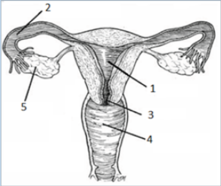
following events:
a. The fertilization takes place.
b. The sex intercourse takes place
c. The zygote develops
d. Follicles develop
e. The opening closes during the pregnancy.
3. What effect do the following hormones have on the size of the follicles?
a. FSHb. LH
4. Answer the following questions:
a. Define the term fertilizationb. The diagram below shows the structure of a human sperm.
i. Explain the part played by the organelle labelled A in the process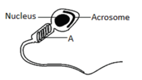
leading to fertilisation.
ii. The acrosome contains an enzyme that breaks down proteins. Describe
the function of this enzyme in the process leading to fertilisation.
5. Study the figure below on menstrual cycle and answer the questions thatfollow:
a. Name the hormones labelled a, b, c and d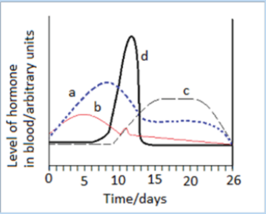
b. Give the likely day of the cycle on which ovulation takes places and give
reason for your answer.
c. What is meant by the term ovulation?d. State any 2 physical features which can prove that a female has ovulated.
6. The chard diagram below shows one way in which twins can be formed:
a. Give the name of the cell X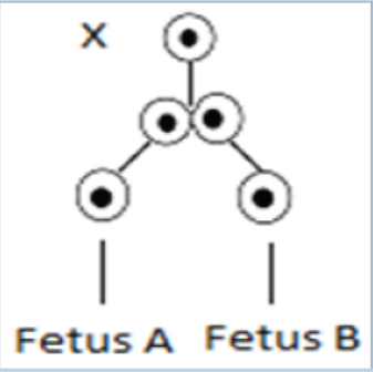
b. Why in this case will the embryo develop into identical twins?
7. Access the events that take place between the following stages in human
female.
a. The time the sperm meet the egg and fertilisation.
b. Fertilisation and implantation.
8. The eggs of birds are relatively much larger than those of mammal. Suggest
reason to account for the difference.
9. Identify the changes (events) occur in the uterus of a woman for menstrual
cycle to take place.10. Discus the main ways by which HIV is transmitted?
UNIT 13 PRINCIPLES OF GENE TECHNOLOGY
UNIT 13: PRINCIPLES OF GENE TECHNOLOGY
Key Unit Competence
Explain the principles of gene technology.
Learning ObjectivesBy the end of this unit, I should be able to:
–Define the term recombinant DNA.
–Explain that genetic engineering involves the extraction of genes from one organism
or the synthesis of genes, in order to place them in another organism(of the same or another species) such that the receiving organism expresses the gene product.
–Describe the properties of plasmids that allow them to be used in gene cloning.
–Explain the use of genes in fluorescent or easily stained substances as markers in gene technology.
–Describe the principles of the Polymerase Chain Reaction (PCR) to clone and amplify DNA(the role of Taq polymerase should be emphasized).
–Describe and explain how gel electrophoresis is used to analyse proteins and nucleic
acids, and to distinguish between the alleles of a gene (limited to the separation of polypeptidesand the separation of DNA fragments cut with restriction endonucleases).
–Explain the roles of restriction endonucleases, reverse transcriptase and ligasesin genetic engineering.
–Explain and outline, how microarrays are used in the analysis of genomes and indetecting mRNA in studies of gene expression.
–Interpret illustrations of the isolation and transfer of genes using plasmids in transgenicorganisms (bacteria, plant or an animal).
–Sequence the processes involved in the extraction and transfer of genes fromone organism to another.
–Interpret charts of the Polymerase Chain Reaction (PCR).
–Relate the mechanism of DNA replication to PCR and the amount of DNAproduced in a given period of time.
–Appreciate that the easy transfer of some plastids from one species of bacteria toanother may carry genes for antibiotic resistance.
–Acknowledge that advances in genetic engineering have enabled manipulation of genesto our advantage.
Introductory activity
Observe the figures below and respond to the questions that follow:
1. State and explain briefly the Chargaff’s rule of bases pairing based on the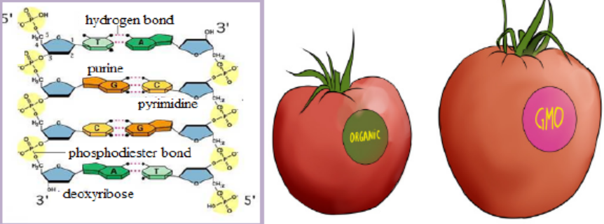
DNA structure shown above.
2. Describe briefly the gene expression starting by the DNA structure shown
above.
3. Summarize the main action done to transform organic tomato intogenetically modified organism (GMO), also called transgenic organism
13.1 Recombinant DNA and enzymes involved in genetic engineering
Activity 13.1
Using textbooks and or internet to answer the following questions.
1. Explain briefly the terms below:
a. Recombinant DNA
b. Transgenic organism
c. Enzyme2. Describe briefly the role of enzymes involved in genetic engineering
13.1.1 Recombinant DNA
A recombinant deoxyribonucleic acid (r DNA) is the DNA that contains genes from
more than one source. Examples of molecules produced from recombinant DNA
and that are important to humans include some pharmaceuticals like human insulin
and antibiotics.
Genetic engineering, also known as recombinant DNA technology or gene cloningor gene technology is the alteration of the genes in a living organism to produce a
genetically modified organism (GMO) with a new genotype. Various kinds of genetic
modification are possible and include:
– Inserting a foreign gene from one species into another in order to form a
transgenic organism,
– Altering an existing gene so that its product is changed and changing geneexpression so that it is translated more often or not at all.
13.1.2 Role of some enzymes in genetic engineering
The enzymes involved in gene manipulation include; restriction endonucleases(restriction enzymes), methylase, ligase and reverse transcriptase.
Restriction endonucleases
Different restriction enzymes, also called restriction endonucleases, exist and cut
the DNA molecule into fragments; their examples are shown in the table 13.1below.Table 13.1: List of some restriction enzymes and their respective recognition sites
Restriction enzymes are named according to the bacteria from which they originate.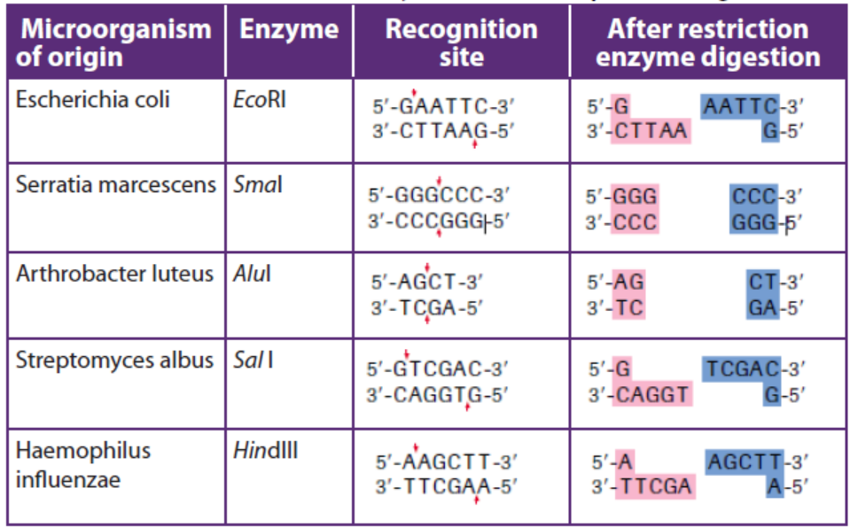
For example, the restriction enzyme BamHI is named as follows:
– B represents the genus Bacillus
– am represents the species amyloliquefaciens
– H represents the strain– I mean that it was the first endonuclease isolated from this strain
A commonly used tool in molecular biology is restriction endonucleases which
are molecular scissors that can cut double-stranded DNA at a specific base-pair
sequence. Each type of restriction enzyme recognizes a characteristic sequence of
nucleotides that is known as its recognition site. Most recognition sites are four to
eight base pairs long and are usually characterized by a complementary palindromicsequence.
For example, the restriction enzyme EcoRI binds to the following base-pair sequence:
5’-GAATTC-3’/3’-CTTAAG-5’. It is palindromic because both strands have the same
base sequence when read in the 5’ to 3’ direction. EcoRI scans a DNA molecule and
only stops when it is able to bind to its recognition site. Once bound, it disrupts,
via a hydrolysis reaction, the phosphodiester bond between the guanine and
adenine nucleotides on each strand. A phosphodiester bond is a covalent bond
located between a two sugar groups and a phosphate group; such bonds form the
sugar-phosphate backbone of DNA and RNA. Subsequently, the hydrogen bonds of
complementary base pairs between the cuts are disrupted. The result is a cut withina DNA strand, producing two DNA fragments where once there was only one.
So, in cleavage of DNA sequence using restriction enzyme EcoRI:EcoRI scans the DNA molecule:
The ends of DNA fragments produced from a cut by different restriction endonucleases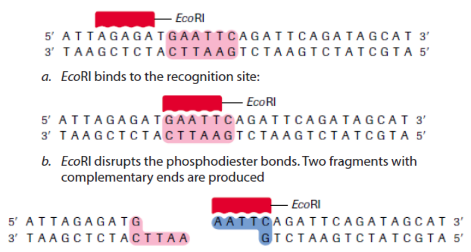
differ, depending on where the phosphodiester bonds are broken in the recognition
site. In the example in Table 13.1, EcoRI produces sticky ends; that is, both fragmentshave DNA nucleotides that are now lacking their respective complementary bases.
These overhangs are produced because EcoRI cleaves between the guanine and
the adenine nucleotide on each strand. Since A and G are at opposite ends of the
recognition site on each of the complementary strands, the result is the overhang.
In few words, sticky ends are fragment end of a DNA molecule with short singlestranded overhangs, resulting from cleavage by a restriction enzyme.
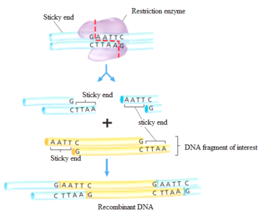
Figure 13.2: Cutting DNA by restriction enzymes and rDNA formation.
b. Methylases
These are enzymes that add a methyl group (CH3) to one of the nucleotides found in
a restriction endonuclease recognition site, altering its chemical composition. They
allow the molecular biologist to protect a gene fragment from being cleaved in anundesired location.
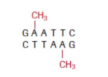
Figure 13.3: At a methylated EcoRI site, EcoRI restriction enzyme is no longer able to cut.
c. DNA ligase
This enzyme repairs broken DNA by joining two nucleotides in a DNA strand. It is
commonly used in genetic engineering to do the reverse of a restriction enzyme that
is to join together complementary restriction fragments. The sticky ends allow two
complementary restriction fragments to harden, but only by weak hydrogen bonds,
which can quite easily be broken by gentle heating. The backbone is still incomplete.
DNA ligase completes the DNA backbone by forming covalent bonds. T4 DNA ligase
is an enzyme that originated from the T4 bacteriophage and which is used to join
together DNA blunt or sticky ends. So, DNA ligase is able to join complementarysticky ends produced by the same restriction enzyme via a condensation reaction:
a. Complementary sticky ends produced by HindIII.
b. Hydrogen bonds form between complementary bases. DNA ligase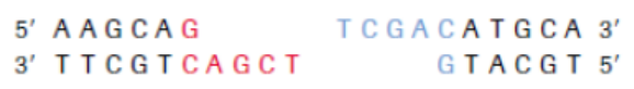
reconstitutes the phosphodiester bond in DNA backbones.
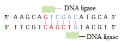
c. If fragments are not complementary, then hydrogen bonds will not form.
d. Reverse transcriptase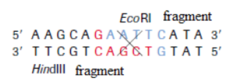
Reverse transcription is a process whereby a mRNA is converted into c DNA
(complementary DNA, also called copy of DNA). It requires the enzymes calledreverse transcriptase. It is shown by this reaction:
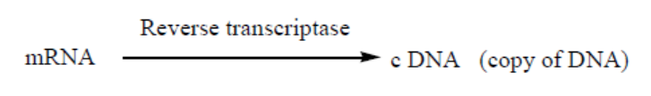
Application 13.1
1. Write the following abbreviations in full: DNA, GMO and RNA.
2. Explain the nomenclature of the enzyme EcoRI.
3. Distinguish between sticky ends and blunt ends.4. Discuss the role of T4 DNA ligase.
13.2 Properties of plasmids and gene manipulation
Activity 13.2
Use different biology textbooks or internet to respond to the following
questions.
1. Identify any 3 properties of plasmids.
2. Explain the role of vectors in genetic engineering.3. Elaborate the main steps of gene manipulation
13.2.1. Properties of plasmids
A plasmid is a genetic structure, in some cells, that can replicate independently
of the chromosomes; it is typically a small circular DNA strand in the cytoplasm
of a bacterium or protozoan. Plasmids are much used in the laboratory duringmanipulation of genes.
Figure 13.4. The structure of the Tumor – inducing plasmid (Ti plasmid) of Agrobacterium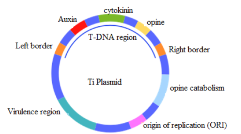
tumefaciens and Agrobacterium rhizogenes
The properties of plasmids are:
– It is big enough to hold the desired gene.
– It is circular (or more accurately a closed loop), so that it is less likely to be
broken down.
– It contains control sequences, such as a transcription promoter, so that the
gene will be replicated or expressed.
– It contains marker genes, so that cells containing the vector can be identified.
Plasmids are not the only type of vector that can be used. Viruses can also be used
as vectors. Another group of vectors are liposomes, which are tiny spheres of lipidcontaining the DNA.
13.2.2. Gene manipulation
Genetic manipulation is a process done to use the genome of an organism in order
to produce desired traits. A genome is the complete set of genes or genetic material
present in an organism.
Genes are pieces of DNA, that carry genetic information which determines all the
characteristics of an individual such as eye colour, size, ability to resist disease, etc.
Each gene contains the information required to build specific proteins needed in an
organism.
The human genome contains 20,687 protein-coding genes.
The overview of gene transfer, resulting in genetically modified organisms (GMO)
also called transgenic organisms such as bacteria or animals or plants havingforeign gene inserted into them, is shown below:
1. Generation of DNA fragments using restriction endonucleases:
– Appropriate restriction endonucleases need to be used to ensure that the
gene fragment in question is excised completely from the source DNA.
– More than one restriction endonuclease may be used at one time.
2. Construction of a recombinant DNA molecule:
– The target gene fragment is ligated to a DNA vector (plasmids are one example)
and is now recombinant DNA.
– The vector can replicate autonomously in an appropriate host organism.
3. Introduction into a host cell:
– Bacterial host cells can be manipulated to take up the recombinant DNA usingelectroporators, gene guns or classical transformation protocols.
– Once the bacterium takes up the recombinant DNA, it is referred to as being
transformed.
4. Selection:
– Cells that have been successfully transformed with the recombinant DNA must
be isolated.
– The desired cells are usually chemically selected by the presence of a marker
(e.g. antibiotic resistance) on the vector.
– Growth of colonies on media containing the chemical indicates successful
transformation of the recombinant DNA vector.
– Individual colonies are isolated from media containing the chemical and are
grown in culture to produce multiple copies (clones) of the incorporated
recombinant DNA. Different gene manipulations are illustrated under the
heading 13.3.
To perform these gene manipulation steps, the genetic engineer needs a tool kitconsisting of:
1. Enzymes, such as restriction endonucleases (restriction enzymes), ligase and
reverse transcriptase
2. Vectors, including plasmids and viruses3. Genes coding for easily identifiable substances that can be used as markers.
Application 13.2
1. Identify the vectors that are used in genetic engineering.
2. Find the components of a genetic engineering tool kit.
3. Differentiate between a gene and a genome.4. Explain the second step of gene manipulation.
13.3 Transfer of genes using plasmids in transgenic organisms
Activity 13.3
Using different Biology textbooks or internet:
1. What is meant by a pathogenic bacterium
2. Explain the role of a gene marker in genetic engineering.
3. Distinguish between bacterial transduction and transformation
4. Explain briefly the steps of formation of a transgenic plant
5. Draw and interpret the chart of about the transfer of DNA from
eukaryotic cell to a bacterial cell using a plasmid.
6. By diagrams, show how a transgenic organism such as a transgenicplant and a clone are produced.
The production of genetically modified organisms (GMO), also called transgenic
organisms, is a multistage process which can be generally summarized as follows:
– Identification of the gene of interest.
– Isolation of the gene of interest.
– Cutting of gene of interest and opening of plasmid with restriction enzymes in
order to have sticky ends
– Associating the gene with an appropriate promoter and poly -A sequence and
insertion into plasmids.
– Multiplying the plasmid in bacteria and recovering the cloned construct for
injection.
– Transference of the construct into the recipient tissue, usually fertilized eggs.
– Integration of gene into recipient genome.
– Expression of gene in recipient genome.– Inheritance of gene through further generations.
13.3.1. Extraction, purification, isolation and transfer of genes usingplasmids into bacteria
The normal gene coding for a particular protein is extracted from an organism; it
is isolated and transferred into a plasmid of a bacterium. This plasmid becomes a
recombinant DNA that is introduced into that bacterium. This bacterium becomes
a transgenic bacterium. An example of the sequence of the processes involved
in the extraction and transfer of genes from one organism to another is illustratedbelow.
Process 1: Extraction and purification of DNA containing an interest gene
is required for a variety of molecular biology applications. Its process issummarized below.
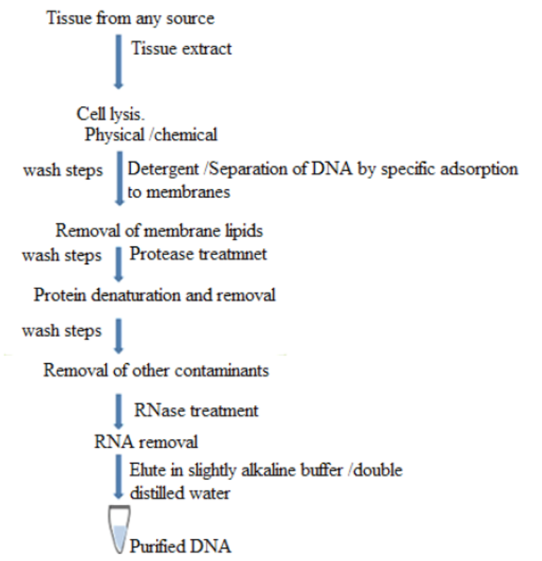
Figure 13.5: Basic steps involved in all DNA extraction methods
The purification of DNA from cell extract occurs in this way:
– The standard way to deproteinize a cell is to add phenol or a 1:1 mixture of
phenol and chloroform.
– The organic solvents precipitate proteins but leave the nucleic acids (DNA and
RNA) in an aqueous solution.
– The result is that is the cell extract is mixed gently with the solvent, and the
layers then separated by centrifugation, precipitated protein molecules are
left as a white coagulated mass at the interface between the aqueous and
organic layers.
– The aqueous solution of nucleic acids can then be removed with a white
pipette.
– Cell extract is treated with protease such as pronase or proteinase K before
extraction.
– These enzymes will break polypeptides into smaller units thus making phenol
easier to remove them.
– The only effective way to get rid of RNA is the use of ribonuclease enzyme
which will rapidly degrade the molecules into ribonucleotide subunits. As
DNA is purified, also its genes are purified.
The Concentration of DNA samples is carried out in this way:
– The most frequently used method of concentration is ethanol precipitation.
– In the presence of salt and a temperature of -20 oC or less absolute ethanol
with efficiently precipitate polymeric nucleic acids.
– With 2 thick solution of DNA, the ethanol can be layered on the top of the
sample.
– A spectacular trick is to push a glass rod through the ethanol into the DNA
solution.
– When the rod is removed, DNA molecules will adhere and be pulled out of thesolution in the form of long fiber.
Figure 13.6. Practical summary of DNA extraction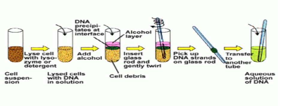
After getting DNA, it is possible to remove the gene from it, by a restriction enzyme, in
order to use it for a particular purpose. For example, normal insulin gene is removedfrom human cell as shown in the figure below.
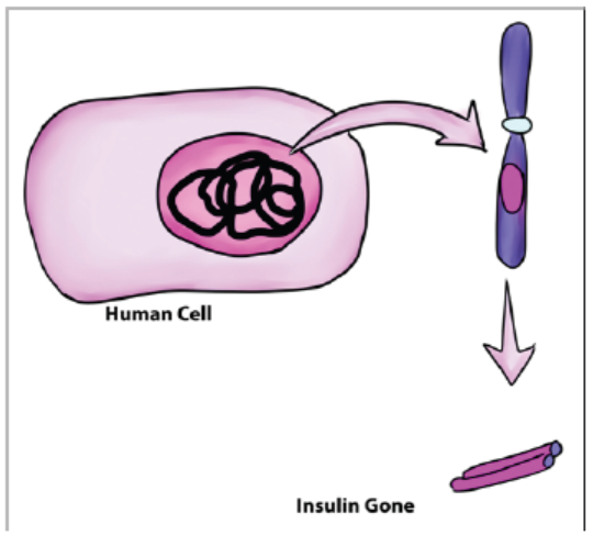
Figure 13.7: Removal of insulin gene from human cell
Process 2: Summary of transfer of insulin gene using plasmids into bacteria
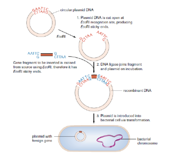
Figure 13.8: A foreign gene is introduced into a plasmid of a bacterium to form a recombinant DNA
The plasmid is now an example of recombinant DNA, which can be introduced into
a bacterial cell to produce numerous copies (clones) of the gene. As the inserted
gene codes for insulin, a hormone that reduces the blood glucose level, and this
gene functions normally as expected, the product (insulin) may also be retrievedand used for therapeutic purposes in which it is given to diabetic people.
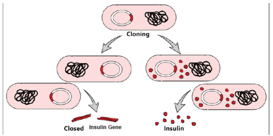
Figure 13.9: Gene cloning after a bacteriumpr otadkuecst iuopn a recombinant DNA (plasmid) and insulin
In nuclear biology and molecular biology, a marker gene is a gene used to determineif a nucleic acid sequence has been successfully inserted into an organism›s DNA.
13.3.2 Use of Agrobacterium tumefaciens to transfer genes in plants
Agrobacterium is a bacterium that uses a horizontal gene transfer (HGT). HGT is
the transfer of DNA between different genomes. HGT can occur in bacteria through
transformation, conjugation and transduction. However, it is also possible for HGT
to occur between eukaryotes and bacteria. Bacteria have three ways of transferringbacteria DNA between cells:
1. Transformation: The uptake and incorporation of external DNA into the
cell thereby resulting in the alteration of the genome.
2. Conjugation: The exchange of genetic material through cell-to-cell contact
of two bacterial cells. A strand of plasmid DNA is transferred to the recipient
cell and the donor cell then synthesis DNA to replace the strand that was
transferred to the recipient cell.
3. Transduction: A segment of bacterial DNA is carried from one bacterial
cell to another by a bacteriophage. The bacteriophage infects a bacterial
cell and takes up bacterial DNA. When this phage infects another cell, it
transfers the bacterial DNA to the new cell. The bacteria can then become a
part of the new host cell.
Agrobacterium also has the ability to transfer DNA between itself and plants
and is therefore commonly used in genetic engineering. The process of usingAgrobacterium for genetic engineering is illustrated in the diagram below.
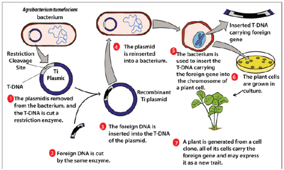
Figure 13.10: Process of formation of a transgenic plant
Summary of formation of a transgenic plant:
– The agrobacterium cell contains a bacterial chromosome and a Tumor inducing
plasmid (Ti Plasmid).
– The Ti plasmid is removed from the agrobacterium cell and a restriction
enzyme cleaves the T-DNA restriction site. The transfer DNA (T-DNA) is the
transferred DNA of the tumor-inducing plasmid of some species of bacteria
such as Agrobacterium tumefaciens
– The T-DNA is transferred from bacterium into the host plant›s
nuclear DNA genome.
– Next foreign DNA, which is also cleaved by the same enzyme, is inserted into
the T -DNA at the site that was cleavage site.
– The modified plasmid is then reinserted in the agrobacterium and the
bacterium inserts the T-DNA, which now carries a foreign gene into the plant
cell.
– The plant cell is then cultured and results in a new plant that has the foreignDNA trait.
13.3.3 Transfer of genes into animals
In reproductive cloning, researchers remove a mature somatic cell, such as a skin
cell, from an animal that they wish to copy. They then transfer the DNA of the donoranimal’s somatic cell into an egg cell, or oocyte, that has had its own DNA-containing
nucleus removed. For example, the cell used as the donor for the cloning of Dolly
sheep was taken from a mammary gland and the production of a healthy clone
therefore proved that a cell taken from a specific part of the body could recreate awhole individual.
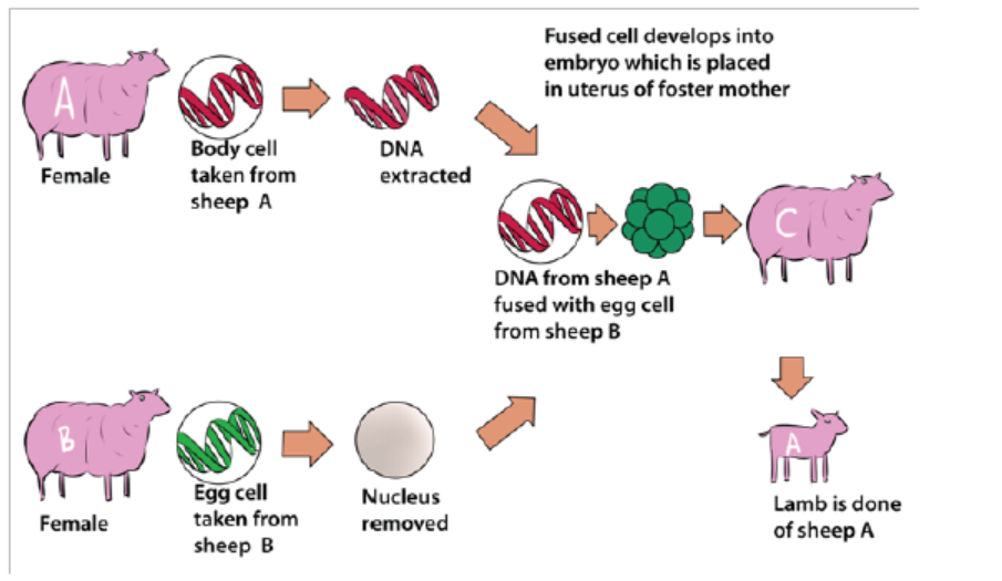
Figure 13.11: The cloning process that produced transgenic Dolly sheep
13.3.4 Transformation of harmless bacteria to pathogenic bacteria andresistant bacteria
A pathogenic bacterium is a bacterium which is capable of causing a disease.
An example of harmful or pathogenic bacterium is Vibrio cholerae which causes
cholera. A harmless bacterium can become pathogenic bacterium due to certain
factors. The discovery of DNA and the genetic code led scientists to determine that
some bacteria were resistant to particular antibiotics because of inserted genes that
rendered bacteria unaffected by the effects of some antibiotics. This gene insertion
can be done naturally between bacteria or artificially by biotechnologists.
Antibiotic resistance, also known as drug resistance, is the ability of bacteria
and other microorganisms to resist the effects of an antibiotic to which they were
once sensitive. Since bacteria are ubiquitous in the colon, conjugation is constantly
occurring. This conclusion has been supported by bacteria in different genera
containing homologous DNA plasmids. Therefore, horizontal gene transfer can occur
between different species or within a population. This can become problematic if
harmful bacteria that have been artificially selected for antibiotic resistance happen
to be in the colon, where bacteria can transfer the resistance gene to other species
of bacteria. Typically, this is not a problem because most bacteria are not harmful,
unless bacteria that are a public health concern happen to receive a resistance gene.
Individuals that have previously taken antibiotics are less responsive to treatmentbecause their bodies contain more antibiotic resistant bacteria.
These bacteria received these genes from disease - causing microbes that transferred
a resistance gene through conjugation or transformation. The harmless bacteria
that are resistant to antibiotics can then pass this gene to harmful bacteria that do
not yet have antibiotic resistance. Thus, horizontal gene transfer allows bacteria
to indirectly become resistant to antibiotics. Transformation and conjugation
contribute to increasing frequencies of antibiotic resistant genes because of genes
transferring between different species. The gene transfer can transform harmlessbacteria into pathogenic bacteria which can cause diseases.
The prevention of antibiotic-resistant infections includes:
– Do not take antibiotics for viral infections.
– Complete your prescribed course of treatment exactly as instructed by your
healthcare provider. Do not stop taking your medicine even if you feel better,
and do not save any antibiotics for future use.
– Do not take someone else’s antibiotics because different kinds of antibioticstreat different types of bacterial infections.
Application 13.3
1. Identify any bacterium involved in formation of transgenic plant.
2. Explain briefly the cloning of a sheep.
3. Describe briefly one cause of antibiotic resistance.
4. Discuss how biotechnologists might transform harmless bacteria topathogenic forms in the course of their studies.
13.4 Non-biological methods of gene transfer
Activity 13.4
Search from Biology textbooks and internets and answer to the following
questions:
1. Identify the non- biological methods of gene transfer
2. Explain the procedure of carrying out biolistics
3. Describe the disadvantages of vacuum infiltration.
Different non-biological methods, also called physical methods or direct methods
of gene transfer exist and include genetic transformation, shock wave-mediated
genetic transformation, electroporation, biolistic, vacuum infiltration, silicon carbidewhisker and laser microbeams.
a. Electroporation
Electroporation is a method of transformation via direct gene transfer. In this
technique, a mixture containing cells and DNA is exposed to very high voltage
electrical pulses (4000 – 8000 Volts/cm) for very brief time periods (few milliseconds).
It results in formation of transient pores in the plasma membrane, through whichDNA seems to enter inside the cell and then nucleus.
Figure 13.12: Electroporation: (a) Diagram showing formation of transient pores in cell membrane on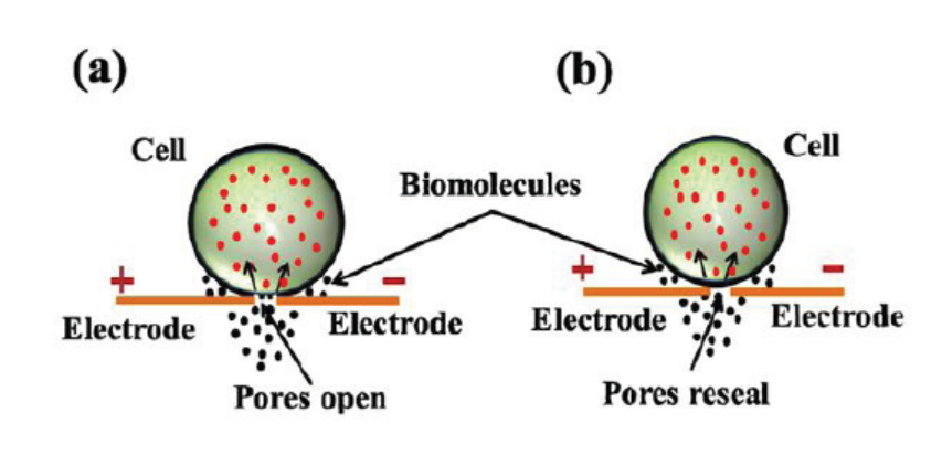
applying electrical pulse, (b) Entry of DNA inside the cell and sealing of pores afterwards.
A suspension of cells with plasmid DNA is taken in an electroporation cuvette
placed between electrodes and electrical pulses are applied. Temporary micropores
are formed in cell membranes which allow cells to take up plasmid DNA leading tostable or transient DNA expression.
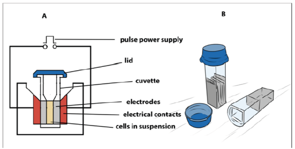
Figure 13.13: (A) Main components of an electroporator (B) Cuvettes used for electroporation
Cells which are arrested at metaphase stage of cell cycle are especially suitable for
electroporation as these cells have absence of nuclear envelope and an unusual
permeability of the plasma membrane. Protoplasts are used for electroporation of
plant cells as thick plant cell walls restrict movement of DNA. The electroporation
method was originally developed for protoplasts, but has given equally good results
with cells and even tissues with easy recovery of regenerated plantlets. Immature
zygotic embryos and embryogenic cells have also been used for electroporation to
produce transgenic maize.
Transformation of protoplast is associated with low transient expression
of transgenes as compared to organized tissues and low regeneration frequency
especially in monocotyledonous plants. The electrical field and chemical substances
applied to disorganize cell walls reduce the viability and capability of division of
protoplasts.
b. Biolistics
Biolistics, also called micro projectile or particle bombardment, is a method wherecells are physically impregnated with nucleic acids or other biological molecules.
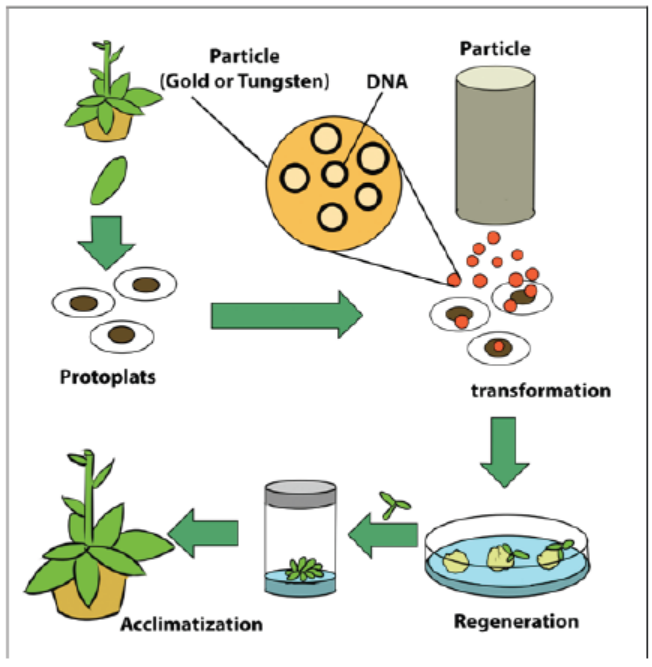
Figure 13.14: Particle bombardment method of plant transformation
The main steps of a biolistic method are:
– Isolation of protoplasts.
– Injection of DNA-coated particles using particle gun.
– Regeneration of transformed protoplasts into plantlets.
– Acclimatization of regenerated plantlets in a greenhouse.
A biolistic particle delivery system is a device for plant transformation where cells
are bombarded with heavy metal particles coated with DNA/RNA. This technique
was invented by John Stanford in 1984 for introduction of DNA into cells by physical
means to avoid the host-range restrictions of Agrobacterium. Agrobacteriummediated
genetic transformation system works well for dicotyledonous plants
but has low efficiency for monocots. Biolistic particle delivery system provides an
effective and versatile way to transform almost all type of cells. It has been proven
to be a successful alternative for creating transgenic organisms in prokaryotes,mammalian and plant species.
c. Microinjection
The process of using a fine glass micropipette to manually inject transgene atmicroscopic or borderline macroscopic level is known as microinjection.
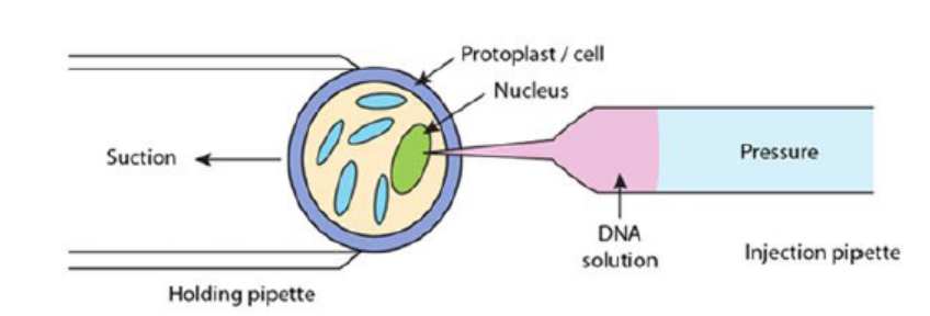
Figure 13.15: Illustration of microinjection method
The transgene, in the form of plasmids, cosmids, phage or PCR products, can be
circular or linear and does not need to be physically linked for injection. Microinjection
involves direct mechanical introduction of DNA into the nucleus or cytoplasm using
a glass microcapillary injection pipette. The protoplasts are immobilized in low
melting agar, while working under a microscope, using a holding pipette and suction
force. DNA is then directly injected into the cytoplasm or the nucleus. The injected
cells are then cultured in vitro and regenerated into plants. Successful examples of
this process have been shown in rapeseed, tobacco and various other plants.
Stable transformants can be achieved through this method but it requires technical
expertise and is a time consuming process. Also, microinjection has achieved only
limited success in plant transformation due to the thick cell walls of plants and a lack of
availability of a single-cell-to-plant regeneration system in most plant species. In this
technique, a traditional compound microscope (around 200x magnification) or an
inverted microscope (around 200x magnification) or a dissecting stereomicroscope
(around 40-50x) is used. The microscope target cell is positioned, cell membrane and
nuclear envelope are penetrated with the help of two micromanipulators. Onemicromanipulator holds the pipette and another holds the micro capillary needle.
The two types of microinjection systems are constant flow system and pulsed flow
system.
– In the constant flow system, the amount of sample injected is determined by
the duration for which needle remains in the cell. The constant flow system is
relatively simple and inexpensive but outdated.
– The pulsed flow system has greater control over the volume of substance
delivered, needle placement and movement and has better precision. This
technique results in less damage to the receiving cell; however, the componentsof this system are quite expensive.
d. Whiskers methods
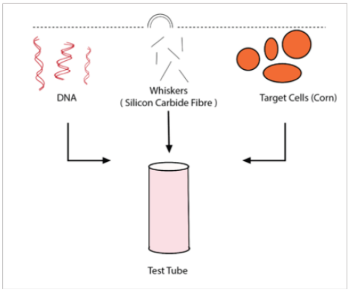
Figure 13.16: Whiskers methods
In this method, silicon carbide fibers are mixed in a vortex with a suspension of
tissue and DNA allowing introduction by abrasion. Maize (Zea mays) and tobacco
(Nicotiana tabacum) tissue cultures were transformed using silicon carbide fibers to
deliver DNA into suspension culture cells. DNA delivery was mediated by vortexingcells in the presence of silicon carbide fibers and plasmid DNA.
e. Vacuum infiltration method
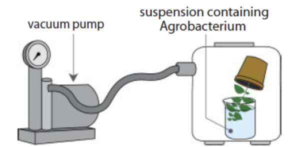
Figure 13.17: Vacuum infiltration methods
In this method, a vacuum pump generates a negative pressure that increasesintercellular spaces allowing the infiltration of Agrobacterium.
f. Laser microbeams method
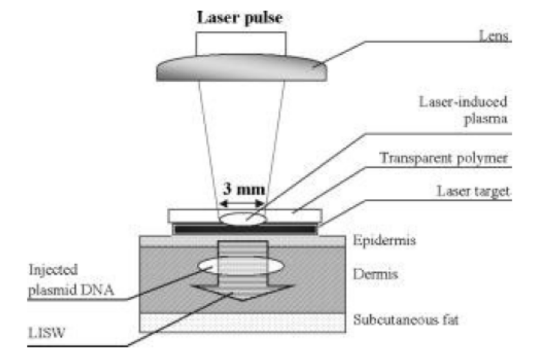
Figure 13.18. Laser microbeams method
In this method, a laser microbeam punctures self-healing holes into the cell wallallowing DNA penetration.
g. Ultrasound method
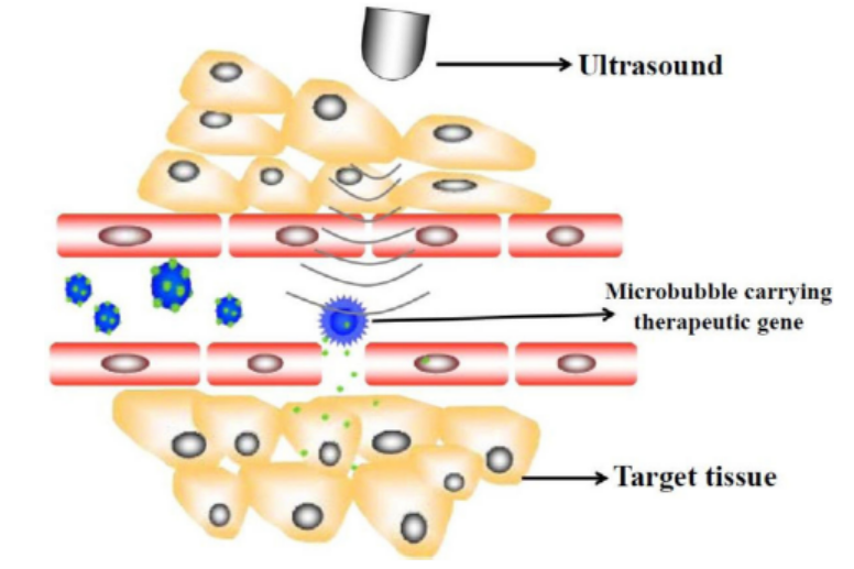
Figure 13.19: Ultrasound method
This method requires that a gene introduces DNA molecules into cells via acoustic
cavitation that temporarily changes the permeability of the cell membrane.
Sonoporation, or cellular sonication, is the use of sound (typically ultrasonic
frequencies) for modifying the permeability of the cell plasma membrane. This
technique is usually used in molecular biology and non-viral gene therapy in
order to allow uptake of large molecules such as DNA into the cell, in a cell
disruption process called transfection or transformation. Sonoporation employs
the acoustic cavitation of microbubbles to enhance delivery of these large molecules.
Sonoporation is under active study for the introduction of foreign genes in tissue
culture cells, especially mammalian cells. Sonoporation is also being studied for use
in targeted gene therapy in vivo, in a medical treatment scenario whereby a patient
is given modified DNA, and an ultrasonic transducer might target this modified DNAinto specific regions of the patient’s body.
h. Shock wave-mediated genetic transformation method
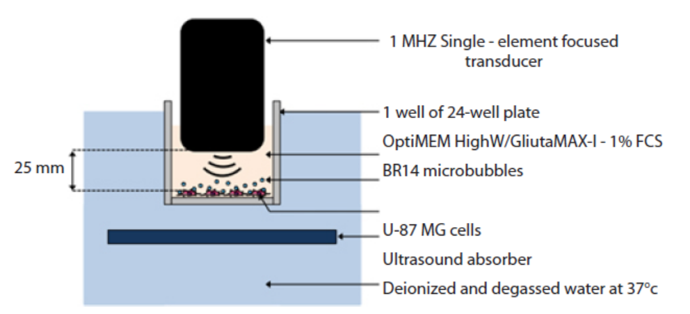
Figure 13.20: Shock wave-mediated genetic transformation method
This method involves sonoporation, based on the use of high frequency ultrasound
(1–10 MHz) in combination with gas microbubbles was introduced as a non-viral
physical method that is currently under evaluation for gene and drug delivery.
Sonoporation involves the treatment of a desired volume of cells in vitro or tissue in
vivo with ultrasound in the presence of microbubbles. These microbubbles, which
are formulated as lipid, albumin or polymer shelled micrometer sized gas bodies
in aqueous suspension, are commonly mixed with cells for in vitro applications or
administered by intravascular or intratissue injection for in vivo applications. The
exposure of microbubbles to ultrasound causes their periodic oscillations and/or
their collapse, under appropriate insonation conditions. It is now known that these
oscillations can induce micro-streaming, shock waves and/or micro-jets that can
affect the integrity of biological barriers (e.g. cell membrane, endothelial barrier). The
use of sonoporation to deliver therapeutic molecules to tissues has been extensively
explored over the past decade.
For example, the loco-regional delivery of anti-tumoral drugs has been reported
and is now under clinical investigation. Sonoporation has been successfully used to
transfer nucleic acids such as DNA into the heart, skeletal muscle, tumors, vessels,
liver and kidney. This method enables exogenous delivery of molecules with minimal
cell or tissue damage, inflammation and/or immunological response. In addition,
ultrasound can be non-invasively targeted to a specific volume of superficial tissues
or deeply embedded organs. Taken together, these properties make sonoporationan innovative and compelling method for gene and drug delivery.
Table 13.2: Summary of some physical methods for genetic transformation of cells
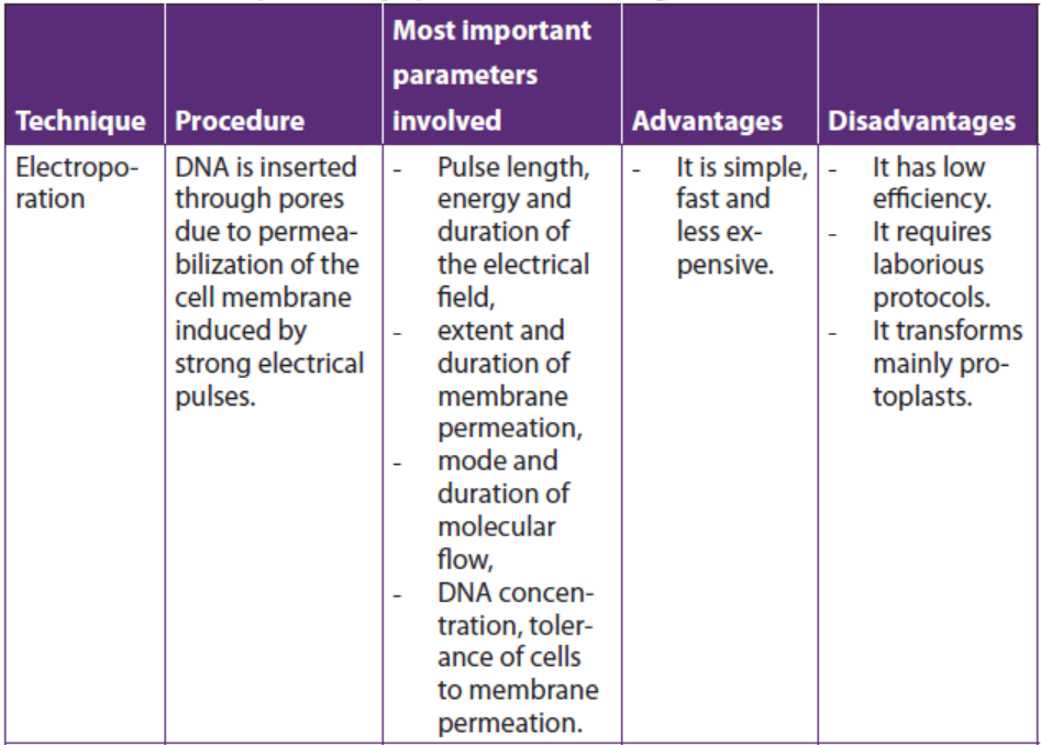
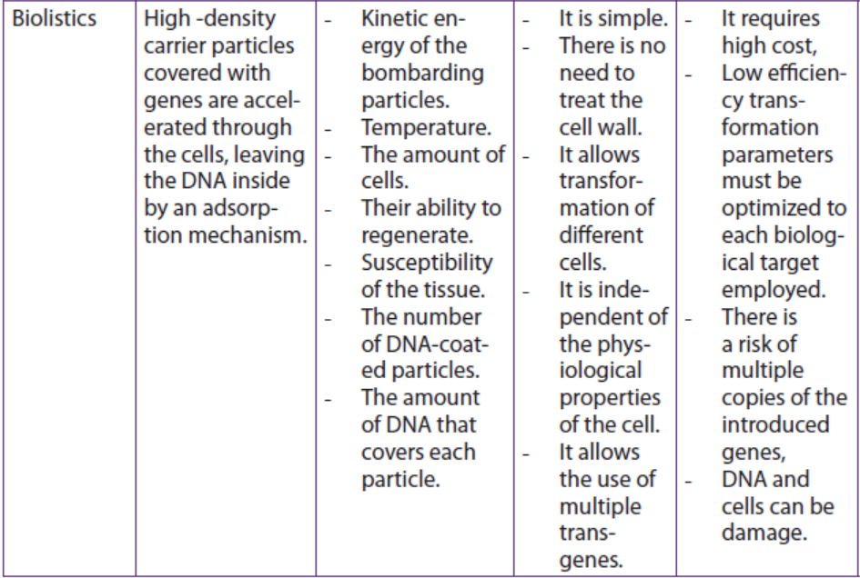
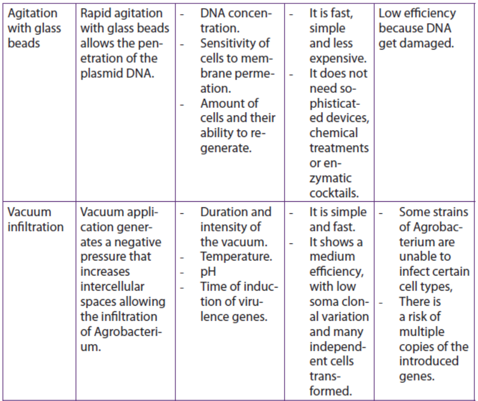
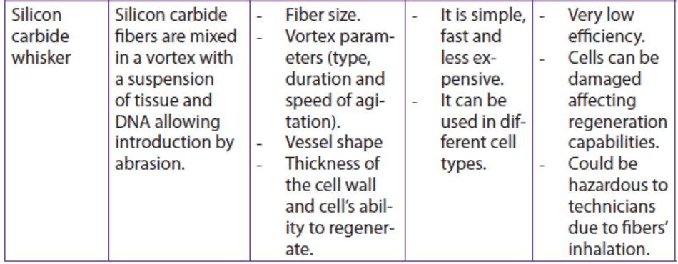
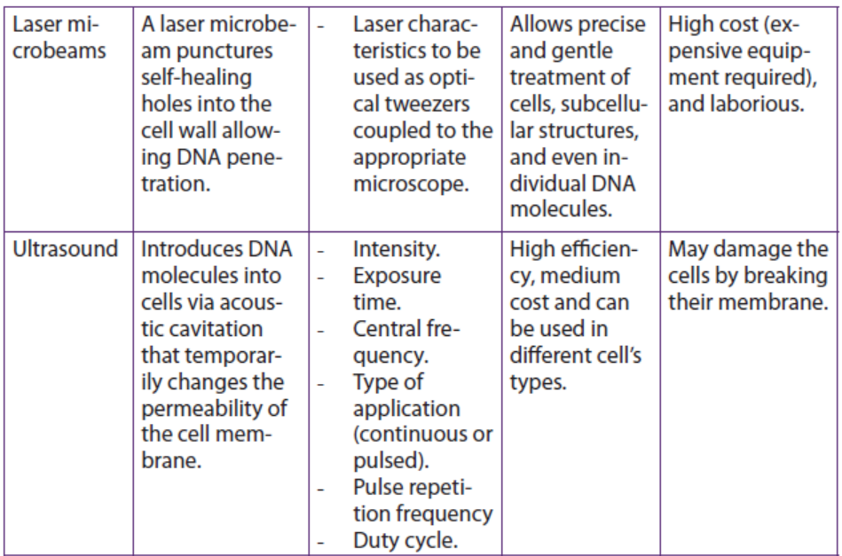
Application 13.4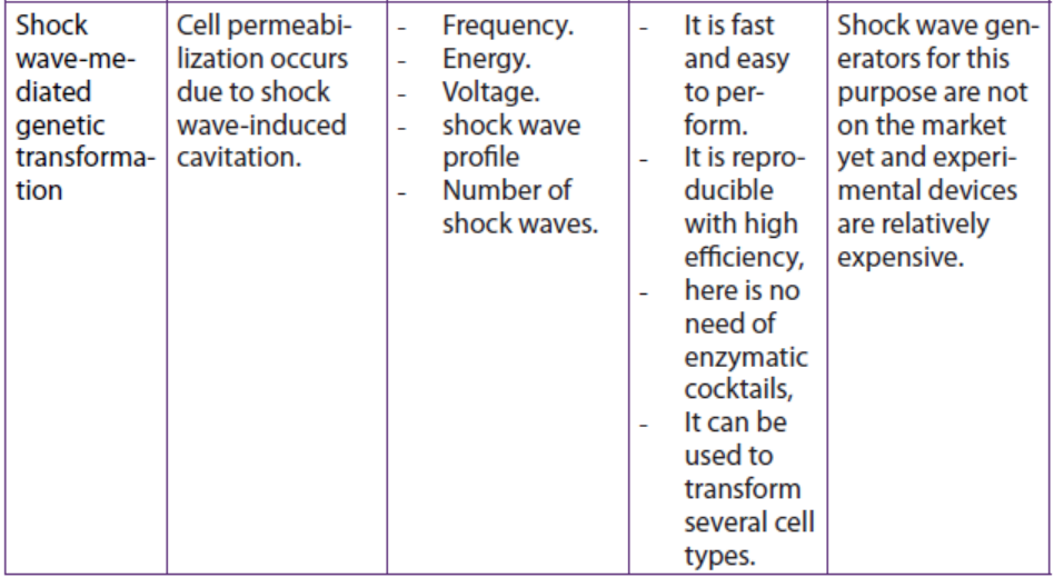
1. Observe the figure below and respond to the following questions.
a. Identify A and B.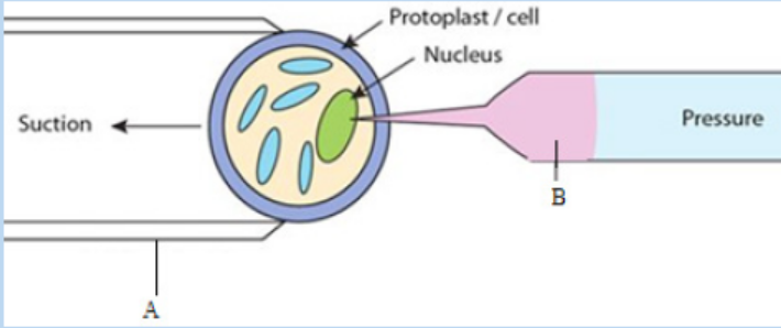
b. Describe briefly the method shown by this figure.
2. Distinguish between ultrasound technique and shock waves technique interms of involved parameters
13.5 Principles of Polymerase Chain Reaction (PCR) in cloningand amplifying DNA
Activity 13.5
Using different Biology textbooks, charts and computer simulations, discuss
the mechanism of artificial DNA synthesis focusing on Polymerase Chain
Reaction (PCR). During your discussion answer to the following questions:
1. Identify the types of artificial DNA synthesis.
2. Analyse the main steps of PCR3. Explain the role of Thermus aquaticus in PCR
Polymerase chain reaction is a technique that uses the enzyme called DNA
polymerase to produce millions of copies of a particular piece of DNA. For simplicity,the two original single strands are not shown after step 3. The main steps of the PCR:
– The DNA duplex is heated to 90 oC to separate the two strands (step 1:
denaturation).
– The mixture is cooled to 60 oC to allow the primers to anneal to their
complementary sequences (step 2: annealing).
– At 72 oC the primers direct the thermostable DNA polymerase to copy each ofthe template strands (step 3: extension or elongation of primers).
The three steps of the PCR are repeated many times to yield many thousands of
copies of the original target sequence. Genes can be cloned by cloning the bacterial
cells that contain them, but this requires quite a lot of DNA in the first place. PCR can
clone (or amplify) DNA samples as small as a single molecule. It is a newer technique,
having been developed in 1983 by Kary Mullis, for his discovery he won the Nobel
Prize in 1993.
The polymerase chain reaction is simply DNA replication in a test tube. If a length
of DNA is mixed with the four nucleotides (A, T, C and G) and the enzyme DNA
polymerase in a test tube, then the DNA will be replicated many times.
Normally, in vivo where DNA replication occurs, the DNA double helix would be
separated by the enzymes DNA gyrase and DNA helicase, but in PCR (in vitro) the
strands are separated by heating to 95°C for two minutes. This breaks the hydrogen
bonds. DNA polymerisation always requires short lengths of DNA (about 20 bases
pair long) called primers, to get it started. In vivo the primers are made during
replication by DNA polymerase, but in vitro they must be synthesised separately
and added at this stage. This means that a short length of the sequence of the DNA
must already be known, but it does have the advantage that only the part between
the primer sequences is replicated. The DNA must be cooled to 40°C to allow theprimers to anneal to their complementary sequences on the separated DNA strands.
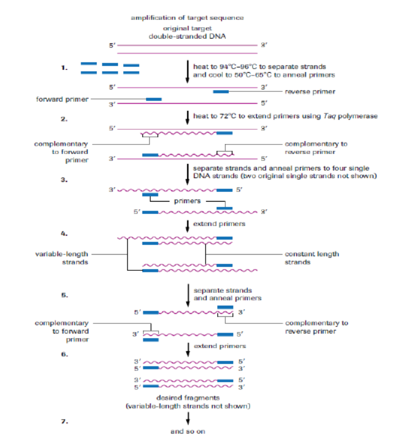
Figure 13.21: Polymerase chain reaction.
The enzyme (Taq polymerase) used in PCR is derived from the thermophilic bacterium
Thermus aquaticus, which grows naturally in hot springs at a temperature of 90°C, so
it is not denatured by the high temperatures in step 2. Its optimum temperature is
about 72°C, so the mixture is heated to this temperature for a few minutes to allow
replication to take place as quickly as possible. Once the primers have annealed, Taqpolymerase, a DNA polymerase, can build complementary strands using free
nucleotides that have been added to the solution. Each original DNA molecule has
now been replicated to form two molecules. The cycle is repeated from step 2 and
each time the number of DNA molecules doubles. This is why it is called a chain
reaction, since the number of molecules increases exponentially, like an explosivechain reaction. Typically PCR is run from 20 to30 cycles.
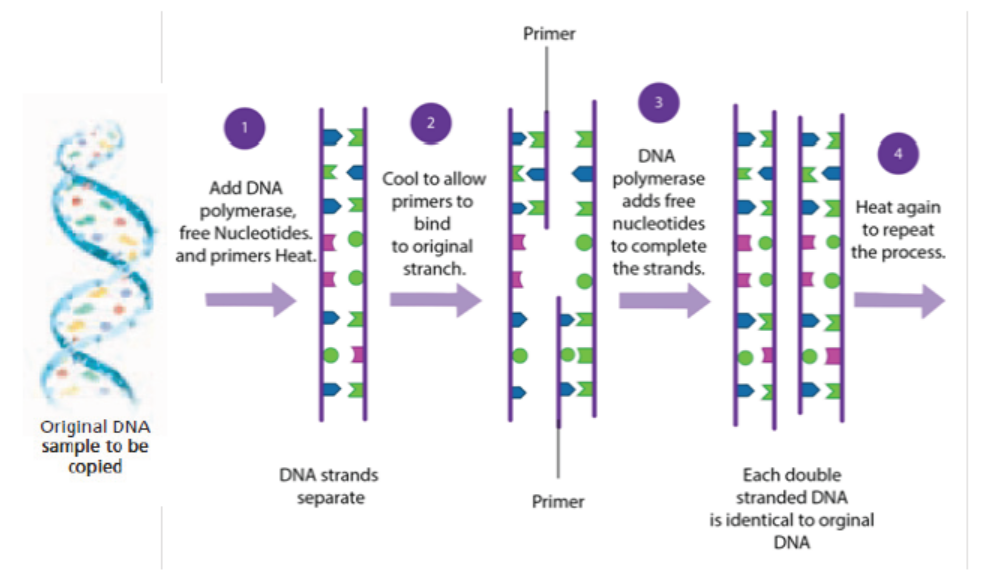
Figure 13.22: Summary of PCR technique
Note that:
Artificial DNA synthesis, sometimes known as DNA printing is a method
in synthetic biology that is used to create artificial genes in the laboratory. The types
of artificial DNA synthesis include recombinant DNA technology, gene purification
and PCR (Polymerase Chain Reaction). All of these types have been described aboveunder headings 13.1, 13.3 and 13.5 respectively.
Application 13.5
1. Explain briefly the artificial DNA printing2. Differentiate between PCR and DNA replication
13.6 Gel electrophoresis
Activity 13.6
Using computer animations and biology textbooks, observe the gel
electrophoresis used to analyse proteins and nucleic acids to distinguish
between alleles of a gene; then answer the following questions:
1. What is meant by gel electrophoresis?
2. Describe briefly the steps taken during separation of DNA, RNA fromthe mixture by use of gel electrophoresis.
Gel electrophoresis is a laboratory technique used to separate mixtures of DNA,
RNA or proteins according to molecular size. In gel electrophoresis, the molecules
to be separated are pushed by an electrical field through a gel that contains smallpores.
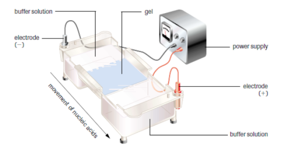
Figure 13.23: Setup of gel electrophoresis.
In a common gel electrophoresis setup, a nucleic acid such as DNA is loaded into
wells at one end of the gel and then migrates toward the positive electrode at the
opposite end. The rate of migration of fragments varies with size. The steps of gel
electrophoresis are shown below.
– The DNA samples are cut with a restriction enzyme into smaller segments of
various sizes. The DNA is then placed in wells made on a thick gel.
– An electric current runs through the gel for a given period of time. Negativelycharged DNA fragments migrate toward the positively charged end of the
porous gel. Smaller DNA fragments migrate faster and farther than longer
fragments, and this separates the fragments by size. The gel floats in a buffer
solution within a chamber between two electrodes.
– The DNA is transferred to a nylon membrane and radioactive probes are added.
The probes bind to complementary DNA.
– The X-ray film is exposed to the radiolabelled membrane. The resulting patternof bands is called a DNA fingerprint.
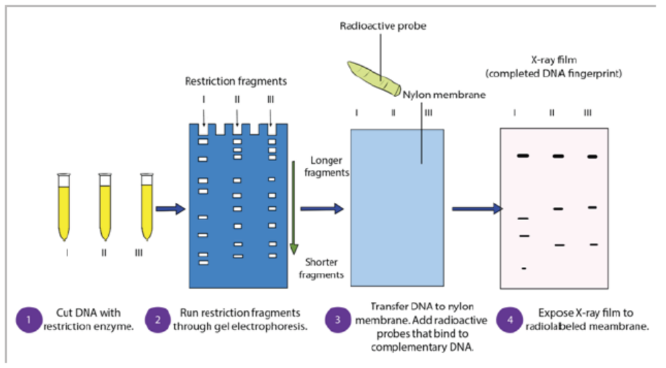
Figure 13.24: Steps of gel electrophoresis
During electrophoresis, DNA fragments migrate through the gel at a rate that is
inversely proportional to the logarithm of their size. The shorter the fragment is,
the faster it will travel because of its ability to navigate through the pores in the gel
more easily than a large fragment can. Larger fragments are hampered by their size.
Hence, the longer a nucleotide chain, the longer it takes for the migration.
Gel electrophoresis takes advantage of DNA’s negative charge. Using direct current,
a negative charge is placed at one end of the gel where the wells are, and a positive
charge is placed at the opposite end of the gel. The electrolyte solution conveys
the current through the gel. The negatively charged DNA will migrate toward the
positively charged electrode, with the shorter fragments migrating faster than the
longer fragments, achieving separation. Small molecules found within the loading
dye migrate ahead of all the DNA fragments. Since the small molecules can be
visualized, the electrical current can be turned off before they reach the end of thegel.
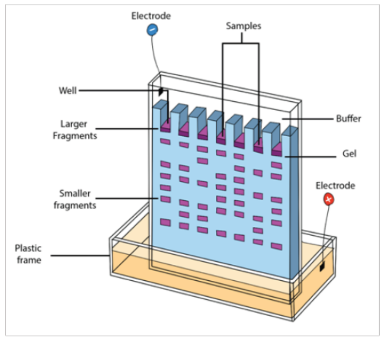
Figure 13.25: Fragments arrangement in gel electrophoresis
Once gel electrophoresis is complete, the DNA fragments are made visible by
staining the gel. The set of fragments generated with a particular restriction enzyme
produces a banding pattern characteristic for that DNA. The most commonly used
stain is ethidium bromide. Ethidium bromide is a flat molecule that fluoresces under
ultraviolet (UV) light and is able to insert itself among the rungs of the ladder of
DNA. When the gel is subjected to UV light, the bands of DNA are visualized because
the ethidium bromide is inserted among the nucleotides. The size of the fragments
is then determined using a molecular marker as a standard. The molecular marker,
which contains fragments of known size, is run under the same conditions (in thesame gel) as the digested DNA.
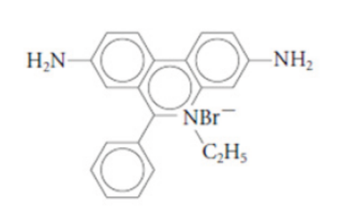
Figure 13.26: Ethidium bromide
Gel electrophoresis is not limited to the separation of nucleic acids but is also
commonly applied to proteins. Proteins are usually run on polyacrylamide gels,
which have smaller pores, because proteins are generally smaller in size than nucleic
acids. Proteins, however, are not negatively charged; thus, when researchers want to
separate proteins using gel electrophoresis, they must first mix the proteins with a
detergent called sodium dodecyl sulfate. This treatment makes the proteins unfold
into a linear shape and coats them with a negative charge, which allows them to
migrate toward the positive end of the gel and be separated. Finally, after the DNA,
RNA, or protein molecules have been separated using gel electrophoresis, bands
representing molecules of different sizes can be detected. The gel electrophoresis is
used for different purposes such as DNA analysis, protein and antibody interactions,testing antibiotics and testing vaccines.
Application 13.6
1. Identify the processes in which gel electrophoresis is used.
2. Describe the importance of restriction enzyme in gel electrophoresis.3. Explain the role of ethidium bromide in gel electrophoresis
13.7 Use of microarrays in the analysis of genomes and in detectingmRNA
Activity 13.7
Using different Biology textbooks, charts and internet; research information
about the use of microarrays in the analysis of genomes, answer to the
following questions.
1. What is microarray in genome analysis?2. Summarize the steps of microarray process in genome analysis.
DNA microarray, also commonly known as RNA chip or gene chip or biochip,
is technique consisting of a two-dimensional arrangement of DNA molecules
representing thousands of cloned genes on a solid surface such as a microscopic slide.
DNA microarray shows active genes that are being expression. Since a microarray
technology has the potential to examine the expression of several genes at a time,
it promises to revolutionize the way scientists study gene expression. For these
reasons, DNA microarrays are considered important tools for discovery in clinicalmedicine.
A basic protocol for a DNA microarray is as follows:
1. Isolate and purify mRNA from samples of interest:
As we are interested in comparing gene expression, one sample usually serves as
control, and another sample would be the experiment (for example, a healthy or
normal cell versus cancer cell).
2. Reverse transcribe and label the mRNA:
In order to detect the transcripts by hybridization, they need to be labeled, and
because starting material may be limited, an amplification step is also used. Labeling
usually involves performing a reverse transcription (RT) reaction to produce a
complementary DNA strand (cDNA) and incorporating a florescent dye that has
been linked to a DNA nucleotide, producing a fluorescent cDNA strand. Disease and
healthy samples can be labeled with different dyes and cohybridized onto the same
microarray in the following step. Some protocols do not label the cDNA but use a
second step of amplification, where the cDNA from RT step serves as a template toproduce a labeled cRNA strand.
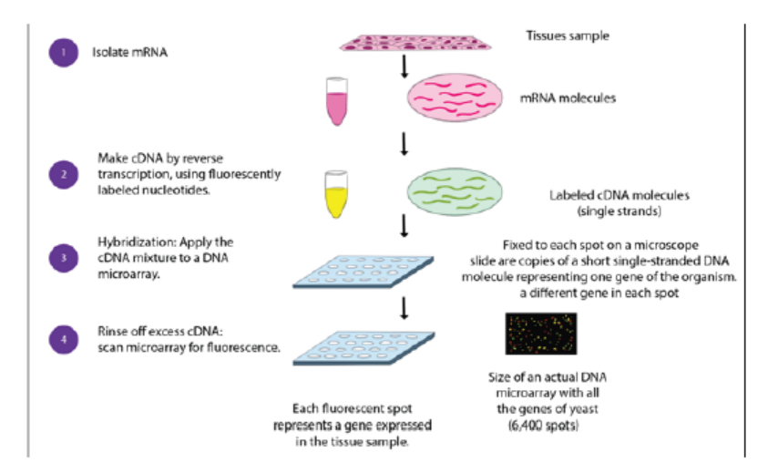
Figure 13.27: Procedure of microarray
3. Hybridize the labelled target to the microarray:
This step involves placing labelled cDNAs onto a DNA microarray where it will
hybridize to their synthetic complementary DNA probes attached on the microarray.
A series of washes are used to remove non-bound sequences. In molecular biology,
a hybridization probe is a fragment of DNA or RNA of variable length (usually 100–
1000 bases long) which can be radioactively labelled. It can then be used in DNA or
RNA samples to detect the presence of nucleotide sequences (the DNA target) thatare complementary to the sequence in the probe.
4. Scan the microarray and quantitate the signal:
The fluorescent tags on bound cDNA are excited by a laser and the fluorescently
labelled target sequences that bind to a probe generate a signal. The total strength
of the signal depends upon the amount of target sample binding to the probes
present on that spot. Thus, the amount of target sequence bound to each probe
correlates to the expression level of various genes expressed in the sample. Thesignals are detected, quantified, and used to create a digital image of the array.
Figure 13.28: A DNA microarray as viewed with a laser scanner. The colours are analysed to show which
genes or alleles are present
Application 13.7
1. Explain the role of reverse transcriptase in microarray technique
2. Describe the DNA probe
3. The figure below shows the microarray experiment in which the nucleicacids from cancer cells and normal cells are involved.
a. Redraw this figure identifying the steps X and Y and the molecule Z.
b. Describe briefly the aim of this experiment.
End of unit assessment 13
1. Choose the letter corresponding to the best answer.
(i) Different enzymes are used in the various steps involved in the production
of bacteria capable of synthesizing a human protein. Which step is
catalysed by a restriction enzyme?
a. Cloning DNA
b. Cutting open a plasmid vector
c. Producing cDNA from mRNA
d. Reforming the DNA double helix
(ii) What describes a promoter?
a. A length of DNA that controls the expression of a gene.
b. A piece of RNA that binds to DNA to switch off a gene.
c. A polypeptide that binds to DNA to switch on a gene
d. A triplet code of three DNA nucleotides that codes for ‘stop’
(iii) Which statement correctly describes the electrophoresis of DNA
fragments?
a. Larger fragments of DNA move more rapidly to the anode than smaller
fragments.
b. Positively charged fragments of DNA move to the anode.
c. Small negatively charged fragments of DNA move rapidly to the cathode.
d. Smaller fragments of DNA move more rapidly than larger fragments.
2. Explain the advantages of using plasmids as vectors.
3. The latest estimate of the number of genes in the human genome is 21 000.
Before the invention of microarrays, it was very time consuming to find out
which genes were expressed in any particular cell.
a. Explain how it is possible to find out which genes are active in a cell at a
particular time in its development.
b. Why is it not possible to use the same technique to find out which genes
are active in red blood cells?
4. Refer to what you have studied on DNA and PCR,
a. How many molecules of DNA are produced from one double-stranded
starting molecule, after eight cycles of PCR?
b. Explain why it is not possible to use PCR to increase the number of RNA
molecules in the same way as it is used to increase the number of DNAmolecules.
5. Complete the following table of the techniques used in gene technology.
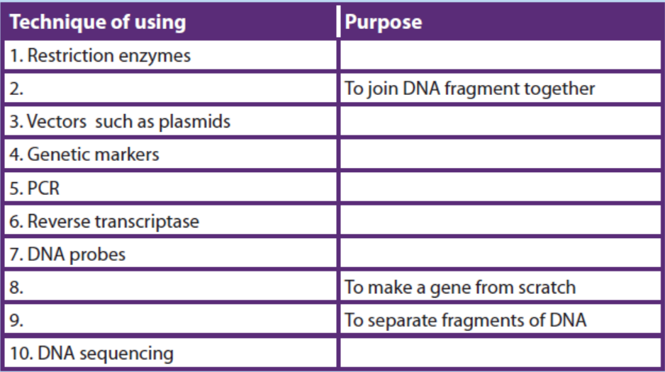
UNIT 14 APPLICATION OF GENE TECHNOLOGY
UNIT 14: APPLICATION OF GENE TECHNOLOGY
Key Unit Competence
Evaluate how gene technology is applied in areas of medicine, forensic science and
agriculture
Learning objectives
By the end of this unit, I should be able to:
– Define the term bioinformatics.
– Outline the role of bioinformatics following the sequencing of genomes, such as
those of humans and parasites, e.g. Plasmodium. (Details of the methods of DNA
sequencing are not required).
– Explain the advantages of producing human proteins by recombinant DNA
techniques. (Reference should be made to some suitable examples, such as
insulin, factor VIII for the treatment of haemophilia and adenosine deaminase
for treating severe combined immunodeficiency (SCID).
– Outline the advantages of screening for genetic conditions. (Reference may
be made to tests for specific genes such as those for breast cancer, BRCA1 and
BRCA2, and genes for haemophilia, sickle cell anaemia, Huntington’s disease
and cystic fibrosis).
– Outline how genetic diseases can be treated with gene therapy and discuss
the challenges in choosing appropriate vectors, such as: viruses, liposomes and
naked DNA, (Reference may be made to SCID, inherited eye diseases and cystic
fibrosis).
– Explain the significance of genetic engineering in improving the quality and
yield of crop plants and livestock in solving the demand for food in the world
e.g. Bt maize, vitamin A enhanced rice (Golden rice TM) and GM salmon.
– Outline the way in which the production of crops such as maize, cotton, tobacco
and rape seed oil may be increased by using varieties that are genetically
modified for herbicide resistance and insect resistance.
– Explain the ethical and social implications of using genetically modified
organisms (GMOs) in food production.
– Interpret a chart on the stages involved in the production of insulin by bacteria.
– Analyse the application of gene technology in agricultural modernisation.
– Research the benefits, hazards and implications of gene technology.
– Appreciate the application of gene technology in medicine, and forensic science
such as the detection of crimes e.g. rape, murder, and paternity disputes.
– Appreciate the application of gene technology in agriculture through the
improvement of crop varieties and animal breeds.
Introductory activity14.1
Observe the plants and animals below and carry out the following activity: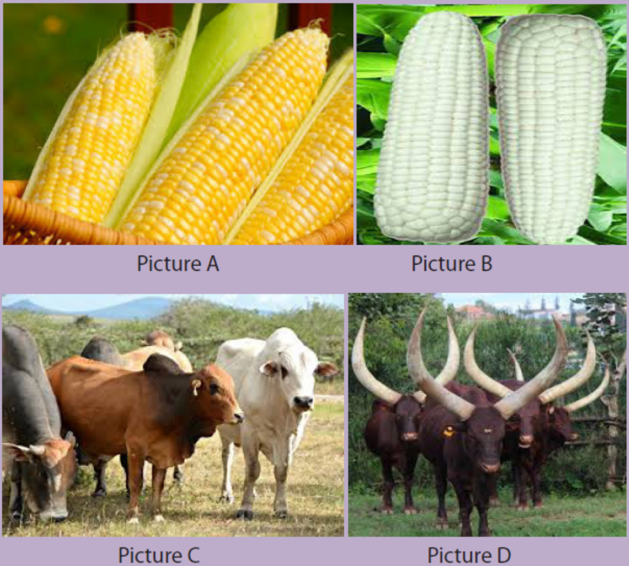
1. Discuss the reasons why the above crops(A and B) and animals (C and D)
present some differences.
2. Is there any benefits of having different varieties of organisms belonging to
the same species?
Techniques used by genetic engineers have been seen in unit 13. What can be
done with these techniques? By far most numerous applications are still as research
tools, and those techniques are helping geneticists to understand complex genetic
systems. Despite all of those types, genetic engineering still has very few successful
commercial applications, although these are increasing each year. The applications
so far can usefully be considered in three groups.
– Gene products using genetically modified organisms (usually microbes) to
produce chemicals, usually for medical or industrial applications.
– New phenotypes using gene technology to alter the characteristics of
organisms (usually farm animals or crops).
– Gene therapy using gene technology on humans to treat a disease.
The biggest and most successful kind of genetic engineering is the production of
gene products. These products are of; medical, agricultural or commercial value.
The table below shows some examples of genetically engineered products that are
already available.
Table 14.1 Examples of genetically engineered products and their uses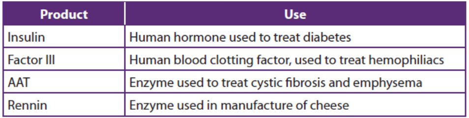
14.1 Bioinformatics
Activity 14.1
Using book or internet to search information about the importance of
Bioinformatics. Thereafter; discuss how the bioinformatics contribute to the
sequencing of genomes. In your discussion focus on human and parasite
genomes like Plasmodium.
Bioinformatics is the collection, processing and analysis of biological information
and data using computer software. In other words, it is the branch of biology that
is concerned with the acquisition, storage, and analysis of the information found in
nucleic acid and protein sequence data. Bioinformatics combines biological data
with computer technology and statistics. It builds up databases and allows links to
be made between them. The databases hold gene sequences of complete genomes,
amino acid sequences of proteins and protein structures.
UniProt (universal protein resource) holds information on the primary sequences
of proteins and the functions of many proteins, such as enzymes. The search tool
BLAST (basic local alignment search tool) is an algorithm for comparing primary
biological sequence information, such as the primary sequences of different proteins
or the nucleotide sequences of genes. Researchers use BLAST to find similarities
between sequences that they are studying and those already saved in databases.
When a genome has been sequenced, comparisons can be made with other known
genomes. For example, the human genome can be compared to the genomes of
the fruit fly, Drosophila, the nematode worm, or the malarial parasite, Plasmodium.
All the information about the genome of Plasmodium is now available in databases.
This information is being used to find new methods to control the parasite. For
example, being able to read gene sequences is providing valuable information in
the development of vaccines for malaria.
Application 14.1
1. What do you understand by the term bioinformatics?
2. Explain the role of bioinformatics following the sequencing of genomes, in
controlling and prevention of malaria.
14.2 Production of human proteins by recombinant DNA technology
Activity 14.2
Observe the figure below, analyse it and do the following: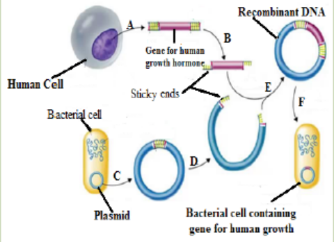
1. What does the above figure represent?
2. Using textbook or internet, interpret what happens in the stages A to E.
Recombinant DNA technology brought about a complete revolution in the way
living organisms are exploited. By transferring new DNA sequences into microbes,
plants, and animals, or by removing or altering DNA sequences in the endogenous
genome, completely new strains or varieties can be created to perform specific
tasks. One of the earliest commercial applications of gene manipulation was the
production of human therapeutic proteins in bacteria. Not surprisingly, the first
such products were recombinant versions of proteins already used as therapeutics:
human growth hormone and insulin. Prior to the arrival of genetic engineering,
human growth hormone was obtained from pituitary glands removed from cadavers
and the insulin was extracted from the pancreas of pigs or cattle.
Production of Insulin
This hormone can be produced by genetically modified bacteria and has been
in use since 1982. The human insulin gene is inserted into bacteria, which then
secrete human insulin. The human insulin produced in this way is purer than insulin
prepared from pigs or cattle that was used before, which sometimes provokes
allergic reactions owing to traces of ‘foreign’ protein. The Genetically Modified(GM)
insulin is acceptable to people with a range of religious beliefs who may not be
allowed to use insulin from cows or pigs. The main advantage of this form of insulin
is that there is now a reliable supply available to meet the increasing demand. The
chart below summarises stages involved in the production of insulin by bacteria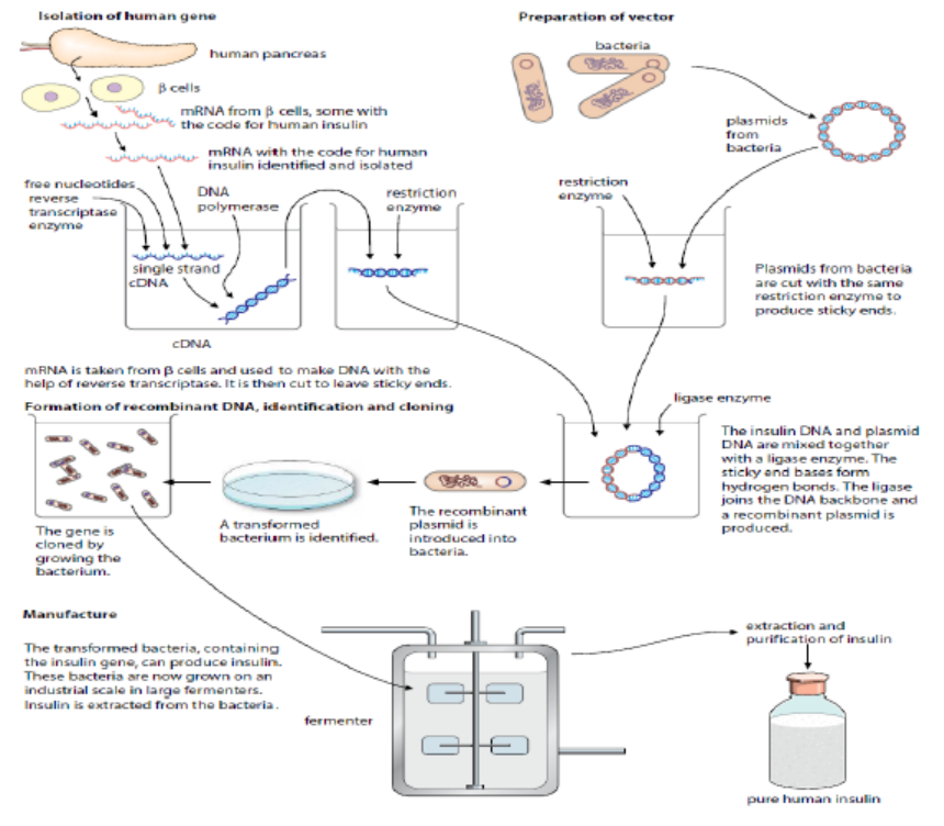
Figure 14.2 Producing insulin from genetically modified bacteria.
There were problems in locating and isolating the gene coding for human insulin
from all of the rest of the DNA in a human cell. Instead of cutting out the gene from
the DNA in the relevant chromosome, these are steps involved in human insulin
production:
– Researchers extracted mRNA for insulin from pancreatic β cells, which are the
only cells to express the insulin gene. These cells contain large quantities of
mRNA for insulin as they are its only source in the body.
– The mRNA was then incubated with the enzyme reverse transcriptase which
comes from the group of viruses called retroviruses. As its name suggests, this
enzyme reverses transcription, using mRNA as a template to make singlestranded
DNA.
– These single-stranded DNA molecules were then converted to doublestranded
DNA molecules using DNA polymerase to assemble nucleotides to
make the complementary strand.
– The genetic engineers now had insulin genes that they could insert into
plasmids to transform the bacterium Escherichia coli.
– When the bacterial cells copy their own DNA, they also copy the plasmids and
the donor genes that plasmids carry. After the cells have grown into colonies,
on an industrial scale in large fermenters insulin is extracted from the bacteria.
14.3 Genetic technology applied to medicine and forensic science
Activity 14.3
Using books or internet search and summarize information about genetic
technologies applied to medical and forensic sciences
1. Discuss about the social and ethical considerations of using gene
testing and gene therapy in medicine.
2. Interpret how gene technology is important in detection of crimes
(such as; rape, theft, and murder) and solving parenthood disputes.
14.3.1 Genetic screening
Genetic screening is the detection of mutations known to be associated with
genetic disorders before they manifest themselves in an individual. This can be done
in adults, in a foetus or embryo in the uterus, or in a newly formed embryo produced
by in-vitro fertilization. For example, an adult woman with a family history of breast
cancer may choose to be screened for the faulty alleles of the genes Brca-1 and Brca-
2, which considerably increase an individual’s chance of developing breast cancer. If
the results are to be positive; the woman may choose to have her breasts removed
(elective mastectomy) before such cancer appears.
Genetic disorders in the human foetus can also be detected using genetic screening
of embryonic cells found in the amniotic fluid during gestation. Such prenatal screens
are available for haemophilia, phenylketonuria, cystic fibrosis, and Duchenne’s
muscular dystrophy. Couples with a family history of genetic disorders who are
at risk of passing mutations on to their offspring are offered genetic counselling
to better prepare for the birth of a child. The most common vectors that are used
to carry the normal alleles into host cells are viruses (often retroviruses) or small
spheres of phospholipid called liposomes.
14.3.2 The ethics of genetic screening
Many people believe that the law is allowing too much, while others think that it
should allow more. For instance, in some countries, the law allowed an embryo
screening for a genetic disease; also some countries allow a successful transplant of
tissue from one person to another. But the law does not allow the addition of an allele
to an egg, sperm or zygote. Other countries have different attitudes and regulations.
For example, a foetus can now be screened for a genetic disease while in the uterus,
using amniocentesis or chorionic villus sampling. From this screening parent can
decide to terminate her pregnancy if the embryo is found to have a genetic disease.
Some parents have decided to terminate pregnancies simply because the child is not
the sex that they want. Pre-implantation genetic diagnosis (PGD) is the technique
that involves mixing the father’s sperm with the mother’s eggs in a dish (In vitro
procedure). The PGD has been also used to select the sex of the embryo that is
chosen to be implanted. Many people think that this sex pre-selection, as it is called,is totally unethical.
14.3.3 Treatment of genetic diseases by gene therapy
Gene therapy is the introduction of genes into suffering individual for therapeutic
purposes. It holds great potential for treating disorders noticeable to a single
defective gene. The first successful gene therapy performed was about the rare
genetic disorder known as severe combined immunodeficiency (SCID). The defect
in SCID involves the inability to make an enzyme, adenosine deaminase (ADA)
which is vital for the functioning of the immune system. These enzymes are made
by a genetically modified; insect larva, the cabbage looper moth caterpillar. This
enzyme is administered to patients while they are waiting for gene therapy or when
gene therapy is not possible. The work on SCID has led to increasingly successfulgene therapies in the last few years, including the followings:
a. Inherited eye diseases
Inherited eye diseases called Leber congenital amaurosis is a form of hereditary
blindness that primarily affects the retina, which is a specialised tissue at the back of
the eye that detects light and colour. People with this disorder typically have severe
vision impairment beginning at infancy. By gene therapy this condition has beenimproved.
b. Haemophilia
Haemophilia is an inherited bleeding disorder where the blood does not clot
properly. It is caused when blood does not have enough clotting factor. Genetically
modified hamster (small furry animal which is similar to a mouse) cells are used by
several companies to produce factor VIII. This protein is essential for blood clotting,
and people who cannot make it are said to have haemophilia. The human gene for
making factor VIII has been inserted into hamster kidney and ovary cells which are
then cultured in fermenters. The cells constantly produce factor VIII which is extracted
and purified before being used to treat people with haemophilia. These people need
regular injections of factor VIII which, before the availability of recombinant factorVIII, came from donated blood.
c. Cystic fibrosis
Cystic fibrosis which is a genetic disorder in which abnormally thick mucus
is produced in the lungs and other parts of the body, is also treated using gene
therapy. Cystic fibrosis is caused by a recessive allele of the gene that codes for a
transporter protein called CFTR (cystic fibrosis transmembrane conductance
regulator). This protein is found in the cell surface membranes of cells in the alveoli
and allow chloride ions (Cl-) to pass out of the cells. The recessive allele codes for a
faulty version of this protein that does not act properly as a chloride ion transporter.
If the normal dominant allele could be inserted into cells in the lungs, the correct
CFTR should be made. In theory this should happen but in practice, there have beenproblem of getting the allele into the cell.
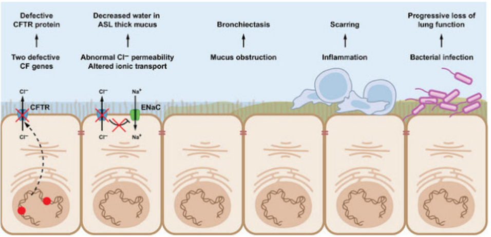
Figure 14.3: Diagram showing the processes involved in the infection of cystic fibrosis in cell lining of lungs
Note that:
There were different trials of gene therapy using different vectors like liposomes and
viruses which were not successful. DNA also has been inserted directly into tissues
without the use of any vector. This so called naked DNA has been used in trials
of gene therapy for skin, muscular and heart disorders. The advantages of using
this method is that, it removes the problems associated with using vectors. Some
proteins are even produced by transgenic animals. Sheep and goats have been
genetically modified to produce human proteins in their milk: human antithrombin
is produced by goats, this protein is used to stop blood clotting human alpha.
Antitrypsin is produced by sheep, this is used to treat people with emphysema.
14.3.4 Application of gene technology in forensic science.
Forensic science deals with the application of scientific methods and techniques
to matters under investigation by a court of law. For most people, forensic science is
synonymous with criminal investigations, but it is also used to resolve civil disputes
such as parenthood disputes.
DNA can be extracted from small sample of the cells found at the scene of the crime,for example in traces of blood, hair or saliva. In cases of rape, semen may be used.
a. Detection of crimes (Rape or murder)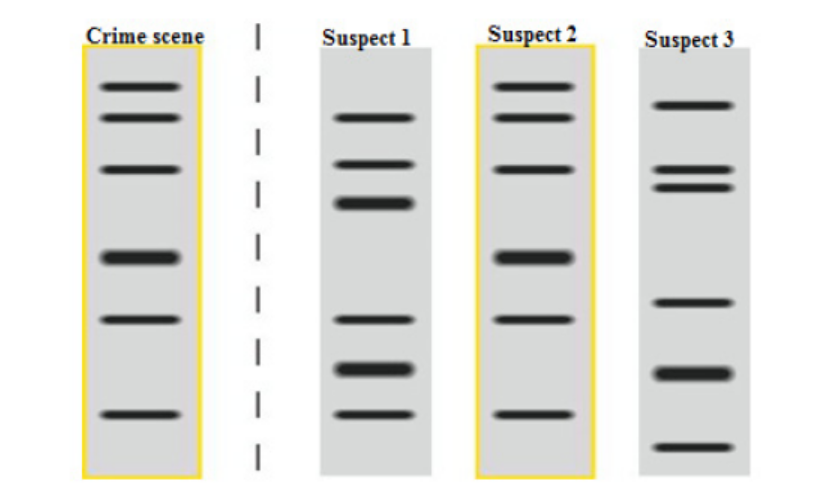
Figure 14.4 Genetic fingerprint of semen or blood (from Crime scene) and the blood of suspect rapists ormurders.
b. In forensic science, DNA fingerprinting is used to match material collected at
the scene of crime to that of suspects. This diagram above( Figure 14.4 ) of the
genetic fingerprints shows semen or blood (specimen from crime scene) found
on the victim and blood samples taken from the suspect rapists or murder.
The fingerprint results show an exact match between semen or blood sample
obtained from the victim and the blood sample of suspect 2. As a result suspect2 is confirmed to be the rapist or a murderer.
c. Paternity test
In perternity tests, DNA of suspected fathers are analysed together with the one
of the child and the mother in order to find out the potential father among the
suspect fathers that has the most DNA common with the child in question. Figure
14.5 shows an example of a Restriction Fragment Length Polymorphism (RFLP) used
to determine which potential father between father 1 and 2 who is the real father of
the child (C). As it is seen on the above figure, the second father tested (F2) seems tohave more DNA in common with the child than of the first farther tested (F1).
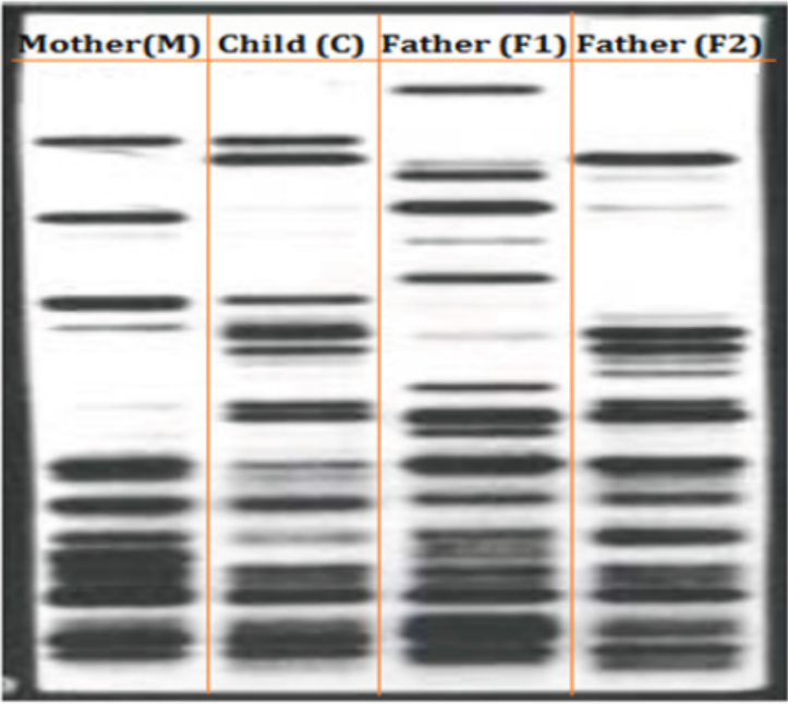
Figure 14.5 A peternity test using the RFLP (Restriction Fragment Length Polymorphism) technique
Application 14.3
1. Identify the advantages of genetic screening.
2. Gene therapy for cystic fibrosis would be successful if only one copy of
the normal allele of the gene was successfully inserted into the cells.Explain why this is so.
14.4 Significance of genetic engineering in improving thequality and yield of crop plants and livestock
Activity 14.4
Visit an agricultural center or research stations available in the area or observe
movies and find out how gene technology is applied in the modernization of
agriculture and livestock farming in Rwanda
1. Focus on the following crops varieties: maize, cassava, irish potatoes,
beans, tomatoes, oranges, mangoes, and avocado.
2. Focus on the following animals: poultry, cattle, goats, sheep, and pigs.
3. Based on your observations, discuss how modified crops and animals
contributed in improving the quality and yield of crop plants andlivestock in Rwanda.
Scientists are working to learn more about the genomes of agriculturally important
plants and animals. For a number of years, they have been using DNA technology
in an effort to improve agricultural productivity. The selective breeding of both
livestock (animal husbandry) and crops has exploited naturally occurring mutations
and genetic recombination for thousands of years. As we described earlier, DNA
technology enables scientists to produce transgenic animals, which speeds up theselective breeding process.
14.4.1 Gene technology and agriculture
Many new products have been developed using this technology. Crops have been
genetically engineered to increase yield, hardiness, uniformity, insect and virus
resistance, and herbicide tolerance. The vast bulk of genetically modified plants
grown around the world are crop plants modified to be resistant to herbicides or
crops that are resistant to insect pests. These modifications increase crop yield. A
few crops, such as vitamin A, enhanced rice, provide improved nutrition
a. Golden rice
Golden rice is a staple food in many parts of the world, where people are poor and
rice forms the major part of their diet. Deficiency of vitamin A is a common and serious
problem; its deficiency can cause blindness. In the 1990s, a project was undertaken
to produce a variety of rice that contained carotene in its endosperm. Genes for the
production of carotene were extracted from maize and the bacterium Pantonoea
ananatis. These genes, together with promoters, were inserted into plasmids. The
plasmids were inserted into bacteria called Agrobacterium tumefaciens. These
bacteria naturally infect plants and so could introduce the genetically modified
plasmid into rice cells. The rice embryos, now containing the carotene genes, weregrown into adult plants.
This genetically modified rice is called golden rice, because it contains a lot of
yellow pigment carotene. The genetically modified rice is being bred into other
varieties of rice to produce varieties that grow well in the conditions in different
parts of the world, with the same yield, pest resistance and eating qualities as theoriginal varieties.
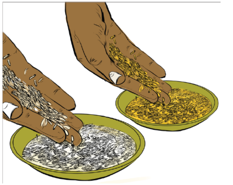
Figure 14.6: Normal rice (white) compared with golden rice (yellow)
b. Herbicide-resistant crops: Oil seed rape
Herbicide-resistant crops called oil seed rape or Brassica napus, is grown in many
parts of the world as a source of vegetable oil which is used as biodiesel fuel, as a
lubricant and in human and animal foods. Natural rape seed oil contains substances
that are undesirable in oil that is to be used in human or animal food. A hybrid, was
made to produce low concentrations of these undesirable substances, called canola
(Canadian oilseed low acid), and this name is now often used to mean any variety of
oil seed rape. Gene technology has been used to produce herbicide-resistant strains.
Growing an herbicide-resistant crop allows fields to be sprayed with herbicide after
the crop has germinated, killing any weeds that would otherwise compete with the
crop for space, light, water or ions. This increases the yield of the crop.
c. Insect pests-resistant plants
Another important agricultural development is that of genetically modified plants
protected against attack by insect pests. Bt maize is genetically engineered (GE)
plant that produces crystal (Cry) proteins or toxins derived from the soil bacterium,
Bacillus thuringiensis (Bt), hence the common name “Bt maize”. Bt maize plant has
revolutionized pest control in a number of countries, but there still are questionsabout its use and impact.
14.5.2 Transgenic animals.
DNA technology enables scientists to produce transgenic animals, which speeds up
the selective breeding process. Creating transgenic animals is aimed at improving
quality and productivity. For instance, to make a sheep with better quality wool, a
pig with leaner meat, or a cow that will mature in a shorter time. Scientists might, for
example, identify and clone a gene that causes the development of larger muscles
(muscles make up most of the meat) in one breed of cattle and transfer it to othercattle or even to sheep.
Genetically modified animals for food production are much rarer than crop plants.
An example is the genetically modified (GM) Atlantic salmon, developed in the USA
and Canada. A growth-hormone regulating gene from a Pacific Chinook salmon
and a promoter from another species of fish (an ocean pout), were injected into a
fertilised egg of an Atlantic salmon. By producing growth hormone throughout the
year, the salmon are able to grow all year, instead of just in spring and summer. As
a result, fish reach market size in about eighteen months, compared with the three
years needed by an unmodified fish. It is proposed to rear only sterile females and
to farm them in land-based tanks. The characteristics of the GM salmon reduce their
ability to compete with wild salmon in a natural environment. Below figure comparesGM salmon the big one, and farm salmon the small; both fish are 18 months.
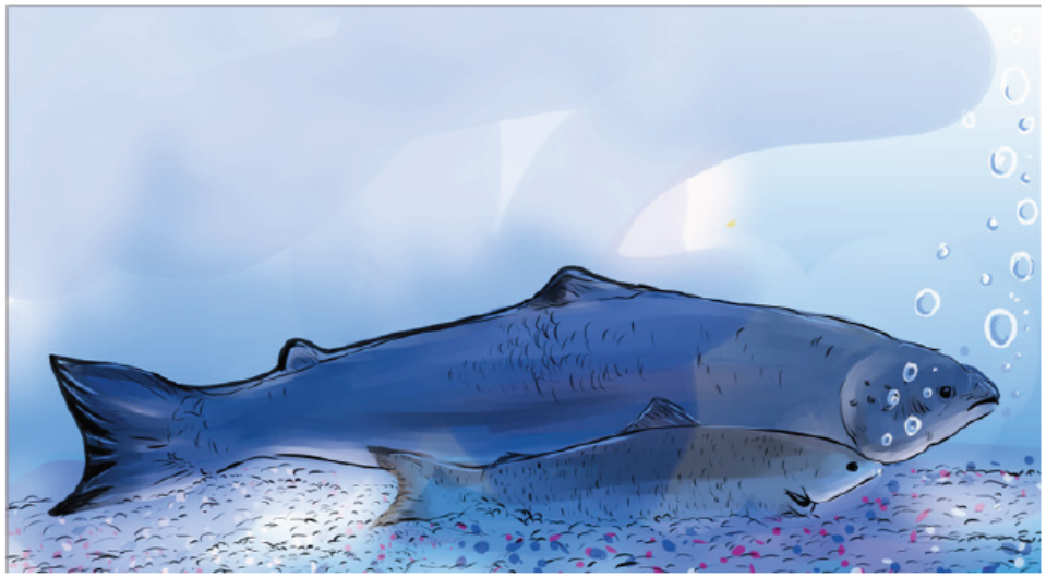
Figure 14.7. Comparison between GM salmon and farm salmon
Application 14.4
1. Explain the meaning of transgenic organisms
2. Why is Bt maize popular with growers?
3. Discuss the process of production of pest resistant plants like Bt cotton,tomato maize corn and rice
14.5 Ethical and social implications of using genetically modifiedorganisms (GMOs).
Activity 14.5
From your daily life experience, discuss the ethical and social implications ofusing genetically modified crops in food production.
Ethics includes moral principles that control or influence a person’s behaviour.
It includes a set of standards by which a community regulates its behaviour and
decides as to which activity is legitimate and which is not. Bioethics may be viewed
as a set of standards that may be used to regulate our activities in relation to the
biological world. Biotechnology, particularly recombinant DNA technology, is usedfor exploitation of the biological world by various ways.
Some genetically modified plants are grown in strict containment of glasshouses, but
a totally different set of problems emerges when genetically engineered organisms
such as crop plants and organisms for the biological control of pests are intended
for use in the general environment. Few countries would object to the growth of
genetically modified crops that produce vaccines for human or animal use, yet there
are people who object to the growth of pro-vitamin A enhanced rice. The majorbioethical concerns pertaining to biotechnology are summarized below:
– When animals are used for production of certain pharmaceutical proteins,
they are treated as factory machines.
– Introduction of a transgene from one species into another species violates the
integrity of species.
– The transfer of human genes into animals or vice-versa is great ethic threat to
humanity.
– Biotechnology is disrespectful to living beings, and only exploits them for the
benefit of humans.
– Genetic modification of organism can have unpredictable/ undesirable effectswhen such organisms are introduced into the ecosystem.
Moreover, most objections are raised against the growth of herbicide-resistant orinsect-resistant crops as follow:
– The modified crop plants may become agricultural weeds or invade natural
habitats.
– The introduced gene may be transferred by pollen to wild relatives whose
hybrid offspring may become more invasive.
– The introduced gene may be transferred by pollen to unmodified plants
growing on a farm with organic certification.
– The modified plants may be a direct hazard to humans, domestic animals or
other beneficial animals, by being toxic or producing allergies.
– The herbicide that can now be used on the crop will leave toxic residues in the
crop.
– Genetically modified seeds are expensive, as is herbicide, and their cost may
remove any advantage of growing a resistant crop.
– Growers mostly need to buy seed each season, keeping costs high, unlike for
traditional varieties, where the grower kept seed from one crop to sow for the
next
– In parts of the world where a lot of genetically modified crops are grown, there
is a danger of losing traditional varieties with their desirable background genes
for particular localities This requires a programme of growing and harvestingtraditional varieties and setting up a seed bank to preserve them.
Application 14.5
1. Write an account on edible GM crops.
2. Discuss ethical and social implications raised against growth ofherbicide-resistant or insect-resistant crops.
End of unit assessment 14
I. Multiple choice questions
1. What is the term used for inserting a healthy copy of a gene into a person
who has a defective gene?
a. Cloning vector
b. gene therapy
c. Recombinant DNA
d. Polymerase chain reaction (PCR)
2. Which is the process used in animal cloning
a. DNA cloning
b. Recombinant DNA
c. Polymerase by nuclear transfer
3. A man and woman, each with a family history of sickle cell disease and no
children, would benefit most by:
a. Prenatal screening
b. Carrier screening.
c. Inherited predisposition screening
d. No screening because they already know their status.
4. DNA technology has many medical applications. Which of the following is
not done routinely at present?
a. Production of hormones for treating diabetes and dwarfism.
b. Production of viral Proteins for vaccines
c. Introduction of genetically engineered genes into human gametes.
d. Prenatal identification of genetic disease genes.
e. Genetic testing for carriers of harmful alleles
5. Which of the following is NOT a use of DNA profiling?
a. Determining if two DNA samples come from the same person.
b. Determining if a child could have inherited their genes from a
suspected father.
c. Determining whether a person has a given genetic disease.
d. None of the above.
II. Structured questions
6. Rearrange the statements below to produce a flow diagram showing the
steps involved in producing bacteria capable of synthesizing a human
protein such as insulin.
a. Insert the plasmid into a host bacterium.
b. Isolate mRNA for insulin.
c. Insert the DNA into a plasmid and use ligase to seal the ‘nicks’ in the
sugar phosphate chains.
d. Use DNA polymerase to clone the DNA.
e. Clone the modified bacteria and harvest the insulin.
f. Use reverse transcriptase to produce cDNA.
g. Use a restriction enzyme to cut a plasmid vector.
7. In the production of bacteria that synthesise human insulin, plasmids acted
as vectors to introduce the gene into the bacterial cells. What were the
vectors used in the production of vitamin A enhanced rice? Explain your
answer.
8. What is genetic modification (GM) of crops and how is it done? Evaluate all
possible hazards of GM crops.
9. Identify genes that have been introduced into GM crops so far and explain
its purpose.
10. Answer the following questions:
a. How does gene therapy differ from genetic screening?
b. Explain why it is easier to devise a gene therapy for a condition caused
by a recessive allele than for one caused by a dominant allele.
11. As a genetic engineer, you have a patient with symptoms that suggest a
hepatitis A infection. The symptoms come and go, but you have not been
able to detect viral proteins in the blood. Knowing that hepatitis A is an RNA
virus, what lab tests could you perform to support your diagnosis? Explain
what the result would mean.
12. Examine the figure, which shows diagrammatic DNA profiles of a mother, herchild and suspected fathers (P, Q and R) of the child.
Identify true biological father of the child. Explain your answer.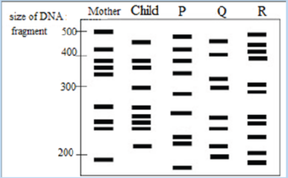
13. Some people need blood transfusions because their blood lucks important
proteins, such as those needed for blood clotting (Factor VIII). People who
receive blood transfusion have some risk of being exposed to disease-causing
viruses. How might genetic engineering eliminate this risk?
14. Bacteria and human beings are very different why is it possible sometimes
possible to combine their DNA and use a bacterium to make a human protein.
15. Describe a potential safety environmental concern with regard to genetically
modified (GM)) crops
16. The figure shows the CFTR (cystic fibrosis transmembrane conductanceregulator) protein in a cell surface membrane
a. Based on the above figure: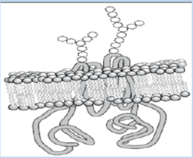
(i) Describe the normal function of the CFTR protein.
(ii) Use the letter E to indicate the external face of the membrane. State
how you identified this face.
b. Cystic fibrosis is caused by a recessive allele of the CFTR gene.
(i) Explain the meaning of the term recessive allele.
(ii) Explain how cystic fibrosis affects the function of the lungs.
c. As cystic fibrosis is caused by a recessive allele of a single gene, it is a good
candidate for gene therapy. Trials were undertaken, attempting to deliver
the normal allele of the CFTR gene into cells of the respiratory tract, using
viruses or liposomes as vectors. Explain how viruses deliver the allele intocells.
UNIT 15 VARIATION
UNIT 15: VARIATION
Key Unit Competence
Explain variation and mutation as a source of biodiversity
Learning objectives
At the end of this unit, I should be able to:
– Explain population traits and types of variation.
– Describe the differences between continuous and discontinuous variation.
– Describe the causes of variation.
– Explain the genetic basis of continuous (many additive genes control
characteristics) and discontinuous variation.
– Explain, with, examples, how the environment may affect the phenotype of
plants and animals.
– Explain why genetic variation is important in selection.
– Interpret graphs of variations in blood groups and height.
– Construct genetic diagrams to show how sickle cell anaemia is inherited.
– Use a t-test to compare the variation of two different populations (see
mathematical requirements for the syllabus).
– Appreciate the significance of genetic variation in selection.
– Express that discontinuous variation results in a limited number of phenotypes
with no intermediates e.g. tongue rolling.– Justify the effect of the environment on the phenotype of plants and animals.
Introductory activity
The diagrams below represent the beetles, maize and giraffes. Observe andanalyze them carefully and answer the following questions.
a. Explain why the beetles, maize and giraffes have different colors?
b. How do you call the biological term indicated by the above diagrams?
15.1 Variation
Activity 15.1
Use the school library and search additional information on the internet, read
the information related to variation. Based on the readings answer to the
following questions:
1. Describe what variation concept is, in your own words.2. Describe the mechanism and importance of variation.
The earth is inhabited by billions of organisms, every one of which is unique.
Individuals belonging to different species are usually easy to distinguish. Members
of the same species may differ only in small ways; but even clones (such as identical
twins) show some differences. The differences between individuals of the same
species are called variation. These differences between cells, individual organisms,
or groups of organisms of any species are caused either by genetic differences
(genotypic variation) or by the effect of environmental factors on the expression
of the genetic potentials (phenotypic variation). Variation may be seen in; physical
appearance (phenotype of individuals), metabolism, fertility, mode of reproduction,behavior, learning and mental ability, and other obvious or measurable characters
15.1.1 Origins of variation
Genotypic variations are caused by differences in number or structure of
chromosomes or by differences in the genes carried by the chromosomes. Eye color,
body form, and disease resistance results by genotypic variations. Individuals with
multiple sets of chromosomes are called polyploid. Many common plants have two
or more times the normal number of chromosomes and new species may arise by
this type of variation.
Variation may be due to either environmental factors or genetic disorders. For
example, the action of sunlight on a light- colored skin may result in its becoming
darker. Such changes have little evolutionary significance as they are not passed
from one generation to the next. Much more important to evolution are the inherited
forms of variation which result from genetic changes. These genetic changes may
be the result of the normal and frequent reshuffling of genes which occurs duringsexual reproduction, or as a consequence of mutations.
15.1.2 Importance of variation
Variation plays different roles such as:
– Make some individuals better fitted in the struggle for existence.
– Help the individuals to adapt themselves according to the changing
environment.
– Produce new traits in the organisms.
– Allow breeders to improve races of useful plants and animals for increased
resistance, better yield, quicker growth and lesser input.
– Constitute the raw material for evolution.
– Give each organism a distinct individuality.
– Species do not remain static. Instead, they are slowly getting modified forming
new species with time.
– Pre-adaptations caused by the presence of neutral variations are extremely
useful for survival against sudden changes in environment, e.g., resistanceagainst a new pesticide or antibiotic.
Application 15.1
Explain the origin and the importance of variation
15.2 Types of variation
Activity 15.2
Use the school library and search additional information on the internet,
read the information related to types of variation. Furthermore, get outside
classroom and then observe species day after day for at least 10 days. You need
to collect data every day from what you observe. You can even use a ruler or
a weighing machine where applicable. based on what you have done before
answer to the following questions:
1. Write in your own words the differences that exist between types of
variation2. Using a genetic cross, show that sickle cell anaemia is inherited
Variation does occur into two categories namely; genetic and phenotypic as
described in detailed below.
Genetic variation
Genetic differences reflect the genotype (the genetic make-up of an
individual organism, an individual ‘s genotype functions as a set of instructions for
the growth and development) of an organism, that is, its genetic make-up. A diploid
organism has two sets of chromosomes and two forms (alleles) of each particular
gene. These alleles may be the same (the organism is homozygous for that gene) or
different (the organism is heterozygous for that gene). If different, one of the alleles
(the dominant allele) may mask the other allele (the recessive allele). The dominant
allele is therefore expressed in either the heterozygous or the homozygous condition.
If an organism is haploid (that is, it has only one set of chromosomes), all its alleles
will be expressed and will be reflected in its observable or measurable characters(the features or traits transmitted from parent to offspring).
There are three primary sources of genetic variation:
1. Mutations are changes in the DNA. A single mutation can have a large effect,
but in many cases, evolutionary change is based on the accumulation of
many mutations.
2. Gene flow is any movement of genes from one population to another and
is an important source of genetic variation.
3. Sex can introduce new gene combinations into a population. This geneticshuffling is another important source of genetic variation.
Why is genetic variation important for evolution?
Variation is one of the main things that drive evolution. First, there are limited
resources available, and there is just not enough; food, water, shelter, etc. available
for all organisms. Second, to make matters worse, most species have many offspring
that can possibly survive. Just think of how many insect eggs are laid compared
to the number that make it to adulthood. This leads to competition for the limitedresources.
Not all individuals in a species are the same. There are variations in; size, speed,
coloration, etc. These small variations can help or hinder individuals in their survival.
These variations are caused by small differences in genes. Organisms that have
helpful variations are more likely to survive. On average, they get more food, get
better shelter, etc. Coloration can help a predator get closer to prey and eat better.
Or, for the prey species, coloration can make it harder for predators to find and eat
it. So, organisms that have helpful variations tend to survive better, and reproduce
more. As they reproduce, their genes (including the helpful genes) become more
common in the gene pool, and these variations spread out more and more.
Phenotypic variation
The measurable physical and biochemical characteristics of an organism, whether
observable or not, make up its phenotype (observable physical or biochemical characteristics
of an individual organism, determined by both genetic make-up and environmental
influences, for example, height, weight and skin color). The phenotype
results from the interaction of the genotype and the environment. The genotype
determines the potential of an organism, whereas the environment factors to which
it is exposed determine to what extent this potential is fulfilled. For example, in humans
the potential height of a person is genetically determined, but a person cannot
reach this height without an adequate diet. Phenotypic variation is of two maintypes: continuous and discontinuous.
a. Continuous variation
Continuous variation is variation which does not show clear cut differences i.e.
it shows a gradual change from one extreme to another. Characteristics such as;
human height and weight show continuous variation, and are usually determined
by a large number of genes (i.e. polygenic) and/ or considerable environmental
influence. Some examples of continuous variation are: Height, weight, heart rate,
finger length, and leaf length. They are also called fluctuating variations because
they fluctuate on either side (both plus and minus) of a mean or average for the
species. Continuous variations are typical of quantitative characteristics. They
show differences from the average which are connected with it through small
intermediate forms. If plotted as a graph, the mean or normal characteristic will
be found to be possessed by maximum number of individuals. The number of
individuals will decrease with the increase in degree of fluctuation. The graph (figure
15.1) will appear to be bell shaped. The variations are already present in differentorganisms or races of a species.
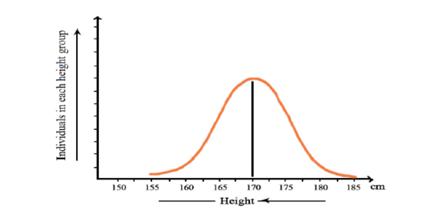
Figure 15.1: Continuous variations or fluctuations in the height of adult human beings
Continuous variations are produced by:
– Segregation of chromosomes at the time of gamete or spore formation.
– Crossing over or exchange of segments between homologous chromosomes
during meiosis.
– Chance combination of chromosomes during fertilization.
Therefore, these variations are also known by the name of re-combinations. They
make an organism better fitted to struggle for existence in a particular environment.
They also enable human beings to improve the races of important plants and
animals. However, they are unable to form a new species.
Continuous variations are of two types:
a. Substantive: They influence appearance including; shape, size, weight and
color of a part or whole of the organism, for example., height, shape of nose,
skin color, color of eyes, hair, length of fingers or toes, yield of milk, eggs, etc.
b. Meristic: They influence the number of parts, for example, number of grains in
an ear of wheat, number of epicalyx segments in Althaea, tentacles in Hydra or
segments in earthworm, etc.
c. Discontinuous variation
Discontinuous variation is variation where there is a clear cut difference with no
intermediates between individuals e.g. blood groups (A, B, AB, or O), Rhesus factor
(+ve or –ve), mice coat colour, gender, eye colour in drosophila, haemophilia, tongue
rolling, flower colour, seed shape, pawpaw tree sex (male or female) etc. Such
variations are represented in a bar graph as shown in Figure 15.2. Such variations
are controlled by a single gene or many alleles of the same gene. Continuous
variations are usually quantitative (they can be measured) whereas discontinuous
variations are qualitative (they tend to be defined subjectively in descriptive terms).
Thus height in humans is a continuous variation given a value in meters, whereas
height in sweet peas is a discontinuous variation described as tall or dwarf. Such
discontinuous variations are not changeable and neither can environment changethem.
Discontinuous variations are caused by:
– Chromosomal aberrations like; deletion, duplication, inversion and
translocation,
– Change in chromosome number through aneuploidy and polyploidy,
– Change in gene structure and expression due to addition, deletion or changein nucleotides.
Figure 15.2: Discontinuous variation of blood groups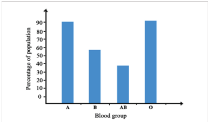
Sickle-cell anaemia an example of discontinuous variation
It is caused by the substitution of a single amino acid in molecular structure of RBCs.
When the oxygen content of an affected individual is low (at high altitude or under
physical stress), the sickle cell Hb deforms the RBCs to a sickle shape. Sickling of the
cells, in turn, can lead to other symptoms.
Individuals who are heterozygous (having a single copy of the allele) for the sicklecell
allele are said to have sickle-cell trait. They carry a normal life but suffer some
symptoms of sickle-cell disease when there is an extended reduction of blood
oxygen. Although the sickle-cell anaemia is lethal for homozygous, the sicklecell
trait (heterozygous) is sometimes considered as an advantage. People who
are heterozygous are resistant to malaria. Thus, in tropical Africa, where malaria iscommon, the sickle-cell allele is both beneficial and an afflicition.
Genotype for sickle cell anemia
Most genes, including the β-globin polypeptide gene, have several different alleles.
For the moment, only the two alleles of this gene are considered. For simplicity, the
different alleles of a gene can be represented by symbols. In this case, they can berepresented as follows:
HbA = the allele for the normal β-globin polypeptideHbS = the allele for the sickle cell β-globin polypeptide
The letters Hb stand for the locus of the haemoglobin gene, whereas the superscripts
A and S stand for particular alleles of the gene. In a human cell, which is diploid, there
are two copies of the β-globin polypeptide gene. The two copies might be: HbAHbA
or HbSHbS or HbAHbS. The alleles that an organism has form its genotype. In this
case, where we are considering just two different alleles, there are three possible
genotypes.Table 15.1: Genotype for sickle cell anemia
Inheriting genes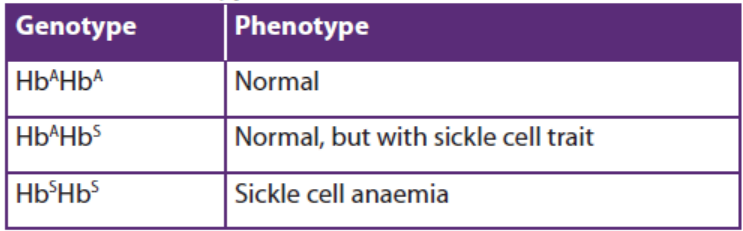
In sexual reproduction, haploid gametes are made, following meiosis, from diploid
body cells. Each gamete contains one of each pair of chromosomes. Therefore, each
gamete contains only one copy of each gene. Think about what happens when
sperm are made in the testes of a man who has the genotype HbAHbS. Each time a
cell divides during meiosis, four gametes are made, two of them with the HbA allele
and two with the HbS allele.
Of all the millions of sperm that are made in his lifetime, half will have the genotype
HbA and half will have the genotype HbS. Similarly, a heterozygous woman will
produce eggs of which half have the genotype HbA and half have the genotype HbS.
This information can be used to predict the possible genotypes of children born to
a couple who are both heterozygous. Each time fertilisation occurs, either an HbAsperm or an HbS sperm may fertilise either an HbA egg or an HbS egg.
Table 15.2: The possible results of genotypes and phenoty
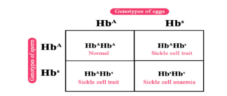
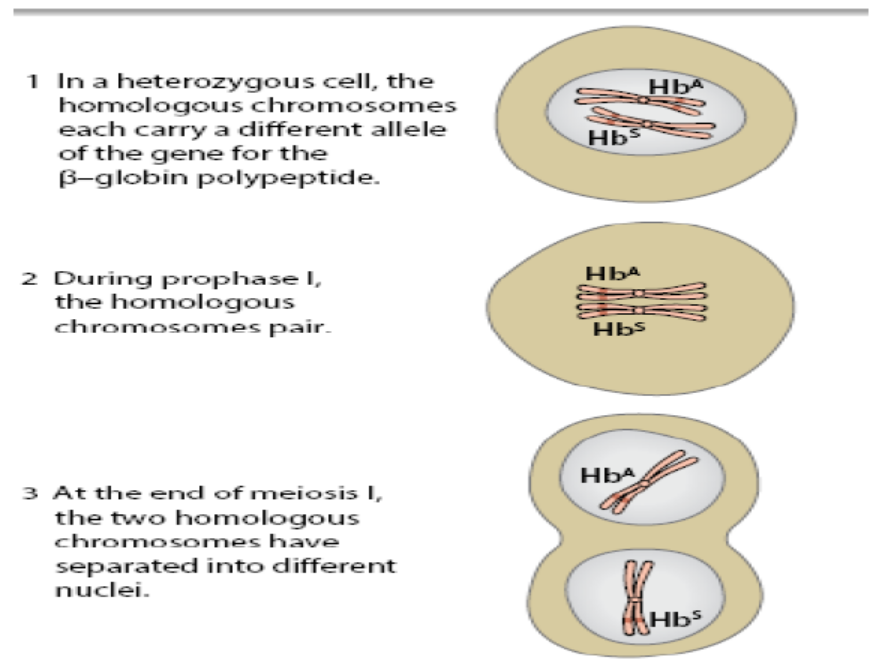
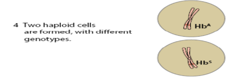
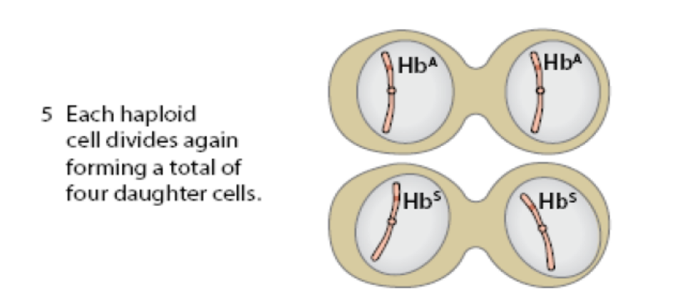
Figure 15.3: Meiosis of a heterozygous cell produces gametes of two different genotypes. Only one pair of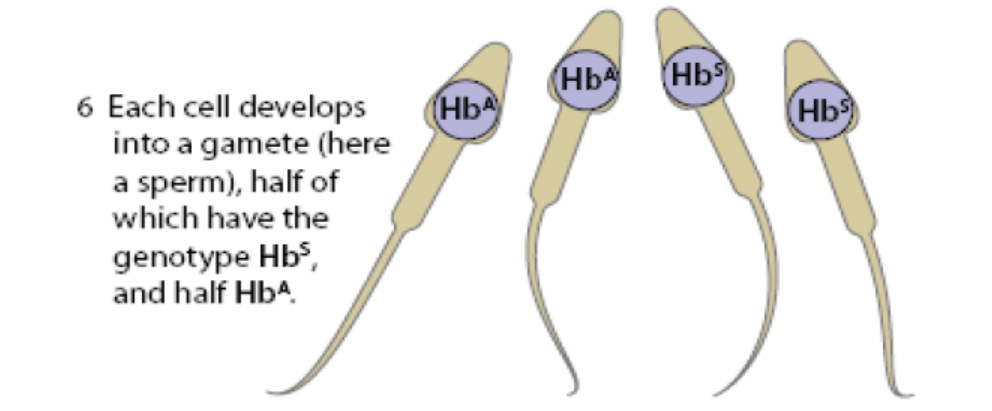
homologous chromosomes is shown.
As there are equal numbers of each type of sperm and each type of egg, the chances
of each of these four possibilities are also equal. Each time a child is conceived, there
is a one in four chance that it will have the genotype HbAHbA, a one in four chance
that it will be HbSHbS and a two in four chance that it will be HbAHbS. Another way of
describing these chances is to say that the probability of a child being HbSHbS is 0.25,
the probability of being HbAHbA is 0.25, and the probability of being HbAHbS is 0.5.
It is important to realize that these are only probabilities. It would not be surprising
if this couple had two children, both of whom had the genotype HbSHbS and so
suffered from sickle cell anaemia.
The major distinctions between continuous and discontinuous variations in
inheritance are as follows:
Continuous variations have the following characteristics:
– The variations fluctuate around an average or mean of species.
– Direction of continuous variations is predictable.
– They are already present in the population.– Continuous variations are formed due to chance segregation of chromosomes
during gamete formation, crossing over and chance pairing during fertilization.
– They can increase adaptability of the race but cannot form new species.
– Continuous variations are connected with the mean or average of the species
by intermediate stages.
– The continuous variations are also called fluctuations.
– When represented graphically, continuous variations give a smooth bell
shaped curve
– They are very common– Continuous variations do not disturb the genetic system.
Discontinuous variations have the following characteristics:
– A mean or average is absent in discontinuous variations.
– The direction of discontinuous variations is unpredictable.
– Discontinuous variations are new variations though similar variations might
have occurred previously.
– Discontinuous variations are produced by changes in genome or genes.
– Discontinuous variations are the fountain head of continuous variations as
well as evolution
– These variations are not connected with the parental type by intermediate
stages.
– Discontinuous variations are also known as mutations or sports.
– A curve is not produced when discontinuous variations are representedgraphically.

– These variations appear occasionally.
– They disturb the genetic system of the organism
Application 15.2
1. Using a table differentiate between continuous and discontinuous
forms of variation.
2. Draw and interpret graphs of variations in blood groups and height.
15.3 Causes of variation in living things
Activity 15.3
Use the school library and internet to search additional information about
cause of variations. Summarize the information in a table. Share and discuss
with your classmates.
a. Crossing over
Genes are interchanged resulting in new chromosomes (recombinants), different
from the parental combination. Chromosomal crossover (or crossing over) is the
exchange of genetic material between homologous chromosomes that results in
recombinant chromosomes during sexual reproduction. Crossing over and random
segregation during meiosis can result in the production of new alleles or new
combinations of alleles. Portions of paired chromosomes may be exchanged to
form new chromosomal and gene combinations in gametes resulting into new trait
combinations in offspring.
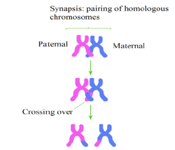
Figure 15.4: Illustration of crossing over
b. Non-disjunction
Non-disjunction results into doubling of the chromosome number due to failure
of chromosomes to segregate during meiosis. This leads to increase in cell size andsubsequent increase in size of various parts of the organism, hence variation.
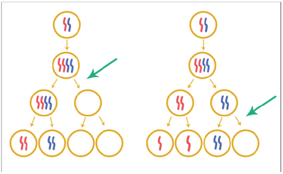
Figure 15.5: illustration of non-disjunction
c. Random fertilization
Random fertilization that results during the fusion of the gametes also contributes
to variation. Gametes are the egg and sperm, or pollen, produced by meiosis. Each
gamete has a unique set of combination of genes. A male gamete can fertilize any of
the female gametes. The fertilization between a male gamete and a female gamete
occurs randomly in the fallopian tube. As a result, each zygote is unique and hence
variation occurs due to the different combination of genes from the male and female
gamete.
The random fusion of gametes is a source of genetic variation in offspring (with the
same parents). For example, a litter of puppies or kitten (bred) by the same father
will show variation between individuals as shown below.
d. Random mating
Random mating involves individuals pairing by chance, not according to their
genotypes or phenotypes. Random mating is a source of variation in a population.
For example, a population in which mating only occur between organisms of similar
phenotypes, such as red beetles mating with red beetles and yellow beetles mating
with yellow beetles, will tend to show less variation than a population where crosses
are random. For example, red beetles mating with yellow beetles.
e. Mutations
Mutations are sudden and permanent changes in the genes and chromosomes
which are then passed on from cell to cell during mitosis. Such changed genes or
chromosomes will produce offspring that differ from parents.
A mutation is also a change in the amount or the chemical structure of DNA. If the
information contained within the mutated DNA is expressed, it can cause a change
in the characteristics of an individual cell or an organism. Mutations in the gametes
of multicellular organisms can be inherited by offspring. Mutations of the body cells
of multicellular organisms (somatic mutations) are confined to the body cells derived
from the mutated cell; they are not inherited. Mutations can happen spontaneously
as a result of errors in DNA replication or errors during cell division, or they can be
induced by various environmental factors (such as certain chemicals, X-rays, and
viral infection). Factors that induce mutations are called mutagens.
f. Independent assortment of homologous chromosomes
This occurs at the time of gamete formation. At the time of gamete formation during
meiosis, the parental chromosomes separate at random hence forming different
gametes with different chromosomes. This independent assortment gives a wide
variety of different gametes and hence individuals.
g. Environmental factors
These variations are not inherited but are due to environmental factors. The
environmental factors bring about only slight modifications in animals but in plants
the modifications are much more conspicuous. This is due to the environmental
effect on the meristems of various parts. A slight change in the meristematic activity
can have permanent effect on the plant. Environment can also change the amount
of flowering and bring about non- inheritable changes in the floral parts.
1. Light
In the absence of light, the plants remain etiolated. Shade produces elongated
internodes and thinner and broader leaves. It increases the succulence of many
vegetables. Strong light, on the contrary, helps in the production of more mechanical
tissue and smaller and thicker leaves. The effect of light has also been observed
by Cunningham in flat fish Solea. The fish habitually rests on left side. It develops
pigmentation and eyes on right side, the side exposed to sun. If left side is exposed
to sunlight in the young fish, both eyes and pigmentation develop on that side.
2. Temperature
Temperature directly affects the metabolic activity of the organisms and rate of
transpiration in plants. Plants growing in hot area show stunted growth of the aerial
parts and greater growth of the root system. Strong sunlight and high temperature
bring about sun-tanning of human skin by production of more melanin for protectionagainst excessive insulation and ultraviolet radiations.
3. Nutrition
The individual provided with optimum nutrition grows best while the under
nourished shows stunted growth. The abundance or deficiency of a mineral salt
produces various types of deformities in plants. A larva of honey bee fed on royaljelly grows into queen while the one fed on the bee bread develops into worker.
4. Water
Plants growing in soils deficient in water or in areas with little rainfall show
modifications in order to reduce transpiration and retain water, e.g., succulence,
spines, reduced leaves, thick coating, sunken stomata, etc. Those growing in humidand moist area show luxuriant growth.
Application 15.3
1. Outline and explain in your own words any 3 environmental factors
that cause variation1. Distinguish the random fertilization from random mating.
15.4 t-test
Activity 15.4
Search from books or internet to have more information on t-test?
Collect measurements from populations of organisms in two varying sites
and use t-tests to distinguish whether or not these are likely to represent twodistinct populations.
The t- test is used to test the statistical significance of continuous variables. The t-test
therefore has less application in genetics and far more in other areas of biology,
such as ecology. The t- test is used when a sample size is relatively small, e.g. Under
30 readings/ figures. The mean and standard deviation of these small samples are
prone to error since a single extreme reading will have a disproportionate effect.
The t- test accounts for this error. For the t-test to be of use, the data used have to
conform to certain conditions, namely they must be related to one another, normally
distributed, have similar variances and the sample size must be small. The t-test canbe expressed as:
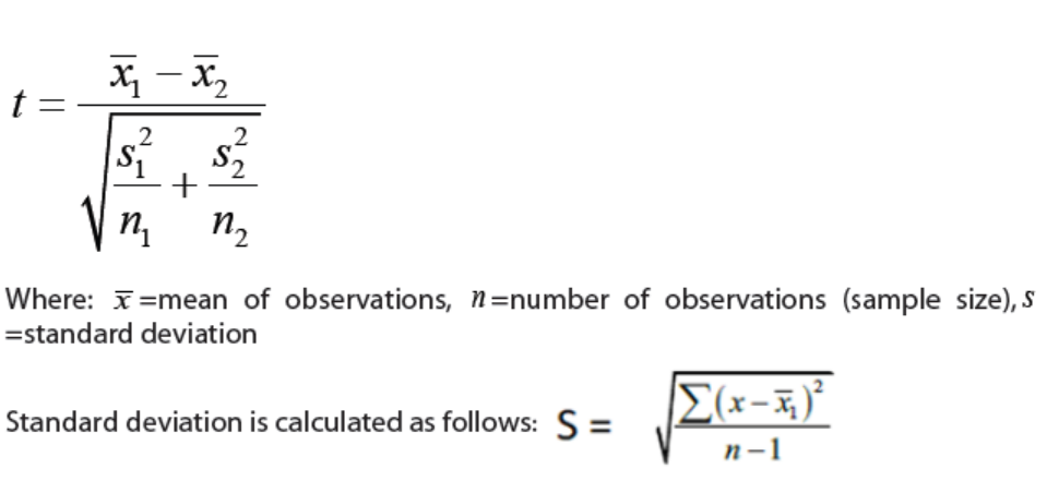
Where the suffixes 1 and 2 refer to samples 1 and 2 respectively
To take an example. A farmer wishes to decide which of two fertilizers gives the best
yield for her crop of wheat. She divides one of her fields into 16 plots, eight of which
she treats with fertilizer 1 and eight with fertilizer 2. The number of tons of wheat
obtained from each plot is given in this table
The first stage of t-test is to calculate the standard deviation for each sample. It is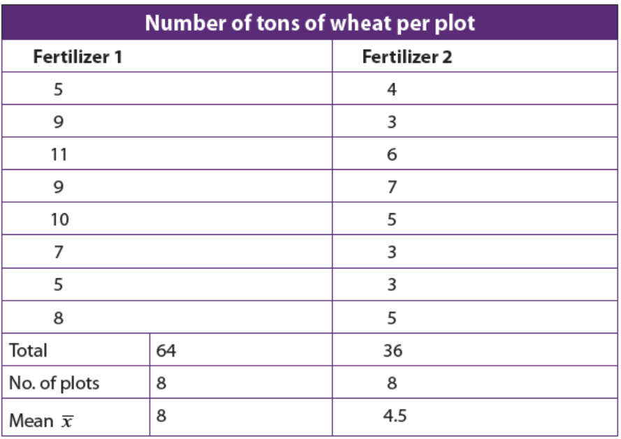
calculated from the mean of each sample (see table 15.3), the deviation of each
reading from the mean (see table 15.4) and the square of this deviation and the sumof squares (see table 15.4)
Table 15.4: The mean of each sample
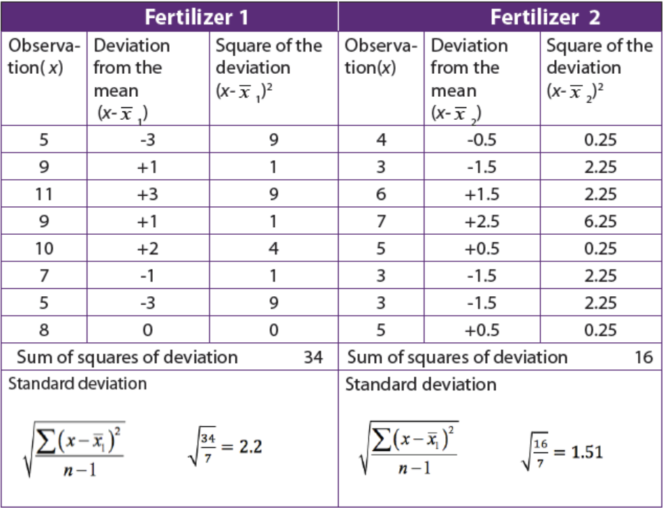
It is now substitute in the equation:
Finally, to discover whether value of 3.68 indicates whether the different readings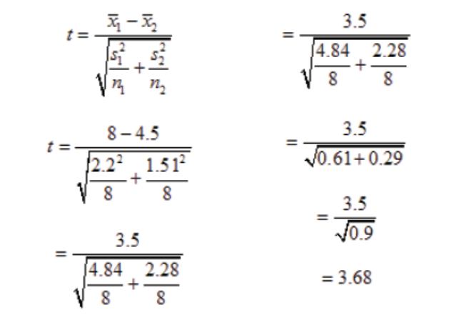
are significant, or merely due to chance, we need to look up 3.68 on a statistical table
called t-table. To do this we need to know the degrees of freedom. This is calculatedaccording to the formula:
Degrees of freedom (v) = (n1 + n2) – 2. In our example: v = (8+8) -2 =14
It is found that looking along the row for 14 degrees of freedom values of 3.68
lies between 2.98 and 4, 14, which corresponds to a probability value of between
0.01 and 0.001. This refers to the probability that chance alone is the reason for the
difference between the two sets of data. In this example, the probability that the
different wheat yields when using our two fertilizers was pure.
Application 15.4
1. Explain how to calculate the t- test.2. Why is t-test important in variation?
End unit assessment 15
Multiple choice questions
1. Which of the following gives rise to genetic variation in a population?
a. Crossing over and independent assortment in meiosis
b. Different environmental conditions
c. Random mating and fertilization
d. Mutation
Choose the best answer
a. 1, 2, 3 and 4
b. 1, 2 and 3 only
c. 1, 3 and 4 only
d. 2, 3 and 4 only
2. Inheritance variations could result
a. high energy radiation
b. geographical isolation
c. environmental factors
d. mutation
Questions with short answers
3. If a diploid organism has two different alleles for the same gene, is it
homozygous or heterozygous?
4. Is weight in human an example of continuous variation or discontinuous
variation?
5. What is a mutagen? Give one example.
Essay questions
6. Explain why variation caused by the environment cannot be passed from
an organism to its offspring.
7. Answer the following questions:
a. Distinguish between continuous and discontinuous variation.
b. Explain the genetic basis of continuous variation.8. The histogram shows the height of wheat plants in an experiment plot.
a. What evidence from the data suggests that there were two strains of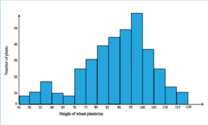
wheat growing in the experimental plot?
b. Based on the figure:
i. Which type of variation is shown by the height of each of the strains
of wheat plants? Give the reason for your answer
ii. Explain why the height of the wheat plants varies between 45 cmand 120 cm.
Practical work
9. Body Mass Index (BMI): Group work
Aim: identify one’s BMI and behave accordingly
Requirement: weighing scale, tap ruler, calculators
The BMI helps identify the weight status of individuals. The BMI is calculatedby:
The values of BMI are ranged into five categories as follow:
Record in a table weight and height of all students in your classroom and thereafter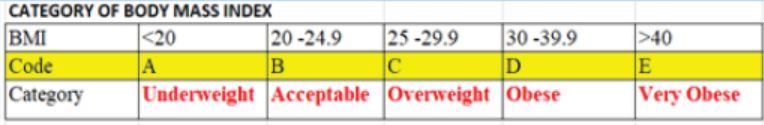
answer the following questions:
a. Calculate the BMI for all the classmates:
b. What health advice would you give to people in each BMI category?
c. Is the BMI genetic or phenotypic variation? Give reason.
d. Is BMI continuous or discontinuous variation? Give reason.
e. Considering your class members’ weight and height, calculate:
i. The standards deviation (S)
ii. The degree of freedom (Df )iii. The t-test (t)
UNIT 16 NATURAL AND ARTIFICIAL SELECTION
UNIT 16: NATURAL AND ARTIFICIAL SELECTION
Key Unit Competence
Explain the role of artificial and natural selection in the production of varieties of
animals and plants with increased economic importance
Learning objectives
By the end of this unit, I should be able to:
– Explain that natural selection occurs as populations have the capacity to
produce many offspring that compete for resources.
– Explain, with examples, how environmental factors can act as either stabilising,
disruptive and directional forces of natural selection.
– Explain how selection, the founder effect and genetic drift may affect allele
frequencies in populations.
– Explain how a change in allele frequency in a population can be used to
measure evolution.
– Describe how selective breeding (artificial selection) has been use to improve
the milk yield of dairy cattle.
– Outline the following examples of crop improvement by selective breeding:
– The introduction of disease resistant varieties of wheat, tomatoes, Irish
potatoes, and rice.
– Inbreeding and hybridization to produce vigorous, uniform varieties of maize
– Interpret graphs on how fur length affects the number of individuals at
different temperatures.
– Use the Hardy-Weinberg principle to calculate allele, genotype and phenotype
frequencies in populations.
– Differentiate between natural and artificial selection.
– Appreciate that the environment has considerable influence on the expression
of features that show continuous (or Quantitative) variation.
– Appreciate the importance of selective breeding (artificial selection) to
improve features in ornamental plants, crop plants, domesticated animals andlivestock.
Introductory activity
Some species of plants and animals such as Dinosaurs no longer exist today
but it is common to find new species such as lemon-orange. According to you,
what would be the cause of some species to disappear and some new speciesto appear?
16.1 Natural selection
Activity 16.1
1. Observe the graphs below, analyse and interpret them and then deducedifferent types of natural selection.
Key: Blue line indicates a given population after natural selection while Red
line indicates a given population before natural selection
16.1.1 Natural selection as a means of evolution as well as capacity
to survive and reproduce
Throughout the lives of the individuals, their genomes interact with their
environments to cause variations in traits from genotypic to phenotypic variations
among the individuals in a population because of differences in their genes.
Individuals with certain variants of the trait may survive and are capable to
reproduce more than less successful individuals with unfavourable characters;
therefore, the population evolves. Over time, this process can result in populations
that specialise for particular ecological niches (microevolution) and may eventually
result in speciation (the emergence of new species also known as macroevolution).
In other words, natural selection is a key process to change organisms and make
them suitable to different environment.
The variants that are best adapted to their natural environment such as abiotic
conditions, predation, competition to food, space, light, water and resistance
against diseases will be selected for survival and can reproduce. By reproduction,
organisms transmit their physical traits contained within their genes or alleles to
their next generation. The individuals that best suited or fitted to the stated before
environmental conditions will have the best chance to survive and produce fertile
offspring due to characteristic features or favourable characteristics that give
them an advantage in the struggle for existence being intraspecific or interspecific
competition. However, those with unfavourable characteristics are more likely to
die due to lack of resources or not having access to resources. The high or birth
rate gives a selective advantage whereas high mortality or death rate gives them aselective disadvantage.
As environmental conditions gradually change, certain characteristics within a
population also gradually change; thus, randomly varying population are favoured,
and natural selection occurs. This is known as the survival of the fittest. The fittest in
evolution is defined as the ability of an organism to pass on its alleles to subsequentgenerations, compared with other individuals of the same species.
16.1.2 Types of natural selection
As it has been mentioned, environment is a responsible agent of natural selection.
Thus, it selects and determines individuals in different ways according to different
types of natural selections. Those natural selections are stabilizing selection,
directional selection, and disruptive selection among other.
a. Stabilising selection
Stabilising selection is a type of natural selection in which a population mean
stabilises on a particular non-extreme trait value as result of genetic diversitydecreases as illustrated in the figure below.
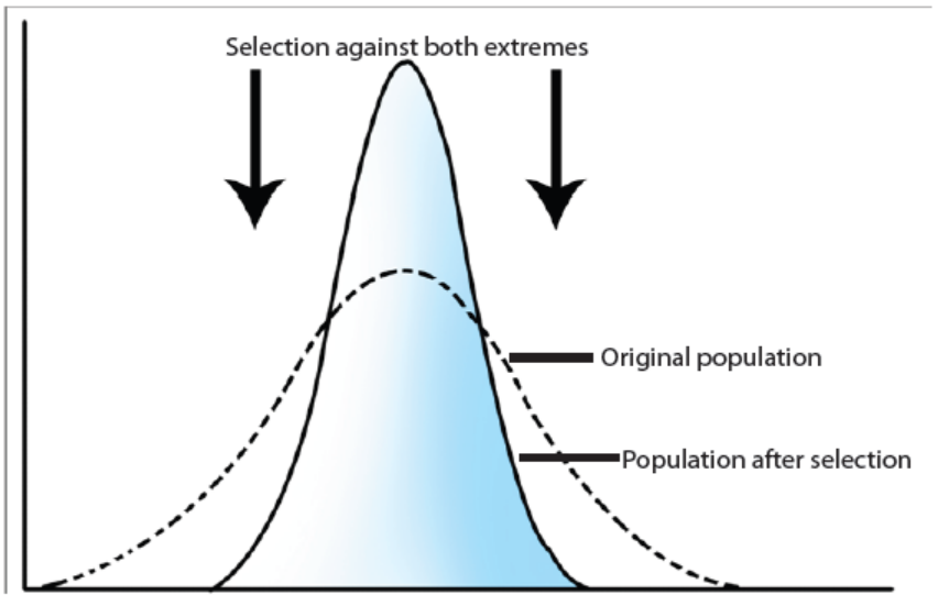
Figure 16.1.a: Illustration of stabilising selection
As illustrated in the above figure, in stabilizing selection, natural selection favours
the individuals in the population with the intermediate phenotypes. These
individuals have greater survival and reproductive success. Individuals with extreme
phenotypes are less adaptive and are therefore eliminated. An example is the newlyborn
human babies who are under 2.27 Kg or over 4.54 kg are less likely to survive
than babies weighing between 2.27 and 4.54 kg. Despite of this, with advances
in medical science, the survival chances of newly-born underweight or overweight
babies have now been improved.
These individuals with extreme phenotypes have greater survival and reproductive
success.
b. Directional selection
Directional selection is a mode of natural selection in which a single or new
fit phenotype is favoured when exposed to environmental changes, causing a
population genetic variance or allele frequency to continuously shift in one directionor one end of the spectrum of existing variation.
Figure 16.1.b: Illustration of directional selection
A classical description of directional selection has been identified in eighteenth and
nineteenth century in England as illustrated in the figure 16.1.b above. Prior to the
industrial revolution, the moths were predominately light in colour, which allowed
them to blend in with the light-coloured trees and lichens in their environment. As
soot/black powder began spewing from factories, the trees darkened and the lightcoloured
moths became easier for predatory birds to spot.
Over time, the frequency of the melanic form of the moth increased because their
darker coloration provided camouflage against the sooty tree; they had a higher
survival rate in habitats affected by air pollution. The result of this type of selection
is a shift in the population’s genetic variance towards the new and fit phenotype.
These individuals with extreme phenotypes have greater survival and reproductivesuccess.
c. Disruptive or diversifying selection
In disruptive selection, both the extreme phenotypes in the population are selected
and become more prevalent. The individuals with extreme phenotypes or endphenotypic
spectrum have greater survival and reproductive success. The disruptive
selection pressure increases the chances of the advantageous alleles to be passed
on to the next generation. By disruptive selection, the intermediate phenotype is
selected against and gradually decreases in number from generation to generation,and may become extinct.
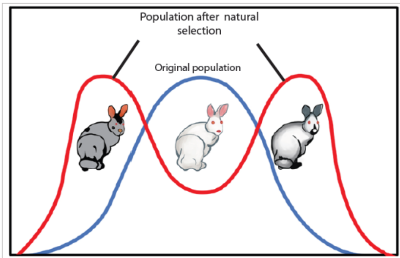
Figure 16.1.c: Illustration of disruptive selection
From the above figure, disruptive selection many generations may cause the
formation of two separate gene pools and the formation of new species.
Disruptive selection is mostly seen in many populations of animals that have
multiple male mating strategies such as; rabbits, mice, and lobsters among others
and is often the source of speciation or drives to speciation.
In rabbits as illustrated in the figure 16.1.c, a hypothetical population in which
grey and Himalayan (grey and white) rabbits are better able to blend with a rocky
environment than white rabbits. Large dominant alpha lobster males obtain mates
by brute force, while small males can sneak in for furtive copulations with the females
in an alpha male’s territory. In this case, both the alpha males and the sneaking males
will be selected for, but medium-sized males, which cannot overtake the alpha malesand are too big to sneak copulations, are selected against.
In scenario case of mice, those living at the beach where there is light-coloured sand
interspersed with patches of tall grass. Light-coloured mice that blend in with the
sand would be favoured, as well as dark-coloured mice that can hide in the grass.
Medium-coloured mice, on the other hand, would not blend in with either the grass
or the sand, thus, would more probably be eaten by predators. The result of this typeof selection, is increased genetic variance as the population becomes more diverse.
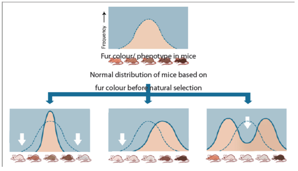
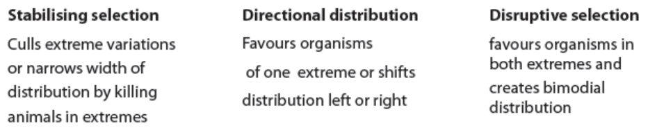
Figure: 16. 1.d: Comparison of three types of natural selection
The three types of natural selection are summarized in the figure 16.1.d above. It
shows populations of species which are selected by the environment particularlythe temperature on fur colour and those which decreases to extinction.
Application 16.1
1. Distinguish among the different of natural selection.
2. Describe what is meant by industrial melanism and how is beneficial to
peppered moth
3. Discuss how natural selection is one way of evolution and allows individualcan survive and reproduce
16.2 Artificial selection
Activity 16.2
From your daily experience and or carry out project work on plants (cabbage,
banana, wheat, maize, tomatoes, irish potatoes, and rice) and animals (cattle
and chicken) at your school or home. Do also research through internet and
textbooks and then answer to the questions below:
1. Discuss what is meant by artificial selection
2. Distinguish between inbreeding and outbreeding selection
3. Discuss how selective breeding or artificial selection has been used to
improve the yield or production of plant crops such as maize, wheat,tomatoes, and rice as well as milk and meat
Artificial selection is selective breeding that occurs when humans instead of
environmental forces select and determine the desirable alleles of plants or animals
to be passed on to successive generations. Artificial selection has been practiced
by humans for several centuries. It has played an important role in the evolution of
modern crop plants, farm animals and domestic pets from the wild ancestors. For
example, farming took place about 7000 years ago. The first crops humans selected
and domesticated include barley and wheat. By artificial selection, some scientists
argue that artificial selection and biotechnology can combine characteristics within
a short period of time that natural selection would require thousands or millions of
years to carry out.
It exerts/influences a directional selection pressure which leads to changes in the
frequencies of alleles and genotypes which have been selected by nature in thepopulation.
16.2.1 Advantages of artificial selection
Some of the advantages of artificial selection are:
– It is the quickest and more certain method of producing offspring for a
desirable characteristic.
– It selects and breeds animals and plants that can adapt and tolerate to live in
certain habitats or different environmental conditions such as heat, cold, day
length, and salinity or pH changes in the soil.
– It produces organisms that are resistant to pests, diseases or herbicides.
– It selects and breeds crop plant such as wheat, barley, rice, and maize plants
for high productivity or yield per unity area.
– It selects and breeds farm animals for better quality and quantity of milk, meat
production and wool quality.
– It leads to plants of fast germination seeds capacity, higher growth rate, early
maturation, better absorption of water, mineral salts or fertilizers. This allows
the planting of the same type of crop two or three times in one season and
therefore increases their production.
– Animals for sports or hobbies such as horses for racing and transport; pigeons
for flight capacity and plumage type; dogs as guardians or for hunting, racing
and appearance; orchids, roses and other flowers to produce more colourful
bloom; koi (a beautiful ornamental fish of striking colours-reds, golds, blues,
yellows, metallic silvers and even greens) fish for appearance from colouredmutants of common food carp are produced.
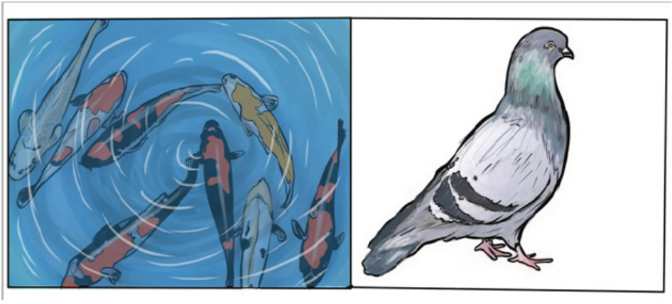
Figures 16.2 (a) Japanese Koi fish (b) Columbia livia of Europe of artificial breeding
16.2.2 Types of artificial selection
Inbreeding and outbreeding are the two distinguishable types of artificial selection.
a. Inbreeding
Inbreeding is the selective crossing between individuals that have a similar genotype
or are more closely related than if they had been chosen at random from the entire
population. Examples of inbreeding include; selfing in plants, mating between
offspring with one of the parents, among siblings or closely related individuals.
It has noticed that after several generations, the force of selection of inbreeding
increases the frequency of homozygous genotypes. Thus, the organism is probably
purebred, or homozygous for the selected characteristics. By inbreeding, organism
tends to maintain the desirable characteristics such as increase the quantity and
quality of milk by jersey cows (high cream content), produce maize plants and others
of uniform height to facilitate mechanical harvesting, increase oil content of linseed
oil to reduce cost of production and extraction, increase yields from plant crop and
livestock, use less land for farming or raising livestock but increase, breading of
horses for racing, and produce varieties of dogs for competition or as security guard
for example.
Even though, inbreeding is advantageous as described in above; it also presents
disadvantages that include:
– After several generations of excessive inbreeding, it results into inbreeding
depression. The inbreed progeny have decreased/loss vigor resulting from
excessive selective inbreeding between closely related organisms which
increases homozygosity (production of individuals with harmful or undesirable
phenotypic characteristics), poor growth and yield and decline in fertility than
non-inbred individuals.
– There is an increased risk of lowered diseases resistance as genetic variation is
reduced. Thus, inbreeding is not encouraged by animal breeders.
b. Outbreeding
Outbreeding is the controlled mating or crossing between distantly related
individuals (plants and animals) with desired characteristics e.g. the cross between
Elaeis guineensis (African oil palm or macaw-fat) variety dura with Elaeis guineensis
variety pisifera to produce the hybrid oil palm Elaeis guneesis variety tenera, with
fruits of high oil content and do not drop off easily. They may come from two breeds
of the same species or may come from different species. Outbreeding is more
advantageous than inbreeding because:
– The progeny also known as hybrids usually show more variation than
progeny produced by inbreeding. The hybrids usually have new and superior
phenotypes and have greater potential to adapt to environmental changes
for example wheat, tomatoes and rice produced by outbreeding are capable
to resist to diseases.
– Increases heterozygosity and new opportunities for gene interaction. Harmful
recessive alleles are masked by dominant alleles.
However, in some cases outbreeding results in hybrid vigour; healthier; or larger
offspring. And the hybrid produced between genetically different species are often
sterile. They do not have sets of homologous chromosomes and meiosis cannotproceed to produce fertile gametes.
Application 16.2
1. Explain how artificial selection is beneficial to man.2. Distinguish between inbreeding from outbreeding.
16.3 Allele frequency and its causes
Activity 16.3
Use available school resources such as internet, library, and teachers; search
information about allele frequency, selection, the founder effect and genetic
drift and or use pictures (a) and (b) given in question of this activity or use bean
seeds of different colour and play a game as instructed:
a. Take 15 bean seeds and then put all in one plastic bottle such as the one of
mineral water or power soap
b. Take other three empty bottles
c. Shake the bottle containing bean seeds and randomly distribute seeds into
the three bottles. Record and discuss the observations
d. Repeat events in step c) at least three times.
e. Draw the conclusion by linking the discussion to what they have read on
allele frequency, founder effect, and genetic drift
Then, do the following:
1. Discuss what is meant by allele frequency
2. Discuss how forces of mutation and natural selection affect the allelefrequencies
3. Analyse the figures below and then describe how the founder effectand genetic drift affect the allele frequencies in populations
16.3.1 Allele frequency in a population as determinant of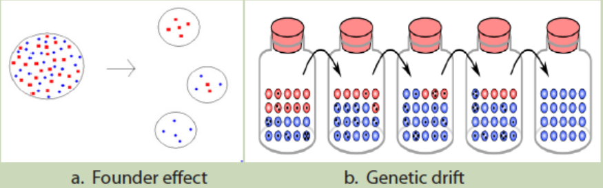
evolution
Genetic variation which confirms evolution is determined by; mutation, natural
selection, the founder effect, and genetic drift among others.
a. Mutation and natural selection
In a particular period, why do some organisms survive while others die? These
surviving organisms generally possess traits or characteristics that bestow / give
them traits or benefits of great value benefits that help them survive (e.g. better
camouflage, mating, faster swimming or running, or digesting food more efficiently)
as discussed before. Each of these characteristics is the result of a mutation or a
change in the genetic code.
Mutations occur spontaneously, but not all mutations are heritable; they are passed
down to offspring only if the mutations in the gametes. These heritable mutations
are responsible for the rise of new traits in a population. Populations or gene pools
evolve as gene frequencies change otherwise individual organisms cannot evolve.
Variation in populations is determined by the genes present in the population’s
gene pool as illustrated in figure below, which may be directly altered by mutation.
In natural selection, those individuals with superior traits will be able to compete
and get more resources as there are more organisms than resources and produce
more offspring. The more offspring an organism can produce, the higher its fitness.
As novel traits and behaviours arise from mutation, natural selection preserves thetraits that confer a benefit.

Figure 16.3a. Mutation and natural selection
As mutations create variation, natural selection gradually affects the frequency of
that advantageous trait in a population.
b. The founder effect
The founder effect occurs when part of a population becomes isolated and
establishes a separate gene pool with its own allele frequencies. When a small
number of individuals become the basis of a new population, this new population
can be very different genetically from the original population if the founders are
not representative of the original. Therefore, many different populations, with very
different and uniform gene pools, can all originate from the same, larger population.
Together, the forces of natural selection, genetic drift, and founder effect can lead tosignificant changes in the gene pool of a population.
Figure 16. 3b. three possible outcomes of the founder effect, each with gene pools separate from the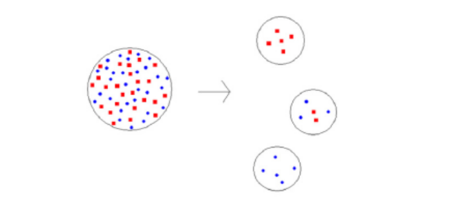
original populations
c. Genetic drift
Genetic drift is an overall shift of allele distribution in an isolated population,
due to random fluctuations in the frequencies of individual alleles of the genes.
When selective forces are absent or relatively weak, gene frequencies tend to drift
or change due to random events. This drift halts when the variation of the gene
becomes “fixed” by either disappearing from the population or replacing the other
variations completely. Even in the absence of selective forces, genetic drift can cause
two separate populations that began with the same genetic structure to drift apartinto the two divergent populations.
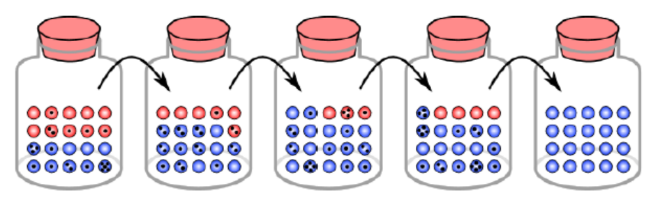
Figure 16.3c. Genetic drift and gene fixation in beetles
In the above simulation, there is fixation in the blue gene variation within five
generations. As the surviving population changes over time, some traits (red) may
be completely eliminated from the population, leaving only the beetles with othertraits (blue).
16.3.2 Allele frequency
Natural selection affects a gene pool by increasing the frequency of alleles that give
an advantage, and reducing the frequency of alleles that give a disadvantage. The
allele frequency (or gene frequency) is the rate at which a specific allele appears
within a population. In population genetics, the term evolution is defined as a
change in the frequency of an allele in a population. Frequencies range from 0,
present in no individuals, to 1, present in all individuals. The gene pool is the sum of
all the alleles at all genes in an interbreeding population.
A gene for a particular characteristic may have several variations called alleles. These
variations code for different traits associated with that characteristic. For example,
in the ABO blood type system in humans, three alleles (IA, IB, or i) determine the
particular blood-type protein on the surface of red blood cells. A human with a type
IA allele will display A-type proteins (antigens) on the surface of their red blood cells.
Individuals with the phenotype of type A blood have the genotype IAIA or IAi, type Bhave IBIB or IBi, type AB have IAIB, and type O have ii.
A diploid organism can only carry two alleles for a particular gene. In human blood
type, the combinations are composed of two alleles such as IAIA or IAIB. Although each
organism can only carry two alleles, more than those two alleles may be present
in the larger population. For example, in a population of fifty people where all the
blood types are represented, there may be IA alleles than i alleles. Population genetics
is the study of how selective forces change a population through changes in alleles
and genotypic frequencies.
Using the ABO blood type system as an example, the frequency of one of the alleles,
for example IA, is the number of copies of that allele divided by all the copies of the
ABO gene in the population, i.e. all the alleles. Allele frequencies can be expressed
as a decimal or as a percent and always add up to 1, or 100 percent, of the total
population. For example, in a sample population of humans, the frequency of the
IA allele might be 0.26, which would mean that 26% of the chromosomes in that
population carry the IA allele. If we also know that the frequency of the IB allele in
this population is 0.14, then the frequency of the i allele is 0.6, which we obtain
by subtracting all the known allele frequencies from 1(thus: 1-0.26-0.14=0.6). A
change in any of these allele frequencies over time would constitute evolution inthe population.
Application 16.3
1. What is allele frequency?
2. Explain how mutation and natural selection are important in gene
frequency?
3. In a situation where a trait is determined by two allele forms. What is
the frequency of each allele form?4. Using illustrations, explain genetic drift and founder effect.
16.4 Study of population genetic variation by Hardy-Weinbergprinciple
Activity 16.4
Use available school resources such as internet, library, search information
about Hardy-Weinberg principle, allele, genotype and phenotype as well as
allele frequency and then do the following:
1. What is Hardy-Weinberg principle
2. If the frequency of a recessive allele is 0.2. What is the frequency of a
dominant allele?
3. Cross one homozygous dominant individual of yellow colour with
one homozygous recessive pea plant of green colour. Calculate both
genotype, phenotype and allele frequencies by using Hardy-Weinbergprinciple if the recessive allele is equal to 0.4.
The Hardy-Weinberg principle is a mathematical baseline way used to estimate
the frequency of alleles, genotypes and phenotypes in a population. The principle
assumes that in a given population, the population is large and is not experiencing
mutation, migration, natural selection, or sexual selection.
The Hardy- Weinberg principle states that the frequency of alleles in a population
can be represented by P + Q = 1, with P equal to the frequency of the dominant
allele and Q equal to the frequency of the recessive allele.
The principle also states that the frequency of genotypes in a population is
represented by
p2 + 2pq + q2 = 1, with p2 equal to the frequency of homozygous dominant
genotype, pq equal to the frequency of the heterozygous genotype, and q2 equal
to the frequency of the Homozygous recessive genotype.
The frequency of alleles can be estimated by calculating the frequency of the
recessive genotype, then calculating the square root of that frequency in order todetermine the frequency of the recessive allele.
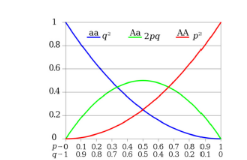
Figure 16.4a: Proportions of two alleles by Hardy-Weinberg principle
By referring to the above chart, by applying the expression of Hardy-Weinberg
principle, if the dominant allele is illustrated below as Y is equal to 0.7 while the
recessive allele noticed as y is equal to 0.3; then by using the Hardy-Weinberg
principle p2+2pq+q2=1, if the number of individuals is given as 500 and number
of alleles in a gene pool is 1000; genotypic and allelic frequencies are calculated asillustrated follow by Y2 + 2Yy + y2 = 1 and p + q = 1 respectively:
16.4.1 Hardy-Weinberg analysis
Figure 16.4b: Illustration showing analysis of Hardy-Weinberg principle and calculation of allele/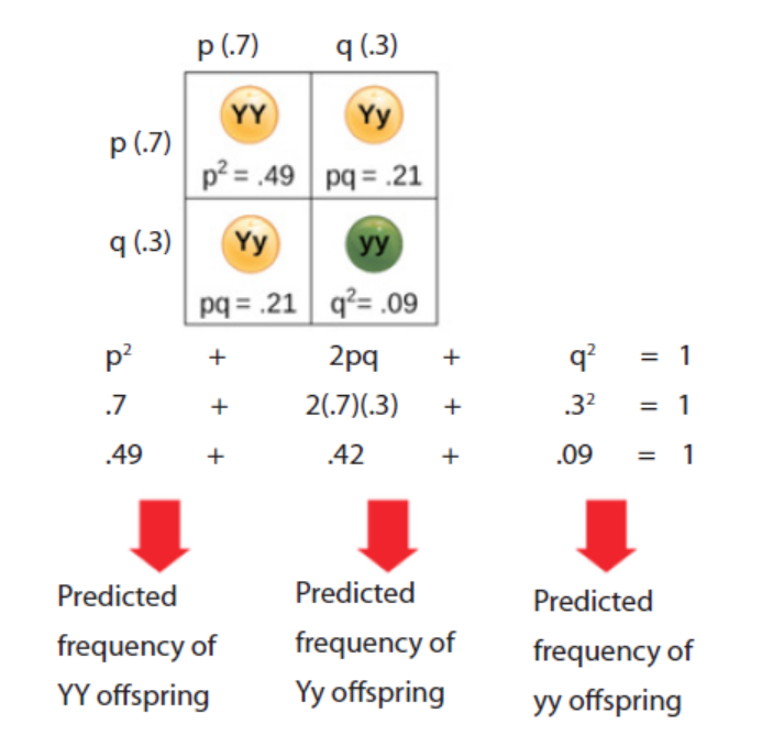
genotypes frequencies
The Hardy-Weinberg principle states that a population’s allele and genotype
frequencies will remain constant in the absence of evolutionary mechanisms.
Ultimately, the Hardy-Weinberg principle models a population without evolution
under the conditions such as; no mutations, no immigration/emigration, no natural
selection, no sexual selection and a large population. Although there is no realworld
population can satisfy all of these conditions, the principle stiff offers a useful
model for population analysis.
16.4.1 Hardy-Weinberg equations and analysis
According to the Hardy-Weinberg principle, the variable p often represents the
frequency of a particular allele, usually a dominant one. For example, assume that p
represents the frequency of the dominant allele, Y, for yellow pea pods. The variable
q represents the frequency of the recessive allele, y, for green pea pods. If p and q are
the only two possible alleles of this characteristic, then the sum of the frequencies
must add up to 1, or 100 percent. This can also be written as p+q=1, if the frequency
of the Y allele in the population is 0.6, then we know that the frequency of the y
allele is 0.4.
From the Hardy-Weinberg principle and the known allele frequencies, we can also
infer the frequencies of the genotypes. Since each individual carries two alleles per
gene (Y or y), we can predict the frequencies of these genotypes with chi square. If
two alleles are drawn at random from the gene pool, we can determine the possibilityof each genotype.
Application 16.4
1. Calculate the allelic, genotypic and phenotypic frequencies:
a. When a tall plant is crossed with a short one
b. When a heterozygous is crossed with another heterozygous
c. When heterozygous is crossed with a dominant homozygous. Note
that 0.2 is given as a value of recessive allele
2. Calculate the phenotype, genotype and allele frequencies of
populations/ hybrids obtained when the crossing is done between YYand Yy individuals. Note that the dominant allele is assumed to be 0.7.
End of unit assessment 16
1. Differentiate between natural selection from artificial selection
2. Some individuals of the swallowtail butterfly scientifically known as
Papilio machaon of the family papilionidae pupate on brown stems
or leaves; others pupate on green stems or leaves. Two distinct colour
forms of the pupae are found, namely brown and green, with very few
intermediates.
a. What type of natural selection does this example show?
b. Explain why the intermediate colour formed would be at selective
disadvantage.
3. Why are heavy-metal tolerant plants rare in unpolluted regions?
4. What effect did the industrial pollution have on the frequency of the C
(melanic) allele within a population of peppered moths.
5. Explain what is meant by heterozygous advantage, using the sickle-cellallele as an example.
UNIT 17 EVOLUTION AND SPECIATION
UNIT 17: EVOLUTION AND SPECIATION
Key Unit Competence
Analyze the relevance of theories of evolution and explain the process of speciation.
At the end of this unit, I should be able to:
– State the general theory of evolution that organisms have changed over time.
– Discuss the molecular evidence that reveals similarities between closely related
organisms with reference to mitochondrial DNA and protein sequence data.
– Explain the causes of present day evolution.
– Explain the role of pre-zygotic and post-zygotic isolating mechanisms in the
evolution of new species.
– Explain how speciation may occur as a result of geographical separation
(allopatric speciation), and ecological and behavioural separation (sympatric
speciation).
– Explain why organisms become extinct, with reference to climate change,
competition, habitat loss and killing by humans.
– Explain large-scale extinctions in earth’s history
– Observe and interpret mitochondrial, DNA and protein sequence data and
investigate the similarities of closely related organisms.
– Relate diagrams of Darwin’s finches to the mechanism of evolution.
– Research evidence for evolution.
– Acknowledge that over the years the theories of evolution have undergone
modifications as more evidence is collected.
– Appreciate that over prolonged periods of time, some species have remained
virtually unchanged, while others have changed significantly and many othershave become extinct.
Introductory activity
1. The coyote, jackal and dingo are closely related species of the dogfamily. Their distribution is shown on the map.
Suggest and explain how these three distinct species evolved from a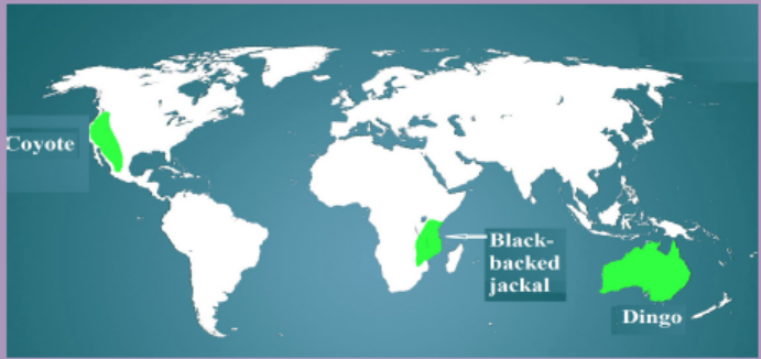
common ancestor.
2. Observe and analyse the pictures below. From your observation and
analysis, do you think there is relationship between individuals? If yes,
which ones? Is there any difference? If so, what does it cause or hascaused it?
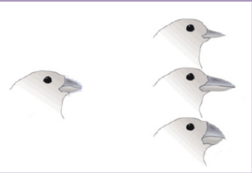
17.1 Theories of evolution
Activity 17.1
Use the school library and search additional information on the internet, read
the information related to evolution
1. Write a short note on the term evolution.2. Identify the importance of studying evolution
Evolution is the process by which new species are formed from pre-existing ones
over a period of time. It is not the only explanation of the origins of the many species
which exist on earth, but it is the one generally accepted by the scientific world
at the present time. Evolution is marked by emergence of new species from preexisting
species and the disappearance of some species. The species that disappear
are said to become extinct.
Studying evolution helps to understand the biological forces that cause organisms
to develop from simple to more complex organisms to the extent of new species
emerging. It also helps to know how different organisms relate to each other andone another.
The evolution is explained through different theories namely; Lamarckism,Darwinism, Neo-Darwinism, and Special creation.
1. Lamarckism/ Lamarckian inheritance theory
Lamarckism is briefly described as follows:
– An organism can pass on characteristics that it acquired to its offspring.
– Organisms evolve overtime due to the environmental factors that act up on
that organism. For example: A giraffe’s neck grows longer overtime becausethe giraffe’s desire for treetop leaves.
Figure 17.1: Lamarck’s giraffe: A giraffe’s neck grows longer overtime because the giraffe’s desire for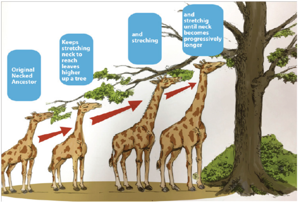
treetop leaves.
a. Assumptions of Lamarck’s theory
– Organisms tend to increase in size as they become more complex to a
predetermined limit.
– When influenced by the environment, body changes can be induced in
organisms.
– Organisms acquire new features because of need.
– Development of an organ and its effectiveness is promoted by its use whereas
its disuse brings about decline.
– Acquired features are inherited by future generations.
b. Merits/Advantages
– Lamarck was able to show that the environment influences the course of
evolution.
– He observed that features are passed down from parents to their offspring.
– He was able to recognize that as organism increase in size, they become more
complex to a predetermined limit. (Predetermine: to determine or decide in
advance)
c. Demerits /disadvantages
– Acquired changes are not heritable as they are influenced by genes.
– Somatic changes are not heritable as they are not passed through reproduction.
– The process of gametogenesis is not related to occupation or their activity.
– Use or disuse of somatic cells does not affect gamete formation.
2. Darwinism/Theory of natural selection
The term Darwinism has been applied to the evolutionary theories of Charles
Darwin (1809-1882). Darwin’s theory of natural selection is important landmark in
the evolutionary process and the origin of species. Darwin’s theory of evolution had
a great impact because it was supported by a wealth of evidence.
According to Darwin’s theory:
– Each species living today arose from a pre-existing species.
– All species have evolved from one ancestral type.
– Natural selection provides the mechanism for one species to change into
another. The main evidence for his first suggestion, which has been called
descent with modification, comes from fossils.
Essential features of Darwin’s theory of natural selection
Charles Darwin conducted extensive research on plants and animals in order to
study the process of evolution. The essential features of the theory Darwin putforward are:
– Overproduction of offspring: All organisms produce large numbers of
offspring which, if they survived, would lead to a geometric increase in the
size of any population
– Constancy of numbers: Despite the tendency to increase numbers due to
overproduction of offspring, most populations actually maintain relatively
constant numbers.
– Struggle for existence: Darwin deduced on the basis of 1 and 2 that members
of the species were constantly competing with each other in an effort to
survive. In this struggle for existence only a few would live long enough to
breed
– Variation among offspring: The sexually produced offspring of any species
show individual variations, so that generally no two offspring are identical.
– Survival of the fittest by natural selection: Among the offspring there will
be some better able to withstand the prevailing conditions. That is, some will
be better adapted (fitter) to survive in the struggle for existence. These types
are more likely to survive long enough to breed.
– Like produces like: Those that survive to breed are likely to produce offspring
similar to themselves. The advantageous characteristics that gave them
the edge in the struggle for existence are likely to be passed on to the next
generation.
– Formation of new species: Over many generations, the individuals with
favorable characteristics will breed, with consequent increase in their numbers.
The development of a number of variations in a particular direction over many
generations will gradually lead to the evolution of a new species.
Darwin’s theory was based on three main observations:
– Within a population are organisms with varying characteristics, and these
variations are inherited (at least in part) by their offspring.
– Organisms produce more offspring than are required to replace their parents
– On average, population numbers remain relatively constant and no population
gets bigger indefinitely.
From these observations, Darwin came to the conclusion that within a population
many individuals do not survive, or fail to reproduce. In his study of birds, he found
that after arriving at the islands, the Finches were dispersed in varied environmental
conditions. In due course of time, the anatomy of birds was modified naturally as anadaptation to the prevailing conditions especially food regimes.
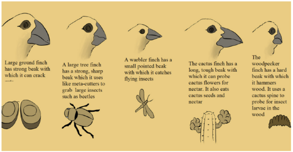
Figure 17.2: Five of Darwin’s finches
• Assumptions of Darwinism
– Most organisms have the potential to produce large number of offspring or
progeny than the environment can support. This leads to still competition as
the numbers of organisms are fairly stable.
– All organisms, even of the same species vary in a few characteristics,
– Only those organisms of a given species with variations that adapt them to the
environment, survive the competition and live. There is survival for the fittest
by natural selection.
– The features favored/selected by nature survive and are inherited. Therefore,
new species may develop by natural selection, which is one of the forces of
evolution.
• Merits of Darwin’s theory of natural selection
– Species always change as the environment changes.
– Species are compared with their ancestors due to presence of similarities in
characteristics.
– Enough data are / can be collected for explaining variation in a population
that may result into formation of a new species.
• Demerits of Darwin’s theory of natural selection
– Not all variations inherited, except for only genetic variations.
– It provides inadequate explanation of existence of many vestigial structures in
organisms.
– Explanation on deleterious mutations that are retained in a population is notadequate.
3. Neo-Darwinism
The modern theory of evolution is called Neo-Darwinism (neo= new) because it
incorporates new scientific evidence, particularly from genetics and molecular
biology. For example, we now know that the variations that are so important in
natural selection come about by random and spontaneous changes in genes,
particularly from mutations in reproductive cells. According to neo-Darwinism,
nature selects those individuals with beneficial mutations and allows them to be
passed to their offspring through reproduction from generation to generation. The
mutations are transmitted within the population and if selected by nature, they may
form a new species.
4. Special creation
It is believed that a special being, God created the universe and all living organisms.
In this theory, heavens and earth were first created. Light, day and night were created
next and subsequently, all living things with human beings the last in the creation. Itshows that there was direct creation of organism with no precursor to life.
Application 17.1
1. Give the biological meaning of evolution
2. How does neo- Darwinism differ from Darwin’s original theory ofevolution?
17.2 Evidence of evolution
Activity 17.2
Use the school library and internet to search and read the information related
to evidence of evolution with particular emphasis on molecular evidence.
Make a table showing that the molecular evidence reveals similarities between
closely related organisms with reference to mitochondrial DNA and proteinsequence data
17.2.1 Palaeontology: the study of fossil
A fossil is the remains of an organism that lived in the past, preserved by a natural
process (for example, in rock, peat, or ice). Fossils include; bones, shells, footprints,
and faeces. Most of fossils are found in sedimentary rocks formed by layers of silt.
Rocks and their fossils can be dated approximately on the basis of how long it takes
for sedimentary rocks to be laid down. However, these estimates are very rough.
More accurate estimates come from measuring the radioactivity of crystals ofigneous rock in the strata.
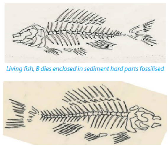
Living fish, A dies enclosed in sediment hard parts fossilised
Figure 17.3: Fossil formation. Fish B becomes a fossil much later than fish A. The deeper the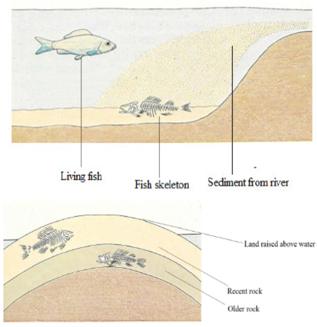
rock layer, the older the fossil.
The level of radioactivity is greatest when the crystals first form. As they age, the
isotopes decay: uranium to lead, and potassium to argon. The older the rock, the less
original radioactive material remains. Fossils can therefore be detailed by analyzing
the amounts of uranium and lead, or potassium and argon, they contain. Potassiumargon
dating is often used to date fossils because potassium is a common element
found in many types of rock, and it decays to argon very slowly. This allows rocks up
to 3000 million years old to be dated. Sometimes younger fossils can be dated byradioactive carbon dating.
17.2.2 Comparative biochemistry and cell biology
The most persuasive evidence that all organisms have evolved from a common
ancestor comes from studies comparing the cell biology and biochemistry of
different organisms, which reveal that:
– The genetic code contained within nucleic acids is almost universal
– Physiological processes vital to all organisms, such as respiration, follow very
similar metabolic pathways.
– ATP is the universal energy currency
The cellular and biochemical details of organisms are quite similar, but any
differences can give an idea of how closely different species are related. Species
that are closely related would be expected to differ only slightly from each other.
Detailed comparisons of DNA, metabolic pathways, key proteins, and organelles
such as ribosomes have been used to work out the evolutionary relationships of
organisms. For example, ribosomes inside mitochondria and chloroplast are similar
to those in bacteria, suggesting that these organelles may have evolved from
bacteria. Mammalian blood proteins can be tested to see how similar they are to
human blood proteins: blood serum from the mammal in question is added to
rabbit serum containing anti-human antibodies
17.2.3 Comparative embryology
Observations have shown that species that are known to be closely related show a
similar embryonic development. Therefore, species that show a similar embryonic
development are assumed to be closely related, even if the adult stages are very
different. For example, echinoderms (the phylum containing starfish and sea urchins)
are believed to be related to chordates (the phylum including vertebrates) becauseof similarities in their early embryonic development.
Figure 17.4: Comparison of embryos from different vertebrates: Although the adults are quite different,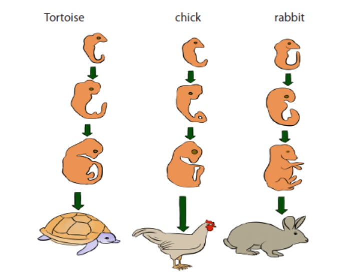
the early embryonic stages are similar
17.2. 4 Comparative anatomy
Comparative anatomy is the study of biological structures in different organisms.
The scientists look at structures that are similar in different organisms or species.
Example: limbs of vertebrates such as human beings, goats and wings of birds are
used for different purposes but they have a basic design structure, this is known as
homologous structure. The forelimbs of humans are for manipulation, fore limbs of
birds (wings) are for flight and fore limbs of a goat are for walking; this shows that
all these animals are from common ancestors. Analogous structures are the ones,
which look different, but they perform similar functions e.g. insect, birds and bats allhave wings used for flight but they have different structural organization.
Figure 17.5: The forelimbs of the following vertebrates show the basic pattern of limb bones with modifications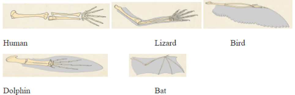
which are adapted to their methods of locomotion.
17.2.5 DNA evidence
Another important line of evidence for evolution comes from DNA analysis. Any
permanent change in form or function of an organism must be preceded by a
change in its DNA. Organisms which have much of their DNA in common must be
closely related, i.e. they have split from a common ancestor comparatively recently
(in geological terms). For example, humans and chimpanzees have 99% of their DNA
in common which suggests a close relationship and relatively ‘recent’ divergencefrom a common ancestor.
Application 17.2
1. By what process do:
a. Analogous structures evolve so that they look alike?
b. Two related but geographically separate groups evolve similar
adaptations independently?
3. Give two pieces of evidence from comparative biochemistry that
support the theory that all species living today are descended from acommon ancestor
17.3 Causes of evolution
Activity 17.3
Use the school library and internet to search and read the information related
to the causes of evolution. Make a list of different causes of evolution and writeshort summary in your own words on the meaning of each cause.
17.3.1 Competition changes in the environment
Imagine that we are plunged into a new Ice Age. The climate becomes much colder,
so that snow covers the ground for almost all of the year. Assuming that rabbits can
cope with these conditions, white rabbits now have a selective advantage during
seasons when snow lies on the ground, as they are better camouflaged (like the
hare in figure 17.6). Rabbits with white fur are more likely to survive and reproduce,
passing on their alleles for white fur to their offspring. The frequency of the allele for
white coat increases at the expense of the allele for agouti. Over many generations,almost all rabbits will come to have white coats rather than agouti.
Figure 17.6: The white winter coat of a mountain hare provides excellent camouflage from predators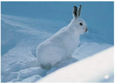
when viewed against snow.
17.3. 2 Mutations
Because they are random events, most mutations that occur produce features that
are harmful. That is, they produce organisms that are less well adapted to their
environment than ‘normal’ organisms. Other mutations may be neutral, conferring
neither an advantage nor a disadvantage on the organisms within which they occur.
Occasionally, mutations may produce useful features. Imagine that a mutation
occurs in the coat colour gene of a rabbit, producing a new allele which gives a
better camouflaged coat colour than agouti. Rabbits possessing this new allele
will have a selective advantage. They will be more likely to survive and reproduce
than agouti rabbits, so the new allele will become more common in the population.
Over many generations, almost all rabbits will come to have the new allele. Such
changes in allele frequency in a population are the basis of evolution. Evolution
occurs because natural selection gives some alleles a better chance of survival
than others. Over many generations, populations may gradually change, becomingbetter adapted to their environments.
17.3.3 Effect of drugs or chemical resistance
Antibiotic resistance is a severe problem throughout the world. For example, some
strains of the common bacterium Staphylococcus aureus are resistant to antibiotics
such as penicillin and methicillin. Penicillin resistance has probably evolved in thefollowing way:
– By chance, a mutation produces an individual bacterium with an allele that
allows it to produce an enzyme, penicillinase, which deactivates penicillin
– This bacterium is immediately resistant to penicillin. (As bacteria have only
one strand of DNA and one copy of each gene, the mutant allele is expressed
immediately and is not masked by a dominant allele.)
– If the population to which the mutant belongs is exposed to penicillin, the
mutant will survive and reproduce whereas those without the mutant will bekilled.
17.3. 4 Industrialization
Many species of organisms, especially insect species, have two or more adult body
forms that are genetically distinct from one another, but which are contained within
the same interbreeding population. This condition is known as polymorphism
(another type of natural selection). The peppered moth (Biston betularia), for
example, has two main forms with different wing colours. One form has pale wings
with dark markings; the other form is called melanic because the wings contain
large amounts of melanin (a black pigment), so they are almost black.
17.3. 5 Gene recombination
Despite these efforts there are still some copying errors and accidental damage,
permanent changes, or mutations. These may be responsible for thousands of
inherited diseases, and mutations that appear in cells throughout the lifetime of
an individual. These may lead to many types of cancer. DNA repair thus becomes
important to prevent mutations and inherited diseases.
17.3.6 DNA Recombination
DNA sequences in cells thus are maintained from generation to generation with
very little change. While this is true, there is evidence that the DNA sequence in
chromosomes does change with time and the DNA gets rearranged over time.
The combination of the genes on the genome may change due to such DNA
rearrangements. In a population, this sort of genetic variation is important to
allow organisms to evolve in response to a changing environment. These DNA
rearrangements are caused by a class of mechanisms called genetic recombination.
a. Homologous DNA recombination
The most important form of genetic recombination is homologous recombination.
The process involves the basic facts such as two double double-stranded DNA
molecules that have regions of very similar (homologous) DNA sequence come
together so that their homologous sequences are in tandem. Then they can “crossover”:
in a complex reaction, both strands of each double helix are broken and the
broken ends are re-joined to the ends of the opposite DNA molecule to re-form two
intact double helices, each made up of parts of the two different DNA molecules.
b. Non homologous DNA recombination
In homologous recombination, DNA rearrangements occur between DNA segments
that are very similar in sequence. A second, more specialized type of recombination,
called site-specific recombination, allows DNA exchanges to occur between DNA
double helices that are dissimilar in nucleotide sequence.
17.3.7 Artificial selection
Over the years, humans have used artificial selection to create crazy specific dog
breeds
Over the past 150 years or so, humans have been specifically mating dogs that look
a certain way to create the animals we now keep as pests via a process known as
breeding. This is artificial selection, where one species (humans) directs the traitsthat get passed down to future generations of another species (dogs).
Application 17.3
Write short summary on industrialization and gene recombination as causes
of evolution.17.4 Speciation
Activity 17. 4
Use the school library and internet and read the information related to
speciation.
1. How does speciation occur?2. How does one species evolve into two or more new species?
Evolution occurs whenever the inherited characteristics of a population or of a
species change over a period of time. When these changes lead to the formation of
one or more new species, speciation has taken place. A species can be defined as
a group of organisms with similar features which can interbreed to produce fertile
offspring, and which are reproductively isolated from other species. The central part
of this and most other definitions of species is that members of the same species
can interbreed to produce fertile offspring. Thus, although donkeys can interbreed
with horses to produce offspring called mules, donkeys and horses are regarded as
separate species because mules are infertile.
Organisms which do not interbreed to produce fertile offspring under normal
circumstances are regarded as reproductively isolated, and they belong to separate
species. Mechanisms that prevent breeding between populations and which can
eventually lead to speciation are called isolating mechanisms. Mechanisms that
prevent the formation of hybrids are called prezygotic isolating mechanisms,Prezygotic (before a zygote is formed) isolating. Mechanisms include:
– Individuals not recognising one another as potential mates or not responding
to mating behaviour
– Animals being physically unable to mate
– Incompatibility of pollen and stigma in plants
– Inability of a male gamete to fuse with a female gamete.
The mechanisms that affect the ability of hybrids to produce fertile offspring are
called postzygotic isolating mechanisms. Postzygotic isolating mechanisms include:
– Failure of cell division in the zygote
– Non-viable offspring (offspring that soon die)
– Viable, but sterile offspring.
The most important isolating mechanism is thought to be geographical isolation, in
which two populations originally of the same species are separated from each other
by a physical barrier such as a mountain, river, or ocean.
Allopatric speciation
When geographical isolation leads to new species being formed, allopatric
speciation is said to have occurred. (Allopatric means literally ‘different countries’.
Any physical barrier that prevents members of different populations from meeting
must inevitably prevent them from interbreeding. Note that although geographical
isolation is the original cause of allopatric speciation, the two isolated populations
diverge so much from each other that when reunited they are unable to interbreed.Other isolating mechanisms now keep the two species from breeding together.
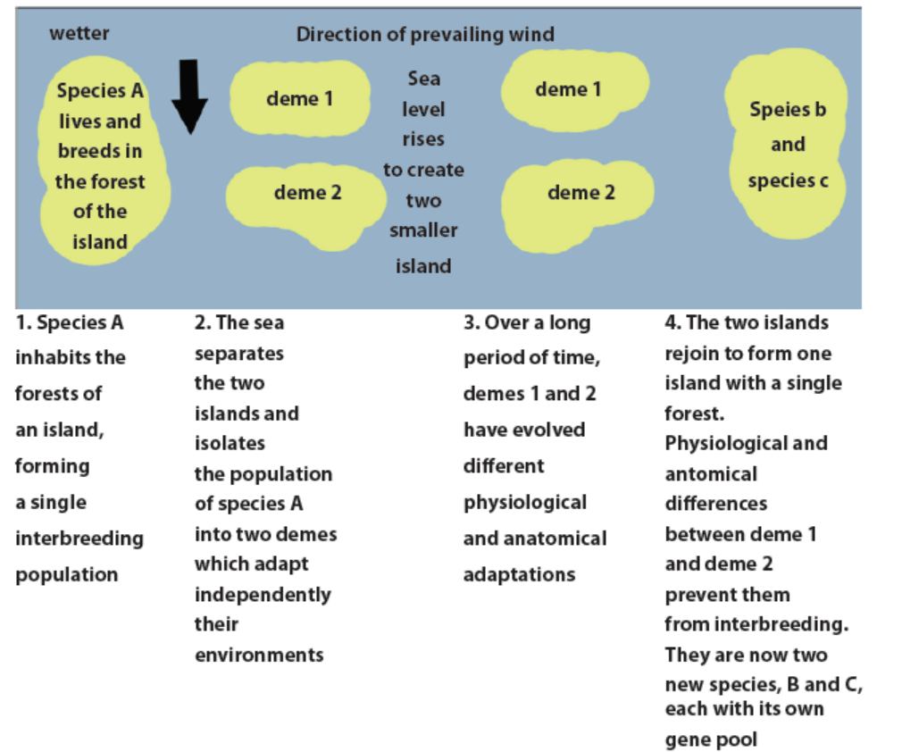
Figure 17.10: A hypothetical example of allopatric speciation
Sympatric speciation
Sympatric literally means. (‘Same country’.) Sympatric speciation occurs when
organisms inhabiting the same area become reproductively isolated into two groupsfor reasons other than geographical barriers. Such reasons might include:
1. The genitalia of two groups may be incompatible (mechanical isolation): It
may be physically impossible for the penis of a male mammal to enter the
female’s vagina
2. The gametes may be prevented from meeting: In animals, the sperm may
not survive in the female’s reproductive tract or, in plants; the pollen tube
may fail to grow.
3. Fusion of the gametes may not take place: Despite the sperm reaching
the ovum, or the pollen tube entering the micropyle, the gametes may be
incompatible and so will not fuse.
4. Development of the embryo may not occur (hybrid inevitability): Despite
fertilization taking place, further development may not occur, or fatal
abnormalities may arise during early growth
5. Polyploidy (hybrid sterility): When individuals of different species breed,
the sets of chromosomes from each parent are obviously different. These
sets are unable to pair up during meiosis and so the offspring cannot
produce
6. Behavioral isolation: Before copulation can take place, many animals
undergo elaborate courtship behavior. This behavior is often stimulated
by the colour and markings on the members of the opposite sex, the callof a mate or particular actions of a partner.
Application 17.4Distinguish between allopatric and sympatric speciation.
17.5 Roles natural selection in speciation
Activity 17.5
Use the school library and internet, read the information related to the roles of
natural selection in speciation.
In your own words, write a short summary on the roles of each type of naturalselection in speciation.
The role of natural selection in evolution
Natural selection leads to evolutionary change when individuals with certain
characteristics have a greater survival or reproductive rate than other individuals in
a population and pass on these inheritable genetic characteristics to their offspring.
Simply put, natural selection is a consistent difference in survival and reproduction
between different genotypes, or even different genes, in what we could callreproductive success.
The reason that natural selection is important is that it›s the central idea, stemming
from Charles Darwin and Alfred Russel Wallace that explains design in nature. It is
the one process that is responsible for the evolution of adaptations of organisms
to their environment. Three essential components of evolution via natural selectioninclude:
1. Genetic Diversity – Populations of individuals are genetically diverse. Even
members of the same species have characteristics that vary from one
individual to the next.
2. Fitness – In any given environment, some individuals have characteristics
that put them at an advantage over individuals who do not possess those
same characteristics.
3. Population Shift – In any given environment, those individuals who have
advantageous characteristics will generally be healthier, live longer,
and leave more offspring than individuals who do not possess those
characteristics. The population will, over time, contain more and more
individuals with the advantageous characteristic, and fewer individualswho do not possess the characteristic.
Application 17.5
1. Some individuals of the Rwandan swallowtail butterfly (Papillio
machaon) pupate on brown stems or leaves; others pupate on green
stems or leaves. Two distinct colour forms of the pupae are found,
namely brown and green, with very few intermediates. Explain why the
intermediate colour forms would be at a selective disadvantage.2. What is the role of natural selection in evolution?
17.6 Mechanism of speciation
Activity 17.6
Use the school library and search additional information on the internet, read
the information related to mechanism of speciation. Write a short report ondifferent mechanisms of speciation
a. Continental drift
The continents which now exist have not always appeared as they do today. At one
time, the earth had a single large land mass called Pangaea. This is thought to have
broken up into two parts, a northern Laurasia and a southern Gondwanaland. Over
millions of years, the two great land masses split up and moved by a process called
continental drift to form our present continents. The theory that these land masses
were once joined is supported by the discovery in Australia, South Africa, South
America, and Antarctica of fossils belonging to the same extinct species. Fossils in
North and South America show differences between the species, suggesting that
these two continents have only joined together relatively recently. Before this,
their fauna (animals) and flora (plants) were geographically isolated and evolved
independently.
Australia shows many excellent examples of species that evolved independently
following its geographical isolation. It is thought that Australia became isolated
about 120 million years ago, when marsupials (mammals without a placenta but
with a pouch in which the young develop) and eutherian mammals (mammals witha true placenta) diverged from a common ancestor
b. Migration
Migration also called gene flow is any movement of individuals, and/or the genetic
material they carry, from one population to another. Gene flow includes lots of
different kinds of events, such as pollen being blown to a new destination or people
moving to new cities or countries. If gene versions are carried to a population where
those gene versions previously did not exist, gene flow can be a very important
source of genetic variation. In the graphic below, the gene version for browncoloration moves from one population to another.
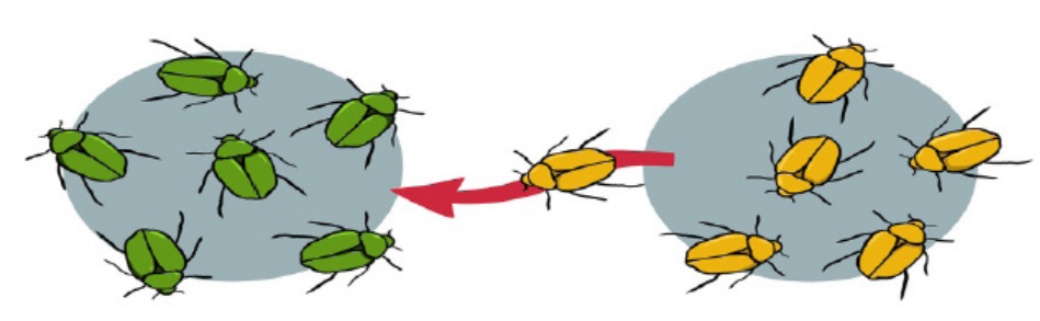
Figure 17.15: Illustration of migration
17.7 Divergent evolution
A single species evolves into several new species that live in different ways.
The five of Darwin’s finches are a good example. There are separate species of
finch in the group, all of which probably evolved from individuals belonging to
one mainland species. The islands have few other bird species. In the absence of
competition, the finches became adapted to fill all the available niches. In particular,
they evolved a wide range of beak sizes and shapes so that they could take advantage
of the food sources on the different islands. The evolution of an ancestral speciesinto different species to fill different niches is called adaptive radiation
17.8 Convergent evolution
Unrelated species independently evolve similarities when adapting to similarenvironments
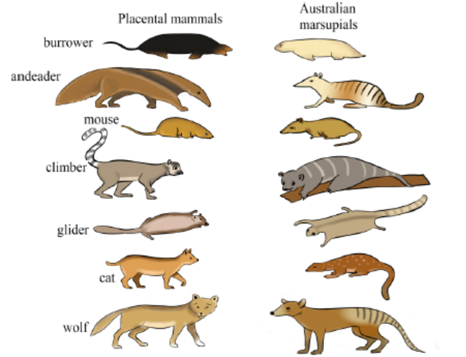
Figure 17.16: Convergent evolution
17.1: Table isolating mechanisms
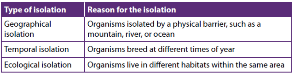
17.9 Extinctions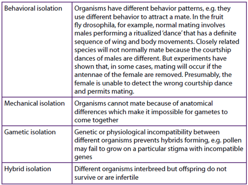
Extinct means that a species that has died out
17.9.1 Causes of Extinction
The single biggest cause of extinction today is habitat loss. Other causes of extinctiontoday include:
– Exotic species introduced by humans into new habitats. They may carry
disease, prey on native species, and disrupt food webs. Often, they can outcompete
native species because they lack local predators.
– Over-harvesting of fish, trees, and other organisms. This threatens their survival
and the survival of species that depend on them.
– Global climate change, largely due to the burning of fossil fuels. This is raising
Earth’s air and ocean temperatures. It is also rising sea levels. These changes
threaten many species.
– Pollution, which adds chemicals, heat, and noise to the environment beyond
its capacity to absorb them. This causes widespread harm to organisms.
– Human overpopulation, which is crowding out other species. It also makes allthe other causes of extinction worse.
17.9.2 Large-scale extinctions in earth’s history
– During the late Precambrian, continents drifted, carbon dioxide levels
fluctuated, and climates changed. Many organisms could not survive the
changes and died out. Others evolved important new adaptations. These
include sexual reproduction, cell specialization, and multi cellularity. The
Precambrian ended with a mass extinction. It paved the way for the Cambrian
explosion.
– The Paleozoic Era began with the Cambrian explosion. It ended with the
Permian extinction. During the era, invertebrate animals diversified in the
oceans. Plants, amphibians, and reptiles also moved to the land.
– The Mesozoic Era is the age of dinosaurs. They evolved from earlier reptiles
to fill niches on land, in the water, and in the air. Mammals also evolved but
were small in size. Flowering plants appeared for the first time. Dinosaurs went
extinct at the end of the Mesozoic.
– The Cenozoic Era is the age of mammals. They evolved to fill virtually all the
niches vacated by dinosaurs. The ice ages of the Quaternary Period of the
Cenozoic led to many extinctions. The last ice age ended 12,000 years ago. Bythat time, Homo sapiens had evolved.
Application 17.6
1. Describe the mechanism of continental drift.
2. Briefly explain why two types of organism may be regarded as separate
species even though they can interbreed to produce fertile offspring
3. Describe the Rwanda policies to overcome the extinction of somespecies
End unit assessment 17
Multiple choice questions
1. A species of finch living on an isolated island shows variation in beak size.
Birds with larger beaks can eat larger seeds. After a period of drought on the
island, large seeds were more plentiful than small seeds and the average
size of the finches’ beaks increased. What explains this increase in size of
beak?
a. Artificial selection acting against finches with small beaks
b. Directional selection acting against finches with small beaks
c. Increased rate of mutation resulting in finches with larger beaks
d. Stabilizing selection acting against finches with the smallest andlargest beaks
2. Which effect of natural selection is likely to lead to speciation?
a. Differences between populations are increased.
b. The range of genetic variation is reduced.
c. The range of phenotypic variation is reduced.d. Favourable alleles are maintained in the population.
Questions with short answers
3. Name two examples of adaptive radiation.
4. What effect did industrial pollution have on:
a. The frequency of the C (melanic) allele within a population of
peppered moths.
b. The rate of mutation of the c allele to the C allele
5. Explain what is meant by heterozygous advantage, using the sickle-cell
allele as an example.
6. Answer the following questions:
a. Distinguish between homologous structures and analogous
structures with specific examples.
b. Name the type of evolution exhibited by comparing:
(i) Flipper of whale and forelimb of desert rat.
(ii) Wing of a bat and wing of butterfly
(iii) Wing of a flamingo and wing of an insect
Essay questions
7. Explain the various evidences of organic evolution.
8. Explain Darwin’s theory of natural selection.
9. What do you understand by Lamarckism? How does it differ from Darwinism?
10. How can you convince that evolution is still in progress?
11. A Darwin and Lamarck contribution to science is unparalleled. Discuss.
12. Explain the importance of modern genetics to the theory of origin ofspecies by natural selection
REFERENCES
Arms, K., Camp, P. S., Jenner, J. V., and Zalisko, E. J. (1994). Biology: A Journey Into Life.
Florida Saunders College publishing, Florida, USA.
Barbara, A., Jean, B, Niamh, G., Douglas W. (2009). Biology, CK-12 Foundation,
California, USA.
Benjamin Cummings. 8th Ed. San Francisco, US. Pp. 142-148.
Campbell, N.A., Reece, J.B., Urry, A.L., Cain, L.M, Wasserman, A.S., Minorksy, V.P and
Jackson, B.R. (2008). Biology. 8th edition. Pearson international, San Francisco, USA.
Fullick, A. (2006). GCSE Biology. AQA Science Exclusively endorsed and approved by
AQA. Nelson Thornes Ltd. Cheltenham
Fullick, A., Mwinshekhe, H., Maddu, M., and Nyabua, S. (2011). A level Biology.
Malaysia Ashton Editorial and Juice Creative, Ashton, Malasia.
Kent, M. (2000). Advanced biology. Anew mainstream text for the new specifications.
Oxford university press, New York, USA.
Kinyua, S., and Mungai, M. (2013). Top Mark Revision Biology. Kenya literature bureau.
Nairobi, Kenya.
L.Hopson, J. H. (2006). Modern Biology . London : A Harcourt Education Company .
Lee Ching (2004). Biology. Vol.11, Pearson Longman, pre- U text STPM. Selangor
Darul Ehsan, Malaysia
Lee Ching and J. Arnasalam (2008). Biology volume 1, Pre-U Text STPM, Pearson-
Longman Malaysia.
Lisa Urry, M.C. (2017). Biology. Pearson Prentice Hall. New York
Mackean, D.G., Hayward D. (2014). Biology. 3rd edition Cambridge International
Examinations, UK
Mader, S. (2004). Biology. 8th Edition. Mc Graw Hill. New York, USA.
Martini, H., Frederic, and Bartholomew, F.E. (2007). Essential of anatomy and
physiology . 4thedution.Pearson Education, San Francisco, USA.
Mary, J., Richard, F., Jennifer, G., Dennis, T. (2014). Biology. 4th edition, Cambridge
International AS and A Level, UK,
Michael Kent, 2000, Advanced BIOLOGY, Oxford. New York
Miller, L. (2006). Prentice Hall Biology. Pearson Prentice Hall.
Ned Z. Elkins, Lawrence W. Parker, Earl Aldon and W.G. Whitford (1984). Responses of
Soil Biota to Organic Amendments in Stripmine Spoils in North western New Mexico,
Journal of Environmental Quality,
Nelson. (2003). Nelson Biology 12. Ontario: Ontario University
Pete, K., Frank, S. (2008). OCR Biology, Heinemann, London, UK.
Reece J.B., Urry L.A., Cain M.L., Wasserman S.A., Minorsky P.V., and Jackson R.B. (2008).
Biology. Pearson
Roberts, J.A., Daniels, W.L., Bell J.C., and Burger J.A. (1988). Early Stages of Mine Soil
Genesis as Affected by top soiling and Organic Amendments. Journal of Soil Science
Society of America.
Taylor, D. (2008). Biological science. Cambridge: Cambridge university press.
Taylor, D.J., Green, N.P.O and Stout, G.W. (1997). Biological sciences. 3rd edition, CambridgeUniversity Press, UK.
Electronic links_
http://www.fao.org/docrep/006/Y4955E/y4955e06.htm
https://www.google.rw/search?q=Animal+cloning&rlz=1C1GGRV_en-
RW752RW752&oq=Animal+cloning&aqs=chrome..69i57.10995j0j7&-
sourceid=chrome&ie=UTF-8
http://vle.du.ac.in/mod/book/print.php?id=11914&chapterid=23696
https://en.wikipedia.org/wiki/Dolly_(sheep)
http://sphweb.bumc.bu.edu/otlt/MPH- Modules/PH/GMOs/GMOs3.html
https://www.google.rw/search?q=t-dna&rlz=1C1GGRV_enRW752RW752&oq=t-DN
A&aqs=chrome.0.0l6.14641j0j7&sourceid=chrome&ie=UTF-8
https://www.omicsonline.org/open-access/genetic-transformation-of-cells-using-
physical-methods-2157-7412-5-237.php?aid=29908
https://www.rxlist.com/antibiotic_resistance/drugs-condition.htm
https://www.lakeforest.edu/live/news/4396-the-role-of-horizontal-gene-transferin-
antibiotic
https://www.ncbi.nlm.nih.gov/pubmed/24201341
https://www.britannica.com/list/extinct-in-the-wild-but-still-around-5-plants-andanimals-
kept-alive-by-humans
https://sites.google.com/a/hvrsd.org/charles-darwin-webquest/post-darwin-populationwww.dictionary.com/browse/natural-selection.