UNIT 7 EXCRETION AND OSMOREGULATION
UNIT 7: EXCRETION AND OSMOREGULATION
Key Unit CompetenceExplain the principles of excretion and osmoregulation
Learning Objectives
By the end of this unit, the student should be able to:
– Describe the structure and role of excretory organs in mammals.
– Dissect, display, draw and label the urinary system of a toad, rat/rabbit etc.
– Describe the detailed structure of the nephron with its associated blood vessels.
– Describe and outline the ornithine cycle and its role in the conversion of
ammonia to urea.
– Describe how the process of ultrafiltration and selective reabsorption are
involved in the formation of urine in the nephron.
– Describe the use of dialysis in kidney machines.
– Describe how kidney transplants are performed.
– Describe the role of hypothalamus, posterior pituitary, ADH and collecting
ducts in osmoregulation.
– Explain the principles of osmoregulation in organisms living in marine,
freshwater and terrestrial habitats.
– Explain dialysis in terms of salt balance, the maintenance of glucose
concentration and the removal of urea.
– Explain why plants do not have specialised excretory organs.
– State the excretory products of plants and how they are eliminated.
– Dissect, display, draw and label the urinary system of a toad, rat/rabbit etc.
– Interpret the ornithine cycle diagram with reference to urine formation.
– Relate adaptations of different organisms to their habitat in terms of
osmoregulation.
– Compare the advantages and disadvantages of kidney transplants with dialysis
machines.
– Support the use of dialysis machine or kidney transplants in solving problems
associated with kidney failure.
– Appreciate the adaptation of organisms to different habitats in relation toosmoregulation.
Introductory activity
Metabolic reactions generate different kinds of wastes. These metabolic
wastes are removed by different organs of our body.
a. Identify any three metabolic waste products of our body.
b. Where are those metabolic wastes produced?
c. What is the name given to the process by which metabolic wastesproducts are removed from the body?
7.1 Structure and functions of excretory organs in mammals
Activity 7.1
Dissection of the rabbit to study the urinary system
Materials required: A mature rabbit, dissecting tray, and dissecting kit,
chloroform.
Safety: Gloves, safety goggles, and apron must be worn at all times. Anyone
not wearing these items will Not dissect. Be sure to follow all lab safety rules.
Procedure
– Place the rabbit in the dissecting tray, ventral side up.
– Tie the legs securely to the corners of the tray by passing a string or rubber
bands (2 bands together) under the tray from front leg to front leg and hind
leg to hind leg.
– Be sure that the specimen is held firmly before you begin dissecting.
– Find the lower edge of the sternum (breastbone) and make an incision
through the skin from that point to the pelvis. This will expose the layers of
the abdominal muscles.
– Strip the skin well back to the sides and examine the muscle layer.
– Using the scissors or the scalpel, make another incision through the muscle
layer. This will expose a thin membrane, the peritoneum, which lines the
abdominal cavity.
– Cut through the peritoneum to expose the abdominal organs.
– Open the abdominal cavity wide by making several lateral cuts and pulling
the skin and muscle layer well to the side.
– Use pins to pin back the cut sections of skin and muscle.
– Discard the digestive organs and examine the kidneys.
– Cut under each kidney and remove it along with the ureter tube.
– Cut a kidney laterally and examine its internal structure.
– You should find a spongy cortex on the other curved side and a hollow pelvis
on the inner concave side. See if you can find the renal blood vessels which
lead to and from the kidneys. Discard the kidneys.Identify the functions of each part of the urinary system.
Excretion the removal of toxic waste products of metabolism from the body. The
term is generally taken to mean nitrogenous wastes such as; urea, ammonia and uric
acid but other materials like carbon dioxide and the bile pigments are also waste
products of metabolism, and their removal is as much a part of excretion as theelimination of urea.
Excretion is an essential process in all forms of life. When cells metabolize or break
down nutrients, waste products are produced. For example, when cells metabolize
amino acids, nitrogen wastes such as ammonia are produced. Ammonia is a toxic
substance and must be removed from the blood and excreted from the body.
Although the kidneys are the main organs of excretion of wastes from the blood,
several other organs are also involved in the excretion, including the; liver, skin, andlungs.
– The large intestine eliminates waste products from the bile synthesis.
– The liver breaks down excess amino acids in the blood to form ammonia, and
then converts the ammonia to urea, a less toxic substance. The liver also breaks
down other toxic substances in the blood, including alcohol and drugs.
– The skin eliminates water and salts in sweat.– The lungs exhale water vapour and carbon dioxide.
The importance of excreting wastes
i. To maintain life processes, the body must eliminate waste products, many of
these which can be harmful. The lungs eliminate carbon dioxide, one of the
products of cellular respiration. The large intestine removes toxic wastes from
the digestive system.
ii. The liver transforms ingested toxins, such as alcohol and heavy metals, into
soluble compounds that can be eliminated by the kidneys.Table 7.1: Metabolic wastes products and their organs of excretion
Kidneys and Excretion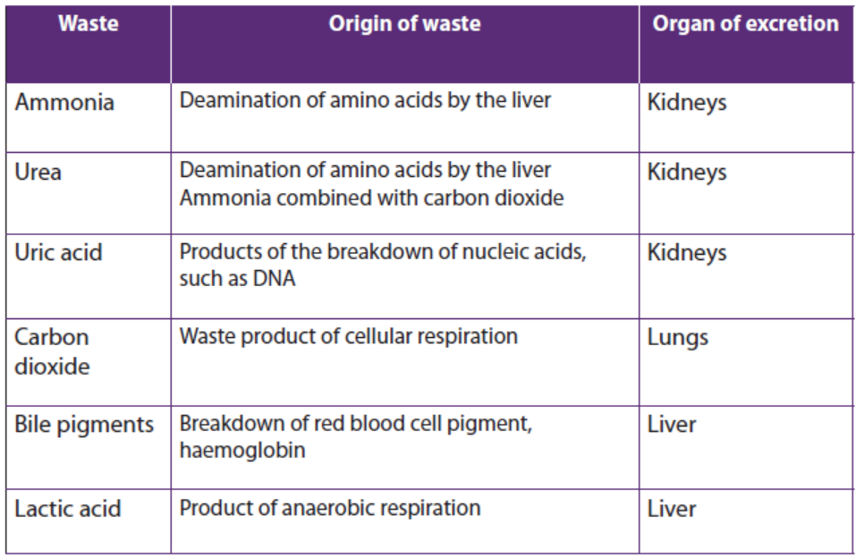
The kidneys are part of the urinary system (Figure 7.1). The kidneys work togetherwith other urinary system organs in the function of excretion
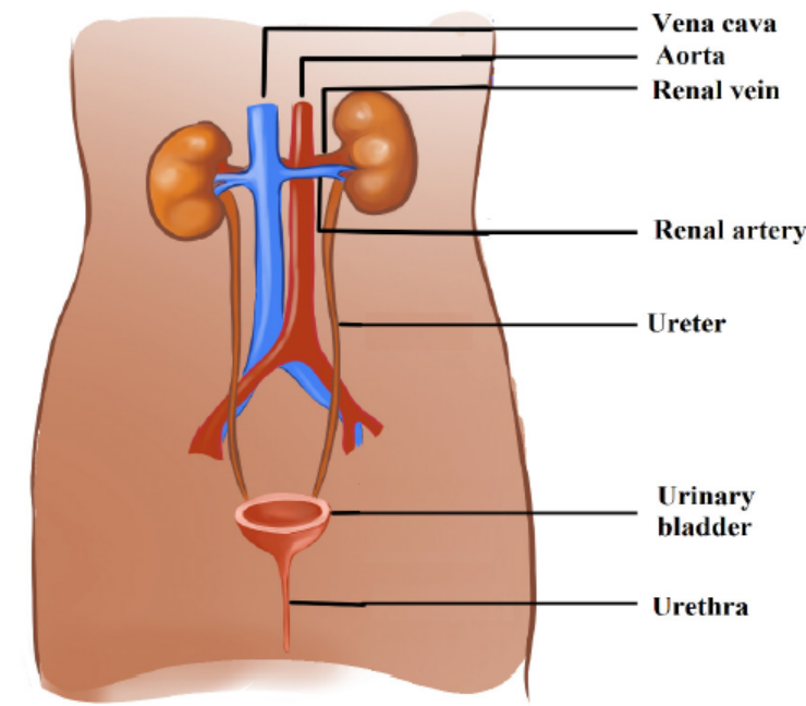
Figure 7.1: The human urinary system
a. The Urinary System
In addition to the kidneys, the urinary system includes the; ureters, bladder, and
urethra. The main functions of the urinary system are to; filter waste products and
excess water from the blood and remove them from the body.
From the kidneys, urine enters the ureters. Each ureter is a muscular tube about
25 centimetres long. Peristaltic movements of the muscles of the ureter send urine
to the bladder in small amount. Ureters carry urine to the bladder. The bladder is a
hollow organ that stores urine. It can stretch to hold up to 500 millilitres. When the
bladder is about half full, the stretching of the bladder sends a nerve impulse to the
sphincter that controls the opening to the urethra. In response to the impulse, the
sphincter relaxes and lets urine flow into the urethra.
The urethra is a muscular tube that carries urine out of the body. Urine leaves the
body through another sphincter in the process of urination. This sphincter and theprocess of urination are normally under conscious control/voluntary system.
b. Kidneys
The kidneys are a pair of bean-shaped, reddish brown organs about the size of a fist.
They are located just above the waist at the back of the abdominal cavity, on either
side of the spine. The kidneys are protected by the ribcage. They are also protected
by a covering of tough connective tissues and two layers of fat, which help cushion
them. Located on top of each kidney is an adrenal gland. The two adrenal glands
secrete several hormones. Hormones are chemical messengers in the body that
regulate many body functions. The adrenal hormone aldosterone helps regulatekidney functions. The functional unit of a kidney is a nephron.
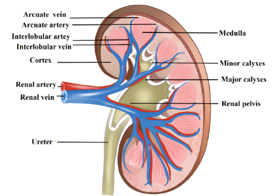
Figure 7.2: Human kidney
Application 7.1
1. What are the functional units of the kidney?
2. What are the main parts of a kidney?
3. Which blood vessel carries filtered blood away from the kidney?4. Which blood vessel brings oxygenated blood to the kidney?
7.2 Structure and the functions of the nephron.
Activity 7.2
1. Download from internet /YouTube and watch a simulation showing the
working of the nephron. 2. Study the diagram below then answer thequestions that follow.
a. Name the structures marked Q, R, S, T, U and W.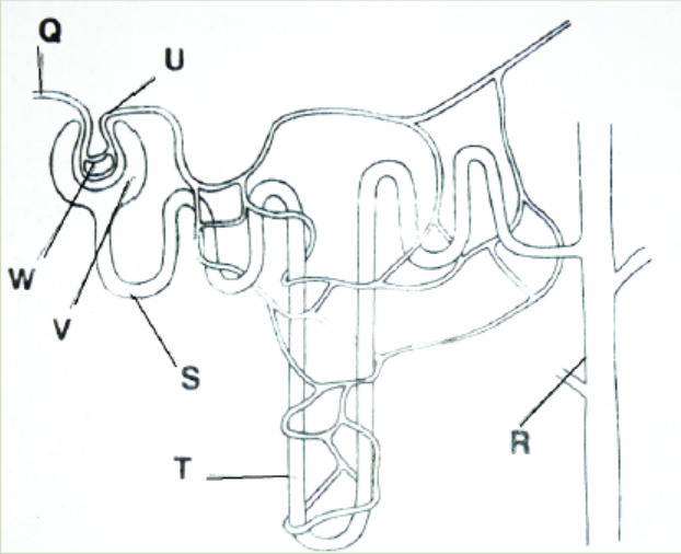
b. When some pressure is applied at W, a fluid appears at V. Name the fluid andstates its contents.
Nephrons are the structural and functional units of the kidneys. A single kidney may
have more than a million nephrons. An individual nephron (Figure 7.3) includes aglomerulus, Bowman’s capsule, and renal tubule.
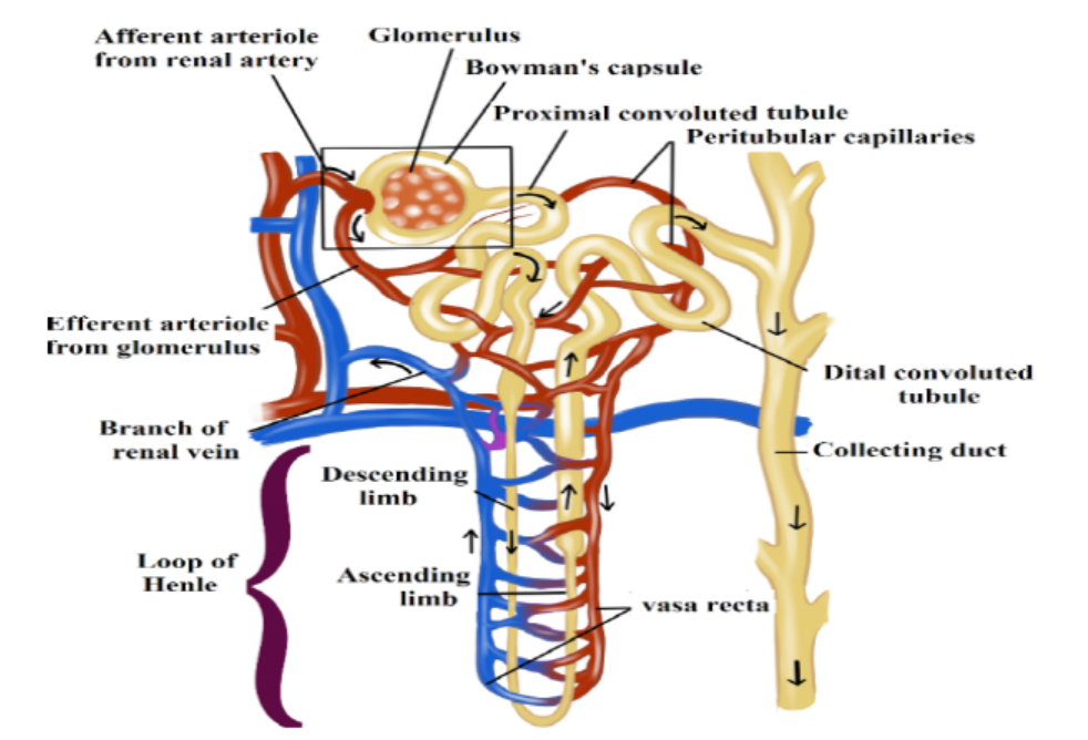
Figure 7.3: The structure of a nephron
a. Parts of the nephron and their functions
– The glomerulus is a cluster of arteries that filters substances out of the blood.
– Bowman’s capsule is a cup-shaped structure around the glomerulus that
collects the filtered substances.
– The renal tubule is a long, narrow tube surrounded by capillaries that reabsorbs
many of the filtered substances and secretes other substances.
b. Ultra-filtration, selective reabsorption and tubular secretion
The renal arteries, which carry blood into the kidneys, branch into the capillaries
of the glomerulus of each nephron. The pressure of blood moving through these
capillaries forces some of the water and dissolved substances in the blood through
the capillary walls and into Bowman’s capsule. Bowman’s capsule is composed of
layers. The space between the layers, called Bowman’s space, fills with the filtered
substances.
The process of filtering substances from blood under pressure in the glomerulus
is called ultra-filtration, while the fluid that collects in Bowman’s space is called
glomerular filtrate. The filtrate is mainly composed of; water, salts, glucose, amino
acids, hormones and urea. Larger structures in the blood including; the protein
molecules, blood cells, and platelets do not pass into Bowman’s space. Instead, theyremain in the main circulation.
From Bowman’s space, the filtrate passes into the renal tubule whose main function
is reabsorption. Reabsorption is the return of needed substances in the glomerular
filtrate back to the bloodstream. It is necessary because some of the substances
removed from the blood by filtration including; water, salts, glucose, and amino
acids which are useful and needed by the body. About 75 % of these substances arereabsorbed in the renal tubule.
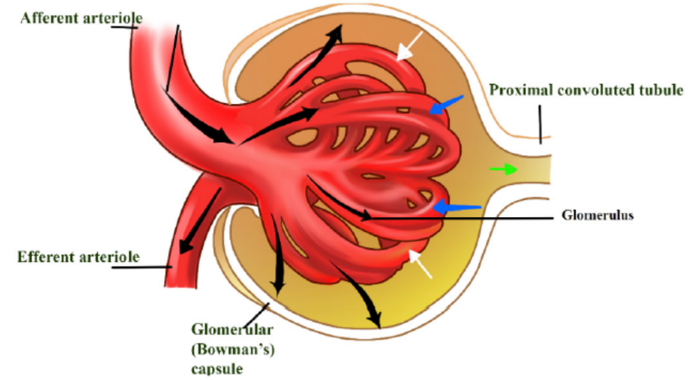
Figure 7.4: The glomerulus
Under conditions in which the kidney conserves as much water as possible, urine can
reach an osmolality of about 1200 milliosmoles (mOsm/L), considerably hypertonic
to blood (about 300 mosm/L). Osmolarity is the solute concentration expressed as
molarity. This ability to excrete nitrogenous wastes with a minimal loss of water is
a key terrestrial adaptation of mammals. The loop of Henle is known as a countercurrent
multiplier. The term counter-current refers to the fact that the fluid flows in
opposite directions in the two sides of the loop, down one side and up in the other.
The multiplier effect is seen by comparing the osmolality of the fluid in the cortexand that in the renal medulla at the hairpin end of the loop.
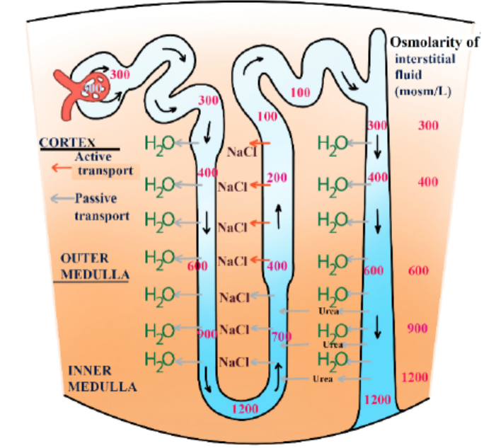
Figure 7.5: Transport of substances across the loop of Henle
The remaining fluid enters the distal tubule. The distal tubule carries the fluid, now
called tubular fluid, from the loop of Henle to a collecting duct. As it transports
the fluid, the distal tubule also reabsorbs or secretes substances such as calcium
and sodium following the influence of hormones (e.g. aldosterone). The process ofsecreting substances into the tubular fluid is called secretion.
Application 7.2
1. What are the main parts of a nephron?
2. In which part of the nephron does each of the following processes
takes place?
a. Ultrafiltration
b. Reabsorption
c. Secretion3. What is the function of the loop of Henle?
7.3 Formation of urine and purification of blood
7.3.1 Urine formation
Activity 7.3
Download from internet/YouTube and watch a movie about the formation of
urine and after answer the questions that follow:
1. Make a list of processes which are involved in the formation of urine.2. What are the main components of urine?
Urine formation depends on three processes including ultrafiltration, selective
reabsorption and secretion/tubular secretion.
a. Ultra-filtration
Each nephron of the kidney has an independent blood supply, which moves through
the afferent arteriole into the glomerulus, a high-pressure filter. Normally, pressure
in a capillary bed is about 25 mm Hg. The pressure in the glomerulus is about 65 mm
Hg. Dissolved solutes pass through the walls of the glomerulus into the Bowman’s
capsule. Although materials move from areas of high pressure to areas of low
pressure, not all materials enter the capsule.
b. Selective reabsorption
The importance of reabsorption is emphasized by examining changes in the
concentrations of fluids as they move through the kidneys. On average, about 600
mL of fluid flows through the kidneys every minute. Approximately 20% of the fluid,
or about 120 mL, is filtered into the nephrons. This means that if none of the filtrate
were reabsorbed the quantity of around 120 mL of urine each minute would be
formed and the amount of at least 1 L of fluids would be consumed every 10 minutes
to maintain homeostasis.
Fortunately, only 1 mL of urine is formed for every 120 mL of fluids filtered into
the nephron. The remaining 119 mL of fluids and solutes is reabsorbed. Selective
reabsorption occurs by both active and passive transport. Carrier molecules move
Na+ ions across the cell membranes of the cells that line the nephron. Negative ions,
such as Cl-and HCO3- follow the positive Na+ ions by charge attraction. Numerous
mitochondria supply the energy necessary for active transport. Reabsorption
occurs until the threshold level of a substance is reached. Excess NaCl remains in the
nephron and is excreted with the urine.
Other molecules are actively transported from the proximal tubule. Glucose and
amino acids attach to specific carrier molecules, which shuttle them out of the
nephron and into the blood. However, the amount of solute that can be reabsorbedis limited. For example; excess glucose will not be shuttled out of the nephron by
the carrier molecules. The solutes that are actively transported out of the nephron
create an osmotic gradient that draws water from the nephron. A second osmotic
force, created by the proteins not filtered into the nephron, also helps reabsorption.
The proteins remain in the bloodstream and draw water from the interstitial fluid
into the blood. As water is reabsorbed from the nephron, the remaining solutes
become more concentrated. Molecules such as urea and uric acid will diffuse from
the nephron back into the blood, although less is reabsorbed than was originallyfiltered.
c. Secretion
Secretion is the movement of wastes from the blood back into the nephron. Nitrogencontaining
wastes, excess H+ ions, and minerals such as K+ ions are examples of
substances secreted.
Even drugs such as penicillin can be secreted. Cells loaded with mitochondria line
the distal tubule. Like reabsorption, tubular secretion occurs by active transport,but, unlike reabsorption, molecules are shuttled from the blood into the nephron.
7.3.2 Formation of urea
The body is unable to store proteins or amino acids, and any surplus is destroyed in
the liver. Excess amino acids which are brought to the liver by the hepatic portal vein,
are deaminated by the liver cells. In this process the amino (NH2) group is removed
from the amino acid, with the formation of ammonia. The amino acid residue is
then fed into carbohydrate metabolism and oxidized with the release of energy.
Meanwhile the ammonia must not be allowed to accumulate because it is highly
toxic even in small quantities. Under the influence of specific enzymes in the liver
cells, the ammonia enters a cyclical series of reactions called the ornithine cycle, in
which it reacts with carbon dioxide to form the less toxic nitrogenous compound
urea. The urea is then shed from the liver into the bloodstream, and taken to thekidney which eliminates it from the body.
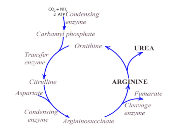
Figure 7.6: The ornithine cycle
Application 7.3
1. The following is a random list of processes that occur in the formation
and excretion of urine once the blood has entered the kidney. Place these
subsequent processes in the correct order:
a. Urine is stored in the bladder
b. Blood enters the afferent arteriole
c. Fluids pass from the glomerulus into the Bowman’s capsule
d. Urine is excreted by the urethra
e. Na+ ions, glucose, and amino acids are actively transported from the
nephronf. Urine passes from the kidneys into the ureters
3. The table below shows the percentage of various components in the blood
plasma in the part labelled A, the fluid in the part labelled B and in the urineof a human.
a. Give a reason why there is no protein in urine.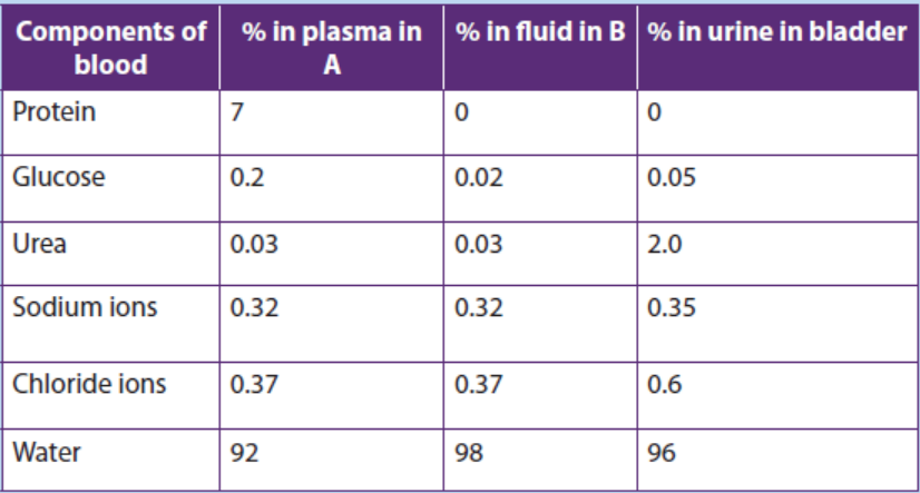
b. Which component of urine shows the greatest percentage increase
in concentration compared to the fluid in B?
c. Give a reason why the component you have named in (ii) above has
the greatest increase in concentration in urine.
d. Suggest with a reason the health condition of the person fromwhom the figure was obtained.
7.4 Role of hypothalamus, pituitary gland, adrenal gland andnephron in varying the blood osmotic pressure
Activity 7.4
Read the following text and answer to the questions that follow: “Water in
essential for all living organisms. People living around lakes and rivers can
drink safely the water without problems but people living around oceans
cannot drink sea water”.
1. Provide an explanation for the possible reason for this.
2. Write on paper the possible endocrine glands involved in this regulation.
3. Make a list showing hormones involved in this regulation.
The body adjusts for increased water intake by increasing urine output. Conversely,
it adjusts for increased exercise or decreased water intake by reducing urine output.
These adjustments involve nervous system and the endocrine system.
7.4.1 Regulation by antidiuretic hormone (ADH)
A hormone called antidiuretic hormone (ADH) helps to regulate the osmotic
pressure of body fluids by causing the kidneys to increase water reabsorption. When
ADH is released, more concentrated urine is produced, thereby conserving body
water. ADH is produced by specialized nerve cells in the hypothalamus, and it moves
along specialized fibres from the hypothalamus to the pituitary gland, which stores
and releases ADH into the blood. Specialized nerve receptors, called osmoreceptors,
located in the hypothalamus detect changes in osmotic pressure when there is a
decrease in water intake or increase in water loss by sweating, causing blood solutes
to become more concentrated. This increases the blood’s osmotic pressure.
Consequently, water moves into the bloodstream, causing the cells of the
hypothalamus to shrink. When this happens, a nerve message is sent to the pituitary,
signalling the release of ADH, which is carried by the bloodstream to the kidneys. By
reabsorbing more water, the kidneys produce more concentrated urine, preventingthe osmotic pressure of the body fluids from increasing any further.
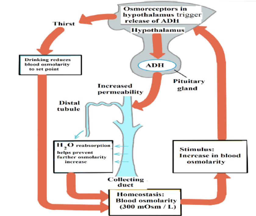
Figure 7.7: Water balance by ADH
7.4.2 Kidneys and Blood Pressure
The kidneys play a role in the regulation of blood pressure by adjusting for blood
volumes. A hormone called aldosterone acts on the nephrons to increase Na+
reabsorption. The hormone is produced in the cortex of the adrenal glands which
lies above the kidneys. Not surprisingly, as NaCl reabsorption increases, the osmotic
gradient increases and more water move out of the nephron by osmosis.
Aldosterone is secreted by the adrenal cortex in response to a high blood potassium
levels, to a low blood sodium levels, or to a decreased blood pressure. When
aldosterone stimulates the reabsorption of Na+ ions, water follows from the filtrate
back to the blood. This helps maintain normal blood volume and blood pressure. In
the kidneys, aldosterone increases reabsorption of Na+ and water so that less is lost
in the urine. Aldosterone also stimulates the kidneys to increase secretion of K+ and
H+ into the urine. With increased water reabsorption by the kidneys, blood volumeincreases.
Application 7.4
1. Describe the mechanism that regulates the release of ADH.
2. Where is the thirst centre located?3. Write ADH in full where is it produced and stored?
7.5 Kidney transplants and dialysis machines
Activity 7.5
Nowadays kidneys diseases are well known and some people with kidney
failure are being treated in different hospitals in our country and abroad.
1. Write the types of treatments you know for the person with kidney
failure.2. Discuss the advantages and disadvantages of such treatments.
Dialysis is a medical procedure in which blood is filtered with the help of a machine.
Blood from the patient’s vein enters the dialysis machine through a tube. Inside the
machine, excess water, wastes, and other unneeded substances are filtered from the
blood. The filtered blood is then returned to the patient’s vein through another tube.
A dialysis treatment usually lasts three to four hours and must be repeated three
times a week. Dialysis is generally performed on patients who have kidney failure.Dialysis helps them stay alive, but does not cure their failing kidneys.
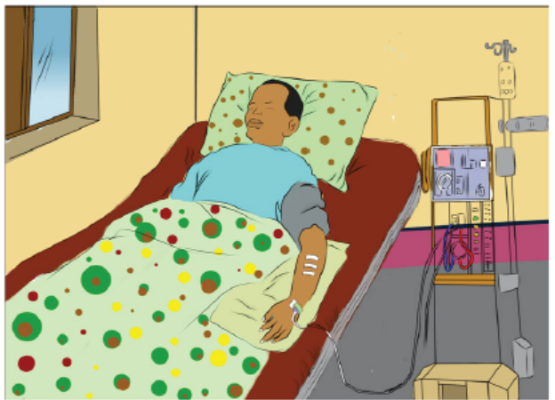
Figure 7.8: Patient under dialysis treatment
Kidney transplants are sometimes performed on people who suffer from severe
renal failure. Usually, the donor has suffered an accidental death and had granted
permission to have his or her kidneys used for transplantation. An attempt is made
to match the immune characteristics of the donor and recipient to reduce the
tendency for the recipient’s immune system to reject the transplanted kidney. Even
with careful matching, however, recipients have to take medication for the rest of
their lives to suppress their immune systems so that rejection is less likely. The major
cause of kidney transplant failure is rejection by the recipient’s immune system.
In most cases, the transplanted kidney functions well, and the tendency for the
recipient’s immune system to reject the transplanted kidney can be controlled. Theadvantages and disadvantages of kidney transplants, compared with dialysis.
Advantages
– The patient can return to a normal lifestyle – dialysis may require a lengthy
session in hospital, three times a week, leaving the patient very tired after each
session.
– The dialysis machine will be available for other patients to use.
Disadvantages
– Transplants require a suitable donor – with a good tissue match. The donor may
be from a dead person, or from a close living relative who is prepared to donate
a healthy kidney (we can survive with one kidney).
– The operation is very expensive.
– There is a risk of rejection of the donated kidney; immunosuppressive drugs
have to be used.– Transplants are not accepted by some religions.
Application 7.5
1. What is the most difficult challenge to overcome in achieving successful
kidney transplants? Provide a reason.
2. Why do you think it is beneficial to humans to have two kidneys ratherthan one? Explain your answer.
7.6 Principles of osmoregulation in marine, freshwater andterrestrial organisms.
Activity 7.6.
Aim: To demonstrate the process of osmoregulation in earthworms and
amphibians
Materials required: earthworms, amphibians (toads or frogs), beaker, salt
solution and tap water.
Procedure:
– Put three earthworms in beaker A containing water from the tap and note
the observations after 20 minutes.
– Put other three earthworms in a beaker B containing concentrated salt
solution and note the observations after 20 minutes.
– Put one amphibian in beaker C containing water from the tap and note the
observations after 20 minutes.
– Put the other amphibian in a beaker D containing concentrated salt solution
and note the observations after 20 minutes.– Prepare a report that explains the above observations.
Organisms in aquatic and terrestrial environments must maintain the right
concentration of solutes and amount of water in their body fluids. This involvesexcretion through the skin and the kidneys.
a. Marine animals
Marine bony fishes, such as the salmon, constantly lose water by osmosis. Such fishes
balance the water loss by drinking large amounts of seawater. They then make use of
both their gills and kidneys to rid themselves of salts. In the gills, specialized chloride
cells actively transport chloride ions (Cl-) out, and sodium ions (Na+) follow passively.
In the kidneys, excess calcium, magnesium, and sulphate ions are excreted with theloss of only small amounts of water.
b. Freshwater animals
The body fluids of fresh water animals must be hypertonic because animal cells
cannot tolerate salt concentrations as low as those of lake or river water. Having
internal fluids with an osmolality higher than that of their surroundings, freshwater
animals face the problem of gaining water by osmosis and losing salts by diffusion
through their gills. Many freshwater animals, including fishes, solve the problem
of water balance by drinking almost no water and excreting large amounts of very
dilute urine. At the same time, salts lost by diffusion and in the urine are replaced bythose found in the food they eat.
c. Land animals
The threat of dehydration is a major regulatory problem for terrestrial plants and
animals. Humans, for example, die if they lose as little as 12% of their body water.
Adaptations that reduce water loss are key to survival on land. Much as a waxy cuticle
contributes to the success of land plants, the body coverings of most terrestrialanimals help prevent dehydration.
Examples are the waxy layers of insect exoskeletons, the shells of land snails, and the
layers of dead, keratinized skin cells covering most terrestrial vertebrates, including
humans. Despite these and other adaptations, most terrestrial animals lose water
through many routes: in urine and faeces, across their skin, and from moist surfaces
in gas exchange organs. Land animals maintain water balance by drinking and eating
moist foods and by producing water metabolically through cellular respiration. A
number of desert animals, including many insect-eating birds and other reptiles,
are well enough adapted for minimizing water loss that they can survive without
drinking water. A noteworthy example is the kangaroo rat loses so little water that
90% replaced by water generated metabolically; the remaining 10% comes from the
small amount of water in its diet of seeds.
Application 7.6
Explain why organisms in aquatic and terrestrial environments need to
maintain the right concentration of solutes and amount of water in their body
fluids?
7.7 Excretion and osmoregulation in protoctista, insects, fish,amphibians and birds.
Activity 7.7
Aim: To demonstrate the process of osmoregulation in a fish.
Materials required: A living fish, bucket, salt solution and tap water.
Procedure:
– Put a fish into a bucket containing water from the tap and note your
observations after 10 minutes.
– Take a fish into a bucket containing concentrated salt solution and note
your observations after 10 minutes.
– Explain your observations.
a. Osmoregulation in protists such as Amoeba
Amoeba makes use of contractile vacuoles to collect excretory wastes, such as
ammonia, from the intracellular fluid by diffusion and active transport. As osmotic
action pushes water from the environment into the cytoplasm, the vacuole moves
to the surface and disposes the contents into the environment.
b. Excretion in insects
Insects and other terrestrial arthropods have organs called Malpighian tubules that
remove nitrogenous wastes and also function in water balance. The Malpighian
tubules extend from dead-end tips immersed in haemolymph (circulatory fluid) to
openings into the digestive tract. The filtration steps which are common to other
excretory systems are absent. Instead, the transport epithelium that lines the tubules
secretes certain solutes, including nitrogenous wastes, from the haemolymph into
the lumen of the tubule.
Water follows the solutes into the tubule by osmosis, and the fluid then passes into
the rectum. There, most solutes are pumped back into the haemolymph and water
reabsorption by osmosis follows. The nitrogenous wastes mainly insoluble uric acid,
are eliminated as nearly dry matter along with the faeces. Capable of conserving
water very effectively, the insect excretory system is a key adaptation contributingto their success on land.
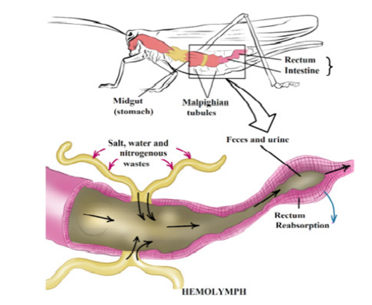
Figure 7.9: Malpighian tubules of insects
c. Excretion in Birds and Reptiles
Most birds live in environments that are dehydrated. Like mammals, birds have
kidneys with juxtamedullary nephrons that specialize in conserving water.
However, the nephrons of birds have loops of Henle that extend less far into the
medulla than those of mammals. Thus, bird kidneys cannot concentrate urine to
the high osmolarities achieved by mammalian kidneys. Although birds can produce
hyperosmotic urine, their main water conservation adaptation is having uric acid as
the nitrogen waste molecule. Since uric acid can be excreted as a paste, it reduces
urine volume.
The kidneys of reptiles having only cortical nephrons, produce urine that is osmotic
or hypo-osmotic to body fluids. However, the epithelium of the chamber called the
cloaca helps conserve fluid by reabsorbing some of the water present in urine and
faeces. Also like birds, most reptiles excrete their nitrogenous wastes as uric acid.
Freshwater fishes and amphibians
Freshwater fishes are hyperosmotic to their surroundings, so they must excrete excess
water continuously. In contrast to mammals and birds, freshwater fishes produce
large volumes of very dilute urine. Their kidneys, which contain many nephrons,
produce filtrate at a high rate. Freshwater fishes conserve salts by reabsorbing ions
from the filtrate in their distal tubules, leaving water behind.
Amphibian kidneys function much like those of freshwater fishes. When in freshwater, the kidneys of frogs excrete dilute urine while the skin accumulates certain
salts from the water by active transport. On land, where dehydration is the most
pressing problem of osmoregulation, frogs conserve body fluid by reabsorbingwater across the epithelium of the urinary bladder.
Marine bony fishes
The tissues of marine bony fishes gain excess salts from their surroundings and
lose water. These environmental challenges are opposite to those faced by their
freshwater relatives. Compared with freshwater fishes, marine fishes have fewer and
smaller nephrons, and their nephrons lack a distal tubule. In addition, their kidneys
have small glomeruli, and some lack glomeruli entirely. In keeping with thesefeatures, filtration rates are low and very little urine is excreted.
Application 7.7
1. What is the importance for birds and reptiles to excrete their nitrogenous
wastes in the form of uric acid?2. Explain how osmoregulation occurs in protozoa such as amoeba.
7.8 Excretion in plants
Activity 7.8
All living organisms carry out the process of excretion. Plants as other living
organisms need to remove the metabolic wastes products outside of their
bodies. Yet plants do not have kidneys and other excretory organs as seen
in animals. Use books from your school library and use internet for further
research to answer the questions that follow:
– Identify and write the structures that are involved in the excretion in plants.
– List the differences between the excretory system of a plant and that of a
human.
– Explain why plants do not have complex organs systems as animals?
Compared to animals, plants do not have a well-developed excretory system to
throw out nitrogenous waste materials. This is because of the differences in theirphysiology. Therefore, plants use different strategies for excretion.
The gaseous waste materials produced during respiration (carbon dioxide) and
photosynthesis (oxygen) diffuse out through stomata in the leaves and through
lenticels in other parts of the plant. Excess water evaporates mostly from stomata
and also from the outer surface of the stem, fruits, etc., throughout the day. This
process of getting rid of excess water is called transpiration. The waste products, like
oxygen, carbon dioxide and water, are the raw materials for other cellular reactions
such as photosynthesis and cellular respiration. The excess of carbon dioxide and
water are used up in this way. The only major gaseous excretory product of plants isoxygen.
Many plants store organic waste products in their permanent tissues that have
dead cells, for example in heartwood. Plants also store wastes within their leaves
or barks, and these wastes are periodically removed as the leaves and barks fall off.
Some of the waste products are stored in special cells or cellular vacuoles. Organic
acids, which might prove harmful to plants, often combine with excess cations and
precipitate out as insoluble crystals that can be safely stored in plant cells. Calciumoxalate crystals accumulate in some tubers like yam.
Aquatic plants lose most of their metabolic wastes by direct diffusion into the water
surrounding them. Terrestrial plants excrete some wastes into the soil around them.
Plants do not have complex excretory systems. This is because of the followingreasons:
– There is very little accumulation of toxic wastes. Often the plant wastes are
utilized by the plant. For example, carbon dioxide is used for photosynthesis
and oxygen for respiration.
– The extra gaseous waste is removed from the plant by simple diffusion through
the stomata and the lenticels.
– Most of the waste substances formed in plants are not harmful and can be
stored in the plant tissues.
– Some plants store other waste such as resins in their tissues in a non-toxic form.
These tissues or organs later fall off the plant.
– Excess water and dissolved gases are removed by the process of transpiration
through the stomata.
– Some plants remove waste products by exudation, for example gums, resins,
latex and rubber.
– In some plants water with dissolved salts oozes out through hydathodes. This
is called guttation.
Note that hydathodes are specialized structures and they are mainly responsible
for secreting water in liquid form. They are generally restricted to the apex or theserrated edges of the margins of leaves.
Application 7.8
1. What are the excretory products produced by plants? State any four.
2. Identify three ways by which plants excrete their waste products.3. What are hydathodes? What are their functions in excretion?
End of unit assessment 7
Multiple choice questions: choose the letter corresponding to the best answer.
1. Glucose is small enough to be filtered from the blood in glomeruli in the
kidney, but is not normally found in the urine. This is because glucose is:
a. Reabsorbed in distal convoluted tubules
b. Reabsorbed in proximal convoluted tubules
c. Reabsorbed along the whole length of the nephrons
d. Respired by cells in the kidney
2. Which of these does not contribute to the process of filtration in the kidney?
a. High hydrostatic blood pressure in glomerular capillaries.
b. Large surface area for filtration.
c. Permeability of glomerular capillaries.
d. Active transport by epithelial cells lining renal tubules.
3. The most important function of the kidney is:
a. Removal of water from the body.
b. Regulating blood composition.
c. Storage of salts in the body.d. Elimination of urea from the blood.
Structured answer questions4. The following diagram shows the nephron.
a. From the diagram above write the number that represents the: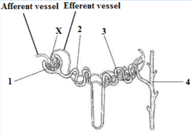
i. Collecting duct
ii. Bowman’s Capsule
b. On the diagram above label the loop of Henle.
c. Name structure X.
d. Compare the blood pressure in the afferent and efferent arterioles
and explain the cause of this difference.
e. Proteins are not present in the glomerular filtrate but amino acids
are. Explain.
f. Compare the urea concentration in the renal artery with that in the
renal vein.g. Name TWO organs that excrete urea.
5. Observe the diagram below and identify the following structures:
a. The structure that filters blood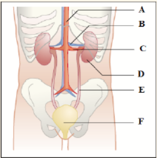
b. The structure that carries urine from the kidney
c. The structure that carries blood containing urea into the kidney
d. The structure that stores urine6. Use the figure below to answer the following:
a. Identify which letters indicate the afferent and efferent arterioles.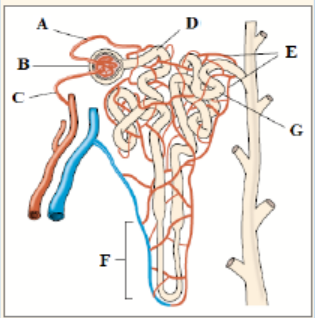
b. Explain how an increase in blood pressure in area (B) would affect
the functioning of the kidney.
c. Explain why proteins and blood cells are found in area (B) but not in
area (D).
d. In which area of the nephron would you expect to find the greatestconcentration of glucose?