UNIT 11 SKELETONS, MUSCLES AND MOVEMENT
UNIT 11: SKELETONS, MUSCLES AND MOVEMENT
Key unit competence
Explain the structure of muscles in relation to movement.
Learning objectivesBy the end of this unit, I should be able to:
– Describe the three main types of animal skeletons.
– Discuss the functions of skeletons.
– State and discuss the advantages and disadvantages of exoskeletons.
– Describe the features of a synovial joint
– Appreciate the role of joints and muscles in bringing about movement.
– Describe the main types of mammalian muscles.
– Compare the structure of cardiac, smooth and skeletal muscle.
– Distinguish between slow twitch and fast twitch fibers.
– Demonstrate the structure and function of the sarcomere.
– Demonstrate the laws of muscle contraction.
– Distinguish between temporal summation and muscle fibre recruitment.
– Explain the role of antagonistic muscles in a joint.
– Adopt the practice of playing sport to develop healthy muscles and bones.
– Appreciate the role of joints and muscles in bringing about movement.
– Describe the ultrastructure of striated muscles with particular reference to the
sarcomere structure.
– Interpret the ultrastructure of striated muscle with particular reference to the
sarcomere structure
– Explain the sliding filament model of muscle contraction, including the roles
of troponin, tropomyosin, calcium ions and ATP.
– Explain the function of a motor unit/ neuromuscular junction/motor end plate.– Illustrate the sliding filament model of muscular contraction.
Introductory activity
Let do the physical exercise of push-up. Do as many as you can. Explain whatallows you to push-up.
11.1 Types of animal skeletons: hydrostatic, exoskeletonand endoskeleton
Activity 11.1.
You are provided with an earthworm and a grasshopper.AW: Put diagrams of both earthworm and grasshopper but without labels.
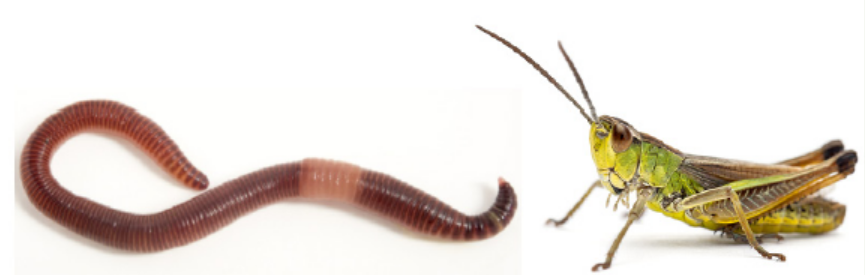
Observe locomotion and then differentiate their skeletons.
Figure 11.1: Structure of earthworm and insect
A support system is made up of those materials that bear the weight of the body,
strengthen its parts and endure all stresses that the body or its parts may be subjected
to during movement. Consequently, the strength of the supporting materials in
an organism is directly related to its size and weight. The strength of a supporting
material itself depends among other things on its length, shape, thickness andstructure.
11.1.1. Types of skeleton
There are three types of skeleton namely hydrostatic skeleton, endoskeleton andexoskeleton.
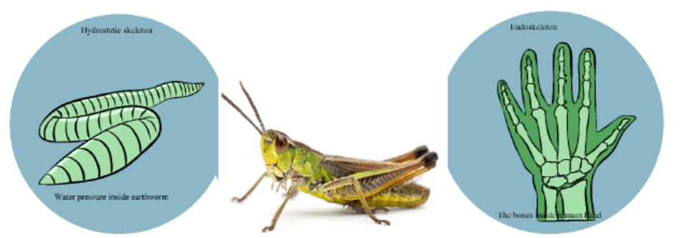
Figure 11.2: Types of skeletons: hydrostatic, exoskeleton and endoskeleton
a. Hydrostatic skeleton
Annelids (earthworm), nematodes (round worms), Echinoderms (starfish and the
sea urchin), cnidarians (Jellyfish), and some other organisms use the hydrostatic
skeleton for movement. This skeleton is found in soft-bodied and cold-blooded
animals having a coelom. This coelom is fluid-filled cavity surrounded by muscles
and the rigidity caused by the fluid. The muscles serve as a supporting structure for
organisms. Hydrostatic skeleton is basically composed of a fluid filled body cavity
surrounded by sets of antagonistic muscles. The hydrostatic skeleton operates on
the principle that water is incompressible and therefore can provide a rigid medium
against which muscles can contact. The hydrostatic skeleton is segmented and
therefore can be used for movement and locomotion. It is also flexible and therefore
allows expansion to allow growth.
However, hydrostatic skeleton presents some disadvantages as it provides relatively
little support and therefore neither supports the animal upright nor their body
weight off the ground. It does not provide strong levels on which powerful muscles
can operate fast locomotion. This coupled with the fact that the body weight is
dragged on the ground makes it unsuitable for fast locomotion. Consequently,
animals depending on hydrostatic skeleton are slow moving. The thin flexible cuticle
associated with it does not properly protect the animal against water loss, because ifthe cuticle was thick and inflexible, it would not allow free movement.
b. Exoskeleton
The exoskeletons also known as cuticles are found in all arthropods It is found
outside the body and forms a protective covering for the animals. It supports as well
as protects the animals. All crustaceans have exoskeleton. Crabs, spiders, lobsters,
insects are all arthropods. Animals with exoskeleton are usually small. This is because
large animals could not be supported by exoskeleton and need bones to support
them. Animals with exoskeleton have a head and abdomen and in some cases, a
thorax. The exoskeleton is soft and thin at the joints where it has to bend. The large
exoskeletons are called shells. Tortoise is one vertebrate animal that has a shell andendoskeleton.
Advantages of the exoskeletons (in movement and protection)
– It is joined and allows muscle attachment which makes it useful in locomotion.
It also forms locomotors devices like legs and wings.
– It maintains the shape of the insect. The shape is an important determinant of
how well movement can take place.
– It prevents water loss by having wax. This has helped insects to adapt to dry
environments.
– It is hard and offers protection of internal organs from mechanical injury,
friction and microbial attack.
– It is usually colored and offers protection from predators through camouflage
and mimicry.
– It is used to form various mouthparts. Mouth parts are adapted for the various
feeding methods in insects and also for various forms of protection especially
biting the enemy.
Disadvantages of the exoskeletons
– Its components are heavy compared to those of similar size in other skeletons.
This affects the locomotion of insects especially those which are big and
explains why large insects cannot fly for long without resting.
– It does not allow continuous growth because of its rigidity; growth has to beintermittent following moulting.
c. Endoskeleton
Mainly made of bones, the endoskeleton is a rigid internal skeleton of vertebrates.
It forms the frame work for the animal. The tissues and muscles are formed around
the skeletal system and the muscular forces are transmitted to this skeleton. It is
composed of mineralized tissues. In phylum Chordata, Porifera and Echinodermata
endoskeleton is present. The animals that come under Phylum Chordata are allvertebrates including human beings.
Advantages of the endoskeletons (in movement and protection)
– It does not restrict growth like the exoskeleton.
– It is relatively light and allows faster locomotion both on land and in the air.
– It is jointed and allows flexibility and movement.
– It maintains the shape and form of the body which allows it to move fast.
– Its skeletal elements (the bones) are metabolically active and synthesize blood
cells some of which offer protection against disease (white blood cells).
– It offers maximum protection to some delicate internal organs e.g. the brain
from mechanical injury.
– It is a stronger skeleton and therefore supports most of the body weight aboveground which allows faster locomotion.
Disadvantages of the endoskeletons in movement and protection.
– It does not completely enclose internal organs and therefore offers less
protection to them from mechanical shock.– It does not protect the animal from water loss.
Table 11.1: A comparative table of animal skeletons: hydrostatic, exoskeletonand endoskeleton
11.1.2. Functions of Bones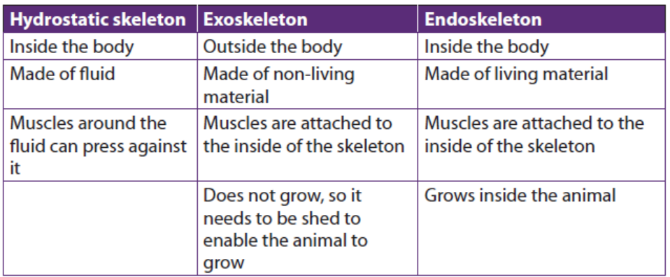
The skeletal system is important for the proper functioning of animal’s body.
In addition to giving shape and form to the body, bones have many importantfunctions as follow:
– Structural support of the body: The skeleton supports the body against the
pull of gravity. The large bones of the lower limbs support the trunk when
standing.
– Protection of internal organs: The skeleton provides a rigid frame work that
supports and protects the soft organs of the body. The fused bones of the
cranium surround the brain to make it less vulnerable to injury. Vertebrae
surround and protect the spinal cord and bones of the rib cage help protect
the heart and lungs.
– Attachment of the muscles: The skeleton provides attachment surfaces for
muscles and tendons which together enable movement of the body.
– Movement of the body: Bones work together with muscles as simple
mechanical lever systems to produce body movement.
– Production of blood cells: The formation of blood cells takes place mostly in
the interior (marrow) of certain types of bones.
– Storage of minerals: Bones contain more calcium than any other organ in the
form of calcium salts such as calcium phosphate. Calcium is released by the
bones when blood levels of calcium drop too low. Phosphorus is also storedin bones.
11.1.3. The human skeleton
Humans are vertebrates, which are animals that have a vertebral column, or
backbone. The study of internal framework of bones and cartilage that is found inside
vertebrates, including humans, is called an endoskeleton. The adult human skeleton
consists of approximately 206 bones. Cartilage is a type of fibrous connective tissue
that is made of tough protein fibers. The function of cartilage in the adult skeleton
is to provide smooth surfaces for the movement of bones at a joint. A ligament is a
band of tough, fibrous tissue that connects bones together. Ligaments are not very
elastic and some even prevent the movement of certain bones. The skeletons of
babies and children have many more bones and more cartilage than adults have.
As a child grows, the extra bones, such as the bones of the skull (cranium), and the
sacrum (tailbone) fuse together, and cartilage gradually hardens to become bontissue.
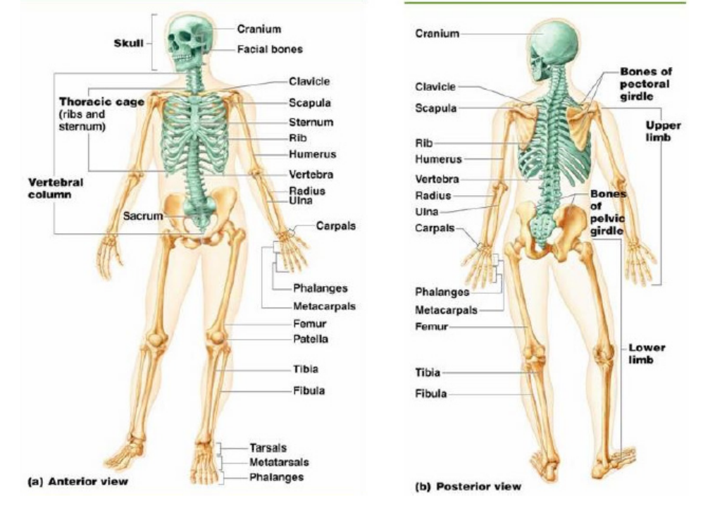
Figure 11.3: The human skeleton
The bones of the skeleton can be grouped in two divisions: the axial skeleton and
appendicular skeleton.
The axial skeleton includes; the bones of the; head, vertebral column, ribs andsternum. There are 80 bones in the axial skeleton.
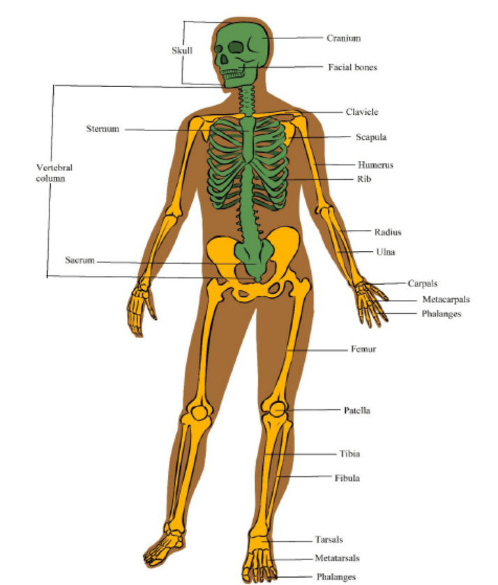
Figure 11.4: Divisions of the human skeleton
a. The axial skeleton
The axial skeleton forms the central axis of the body. It consists of the skull, the
vertebral column, the ribs and the sternum or breastbone. There are 80 bones inaxial skeleton.
i) The Skull
The skull consists of 28 different bones including the ossicles of the ear. The bones
of the skull can be divided into two main groups: the cranium which encloses and
protects the brain and the facial bones. The cranium is a rigid structure with an
opening, the foramen magnum (literally large hole) where the spinal cord enters.
ii) The Vertebral column
The vertebral column forms the central part of the skeleton. It supports the skull
and protects the spinal cord. It also serves as attachment for the ribs, the pectoral
and pelvic girdles. The vertebral column consists of separate bones, the vertebrae.
Because the separate vertebrae are attached to each other by means of fibrous
cartilaginous discs they form a flexible column. Each vertebra has articular surfaces
above and below, which allow articulation movement between them.
The vertebral column of 33 vertebrae is divided into five regions according to their
position and structure. The five regions consist of: seven cervical (neck) vertebrae,
twelve thoracic (chest) vertebrae, five lumbar vertebrae (vertebrae of the lower
back), five fused sacral vertebrae (vertebrae of the pelvic region), and four fused
vertebrae of the coccyx. The first two cervical vertebrae are known as the atlas and
axis. They are specially adapted to support the skull and to enable it to move. Theydiffer from the structure of the typical vertebra in certain respects species.
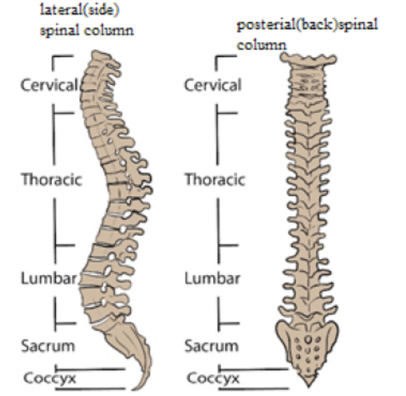
Figure 11.5: The Vertebral Column
A typical vertebra consists of the centrum (or body), a neural arch, a neural spine,two transverse processes and four articular processes with articulating surfaces. The
centrum is the front part (anterior) and consists of a solid piece of spongy bone
encircled by a layer of compact bone. The upper and lower surfaces are flat and
rough and provide attachment for the cartilaginous discs. These surfaces allow alimited degree of movement. The posterior (back) part is called the neural arch.
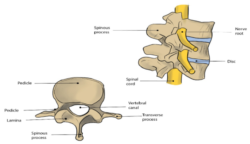
Figure 11.6: The structure of a vertebra
iii) The sacrum and the coccyx
The sacrum is roughly triangular in shape and consists of 5 fused vertebrae. It lies
between the hip bones with which it articulates. Horizontal ridges indicate the
divisions between the fused vertebrae. At the ends of these ridges are openings
which allow nerves and blood vessels to pass through. The coccyx consists of 4
fused tail vertebrae which are small and have a relatively simple structure. They do
not resemble the structure of a typical vertebra and the muscles of the buttocks areattached to the coccyx.
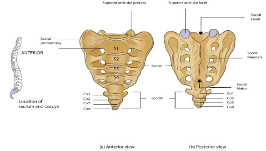
Figure 11.7: The Sacrum and Coccyx
iv) The Ribs
Twelve pairs of ribs articulate with the 12 vertebrae of the thoracic region. The ribs
are flat and narrow bones with a distinctive bow-shaped curve. Each rib consists of
a head or capitulum, a small tubercle (which is a short distance back from the head)
and the shaft. The tubercle fits into and articulates with the articulating facets on the
transverse process. All ribs articulate with thoracic vertebrae. True ribs (first seven
pairs) articulate directly with sternum by means of costal cartilages. Ribs 8 to 10
attach to the costal cartilage of rib 7, and ribs 11 and 12 do not attach to anything at
the distal end but are embedded in thoracic muscle. Ribs 8 to 12 are therefore called
false ribs, and ribs 11 and 12 are also called floating ribs for lack of any connectionto the sternum.
Figure 11.8: Diagram to illustrate the attachment of the ribs to the thoracic vertebrae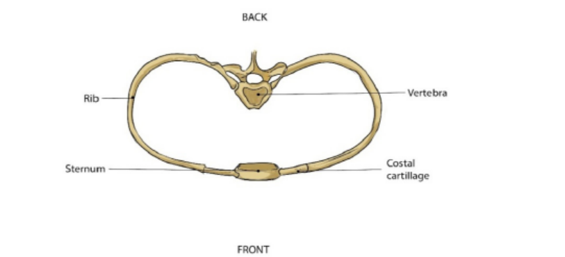
and sternum
b. The appendicular skeleton
The appendicular skeleton consists of the girdles (clavicle, scapula and pelvis) and
the skeleton of the limbs (arms and legs) There are approximately 126 bones in the
appendicular skeleton. Limbs are connected to the rest of the skeleton by girdles.
The upper (anterior) limbs are attached to the pectoral (shoulder) girdle and the
lower (posterior) limbs are attached to the pelvic (hip) girdle. The pectoral girdle
consists of the clavicle (collar bone) and scapula (shoulder blade). The pelvic girdle
consists of two pelvic bones (hipbones) that form the pelvic girdle. The vertebral
column attaches to the top of the pelvis; the femur of each leg attaches to the
bottom. The humerus is joined to the pectoral girdle at a joint and is held in place bymuscles and ligaments.
i) The pectoral (shoulder) girdle
The Pectoral girdle consists of two shoulder blades (scapulae) and two collar bones
(clavicles). These bones articulate with one another, allowing some degree ofmovement.
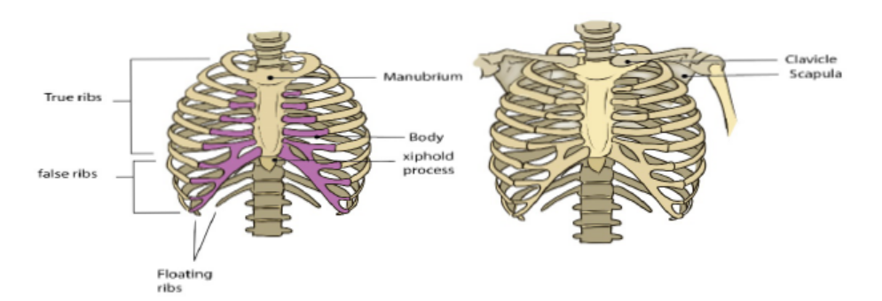
Figure 11.9: The thoracic cage and the pectoral girdle
ii) The pelvic (hip) girdle
The pelvic girdle consists of two large and sturdy hip bones. Each hip bone consists
of three fused bones namely the ilium, ischium and the pubis. The ilium is the largest
of the three and forms the upper part of the hip bones. The sacrum fits like a wedge
posteriorly between the two hip bones. The sacrum has a large, flat articular surface
on each side for articulation with the ilia. The ischium forms the inferior part of the
hip bone and the pubis at the central in front. The two pubic bones are attached in
the middle, on the front side by a symphysis which consists of fibrocartilage and
ligaments, the pubic symphysis. The two hip bones and the sacrum form a complete
bony ring, the pelvis. On the outer side of the point where the fused bones meet,
there is a deep hip socket into which the head of the femur fits. This is called theacetabulum.
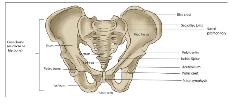
Figure 11.10: The pelvic girdle
The pelvic girdle forms a strong support for the attachment of the limbs. Strong
muscles of the back, the legs and the buttocks are attached to it. It protects some of
the internal organs. In females it forms a strong basin-like structure for supportingand protecting the developing foetus during child-bearing.
Application 11.1
1. What are the three main types of animal skeletons?
2. What are advantages and disadvantages of exoskeletons?
3. What is the difference between hydrostatic, exoskeleton and endoskeleton
skeletons?
4. What is importance of skeletal system human body apart from giving shapeand form to the body?
11.2 Types of joints
A joint is the junction between two or more bones. There are three major types ofjoints:
Activity 11.2Observe the joints below and answer questions that follow:
1. Why the joints of the skull are basically described as immovable joints?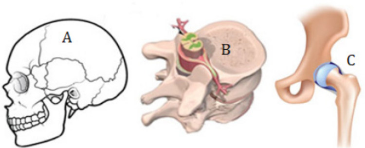
2. Why the joint in B is described as semi-movable?3. Describe the types of joints in B and C.
11.2.1 Immovable or Fused joints or sutures
These joints include the skull, sacrum, pelvis, and coccyx. As the name suggests,
these joints are points where joints fuse or grow together. The place where they growtogether is called the suture. These joints provide strength, support, and protection.
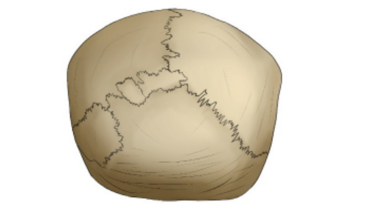
Figure 11.12: The fused joint
11.2.2 Slightly moveable joints
These joints are located between the vertebrae of the upper spine. There is cartilage
within the joints. They help pad and protect the bones. The bones are held together
by ligaments. The ligaments are tightly bound and limit the movement of the bones.This protects the spinal cord.
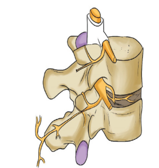
Figure 11.13: The slightly moveable joint
11.2.3 Freely moveable or synovial joints
At these joints the ends of the bones are covered with cartilage and there is a cavity
that separates the bones. The bones are held in place by ligaments which stop
the bones from moving too much. In addition to the ligaments the two bones are
joined together by sleeve-like capsule. The capsule encloses the synovial cavity. The
outer layer of the capsule is composed of ligaments. The inner layer of the capsule
is the synovial membrane. The synovial membrane secretes the lubricating synovial
fluid. Lubrication is essential to prevent frictional wear and tear. The cartilage at the
contact ends of the bones also reduces friction. The cartilage pads also act as shockabsorbers against mechanical damage.
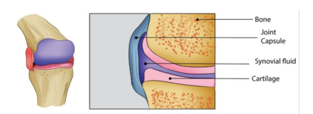
Figure 11.14: The synovial joint
11.2.4 There are four classes of synovial joints:
i) Gliding: The bones of these joints move across each other, back-and-forth and
side-to-side. Examples are between the carpals of the wrist and tarsals of the
ankle.
ii) Pivot: These joints allow a turning movement. Examples are between the first
and second vertebras when turning the head, between the ulna and the radius
of the lower arm when turning the palm of the hand up or down.
iii) Hinge: These joints allow movement in one plane during flexion and extension.
They act, as the name implies, like the hinge of a door. Examples are bending the
elbow or knee.
iv) Ball and Socket: This type of joint permits movement in three planes, i.e., in alldirections. Examples are the shoulder and hip joints.
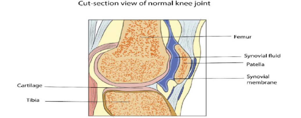
Figure 11.15: Cut-section view of normal knee joint.
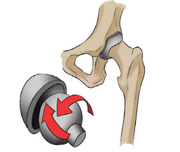
Figure 11.16: Ball and Socket joint.
Table 11.2: Summary of the types of joints
Application 11.2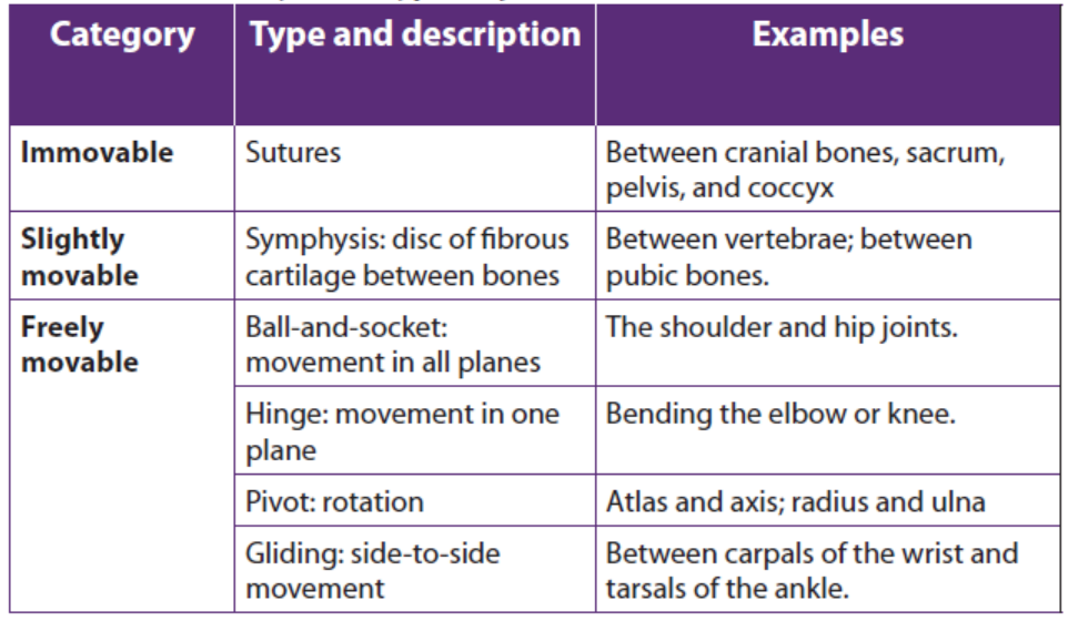
1. What is a joint?
2. Distinguish between fused joints and slightly moveable joint.3. What are the differences between the types of Synovial Joints?
11.3 Types of muscles: cardiac, smooth and skeletal muscle
Activity 11.3
Dissection of a frog / toad heart and observation of myogenic contraction.
Materials required
Dissection pan with 4 needles, 20 ml of physiological liquid (Ringer’s solution),
plastic eye-droppers, suture needle with thread attached, razor blade,
magnifying hand lens, pins, chloroform, cotton wool, frog or toad, bell jar,forceps, glass beaker, gloves, and water
Procedure
– Collect a living frog or toad from the nearest swamp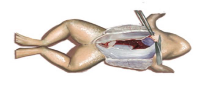
– Prepare 20ml of Ringer’s liquid in a glass beaker
– Put the cotton wool imbibed of 10 ml of chloroform in the bell jar
– Put your frog in the bell jar for 5 minutes, then remove it
– Lay your frog dorsally and fix its four limbs with pins on the dissection dish
– Carry out the longitudinal section from the abdomen to the chest usingsurgical blade (razor blade) or scissor.
There are 3 types of muscle: skeletal, smooth, and cardiac.
a. Skeletal Muscle
Skeletal muscle, as its name implies, is the muscle attached to the skeleton. It is also
called striated muscle. The contraction of skeletal muscle is under voluntary control.
These muscles are mainly responsible for movement of the body. Other purposes
are posture maintenance, support of the joints, and heat production. While its
contraction is fast and strong, skeletal muscle tires easily.
b. Smooth Muscle
Smooth muscle is found in the walls of all the hollow organs of the body (except the
heart). Its contraction reduces the size of these structures. Thus it regulates the flow
of blood in the arteries, moves your breakfast along through your gastrointestinal
tract, expels urine from your urinary bladder, sends babies out into the world from
the uterus, and regulates the flow of air through the lungs. The contraction of smooth
muscle is not under voluntary control. It is called involuntary muscle. It contracts
slowly and is slow to tire.
c. Cardiac Muscle
Your heart is made of cardiac muscle. This type of muscle only exists in your heart.
Unlike other types of muscle, cardiac muscle never gets tired. It works automatically
and constantly without ever pausing to rest. Cardiac muscle contracts to squeezeblood out of your heart, and relaxes to fill your heart with blood.
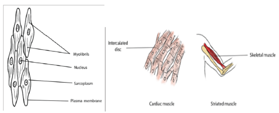
Figure 11.18: structure of three types of muscles
Application 11.3
Use prepared slides or charts of the three types of muscles and compare theircharacteristics.
11.4 Universal characteristics of muscles
Activity 11.4
Have you ever been injected medicine by intramuscular pathway? Or have
you been tested a rapid test of Covid-19?
In both cases the doctor or nurse advises in advance to keep calm so that
the muscle can relax and allow smooth flow of drugs through the muscle or
smooth flow of the strip through the nasal cavity.Why everybody including those without fear tend react on the same way?
The functions of muscle tissue are: movement, stability, control of body openings
and passages and heat production. To carry out those functions, all muscle tissuehas the following characteristics:
a. Responsiveness or excitability
Responsiveness is a property of all living cells, but muscle and nerve cells have
developed this property to the highest degree. When stimulated by chemical signals
(neurotransmitters), stretch, and other stimuli, muscle cells respond with electrical
changes across the plasma membrane.
b. Conductivity
Stimulation of a muscle fiber produces more than a local effect. The local electric
change triggers a wave of excitation that travels rapidly along the muscle fiber and
initiates processes leading to muscle contraction.
c. Contractility
Muscle fibers are unique in their ability to shorten substantially when stimulated.
This enables them to pull on bones and other tissues and create movement of the
body and its parts.
d. Elasticity
When a muscle cell is stretched and the tension is then released, it recoils to its
original resting length. Elasticity refers to the tendency of a muscle cell (or other
structures) to return to the original length when tension is released.11.4.1 Muscle contraction
Activity 11. 4.1
Using internet search simulations demonstrating the structure and functioning
of the sarcomere during muscle contraction with reference to sliding filamenttheory.
The excitability or the power of responding to an adequate stimulus is an innate
property of the muscle. When a brief stimulus is given, the muscle contracts and thisis followed by a wave of relaxation. This phenomenon is called a muscle twitch.
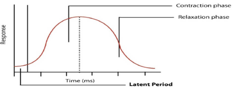
Figure 11.19: A muscle twitch
The Figure 11.19 shows a typical muscle curve of a skeletal muscle in response to
single stimulation. The muscle curve can be recorded with the help of a kymograph.
The curve indicates three phases: the latent phase, the contraction phase and the
relaxation phase. The period between the stimulus and beginning of contraction
is called the latent phase which lasts for about 0.01 second. During this period
chemical changes take place as a result of the stimulus. Latent period is required
for traversing the excitation along the nerve and the neuromuscular junctions. The
duration of the latent period varies with the species and depends on the type of
muscle, temperature and condition of the muscle.
The contraction phase during which the muscle actually contracts lasts for about
0.04 second in case of frog muscle. Shortening of the muscle takes place due to
chemical events which will be described in some details later. The third phase or
the relaxation phase lasts for about 0.05 sec. The total time taken by a single muscle
contraction is about 0.1 sec which varies with the temperature. At low temperature
contractions are prolonged, whereas with rising temperature the duration ofcontractions becomes shorter.
a. Muscle twitch, summation, and tetanus
A single action potential to the muscle fiber of a motor unit produces a muscle
twitch, a rapid and unstained contraction. If the impulses are applied to a muscle in
rapid succession through several motor units, one twitch will not have completely
ended before the next begins. Since the muscle is already in a partially contracted
state when the second twitch begins, the degree of muscle shortening in the second
contraction will be slightly greater than the shortening that occurs with a single
twitch. There are two types of twitch which are slow-twitch muscles and fast-twitchfibers.
– Slow-twitch are slower-contracting fibers but they are very efficient at using
oxygen to create energy without lactic acid build-up. These fibers are used for
high-endurance events like marathons.
– Fast-twitch fibers are white fibers, that contract very quickly making them
very strong and explosive but they also tire out very easily. The additional
shortening due to the rapid succession of two or more action potentials is
termed summation. At high stimulation frequencies, the overlapping twitchessum to one strong, steady contraction called tetanus.
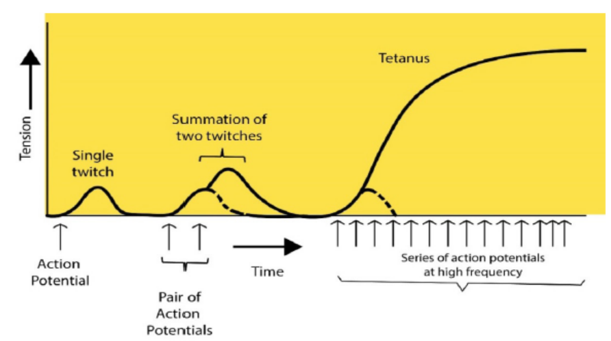
Figure 11.20: Patterns of muscle twitch, summation and tetanus
The graph 11.19 compares the tension developed in a muscle fiber in response to a
single action potential in a motor neuron, a pair of action potentials, and a series of
action potentials. The dashed lines show the tension that would have developed ifonly the first action potential had occurred.
b. Tetanic contractions
During normal activity such as locomotion, muscular contractions are not merely
twitches lasting for a second or a fraction of it. They are sustained for a longer period
during continued activity and exhibit compound or tetanic contractions. This can
be experimentally demonstrated by applying a number of stimuli to a muscle-nerve
preparation in rapid succession with little interval between successive stimuli, the
resulting contractions tend to fuse to give a maximum contraction. This sustained
contraction is called complete tetanus which, however, varies with the kind of
muscle and its condition. If repetitive stimuli are applied to muscle with long periods
of interval, the individual contractions can be seen because of little relaxation. Thiscondition is known as incomplete tetanus.
More interesting information is available about the tetanus. When a muscle is in tetany,
a musical note is produced by it which can be heard with the help of a stethoscope.
The pitch of the note is indicative of the vibrations that are produced at a rate
corresponding to the rate of application of stimuli. Most of the voluntary contractions
are of tetanus types which are produced by a series of nerve impulses arrivingin the muscle from the central nervous system.
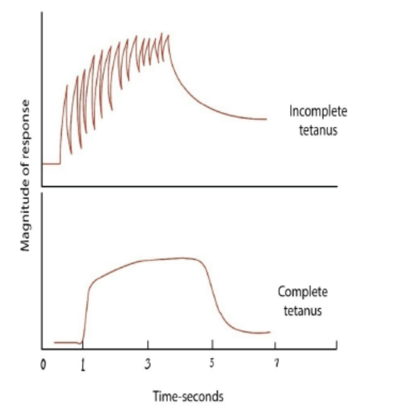
Figure 11.21: Diagram showing the condition of tetanus
a) The neuromuscular junction
This is a special kind of synapse where a motor nerve and muscle tissue meet. The
membrane of the muscle fiber, the sarcolemma is very folded in this region and
forms a structure known as an end plate. Electron microscopy shows us that the
structure of the neuromuscular junction is remarkably similar to that of any other
synapses. The end of the motor nerve is full of mitochondria and synaptic vesicles
which contain acetylcholine/neurotransmitter substances.
It appears that when an impulse arrives at the end of the motor neuron, it increases
permeability of the pre-synaptic membrane to calcium ions in the synaptic cleft. The
electrical impulse gets changed into a chemical message and gets stored into the
synaptic vesicles. The calcium ions then push the vesicles to fuse with the presynaptic
membrane thus discharging their neurotransmitter substances by exocytosis. The
neurotransmitter then diffuses through the synaptic cleft and get attached onto
receptor sites on the sarcolemma. This causes the sodium gated channels to open
thus causing a generator potential to be setup in the sarcolemma. If it reaches thethreshold, an impulse is fired into the muscle fiber.
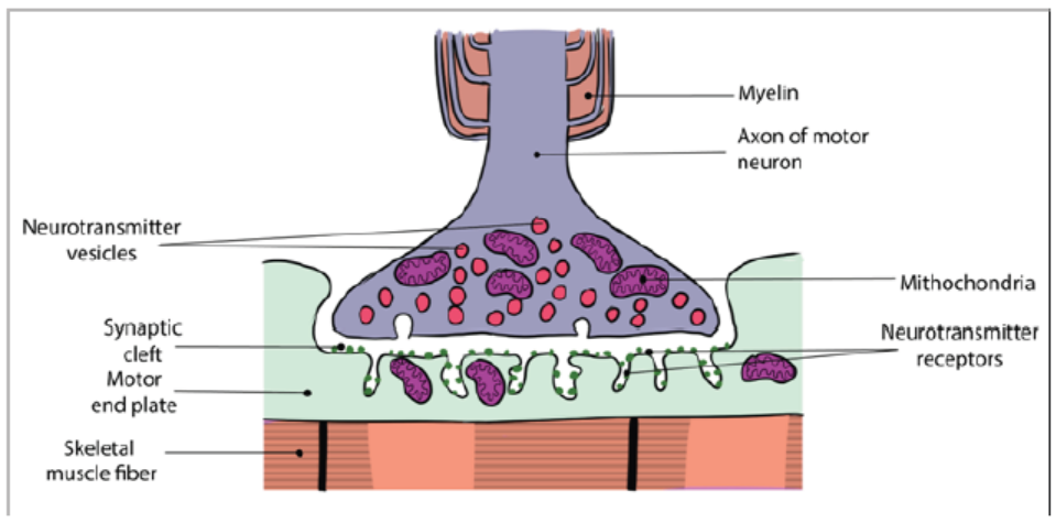
Figure 11.22: The neuromuscular junction
11.4.2 Laws of muscle contraction
A muscle contraction occurs when a muscle fiber generates tension through the
movement of actin and myosin. The sarcomere is the functional unit of muscle
contraction; it reaches from one Z-line to the next. In a relaxed muscle, the actin (thin
filament) and myosin (thick filament) overlap. In a muscle contraction, the filaments
slide past each other, shortening the sarcomere. This model of contraction is calledthe sliding filament mechanism.
Activity 11.4.2
Use of computer aided simulations to demonstrate the laws of musclecontraction (all or nothing, temporal summation and muscle fibre recruitment)
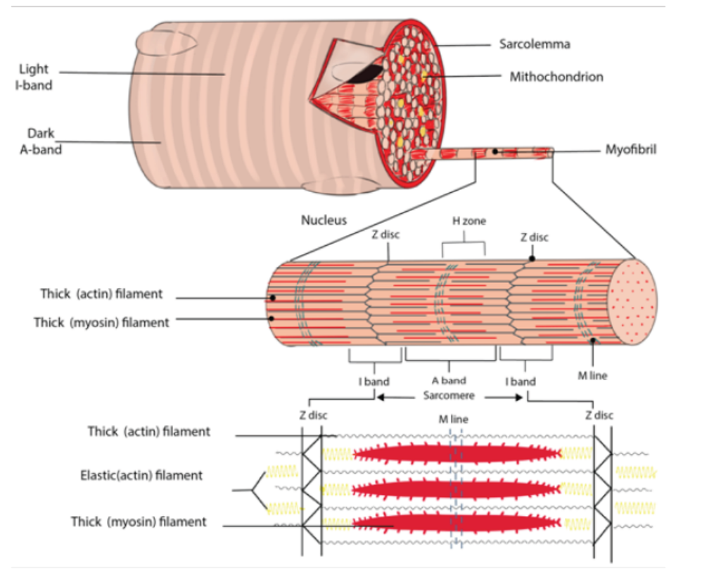
Figure 11.23: The sarcomere
Each muscle fiber contains cellular proteins and hundreds or thousands of myofibrils.
Each myofibril is a long, cylindrical organelle that is made up of two types of
protein filaments: actin and myosin. The actin filament is thin and threadlike; the
thin actin filaments are anchored to structures called Z lines. The region from one Z
line to the next makes up one sarcomere and the myosin filament is thicker. Myosin
has a head region that uses energy from ATP to walk along the actin thin filament.
The overlapping arrangement of actin and myosin filaments gives skeletal muscle
its striated appearance. When each end of the myosin thick filament moves along
the actin filament, the two actin filaments at opposite sides of the sarcomere are
drawn closer together and the sarcomere shortens. When a muscle fiber contracts,all sarcomeres contract at the same time, which pulls on the fiber ends.
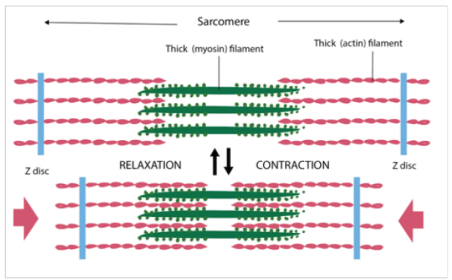
Figure 11.24: Muscle contraction
When each end of the myosin thick filament moves along the actin filament, the two
actin filaments at opposite sides of the sarcomere are drawn closer together and
the sarcomere shortens. In the contacted sarcomere, the A bands do not change in
length, but the I bands shorten and the H zone disappears. This behaviour can be
explained by the sliding filament model of muscle contraction.
How motor unit summation develops muscle tension
A skeletal muscle is an organ composed of multiple muscle cells or fibers, just like
any organ is made up of a whole bunch of cells. These fibers are arranged in motor
units, each of which is composed of a single motor neuron and all the muscle fibers
that that motor neuron innervates. Each motor unit contracts in an all-or-none fashion.
In other words, if the motor neuron is excited, it will stimulate all of the musclefibers to contract - that is, all of the muscle fibers within that particular motor unit.
11.4.3 Antagonistic skeletal muscles
Activity 11.4.3
Observe the following biceps and triceps muscles through the books and
internet and note down your observations (shortening and thickening of theantagonistic muscles).
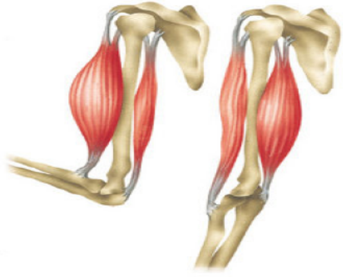
Antagonistic muscles are pairs of muscles. The action of one member is opposite to
that of the other member. Muscles can contract but they do not have the ability to
lengthen (stretch) themselves. They are arranged in pairs such that after one muscle
or muscle group contracts, a skeleton transfers the movement to stretch another
muscle or muscle group. The pairs of muscles that stretch each other are said to beantagonistic.
The biceps and triceps muscles of the arm are an example of an antagonistic pair.
Contraction of the biceps moves the arm toward the body and stretches the triceps.
Contraction of the triceps extends the arm and stretches the biceps. In this example
the bicep is said to be the flexor while the triceps is the extensor. Extensors are notas strong as flexors.
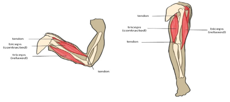
Figure 11.25: The antagonistic skeletal muscles
11.4.4 Movement in animals
Locomotion refers to the movement that causes a progression from one place to
another. There are several different types of locomotion exhibited by the animal
kingdom. It could either be active or passive. Sessile are animals that spend most
of their adult life in one place. Animals that move around are called motile. Corals,
sponges are examples of sessile organisms.
The act of flying is called aerial locomotion. Many organisms including; birds, insects,
bats, flying squirrels, many aquatic species and some amphibians including frog
have learnt to fly or glide.
Arboreal locomotion refers to species that live in and move through trees. Leopards
are good climbers that can climb up the tree along with their hunted prey to
keep them safe from other predators. The challenges of arboreal locomotion
include walking on narrow branches, moving up and down the inclines, balancing,
swinging with arms from one handhold to another and crossing gaps. Cats, parrots,
chameleons, goats, lizards and tree snakes are few examples of arboreal animals.
The movement on water is called aquatic locomotion. This involves swimming or
walking on the bottom surface of sea or ocean. Fish, ducks, bacteria, turtles, flat
worms, inchworms, leeches are organisms that can move through a liquid medium.
Most terrestrial animals move about using cursorial locomotion. Running
adaptation of different animals is referred to as cursorial locomotion. Forelimbs and
hind limbs play different roles in cursorial four-footed animals. These animals are
accustomed to long distance running at high speeds rather than high acceleration
over short distances. Cheetahs, wolves, ostriches are known for their cursorial
locomotion. Movement of animals that dig and live underground possess is called
fossorial locomotion. Such animals penetrate soil, wood or stone. Many soft bodiedinvertebrates, moles, earthworms and sea cucumbers are examples of organisms
with fossorial locomotion. Animals using hopping or jumping to move possess
saltatorial locomotion. Kangaroos, rabbits and few rodents exhibit saltatorialmotion.
Application 11.4
The diagram below is a drawing of the forelimb skeleton and part of theshoulder girdle of a mammal (rabbit).
Complete the diagram to show the following muscles and their attachments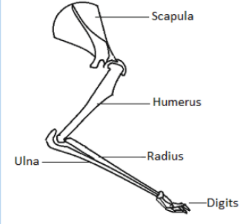
to the skeleton.
1. A muscle which contracts in preparation for landing after a leap. Label
this muscle X.
2. A muscle which will extend the digits. Label this muscle Y.
3. A muscle which will contract in response to a painful stimulus applied
to the forefoot. Label this muscle Z.11.5 Ultrastructure and functioning of striated muscle
Activity 11.5
Use the books from the school library and search further information from theinternet. Discuss Ultrastructure and functioning of striated muscle.
a. Ultrastructural appearance of skeletal muscle
The striated appearance of skeletal muscle fibres arises due to the organization
of two contractile proteins or myofilaments. The functional unit of contraction in a
skeletal muscle fibre is the sarcomere, which runs from Z line to Z line. A sarcomereis broken down into a number of sections:
– Z line – Where the actin filaments are anchored.
– M line – Where the myosin filaments are anchored.
– I band – Contains only actin filaments.
– A band – The length of a myosin filament, may contain overlapping actin
filaments.
– H zone – Contains only myosin filaments.
A useful acronym is MHAZI – the M line is inside the H zone which is inside the Aband, whilst the Z line is inside the I band.
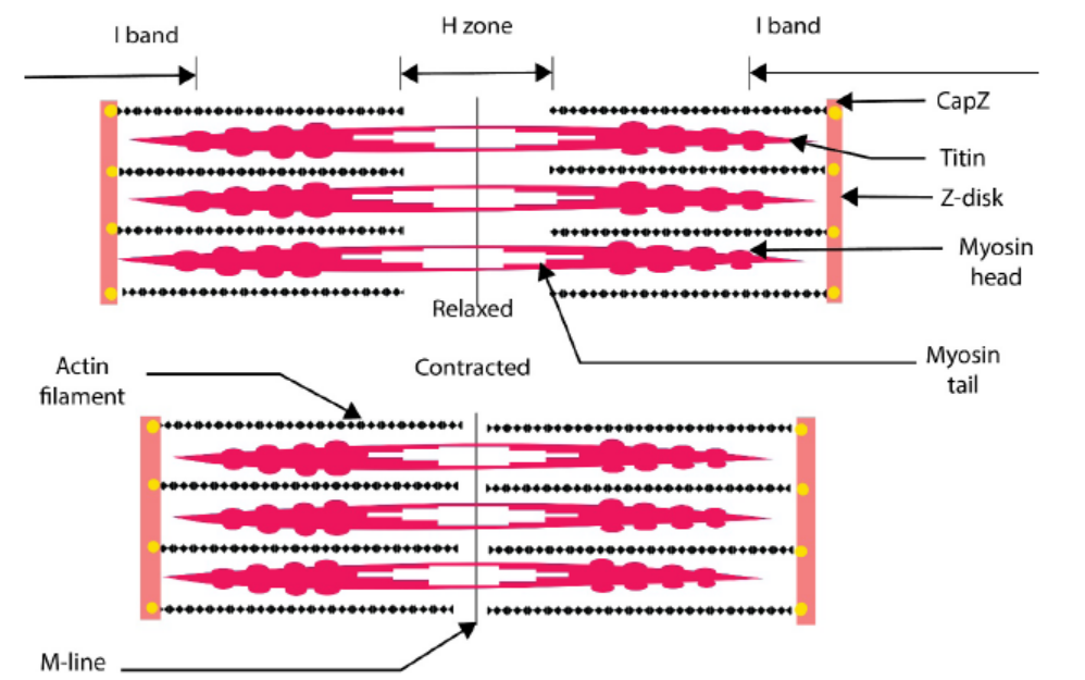
Figure 11.26: The sarcomere in contraction and relaxation
a. Function of striated muscles
Based on their fibrous and dense tissues, their main function is movement through
continuous contraction and relaxation. These muscles also help in; maintainingposture, stabilizing skeletal joints and producing body heat.
Application 11.5
1. Write on your own word the ultrastructure of muscle.
2. How many contractile proteins or myofilaments which constitute the
skeletal muscle fibres?3. What is the function of striated muscle?
11.6 Sliding filament theory of muscle contraction
Activity 11.6
Use the books from the school library and search further information from the
internet. Read and make summary about the sliding filament theory of musclecontraction.
The widely accepted theory of how muscles contract is called the sliding-filament
model also known as the sliding filament theory. According to this model, neither the
thin filaments nor the thick filaments change in length when the muscle contracts.
At rest, there is a low concentration of Ca2+ ions in the sarcomere, and the tropomyosin
blocks the actin sites to which myosin can bind. Upon arrival of an impulse, the
synaptic vesicles release their neurotransmitter substance (e.g. acetylcholine, Ach)
into the synaptic cleft. When Ach attaches on specific receptor sites, it causes the
release of Ca2+ ions from the triad vesicles into the sarcoplasm. Ca2+ ions bind to
Troponin-Complex which is protein that is integral to muscle contraction in skeletal
muscle and cardiac muscle, but not smooth muscle.
Once activated, the myosin head moves out and binds to actin, forming an action
myosin cross-bridge. The hydrolytic breakdown of ATP accompanies cross-bridge
formation and energy released causes the myosin head to pull the actin filament
towards the centre of the sarcomere. This leads to the shortening of the sarcomere
length and the overall contraction of the skeletal muscle. Cross-bridge formation
and breakage is repeated many times and on each occasion a new bridge is formed
between myosin head and another actin subunit further along the myofibril. After
stimulation, an active cation pump returns the Ca2+ ions to the triad vesicles; the
reduction in the level of Ca2+ ions in the sarcoplasm occurs and relaxation of thesarcomere begins.
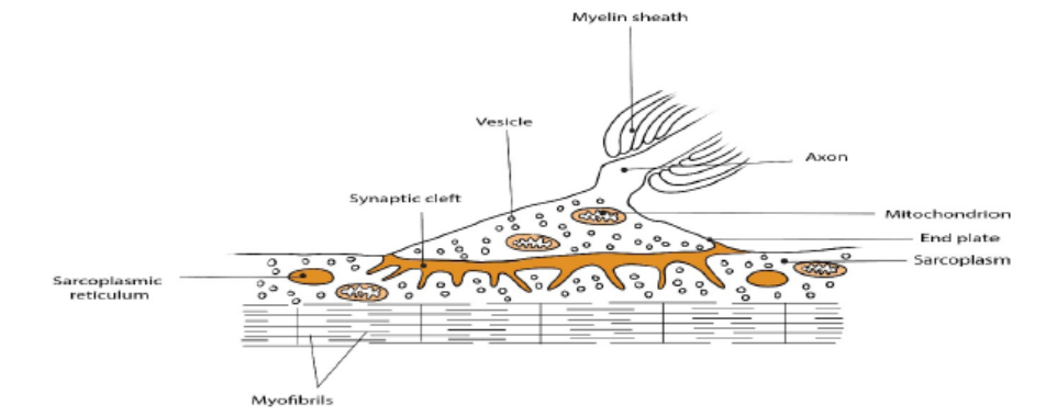
Figure 11.27: Neuromuscular junction or end plate
– When a muscle contracts, all the ATP present is rapidly used up. Replenishment
of ATP occurs when ADP and Pi are converted to ATP by phosphocreatine
breakdown. Later, after contraction has ceased, phosphocreatine is
reconstituted by ATP regeneration by energy from oxidation of fatty acids and
glycogen.
– In presence of adequate stimulus, the fibre contracts maximally. No further
increase in strength of stimulus will produce a stronger contraction this is
called all-or nothing response. A latent period of 0.05 seconds elapses prior to
muscle contraction.
– Contraction last for 0.1 second and is followed by a 0.2 second period of
relaxation. During this time, an absolute refractory is allowed by a relative
refractory period.
– When another stimulus is applied while the muscle is still responding to the
first stimulus, mechanical summation occurs whereby a second contraction
of greater force is caused. A rapid series of stimuli provokes a continued
contraction called tetanus. Tetanus ends when the muscle fatigues.
– If a muscle becomes very active, the respiratory and blood systems are unable
to supply sufficient oxygen for the muscle’s need. Consequently, pyruvic acid
is converted to lactic acid by the addition of H+ ions and the muscle builds up
an oxygen debt. Removal of lactic acid occurs when activity slows down or
ceases.
– The refractory period is the time after receiving a stimulus during which anerve or muscle cell cannot respond to further stimuli.
Application 11.6
1. Explain the sliding filament model of muscle contraction, including the
roles of troponin, tropomyosin, calcium ions and ATP.
2. Describe a neuromuscular junction?
3. What is the function of motor neurons?
4. Draw a well labelled diagram of sliding filament model of muscularcontraction.
End of unit assessment 11
1. What is the basic reason for the fact that animals show locomotion
whereas plants do not?
2. Briefly explain the role of each of the following in a mammalian locomotion:
3. Bones
a. Joints
b. muscles
4. What is meant by endoskeleton?
5. Outline the main functions of the endoskeleton.
6. Explain the various types synovial of joints.
7. In relation to antagonistic muscles, explain how it is possible to lift and
lower an object with your hands.
8. Outline the functions of fused joints and give an example.
9. What are the functions of muscle tissue?
10. What is the meaning of MHAZI in skeletal muscle fibres?
11. Explain what happened in refractory period in the sliding filament theory
of muscle contraction.
12. Explain what happened when mortar impulse reaches the end plate, the
vesicles release acetylcholine into the synaptic cleft of the end plate.
13. Draw a well labelled diagram of human skeleton.14. How does the structure of a muscle cell type relate to its function?