UNIT 10 HORMONAL COORDINATION IN ANIMALS
UNIT 10: HORMONAL COORDINATION IN ANIMALS
Key Unit Competence
To be able to identify the location and function of endocrine glands in the body.
Learning objectives
By the end of this unit, I should be able to:
– Define hormones.
– Explain why hormonal balance is necessary for coordinating the functions
in the body.
– Describe the principle of the negative feedback mechanism by which
hormones produce their effects on target cells.
– Describe the structure and function of the endocrine system.
– Explain the effects of hormonal imbalances.
– Compare and contrast the actions of the endocrine and nervous systems.
– Draw and interpret the flow chart of negative feedback mechanisms.
– Appreciate the role of hormones in the growth and development oforganisms.
Introductory activity
At a given time, there are certain changes which occur in the body especially
during puberty. As girls and boys enter the period of puberty, they start to
develop remarkable differences in physical appearance and in their behaviour.
1. What do you think to be the causes of such changes?
2. What are some changes which can be observed in boys and not in girls
and vice versa?
3. Which the organs do you think are responsible for producing such
changes?
4. What will be the causes if some of those changes do not appear in a boyor in a girl?
10.1 Structure and function of the endocrine system in humans
Activity 10.1
By using different books from the school library and a chart showing different
endocrine glands, discuss the following:
1. What are endocrine glands?
2. Draw and locate the following endocrine glands
a. The adrenal glands
b. The pancreas
3. What are the hormones produced by the pancreas and their functions?4. Why the pituitary gland was once described as a master gland?
A hormone is an organic substance which is produced in minute quantity by an
endocrine gland, transported by blood to other parts or organs of the body where it
exerts maximum effects. Such parts of the body or organs are called target organs.
The word endocrine means internal secretion and endocrine glands are therefore
glands of internal secretion. Since they shed their secretion into the bloodstream,
they have no ducts and are hence known as ductless glands.
Hormones are released into the blood stream as a result of:
1. Stimulation of the endocrine gland directly by the nervous system e.g. the
sympathetic nervous system causes secretion of adrenaline by the adrenal
medulla.
2. The levels of particular metabolites in the blood e.g. glucose levels trigger
the release of insulin.
3. Presence of other hormones called releasing hormones mostly produced in
anterior pituitary e.g. TSH stimulates the release of thyroxin by the thyroid
gland.
4. Environmental changes such as high or low temperatures effects activities
of the pituitary gland.5. Animals’ general mental state does affect the activity of the pituitary.
Once in the bloodstream, the hormones are carried around the body, bringing
about responses in various places. Structures that respond to them are called targetorgans. A hormone is a chemical messenger having the following properties:
– It travels in the blood
– It has its effect at a site different from the site where it is produced. The site
where it has effect is called the target, while itself is called messenger
– It fits precisely into receptor molecules in the target like a key in a lock. It is
therefore specific for a particular target;– It is a small soluble molecule;
It is effective in low concentrations.
Hormones fulfill many functions. These include:
1. Regulation of growth and development.
2. Controls homeostasis e.g.in osmoregulation/thermo regulation etc
3. Regulation of metabolism e.g. digestion storage and utilization of food
substances.
4. Development of the skin coloration.
5. Enabling the body to withstand shock, tension wounding etc. and to
recover from it.
6. Together with the nervous system it provides for effective responses to all
kinds of stimuli both internal and external.
The endocrine glands include; the pituitary, thyroid, parathyroid, adrenal, and pineal
glands (Figure 10.1 below). Taken together, all endocrine glands and hormone-
secreting cells constitute the endocrine system.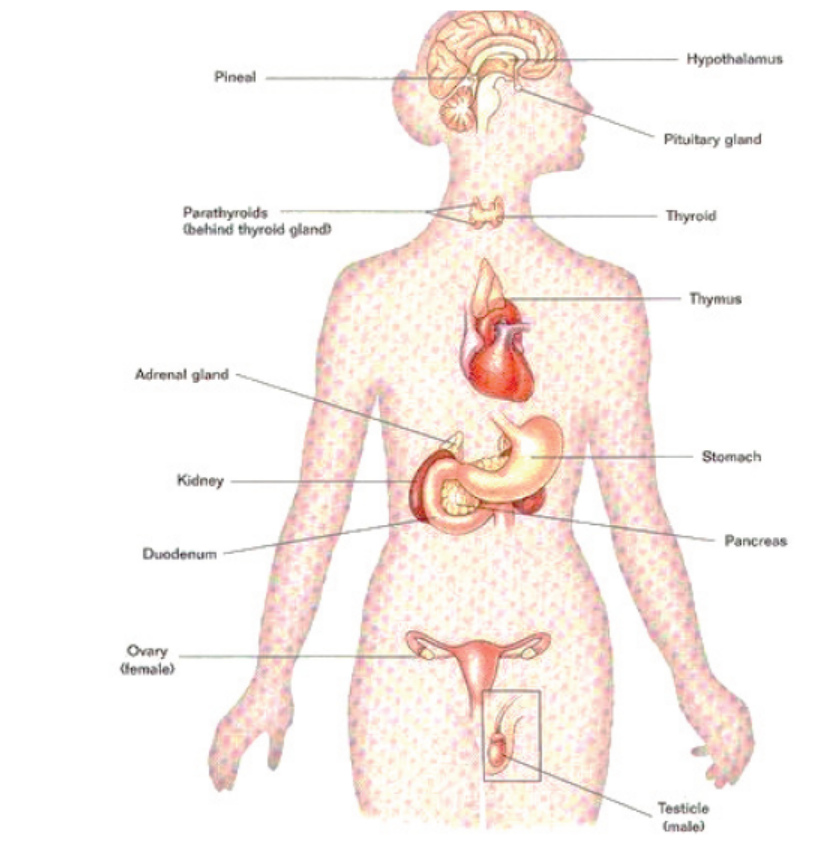
Figure 10.1: Major endocrine glands
a. The pituitary gland
The pituitary gland which was formerly called the master gland hangs from the
base of the brain by a stalk and is enclosed by bone. As shown in figure 10.2, the
pituitary gland consists of a hormone-producing glandular portion called anterior
pituitary and a neural portion called posterior pituitary, which is an extension of
the hypothalamus. The hypothalamus now called the master gland, regulates the
hormonal output of the anterior pituitary and synthesizes two hormones that it
exports to the posterior pituitary for storage and later release. Most anterior pituitary
hormones exhibit a diurnal rhythm of release, which is subject to modification bystimuli influencing the hypothalamus.
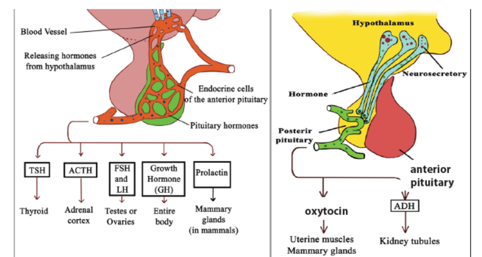
Figure 10.2: Pituitary and hypothalamic secretions
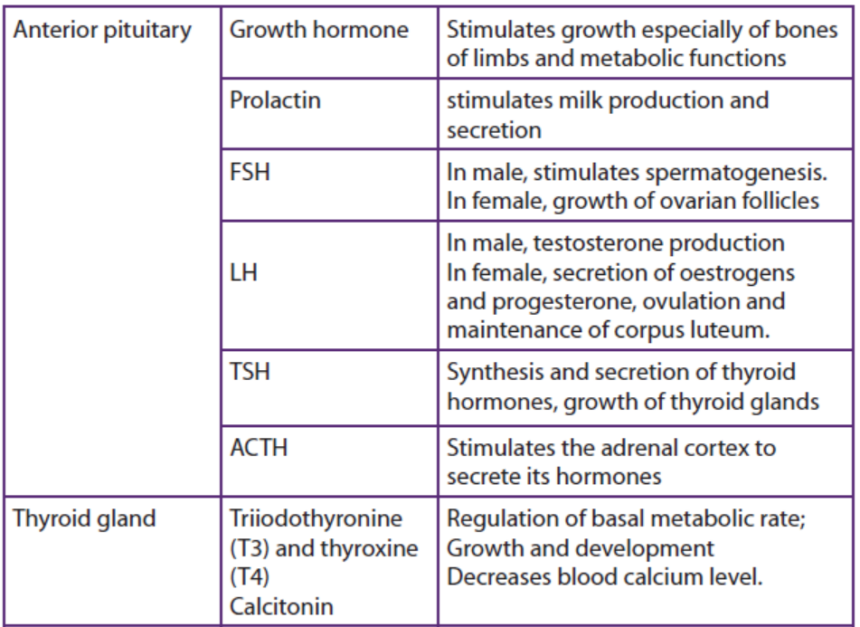
The following are the hormones produced by the anterior pituitary gland;
– Growth hormone (GH) or Somatotropic hormone: is a hormone that
stimulates growth of all body tissues especially skeletal muscle and bone.
GH; mobilizes the use of fats, stimulates protein synthesis, and promotes
glucose uptake and metabolism.
– Thyroid-stimulating hormone (TSH) This hormone causes thyroid glands
to secrete thyroxin. The secretion of TSH is controlled by levels of thyroxin
in blood. TSH also stimulates growth of thyroid gland.
– Adrenocorticotropic hormone (ACTH) stimulates the adrenal cortex to
release its hormones. ACTH release is triggered by corticotropin-releasing
hormone (CRH) and inhibited by rising glucocorticoid levels.
– The gonadotropins: follicle-stimulating hormone (FSH) and luteinizing
hormone (LH) regulate the functions of the gonads in both sexes.
– Prolactin (PRL) promotes the production of milk in human’s females. Its
secretion is triggered by prolactin-releasing hormone (PRH) and inhibited
by prolactin-inhibiting hormone (PIH).
The following are the two hormones released from the posterior pituitary gland:
– Oxytocin: It stimulates powerful contractions of the uterus, which trigger
labour and delivery of an infant, and milk ejection in nursing women. Its
release is mediated reflexively by the hypothalamus and represents a
positive feedback mechanism.
– Antidiuretic hormone (ADH) stimulates the kidney tubules to reabsorb
and conserve water, resulting in small volumes of highly concentrated urine
and decreased plasma osmolality.
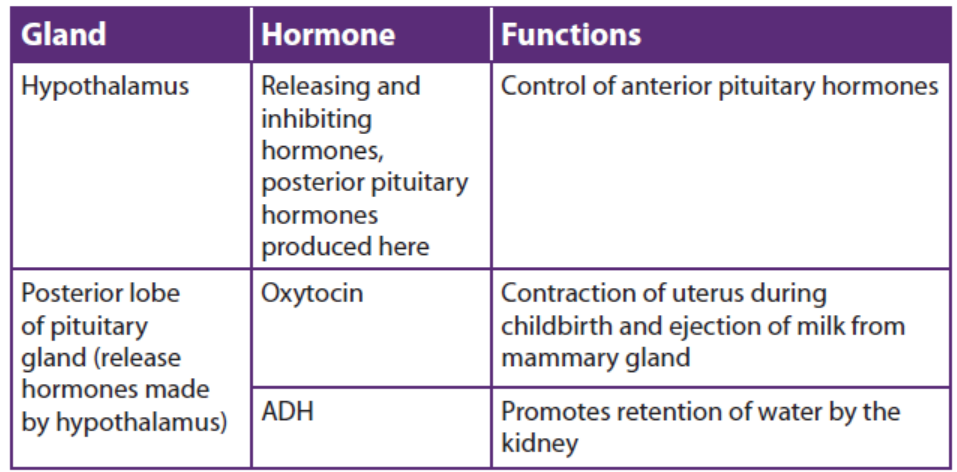
b. The hypothalamus
The hypothalamus plays an important role in integrating the endocrine and
nervous systems. The region of the lower brain receives information from nerves
throughout the body and from other parts of the brain thus initiates endocrine
signals appropriate to environmental conditions. The reason it is called a master
gland is that a set of neurosecretory cells in the hypothalamus exerts control over
the anterior pituitary by secreting two kinds of hormones into the blood: Releasing
hormones which make the anterior pituitary to secrete its hormones and inhibiting
hormones that make the anterior pituitary stop secreting hormones. Every anterior
pituitary hormone is controlled at least by one releasing hormone and some have
both a releasing and an inhibiting hormone.
The posterior pituitary remains attached to the hypothalamus. It stores and releases
two hormones that are made by a set of neurosecretory cells in the hypothalamus.
c. Thyroid gland
The thyroid gland is located in the anterior throat. Thyroid follicles store colloid
containing thyroglobulin, a glycoprotein from which thyroid hormone is derived.
Thyroid hormone (TH) includes thyroxine (T4) and triiodothyronine (T3), whichperform the following tasks;
– Control the basal metabolic rate.
– Increase the rate of cellular metabolism. Consequently, oxygen use and
heat production rise.
Calcitonin, is another hormone produced by the thyroid gland in response to rising
blood calcium levels. Its role is to decrease blood calcium levels by inhibiting bonematrix reabsorption and enhancing calcium deposit in bone.
d. Parathyroid glands
The parathyroid glands are located on the dorsal aspect of the thyroid gland and secrete
parathyroid hormone (PTH), which causes an increase in blood calcium levels by;
– Increasing the rate of calcium reabsorption by the kidney at the expense of
phosphate ions.
– Increasing the rate of calcium absorption from the gut.– Causing the release of calcium reserves from the bones.
PTH is antagonistic calcitonin. PTH release is triggered by decreasing blood calciumlevels and is inhibited by increasing blood calcium levels.
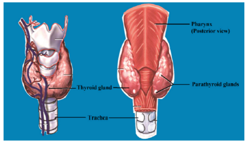
Figure 10.3: The location of the thyroid and the parathyroid glands
e. Pancreas
The pancreas is an organ located in the abdomen close to the stomach and is both
an exocrine and an endocrine gland. The endocrine portion (islets of Langerhans)
releases insulin and glucagon hormones. Glucagon is released by alpha (α) cells
when glucose levels in blood are low. Glucagon stimulates the liver to convert stored
glycogen to glucose thus increasing glucose levels. Insulin is released by beta (β)
cells of the islets of Langerhans when blood levels of glucose are rising. It increases
the rate of glucose uptake and causes the conversion of glucose to glycogen.
f. Gonads
The ovaries of the female which are located in the pelvic cavity, release two main
hormones. Secretion of oestrogen by the ovarian follicles begins at puberty under
the influence of FSH. Oestrogen stimulates maturation of the female reproductive
system and development of the secondary sex characteristics. Progesterone
is released in response to high blood levels of LH. It works with oestrogen in
establishing the human menstrual cycle. The testes of the male begin to produce
testosterone at puberty in response to LH. Testosterone stimulates the maturation
of the male reproductive organs, development of secondary sex characteristics, and
the production of sperm by the testes.
g. Adrenal Glands (Suprarenal Glands)
A fresh adrenal gland section shows a bright yellow cortex, making up about 80% of
the organ, and a more reddish-grey medulla. The endocrine activities of the adrenalcortex and the adrenal medulla differ both in development and function.
• Adrenal cortex
Adrenal cortex makes mineralocorticoids (such as aldosterone and cortisol). Cortisol
raises blood glucose level whereas aldosterone stimulates the reabsorption of Na+
and excretion of K+ in kidney.
• Adrenal Medulla
The adrenal medulla makes two hormones epinephrine (adrenaline) (80 %) and
norepinephrine (noradrenaline) (20 %). Epinephrine and norepinephrine are released
into the bloodstream during stress and they act on the whole organism by preparing
it for increased energy use. Both hormones, for instance, activate the liberation of
fatty acids from fat depots and liberate glucose from glycogen storage in the liver
(producing a rise in the blood sugar level). They raise the blood pressure and stroke
volume of the heart and may lead to vasoconstriction in certain defined areas.
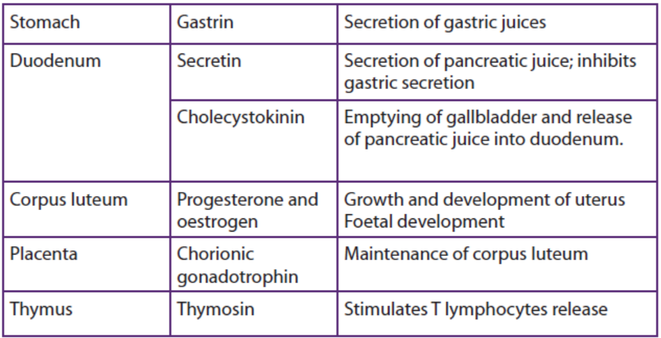
h. Other hormone-producing structures
Many body organs not normally considered endocrine organs contain isolated cell
clusters that secrete different hormones. Examples include the; gastrointestinal tract
organs (gastrin, secretin, and others), the placenta (hormones of pregnancy such as
oestrogen, progesterone, and others) and the kidneys (erythropoietin and renin).
Table 10.1: Major human endocrine glands, their functions and the control oftheir secretions
Application 10.1
1. What are the hormones produced by the thyroid glands?
2. What are the functions of the hormones stored and released by the
posterior pituitary gland?3. By which means are hormones transported in our body?
10.2 Principles of the negative feedback mechanism of hormonalaction.
Activity 10.2
1. The amount of urine produced varies according to the amount of water
consumed. Make a list of events that may occur in the following cases for the
human body:
a. Two days without drinking water
b. When you have drunk 1 litre of water per day
c. How can you explain the above observations?
2. Make short notes on what happens to your body if the level of sugars decreasesin the blood?
Feedback mechanisms are necessary in the maintenance of homeostatic mechanisms.
All homeostatic control mechanisms have at least three interdependent componentsfor the variable being regulated that work together i.e.
a. The receptor is the sensing component that monitors and responds to
changes in the environment. When the receptor senses a stimulus, it sends
information to a control center, the component that sets the range at which
a variable is maintained.
b. The control center determines an appropriate response to the stimulus. In
most homeostatic mechanisms, the control center is the brain.
c. An effector, which can be muscles, organs or other structures that receive
signals from the control center. After receiving the signal, a change occurs
to correct the deviation by either enhancing it with positive feedback ordepressing it with negative feedback.
The homeostatic mechanisms in mammals require information to be transferred
between different parts of the body. There are two coordination systems in mammals
that control this: the nervous system and the endocrine system.
– In the nervous system, information in the form of electrical impulses is
transmitted along nerve cells (neurons).
– The endocrine system uses chemical messengers called hormones thattravel in the blood, in a form of long-distance cell signalling.
10.2.1 Positive feedback mechanisms
These mechanisms are designed to accelerate or enhance the output created by
a stimulus that has already been activated. The positive feedback mechanisms are
designed to push levels out of normal levels. To achieve this purpose, a series of
events initiates a cascading process that builds to increase the effect of the stimulus.
This process can be beneficial but is rarely used by the body due to risks of the
acceleration’s becoming uncontrollable.
Examples include; the accumulation blood platelets which in turn causes blood
clotting in response to a break or tear in the lining of blood vessels. The release of
oxytocin to intensify the contractions of the uterus that take place during childbirth.
Another example of a positive feedback mechanism is the production of milk by
a mother for her baby. As the baby suckles, nerve messages from the mammary
glands cause the mother’s pituitary gland to secrete a hormone called prolactin.
The more the baby suckles, the more prolactin is released, which stimulates further
milk production by the mother’s mammary glands. In this case, a negative feedback
mechanism would not be helpful because the more the baby nursed, the less milkwould be produced.
12.2.2 Negative feedback mechanisms
These are mechanisms concerned with keeping changes in the factor within narrow
limits. Here, an increase in a factor (input) e.g. hormone levels results in somethinghappening that makes the factor decrease (output).
An example of negative feedback mechanism is regulation of thyroxine levels i.e. The
shedding of thyroxine into blood stream is triggered by thyrotropin releasing factor
(TRF) produced by the hypothalamus of the brain. TRF passes to the pituitary gland
along the blood vessels stimulating the anterior pituitary gland to produce Thyroid
stimulating hormones (TSH). TSH then stimulates the thyroid gland to produce
thyroxine into blood. A slight excess of thyroxine in blood detected by hypothalamus,
inhibits the anterior lobe of the pituitary gland which responds by secreting less
TSH. This in turn reduces the activity of the thyroid gland, leading to decrease in
the amount of the thyroxine produced. This removes the inhibitory influence on thepituitary so that more thyroid stimulating hormone will be produced again.
Table 10.2: Negative and positive feedback compared
Application 10.2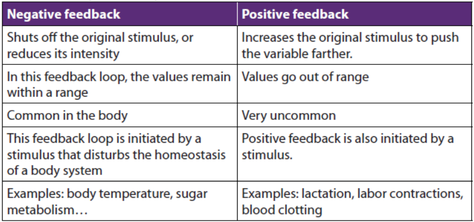
1. State any two examples of positive feedback.2. Why is the positive feedback not useful in many homeostatic mechanisms?
10.3 Effects of hormonal imbalances
Activity 10.3
1. You may know people suffering from diabetes mellitus or you may have heard
about the disease from the radio or from a newspaper.
a. Collect information about the cause of this disease.
b. Predict the ways this disease can be treated.
2. Observe carefully figure 10.4 below and suggest the type of disorders thefollowing people may be suffering from:
The disorders of the endocrine system often involve either the hypo-secretion (hypo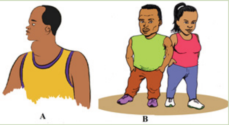
means too little or under), inadequate release of a hormone, or the hyper-secretion
(hyper means too much or above), excessive release of a hormone. In other cases,
the problem is faulty hormone receptors, an inadequate number of receptors, or
defects in second-messenger systems. Because hormones are distributed in the
blood to target tissues throughout the body, problems associated with endocrinedysfunction may also be widespread.
10.3.1. Pituitary Gland Disorders
a. Pituitary dwarfism, gigantism, and acromegaly
Several disorders of the anterior pituitary involve human growth hormone.
Hyposecretion of human growth hormone during the growth years slows bone
growth, and the epiphyseal plates close before normal height is reached. This
condition is called pituitary dwarfism. Other organs of the body also fail to grow,
and the body proportions are childlike. Treatment requires administration of humangrowth hormone during childhood, before the epiphyseal plates close.
Hypersecretion of human growth hormone during childhood causes gigantism, an
abnormal increase in the length of long bones. The person grows to be very tall,
but body proportions are about normal. Hypersecretion of human growth hormoneduring adulthood is called acromegaly.
b. Diabetes insipidus
The most common abnormality associated with dysfunction of the posterior pituitary
is diabetes insipidus. This disorder is due to defects in antidiuretic hormone (ADH)
receptors or an inability of the pituitary gland to secrete ADH. A common symptoms
of diabetes insipidus are: excretion of large volumes of urine resulting in dehydration
and thirst. Bed-wetting is common in afflicted children. Because so much water is
lost in the urine, a person with diabetes insipidus may die of dehydration if deprived
of water for only one day. Treatment of diabetes insipidus involves the injection ofADH into the body.
10.3.2. Thyroid gland disorders
Thyroid gland disorders affect all major body systems and are among the most
common endocrine disorders. Congenital hypothyroidism or the hyposecretion
of thyroid hormones that is present at birth has devastating consequences if not
treated quickly. Previously termed cretinism, it causes severe mental retardation and
stunted bone growth. At birth, the baby typically is normal because lipid-soluble
maternal thyroid hormones crossed the placenta during pregnancy and allowednormal development.
Hypothyroidism during the adult years produces a disorder called myxoedema. An
indication of this disorder is oedema (accumulation of interstitial fluid) that causes
the facial tissues to swell and look puffy. A person with myxoedema has a slow
heart rate, low body temperature, sensitivity to cold, dry hair and skin, muscular
weakness, general lethargy, and a tendency to gain weight easily. Because the brain
has already reached maturity, mental retardation does not occur, but the person
may be less alert.
The most common form of hyperthyroidism is Graves’ disease which is an autoimmune
disorder in which the person produces antibodies that mimic the action of thyroidstimulating
hormone (TSH). The antibodies continually stimulate the thyroid gland
to grow and produce thyroid hormones. A primary sign is an enlarged thyroid,
which may be two to three times its normal size. Graves’ patients often have a
peculiar oedema behind the eyes, called exophthalmos, which causes the eyes to
protrude. Treatment may include surgical removal of part or all of the thyroid gland
(thyroidectomy), the use of radioactive iodine to selectively destroy thyroid tissue,
and the use of anti-thyroid drugs to block synthesis of thyroid hormones. A goitre
is simply an enlarged thyroid gland. It may be associated with hyperthyroidism,hypothyroidism or by the lack of iodine.
10.3.3. Parathyroid gland disorders
Parathyroid gland disorders cause the hypoparathyroidism due to the too little
parathyroid hormone leading to a deficiency of blood Ca2+, causing neurons and
muscle fibres to depolarize and produce action potentials spontaneously. This leads
to twitches, spasms, and tetany (maintained contraction) of skeletal muscle. The
main cause of hypoparathyroidism is accidental damage to the parathyroid glands
or to their blood supply during thyroidectomy surgery.
Hyperparathyroidism or an elevated level of parathyroid hormone, most often
is due to a tumour of one of the parathyroid glands. An elevated level of PTH
causes excessive resorption of bone matrix, raising the blood levels of calcium and
phosphate ions and causing bones to become soft and easily fractured. High blood
calcium level promotes formation of kidney stones. Fatigue, personality changes,
and lethargy are also seen in patients with high levels of parathyroid hormone.10.3.4. Adrenal gland disorders
a. Cushing’s syndrome
Hypersecretion of cortisol by the adrenal cortex causes an endocrine disorder
known as Cushing’s syndrome. The condition is characterized by breakdown
of muscle proteins and redistribution of body fat, resulting in thin arms and legs
accompanied by a rounded moon face and buffalo hump on the back. Facial skin is
flushed, and the skin covering the abdomen develops stretch marks. The person also
bruises easily, and wound healing is very slow. The elevated level of cortisol causes
hyperglycaemia, osteoporosis, weakness, hypertension, increased susceptibility to
infection, decreased resistance to stress, and mood swings.
b. Addison’s disease
Hyposecretion of glucocorticoids and aldosterone causes Addison’s disease (chronic
adrenocortical insufficiency). The majority of cases are autoimmune disorders in
which antibodies cause adrenal cortex destruction or block binding of ACTH to
its receptors. Pathogens, such as the bacterium that causes tuberculosis, also may
trigger adrenal cortex destruction. Symptoms, which typically do not appear until
90% of the adrenal cortex has been destroyed, include mental lethargy, anorexia,
nausea and vomiting, weight loss, hypoglycemia, and muscular weakness. Loss of
aldosterone leads to the elevated potassium and decreased sodium in the blood,low blood pressure, dehydration, decreased cardiac output and even cardiac arrest.
10.3.5. Pancreas disorders
The most common endocrine disorder is diabetes mellitus caused by an inability
to produce or use insulin. According to the diabetes atlas of 2018, the prevalence
of diabetes in Rwanda is about 3.16% of the population with 1,918 diabetes
related deaths per year. Its complications can lead to heart attack, stroke, blindness,kidney failure and lower limb amputation.
Because insulin is unavailable to aid transport of glucose into body cells, blood glucose
level is high and glucose is found in the urine, the process known as glycosuria.
The cardinal signs of diabetes mellitus are; polyuria (excessive urine production due
to an inability of the kidneys to reabsorb water), polydipsia (excessive thirst) andpolyphagia (excessive eating).
Application 10.3
1. Which disorders are caused by:
a. Hypersecretion of GH in children?
b. Hyposecretion of insulin?
c. Hypersecretion of thyroid hormones?
2. What are some symptoms of?
a. Diabetes mellitus?b. Grave’s disease?
10.4 Comparison of hormonal and nervous systems
Activity 10.4
Use your knowledge of the nervous system and the endocrine system to
answer the questions that follow
1. Discuss the similarities between the structure and functioning of nervous
and hormonal systems.
2. Discuss the differences between the structure and functioning of nervousand hormonal systems.
The nervous and endocrine systems act together to coordinate functions of all our
body systems.
A basic similarity between the endocrine system and the nervous system is that both
provide means of communication within the body of an organism. Both involve
transmission of a message which is triggered by a stimulus and produces a response.
Several chemicals function as both neurotransmitters and hormones including
norepinephrine. Some hormones such as oxytocin are secreted by neuroendocrine
cells; neurons that release their secretions into the blood. The target organs of a
hormone are equivalent to nerve’s effectors.
Similarities
1. Both provide a means of communication and coordination in the body.
2. In both the information transmitted is triggered by a stimulus and produces
a response.
3. Both involve chemical transmission.4. Both are controlled by the brain.
The main differences between the two systems concern the nature of the message.
In the endocrine system, the message takes the form of a chemical substance
transmitted through the blood stream. In the nervous system it is a discrete-all or
none action potential transmitted along a nerve fibre. All other differences arise
from this fundamental one. They can be listed as follows:
– Because of the comparatively high speed at which impulses are transmitted
along nerves, nerves responses are generally transmitted more rapidly than
hormonal ones.
– Since it is conveyed by the bloodstream, there is nothing to stop a hormone
being carried to every part of the body. Nervous impulses however are
transmitted by particular neurons to specific destinations.
– As a result, hormones are often widespread, sometimes involving the
participation of numerous target organs. In contrast, nervous responses
may be much localized, involving perhaps the contractions of only one
muscle.
– Hormonal responses frequently continue over a long period of time. Obviousexamples of such long-term responses are growth and metabolism.
A comparison between nervous and endocrine system is summarized in the table
10.3Table 10.3: Comparison between nervous and endocrine system
Application 10.4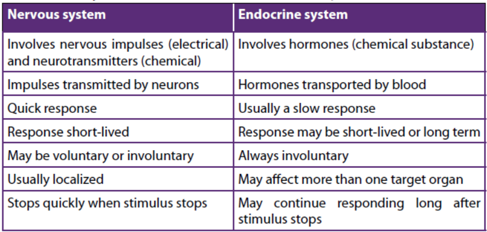
Describe a short term effect of the endocrine system and a long term effect ofthe nervous system.
End of unit assessment 10
Multiple choice questions: from question 1 to 5, choose the letter
corresponding to the best answer
1. What are the chemical messengers of the endocrine system called?
a. Neurons
b. Hormones
c. Blood cells
d. Carbohydrates
2. Endocrine glands
a. Function only after puberty
b. Function only before puberty
c. Release products through ducts
d. Release products into bloodstream
3. X and Y are hormones. X stimulates the secretion of Y, which exerts negative
feedback on the cells that secrete X. Suppose the level of Y decreases. What
should happen immediately afterwards?
a. Less X is secreted
b. More X is secreted
c. Secretion of Y stops
d. Secretion of X stops
4. Which one of the following hormones is secreted by the neurosecretory cells in
mammals?
a. Adrenaline
b. Antidiuretic hormone
c. Insulin
d. Thyroxin
5. Select the hormone INCORRECTLY paired with its target.
a. TSH - thyroid gland
b. LH - ovary or testis
c. ACTH - anterior pituitaryd. ADH - kidney
6. A number of metabolic diseases in mammals arise as a result of abnormalendocrine function. Complete the table below concerned with this:
7. During the control of blood sugar in a mammal two antagonistic hormones are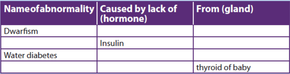
employed. Fill in the table about them
8. Name the hormone involved in the functions described below and the name of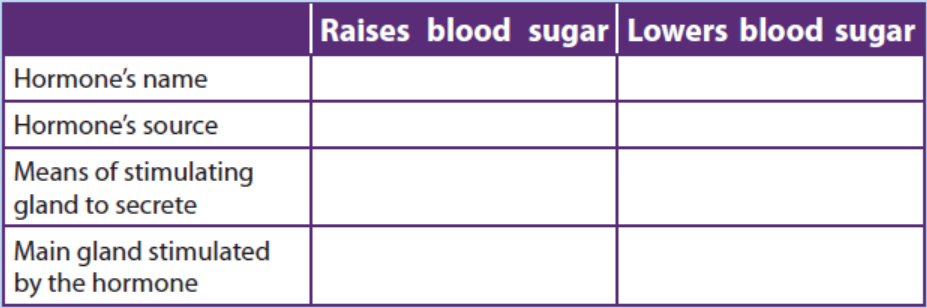
the gland which produces it:
a. Controls reabsorption of Na+ in the kidney.
b. Increases the permeability of convoluted distal tubule and collecting
duct.
c. Increases heart rate.
d. Increases blood glucose level.
e. Decreases blood glucose level.
f. Repair and growth of the endometrium.
g. Stimulates the anterior pituitary gland to release FSH.
h. Stimulates contraction of the uterus.
i. Stimulates the mammary glands to secrete milk.
9. What is the difference between diabetes mellitus and diabetes insipidus? What
are the characteristic signs of diabetes insipidus?
10. Use the following to describe a negative feedback mechanism: TSH, TRH,decreased metabolic rate, thyroxine and T3.