Unit 9 Gas Exchange in Animals
Key Unit Competence
To be able to describe structures of gas exchange in different groups of animals.
LEARNING OBJECTIVES
At the end of this unit, the learner will be able to:
• Describe the tracheal system of insects and relate to its function.
• Describe the structure of the gills in relation to function.
• Explain the significance of counter current flow in bony fish.
• Dissect an insect, fish and a small mammal to study gaseous exchange organs.
• Relate the structure of gas exchange organs to function.
• Differentiate between the gaseous exchange in bony fish and that in cartilaginous fish.
• Describe the mode of gaseous exchange in amphibians.
• Describe the structure of the human gas exchange system.
• Appreciate the similarities and differences in gas exchange surfaces of animals.
• Interpret a graph of human lung volumes measured with a spirometer.
• Calculate the volume of air in the lungs and in the alveoli.
• Describe the distribution of tissues within the trachea, bronchi, bronchioles and alveoli and
relate each tissue to its function.
• Explain the mechanism of ventilation in humans.
• Explain the process of gas exchange in alveoli with emphasis on diffusion.
• Describe the role of the brain in controlling gas exchange in humans.
• Appreciate the role of the brain in controlling gas exchange.
• Define terms related to the lung capacities (tidal, reserve volume, vital capacity, residual
volume, and dead air space).
• Describe how a spirometer can be used to measure vital capacity, tidal volume, breathingrates, and oxygen uptake.
INTRODUCTORY ACTIVITYObserve the following images and answer the following
1. According to your observation what is happening to each living organism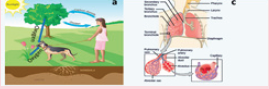
2. What are the types of gas the dog is giving out?
3. You are inhaling and exhaling as shown on the figure b. Search and find the description ofthe pathway through which that air is passing in your body
9.1 INTRODUCTION
ACTIVITY 1
Place your hands on your chest: You can feel the chest moving up and down. You know it is dueto breathing. How do you breathe? What happens during breathing? Discuss.
Air breathing animals (aerobic) require a continuous supply of oxygen for various metabolic
activities. They also require continuous removal of carbon dioxide formed as a by-product
of these metabolic activities. This process of gas exchange is vital for their survival. This
continuous ‘exchange’ of oxygen and carbon dioxide with the animal and the environment is
known as gas exchange. For a surface to be able to exchange gases in living system, it should
be moist, have large surface area, and highly vascular i.e., richly supplied with blood vessels.
Exchange of gases through the biological membrane occurs by a process known as diffusion.
Diffusion is movement of gas molecules from a region of higher concentration to a region oflower concentration.
9.2 GASEOUS EXCHANGE IN INSECTS
Insects have a specialised system of ‘tubes’ called the tracheal system for exchange ofgases (Figure 9.1). This system consists of a vast network of cuticular (i.e., made of chitin, a
(a) The respiratory system of an insect consists of branched internal tubes. The largest tubes, called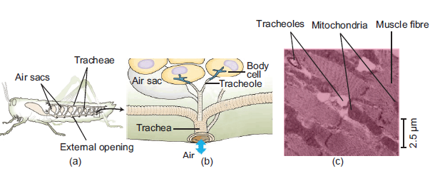
tracheae, connect to external openings spaced along the insect’s body surface. Air sacs formed
from enlarged portions of the tracheae are found near organs that require a large supply of oxygen.
(b) Rings of chitin keep the tracheae open, allowing air to enter and pass info smaller tubes called
tracheoles. The branched tracheoles deliver air directly to cells throughout the body. Tracheoles
have closed ends filled with fluid (blue-gray). When the animal is active and using more O2, most
of the fluid is withdrawn into the body. This increases the surface area of air-filled tracheoles incontact with cells.
(c) The TEM above shows cross sections of tracheoles in a tiny piece of insect flight muscle. Each ofthe numerous mitochondria in the muscle cells lies with about 2.5 μm of a tracheole.
Figure 9.1: Tracheal system in insect (Grasshopper). Note that the fine tubes called bronchiolesinnervating at cellular level. (Source: Campbell Biology, 2011)
long-chain polymer of an N-acetyl glucosamine) tubes penetrating to almost each individual cells
of the body. This system serves two functions: it brings air into the body, and also distributes it
to the cells. This pattern of tracheal system is very much similar to the system of blood vesselsin higher animals.
Air enters the tracheal system of the animal through special openings called spiracles. These
are present mostly on the lateral side of the animal. These are usually guarded by valves,
operated by muscles and sometimes provided with filters. Tracheal tubes are invaginations
(infoldings) of the body surface. Thus, their walls are similar in structure and composition to the
general body surface (integuments) of the animals. Sometimes larger tracheas have thickenings
called taenidia. These are spiral cuticular layers which give strength and elasticity. The tubes
become progressively smaller and thinner to form tracheoles or air capillaries (Figure 9.2). The
smaller tubes may have incomplete taenidial support. They have a diameter of less than 1 μm
(1 μm = 1×10⁻⁶ m). Tracheoles are the most important physiological unit of this gas exchange
system. It is because they make numerous close contacts with the individual cells for gas
exchange to take place. They sink into cell’s plasma membrane bringing oxygen very close to
the mitochondria of the cells.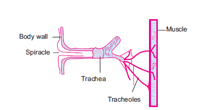
Figure 9.2: Detail structure of the tracheal system in insects
9.2.1 Mechanism of Ventilation in Insects
Normally there is no active ventilation in most tracheates (i.e., animals possessing trachea).
Many of the tracheates (like onychophora, myriapoda, and insect larvae and pupae) depend on
simple diffusion of gases in the air tubes. But ventilation and control of direction and volume
of the air flowing through the system is present in adult insects. This is because adult insects
are larger and so have higher metabolic rate which demands more oxygen. The spiracles and
air sacs help the insect in ventilation and creating unidirectional flow of air. In grasshopper,
thoraxic spiracles are used for inspiration while abdominal spiracles are used for expiration. This
creates a unidirectional flow of air. Air sacs greatly increase the efficiency of ventilation. These
are balloon-like structures of the trachea with a variety of size and shape. Active ventilation
is brought about by rhythmic contraction and relaxation of body walls. This forces the air
movement in and out of the tracheal system. Dorso-ventral flattening of abdomen is observed
in grasshopper and beetles.
ACTIVITY 2
Aim: To dissect an insect (cockroach or grasshopper) and study its tracheal system.
Materials Required: Cockroach, dissecting microscope, surgical scissors, chloroform,
forceps, scalpels, pins, dissecting tray etc.
Procedure:
1. Obtain a live cockroach (Periplaneta americana) and anaesthesize it with chloroform.
2. Locate the position of spiracles on thorax and abdomen and record their position by making
a rough sketch on the record book.
3. Pin the animal on the dorsal side with the ventral surface facing upwards on a dissecting tray.
4. Carefully remove the abdominal sterna (exoskeleton covering of the abdomen) without
disturbing the internal tissues.
5. Remove the fat bodies and reproductive organs carefully to expose the tracheal system.
Observation:
The tracheal system should be easily identified by its silvery appearance due to entrapped air in
it. Can you locate the taenidia? Label the different parts of the tracheal system. Notice that in
grasshopper, thoraxic spiracles are used for inspiration while abdominal spiracles are used forexpiration.
9.3 GASEOUS EXCHANGE IN FISH: GILLS
Gills are typical respiratory organs of aquatic animals, including fishes. Gills range in shape and
size. It may be finger-like projections or simple epithelial extensions. Gills are more developed
in fishes. Fish gills consist of thousands of highly specialised gill lamellae enclosed in a gill
cavity. The gill cavity is covered by an operculum and continuously ventilated by flowing water.
Respiration through gills is also known as branchial respiration. All gill surfaces are provided
with a dense network of thin capillary vessels and supported by skeletal elements called the
branchial arches.
Types of gills
Gills can be of two types:
External gills: These gills are exposed to the environment and not enclosed within a pouch or
cavity. They are found in the larvae of many vertebrates, including lungfishes, actinopterygians,
and amphibians.
Internal gills: Gills are covered and protected laterally by soft skin folds, like the interbranchial
septum in cartilaginous fishes, or by a firm operculum in many bony fishes. They are found
within pharyngeal gills slits or pouches of most cartilaginous and bony fishes.
In cartilaginous fishes, the gills are found on the lateral side of the branchial arch (Figure 9.3).
Gills are usually five pairs in number. They are located in vertical, anterioposteriorly compressed
branchial chambers or gill pouches. Each branchial pouch is separated from each other by a
stout interbranchial septum. This septum is made up of fibro-muscular tissue with blood vessels.
A branchial pouch communicates to exterior with the help of narrow external branchial
aperture or gill slits. Each gill has a central partition called the interbranchial septum. Within
this septum, a stiff structure called gill ray gives support to the gills. This septum is covered
on each face by primary lamellae or gill filaments. Gill filaments are series of raised thin,
highly vascular horizontal lamellar folds of the interbranchial septum. The primary lamellae
are again made up of standing rows of secondary lamellae. Water flows across their sides toirrigate the gills.
Figure 9.3: Structure of gills in shark. (Source: Kardong, Vertebrates: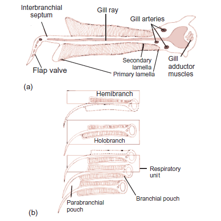
Comparative anatomy, Function, Evolution, 2012)\
When gill lamellae are present on both anterior and posterior sides of a septum, it is called
a holobranch or complete gill. However, when lamellae is present on only one face, it is
called a hemibranch (Figure 9.4). Facing plates of lamellae on adjacent gills constitute a
respiratory unit. A branchial pouch therefore consists of posterior hemibranch of one gill andanterior hemibranch of the succeeding gill.
The pharyngeal structural region in bony fishes is almost similar to that of cartilaginous fishes.
The gill/branchial chamber on each side is covered by a fold of integument called the operculum
(gill covering). It is supported by four opercular bones. The operculum protects the branchial
arches and its gill lamellae and also helps in gill ventilation. There are five pair of gill pouches
and four pairs of holobranchs or complete gills. In cross section, each gill is V-shaped and
composed of primary lamellae (gill filaments) that are subdivided into secondary lamellae andsupported on a branchial arch (Figure 9.5).
ACTIVITY 3
Aim: To observe and study the structure of gills in freshwater fish.
Materials Required: Fish (Tilapia), dissecting microscope, surgical scissors, chloroform,
forceps, scalpels, pins, dissecting tray, etc.
Procedure:
1. Obtain a live fish Tilapia from a local fish market and anaesthesize it with chloroform.
2. Locate the gill on the lateral side of the head by lifting the operculum. Cut out the operculum
and expose the red coloured gills.
3. Dissect out the gills and observe it under the microscope.
4. Draw a detail sketch structure of the gill and it lamellae and label it different parts with the
help of your teacher.Observation: Can you see the gill lamellae and the gill rakers?
9.3.1 Mechanism of Gill Ventilation
ACTIVITY 4
Aim: To observe and study the ventilation mechanism in fish.
Materials Required: Fish in an aquarium, notebook and pencil timer, etc.
Procedure:
Observe the movement of the mouth and operculum of a fish in the aquarium. Note the number
of times the mouth and operculum opens in a minute and record it in your notebook.Observation: Record the rate at which ventilation occurs in the fish.
Ventilation rate is much higher in aquatic animals than air breathing animals. This is because
water has lower oxygen and greater density than air. So, more ventilation is required for oxygen
uptake. This is achieved in gills by having a unidirectional flow of water. Ventilation of fish
gills is achieved by rhythmic movement of various muscles. This generates a continuous
current of water through the gills. The muscular pump of the buccal cavity actively drives
water across the internal gills bringing about ventilation. First the mouth is opened, buccal
floor drops and the pharyngeal floor lowered at the same time. This creates a vacuum inside
pharyngeal cavity. At the same time, external branchial openings are closed. This results in
water rushing into the pharyngeal cavity through the mouth. Now mouth gets closed and the
external aperture opens. This makes water flow out through the branchial apertures. As water
passes through the gills, it washes the gill lamellae. Exchange of gases takes place betweenthe blood flowing in the gill lamellae and the water current.
Figure 9.4: Gill ventilation in sharks. Lateral (a, c) and frontal view (b, d). Relative positive and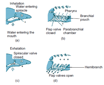
negative pressures indicated by + and –, respectively (Source: Kardong, Vertebrates: Comparativeanatomy, Function, Evolution, 2012)
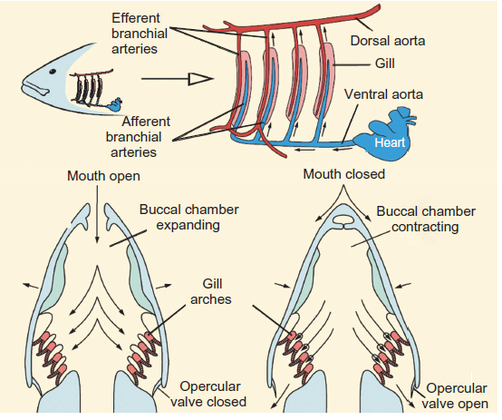
Figure 9.5: Mechanism of gill ventilation in Tilapia
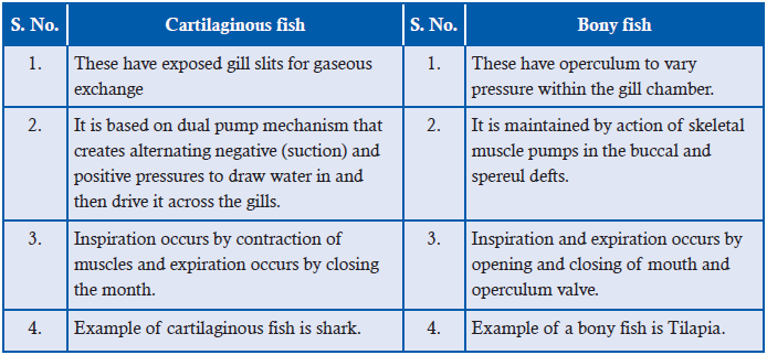
Difference between gaseous exchange of cartilaginous and bony fish
9.4 THE COUNTER CURRENT MECHANISM
ACTIVITY 5
To study counter current flow and parallel flow.
Use the internet or textbooks to make a chart diagram of countercurrent and concurrent flow
of fluids. Using arrows of different colours indicate the direction of flow of fluid in both the
flow system. Download videos/animations from the internet depicting the countercurrent flow
and exchange of gases through this system. Ask each of the students to give an advantage to
each of countercurrent flow over concurrent flow.
The high efficiency of fish gills, especially teleost in gas exchange is due to the presence of a
counter current flow of blood and water through the system. In ‘counter current’ system, twochannels in close proximity carry fluids in opposite directions (Figure 9.6). In such a system,
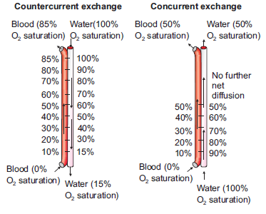
Figure 9.6: Current and counter current fluid flow.
equilibria will be established in the concentration of any permeable materials under two conditions
i.e., if the channels walls are freely permeable to the particular materials and if the channels are
long enough. The flow of blood in the gill lamellae and nearby water is a countercurrent type.
In most fish gills, the blood in the secondary lamellae flows in one direction and water flows
in the opposite direction. This establishes a countercurrent exchange system. As we can see in
Figure 9.7, the counter current system maintains a continuous gradient of oxygen concentrationbetween blood and water which is not found in case of concurrent exchange system.
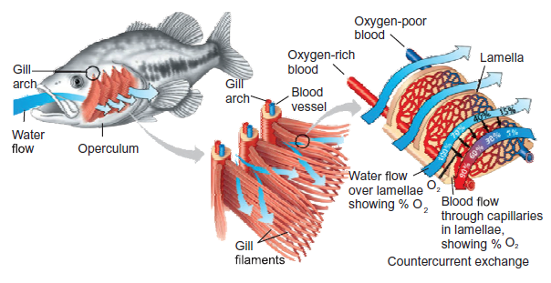
Figure 9.7: Countercurrent mechanism of gas exchange in bony fishes
This countercurrent flow maximizes difference in oxygen (and carbon dioxide) concentration
between water and blood. Countercurrent exchange arrangement results in blood always
being exposed to water with a higher oxygen concentration. A diffusion gradient is, therefore,
maintained across the surface of gill. Blood in gill lamellae capillaries contains less oxygen
and more carbon dioxide as it comes from different tissues after metabolism. However, the
water ventilating the gills has a greater concentration of oxygen compared to that of blood.
Hence, oxygen diffuses readily from water to blood in capillaries of gill lamellae continuously
till equilibrium is maintained. Due to the presence of a countercurrent exchanger system, a
continuous difference in the concentration of the gases is maintained all throughout the lengthof the gill lamellae, and therefore, a continuous efficient gas exchanger system is obtained.
9.4.1 Significance of Countercurrent Mechanism in Bony Fishes
• A larger difference in PO2 (i.e., partial pressure of O2; the pressure of a specific gas in a
mixture is called its partial pressure) can be maintained across the exchange surface. The
larger the difference, the more the exchange of gases; thus, allowing more transfer of gas.
• The system is so efficient that in some teleost 85% of oxygen may be extracted from water
passing over the gills using this system.
• This type of exchanger is also found in temperature control system of cold arctic animals,
in air bladders of fish and even in the kidneys of vertebrates.
• A few fish have some warm tissues. For example, Tuna have warm muscles, eyes, andbrains. This is only possible because of a countercurrent blood supply to selected tissues.
APPLICATION 9.1
1. Complete with appropriate terms:
(i) Two gases involved in gas exchange are ..................... and .....................
(ii) The high efficiency of tedeost gills is due to .....................
(iii) ..................... fish have exposed gill slits.
(iv) Taenidia are thickening of larger trachea in .....................
(v) Active ventilation in insects is brought about by ..................... and .....................
2. Explain why large , active organisms need special surfaces for exchange3. Describe and explain the features that make an exchange surface efficient
9.5 GASEOUS EXCHANGE IN AMPHIBIANS
ACTIVITY 6
Aim: To observe a live frog or toad in a glass tank and discuss its gas exchange surfaces.
Materials Required: A live frog/toad, aquarium/glass tank, notebook and pen etc.
Procedure:
1. Obtain a live frog and put it in an aquarium or glass tank slowly. Take care to handle the
animal gently and the animal should not be harmed.
2. Now observe carefully how it keeps itself ventilated and frequently comes to the surface,
etc.
3. While on land observe carefully the movement of the buccal chamber for ventilation.
4. Touch the surface of the frog and examine the skin whether it is dry or wet.
Observation:
Did you observe the wet slimy condition of the skin of the frog? Try to explain why is it so? Also
note the continuous ventilation of the lungs when in land by alternate lowering and raising ofthe buccal chamber.
Amphibians use the moist skin, gills or the lungs for gas exchange (Figure 9.8). Gas exchange
occurring through the skin is known as cutaneous respiration. In some larval Salamanders and
adult, external gills are also used for respiration. Modern amphibians rely heavily on cutaneous
respiration. Sometimes, they develop accessory skin structures to increase the surface areaavailable for gas exchange.
Figure 9.8: Cutaneous respiratory structures in some amphibians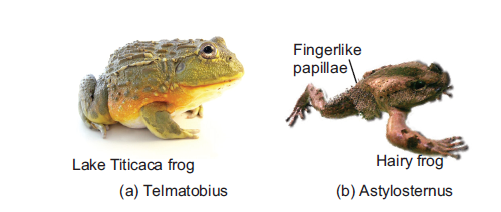
In salamanders of the family Plethodontidae, adults do not have lungs and gills. They depend
entirely on cutaneous respiration for metabolism. Lake Titicaca frog, Telmatobius culeus, has
prominent loose skinfolds on its back and limbs for cutaneous respiration. In the male hairy frog,
Astylosternus robustus, numerous papillae appear on its sides and hindlimbs during the breeding
season, forming supplementary respiratory organ (Figure 9.9). The amphibian skin is thin,
moist, and rich supplied with capillaries making it best suited for gas exchange by diffusion.
In aquatic amphibians, pharyngeal slits often persist with internal gills. Feathery external gills
are often present, especially among larval amphibians.
Amphibian larvae like Salamander larvae typically have both internal and external gills. Pumping
action of the throat irrigates the internal gills with a unidirectional stream of water across
their surfaces. Feathery external gills are held out in the passing current of water. In modern
amphibians, ventilation depends not on ribs but on pumping movements of the throat to irrigate
gills or fill lungs.
Most adult amphibians have lungs for breathing air (Figure 9.10). Normally, the respiratory
surface within the lungs on the anterior region is more developed than the posterior along
the inner walls. The inner surface of lungs forms partitions and divides to increase the
surface area for gas exchange. Such a surface is called septal. The interconnecting septa
divide the internal wall into compartments called faveoli. These faveoli open into the central
chamber within each lung. Faveoli differ from the alveoli of mammalian lungs. Alveoli
are found at the end of a highly branched tracheal system but faveoli are not. Faveoli are
internal subdivisions of the lung wall that open into a common central chamber. Inspired
air travels along the trachea into the central lumen of the lung and from here diffuses into
the surrounding faveoli. Capillaries located within the thin septal walls of the faveoli takeup oxygen and give up carbon dioxide.
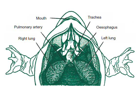
Figure 9.9: Structure of lungs in frog (amphibians).
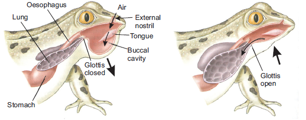
Figure 9.10: Mechanism of ventilation in frog (amphibians)
APPLICATION 9.2
1. Complete with appropriate terms;
(i) Gas exchange occurring through skin is called .............. respiration.
(ii) Amphibians develop .............. to increase surface area for gaseous exchange.
(iii) Partitions of inner lungs in Amphibians are known as ..............2. State 3 ways in which the structure of the lungs allows efficient gas exchange
9.6 GASEOUS EXCHANGE IN HUMANS
Higher vertebrates including humans have specialized organs called lungs for gas exchange.
The process of gas exchange in the body, called respiration, has three basic steps:
1. Pulmonary ventilation or breathing is the inhalation (inflow) and exhalation (outflow) of
air and involves the exchange of air between the atmosphere and the alveoli of the lungs.
2. External (pulmonary) respiration is the exchange of gases between the alveoli of the
lungs and the blood in pulmonary capillaries across the respiratory membrane. In this
process, pulmonary capillary blood gains O2 and loses CO2.
3. Internal (tissue) respiration is the exchange of gases between blood in systemic
capillaries and tissue cells. In this step, the blood loses O2 and gains CO2. Within cells,
the metabolic reactions that consume O2 and give off CO2 during the production of
ATP are termed cellular respiration.9.6.1 Structure of Gaseous Exchange in Humans\
ACTIVITY 7
Make a computer model/simulation of the human respiratory system. This can also be
downloaded from the internet. Use the internet to search videos, graphics and simulation or
animations showing the different parts and surfaces of the gas exchanges system in humans.
Also study the process of gas exchange and the mechanism of ventilation. Now, a smallpresentation on the same topic. You can also clay models of the respiratory system in humans.
Air is inhaled through the nose into the pharynx (throat). Pharynx is a common passage for
both air and food. The pharynx branches into two tubes, the oesophagus or food pipe and
the larynx. The larynx is part of the airways and it houses the vocal cords. The nose, mouth,
pharynx, and larynx are also called the upper airways. The larynx opens into a long tube,
the trachea. The trachea then branches into two bronchi, the right primary bronchus enters
the right lung and the left primary bronchus enters the left lung. The walls of the trachea and
bronchi contain cartilage, which supports them and gives them their characteristic cylindrical
shape. The right primary bronchus is more vertical, shorter, and wider than the left. Within
each lung, the bronchi branch continuously into narrower, shorter, and more numerous tubes,more than 20 generations of branching (Figure 9.11a).
The primary bronchi divide to form smaller bronchi which are known as the secondary (lobar)
bronchi, one for each lobe of the lung. The secondary bronchi continue to branch, forming still
smaller bronchi called tertiary (segmental) bronchi. Tertiary bronchi divide to form smaller
bronchioles. Bronchioles are without cartilage. Alveoli (explained later) first begin to appear in
them attached to their walls. Alveoli normally form grapelike clusters terminally. The airways
are surrounded by smooth muscle whose contraction or relaxation can alter airway radius.
Bronchioles in turn branch repeatedly, and the smallest ones branch into even smaller tubes
called terminal bronchioles. This extensive branching from the trachea resembles an inverted
tree and is sometimes commonly referred to as the bronchial tree.
The lung is a paired cone-shaped organ in the thoracic cavity (Figure 9.11(a)). The lungs
extend from the diaphragm to just slightly superior to the clavicles (collarbone). They are
guarded by the ribs anteriorly and posteriorly. The mid region of left lung also has concavity
called the cardiac notch, in which the heart lies. This makes the left lung about 10% smaller
than the right lung. Each lung is divided into several lobes; three lobes in right and two in
left lungs. Tiny air containing sacs called alveoli (singular, alveolus) arranged like bunch of
grapes at the end of each bronchioles are the respiratory unit of the lungs (Figure 9.11 (b)).
Alveoli are approximately 300 million in number in an adult and are the actual sites for gasexchange.
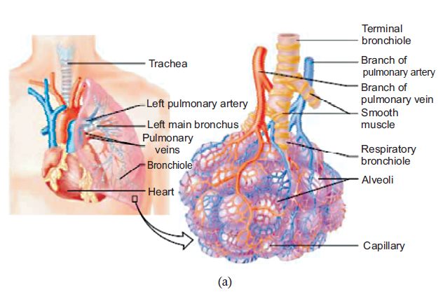
Figure 9.11: (a) Detail structure of human respiratory system: the lungs and the alveoli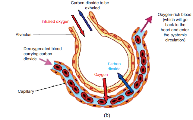
(Source: Vander et. al., Human Physiology, 2001) (b) gas exchange in the alveoli
Each lung is enclosed and protected by a double-layered serous membrane called the pleural
membrane. It consists of two layers: the outer parietal pleura and the deeper visceral pleura.
The space between the two is called the pleural cavity and contains a small amount of lubricating
fluid secreted by the membranes. The important function of this pleural fluid is to reduce friction
between the membranes during breathing movement.
9.6.2 Functions of Tissues within the Gas Exchange System
The respiratory system consists of four main layers (Figure 9.12):
(i) The respiratory mucosa (epithelium and supporting lamina propria)
(ii) Submuscosa
(iii) Cartilage and/or muscle layer(iv) Adventitia
Figure 9.12: Diagrammatic representation of cross section of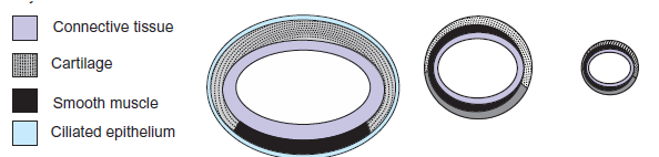
airways showing distribution of tissues (not to scale)
Trachea
The trachea is a wide flexible tube. The respiratory mucosa and submucosa are adapted to warm
and moisten the air, and to trap particles in mucous. It consists of pseudostratifed columnar,
ciliated epithelium with mucous secreting goblet cells. It has twenty C-shaped rings of hyaline
tracheal cartilage which supports the trachea and keeps its lumen open. The gaps between the
rings of cartilage are filled by a bundle of smooth muscle (trachealis muscle) and fibroelastic
tissue. These structures together gives flexibility for ventilation. Adventitia is the outermost
fibroelastic connective tissue layer.
The respiratory mucosa is made up of the epithelium and supporting lamina propria. The
epithelium is tall columnar pseudostratified with cilia and goblet cells. Lamina propia lies
underneath the epithelium. It contains elastin and has a supporting role. Blood vessels warm
the air. The sub-mucosa contains mixed sero-mucous glands. The watery secretions from the
serous glands humidify the inspired air. The mucous, together with mucous from the goblet
cells traps particles from the air which are transported upwards towards the pharynx by the
cilia on the epithlium. This helps to keep the lungs free of particles and bacteria. There are lots
of seromucous glands in the submucosa layer.
The epithelial surfaces of the airways upto the end of the respiratory bronchioles have cilia that
constantly beat toward the pharynx. They also contain mucous secreting glands (Figure 9.13).
This mucous keeps the lungs clear of particulate matter and the many bacteria that enter the
body on dust particles. Macrophage present in the airways and alveoli also protect againstinfection.
Bronchi
Bronchi have the same basic structure as trachea. A few differences are respiratory epithelium
are less tall than that of trachea and contains fewer goblet cells. Lamina propia has more elastic
tissue. Muscularis mucosae begin to appear in lamina propia and submucosa. There are fewer
submucosal glands and cartilage is in plates. There is less cartilage in the tertiary bronchi, Itdoes not completely encircle the lumen.
Bronchioles
The tertiary bronchii branch into bronchioles. They have a diameter of 1mm or less, and the
wall structure changes. There is no cartilage and no glands. The ring of smooth muscle is
arranged in discrete bundles with a variety of organisations. The epithelium is made up of
ciliated columnar cells in larger bronchioles, or nonciliated in smaller bronchioles. There are
no goblet cells, but there are cells called Clara cells. These are secretory cells and they secreteone of the components of surfactant.
Terminal Bronchioles
The final branches of the bronchioles are called terminal bronchioles. These have a layer smooth
muscle surrounding their lumens. Stimulation of the vagus nerve (parasympathetic) causes the
smooth muscle to contract, and reduce the diameter of the terminal bronchioles. Small sacs are
found extending from the walls of the terminal bronchi called respiratory bronchioles. These
are lined by a ciliated cuboidal epithelium, and some non-ciliated cells called clara cells. Therespiratory bronchii have a few single alveoli off their walls.
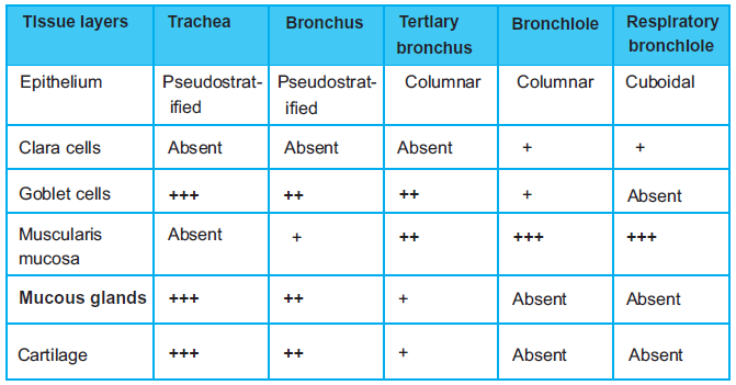
Figure 9.13: Table showing different tissue layers of the gas exchange system
Alveoli
The alveoli are the sites of gas exchange with the blood. The wall of the air-facing surface(s)
are lined by type I alveolar cells which is a one cell thick, continuous layer of flat epithelial
cells. Type II alveolar cells are thicker specialized cells producing a detergent-like substance
called surfactant and they are interspersed between type I cells. In some of the alveolar walls,
pores are present which permit the flow of air between alveoli.
The alveolar walls contain capillaries and a very small interstitial space, made of interstitial
fluid and a loose meshwork of connective tissue. However, the interstitial space is absent
altogether at most places and the basement membranes of the alveolar-surface epithelium and
the capillary-wall endothelium fuse. As a result, the blood within an alveolar-wall capillary
is separated from the air within the alveolus by an extremely thin barrier around 0.2 μm. The
branching of bronchioles and the vast number of alveoli collectively increases the respiratory
surface area to as much as 80 square metres. The extensive surface area of alveoli in contact
with capillaries and the thin barrier results in the rapid exchange of large quantities of oxygen
and carbon dioxide by diffusion.
APPLICATION 9.3
1.Complete with appropriate terms:
(i)Tiny air containing sacs found in the human gaseous exchange system are................ .
(ii) ................ is a wide flexible tube in human gas exchange
(iii) Respiratory mucosa in humans is made up of ................ and ................
(iv) Alveoli are lined up by ................ and ................2.Explain why the barrier to diffusion must be as thin as possible
9.6.3 Mechanism of Ventilation (Breathing)
Inspiration (inhalation or breathing in) is the movement of air from the external environment
through the airways into the alveoli during breathing. Expiration (exhalation) is movement inthe opposite direction. An inspiration and an expiration constitute a respiratory cycle.
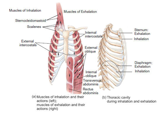
Figure 9.14: Structure and mechanism of lung ventilation in human
Inhalation: Air will move into the lungs when air pressure inside the lungs is less than that
of outside (atmospheres). Expansion of the lungs increases the volume and so the pressureinside the lungs decreases. Expansion of the lungs during normal quiet inhalation is achieved
by contraction of the diaphragm and external intercostals which are the main muscles of
inhalation (Figure 9.14). The diaphragm is the dome-shaped skeletal muscle that forms the
floor of the thoracic cavity. Contraction of the diaphragm causes it to flatten, lowering its
dome. This increases the vertical diameter of the thoracic cavity. Around 75% of air enters
the lungs by this action. Also contraction of the external intercostals elevates the ribs resulting
in an increase in the volume of the chest cavity. About 25% of the air that enters the lungs
during normal quiet breathing is due to this action. As the volume of the lungs increases and
the pressure inside the lungs (alveolar or intra-pulmonic pressure) decreases and atmosphericair rushes into the lungs.
Exhalation: On the other hand if the volume of the lungs decreases, pressure inside the lungs
increases. As a result, air rushes out of the lungs resulting in exhalation or expiration. However,
normal exhalation during quiet breathing, unlike inhalation, is a passive process because no
muscular contractions are involved. Exhalation results from elastic recoil of the chest wall
and lungs. Elastic recoil is the natural tendency of the chest wall and the lungs to spring back
after they have been stretched. The inspiratory muscles relax with the start of exhalation.
Diaphragm and external intercostal muscles also relax resulting in decrease in volume of the
lungs, causing air to move out of the lungs. Interestingly, exhalation becomes an active process
(requiring energy supply) only during the time of forced exhalation (for example during heavy
exercise etc). During these times, the muscles of exhalation are the abdominals and internalintercostals muscles which contract to increase pressure in the abdominal region and thorax.
Gas Exchange in Alveoli
Alveoli are the respiratory units of lungs. The alveolar and capillary walls together form
the respiratory membrane. The exchange of gases in the alveoli and between the air
spaces in the lungs and the blood takes place by diffusion across this respiratory membrane
(Figure 9.15).
The pressure of a specific gas (x) in a mixture is called its partial pressure (Px). The difference
in partial pressures determines the movement of O2 and CO2 between the atmosphere and
lungs, between the lungs and blood, and between the blood and body cells. Gas diffuses across
a permeable membrane from an area where its partial pressure is higher to the area where its
partial pressure is low and the rate of diffusion is directly proportional to the difference inpartial pressure.
Figure 9.15: Detail structural components of an alveolus: (a) section through alveolus and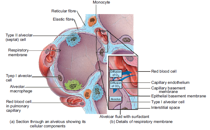
(b) details of respiratory membrane
As stated earlier, external respiration or pulmonary gas exchange is the diffusion of O2 from
air in the alveoli of the lungs to blood in pulmonary capillaries and the diffusion of CO2 in the
opposite direction. In this process, blood picks up O2 from alveolar air and unloads CO2 into
alveolar air as it flows through pulmonary capillaries. In a resting person, PO2 is 105 mmHg
in the alveolar air which is higher than that of blood in pulmonary capillaries, where it is only
40 mmHg. This results in diffusion of O2 from alveolar air into pulmonary capillaries.
However, CO2 diffuses in the opposite direction because the PCO2 of deoxygenated blood is
45 mmHg in a resting person, and the PCO2 of alveolar air is 40 mmHg. Hence, carbon dioxide
diffuses from deoxygenated blood into the alveoli until the PCO2 of the blood decreases to40 mmHg.
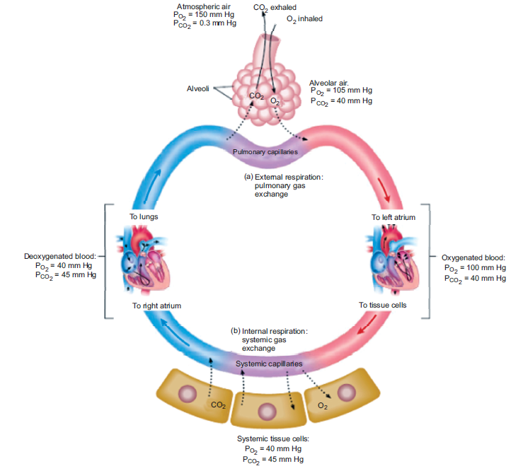
Figure 9.16: Exchange of gases in alveoli of humans
As a result of this diffusion, the capillary blood PO2 rises while its PCO2 falls. This process of
diffusion continues as long as there is difference in partial pressure of the two gases between
the two sides. An equilibrium is reached well before the end of the capillaries because blood
flow in the capillaries is slow and gas exchange is rapid. Oxygenated blood now leaves the
pulmonary capillaries to return to the heart from where it is pumped into the systemic arteries.
The exchange of O2 and CO2 between systemic capillaries and tissue cells is called internalrespiration or systemic gas exchange (Figure 9.16).
9.6.4 Lung Volume and Capacities
Tidal volume: It is the volume of air entering the lungs during a single inspiration during
normal quiet breathing. It is about 500 ml. It is approximately equal to the volume leaving onthe subsequent expiration.
Inspiratory reserve volume: The maximal amount of air that can be increased above the resting
tidal volume during deepest/forced inspiration is termed the inspiratory reserve volume. It is
about 3000 ml in average adult males which is sixfold greater than resting tidal volume and1900 ml in average adult females.
Expiratory reserve volume: The 500 ml of air inspired with each resting breath adds to and
mixes with the much larger volume of air already in the lungs, and then 500 ml of the total is
expired. However, through maximal active contraction of the expiratory muscles i.e., forced
expiration, it is possible to expire much more of the air remaining after the resting tidal volume
has been expired; this additional expired volume is termed the expiratory reserve volume(about 1500 ml).
Residual volume: Even after a maximal active expiration, approximately 1000 ml of air
still remains in the lungs. This is because the subatmospheric intrapleural pressure keeps the
alveoli slightly inflated, and some air also remains in the non-collapsible airways. This volume,
which cannot be measured by spirometry, is called the residual volume and amounts to about1200 ml in males and 1100 ml in females.
Vital capacity: It is the maximal volume of air that a person can expire after a maximal
inspiration. It is a useful clinical measurement for detecting various respiratory system related
conditions. It is the sum of inspiratory reserve volume, tidal volume, and expiratory reservevolume (4800 ml in males and 3100 ml in females).
Inspiratory capacity is the sum of tidal volume and inspiratory reserve volume
(500 ml + 3100 ml = 3600 ml in males and 500 ml + 1900 ml = 2400 ml in females).
Total lung capacity is the sum of vital capacity and residual volume
(4800 ml + 1200 ml = 6000 ml in males and 3100 ml + 1100 ml + 4200 ml in females).
However, in a normal adult only 70% (=350 ml) of tidal volume reaches the respiratory zone
because of the presence of anatomical dead space. Dead Space refers to the conducting airways
which have a volume of about 150 ml. Exchanges of gases with the blood does not occur in
this 150 ml of the airways. It occurs only in the alveoli. Since these airways do not permit gasexchange with the blood, the space within them is termed the anatomic dead space. Thus, the
volume of fresh air entering the alveoli during each inspiration equals the tidal volume minus
the volume of air in the anatomic dead space.
Alveolar ventilation: The total volume of fresh air entering the alveoli per minute is called the
alveolar ventilation which is given by,
Alveolar ventilation (ml/min) = (Tidal volume – Dead space) × Respiratory rate
(ml/breath) (ml/breath) (breath/min)
= (500 – 150) ml/breath × 12 breath/min= 350 × 12 = 4200 ml/min.
ACTIVITY 8
Calculate pulmonary ventilation (PV) and alveolar ventilation (AV) from the data provided.
(i) Tidal volume = 550 ml, Dead space = 185 ml, Respiratory rate = 17/min, inspiratory
reserve volume = 2500 ml, tidal volume = 550, and expiratory reserve volume = 1450.
(ii) Tidal volume = 600 ml, Dead space = 195 ml, Respiratory rate = 15/min, inspiratory
reserve volume = 2800 ml, tidal volume = 600, and expiratory reserve volume = 1350.
(iii) Tidal volume = 550 ml, Dead space = 175 ml, Respiratory rate = 20/min, inspiratory
reserve volume = 2500 ml, tidal volume = 500, and expiratory reserve volume = 1500.
Hint: What is formula for calculation of pulmonary ventilation (PV) and alveolar ventilation(AV)?
Importance of Lung Capacities
These pulmonary function tests are useful diagnostic tools:
• An examination of ventilation function of lungs is necessary for evaluation of functional
properties of human respiratory system.
• It is used for estimation of defects in respiratory system and also for consideration of fitness
load in sports medicine.
• Various respiratory disorders may be diagnosed by comparison of actual and predictednormal values for a patient’s gender, height, and age.
APPLICATION 9.4
1. Complete with appropriate terms:
(i). Maximum volume of air that a person can expire after a maximal inspiration is called
................ .
(ii). Diffusion of O2 from air to expillaries and CO2 in opposite direction is called ...............
(iii). During .................. volume of lungs increases in humans.2. Describe how a steep diffusion gradient is achieved in lungs
Spirometry
The spirometer is an apparatus for measuring inspired and expired volumes during breathingand the respiratory rate (Figure 9.17). The record is called a spirogram (Figure 9.18).
Figure 9.17: (a) A basic model of a spirometer; and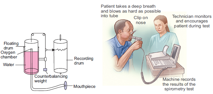
(b) a patient taking readings on a modern spirometer
Use of Spirometer to Measure Ventilation Rate
The lung volumes and capacities can be measured by routine spirometry. A typical spirometer
is a tube like instrument with an open end called the mouthpiece. The spirometer (Figure 9.17a)
consists usually of a water-filled tank with a bell shaped floating device. A tube connects the
air space within the spirometer with the airways of the person whose lung volumes is being
measured. A counterweight is placed on the bell. The position of the bell indicates how much
air is in the spirometer and is calibrated in volume units. A person under the test blows air into
it after deep breath. Usually, the airway through nose is shut or blocked using a clip so that
air can only enter or leave through the mouth. Inhalation is recorded as an upward deflection,
and exhalation is recorded as a downward deflection. The bell on the spirometer rises when
the person blows into the device (expiration), and falls during inspiration. If the spirometer
is equipped with a recording device (spirograph), it can also be used for graphic measurement
of the total ventilation per unit time. Based upon the reading indicated corresponding to each
breathing in or out, an expert physician can diagnose the health of the person’s lungs and detect
disorder if any. Nowadays, the instrument is integrated with a computer system to accuratelymonitor the readings and give instant results.
Figure 9.18: Spirogram of lung volumes and capacities in a healthy man and woman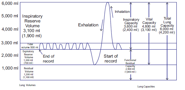
(within parentheses). The spirogram is read from the right(i.e., start of record) to the left (i.e., end of record)
ACTIVITY 9
Aim: To design a model of the spirometer based on its main features.
Materials Required:
1. 2–3 litre empty soda/cold drink bottle with cap
2. One or two feet long piece of plastic tubing
3. One measuring cup with units in millilitres
4. One bucket or pan that can hold more than 3 litres of water5. One permanent marker
Procedure:
1. Marking measuring lines on the bottle: Add 500 ml of water to the soda/cold drink
bottle using the measuring cup and mark a line with the marker at the top of the water
level. Repeat this until the bottle is full. When the bottle is full, put the cap on the
bottle.
2. Add sufficient water to the bucket or pan to submerge the soda bottle.
3. Invert the soda bottle and submerge it in the bucket, and remove the cap under the water.
4. Open the bottle underwater to prevent any unwanted air from entering the bottle.
5. Place one end of the tubing into the soda bottle in the water, and leave the other end outside
of the water.
6. Use the tubing to blow into, for determining lung capacity.
7. Remember to place the bottle in the water upside down before removing the cap.
8. Don’t forget to insert one end of the hose in the bottle after you open the cap
underwater9. Before you exhale into the tubing, your spirometer should resemble the picture below.
10. Using the spirometer to obtain the readings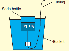
(i) One student holds the bottle to keep it from flipping over. Another student inhales
normally and then exhales the air normally into the tubing connected to the
spirometer. Note: Do not blow out all the “extra” air in your lungs.
(ii) Note the amount of air you exhaled, remembering that each line on the bottle
represents a half litre, starting from the top down.
(iii) Record this volume, it is your “tidal volume.” The tidal volume is the amount of air
that you normally breathe in and out.
(iv) Refill the bottle with water and reinsert the tubing. One student holds the bottle while
another take a few normal breaths initially. This is to get a good reading in the next
step. Then inhale as much air as you can and exhale this air into the end of thetubing outside of the water.
(v) Again note the amount of air you exhaled by looking at the lines on the soda bottle.
(vi) This volume is your “inspiratory reserve.” The inspiratory reserve is the amount of air
that your lungs can hold in.
(vii) Refill the bottle with water and reinsert the tubing. One student holds the bottle while
the other takes a few normal breaths to get himself back to a normal breath. Then
exhale as much air as you can into the end of the tubing outside of the water.
(viii) Note the amount of air you exhaled by looking at the lines on the soda bottle.
(ix) This is your “expiratory reserve.” The expiratory reserve is the amount of air that your
lungs can blow out after a normal breath.
Observations:
The vital capacity is the greatest change in volume that can occur in the lungs.
Inspiratory Reserve + Expiratory Reserve + Tidal Volume = Vital Capacity
ACTIVITY 10
Aim: To use the illustrations of spirometer trace to define tidal volume, inspiratory reserve
volume, expiratory reserve volume, vital capacity and residual volume.
Materials Required: Notebook, pen, pencil etc.
Procedure:
1. First write down the definitions of tidal volume, inspiratory
reserve volume, expiratory reserve volume, vital capacity
and residual volume.
2. Using a colour pencil/pen note try to locate the tidal
volume in the spirometer trace provided.
3. Perform step 2 above for inspiratory reserve volume,
expiratory reserve volume, vital capacity and residual
volume.
4. Give a logical explanation for your labelling.
5. Show the labelled spirometer trace to your teacher and explain your results. Ask for correctionsif any.
9.7 NERVOUS CONTROL OF BREATHING
ACTIVITY 11
Inform the class about the function or role of a particular part of the brain controlling the
process of respiration in humans viz. the medullary rhythmicity area and the pneumotaxic area
in the pons.
Breathing depends entirely upon cyclical respiratory muscle excitation of the diaphragm and
the intercostal muscles by their motor nerves as a result of nerve impulses transmitted to them
from centres in the brain. When a burst of action potentials is initiated in the nerves to the
inspiratory muscles, these muscle contracts and inspiration occurs. When these action potentials
stop, the inspiratory muscles relax, and expiration occurs as the elastic lungs recoil. Similarly,
in situations when expiration is facilitated by contraction of expiratory muscles, the nerves to
these muscles, begin firing during expiration. This neural activity is primarily controlled byneurons in the medulla oblongata.
The respiratory centre is the cluster of neurons located bilaterally in the medulla oblongata
and pons of the brain stem. It can be divided into three areas on the basis of their functions(Figure 9.19):
1. The medullary rhythmicity area in the medulla oblongata:
• Controls the basic rhythm of respiration.
• There are inspiratory and expiratory areas.
• Nerve impulses generated in the inspiratory area establish the basic rhythm of
breathing during quiet breathing by causing contraction of external intercostal muscle.
• The neurons of the expiratory area remain inactive during quiet breathing. However,
during forceful breathing nerve impulses from the inspiratory area activate the
expiratory area.
• Impulses from the expiratory area cause contraction of the internal intercostal and
abdominal muscles, which decrease the size of the thoracic cavity and causes forceful
exhalation.
2. The pneumotaxic area in the pons:
• Transmits inhibitory impulses to the inspiratory area.
• The major effect of these nerve impulses is to help turn off the inspiratory area before
the lungs become too full of air.
• In other words, the impulses shorten the duration of inhalation. When the pneumotaxicarea is more active, breathing rate is more rapid.
3. The apneustic area in the lower pons:
• This area sends stimulatory impulses to the inspiratory area that activate it and prolong
inhalation.
• The result is a long, deep inhalation.• When the pneumotaxic area is active, it overrides signals from the apneustic area.
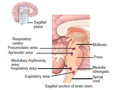
Figure 9.19: Respiratory centre in the human brain
In addition to the above, there are ‘Pulmonary stretch receptors’ in the smooth-muscle layer
of the airway. They respond to stretch stimulus on this muscles. Whenever there is large lung
inflation, they are activated. Electric signals in the afferent nerve fibres from the stretch receptors
travel to the brain and inhibit the medullary inspiratory neurons. This phenomenon is known as
the Hering-Breur inflation reflex. Thus, inspiration is terminated by feedback from the lungs.
However, this pulmonary stretch-receptor reflex plays a role in setting respiratory rhythm onlyunder conditions of very large tidal volumes, for example in rigorous exercise
APPLICATION 9.5
1.Complete with appropriate terms:
(i)................ is used to measure inspired and expired volumes of air.
(ii) Respiratory centre is a cluster of neurons located bilaterally in ................ and ................
of brain stem.
(iii) ................ in brain prolongs inhalation.2. Explain the importance of the cartilage found in the trachea and bronchi
9.8 SUMMARY
• Aerobic animals require a continuous supply of oxygen for metabolic processes and also
removal of metabolic waste (CO2) from its body.
• This is achieved by developing a complex system of gas exchange in every animal.
• Gases exchange takes place by the process of diffusion where it moves from a place of
higher concentration to a place of lower concentration.
• Small invertebrates like insects have a vast network of ‘tubes’ made of chitin called the
tracheal system spread all over their body which is used for exchange of gases.
• The tubes or trachea branches and interbranches to form fine tubes called tracheoles
innervating tissues at cellular level. Air enters and leaves through openings called spiracles.
• In aquatic animals like fish and some amphibian larvae exchange of gases takes place
through special structures called gills.
• Gills can be external or internal depending on its location in the body. Gills are highly
vascular, thin and always ventilated with water.
• A holobranch or complete gill refers to a branchial arch and the lamellae on both
anterior and posterior faces of its septum. A gill arch with lamellae on only one face is
a hemibranch.
• Ventilation of gills in fish is achieved by the coordinated action of the buccal cavity and
the operculum or gill cover.
• Countercurrent mechanism of gas exchange is present in gills of teleost. It is a very
efficient mechanism of gas exchange and almost 85% of oxygen is extracted from water.
• Amphibians can respire through skin (cutaneous respiration), gills or the lungs.
• Exchange of gas in the skin, gills or the lungs takes place by diffusion of gas (O2) from
air or water to the blood capillaries in the skin or the septal walls of faveoli.
• In humans, exchange of gases takes place through the lungs. The lungs are elastic
structures. The lungs, the airways leading to them, and the chest structures responsible
for movement of air into and out of the lungs.
• The conducting zone of the airways consists of the trachea, bronchi, and terminal
bronchioles. The respiratory zone of the airways consists of the alveoli, which are the
sites of gas exchange.
• The alveoli are lined mostly by type I cells along with some type II cells, which produce
surfactant.
• The lungs are covered by pleura and between the two pleural layers is an extremely thin
layer of intrapleural fluid.
• During inspiration, the contractions of the diaphragm and inspiratory intercostal muscles
increase the volume of the thoracic cage causing atmospheric air to rush into the lungs.
• During expiration, the inspiratory muscles cease contracting, allowing the elastic recoil
of the chest wall and lungs to return them to their original between-breath size resulting
in the air moving out of the lungs through the nose.
• The vital capacity is the sum of resting tidal volume, inspiratory reserve volume, and
expiratory reserve volume.
• Gases diffuse from a region of higher partial pressure to a region of lower partial pressure.
Exchange of gases in lungs and tissues takes place though the process of diffusion because
of the differences in partial pressures of gases.
• There is net diffusion of oxygen from alveoli to blood and of carbon dioxide from blood
to alveoli when systemic venous blood flows through the pulmonary capillaries.
• In certain conditions like when the alveolus capillary surface area is decreased or when
the alveolar walls thicken inadequate gas exchange between alveoli and pulmonary
capillaries may occur
9.9 GLOSSARY
• Alveoli: The plural of alveolus. The alveoli are firy air sacs within the human lungs
where exchange of gases takes place.
• Breathing: The process of taking air in and expelling it from lungs through the nose or
mouth.
• Bronchi: The plural of bronchus. It is any of the major air passages of lungs which
diverge from windpipe.
• Counter current mechanism: Maintenance of equilibria in the concentration of any
permeable materials under two conditions.
• Cutaneous respiration: The process of respiration through skin.
• Exhalation: It is the process or act of exhaling out air, generally CO2.
• Gills: These are the paired respiratory organ of fish and some amphibians, by which
oxygen is extracted from water.
• Inhalation: It is the process or act of inhaling in air, generally O2.
• Paveoli: A small pit or cavity resembling a cell of a honey comb alveola.
• Spirometer: An apparatus for measuring inspired and expired volumes of air during
breathing.
• Trachea: A large membranous tube of cartilage extending from larynx to bronchial tubes
and conveying air to and air from the lungs.• Ventilation: The bodily process of inhalation and exhalation.
END UNIT ASSESSMENT 9
Do all these exercises in your exercise book.
I. Choose whether the following statements are True (T) or False (F)
1. Insects have a specialised system of ‘tubes’ called the tracheal system for exchange
of gases.
2. There is active ventilation in most treacheates (i.e., animals possessing trachea).
3. Fish gills consist of thousands of highly specialised gill lamellae enclosed in a gill
cavity.
4. Amphibians use the moist skin, gills or the lungs for gas exchange.
5. Modern amphibians do not rely heavily on cutaneous respiration.
6. Most adult amphibians have lungs for breathing air.
7. Internal (tissue) respiration is the exchange of gases between blood in systemic
capillaries and tissue cells.
8. Alveoli are the structure for gas exchange in humans.
9. The apparatus for measuring inspired and expired volumes during breathing is a
spirometer.
10. The sum of inspiratory reserve volume, tidal volume, and expiratory reserve volumeis called residual volume.
II. Long Answer Type Questions
1. Describe the tracheal system of insects and relate to its function.
2. Describe the structure of the gills in relation to its function.
3. In your own words, explain the significance of counter current flow in bony fish.
4. Describe the mode of gaseous exchange in amphibians.
5. Describe the structure of the human gas exchange system.
6. Describe the distribution of tissues within the trachea, bronchi, bronchioles and
alveoli and relate each tissue to its function.
7. Explain the mechanism of ventilation in humans.
8. Explain the process of gas exchange in alveoli with emphasis on diffusion.
9. Describe the role of the brain in controlling gas exchange in humans.
10. Define the following terms related to the lung capacities:
(i) Tidal volume (ii) Reserve volume
(iii) Vital capacity (iv) Residual volume
(v) Dead air space
11. Describe how a spirometer can be used to measure vital capacity, tidal volume,
breathing rates, and oxygen uptake.
12. Calculate vital capacity and alveolar ventilation from the data provided.
Tidal volume = 550 ml, Dead space = 185 ml, Respiratory rate = 17/min, inspiratory
reserve volume = 2500 ml, tidal volume = 550, and expiratory reserve volume = 1450.
13. What contribution does exchange of gases make on global warming? Discuss your
answer with relevant data. Also throw light on the dialect “Global warming: a mythor truth.”