Topic outline
Unit 1 Interdependence between Organisms within their Environment
Key Unit Competence
To be able to explain complex relationships between organisms within their environment.
LEARNING OBJECTIVES
At the end of this unit, learners should be able to:
• Explain the various interactions of organisms in nature.
• Appreciate the relationships existing among the organisms within their environment.
• State the significance of organisms’ interactions in nature.
• Explain the terms interspecific and intraspecific competition.
• Compare interspecific and intraspecific competition.
• Describe the adaptations of predators to catch and kill prey and adaptations of prey to
avoid
• predators.
• Interpret graphs for predator-prey relationships.
• Classify examples of species interactions, e.g., competition, predation, parasitism,
commensalism, and mutualism.• Recognise the role of saprophytes in mineral recycling.
INTRODUCTORY ACTIVITY
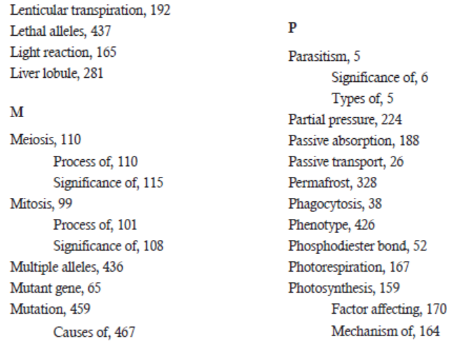
• Have you ever visited Akagera national park?
• Any visitor is impressed by the organization of wildlife Akagera National park. Wen
lions come they alert one another.
• Analyze the photograph below from Akagera national park to see a community ofherbivores grazing.
• At what extent can you say that there is interdependence among these organisms?
1.1 INTERRELATIONSHIP AMONG THE ORGANISMS AND THEIR EFFECTS
ACTIVITY 1.1
Watch a movie on wildlife.
1) What interactions are there among organisms observed?2) State why biological interactions are important and how they help the ecosystem.
No organism exists in an absolute isolation. Every organism interacts with other organisms
within a community. Thus, different organisms interacting with one another within a community
forms a concept called biological interactions or interrelationship among organisms. These
interactions among the organisms can be beneficial or harmful or even neutral. They have the
potential to influence and mould the structure, growth, and maintenance of populations within
a community. Moreover, in some cases, these interactions may result into long-term ecologicaland evolutionary changes among the individuals participating in these interactions.
S5 Biology
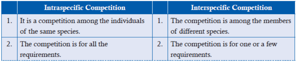
These interactions may involve individuals of the same species or different species. When
the interactions involve individuals of the same species, it is called intraspecific interaction.
On the other hand, when the interactions involve individuals of different species, it is called
interspecific interaction.
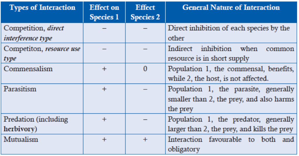
Biological interactions can be generally classified into different categories based on whether the
effects of interactions are beneficial, harmful or neutral for each of any two species. Thus based
on these criteria, biological or population interactions may be divided into basic interactions
and relationships. All the interactions are indicated by signs such as +, + or –,–, or +,–, even
0, 0. The sign (+) indicates that a particular species is benefitting from the interactions. The
sign (–) indicates that a particular species in the interactions is being harmed, While sign (0)
indicates neutral position where it is neither benefited nor harmed in the interactions. The
important species interactions are:Table 1: Interrelationship among the organisms
APPLICATION 1.1
1. Epiphytes are any plants that grow upon another plant or object merely for physical
support. Can you conclude that epiphytes are parasitic plants? Give reason2. Herbivory can be separated from the predation. Discuss
1.2 INTER AND INTRASPECIFIC RELATIONSHIPS AMONG THE ORGANISMSAND THEIR SIGNIFICANCES
ACTIVITY 1.2
You are now familiar with the words intra and interspecific. Can you cite some examples of
interactions from your surroundings distinguishing these terms? Discuss and note down howmany examples you can point out. Also discuss the severity of types of competition.
1.2.1 Competition (–,–)
Competition refers to the interaction of two organisms striving for the same resource. Generally,
competition is of two types: intraspecific and interspecific competition. In both types of
competitions,
the two or more species competing for the same resource inhibit one another
directly or indirectly. That is why they are denoted as (–,–) signs.
(a) Intraspecific Competition
Intraspecific competition is a competition where individuals of the same species compete for
the same limited resources in an ecosystem. The resources could be food, water, space, light,
mates or any other resource which is required for survival.
Significance of Intraspecific Competition
Intraspecific competition acts as an important regulator of population size, meaning
successful individuals will survive while unsuccessful individuals will die. It can also be
called population density dependent regulator. Moreover, since intraspecific competition
results individuals with different reproductive success, it can be a selective factor inevolution.
(b) Interspecific Competition
Interspecific competition is a type of competition in which individuals of different species
compete for the same limited resources in an ecosystem. The resources could be food, space,
light, water, etc. In this kind of interaction, populations of the two or more species are affectedadversely.
Significance of Interspecific Competition
Structuring ecological communities
Gause’s exclusion principle states that the species with identical ecological requirements cannot
coexist over a long period of time. The less-fit species in the competition will be replaced by
the better-fit species. Thus, in such situations, where interspecific competition is intense, the
competition acts as one of the most important factors in structuring ecological communitiesand also as an agent of natural selection.
Character displacement
Competition can cause species to evolve differences in traits. The characteristics that enable
an organism to reduce competition will function to improve fitness; therefore, influencing the
evolution of characteristics related to the acquisition of resources.
Example: Two Darwin finches of the Galapagos Islands. The medium ground finches
Geospizafortis and the small ground finches G. fuliginosa. When both the species live on
separate isolated islands, they possess similar but overlapping beak size. However, when they
live on the same island, the beak size of the medium ground finch is much larger than that of
the medium ground finches that live on isolated island. Similarly, the beak size of ground finch
is smaller than that of the ground finches that live on isolated island.
Under the pressure of competition for food on the same island, selection favours medium
ground finches to have a large beak size to eat larger seeds; and selection favours small
ground finches to have small beak size to eat smaller seeds. Therefore, when the shift involves
changes in features of the species’ morphology, behaviour, or physiology, it is referred to as
character displacement.
Studies of character displacement are important because they provide evidence that competition
plays a very important role in determining ecological and evolutionary patterns in nature. Thisis also known as the evolution of specialization.
Difference between Intraspecific and Interspecific Competition
1.2.2 Parasitism (+,–)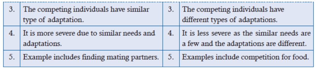
Parasitism describes a relationship between two organisms where one benefits and the other
is harmed. A parasite is an organism that benefits from the relationship, while a host is the
one which is harmed in the relationship. Parasites can be a number of things, including plants,animals, and even viruses and bacteria.
Types of Parasitism
Parasites are classified by how they interact with their host. Overall, parasites are much smallerthan their hosts and reproduce at a faster rate.
(a) Ectoparasites
The term “ecto” in Greek means outside. Therefore, parasites that live
on their host are termed ectoparasites. Examples of ectoparasites
are fleas, ticks, and mites (Figure 1.1). These parasites live on largeranimals, like cats, dogs and deer.
(b) Endoparasites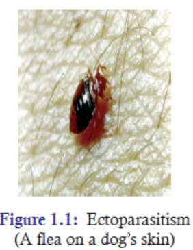
Similarly, the term “endo” in Greek means inside. Parasites that live inside their host are termed
endoparasites. These include the things like parasitic worms,
bacteria, and viruses. Tapeworms are endoparasites. They live in
human intestines where they feed on the partially-digested food in
their host’s intestines. It is a fully protected environment and they
grow and thrive in these conditions.
The tapeworms have no digestive system of their own, but absorb
nutrients through their skin from partially digested food as they pass
through the host (Figure 1.2).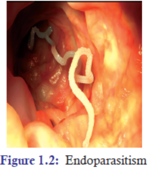
Tapeworms are parasitic worms and are most often referred to as just parasites. They literallysurvive through their host’s nutrients. Parasites need hosts to survive.
Significance of Parasitism
1. Parasitism alters the behaviour and morphology of their hosts. This alteration increases
the chance of being preyed by the predators thereby assisting the parasites to move from
one host to another to complete their life cycle.
2. Parasitism promotes coexistence in biodiversity. Usually in an ecosystem, a competitively
dominant species out-competes a competitively inferior species and doesn’t allow
coexistence with this species. However, parasites reduce the competitive ability of the
dominant species in a biodiversity and, thereby, allow a competitively inferior species to
exist together with a dominant species.
3. Parasitism affects the keystone species and modifies the structure of the ecosystem.
In an ecological community, the effect of parasitism is the strongest when the hosts arekeystone or dominant species with crucial functions in an ecosystem.
4. Parasitism leaves parasite with no responsibility. A social parasite is a parasite that takes
advantage of the interaction of other organisms. The best example of a social
parasitism is brood parasitism. This is an interaction where the parasite, typically
a bird, deposits its eggs in the nest of another species (Figure 1.3). The host
(another species) then ‘babysits’ the egg in place of the parasite (bird), allowing
the parasite to deposit eggs in other nests instead of spending time hatching theirown young.
This therefore leaves the parasite with no responsibility of rearing their young. And it gives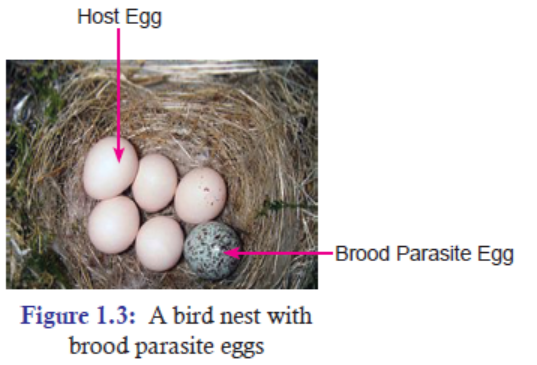
them more time to focus on other things such as producing more offspring. Most species of
cuckoo bird are brood parasites, and there is even one species of fish (spotted catfish) whichparasitizes the ‘nest’ of another fish species!
APPLICATION 1.2
1. Complete the blanks with correct missing terms:
(a) ................ is the competition where individuals of the same species compete.
(b) .................... states that two species requiring same ecological requirements cannot
occupy the same ecological niche.
(c) .................... is a parasite that takes advantage of the interaction of other organism.
(d) Parasites that live ............................ are called ectoparasites.
2. Differentiate between carnivores and herbivores.3. Discuss the significance of predation
1.3 PREDATION
ACTIVITY 1.3
Look out for predator and prey relationships in wildlife channels. You can also watch movies
exhibiting these relations on the following link:.
Observe and discuss the following questions while you watch:
• Why is predation important?
• Giving examples of food chain, name predators and prey.• Why does different predators have different preys?
Predation is an interaction between the two species, i.e., predator and prey, in which one
species (predator) uses another species as food (prey). In other words, one organism kills and
consumes another. Predation influences the distribution, abundance and diversity of species
in ecological communities.
Types of PredationGenerally, predation can be divided into:
(a) Carnivory
Carnivory takes place when a predator consumes meat,
rather than plants, and consequently kills its prey.
Organisms that prefer meat to plants are accordingly called
carnivores. The example of the lion hunting the buffaloesis called carnivory (Figure 1.4). In this type of predation
a predator kills its prey more or less immediately. Other examples are a shark eating a tuna or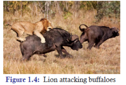
a Venus fly trap consuming a fly.
(b) Herbivory
Herbivory is the act of animals eating plants. Or when an animal uses
a plant as food, it is called herbivory. Example, when a deer eats grass,
the plant is the prey and the animal the predator (Figure 1.5). Additionally,
organisms do not have necessarily to be larger than their prey to be successful
predators. Venomous snakes are able to take advantage of a variety
of large prey items because an injection of venom can be quite fatal.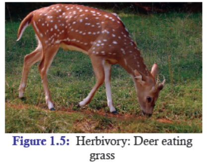
Predation can also occur as parasitism, in which the
prey is a host that supports a parasite, such as a virus. In
this case, the prey may be harmed but not killed outright like the antelope. Unlike carnivory, a
parasite feeds for an extended period on a living host. For example—a tapeworm living in thebody of a deer or a mistletoe “feeding” on a mesquite tree.
Not all predators are animals. Carnivorous plants, such as the Venus fly trap and the pitcher
plant, consume insects. Pitcher plants catch their prey in a pool of water containing digestive
enzymes, whereas the Venus fly trap captures an insect between the two lobes of a leaf and
seals the insect inside with digestive enzymes. These plants absorb nutrients from the insectsas they become available during digestion.
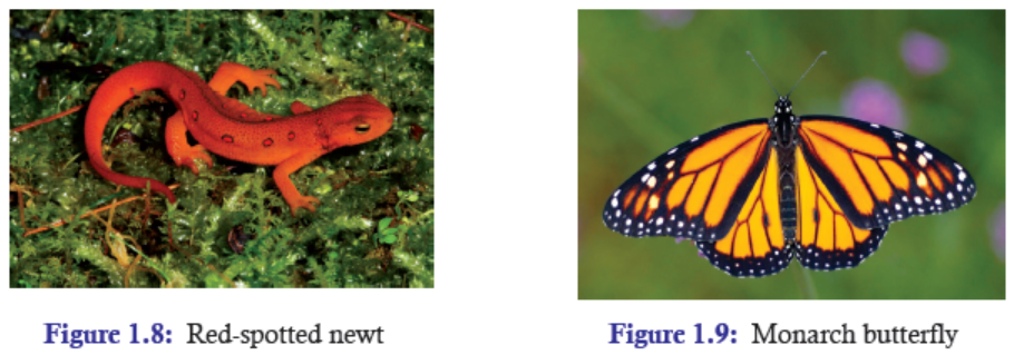
Predation and Adaptation
Adaptation in Predator Species
Based on their experience, predators also undergo certain adaptations to be an efficient hunter
or killer. These adapted traits are passed down from generation to generation. Predators exhibit
traits such as sharp teeth (Figure 1.6), claws, and venom that enhance their ability to catch
food. They also possess extremely acute sensory organs that help them to find potential prey.
Depending upon the requirement that arises, predators also adapt themselves to become much
more efficient. Examples of some adapted animals are:
(a) The ability of raptors to spot potential prey from over a kilometre away.
(b) The acute sense of smell of moles.
(c) The ability of owls to locate mice by sound.
(d) The ability of pit vipers to sense body heat while tracking prey.(e) The ability of bats and dolphins to echolocate.
Predators catch their prey either by pursuing potential prey or by ambushing them. Organisms
that give chase are capable of short bursts of speed like Cheetah (Figure 1.7). Those that lie inwait tend to be camouflaged to avoid detection.
Adaptation in Prey Species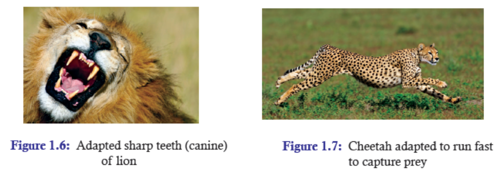
In the same way, as much as predator adapts itself to capture prey, preys also adapt as much
as possible to escape from the predators. Many, such as leaf insects, moths, a variety of frogs
and small lizards, and herbivorous mammals, are cryptically coloured to make them moredifficult to see.
Behaviourally, they freeze after detecting the presence of a predator. This lack of movement
helps them better blend in with their background and inhibits the ability of the predator to
find them. But when the predators venture too close, prey will take flight, running or flying
to escape. When a chase ensues, prey will typically survive if they stay out of reach until the
predator gets tired.
Some species take extra time by distracting the predator. Examples include moths that flash
brightly coloured hind-wings, lizards that drop their tails, and insect larvae that dischargeslime. Such actions surprise the predator and give the prey a few extra moments to escape.
Mimicry
Some prey exhibit bright colouration signalling as poisonous individuals. Such aposematic
colouration helps prevent predation by signalling to potential predators that the vividly-coloured
individual is toxic. Toxins may be manufactured within the body, as with the red-spotted newt
(Figure 1.8), or they may be acquired passively via consumption of toxic plants, as with the
monarch butterfly (Figure 1.9).
Not all the species that exhibit vivid colouration are truly toxic. Some have evolved patterns
and colours that mimic those of toxic species. Examples of such Batesian mimicry include
the extraordinarily polymorphic Papiliodardanus swallow tail butterfly in southern Africa and
Madagascar. Females of this species occur in a wide variety of physical appearances, nearly all
of which mimic distasteful species of the Danaeus and Amauris genera with which they co-occur(Figure 1.10).
Figure 1.10: Batesian mimicry—Non-toxic Papiliodardanus swallow tail butterfly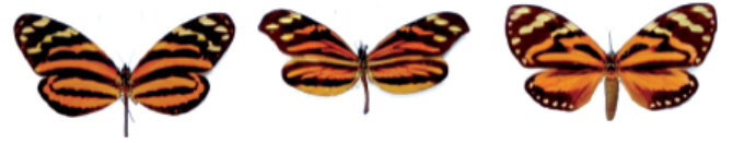
females occur in a variety of forms, each of which mimics the physical appearance
of toxic species. Palatable butterflies (middle column) mimic the warningcolouration of poisonous butterfly species on the left and right butterflies
Adaptation in Herbivory
Herbivory is the consumption of plant material by animals, and herbivores are animals adapted
to eat plants. As in predator-prey interactions, this interaction drives adaptations in both theherbivore and the plant species it eats.
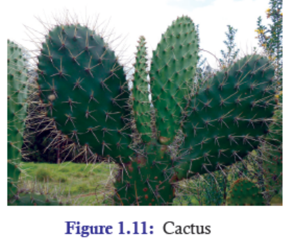
Adaptation in Plants
Though plants cannot move like animals, they also
develop certain mechanism to escape from herbivores. For
example, plants have evolved defences, including thorns
(Figure 1.11) and chemicals, to keep themselves away
from being eaten by herbivores. Scientists have identified
thousands of plant chemical defense compounds,
including familiar compounds such as nicotine andcocaine.
Adaptation in Herbivores
To counteract the adaption of plants and maximize the nutrient intake, herbivores also
have adapted themselves that allow them to determine which plants contain fewer defensivecompounds and more high-quality nutrients.
Some insects, such as butterflies, have chemical sensors on their feet that allow them to taste
the plant before they consume any part of it. Mammalian herbivores often use their keen sense
of smell to detect bitter compounds, and they preferentially eat younger leaves that containfewer chemicals.
Predator-Prey Relationships (Cycle)
Predator-prey relationships are characterized by oscillation of both predator and prey populations
over a period of time. By oscillation, we mean there is a regular pattern of increase and decrease
of populations of both predator and prey (Figure 1.12). Generally, the predator is a carnivore,
while the prey is a herbivore. However, this general truth may vary depending upon the kind
of predator-prey interactions. For example, parasites become predator when they feed on theirhost (prey); herbivores become predator when they feed on plants (prey).
The main reason of oscillation is that as the predator population increases, it progressively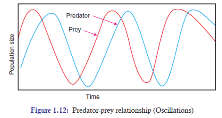
consumes larger number of prey until the prey population starts to decline. Then the declining
prey population no longer supports the large increasing predator population. As the prey
population declines, the predator now faces a food shortage, and many of them starve or fail to
reproduce. As a result, the predator population declines sharply to a point where the reproduction
of prey more than balances its losses through predation. Eventually, the population of prey
increases, which is followed by an increase in the population of predators. In this manner, there
is a regular pattern of increase and decrease in the population of both prey and predator overa time period (Figure 1.12).
Significance of Predation
Predation Prevents a Single Species from Becoming Dominant
A keystone predator is a species that reduces the density of the strongest competitors in a
community. These keystone predators may feed on the dominating prey species and prevent it
from becoming dominant. Thus, they are tied up to the balance of organisms in a particular
ecosystem. Addition or removal of these keystone predators can have drastic cascading effects
on the equilibrium of many other populations in the ecosystem. For example, in grassland,
herbivores (grazers) may prevent as single dominant species from taking over.
Predation can Either Increase or Decrease Species Richness
In an ecological community where predator and prey exist together, predator has the ability to
either increase or decrease the number of prey species. The predator to change the number of
prey depends on the favourability of the environment and also on whether prey is a competitively
dominant species or competitively inferior species in a community. When keystone predator
feeds on dominant prey, it generally promotes species richness by releasing the inferior prey
species to coexist with the dominant species.
Experiment: In an experiment, Paine and others introduced keystone predator Pisaster, a sea
star, in a community (Figure 1.13). This sea star feeds on mussel. In due course of time, they
found out that this predator helped in maintaining species diversity by preventing competitive
exclusion of weaker competitors. Moreover, predation by Pisaster was a key factor in maintaining
populations of at least seven other species. In fact, it was Paine and others who have generatedthe concept of keystone predators.
Predation as Source of Natural Selection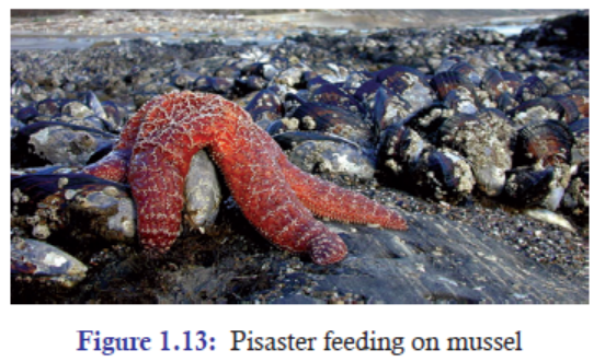
Predation is an important factor of moulding evolution of traits for both predators and prey
species. Natural selection favours the fittest individuals in a community. Thus, the process of
natural selection favours predators that are more efficient in capturing prey than the less efficient
predators. In the same way, the process of natural selection favours prey species that are more
efficient in escaping or deterring predators than the less efficient prey species.
On the one hand, predators impose strong selective force on their prey to evolve into the most
efficient prey against the predators. On the other hand, prey species also counter-impose strong
selective pressures on their predators to evolve into the most efficient predator against the prey.
Since these selection forces are working side-by-side on both predator and prey, these two parties
evolve together. Thus, coevolution is evident. The process of evolution taking place side-by-side
on two closely associated species is called coevolution.
For example: Natural selection process selects faster foxes that can hunt rabbits efficiently.
Simultaneously, natural selection process also selects faster rabbits that can run fast to escape
efficiently from the foxes. The process of selecting the most efficient predator and prey can go
on and on.
APPLICATION 1.3
1). Complete the sentence with the correct assertion:
a) . Thorn is an adaptation of plant against ................................. .
b). Process of evolution taking place side-by-side on two closely associated species is called
a ........................................ .
c) . ................................. prevents a single species from becoming dominant.
d) . Some ................................. exhibit bright colouration signalling as poisonous individuals2) . You are provided with the following Predator-prey data (Snowshoe hare and lynx).
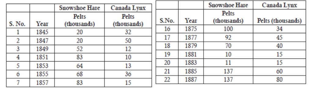
a) Plot the above predator-prey data on a graph paper.
b) Discuss and interpret your graph whether it follows the predator-prey pattern
(oscillation).
c) In the year 1863, the snow hare population was high. What can you say about lynx
population?
d) In 1845, the predator lynx population was more than the prey but gradually it
reversed. What inference can you draw from the change?1.4 MUTUALISMS, COMMENSALISM AND SAPROPHYTISM
ACTIVITY 1.4
Observe the following picture and answer the questions that follow
a) For each animal from A and B, say if it gets benefit (-), it is harmed (-) or remains neutral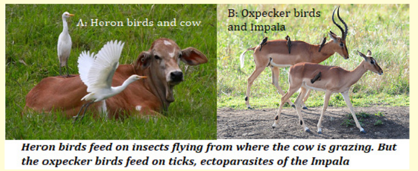
(-)
b) What is the name appropriate to each of the relationship A and B above?
c) It is found that mushrooms often develop on decaying manure of cow and Impala.What is the name appropriate to that type of relationship?
1.4.1 Mutualism (+, +)
It is an interaction of two or more species where the interacting species mutually benefit
from each other. And these interacting species mutually benefit from each other so much
that they become completely dependent on one another. They cannot survive and thrive
without each other. That is the reason why this interaction is termed as mutualism or obligate
symbiosis. Mutualism seems to replace parasitism as ecosystems evolve towards maturity,
and it seems to be especially important when some aspect of the environment is limiting(such as water or infertile soil).
Examples: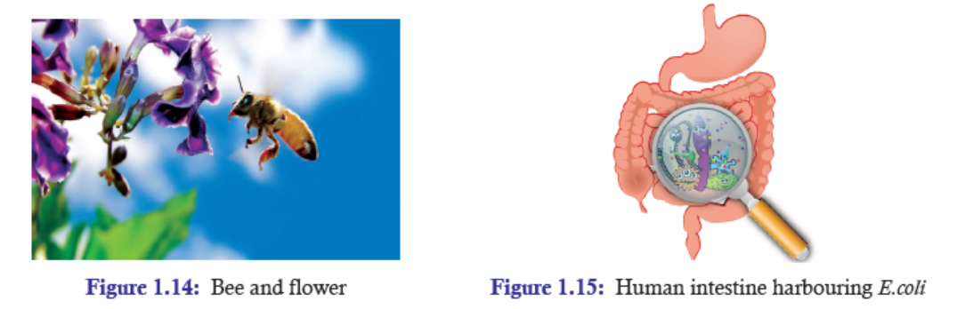
Bees and flowers: Bees depend on flowers for food in the form of nectar and pollen. And
the flowering plants depend on bees or other pollinators to carry their male reproductive cells
specifically to the female parts of other flowers of the same species. In this way, bees depend
on flower for food, while flower depends on bees for pollination (Figure 1.14).
Humans and E.coli: Inside our own bodies, there are hundreds of different types of bacteria
that live just in our large intestine. Most of these are uncharacterized, but we do know a
lot about E.coli, which is one of the normal bacteria found in all human large intestines
(Figure 1.15). Humans provide E.coli with food and a place to live. In return, the E.coli produce
vitamin K and make it harder for pathogenic bacteria to establish themselves in our large
intestine. Whether or not most of the other species of bacteria found in our digestive tract aid
in digestion, absorption, or vitamin production isn’t completely known, but they all make itharder for invasive pathogens to establish a foothold inside us and cause disease.
Significance of Mutualism
Mutualism is a type of symbiosis, which means living together. The most important impact
of mutualism is that the species which cannot survive individually, can survive by partneringwith other individual species. By living together and depending upon each other, they could
overcome harsh and unfavourable conditions and thrive in the ecosystem. Mutualism thus helps
in moulding or structuring community towards better species interactions.
As seen in the above example, E.coli alone might not be able to survive efficiently in the other
environment like they do in the stomach of human beings. In the same way, we humans
might not get the same benefits that we get from E.coli if instead, we harbour other pathogenic
organisms. Thus, in a situation where an individual species cannot survive by itself, mutualism
gives a better chance of survival and reproduction.
1.4.2 Commensalism (+, 0)
It is an interaction of two or more species in which one species is benefited while the other
species is neutral or is not benefited. The species which is benefitted is designated with “+” sign,
while the species which is neutral is designated as “0”. In commensalism, the species which
is unaffected is the host. The species that benefit from the association is called commensal.
Commensal may obtain nutrients, shelter, support or locomotion from the host species. Normally,
commensal relation is often between a larger host and a smaller commensal. Moreover, during
the interaction, the host remains unchanged, whereas the commensal species may show greatmorphological adaptation.
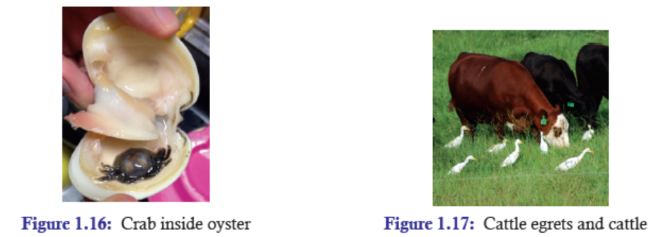
Examples:
Oysters sometimes have a small, delicate crab (Figure 1.16) in the mantle cavity. These crabs
are usually commensal, although sometimes they overdo their guest status by partaking of the
host’s tissues.
Another example is, the cattle egret follows cattle (Figure 1.17), water buffalo, and other
large herbivores as they graze. The herbivores flush insects from the vegetation as they move,
and the egrets catch and eat the insects when they leave the safety of the vegetation. In thisrelationship, the egrets benefit greatly, but there is no apparent effect on the herbivore.
Significance and Criticism of the Concept
The associations between two populations of species that result in positive effects are exceedingly
widespread and are important in determining the function and structure of populations and
communities.
Some biologists argue that the commensal in commensalism must be likely mutualistic or
parasitic in a small scale which is undetected. And it is unlikely that the host is also completely
not harmed or neutral. Example: Epiphytes intercepting substantial amounts of nutrients from
the host plant must be affecting the host in some other way which might be unnoticed.
1.4.3 Saprophytism
In Greek, sapro-(“putrid matter”) + phyte (“plant, growth”). The condition of certain living
organisms feeding and living on dead organic matter is simply called saprophytism. It is generally
exhibited by saprophytes. Saprophytes are living organisms which feed on dead organic matter
such as dead plant or animal tissue. In this regard, they are detrivores. They break down organic
matters in simpler forms that can be taken up and recycled by plants. Thus, they play a very
important role in soil biology. Examples include most fungi (Figure 1.18 (a and b), bacteria,and some orchids.
The term “saprophyte” is a misnomer. By definition, “Phyte” means a plant, and bacteria and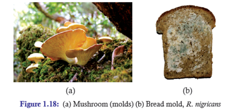
fungi are not categorized as plants. Most of the saprophytes lack chlorophyll, and therefore,
cannot perform photosynthesis. Thus, they depend on the food energy they absorb from the
decaying organic matters. This means that they are heterotrophs and are considered consumers
in the food chain.
They are characterized by their use of a particular kind of digestion mechanism, called extracellular
digestion. In this process, they secrete digestive substances into the surrounding
environment through which they break down organic matter into simpler substances. The
nutrient-rich broken organic substances are then directly absorbed through the membrane of
the organism’s cells and are metabolized.
One of the most common saprophytic fungi belongs to Rhizopus family. These fungi have an
extensive network of hyphae, similar to tiny roots, which grow through the organic matter.
They grow in a network called a mycelium. Mycelium helps the fungus to penetrate into the
organic matter where the hyphae secrete digestive enzymes and absorb the resulting nutrients.
The most common form of Rhizopus is bread mold, R. nigricans (Figure 1.19). These fungi
can also be seen in fruits, especially stone fruit, and vegetables, faeces and the soil. The fungus
grows very rapidly, maturing within four days, though some fungi might take longer period. The
fungus needs a warm, moist environment to thrive. People can prevent or save from Rhizopus
infection by keeping food under refrigeration or in the freezer so that the spores never get a
chance to grow.
Significance of Saprophytism
Many micro saprotrophs and other decomposers, involving insects, snails and beetles help in
recycling valuable nutrients from dead organic matter which is released back into the soil to be
reabsorbed by plants. For example— in a rainforest ecosystem, to promote healthy rainforest,
nutrients such as iron, calcium, potassium and phosphorous are essentially required. The
decomposers derive these essential nutrients from decaying organic matters and then releaseinto the soil where the plants reabsorb it again.
APPLICATION 1.4
Complete the sentence with the correct assertion
(i) Mutualism ........................ both organisms.
(ii) (+, 0) sign exhibits ............................
(iii) ..................... helps in nutrient recycling.(iv) Cattle egret follows water buffalo and exhibits .....................
1.5 SUMMARY
• The basic species interactions are competition (direct interference type), competition
(resource use type), commensalism, parasitism, predation, mutualism and saprophytism.
♦ Competition
• Competition is an interaction of two organisms striving for the same resource. It is of
two types: Interspecific competition is a competition of individuals of the same species
competing for a limited resource, while intraspecific competition is a competition of
different species competing for a limited resource.
• Competition helps in structuring ecological communities and also plays an important
role in character displacement.
♦ Parasitism
• Parasitism is a relationship between two organisms where one benefits and the other
is harmed. The two types of parasitism are: Ectoparasite and endoparasite. A social
parasite is a parasite that takes advantage of the interaction of other organisms.
• Parasitism alters the behaviour and morphology of their hosts; it promotes coexistence
in biodiversity; it affects the keystone species and modifies the structure of ecosystem.
♦ Predation
• Predation is an interaction between species in which one species (predator) uses another
species as food (prey). It can be divided into: Carnivory, parasitism, cannibalism, herbivory.
• Predation prevents a single species from becoming dominant; it also either increases or
decreases species richness; and it acts as a source of natural selection.
♦ Mutualism
• Mutualism is an interaction of two or more species where the interacting species mutually
benefit from each other so much that they become completely dependent on one another.
Example: Bees and flower.
• Mutualism helps in moulding or structuring community towards better species interactions.
♦ Commensalism
• Commensalism is an interaction of two or more species in which one species is benefited
while the other species is neutral or is not benefited. Example: Cattle egret and cattle.
• It helps in determining the function and structure of populations and communities.
♦ Saprophytism
• Saprophytism is a condition of certain living organisms feeding and living on dead organic
matters. Example: Mold (mushroom).
• Many micro saprotrophs and other decomposers, involving insects, snails, beetles, etc.,
help in recycling valuable nutrients such as iron, calcium, potassium and phosphorous
from dead organic matter which is released back into the soil to be reabsorbed by plants.
1.6 GLOSSARY
• Biological interactions: It is a process of different organisms interacting with one another
within a community.
• Brood parasitism: This is an interaction where the parasite, typically a bird, deposits its
eggs in the nest of another species. The host (another species) then ‘babysits’ the egg in
place of the parasite (bird), allowing the parasite to deposit eggs in other nests instead
of spending time hatching their own young.
• Carnivore plants: Not all predators are animals. Carnivorous plants, such as the Venus
fly trap and the pitcher plant, consume insects. Pitcher plants catch their prey in a pool
of water containing digestive enzymes, whereas the Venus fly trap captures an insect
between the two lobes of a leaf and seals the insect inside with digestive enzymes.
• Commensalism: It is an interaction of two or more species in which one species is
benefited while the other species is neutral or is not benefited.
• Competition: It refers to the interaction of two organisms striving for the same resource.
Generally, competition is of two types: intraspecific and interspecific competition.
• Gause’s exclusive principle: It states that two species with identical ecological requirement
cannot occupy the same ecological niche.
• Herbivory: It is an act of animal eating plants. Or when an animal uses a plant as food,
this is called herbivory. Example, when a deer eats grass, the plant is the prey and the
animal the predator.
• Interspecific competition: It is a type of competition in which individuals of different
species compete for the same limited resources in an ecosystem.
• Interspecific interaction: The interaction which involves individuals of the different
species.
• Intraspecific competition: It is a competition where individuals of the same species
compete for the same limited resources in an ecosystem.
• Intraspecific interaction: The interaction which involves individuals of the same species.
• Keystone predator: A keystone predator is a species that reduces the density of the
strongest competitors in a community.
• Mutualism: It is an interaction of two or more species where the interacting species
mutually benefit from each other.
• Oscillation: It is a regular pattern of increase and decrease populations of both predator
and prey.
• Parasitism: It is a relationship between two organisms where one benefits and the other
is harmed.
• Predation: Predation is an interaction between species in which one species (predator)
uses another species as food (prey); one organism kills and consumes another.
• Saprophytism: The condition of certain living organisms feeding and living on dead
organic matters is called saprophytism. In Greek, sapro-(“putrid matter”) + -phyte (“plant,growth”).
END UNIT ASSESSMENT 1
I. Choose whether the given statements are True (T) or False (F)
1. Organisms’ interaction does only harm.
2. Commensalism harms both species.
3. Competing for food is an example of interspecific competition.
4. Herbivory is the act of predation.
5. Predation never promotes species richness.
6. There is a regular pattern of increase and decrease population in oscillation.
7. Parasitism doesn’t promote coexistence of biodiversity.
II. Multiple Choice Questions
1. Both species are denoted by (+, +) in
(a) Mutualism (b) Saprophytism(c) Commensalism (d) Protocooperation
2. When two species compete for a shared resource, it is called
(a) Predation (b) Exploitative competition
(c) Interference competition (d) Apparent competition
3. Adaptations of a predator are
(a) Sharp teeth of lion
(b) Acute sense of smell of moles
(c) Echolocation of bats
(d) All the above
4. Mineral recycling in a rainforest is done by a
(a) Saprophyte (b) Commensal
(c) Predator (d) Ectoparasite
5. Brood parasitism is an interaction where
(a) A parasite kills the host
(b) A parasite lives in the host
(c) A parasite deposits its sperms to the other species’ nest
(d) A parasite deposits its eggs to the other species’ nest
6. In sexual cannibalism, normally
(a) Males eat females (b) Males eat the younger males
(c) Females eat males (d) Females eat the younger females
7. A flea on a dog is an example of
(a) Parasitism (b) Commensalism
(c) Predation (d) Coevolution
8. Saprophytes are
(a) Predators (b) Plants
(c) Parasites (d) Detrivores
9. A commensal is
(a) species that benefits association
(b) species that benefits from the association
(c) species that is negatively affected from the association
(d) species that negatively affects the association
10. The interaction of bees and flowers is an example of
(a) Protocooperation (b) Commensalism
(c) Mutualism (d) None of theseIII. Long Answer Type Questions
1. Giving suitable examples, explain the various interactions of organisms in nature.
2. Giving examples, describe in your own words, the adaptations of predator species
to catch and kill prey and the adaptions of prey species to avoid predators.
3. What are saprophytes? With one example, describe how saprophytes help in recycling
minerals.
4. Briefly compare interspecific and intraspecific competitions with suitable examples.
5. Draw a predator-prey relationship graph and interpret it.
6. Give two examples of the following:
(a) Predation (b) Parasitism
(c) Commensalism (d) Mutualism
7. How does interrelationship among organisms commit for a sustainably developed
environment? Cite examples to support your answer.
8. With examples, state in your own words, the significance of organisms’ interactionsin nature.
Unit 2 Transport Across the Cell Membrane
Key Unit Competence
To be able to explain the physiological processes by which materials move in and out ofcells and the significance of these processes in the life of organisms.
LEARNING OBJECTIVES
At the end of this unit, the learner will be able to:
• Describe and explain the processes and significance of movement in and out of the cell
mentioned in the content.
• Recall that the increasing size of organisms is constrained by its ability to obtain resources
through diffusion across the cell surface and its ability to move substances out of cells.
• Explain the movement of water between cells and solutions with different water potentials
and explain the effects on plant and animal cells.
• Carry out an investigation on simple diffusion using plant tissues and non-living materials.
• Apply the knowledge of hypertonic environments in food preservation by salting.
• Research adaptations of plants and animals to salty habitats.
• Interpret and present data in graphic and table form on the effects of varying concentrations
of : e.g., sugar and salt on plant and animal tissues.
• Appreciate the importance of movement of substances across cells.
• Show concern when exposing living organisms to concentrated media.• Distinguish between endocytosis and exocytosis.
INTRODUCTORY ACTIVITY
A cell is like a fenced factory that produces goods. Apart from goods produced, there are waste
products that should be rid of the factory and on the other hand there is permanent need of
raw materials that should be used by the factory. Therefore, though fenced, the factory is not an
isolated system. It needs exchange with the surrounding environment.
1) In the cell context, what does mimic the fence?
2) State any two materials which should enter the cell
3) State any two material which the cell should get rid of4) Tell your peer how the cell exchanges materials with its environment.
2.1 GRADIENTS ACROSS THE PLASMA MEMBRANE
ACTIVITY 2 .1
1) Use scientific reasoning to prove that the old man is right but the young man is wrong.
2) Make research and bring diagrams that illustrate the following concepts:
(a) Isotonic solutions(b) Hypertonic vs hypertonic solution.
The internal environment of a cell is maintained differently from that of its external environment
by a thin surface membrane called the cell or plasma membrane. The plasma membrane is a
lipid bilayer with phosphate heads and fatty acid tails. It governs the entry and exit of molecules
and ions. This property of the plasma membrane that regulates the exchange occurring between
the cell and its medium is referred to as “cell permeability”.
A cell membrane, therefore, determines which substances can enter the cell (comprising those
which may be important for the vital activities within the cell) and also regulates the outflow
of substances (consisting of excretory waste and water). This feature of the membrane not
only maintains difference in the composition of intracellular and extracellular fluid, but also
establishes a balance in their osmotic pressure. Therefore, a membrane may be permeable to
some substances while impermeable to others. This is called “selective permeability”. The
lipid bilayer of the membrane is permeable to non-polar and uncharged molecules such as O2,
CO2, steroids but is impermeable to charged or polar molecules and ions like glucose. It may
be slightly permeable to water and urea though being polar molecules due to their smaller size.
A concentration gradient refers to difference in the concentration of a substance from one
region to another across a plasma membrane. In such a case, the solute will have a tendency
to move from a region of high to that of low concentration.Due to the distribution of positively and negatively charged ions, the inner surface of plasma
membrane is more negatively charged than the outer. This difference in the electrical charge
between two regions creates an electrical potential and since this is established across a plasmamembrane, it is termed as membrane potential (Figure 2.1).
Figure 2.1: Movement of particles or ions with respect to concentration and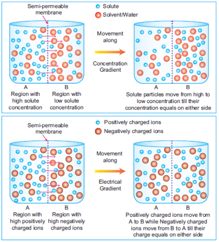
electrical gradient across a semi-permeable membrane
The occurrence of concentration gradient and membrane potential helps in the movement
of substances across plasma membrane. Substances tend to move down the concentration
gradient (downhill movement) known as passive movement, i.e., from high to low
concentration. Similarly, a negatively charged molecule will tend to move to positively
charged region and vice-versa. Therefore, these two parameters greatly affect the ion
movements across the membrane (by diffusion as explained in the following section) and
together constitute the electrochemical gradient.
2.2 MOVEMENT OF SUBSTANCES ACROSS PLASMA MEMBRANE
ACTIVITY 2 .2
1. From O-level you studied many types of transport across the cell membrane. Use available
resources to fin
d out short note on each of the following processes:
Simple diffusion – facilitated d
iffusion – active transport – endocytosis – Phagocytosis
2. Carry out the following experiment and thereafter answer questions that follow:
A. Put a crystal of KMnO4 in a beaker containing 200ml of water. Write your observation
after 5 – 10 – 20 minutes.
B. Take the activity 3 from the SBC. Take the activity 5 from the SB
As mentioned before, plasma membrane is selectively permeable. Figure 2.2 enlists all theprocesses involved in movement of substances across cells.
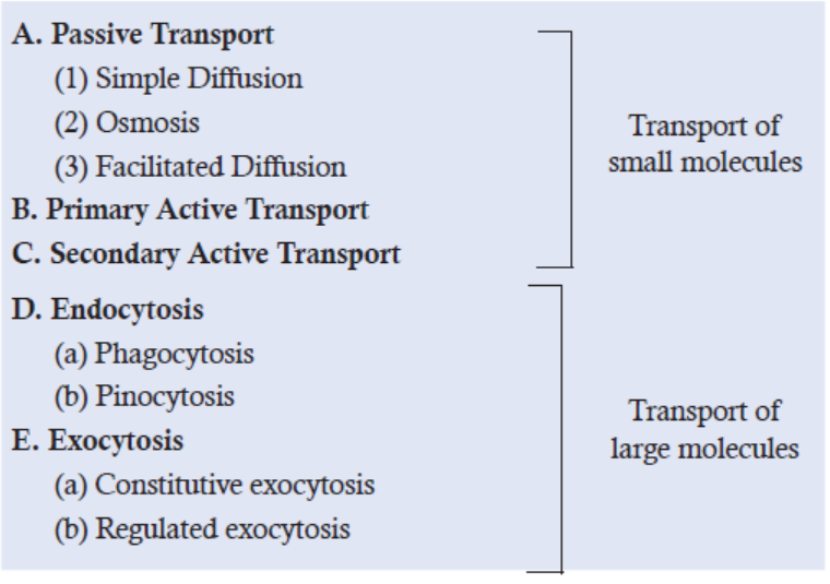
Figure 2.2: Processes involved in the transport of molecules across plasma membrane
Transport mechanisms can be broadly classified into two types:
Passive Transport—It involves the movement of molecules along the electrochemical gradientwithout the use of ATP (Downhill transport). Occurs by diffusion or osmosis.
Active Transport—It drives the molecules against their electrochemical gradient by hydrolysisof ATP (Uphill transport).
2.3 PASSIVE TRANSPORT
Below is an account of different means of transport across the plasma membrane:
2.3.1 Simple Diffusion
It is the simplest mechanism in which a molecule dissolves in the phospholipid bilayer, diffuses
across it and then dissolves in the aqueous solution present on the other side of the cell
membrane. It neither requires ATP nor any protein. The direction of movement is determined
by the concentration gradient (i.e., molecules flow from a region of higher concentration to a
region of lower concentration) or electrical gradient. Therefore, any molecule that is soluble
in the phospholipid layer is capable of crossing the plasma membrane. This is the reason why
only small, relatively hydrophobic (water repelling) molecules (example - benzene), gases (O2,
CO2) and even small polar, uncharged molecules diffuse easily across the plasma membrane
while other larger molecules are restricted.
Diffusion is also regarded as the random mixing of particles in which the particles continue to
move down their concentration gradient until an equilibrium is reached and the particles areevenly distributed throughout the solution (refer to Figure 2.1).
ACTIVITY 2.3
Aim: To investigate simple diffusion using plant tissues and visking tubing.
Materials Required: Visking tubing with capillary, Beaker with water, Sucrose solution (10%),
pieces of beetroot.
Background: Visking tubing is a partially permeable
artificial membrane that mimics the cell membrane,
i.e., it allows
only smaller molecules (e.g., water
molecules, glucose etc.) to pass through while restricts
the larger ones (e.g., starch). In simple diffusion,
the direction of movement
is determined by the
concentration gradient and the molecules
flow from a
region of its higher concentration to a region of lower
concentration (or along electrical gradient).
Procedure:
1. Fill the visking tubing with sucrose and starch solution.
2. Fill a beaker with water and glucose. Now, put some pieces of beetroot into the visking
tubing
3. Partly submerge the visking tubing into the beaker.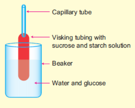
4. Observe the change in the level of liquid in the capillary tube attached to the visking tubing.
5. Observe the diffusion of red pigment from a region of high concentration in the vacuoles to
a region of low concentration in the solution outside the pieces of beetroot.
Discussion:
1. Compare the change in water level (in the capillary) with what you have studied in theory.
2. Discuss your observation.
3. What would you expect if instead of water you had taken 20% sucrose solution in the beakerand performed the same experiment?
Factors Affecting the Rate of Diffusion
1. The greater the concentration difference between the two sides of the membrane, the
faster is the rate of diffusion.
2. As the temperature increases, the rate of diffusion increases.
3. Smaller molecules have faster rate of diffusion while the ones with larger mass, diffuse
slowly.
4. The larger the surface area of membrane available for diffusion, the higher is the rate of
diffusion.
5. The greater the distance across which diffusion is to occur, the longer it takes for molecules
to pass through.
Significance of Diffusion
Diffusion plays an important role in living systems. Below are a few examples where its diverse
significance can be understood.
1. In the human body, nutrients (in the form of ions and small molecules) are absorbed
from the food by the surrounding blood cells in the vessels by way of diffusion.
2. In the lungs, CO2 diffuses out of blood in alveolar sacs whereas O2 (present in high
concentration in the inhaled air) diffuses into the cells in the blood vessels (with low O2concentration).
3. Cutaneous respiration (through skin) is the
most common mode of respiration in lower
non-chordates wherein gases directly diffuse
through the air into the surface epitheliumof the organisms.
4. The eyes lack a great number of blood vessels (which carry oxygen) and therefore, needs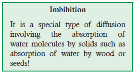
an extra supply of oxygen. The atmosphere provides this extra needed oxygen, which is
taken up by the eye through direct diffusion of O2 into the cornea, the hard outer covering
on the eye. In absence of diffusion, the eyes would dry out.
5. For medicines taken orally as pills, the medicine must somehow find its way into the
bloodstream. Once in the stomach, the medicine from the pill is absorbed into the lining
of the stomach and then into the bloodstream, both by the process of diffusion.
6. Gaseous exchange during the process of respiration and photosynthesis takes place with
the help of diffusion.
7. Transpiration or loss of water from the aerial parts of the plants involves the process of
diffusion.
8. Diffusion is involved in passive uptake of mineral salts.
9. Odour of the flowers to attract the pollinating animals, spreads in the air by diffusion.10. Diffusion plays an important role in imbibition and osmosis.
2.3.2 Osmosis
In osmosis, the movement of water (solvent) occurs due to the difference of chemical potential
(water potential in case of water) on the two sides. The kinetic energy or free energy possessed
by the molecules of a substance is called chemical potential. The chemical potential of water
is called water potential. The chemical potential of pure water (solvent) is higher than that
of the same in a solution. Presence of solute particles decreases the chemical potential (free
energy) of water. The lowering of chemical potential (free energy) is due to attraction and
collision between solvent (water) and solute molecules. Thus, in terms of thermodynamics,
‘Osmosis is the movement of water or solvent molecules from the region of their higher chemical potentialto the region of their lower chemical potential across a semipermeable membrane’.
When a cell is placed in a solution containing a solute (e.g., salt or sugar) dissolved in water,
any of the three conditions may arise (Figure 2.3):
• If the medium is hypotonic with respect to
the cell, i.e., if it has solute concentration
lower than the cell interior, water will tend to
move into the cell. This may lead to swelling
of the cell and even cause it to burst. The cell
is termed turgid. For example, red blood cells
when placed in a hypotonic solution, causehemolysis
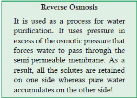
Figure 2.3: Movement of water molecules when a cell is placed in three types of solution—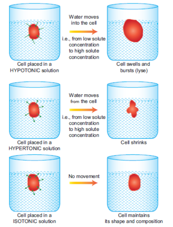
Hypotonic, Hypertonic and Isotonic with respect to the cell
• If the medium is hypertonic with respect to the cell, i.e., has high solute concentration
than the cell interior, then water will tend to move out of the cell into the medium. This
would cause the cell to shrink in size. For example, a plant cell when placed in hypertonic
solution, shows plasmolysis in which the plasma membrane shrinks and becomes widely
separated from the cell wall.
• If the medium is hypotonic with respect to the cell, i.e. the concentration of solutes in
the cytosol is higher than that of the solution. In this condition, water diffuses into the
cell due to osmotic pressure and the cell becomes turgid, or bloated.
• If the medium is isotonic with respect to the cell, i.e., the solute concentration is equal to
that in the cell, the net movement of water across the membrane would be zero. The cell
size and concentration would remain constant. The cell is termed flaccid. For example,0.9% solution of NaCl is nearly isotonic to blood serum.
Difference between Turgidity, Flaccidity and Plasmolysis
Food Preservation by Salting using Hypertonic Solutions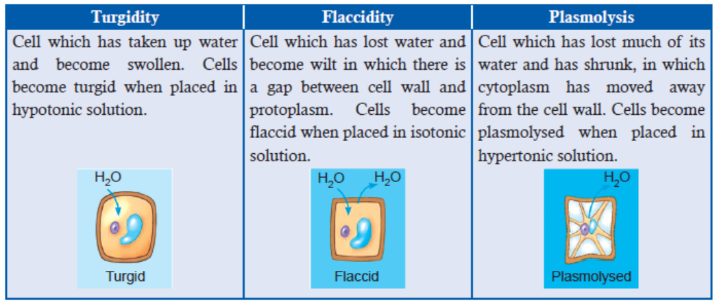
When a cell is placed in a hypertonic solution, water actually flows out of the cell into the
surrounding solution thereby causing the cells to shrink and lose its turgidity. Hypertonic
solutions are used for antimicrobial control.
Salt and sugar are used to create hypertonic environment for microorganisms and are commonly
used as food preservatives.
“Salting is the preservation of food with dry edible salt. It is related to pickling (preparing
food with brine, i.e., salty water). It is one of the oldest methods of preserving food, and two
historically significant such foods are dried and salted cod (usually referred to as salt fish) and
salt-cured meat. Salting is used because most bacteria, fungi and other potentially pathogenic
organisms cannot survive in a highly salty environment, due to the hypertonic nature of salt.
Any living cell in such an environment will become dehydrated through osmosis and die orbecome temporarily inactivated.”
Salting Methods
• Cut your vegetables up in pieces before you put them into the salt water to preserve food
by salt-curing. As you chop a vegetable and put it into the salt water, it makes its own
juice. Nowadays, you might want to use a smaller container. Just make sure the water
has plenty of salt added. Let the vegetables stand in the salt water for at least 10 days in
order to “pickle.” Pickle simply means preserved in brine. Then cover tightly with a lid.
• Preserve meats by salt-curing. Rub meat completely with salt pellets and allow it to cure
for 4 to 8 weeks. At the end of this time, the meat will be almost dry. It can be stored thisway for a long time. This method is called “dry-curing.”
• Soak meat in a solution of brine for a period of 3 to 4 weeks. It will be ready to eat, but
it won’t last long this way. You can also use a syringe to inject brine into the muscles
of the meat in order to preserve the food by salt-curing. It will be ready to eat in 2 to 3
weeks. Just remember that these wet methods of salt-curing meat do not preserve it aslong as the dry method does.
ACTIVITY 4
Aim: To investigate and present the effects of immersing plant tissue in solutions of different
water potentials and using the results to estimate the water potential of tissue.
Materials Required: Potatoes (plant tissue), Cork borer, Measurement scale, Knife/Blade,
Boiling tubes, Aluminium foil, Graph paper, Sucrose solution (0.0M, 0.2M, 0.4M, 0.6M, 0.8Mand 1M), Water.
Procedure: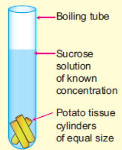
1. Using a cork borer, cut cylinders of potato tissue.
2. Slice the cylinders into 4 cm length each. This is the initial length (Li).
3. Take sucrose solutions of different concentration in boiling tubes
and label them.
4. Immediately add 3 potato tissue cylinders in each boiling tube.
5. Seal the tube with an aluminium foil paper to prevent water loss by
evaporation.
6. Leave the tube rack aside for 1h.
7. Measure the length of the cylinders in each tube. This is the final length (Lf).
8. Calculate the % change in length for each using the formula -
% change in length = [(Lf – Li)/ Li] × 100.
9. Calculate the average of the three readings obtained for each tube.
10. Plot a graph of mean % change in length versus the sucrose concentration used.
11. Draw the best fit line for all the points obtained.
12. Using the graph, determine the sucrose concentration at which the tissue showed no change
in its length. This is the water potential of the potato tissue used (in terms of molarity).
Discussion: Discuss the following questions after observing the results drawn.
• What happened to the potato tissue cylinders? Did they swell or shrink?• Which process do you think brought the change?
Osmosis in Animal Cells
When there is more water outside an animal cell than inside or animal cell is kept in hypertonic
solution, more water particles will enter the cell than leave. This will lead to swelling of the
cytoplasm and push
it outwards. Consequently, the cell membrane will stretch and finally the
cell will burst. On the other hand, when there is less water outside the cell (hypotonic solution)
in comparison to inside, more water molecules move out of the cell and finally the cell will
shrink. Therefore, both the conditions are harmful for animal cells, so, the water concentration
surrounding the animal cell must be kept constant for the cells to carry out their functions
normally.
Process of Osmosis in Plant Cells
Unlike the animal cells, the plant cells do not have the ability to osmoregulate the water that
enters the cells. Therefore, the water tends to move into the cells continuously due to the water
potential. Water Potential is a key concept for understanding the movement of water, i.e., the
plant-water relation. Water molecules (or molecules in gaseous state) show random movement
as they possess kinetic energy. Therefore, as the concentration of water in a solution increases,
the kinetic energy or its water potential increases. Hence, when two solutions are kept in close
contact, water molecules with higher kinetic energy tend to move towards the one with lower
kinetic energy. Pure water has the highest water potential which at room temperature in absence
of any pressure is zero. If solutes are added to water, its water potential decreases because the
number of water molecules with kinetic energy would tend to be low. This magnitude of decline
in water potential due to presence of solutes is referred to as the solute potential.
The continuous uptake of water by the plant cells causes the cells to swell to the limit when the
hydrostatic pressure within the cell prevents any more water to get in. This pressure is known as
Osmotic pressure and the cells are said to be turgid. One of the critical functions of plant cell
walls is thus to prevent cell swelling as a result of this osmotic pressure. In contrast to animal
cells, plant cells do not maintain an osmotic balance between their cytosol and extracellular
fluids. Consequently, osmotic pressure continually drives the flow of water into the cell. This
water influx is tolerated by plant cells because their rigid cell walls prevent swelling and bursting.
Instead, an internal hydrostatic pressure called Turgor pressure builds up within the cell,
eventually equalizing the osmotic pressure and preventing the further influx of water. Turgor
pressure is responsible for much of the rigidity of plant tissues. In addition, turgor pressure
provides the basis for a form of cell growth that is unique to plants. In particular, plant cells
frequently expand by taking up water without synthesizing new cytoplasmic components. Cell
expansion by this mechanism is signalled by plant hormones (auxins) that activate certain
proteins which allow turgor pressure to drive the expansion of the cell in a particular direction.
As this occurs, the water that flows into the cell accumulates within a large central vacuole, so
the cell expands without increasing the volume of its cytosol. Such expansion can result in a
ten to hundred fold increase in the size of plant cells during development. The pressure exerted
on the contents of a plant cell by the cell wall that is equal in force and opposite in directionto the turgor pressure is known as wall pressure
Adaptations of Plants and Animals to Salty Conditions
Plants in salty areas take up more salt from the soil resulting in an increase in salt concentration
in the cells and thus maintaining a water potential that is more negative than that of the soil.
The difference in osmotic potential between plant cells and soil water leads to the movement
of water into the cells through the cell membrane via osmosis. Water is evaporated from the
leaves. This also helps the movement of water from the roots up the stem to the leaves. Some
plants restrict the opening of stomata to conserve their water in salty conditions and some turn
down leaves to decrease surface area of evaporation. Plants have glands to store salt which
burst when concentration of salt increases and causes the release of salt to the soil again. Some
plants regulate salt levels by transporting sodium and chloride ions into the central vacuole.
High salt concentration in the vacuole causes more water uptake and swelling. Some plants
avoid salt stress by releasing leaves in which excess sodium chloride accumulates in petioles.
Animals adapt to the salty conditions very well as plants. For example, fishes in salt water
intake a lot of water and reduce the loss of water by excreting less amount of urine by having
a kidney with relatively few small glomeruli. Fishes also have chloride secretory cells on their
gills which actively transport salts from the blood to the surroundings. Salt glands are also found
in other animals inhabiting salty conditions. Therefore, specially developed kidneys, gills, andbody functions help equalizing salt concentrations across membranes through osmosis.
Significance of Osmosis
Listed below are a few examples that illustrate the importance of osmosis:
1. Osmosis is of prime importance in living organism, where it influences the distribution
of nutrients and the release of metabolic waste products. Living cells of both plants and
animals are enclosed by a partially-permeable membrane called the cell membrane, which
regulates the flow of liquids and of dissolved solids and gases into and out of the cell.
2. It helps maintain the pressure on either side of the cell membrane thereby preventing the
c ells to become turgid and burst or to become flaccid.
3. Plant roots absorb water and minerals from soil and take it upwards to the leaves and
other plant parts which are essential for plant growth.
4. Purification of blood by kidneys also involves osmosis. Osmosis maintains the balance
of inter- and intracellular fluids.
5. Reverse osmosis is used to purify water.
6. Plants wilt when watered with salt water or provided too much of fertilizer as this makes
the soil hypertonic than the plant roots and disrupts water uptake.
But osmosis may also be harmful, especially, in case of marine and freshwater fishes which haveto constantly regulate the movement of water out or into their body (called osmoregulation).
1) Complete the sentence with the correct word
(a) In hypotonic solution, a cell ................................. .
(b) In lungs, CO2 diffuses out of blood by the process of ............................ .
(c) Purification of blood by kidneys takes place by the process of .................. .
(d) The pressure exerted by plants’ cells on the cell wall is ............................. .
(e) The larger the surface area of the membrane, the ........................ is the rate of diffusion.
2) (a) what is the key difference between active and passive transports?(b) Explain briefly how salting is an efficient way of conserving meat
2.4 ACTIVE TRANSPORT
ACTIVITY 4
Active transport is the movement of dissolved molecules into or out of a cell through the cell
membrane, from a region of lower concentration to a region of higher concentration. The
particles move against the concentration gradient, using energy released during respiration.
Use the search engine to find out simulation that illustrates the following:
a) The membrane proteins / Carrier proteins are involved
b) ATP is usedc) Molecules or ions move against their concentration gradient
Active transport is the movement of ions or molecules from a region of lower concentration
to higher concentration across the plasma membrane (Uphill transport). For this, the energy isprovided either by another coupled reaction or by direct hydrolysis of ATP.
2.4.1 Process of Active Transport
(i) Primary Active Transport: It involves direct hydrolysis of ATP. Example includes ion
pumps, for example, Na+ – K+ pump (Na+ – K+ ATPase), responsible for maintaining
gradients of ions across the plasma membrane (Figure 2.4); Ca2+ ions are actively
transported across the plasma membrane with the help of Ca2+ pump which is powered
by ATP hydrolysis, and; H+ ions are actively transported out of the cells by ion pumpsin plasma membranes of bacteria, yeasts and plant cells.
(ii) Secondary Active Transport (Active Transport Driven by Ion Gradients): Molecules are
transported against the concentration gradient not using energy derived directly from ATP
hydrolysis but coupled with the movement of second molecule in an energetically favourable
direction, i.e., from higher concentration to lower concentration. For example, glucose is
transported with the coupled transport of Na+ ions. Na+ gradient is responsible for transportof glucose against concentration gradient from the intestinal lumen to the cell.
Figure 2.4: Working of Na+ – K+ pump. The concentration of Na+ is more outside than inside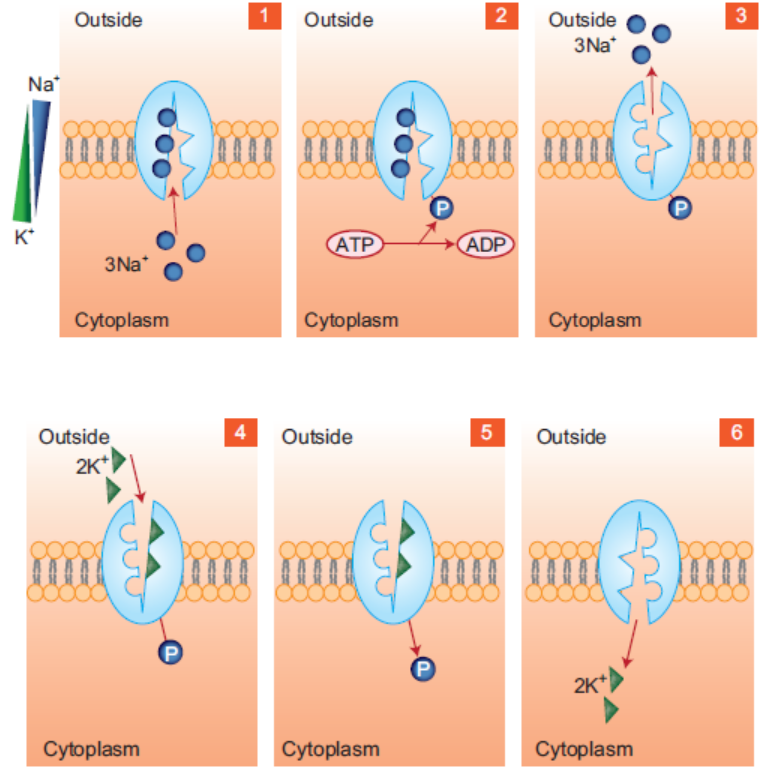
while that of K+ ions is more inside. Steps involved — (1) 3 Na+ ions bind to the pump facing the
cytoplasm, (2) Binding of Na+ promotes ATP hydrolysis and thus phosphorylation of pump,
(3) Conformational change in transporter causes it to face outwards and low binding affinity for
Na+, so 3 Na+ released outside, (4) High binding affinity for K+, so 2 K+ ions bind to pump,
(5) Binding of K+ promotes dephosphorylation and therefore, conformational change in pump, and
(6) Low affinity for K+, so 2 K+ ions are released into the cytoplasm
The transporter simultaneously binds to one molecule of glucose and two ions of Na+. Energetically
favourable movement of Na+ drives the uptake of glucose against its concentration gradient.The coordinated uptake of molecules may be symport, uniport and antiport.
(a) Symport: When two molecules transport in the same direction, e.g., coordinated uptake
of glucose and Na+ (Figure 2.5).
(b) Uniport: Transport of only a single molecule, e.g., diffusion of glucose.
(c) Antiport: When two molecules are transported in the opposite direction, e.g., Na+– Ca2+
antiporter transports Na+ into the cell and Ca2+ out (Figure 2.5). Another example is
Na+–H+ , which transports Na+ into the cell with the export of H+, thereby removingexcess of H+ and preventing acidification of cell cytoplasm.
Figure 2.5: A (1 and 2) - Symport of 1 molecule of glucose with 2 molecules of Na+ ions
by secondary active transport. Note that the two molecules are transported in the same direction.
B-Antiport of 1 molecule of Na+ into the cell and 1 molecule of Ca2+ out of the cell. Note thatthe two molecules are transported in opposite direction
2.4.2 Factors Affecting the Process of Active Transport
It is known that active transport is carried out with the help of pumps. There are two factors
which importantly affect the active transport, including the rate of transport by individual active
transporters and the number of active transporters present in the membrane or in another term
the surface area.
Furthermore, the rate of transport by individual transporter in turn depends upon the nature
of transporter, electrochemical gradient or electrochemical driving force on either side of the
membrane, and the conditions under which a transporter must operate.
2.4.3 Significance of Active Transport in Organisms
(i) In the intestinal lining, glucose is absorbed by active transport from a lower concentrationto a higher concentration in the cells lining the intestine.
ACTIVITY 4
Aim: To interpret data on movement of solvents and ions in and out of the cell in the given
graph.
Materials Required: Given data and the plotted graph.
Background experiment of the given graph: Using a cork borer, cylinders of potato tissue
were cut. The cylinders were sliced into 5 cm length each and weighed. This is the initial mass
(Mi). Different concentrations of salt solutions (0%, 0.2%, 0.4%, 0.6%, 0.8% and 1% NaCl) was
taken in different boiling tubes. 1 potato tissue cylinder was added to each boiling tube. The
tube was sealed and left aside for 24h. Next day, the weight of each cylinder was measured to
obtain the final mass (Mf). The change in weight was then calculated by subtracting Mi fromMf. When the data was plotted on a graph paper, it gave the below shown result.
Procedure:
1. Read and understand the background experimental details and the graph thoroughly.
2. Based on your understanding, interpret the result in terms of answering the below mentioned
questions:
Question 1: What pattern do you observe with respect to some potatoes gaining water and
some losing water? Why?
Question 2: What concentration of salt is isotonic to the potato tissue? Why?
Question 3: Which of the salt concentrations are hypotonic and hypertonic with respect to
potato tissue?
Question 4: What is the effect on the size and weight of the tissue when it is placed in a
hypotonic, hypertonic and isotonic solution?Discussion: Discuss your interpretation
APPLICATION 2.2
1. Define in your own term active transport
2. Differentiate between facilitated diffusion and active transport3. Discuss the significance of active transport in animals.
2.5 ENDOCYTOSIS AND EXOCYTOSIS
ACTIVITY 2.5
Conduct the culture of Paramecium using decaying leaves (e.g. cabbage leave).
After three days, mount on the glass slide a drop sample from the top layer.
Observe under the microscope the movement and feeding of paramecia.
Share your observation.
1) Explain how Paramecia feed2) What name would you give to the feeding process of paramecia?
Christian de Duve (1963) coined the term “endocytosis” which is responsible for ingestion of
large particles (such as bacteria), macromolecules and fluids into the cell in the form of small
vesicles. Unlike all the above mentioned processes involved in transport molecules, endocytosis
is the only means by which large molecules or particles can be taken up by the cell, especially
in eukaryotes. It is further categorized into:
Phagocytosis - Also called “cell eating”. It involves ingestion of bacteria, cell debris or evenintact cells.
Pinocytosis - Also called “cell drinking”. It involves uptake of fluids by the cell.
2.5.1 Phagocytosis
This serves as a means of food capturing by bacteria and many protozoans while in eukaryotic
cells it serves as a defense mechanism to fight against harmful microorganisms and even to get
rid of the cells that have stopped functioning normally or are aged. In mammals (such as man),
macrophages (of spleen and liver) and neutrophils are key components of the immune systemthat show phagocytosis and are therefore also called “professional phagocytes” (Figure 2.6).
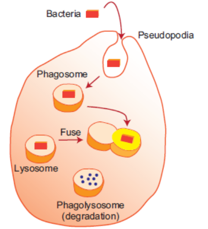
Figure 2.6: Process of phagocytosis showing the ingestion of a bacteria by a cell
2.5.2 Pinocytosis
It is also called “fluid endocytosis” and is used primarily for the uptake of extracellular fluids. It
is a non-specific process which involves engulfing of either pre-dissolved or already broken down
substances. This non-specificity in the ingested substance distinguishes it from phagocytosis
which takes up specific substances from the exterior. Also, phagocytosis involves breakdownof the particle which is lacking in case of pinocytosis (Figure 2.7).
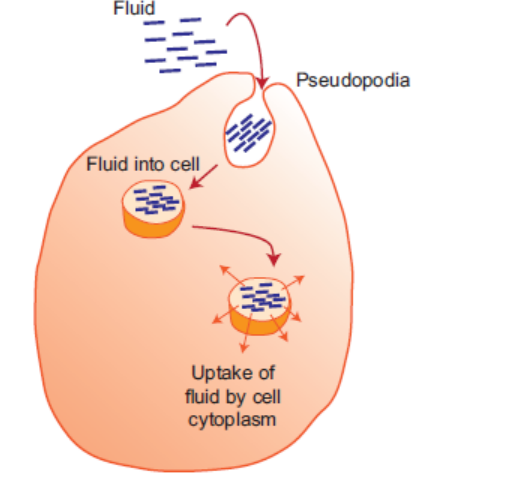
Figure 2.7: Process of pinocytosis showing the uptake of fluid by a cell
EXOCYTOSIS
Unlike endocytosis in which macromolecules or fluids are taken into the cell, exocytosis results
in secretion or release of substances out of the cell. It also involves membrane enclosed secretory
vesicles which are formed within the cell and fuse with the plasma membrane to drain off allits contents into the surrounding medium. (Figure 2.8)
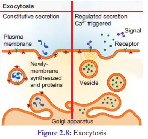
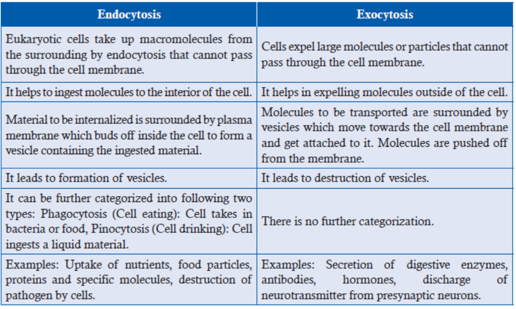
Table 2.1: Comparision of Endocytosis and Exocytosis
APPLICATION 2.3
1. Complete the sentence with the correct word
(a) The process of cell drinking is known as ..................... .
(b) Ca2+ ions are required for .............................. .
(c) When two molecules are transported in opposite direction, it is .................... .
(d) ............................ involves ingestion
2. The Amoeba is a single-celled organism that lives in water. Describe how it engulfs particlesof food by endocytosis.
2.6 SUMMARY
• Every cell is surrounded by cell or plasma membrane which regulates the movement or
exchange of ions or molecules between the cell and its medium. This property of cell is
called cell permeability.
• The presence of concentration and membrane potential (together called electrochemical
gradient) helps in the movement of substances across the membrane.
• Plasma membrane mediates transport of smaller molecules by passive and active
transport whereas larger molecules are transported by endocytosis.
• In passive transport, ions/molecules move from higher concentration to lower
concentration which includes diffusion and osmosis and there is no utilization of energy.
• Simple diffusion is the movement of small hydrophobic molecules from higher
concentration
to lower concentration by dissolving in phospholipid bilayer till equilibrium
is reached.
• Osmosis is a movement of water molecules from low solute concentration to high solute
concentration side (or from higher solvent concentration to lower solvent concentration).
• Active transport is the movement of ions/molecules from lower concentration to higher
concentration. It is of two types: Primary and Secondary active transport. The former
involves direct utilization of energy in the form of ATP hydrolysis while the later involves
movement of molecules against concentration gradient but coupled with the movement
of a second molecule in an energetically favourable direction without direct utilization
of ATP. The movement may be symport, uniport or antiport.
• Endocytosis is the ingestion of large particles such as bacteria, macromolecules and fluids
into the cell in the form of small vesicles. It is further of two types, viz., phagocytosis
(cell eating, engulfing of solid particles) and pinocytosis (cell drinking, uptake of liquidfluids).
2.7 GLOSSARY
• Active transport: Transport of moleculer across cell membrane utilizing energy in the
form of ATP.
• Diffusion: It is the movement of solutes from higher concentration to lower concentration
through semi-permeable membrane.
• Endocytosis: Cells engulf molecules which cannot pass through the plasma membrane.
• Exocytosis: Cells expel molecules which cannot pass through the plasma membrane
by exocytosis.
• Osmosis: It is a movement of water molecules from ion solute concentration to higher
solute concentration.
• Osmotic potential: The potential of water molecules to move from a hypotonic solution
to a hypertonic solution across a semi-permeable membrane.
• Passive transport: Transport of ions and molecules through cell membrane without
utilization of energy.
• Phagocytosis: It is a food capturing process of bacteria and protozoans. In eukaryotes,
this process is used for remolving aged cells or those which have stopped functioning.
• Pinocytosis: It is a non-specific process which involves engulfing of either pre-dissolved
or already broken down substances.
• Plasmolysis: It is the process in which cells lose water in a hypertonic solution. Plasmolysis
can lead to cell’s death.
• Wall pressure: The pressure exerted by water inside the cell against the cell wall. It is
also called Turgor pressure.
• Water potential: It is the measure of potential energy in water. It drives the movement
of water through plants.
END UNIT ASSESSMENT 2
I. Choose whether the given statements are True (T) or False (F)
1. Passive transport occurs by diffusion or osmosis.
2. Simple diffusion involves uphill transport of ions or molecules.
3. Osmosis is the movement of water or solvent molecules from the region of their higher
chemical potential (free energy) to the region of their lower chemical potential (free
energy) across a semipermeable membrane.
4. Not all transport mechanisms occurring across a cell membrane require ATP utilization.
5. Molecules or substances that are large in size are transported across the membrane
by active transport.
6. In the human body, nutrients (in the form of ions and small molecules) are absorbed
from the food by the surrounding blood cells in the vessels by way of osmosis.
7. Purification of blood by kidneys involves diffusion.
8. Reverse osmosis is used to purify water.
9. In the intestinal lining, glucose is absorbed by active transport from a lower
concentration to a higher concentration in the cells lining the intestine.10. Salting is one of the oldest methods of preserving food.
II. Multiple Choice Questions
1. ………………….. is the movement of ions or molecules from a region of lower
concentration to higher concentration across the plasma membrane.
(a) Active transport (b) Passive transport
(c) Pinocytosis (d) Exocytosis
2. In the absence of ………….. eyes would dry out.
(a) osmosis (b) diffusion
(c) endocytosis (d) exocytosis
3. Gaseous exchange during the process of respiration and photosynthesis takes place
with the help of
(a) osmosis (b) diffusion
(c) endocytosis (d) exocytosis
4. Transpiration involves the process of
(a) osmosis (b) diffusion
(c) endocytosis (d) exocytosis
5. ……………………… is important for the transport of nutrients into the cells and
toxic substances out of the cell.
(a) Active transport (b) Passive transport
(c) Pinocytosis (d) Exocytosis
6. For transport by simple diffusion,
(a) Particles should be small in size (b) Particles should be soluble in lipid
(c) Both of the above (d) None of the above
7. Which of the following transport mechanisms describes the process by which a
macrophage engulfs bacteria?
(a) Passive transport (b) Active transport
(c) Endocytosis (d) Transcytosis
III. Long Answer Type Questions
1. In your own words, explain the processes by which materials move in and out of cells.
2. Give four examples, showing significance of diffusion in living systems.
3. Give four examples, showing the importance of osmosis in living systems.
4. In your own words, explain the significance of Active transport in living organisms.
5. With examples, explain how can you apply the knowledge of hypertonic environments
in food preservation by salting?
6. How do plants and animals adapt to salty conditions?7. Distinguish between endocytosis and exocytosis giving suitable examples.
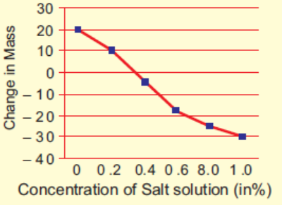
Unit 3 Chromosomes and Nucleic Acids
Key Unit Competence
To be able to describe the structure of a chromosome and how DNA is folded into achromosome
LEARNING OBJECTIVES
At the end of this unit, the learner will be able to:
• Describe the composition of chromosomes and the structure of nucleotides.
• Use of complementary base pairing to write the sequence for messenger RNA and the first
DNA codes for three base codons.
• Appreciate the importance of the presence of DNA in chromosomes.
• State how nucleotides pair.
• Describe the structure of DNA and RNA.
• Explain that the structure of the DNA molecule is described as a ladder twisted into a spiral.
• Draw the structure of DNA (6-10 base pair sequence).
• Explain the Watson-Crick hypothesis of the nature of DNA.
• Research on how Watson and Crick determined the nucleotide base pairing pattern.
• Outline the significance of telomeres in permitting continued replication.
• Acknowledge the role of telomeres in preventing the loss of genes and its relation to the
development of cancer.
• Distinguish between RNA and DNA.
• Describe the nature of genes.• Describe the structure of a genetic code.
INTRODUCTORY ACTIVITY
The genetic characters are passed from parents to offspring through the genes found in the
reproductive cells.
Conduct a research available resources to answer the following questions:
1. What is the relationship between a gene, DNA, and Chromosomes?
2. Draw and label the structure of a chromosome3. What do know about nucleic acids?
3.1 COMPOSITION OF CHROMOSOME
ACTIVITY 1
Aim: Observe the structure of chromosomes at prophase stage of mitotic cell division.
Materials Required:
1. Permanent slide of prophase stage of mitotic cell division.
2. Compound microscope
Background: Prophase is the first phase of mitosis. At this phase, no nucleolus is seen; the
chromosomes start condensing. Thread-like structure of chromosomes is seen under compoundmicroscope
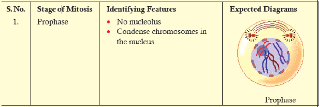
Procedure:
1. Take the permanent slide.
2. Place it on the stage of the compound microscope.
3. Observe the prophase stage of mitotic cell division.4. Draw a well-labelled diagram of the structure of prophase stage chromatin.
Discussion:
1. Is there any difference between the structure of chromatin which you have observed and
what you have learned in the theory class?
2. What can you say about the nuclear membrane and spindle fibres in prophase stage observedunder a compound microscope?
Chromosomes are made of long aggregates of genes formed from condensed chromatin.
Chromatin is made up of DNA, proteins, RNA and other macromolecules. It is located in the
nucleus of a cell.
Deoxyribonucleic acid (DNA) is the storehouse of genetic information in the cell. A complete
set of an organism’s DNA is called a genome. And a gene is a segment of DNA that encodes
for a particular trait. Chromosomes are the structures that hold genes; they are made up of
strands of DNA tightly wrapped around histone proteins. Chromosome is basically composed
of three components—
(A) Nucleotides, (B) Histones proteins and (C) Non-histones proteins.
A. Nucleotides
The monomers that make up Deoxyribonucleic acid (DNA) and Ribonucleic acid (RNA) are
called nucleotides. Nucleotide has three components:
1. Pentose (five-carbon) sugar,
2. Nitrogenous (nitrogen-containing) base, and3. Phosphate group.
Pentose Sugar
Pentose sugar has five carbon atoms, which are numbered 1’ to 5’ respectively (Figure 3.1).
In DNA, the pentose sugar is deoxyribose: a hydrogen atom (H) is attached at the 2’ carbon
position. In RNA, the pentose sugar is ribose: hydroxyl group (OH) is attached at the 2’ carbonposition.
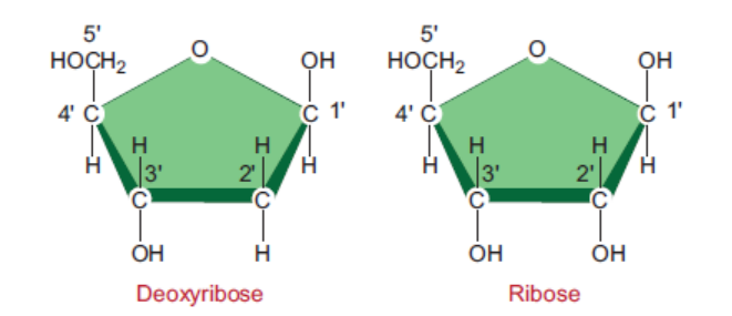
Figure 3.1: Structures of deoxyribose and ribose in DNA and RNA
Nitrogenous Bases
There are two classes of nitrogenous bases—Purines and Pyrimidines. Purines are ninemembered,
double-ringed structures (Figure 3.2). In these purines, the carbons and nitrogens
are numbered 1 to 9. There are two purines—Adenine (A) and Guanine (G). Pyrimidines
are six-membered, single-ringed structures (Figure 3.2). The carbons and nitrogens in these
pyrimidines are numbered 1 to 6. Pyrimidines are of three types—Thymine (T), Cytosine (C),and Uracil (U) (Figure 3.2).
Figure 3.2: Structures of nitrogenous bases in DNA and RNA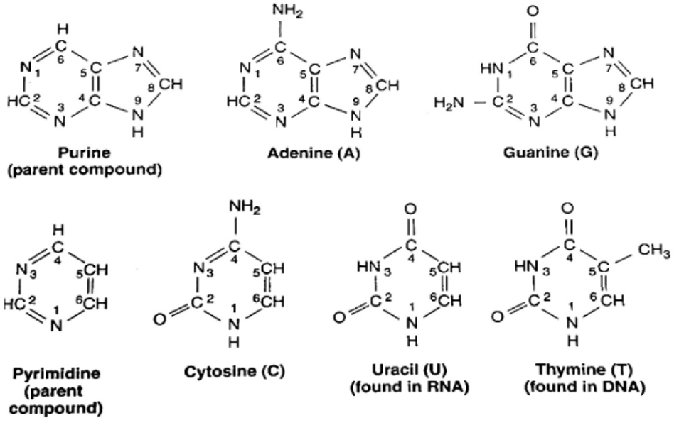
Table 3.1: Difference between DNA and RNA base composition
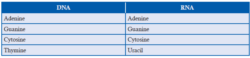
B. Histones Proteins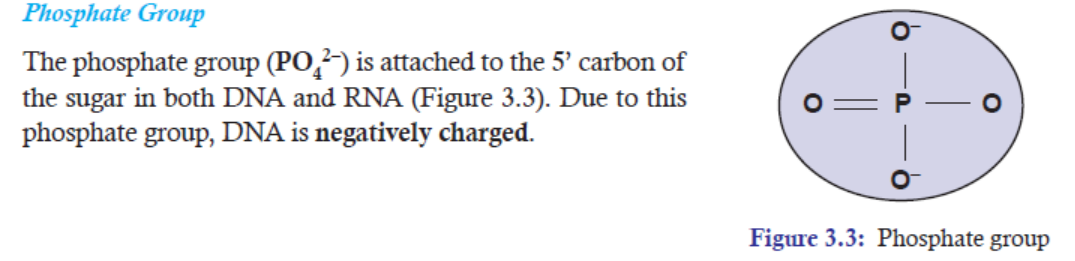
Histone proteins play an important role in organizing the physical structure of the chromosome.
They are most abundantly found in chromatin where they are wrapped around by DNA strands.
Moreover, they are small basic proteins with a net positive charge that assist their binding to
the negatively charged DNA (due to phosphate groups which are negatively charged).
In eukaryotes, there are five main types of histone proteins
(Figure 3.4). They are—H1, H2A, H2B, H3, and H4. H1
is loosely attached to the rest of the histone core proteins.
That is why H1 can be easily separated from the rest of the
histone proteins. And two each of H2A, H2B, H3, and H4
form core of eight histone proteins. These core proteins are
also called histone octamers. A strand of DNA measuring
147 bp segments wraps around this histone octamers for
about 1.7 times. Each nucleosome is connected by a strand
of DNA called linker DNA. For example, Human linkerDNA ranges from 38-53 bp long.
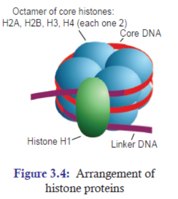
C. Non-histones Proteins
Excluding histone proteins, the rest of the proteins associated with DNA come under the
category of non-histone proteins. Non-histone proteins in so many ways are different from
histone proteins. Some of the differences are:
1. The number of non-histone proteins is much lesser than histone proteins.
2. Non-histone proteins are acidic proteins, which are negatively charged.
3. They play important role in the process of DNA replication, DNA repair, transcription
gene regulation, and recombination.
4. They vary in number and type from cell type to cell type within an organism at differenttimes in the same cell type, and from organism to organism.
3.1.1 The Importance of the Presence of DNA in Chromosomes
(a) Protection
The packaging of DNA in chromosomes helps in protecting DNA from being damaged.
(b) Conserve Space
If we take the DNA from all the cells in a human body and line it up, end to end, it would
form a strand 6000 million miles long! In order to store this very long important material,
DNA molecules are tightly packed around proteins called histones to make structures called
chromosomes.
The packaging of DNA in chromosomes helps in conserving space in cells. Approximately,about two metres of human DNA can fit into a cell that is only a few micrometres wide.
Figure 3.5: To better fit within the cell, long pieces of double-stranded DNA are tightly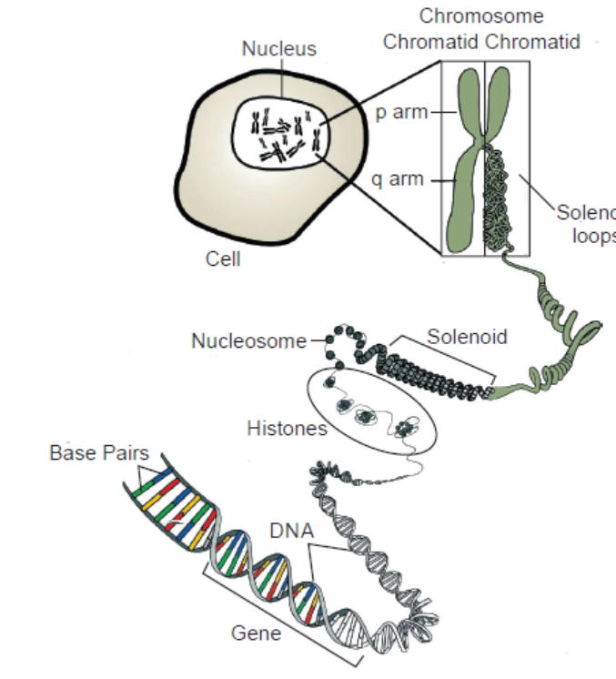
packed into structures called chromosomes
(c) Control of Gene Expression
Chromatin is a complex of DNA and proteins that forms chromosomes within the nucleus
of eukaryotic cells. In its extended form, chromatin looks like beads on a string (Figure 3.5)
under the microscope. The beads are called nucleosomes, while the link between them is a
strand of DNA.
The packaging of DNA in chromatin form helps in controlling gene expression. Highly
compacted chromatin is not accessible to the enzymes involved in DNA transcription,
replication, or repair.
Chromatin has two main regions. The less condensed regions of chromatin are the regions
where active transcription takes place. This region is called euchromatin. On the other hand, the
condensed region of chromatin is where transcription is inactive or is being actively inhibitedor repressed. This region is called heterochromatin.
APPLICATION 3.1
Use the available resource to conduct research on the composition of chromosome. Thereafter,
answer the following questions:
1) What makes up a chromosome2) What is the difference between histone and non-histone proteins?
3.2 STRUCTURE OF NUCLEOTIDES
ACTIVITY 3.2
Use search engine to find out:
1. How the two strands in DNA join or stick together to form double stranded structure?2. Why adenine and guanine don not pair when the two strands join?
3.2.1 Nucleotide
A basic unit of nucleotide is made up of pentose sugar, a nitrogenous base, and a phosphate sugar.
However, a combination of only a pentose sugar and nitrogenous base, without phosphategroup, is called nucleoside.
In DNA and RNA, bases are covalently bonded to the 1’ carbon of the pentose sugar. The purine
and pyrimidines bases attached to pentose sugar from different positions of their nitrogen bases.
Purine bases use the 9th position of nitrogen to attach with 1’ carbon of pentose sugar, while
pyrimidine bases use the 1st position of nitrogen to attach with 1’ carbon of pentose sugar.
In both DNA and RNA, the phosphate group (PO4 2–) attaches to the 5’ carbon of pentosesugar.
Thus, by attaching phosphate group to a nucleoside yields a nucleoside phosphate or
nucleotide.
In DNA, the complex of deoxyribose, nitrogenous base and phosphate group is called
DNA nucleotide (a deoxyribonucleotide) (Figure 3.7), whereas in RNA, the complex of
ribose, nitrogenous base and phosphate group is called RNA nucleotide (a ribonucleotide)(Figure 3.6).
3.2.2 Phosphodiester Bond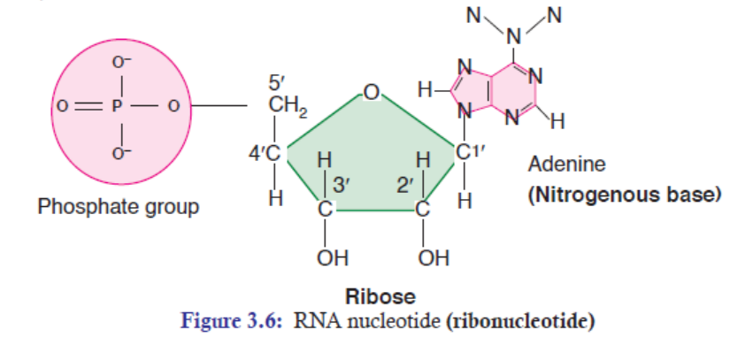
Two nucleotides are covalently joined together by a bond called phosphodiester bond
(Figure 3.8). In phosphodiester bond, the phosphate group, which is attached on 5’ of
one nucleotide, forms a bond with the 3’ carbon of another nucleotide. In this way, many
phosphodiester bonds are formed in between sugar and phosphate groups. The repeated sugarphosphate-
sugar-phosphate backbone is a strong one. Because of this strong backbone, DNAand RNA are stable structures.
5′ end
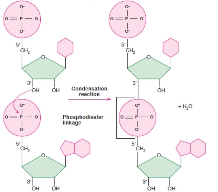
3′ end
Figure 3.8: A figure showing phosphodiester bond formation (between two ribonucleosides)
3.2.3 Polarity
Polynucleotide chains have polarity. On one end, there is a 5’ carbon with a phosphate group
(PO4
2–). On the other end, there is a 3’ carbon with a hydroxyl group (OH) on it (Figure 3.8).
The ends of polynucleotide are frequently referred to as the 5’ end and the 3’ end.
3.2.4 Chargaff ’s Rules: The Rules of Base Pairing
Erwin Chargaff’s rules state that DNA of all organisms should have a 1:1 ratio of pyrimidine and
purine bases. Thus, the amount of adenine (A) is equal to that of thymine (T); and the amount
of guanine (G) is equal to that of cytosine (C). This equivalence of purine and pyrimidine bases
is known as Chargaff ’s rules. This pattern is found in both strands of DNA.Table 3.2: Difference between purine and pyrimidine
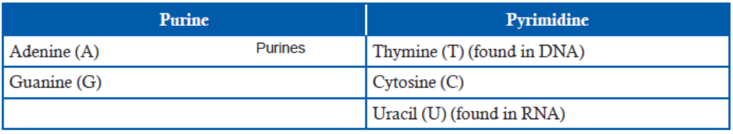
The specific base pairing of A-T bases and G-C bases is called complementary base pairs. For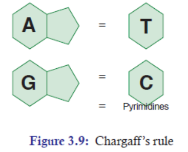
example, if one strand of DNA sequence is 5’-ATATCCGGAT-3’, then the opposite strand of
DNA sequence will be 3’-TATAGGCCTA-5’. Thus, by using the rules of base pairing, once
we have the sequence of at least DNA strand, we can find out the opposite base sequence ofthat DNA.
APPLICATION 3.2
1) Complete the sentence with the correct word:
(a) ............................ is a segment of DNA that encodes for traits.
(b) The two Purines are ............................... and ................................... .
(c) Uracil is present in .................................. .
(d) Two nucleotides are covalently joined by ..................................... .
(e) A-T bases and G-C bases are called ................................... base pairs.
2) What is the maximum number of hydrogen bonds in a length of DNA containing 700 basepairs?
3.3 STRUCTURE OF NUCLEIC ACIDS—DNA AND RNA
ACTIVITY 3.3
Aim: Design the structure of DNA molecule and complimentary
base pairing using plastic
model shapes or homemade kits.
Materials Required:
1. Plastic models of pentose sugars, phosphate groups, nitrogenous bases (A, G, T, and C).
Or
2. Homemade kits of pentose sugars, phosphate groups, nitrogenous bases (A, G, T, and C).
Procedure:
1. Before you start the activity, study the composition of chromosome (DNA) properly.
2. By using the plastic models, construct the structure of DNA.
3. Or in the same way, by using the plastic models, make the complementary base pairs of
nucleotides (A, G, T, and C).
4. Once the construction of the structure of DNA and the complementary base-pairing are
ready, give a presentation to the class.
Note: 1. Remember the Chargaff ’s rule of base pairing concept.2. Remember the structure of DNA.
In the structure of DNA, the strong electronegative atom is the Oxygen (O) and Nitrogen (N),
while H atom has positive charge. In the structure of DNA (Figure 3.10), thymine and adenine
have two hydrogen bonds; while guanine and cytosine have three hydrogen bonds. Hydrogen
bonds or interactions play a very important role in binding the bases of the opposite strands in
the DNA. Though RNA is not the genetic material in most of the cases, both single-stranded
and double-stranded RNAs are the genomes of certain viruses. RNA double-stranded molecules
show structural similarity to that of double-stranded DNA molecules. The similarities are:
(a) Both have anti-parallel strands.
(b) Both have sugar-phosphate backbones on the outside of helical molecule.
(c) In both the cases, in the middle of the helix, a complementary base pairing is formed byhydrogen bonds.
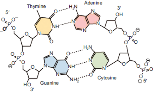
Figure 3.10: Structure of DNA [See the hydrogen bonds between the four bases (T-A, G-C)]
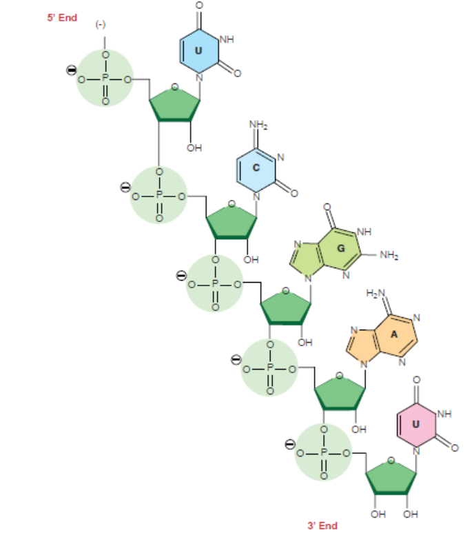
Figure 3.11: Chemical structure of RNA
APPLICATION 3.3
1) Draw the structure of a DNA sequence made of 3 pairs of nucleotides
2) Compare DNA and RNA
3.4 THE WATSON-CRICK HYPOTHESIS OF THE NATURE OF DNA/THESTRUCTURE OF DNA
ACTIVITY 3.4
In 1953, James D. Watson, an American molecular biologist, and Francis H.C.Crick, a British
molecular biologist, proposed a model for the physical and chemical structure of the DNAmolecule.
Make research from available resource to explain why DNA is described as a Twisted LadderStructure.
In 1953, James D. Watson, an American molecular biologist, and Francis H.C.Crick, a British
molecular biologist, proposed a model for the physical and chemical structure of the DNA
molecule. Today, their model is known as double helix model of DNA or simply the Structureof DNA.
The main features of Watson and Crick double helix model (Figure 3.12) of DNA are:
1. Two polynucleotide chains wind around each other in a right-hand double helix(Figure 3.12).
2. The two polynucleotide chains run side-by-side in an antiparallel fashion. This means
that one strand of DNA will orient itself in a 5’ -3’ direction, whereas, the other strand
will orient itself alongside the first one in a 3’-5’ direction. In this way, the two strandsare oriented in opposite directions (Figure 3.13).
3. On one hand, the sugar-phosphate backbones lie outside of the double helix. On the
other hand, the bases orient themselves toward the central axis of the double helix
structure.
The bases of one strand are bonded with the bases of the other strand of double helix
by hydrogen bonds. These bonds are weak chemical bonds. Since hydrogen bonds are
relatively weak bonds, the two strands can be easily separated by heating the DNA. The
bonding of these bases in the double helical structure follows the Chargaff ’s base pairingrules. For example—Adenine (A) will form two hydrogen bonds with Thymine (T).
Similarly, Guanine (G) will form three hydrogen bonds with Cytosine (C). This specificbase paring is called complementary base pairing (Figure 3.12).
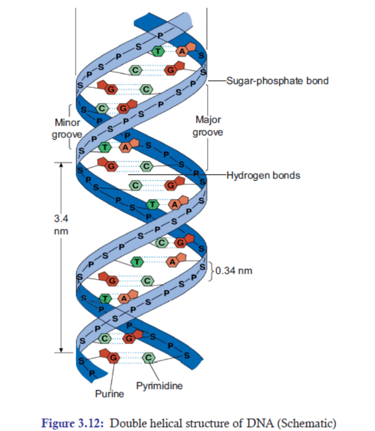
4. The distance between adjacent bases is 0.34 nm in the DNA helix. A complete turn of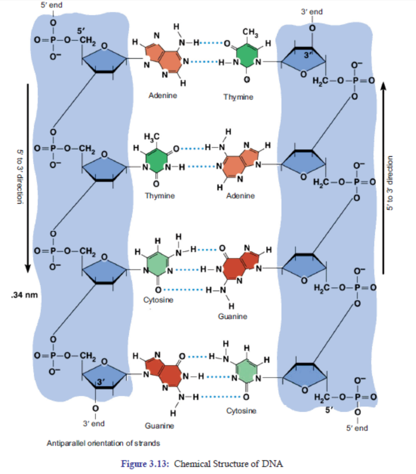
the helix takes 3.4 nm. One complete turn, which is 360° turn, accommodates 10 base
pairs (bp). And the diameter of the helix is 2 nm (Figure 3.13).
5. There are major and minor grooves in the double helix. The two sugar-phosphate
backbones of the double helix are not equally spaced from one another along the helical
axis, because of the way the bases bind with each other. As a result, there is an unequal
size of grooves between the backbones. The wider groove is called major groove; rich in
chemical information. The narrower groove is called minor groove; less rich in chemical
information (Figure 3.13).
3.4.1 DNA is also Described as a Twisted Ladder Structure
A typical ladder has two long wooden or metal side strands or pieces between which a series of
rungs or bars are set in suitable distances (Figure 3.14). In the structure of DNA, the pentose
sugars and phosphate groups make up the “long two side strands or pieces” of a typical ladder.
And the A-T and G-C base pairs which are bonded by hydrogen bonds make up the “rungs
or bars”of a typical ladder (Figure 3.15).
But unlike a typical ladder which is straight, the two strands of DNA are twisted into spiral.
Scientists call this a double helix. DNA also folds and coils itself into more complex shapes. The
coiled shape makes it very small. In fact, it is small enough to easily fit inside any of our cells. If
a DNA from a cell is unfolded, it would stretch out to a length of about six feet. The structural
twisted nature of DNA has been attributed to enhance its stability and strength. Thus, for thesesimple similarities with a typical ladder, DNA is also referred to as a twisted ladder structure.
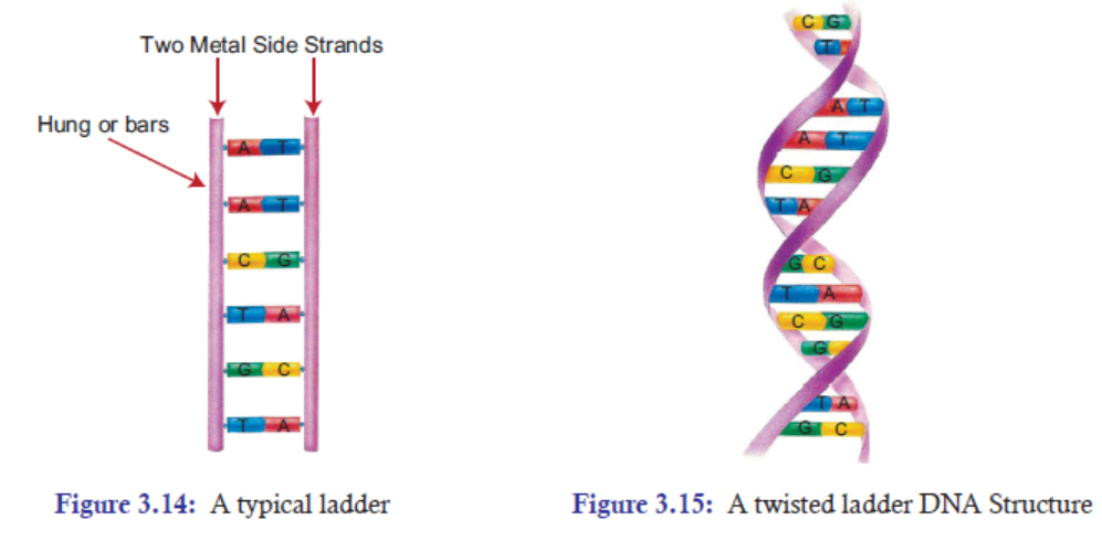
APPLICATION 3.4
1. Complete the sentence with the correct word
(a) Watson and Crick proposed the model of .................................... .
(b) Enzyme ..................................... maintains the length of telomere.
(c) ...................................... can be used to cure cancer.
(d) ..................................... bonds are seen in both DNA and RNA.
(e) ...................................... directs synthesis of proteins in the body.
2. Differentiate the structure of DNA to that of real ladder.
3. What would be the side effect of untwisting the DNA
3.5 SIGNIFICANCE OF TELOMERE IN PERMITTING CONTINUEDREPLICATION
3.5.1 What is Telomere?
A telomere is a region of repetitive nucleotide sequences at each of a chromosome. It protects
the end of the chromosome from being deleted or from fusion with neighbouring chromosomes.
In vertebrates, the repetitive sequence of nucleotides in telomeres is TTAGG. In humans, thissequence is repeated about 2500 times.
3.5.2 How is the Length of Telomere Maintained?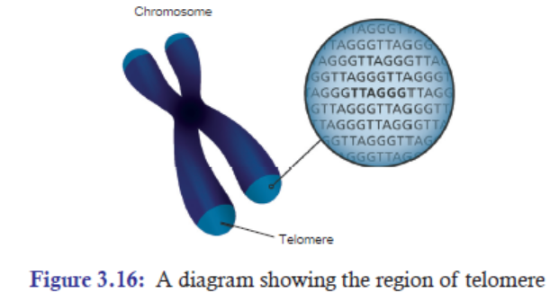
In the process of DNA replication, the length of chromosome (precisely the length of telomere) is
shortened by about 25-200 bases per replication cycle. Elizabeth Blackburn and Carol W. Greider
have shown that the enzyme telomerase maintains the length of chromosome by adding telomere
repeats (TTAGGG) at 3’ end overhang, which serves as template to previous DNA replication
(Figure 3.17). The complementary sequence (5’ end) to this newly synthesized telomere is thenadded by the regular replication machinery.
3.5.3 Significance of Telomere in Replication
1. Telomeres help in organising the chromosomes in the nucleus of the cell.
2. Telomeres protect loss of important genes: During DNA replication, the chromosomes are shortened
by about 25-200 bases per replication. If this process of shortening the chromosomes continues,
there will be loss of important genes. However, fortunately, the ends of the chromosomes are
protected by telomeres; and telomeres are non-coding regions. Thus, even if there is loss at the
tip of chromosomes in every round of replication, the loss of telomeres (non-coding regions)doesn’t affect the important genes
3. Telomeres prevent the end of chromosomes from fusing with its neighbouring chromosomes.
Telomerase has an associated RNA that complements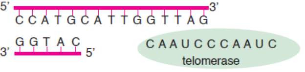
the 3’ overhang at the end of the chromosome.
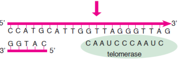
The RNA template is used to synthesize the complementary strand.
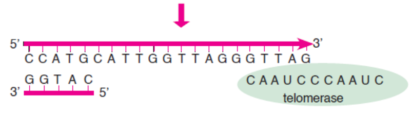
Telomerase shifts and the process is repeated.
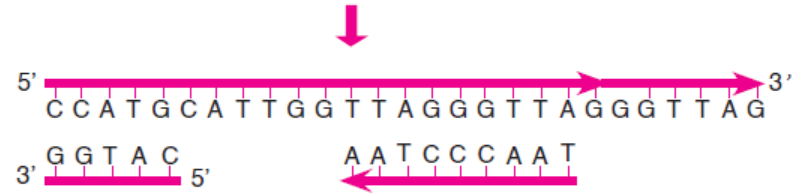
Primase and DNA polymerase synthesize the complementary strand.
Figure 3.17: Synthesis of telomeric DNA by telomerase
Telomeres and Ageing
Telomeres are thought to be related to ageing. Newborn babies are reported to have telomeres
ranging from around 8000 to 13,000 base pairs. These base pairs tend to decline by around
20-40 every year. Thus, by the time someone is 40-year-old, they could have lost up to 1600 base
pairs from their chromosomes. However, no significant shortening of telomeres is observed in
old people. It has been observed that telomerase is typically active in germ cells and adult stem
cells, but is not active in adult somatic cells.
Telomeres and Cancer
Cancer cells are characterized by their rapid and uncontrollable division of cells. These
cells have active telomerase to help them divide uncontrollably and become immortal. In
the absence of telomerase, the cancer cells would become inactive and would stop dividing
resulting into death of the cancer cells. Cancer therapies can take advantage of this concept by
designing drugs that can inhibit telomerase activity, thereby killing the cancer cells. Telomere
biology is an important aspect of human cancer. Many scientists are hoping and working
hard to understand the best way to use anti-telomerase therapy and advance the treatmentof cancer.
3.5.4 Major Differences between DNA and RNA
The three major structural differences of RNA from that of DNA are:
1. RNA contains ribose sugar instead of 2’-deoxyribose. It means that ribose has a hydroxyl
group (OH) at the 2’ position, whereas, deoxyribose has hydrogen (H) at 2’ position in
pentose sugar.
2. RNA has Uracil (U), whereas DNA has thymine (T).
3. Unlike DNA, which consists of two polynucleotide chains, in most cases, RNA is foundin a single polynucleotide chain.
Table 3.3: Differences between DNA and RNA
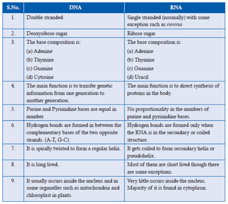
APPLICATION 3.5
Complete the sentence with the correct word
(i) Watson and Crick proposed the model of .................................... .
(ii) Enzyme ..................................... maintains the length of telomere.
(iii) ...................................... can be used to cure cancer.
(iv) ..................................... bonds are seen in both DNA and RNA.(v) ...................................... directs synthesis of proteins in the body.
3.6 NATURE OF GENES
ACTIVITY 3.6
Just like a person has his/her own character and personality, genes also have their unique
characters or nature. By nature we mean the inherent character or basic constitution of a gene.
So, find out the nature of genes from the internet. Make a report on the same and present tothe class.
1. A complete set of an organism’s DNA is called genome. A gene is a segment of DNA
that encodes for a particular trait. For example—black hair, brown hair etc.
2. Chromosomes are the structures that hold genes; they are made up of strands of DNA
tightly wrapped around histone proteins.
3. Genes are located on the chromosomes.
4. In the chromosome, a gene is found in a pair or
alternative forms called alleles. An allele is one of
two or more versions of the same gene or gene locus.
Two alleles for each gene, one from each parent, are
passed on to offspring. Homozygous pair refers to two
of the same alleles (Figure 3.18); and heterozygous
pair refers to two different alleles.
5. Each gene allele occupies a specific position in each
chromosome called locus (plural- loci).
6. Alleles are either dominant or recessive. Dominant
allele will be expressed wherever it is present, even if
it is paired with recessive allele. But recessive allele is
expressed only when it is paired with another recessive
allele.
7. When two or more alleles are present in a gene, the
condition is called multiple alleles. Example: Human blood types. The ABO Blood Type
in human beings is determined by three alleles. IA, IB, i. Both IA and IB are codominant
alleles. They are dominant to the allele, “i.” Allele “i” is recessive (Table 3.4).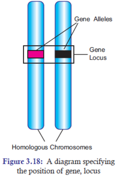
Table 3.4: Human blood type
8. The gene may change its phenotypical (trait) expression due to sudden change in its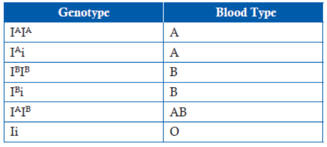
genetic composition. The changed gene is known as mutant gene. The phenomenon of
change in genetic composition is known as mutation.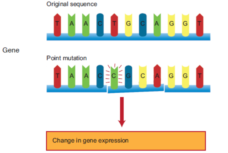
Figure 3.19: Mutation in a DNA sequence. [T base is replaced by C base]
9. Genes duplicate themselves very accurately by DNA replication. DNA replication is the
process of producing two identical DNA replicas from one original DNA molecule duringcell cycle. It occurs in all living organisms and is the basis for biological inheritance.
10. Central Dogma:
The central dogma of molecular biology is an explanation of the flow of geneticinformation, from DNA to RNA, to make a functional protein within a biological system.
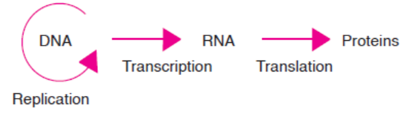
Figure 3.20: Central Dogma-information flow in a biological system
11. Split Genes:
In most eukaryotes, the genes are not continuous. Rather, the exons, the coding regions, in
the m-RNA are interrupted by several distinct units of non-coding regions called intron.
Since, the exons are split by introns, such genes are called split genes or mosaic genes or
discontinuous genes. The intron regions are removed in the latter stage of transcriptionby a process called splicing. However, introns are absent in prokaryotes.
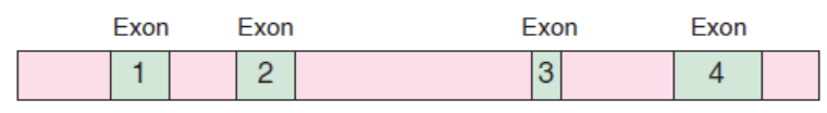
Figure 3.21: A diagram showing m-RNA regions of exons and introns
12. Genetic Code:
In the process of translation, ribosome reads the sequence of the m-RNA in a group of
three nucleotides called codons or nucleotide triplets. Thus, the genetic code is the set
of rules by which information encoded in the form of codons or nucleotide triplets in
the m-RNA is translated into proteins by living cells using ribosome machinery. Each
codon specifies a particular amino acid with some exceptions.
13. One Gene/One-Polypeptide Hypothesis:
George Beadle and Edward Tatum came up with the idea that each gene encodes the
structure of one enzyme. This idea was called the one-gene/one-enzyme hypothesis.
However, presently it is known that many enzymes have multiple polypeptide subunits,
and each subunit is encoded by a separate gene. This relationship is now referred to as
the one gene/one-polypeptide hypothesis.
APPLICATION 3.6
1. What is meant by:
a) Codon
b) Central Dogma
2. How would you summarize the hypothesis of George Beadle and Edward commonlyknown as one gene/one-polypeptide hypothesis?
3.7 GENETIC CODE
ACTIVITY 7
1. Discuss the following in the class.
The genetic code (codons) in m-RNA code for amino acids; and there are 64 possible codons
(sense and nonsense codons). But there are only 20 standard amino acids. How is it possible
that there are excess codons present to code for only 20 amino acids?2. Make a report on it and present it to the class.
Genetic Code
The genetic code is the set of rules by which information encoded in genetic material (DNA
or RNA sequences) is translated into proteins (amino acid sequences) by living cells using
ribosome machinery. In other words, the genetic code is a set of rules that specify how the
nucleotides sequence (ATGC) of an m-RNA is translated into the amino acid sequence of a
polypeptide chain.
The Structure of Genetic Code
The structure of genetic code is related to a series of exciting discoveries.
It was George Gamow (1954), a physicist, who argued that since there are only 4 bases and
if they have to code for 20 amino acids, the code should constitute a combination of bases.
In order to code for all the 20 amino acids, he suggested that the code should be made up
of three nucleotides (triplet code). The permutation and combination of three nucleotides
43 (4 × 4 × 4) would generate 64 codons. Proving that codon was triplet (i.e., three nucleotides)
was quite a challenging task. But the chemical method developed by Har Govind Khurana
for synthesizing RNA molecules with defined combinations of bases (homopolymers and
copolymers), and Marshall Nirenberg’s cell free system for protein synthesis finally helped
the genetic code to be deciphered. In the 1968, both of them, Marshall Nirenberg and Hare
Gobind Khurana along with Robert Hollye were awarded Nobel Prize in Physiology and
Medicine. Finally, a checker board for genetic code (Figure 3.22) was prepared which is asfollows.
Figure 3.22: Genetic Code: The first, second and third bases as read from 5’ to 3’ direction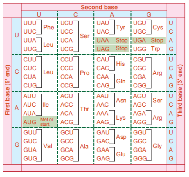
constitute the triplet code in RNA. The codon AUG specifies methionine and is usually the
starting point for protein synthesis. The word ‘stop’ indicates codons serving as signals to
terminate protein synthesis. For each amino acid more than one codon have been identified.
It would be clear from the Figure that while the first and second bases remain the same for a particularamino acid, the third base can be different
APPLICATION 3.7
1. Complete with the correct word:
(a) The phenomenon of change in genetic constitution is called ......................
(b) Two of the same alleles result in ............... pair.
(c) Alleles can be ......................... or ............................ .
(d) The sense codon AUG, is a ............................ codon.2. What is meant by non-sense codons? State them with reference to eukaryotes.
3.8 SUMMARY
• Chromosome is composed of three main components: Nucleotides, histones proteins,
and non-histones proteins.
• Nucleotide is subdivided into pentose sugar, nitrogenous bases, and phosphate groups.
• The presence of DNA in chromosomes is important for three main reasons—Protection
from damage, conserve space, and control of gene expression.
• Nucleotide is made up of pentose sugar, nitrogenous bases, and phosphate groups;
whereas, nucleoside is made up of pentose sugar and nitrogenous bases.
• Phosphodiester bond connects the phosphate group, which is attached on 5’ of one
nucleotide, with the 3’ carbon of another nucleotide. This bond is a strong bond. That
is why DNA is a stable structure.
• Polynucleotide chains have polarity. On one end, there is a 5’ carbon with a phosphate
group. On the other end, there is a 3’ carbon with a hydroxyl group on it.
• Chargaff ’s rules state that DNA of all organisms should have a 1:1 ratio of purine
(A, G) and pyrimidine (T, C) bases. The specific base pairing of A-T bases and G-C bases
is called complementary base pairs.
• In 1953, Watson and Crick proposed the double helix structure of DNA.
• The two strands of DNA are anti-parallel; the bases on both strands are bonded by
hydrogen bonds in line with Chargaff ’s rules. DNA has major and minor grooves.
• DNA is also described as twisted ladder structure.
• RNA has a hydroxyl group at 2’ carbon of pentose sugar. It has a uracil base instead of thymine.
• Unlike DNA, RNA is not the genetic material of many organisms except for few viruses.
• DNA is double stranded while RNA is normally single stranded; DNA transfer genetic
material while RNA is involved directing the synthesis of proteins.
• A telomere is a region of repetitive nucleotide sequences at each of the tip of chromosomes.
• Telomere protects important genes from being deleted, and thus allows a continued
replication.
• Telomere regions are synthesized by a telomerase enzyme.
• Telomeric regions are important in ageing and cancer treatment.
• A gene codes for a specific trait.
• A particular gene can be present in two versions called alleles. When more than two
versions of gene are present, it is called multiple alleles.
• Alleles can either be dominant or recessive.
• Genes duplicate themselves through the process of DNA replication.
• Genes are copied from DNA to RNA through a process called transcription.
• Message in the m-RNA is translated into proteins through a process called translation.
• Many enzymes have multiple polypeptide subunits, and each subunit is encoded by a
separate gene. This relationship is called one gene/one-polypeptide hypothesis.
• It is the set of rules by which information encoded in genetic material (DNA or RNA
sequences) is translated into proteins (amino acid sequences) by living cells.
• Out of 64 codons, 61 codons are sense codons and 3 codons are non-sense codons.
• A codon is made up of three nucleotides or triplets.• Genetic code is almost universal; it shows degeneracy.
3.9 GLOSSARY
• Alleles: An allele is one of two or more versions of the same gene or gene locus.
• Central dogma: The flow of genetic information, from DNA to RNA, to make a functional
protein within a biological system.
• Chargaff ’s rule: It is a rule that states that DNA of all organisms should have a 1:1
ratio of pyrimidine and purine bases. Thus, the amount of adenine (A) is equal to that
of thymine (T); and the amount of guanine (G) is equal to that of cytosine (C).
• Chromosomes: They are the structures that hold genes; they are made up of strands of
DNA tightly wrapped around histone proteins.
• Gene: A gene is a segment of DNA that encodes for a particular trait.
• Genetic code: It is the set of rules by which information encoded in the form of codons or
nucleotide triplets in the m-RNA is translated into proteins by living cells using ribosome
machinery.
• Histone proteins: They are proteins that play an important role in organizing the physical
structure of the chromosome.
• Multiple alleles: The condition where two or more alleles are present in a gene. The
ABO Blood Type in human.
• Nucleoside: A combination of only a pentose sugar and nitrogenous base, without
phosphate group, is called nucleoside.
• Nucleotides: The monomers that make up deoxyribonucleic acid (DNA) and ribonucleic
acid (RNA) are called nucleotides. Nucleotide has three components:—
Pentose (fivecarbon)
sugar, Nitrogenous (nitrogen-containing) base, and Phosphate group.
• Phosphodiester bond: It is a chemical bond where the phosphate group, which is attached
on 5’ of one nucleotide, forms a bond with the 3’ carbon of another nucleotide. Many
phosphodiester bonds are formed in between sugar and phosphate groups.
• Split genes: The exons, the coding regions, in the m-RNA are interrupted by several
distinct units of non-coding regions called intron. Since the exons are split by introns,
such genes are called split genes or mosaic genes or discontinuous genes.
• Telomerase enzyme: It is an enzyme which maintains the length of chromosome by
adding telomere repeats (TTAGGG) at 3’ end overhang, which serves as template on
previous DNA replication.
• Telomere: A telomere is a region of repetitive nucleotide sequences at each of a
chromosome.
END UNIT ASSESSMENT 3
I. Choose whether the following statements are True (T) or False (F)
1. In DNA, the pentose sugar is ribose.
2. RNA has a hydrogen atom at 2’ carbon position.
3. Pyrimidine is a single-ringed structure.
4. DNA contains adenine, thymine, guanine, and cytosine.
5. Out of these five histone proteins, H1 is loosely attached to the rest of the histone
core proteins.
6. Non-histone proteins are acidic proteins.
7. Purine bases use its 9 position nitrogen to attach with 1’ carbon in pentose sugar.
8. DNA is left-handed double helix.
9. UAG, UGA and UAA nucleotides are stop codons.
10. In genetic code, degeneracy means degeneration of DNA.
II. Multiple Choice Questions
1. Codon is a group of
(a) 2 nucleotides (b) 3 nucleotides
(c) 4 nucleotides (d) 5 nucleotides
2. Newborn babies have telomeres ranging from around
(a) 8,000 to 13,000 base pairs (b) 8,000 to 16,000 base pairs(c) 8,000 to 12,000 base pairs (d) None of these
3. Split genes are
(a) Genes with splitting chromosomes
(b) Genes separated from one another
(c) Genes where exons are interrupted with introns
(d) Genes where introns are interrupted with exons
4. Mutant gene is
(a) A gene with different mother genes
(b) A gene where nucleotide sequence has changed due to mutation
(c) A gene where different genes exist together
(d) A gene of different shape and size
5. Splicing is process of
(a) Removing exons (b) Removing introns
(c) Removing coding genes (d) Removing DNA
6. Recessive allele will express only when
(a) It occurs with dominant alleles (b) It occurs with other recessive allele
(c) It is absent (d) It is present with proteins
7. DNA Replication is the process of
(a) Copying DNA from RNA (b) Copying DNA from proteins
(c) Copying DNA from DNA (d) Copying DNA from ribosome
8. Nitrogenous bases of the two strands of DNA are linked with
(a) Hydrogen bonds (b) Covalent bonds
(c) Ionic bonds (d) Phosphodiester bonds
9. DNA does not have
(a) Adenine (b) Cytosine
(c) Guanine (d) Uracil
10. Gene codes for
(a) Polypeptides (b) Blood
(c) Specific trait (d) Specific genome
III. Long Answer Type Questions
1. In your own words, describe the composition of chromosomes.
2. List at least three differences between the structures of DNA and RNA.
3. Why is DNA important in chromosomes?
4. What is telomere? Give the significance of telomere in replication and its importance
in cancer treatment.
5. Describe structure of a Genetic code.
6. In your own words, explain why the structure of DNA is described as a ladder twisted
into a spiral.
7. Draw the structure of DNA having at least 6 base pair sequence.
8. How did Watson and Crick determine the nucleotide base pairing pattern? Explain
in your own words.
9. In your own words, describe the nature of genes.
10. (i) Identify the structure shown in figure.
(ii) Write the measurement (distance) of the parts marked (1), (2), (3), (4) and (5).
(iii) How many H-bonds are there at the place marked (6)?
(iv) How many different forms of the shown structure have been reported to occur
in the living organisms? Give their names.
(v) Which of them has/have left handed spiral and which of them right handed
spiral?(vi) Mention any 2 other special features of the form having left handed spiral.
11. Explain the role of nucleic acids in detecting HIV-AIDS. Describe NAT and also tell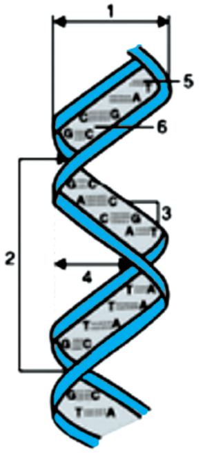
why NAT is not suitable for detecting ultra-law HIV-I DNA and RNA within hostcellular compartments.
Unit 4 DNA Replication
Key Unit Competence
To be able to explain the process of DNA replication and its significance to livingorganisms.
LEARNING OBJECTIVES
At the end of this unit, the learner will be able to:
• Determine how the structure of DNA enables it to reproduce itself accurately.
• Appreciate the importance of proper DNA replication.
• State semiconservative replication as a process by which DNA unzips and each new molecule
of DNA (daughter DNA) contains one intact strand from the original DNA (parent DNA)
and one newly synthesised strand.
• Apply knowledge of complementary base pairing in DNA to interpret Meselson and Stahl’s
experiment to test different hypothetical models for DNA replication using E.coli grown in a
heavy nitrogen (15N) medium.
• Acknowledge improper DNA replication would result into genetic changes in the nucleus
that would have both positive and negative effects on organisms. For example, changes in
the metabolism of cells, variation that can result into evolution and mutations that may lead
to death.
• State the role of enzymes involved in replication of DNA.
• List the ingredients used to make DNA in a test tube.
• Describe how semi-conservative replication of DNA takes place.
• State that conservative and dispersive replications are other hypothesis for DNA replication.• Explain the importance of DNA replication in organisms.
DNA replication is the process by which DNA makes a copy of itself during cell division.
It produces two identical replicas from one original DNA molecule. This biological processoccurs in all living organisms and is the basis for biological inheritance.
INTRODUCTORY ACTIVITY
Mr Aaron is a senior teacher of Biology. He said: “ I used to illustrate the process of
DNA replication from the diagram below and my students could interpret the process withconfidence. However, a question often came back to me …”
1. Make research from internet or library to explain how this diagram can serve to
explain the process of protein synthesis
2. Mr Aaron said a question often came back from students. On your side, what
challenge you face by using such a diagram?
4.1 MODELS OF DNA REPLICATION
ACTIVITY 4.1
Look in books and on the internet how DNA replicates. Research on semi-conservative model
of DNA replication. Also, try to find other models of DNA replication and present yourfindings to class.
4.1.1 Semi-conservative Model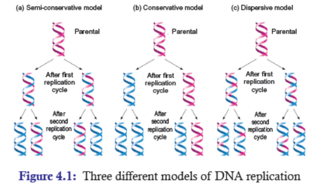
In 1953, Watson and Crick proposed their classic paper postulating a double helix for DNA.
A month later, they published another paper suggesting how such base-paired structures in DNA
might duplicate itself. The essence of Watson and Crick suggestion is that if DNA molecule
was untwisted and the two strands get separated, each strand could act as a template for
the synthesis of a new complementary strand of DNA. And this new complementary strand
could then be bound to the parental strand of DNA. This model replication is known as the
semiconservative model. It is because half of the parent strand of DNA is retained by newly
formed daughter DNA strand (Figure 4.1).
4.1.2 Other Two Models of DNA Replication
Apart from semiconservative replication model, two other models for DNA replication werealso proposed at that time. The two other DNA replication models are:
Conservative DNA Replication Model
In this model, the two parental DNA strands come together right after replication; and as a
whole, these two parental DNA strands serve as template for the synthesis of completely new
daughter DNA strands. As a result, one daughter DNA molecule contains parental DNA strands,
while the other daughter DNA molecule contains newly synthesized DNA strands (Figure 4.1).
Dispersive DNA Replication Model
In this model, the parental double helix is broken or cleaved into double-stranded DNA that acts
as templates for the synthesis of new double helix molecules. The segments then reassemble into
complete DNA double helices, each with parental and daughter DNA segments interspersed.
After the replication, although the two daughter DNA molecules are identical in their basepair
sequence, the parental double stranded DNA has become dispersed throughout both inthe daughter DNA molecules (Figure 4.1).
4.2 IMPORTANCE OF DNA REPLICATION
Genes duplicate themselves very accurately by DNA replication. The three main importance
of DNA replication are:
Reproduction—One of the most fundamental properties of all living things is the ability to
reproduce. It is through reproduction that parents faithfully pass on their genetic information
specifying their structure and function to their young ones. At organism level, organisms
reproduce either by asexual or sexual reproduction methods. At cellular level, cells duplicate
by cellular division. And at the genetic level, the genetic material duplicates by DNA replication.
Repair—DNA is the centre of instructions that govern the cell’s protein production, growth,
and many other activities in the cells. With this enormity of precise responsibility, any minor
mistakes in the replication process can bring potentially harmful changes in the cell’s behaviour or
for that matter, the whole organism. Therefore, DNA employs various error repair mechanisms
to ensure accurate DNA replication.
Growth—DNA Replication is required for the growth of organisms. DNA replication occurs
in two different forms of cellular division. They are mitosis and meiosis. In mitosis, a single
parent cell divides and gives rise to two identical daughter cells. Each of the daughter cells
has the exact amount of genetic material. For example – Growth of limbs, organs, hair etc.
On the other hand, in meiosis, cells divide and give rise to two haploid sex cells. Thus, DNAreplication plays a vital role in both mitosis and meiosis.
APPLICATION 4.1
1. State the three hypotheses that support the mechanism of DNA replication
2. What is the importance of DNA replication?
4.3 EXPERIMENTAL EVIDENCE OF SEMI-CONSERVATIVE DNAREPLICATION
ACTIVITY 4.3
If semi-conservative model is the correct model of DNA replication, research and present
your case by supporting it with experimental evidence. Also tell how semi-conservative modeldisproved other models of replication.
In 1958, Matthew Meselson and Franklin Stahl performed the experiment to test and prove
that DNA replication is semi-conservative. In their experiment, they used two isotopic forms
of nitrogen, 14N (light) and 15N (heavy), to distinguish newly synthesized strands of DNA from
old strands.
Initially, Meselson and Stahl grew E.coli (bacteria) for many generations in a medium containing
15N-labelled ammonium chloride (15NH4Cl) to incorporate this heavy isotope of nitrogen into
their DNA molecule. As expected, the DNA strands in the bacteria had 15N-15N (heavy) DNA(Figure 4.2).
In the second stage, they transferred the 15N-labelled bacteria to a medium containing nitrogen in
the normal 14N form (light). Then the bacteria were allowed to reproduce for several generations.
Since, the bacteria were grown in the normal 14N form, the entire newly synthesized DNA after
the transfer was now labelled with 14N.
Samples of E.coli were taken at various time periods as the bacteria continued to reproduce
in the medium. The DNAs from these bacteria were extracted and analysed to determine its
density. They determined the density of extracted DNAs by using equilibrium density gradient
centrifugation technique. This technique uses Cesium Chloride (CsCl), a heavy metal salt
that forms solutions of very high density. Thus, they analysed the extracted DNA by simplymixing it with a solution of cesium chloride and then centrifuged at high speed.
As a density gradient of cesium chloride is established by the centrifugal force, the DNA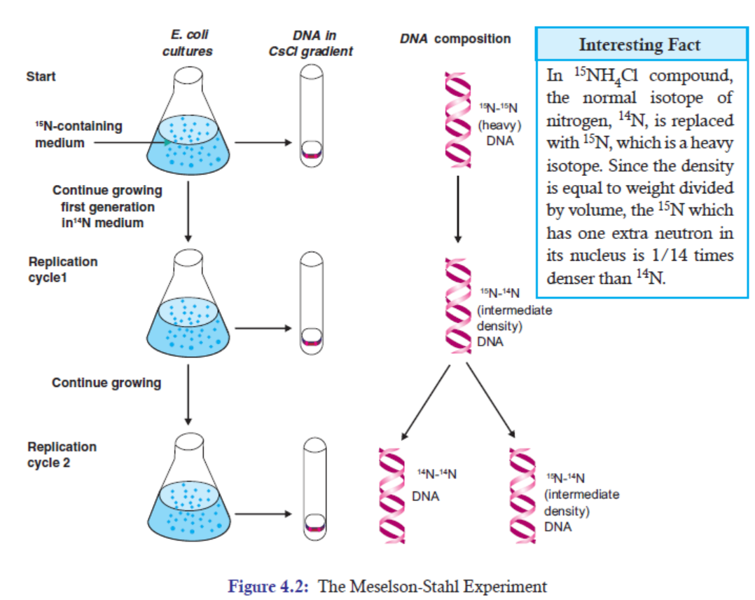
molecules float “up” and sink “down” within the gradient to reach their equilibrium density
positions. The difference in density between the heavy (15N) DNA and the light (14N) DNA
causes DNA molecules to rest at different positions by forming bands in the gradient
(Figure 4.2).Final Observations
First Generation (After One Replication Cycle)
When the observation was made after one replication cycle in the 14N medium, the entire DNA
had a density that was exactly intermediate between that of 15N-15N DNA and that of 14N-14N
DNA. The intermediate composition was 15N-14N DNA.
Second Generation (After Two Replication Cycles)
Again when the observation was made after two replication cycles, half of the DNA was that
of intermediate density (15N-14N DNA) and half was that of the density of 14N-14N DNA.
The observations (Figure 4.2) made in this experiment exactly tested and proved the predication
of the semi-conservative model. Therefore, through this experiment it has been known that
DNA replication follows semi-conservative model. At the same time, it disproved the claimthat DNA replication follows either conservative or dispersed replication models.
APPLICATION 4.2
1) Complete the following sentences by correct missing words:
(a) .................. is the process by which DNA makes a copy of itself.
(b) ................ and ................. proposed the semi-conservative model of DNA replication.
(c) .................. and .................. are two other models of DNA replication.
2) Why In their experiment, Matthew Meselson and Franklin Stahl used N-15 (e.g. 15NH4Cl)Instead of N-14 alone?
4.4 ENZYMES AND PROTEINS INVOLVED IN DNA REPLICATION
ACTIVITY 4.4
Different parts of a car function together to smoothly and efficiently run a car. DNA replication
is also like a running car. It requires different components or parts to smoothly and efficiently
carry out DNA replication. Now, find out and discuss the enzymes and proteins involved inDNA replication. Make a table suggesting their roles and importance.
4.4.1 DNA Polymerase
In 1955, Arthur Kornberg and his colleagues were the first ones to identify an enzyme that
could synthesize DNA. Back then this enzyme was originally called Kornberg enzyme. But
now it is called DNA polymerase I. And the term DNA polymerase by definition encompasses
enzymes that catalyses the synthesis of DNA.
There are five DNA polymerases.
1. DNA polymerase I
2. DNA polymerase II
3. DNA polymerase III
4. DNA polymerase IV5. DNA polymerase V
4.4.2 DNA Helicase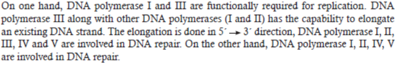
DNA helicase is an enzyme that unwinds or unzips the double stranded DNA by breaking thehydrogen bonds between the complementary bases.
The action of DNA helicase can be compared with a zipper. When we open a zip, the zipper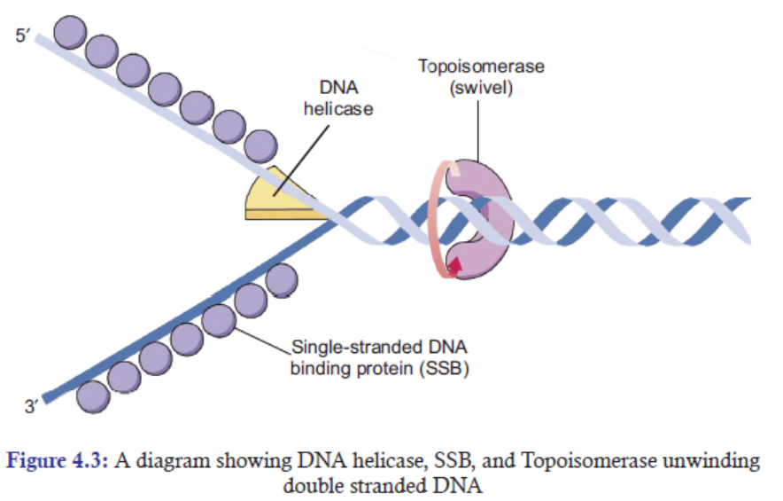
runs on a zip and makes a Y-shape structure with the two strands of interlocking teeth. In the
same way, DNA helicase unzips the double stranded DNA and form a Y-shaped fork knownas a replication fork.
4.4.3 Single-strand DNA-binding Proteins (SSB)
In DNA replication when helicase unwinds the double stranded DNA, the two separating
strands of DNA have the tendency to reform or reanneal into double stranded DNA. A protein
called single-strand DNA-binding (SSB) proteins bind to each single-strand DNA and stabilise
them, so that the separating two strands of DNA do not reform double stranded DNA by
complementary base pairing (Figure 4.3).
4.4.4 DNA Ligase
At the end of DNA replication right after the DNA Pol I is removed and replaced all the RNA
primer nucleotides with DNA nucleotides, normally as single-strand nick (gap) is left between
the two DNA fragments (Figure 4.4). This nick is the point where the sugar-phosphate backbone
between adjacent nucleotides is unconnected. So, what DNA ligase does is to join the two
fragments resulting into a longer and continuous DNA strand.
Chemically, DNA ligase catalyses the formation of a phosphodiester bond between the 3′-OHand the 5′-phosphate groups on either side of a nick. As a result, it seals the nick (gap).
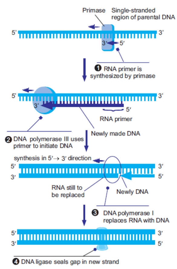
Figure 4.4: A flow diagram showing DNA ligase sealing the gap in a new DNA strand
APPLICATION 4.3
1) State the role of each of the following enzymes and proteins in the replication of DNA.
a) DNA Polymerase
b) Primase
c) DNA Helicase
d) Topoisomerase
e) Single-strand DNA-binding Proteins (SSB)
f) DNA Ligase2) Why In their experiment, Matt
4.5 MECHANISM OF DNA REPLICATION
ACTIVITY 4
Scientists have managed to carry out DNA replication in vitro.
They also realized that there is a slight difference between the DNA replication in eukaryotic
cells and in Prokaryotic cells.
Conduct research from available resource and answer the following:
1) What are the requirements to carry out DNA replication in vitro?2) What is the difference between the DNA replication in Prokaryotes and Eukaryotes?
List of the Ingredients used to Make DNA in A Test Tube
Having studied in the vitro DNA synthesis reaction in detail, the scientists have found out
that in order to synthesize DNA, the following components are required for making DNA in
a test tube.
1. A DNA template: The template (original) DNA that is to be copied. A template is a
molecule which is used to make a complementary DNA molecule in the DNA synthesis.
Normally, the two parental DNA strands act as DNA templates.
2. dNTPs (deoxynucleotides): A mixture of four deoxyribonucleoside 5’-triphosphate
(dNTPs) precursors namely: dATP, dGTP, dTTP, and dCTP. These dNTPs are the
precursors for the nucleotide formation in DNA. Without these dNTPs, new DNA cannot
be synthesized.
3. DNA polymerase I (DNA Pol I): An enzyme to carry out DNA synthesis.
4. A primer: A primer is a short DNA sequence that will bind with the single parent DNA
strand and start the DNA synthesis reaction. Without primer, the coming nucleotides
cannot directly base-pair with parent DNA template.
5. Magnesium ions (Mg2+): It is required for DNA polymerase activity to run optimally.
6. Buffer: Provides a suitable chemical environment for optimum activity and stability ofthe DNA polymerase.
4.5.1 Mechanism of DNA Replication in Prokaryotes
Prokaryotic cells are quite simple in structure. They have no nucleus, no organelles and a small
amount of DNA in the form of a single, circular chromosome. Example is Escherichia coli
(E.coli). The mechanism of DNA Replication can be discussed clearly in the following points.
1. DNA replication starts when DNA helicase unwinds or unzips the DNA at the origin
of replication in both the directions.
2. The locally denatured segment of DNA is called the replication bubble. The two separated
parent DNA strands are called template strands.
3. A Y-shaped structure which is formed when DNA unwinds or unzips to expose the
two parent single template strands for DNA replication is called as a replication fork
(Figure 4.5). In most of the cases, replication forks are formed in both sides and, thus,
move simultaneously in two opposite directions. In other words, the movement of
replication fork is bidirectional (Figure 4.5).
4. In the next step, DNA helicase recruits DNA primase enzyme. Primase enzyme synthesizes
a short RNA primer (about 5-10 nucleotides) on the two template strands through whichnew nucleotides are added by DNA polymerase III.
5. The denaturing or separating two strands of DNA have the tendency to reform (reanneal)
double stranded DNA. A protein called single-strand DNA-binding (SSB) proteins
bind to each single-strand DNA and stabilise them, so that the separating two strandsof DNA do not reform double stranded DNA.
6. As the two single-strands of DNA are held in Y-shaped position and are stabilized by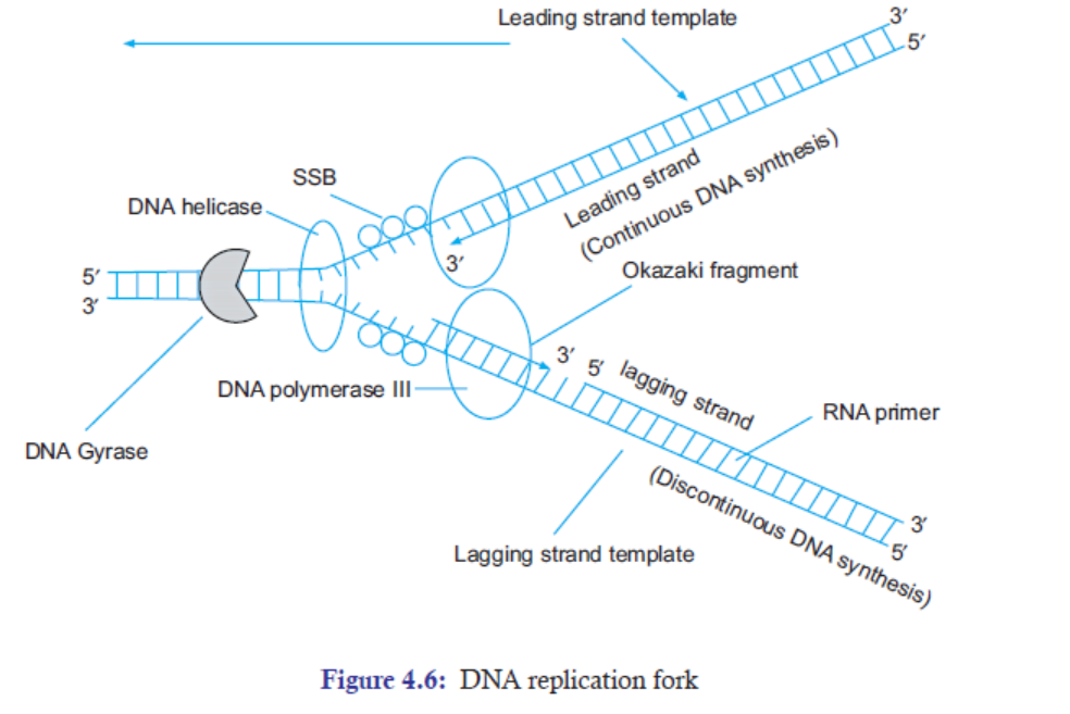
SSB proteins, the DNA polymerase III now comes and starts adding nucleotides by
forming phosphodiester bonds at the 3′-OH of the primer.
7. The DNA polymerase III can add nucleotides only in 5′-3′ directions. However,
the two strands of DNA run in opposite directions forming a polarity. Thus, to
maintain the 5′-3′ polarity of DNA synthesis on both of the two single templates,
DNA is simultaneously made in opposite directions of the two template strands
(Figure 4.6).
(a) The new DNA strand that is synthesized in the same movement of the replication
fork is called the leading strand. This strand requires a single primer for the complete
DNA replication.
(b) On the contrary, the other new DNA strand that is synthesized in the opposite
direction of the movement of the replication fork is called the lagging strand. This
strand requires primers again and again. Therefore, the newly synthesized DNA
strand is discontinuous in nature. And the newly synthesized fragments of DNAon lagging-strand are called Okazaki fragments.
8. The unwinding of the DNA to form a replication fork creates a tension which is relaxed
by DNA gyrase or topoisomerase.
9. At the end of the DNA replication, the RNA primers on the previous Okazaki fragments
are removed by DNA polymerase I.
10. After the DNA polymerase I left, a single-stranded nick is left at the site of the
removal of primer. DNA ligase seals the nick. This completes the process of DNAreplication.
4.5.2 Mechanism of DNA Replication in Eukaryotes
Eukaryotic cells have nucleus, multiple organelles and more DNA arranged in multiple,
linear chromosomes. Examples–Yeast (Saccharomyces cerevisiae), Humans. DNA replication in
eukaryotes (having linear chromosomes) is initiated at multiple sites of origin of replication
(Figure 4.7). In yeast cells, replicators are approximately 100 bp sequence called autonomously
replicating sequences (ARS).
The origin of replication or replicator is located at the centre of each replicon. Replicator is
a DNA sequence that directs the initiation of replication. The initiation of DNA synthesis at
the replicator takes place by a mechanism involving several groups of initiator proteins.
• Firstly, origin recognition complex (ORC), a multi-subunit protein complex, binds to a
replication origin.
• Secondly, mini-chromosome maintenance (MCM) proteins bind to the replication origin.
MCM proteins include several DNA helicases that unwind the double helix.
• Thirdly, helicase loaders, a third set of proteins, mediate binding of MCM proteins to originof origin recognition complex (ORC).
Pre-replication Complex
The complete group of DNA-bound protein is now called as a pre-replication complex. Andat this stage, the DNA is said to be “licensed” for replication.
Replication Bubble (Multiple)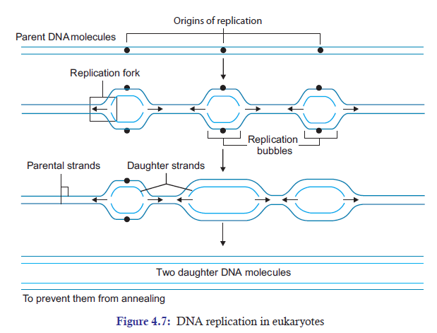
Initiation of DNA synthesis at the origin of replication is followed by formation of replication
bubble. This bubble is formed by two replication forks that begin to synthesize DNA in opposite
directions away from the origin (Figure 4.7). And these bubbles grow in size as replication proceeds
in both 5’ and 3’ end directions. Wherever the growing replication bubble of one replicon meets
the replication bubble of an adjacent replicon, the DNA synthesized by the two replicons is joined
together. Eventually, the DNA synthesized at numerous replication sites is linked together to formtwo double stranded daughter molecules
Significance of Multiple Replicons
In humans, the haploid genome has 24 chromosomes. These chromosomes consist of about
3 billion base pairs long. And eukaryotic chromosome is 25 times longer than the prokaryotic
chromosome. Moreover, the movement of replication fork is much slower in eukaryotes than
in prokaryotes. In this kind of condition, if eukaryotic chromosome has only one origin of
replication or replicator per chromosome, replication of each chromosome would take many
days. So, the question is how does the eukaryotic chromosome replicate faster despite havinga huge amount of chromosomes?
The answer lies in two main characters of the eukaryotic chromosomes. And they are:
(a) DNA replication is initiated at many origins of replication throughout the genome.
(b) DNA replication is bidirectional in nature. In other words, the replication forks move intwo directions at a time.
4.5.3 The Rate of Replication in Prokaryotes and Eukaryotes
Prokaryotes
The genome E.coli consists one replicon with a size of 4.6 Mb (million base pairs, the entire
genome size). The rate of each replication fork movement is about 1000 base pair (bp) per
second. With this rate, E.coli takes about 42 minutes to replicate its entire chromosome.
Eukaryotes
The eukaryotic genome consists of multiple replicons. For example, in humans, there are
about 10,000-100,000 replicons for an average of 30-300 kb (1000 base pairs). And the rate of
replication fork movement is about 100 bp per second. Thus, it takes about 8 hours to replicatethe entire genome.
APPLICATION 4.4
1. Complete the sentence with correct word:
(a) The full form of ORC is ......................... .
(b) The tension ahead of the replication fork is relaxed by ......................... .
(c) The newly synthesized strand is ......................... in nature.
(d) It takes about ......................... hours to replicate the entire genome in Eukaryotes.
(e) DNA replication in Eukaryotes is ......................... in nature.
2. Amoeba and Vibrio cholera are both unicellular human endoparasites. Giving reason,explain which one will undergo rapid DNA replication in vitro.
4.6 MECHANISMS THAT ENSURE ACCURACY OF DNA REPLICATION
ACTIVITY 4.6
It is known that replication is standard. The daughter DNA resemble to the mother DNA.
Carry out a research from available resource to answer the following questions:
1) What factors allow to have accurate DNA replication?2) Discuss what would be outcome of improper DNA replication.
(A) Complimentary Base Pairing
The nitrogen bases of DNA follow the Chargaff ’s rule of base pairing. In simple words, this rule
says that Adenine (A) base pairs with Thymine (T); Guanine (G) base pairs with Cytosine (C).
This base pairing is very strict and accurate. Thus, the complementary base pairing directs theDNA to replicate very accurately and prevents any mistake to occur.
Base pairing between purines and pyrimidines is possible because of hydrogen bonds. We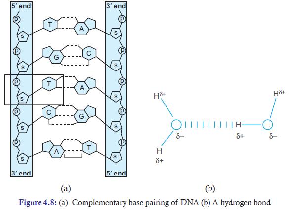
can simply define hydrogen bond as the attractive force between the hydrogen attached to
an electronegative atom of one molecule and an electronegative atom of a different molecule
(Figure 4.8 b). In the structure of DNA, both the strong electronegative atoms, oxygen (O)
and Nitrogen (N), are partially negatively charged (d–), while the hydrogen (H) has the partial
positive charge (d+). Hydrogen bonds or interactions play very important role in binding thetwo bases of the opposite strands in the DNA.
(B) Semi-conservative Nature of DNA
In DNA replication, two of the original strands of DNA act as templates for new DNA to be
synthesized. So, when the new strands of DNA are synthesized, they are just the complimentary
bases of the two original template strands of DNA. In this way, original sequence of DNA is
semi-conserved with the two original strands of DNA. Thus, the semi-conservative nature ofDNA makes the DNA replication highly accurate.
(C) Proofreading
DNA Pol I and DNA Pol II polymerase enzymes have 3′-to-5′ exonuclease activity, which
means that they can remove incorrectly inserted nucleotides from the 3′ end to 5′ end of the
DNA chain. Thus, they play important role in proofreading mechanism. The insertion of
incorrect nucleotides by both DNA Poly I and DNA Poly III occurs at a frequency of one base
in a million (10–6).
When incorrect nucleotides are inserted in the newly synthesized DNA, the 3′-5′ exonucleases
move backward and remove the incorrect nucleotide from the newly synthesized DNA. Then
they resume the forward movement and insert the correct nucleotides in place of the incorrect
nucleotides. With this proofreading mechanism, the chances of error occurrence in DNAreplication is reduced to one base in a billion (10–9) instead of one base in a million (10–6).
(D) Mismatch Repair
After DNA Replication if there are any mismatched base pairs on the newly synthesized strand,
it can be corrected by methyl-directed mismatch repair. In contrast to proofreading mechanism
where only one base is repaired by DNA polymerase, the mismatch repair mechanism can
replace about thousand bases to make one repair. The Mut family of enzymes plays an importantrole in mismatched repair.
Main Differences Between Prokaryotic and Eukaryotic DNA ReplicationTable 4.1: Differences between prokaryotic and eukaryotic DNA replication
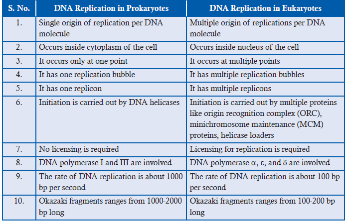
4.7 EFFECTS OF IMPROPER DNA REPLICATION
A. Mutation: A Key to Variability and Evolution
DNA alterations are occasionally beneficial because DNA base-sequence changes, or mutations,
provide the genetic variability that is the raw material of evolution. In other words, mutation
provides variation that can result into beneficial evolution.
Example: Resistance of Bacteria to Drug. The Esherichia coli (bacterium) lives in colon of human
beings. Initially if they are exposed to chloramphenicol, an antibiotic drug, they die. When
Cavilli and Maccacro (1952) exposed the bacteria to high concentration on chloramphenicol,
they found out that the bacteria have mutated and were 250 times resistant to antibiotics. In
this case, mutation has provided variability in the bacterial population to evolve into a new
species which has high survivability against the antibiotic drugs. Thus, mutation is beneficial
for the bacterial population.
B. Lethal Mutation: Mutation that May Lead to Death
One of the best examples is Tay-Sachs disease (TSD). TSD is a fatal autosomal recessive
genetic disorder. It is caused by a mutation in the Hexosaminidase A (alpha polypeptide)
[HEXA gene]. A genetic mutation is a permanent alteration in the DNA sequence that makes up
a gene; this mutation is lethal. It mostly occurs in children and leads to progressive destruction
of the nervous system. When a child with Tay-Sachs reaches the age of three or four years, the
nervous system is severely affected. Eventually, death occurs by the age of five years.
Mutation in Hex-A Gene Causes Tay-Sachs: Tay-Sachs disease results from defects in a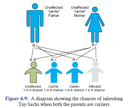
gene on chromosome 15 due to mutation. The gene located on chromosome 15 codes for the
production of the enzyme Hex-A. In normal people, either or both Hex-A genes are active.Thus, the synthesis of this enzyme prevents the abnormal build-up of the GM2 ganlioside lipid.
APPLICATION 4.5
1. Complete the following sentence with correct words:
(a) ............................ is an example of lethal mutation.
(b) ............................... plays an important role in proofreading mechanisms.
(c) Alkaptonuria is also known as ................................... disease.
(d) ...................................... provides variation that can result into beneficial evolution.
(e) Base pairing in DNA occurs due to ................................... bonding.2. Write short not about Tay-Sachs disease (TSD).
4.8 SUMMARY
• DNA replication is the process of producing two identical DNA replicas from one original
DNA molecule.
• DNA replication plays an important role in reproduction, DNA repair and growth of
organisms.
• DNA replicates semi-conservatively, where the two original strands act as template while
the other two strands are newly synthesized.
• The other two models of replication are conservative and dispersive DNA replications.
• Meselson and Franklin Stahl performed the experiment to test and prove that DNA
replication is semi-conservative.
• Enzymes and Proteins Required for DNA Replication are:
— DNA polymerase I and III are functionally required for replication. But DNA
polymerase I, II, IV, V are involved in DNA repair.
— DNA helicase has the role of unzipping or unwinding the double strand structure of
DNA.
— DNA gyrase serves as a main swivel that prevents supercoiling of the DNA ahead
of the replication fork.
— SSB proteins relax the tendency of the two separated DNA strands to reform double
stranded DNA.
— DNA ligase seals the nick at the end.
• The list of the ingredients to make DNA in a test tube involves : a DNA template, dNTPs,
DNA polymerase I, primers, magnesium ion, and buffer solutions.
• In Prokaryotes, DNA replication starts with DNA helicase unwinding or unzipping
the double stranded DNA. Replication forks are formed, then, primers bind on the two
separated strands of DNA. DNA polymerase III starts synthesizing new DNA strands
on both the strands at 5′-3′ directions.
• The synthesis of DNA is discontinuous and thus produces numerous small Okazaki
fragments.
• DNA polymerases I and III have 3′-5′ exonuclease activity. Thus, they remove the primers
and replace the gap with complementary nucleotides.
• In Eukaryotes, replication takes place at multiple sites of origin of replications. In yeast
cells, replicators are approximately 100 bp sequences called autonomously replicating
sequences (ARS).
• Many replicons are formed.
• DNA replication is initiated by multiple proteins.
• Many replication bubbles are formed.
• The main polymerase enzymes involved are: DNA polymerase a, bd, e; they have different
roles.
• The eukaryotic cells have chromosomes 25 times longer than the prokaryotes. Prokaryotes
have only one replicon. Eukaryotes have multiple replicons, which help in replicating
faster than if there were only one replicon.
• The rate of replication in prokaryotes is about 1000 bp per second, whereas the rate of
replication in eukaryotes is about 100 bp per second.
• There are basically four mechanisms that ensure accuracy of DNA replications:
Complimentary base pairing, semiconservative nature of DNA, proofreading, and
mismatched DNA repair.
• Normally uncorrected mistakes in DNA replication are repaired by DNA repair
mechanism. But in very rare cases mistakes are not corrected, leading to mutation.
• On the positive side, mutation can bring species variation and evolution. On the negativeside, mutation can bring defects in metabolic pathway and may also cause death.
4.9 GLOSSARY
• 3′-to-5′ exonuclease activity: DNA Pol I and DNA Pol II polymerase enzymes have
this activity, which means that they can remove incorrectly inserted nucleotides from the
3′ end to 5′ end of the DNA chain.
• Alkaptonuria: It is a rare inherited genetic disorder in which the body cannot process
the amino acids phenylalanine and tyrosine.
• Chargaff ’s rule: This rule says that adenine (A) base pairs with thymine (T);
Guanine (G) base pairs with cytosine (C).
• DNA fyrase: It is an enzyme which serves as a swivel that prevents supercoiling of the
DNA ahead of the replication fork.
• DNA helicase: It is an enzyme which unwinds the double stranded DNA.
• DNA ligase: It is an enzyme which joins the two DNA fragments into a continuous
DNA strand.
• DNA polymerase: It is an enzyme that catalyzes the synthesis of DNA.
• dNTPs: It is a mixture of four deoxyribonucleoside 5′-triphosphate precursors namely:
dATP, dGTP, dTTP, and dCTP.
• Eukaryotes: Eukaryotic cells have nucleus, multiple organelles and more DNA arranged
in multiple, linear chromosomes. Examples: Yeast, Humans.
• Mutation: It is a process by which nucleotide sequence (or base pairs) of DNA is altered.
• Primase: It is an enzyme which synthesizes primer.
• Primer: A primer is a strand of short nucleic acid sequences (generally about 10 base
pairs) that serves as a starting point for DNA synthesis.
• Prokaryotes: They have no nucleus, no organelles and a small amount of DNA in the
form of a single, circular chromosome. Example is Escherichia coli (E.coli).
• Replication fork: It is a Y-shaped structure which is formed when DNA unwinds to
expose the two parent single template strands for DNA replication.
• Semi-conservative model: During DNA replication, each strand of DNA acts as a template
for the synthesis of a new complementary strand of DNA. And this complementary strand
of DNA could then be bound to the parental strand of DNA.
• Single-strand DNA-binding proteins (SSB): These are stabilizing proteins that bind on the
separating two stands of DNA so that they do not reform into double stranded DNA structure.
• Tay-sachs disease (TSD): TSD is a fatal autosomal recessive genetic disorder. It is caused
by a mutation in the Hexosaminidase A (alpha polypeptide) [HEXA gene].
END UNIT ASSESSMENT 4
I. Choose whether the given statements are True (T) or False (F)
1. In conservative DNA replication model, two identical daughter DNA strands are
formed.
2. Meselson and Stahl experiment proved dispersive DNA replication model after two
replication cycles.
3. Without dNTPs, new DNA cannot be synthesized.
4. In Chargaff'’s rule of base pairing. Adenine base pairs with Cytosine.
5. DNA polymerase I has 3′-5′ exonuclease activity.
6. Tay-Sachs disease (TSD) is a fatal autosomal dominant genetic disorder.
7. Topoisomerase II acts as a swivel to prevent supercoiling of DNA.
8. Mutation can be both beneficial and lethal.
9. Synthesis of DNA in lagging strand is discontinuous.10. Primer is required again and again in the leading strand of DNA.
II. Multiple Choice Questions
1. DNA replication plays an important role in
(a) reproduction (b) growth of organisms
(c) DNA repair (d) All of the above
2. Meselson and Stahl are known for
(a) Complementary base pairing
(b) Genetic Code
(c) Semi-conservative DNA replication
(d) Conservative DNA replication
3. DNA helicase has a role to
(a) Seal the nick at the end of DNA replication.
(b) Relax the tendency of DNA to reform double helix structure.
(c) Unwinds or unzips double stranded DNA.
(d) Cut DNA randomly.
4. Which of the following is not an ingredient to make DNA in a test tube?
(a) dNTPs (b) Primers
(c) Magnesium ion (d) Calcium ion
5. The mechanisms that ensure accuracy of DNA replications are
(a) complementary base pairing (b) Proofreading
(c) Mismatched DNA repair (d) All of the above
6. This happens when mistakes are not corrected by DNA repair mechanism.
(a) Mutation (b) Replication
(c) Repair (d) Growth
7. Syntherized fragments of DNA on lagging strand are
(a) Origin complex (b) Mini chromosome
(c) Okazaki fragments (d) Autonomously replicating sequence
8. Mut family protein is important in
(a) Semi-conservative DNA replication
(b) Mismatched repair
(c) Hydrogen bonding
(d) Organizing DNA structure
9. DNA polymerase d appears to perform
(a) Synthesis of DNA on the lagging strand
(b) Synthesis of DNA on the leading strand
(c) Prevention of supercoiling in DNA
(d) Unwinding of DNA
10. The main initiator proteins of DNA replication in eukaryotes consist of three proteins
except
(a) Origin recognition complex
(b) Helicase loaders
(c) Single-stranded binding proteins(d) Minichromosome maintenance proteins
III. Long Answer Type Questions
1. State how complementary base pairing in DNA devises the Meselson and Stahl’s
semiconservative model of replication.
2. Explain different types of DNA replication models.
3. Describe the roles of enzymes involved in DNA replication.
4. Explain the mechanism to show how the structure of DNA enables it to reproduce
itself accurately.
5. Describe the importance of DNA replication in organisms.
6. (i) Identify the structure shown in figure.
(ii) Write the label (a) to (d).
(iii) Describe the process wherein DNA unzips and each new molecule of DNAforms daughter DNA.
7. List out the ingredients used to make DNA in a test tube.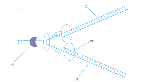
8. Mutation are the ultimate fuel for evolution. Describe mutations as a key genetic
factor in both positive and negative terminology of biology.
Also tell how some people get more sick from bacteria and viruses than others. What
role does our genes play to get that difference?
9. DNA replicates and form a part of HIV-cure-related research. Investigate on thevarious aspects of DNA replication detecting HIV DNA.
Unit 5 Cell and Nuclear Division
Key Unit Competence
To be able to describe the stages of the cell cycle and explain the significance of cell andnuclear division in organisms.
LEARNING OBJECTIVES
At the end of this unit, the learner will be able to:
• Describe the main stages of the cell cycle, including: interphase (growth and DNA replication),
mitosis and cytokinesis.
• Explain what is meant by homologous pairs of chromosomes.
• Explain the meaning of the terms haploid and diploid.
• Describe the process of mitosis and meiosis.
• Outline the significance of mitosis in cell replacement and tissue repair by stem cells.
• State that uncontrolled cell division can result in the formation of a tumour.
• Define meiosis as reduction division in which the chromosome number is halved from
diploid to haploid.
• Explain the need for reduction prior to fertilisation in sexual reproduction.
• Outline the role of meiosis in gametogenesis in humans and in the formation of pollen grain
and embryo sacs in flowering plants.
• Explain how crossing over and random assortment of homologous chromosomes during
meiosis and random fusion of gametes at fertilization leads to genetic variation, including
the expression of rare recessive alleles.
• Interpret data related to time for different cell cycles to identify tissues from which the cells
came.
• Apply knowledge of mitosis to predict which set of cells came from and which part of the
plant and where other cells have come from.
• Make a table showing the phases of the cell cycle mentioning one important event that
occurs at each phase.
• Compare mitosis and meiosis.
• Appreciate the importance of effective cell division.• Show concern to individuals with physical disabilities like Down’s syndrome.
INTRODUCTORY ACTIVITY
A teenage girl got undesired pregnancy when she could not believe. She said: I am 12 years
old. I never got any menstrual period before meeting the boy who pregnant me. Three
month later my stomach was swollen but after pregnancy testing, the doctor told me, you
are pregnant, and your baby is growing well by cell divisions.
1. Can you explain how the baby can grow and yet the cells divide?
2. What types of cell division took place there in this case study? Support youropinion.
5.1 HAPLOID AND DIPLOID CONDITIONS OF THE CELL CYCLE
ACTIVITY 5.1
We know that human beings have 46 chromosomes in total. If it is so, will the number of
chromosomes increase when gametes from father and mother fuse together during fertilizationto form a zygote. Discuss and come out with valid points to support your arguments.
Cell cycle is one of the most important biological events in organisms. Every actively dividing
cell undergoes cell cycle. The most prominent events in cell cycle consist of nuclear division
(karyokinesis) and cytoplasm division (cytokinesis). During cell cycle, chromosomes undergomany changes such as replication, uncoiling, condensation, pairing, cross over, and separation.
A karyotype is a visual representation of the chromosomes within a single cell where thenumber of chromosomes, their arrangement, size and structure can be analysed (Figure 5.1).
The chromosome pairs, one from each parent, which is similar in length, gene position, and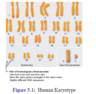
centromere location, is called as a homologous chromosome. In humans, for example, the
23 chromosome pairs are homologous chromosomes. On the contrary, the chromosomes
that contain different genes and do not pair during meiosis are called as non-homologous
chromosomes. For example: In the human karyotype, chromosome 1 and chromosome 2 arenon-homologous.
Chromosomes are of two types: (a) autosomes (b) sex chromosomes. Out of 23 pairs of
chromosomes, one pair of chromosomes is sex chromosome. And chromosomes other than
sex chromosome are called autosomes. The cells that are involved in reproduction are called
gametes (Sperm and eggs). The cells that are not involved in gamete formation are calledsomatic cells (Muscle cells, liver cells).
The cells which contain two complete sets (2n) of chromosomes are called diploid cells. These
cells are formed by the fusion of two haploid gametes, one comes from the female parent and
the other comes from the male parent. For example, all the somatic cells are diploid cells. In
contrast, cells that contain only one complete set (1n) of chromosome are called as haploid
cells. Example is gamete cells in humans. Two separate haploid gametes, one from male parent
(sperm, 1n) and another from female parent (ovum, 1n), come together and fuse to form a
zygote, which is a diploid (2n).
5.2 MITOSIS AND ROLE OF MITOSIS IN LIVING ORGANISMS
Cell cycle is a cyclical event of cell growth, mitosis, and cell division. All somatic cells of an
organism’s body divide by mitosis. The cell cycle basically consists of two phases: MitoticPhase and Interphase.
1. Mitotic (M) Phase: It includes mitosis and cytokinesis. Mitosis is the division of nucleus
while cytokinesis is the division of cytoplasm. As result of mitotic phase, one cell divides
into completely two identical daughter cells.
2. Interphase: A typical cell spends most of its time in interphase. It accounts for about
90% of the whole cell cycle. Interphase involves growth and DNA replication processes.
It is further divided into (Figure 5.2):
(a) G0: It is a resting phase and it can be temporary or permanent.
(b) G1-Phase: It is also called the first growth phase or post mitotic gap phase.
During this phase, the cell grows in size and there is an active synthesis of RNA and
proteins. In this phase, the cell carries out its physiological functions and prepares
the machinery needed for the cell to proceed to the next stage. A large number
of nucleotides, amino acids for histone synthesis and energy rich compounds are
formed. Cell organelles also increase in number. However, it shows no change inits DNA content.
(c) S-Phase: It is also called synthetic phase. In this phase, DNA molecule of each
chromosome replicates by the synthesis of a new DNA on the template of the
existing DNA. Thus, the DNA content doubles and duplicate set of genes are
formed. Along with DNAs, chromatin fibres also replicate. As chromatin fibres
are elongated chromosomes, each chromosome comes to have two chromatin
threads or sister chromatids which remain attached at a common point called
centromere. The cell thus retains the original diploid (2n) chromosome number but
now has a duplicate set of genes. S-phase is also called invisible phase of mitosis,
since in this phase chromosome prepares themselves for equitable distribution
later on. The centriole also divides into two centriole pairs in the cells containing
the same.
(d) G2-phase: It is also called the second growth phase or premitotic gap phase. The
synthesis of RNA and protein continues in this phase and the cell prepares itself to
go into the mitotic phase. The phase produces macromolecules for multiplication
of cell organelles, spindle formation and cell growth. During G2-phase, a cell
contains double DNA content, i.e., 1C to 2C for haploid cells and 2C to 4C for
diploid cells.
Data Related to Time for Different Cell Cycles
The time consumed by each stage in the cell cycle varies from organism to organism. In human
beings, one round of cell cycle takes 24 hours. The relative time division is (Figure 5.2):
(a) G1 phase takes about 5-6 hours.
(b) S phase takes about 10-12 hours.
(c) G2 phase takes about 4-6 hours.(d) M phase takes about less than one hour.
G1 phase is of the most variable length in the cell cycle. Normally, S phase is the longest
phase. Cells such as muscle and nerve cells remain in the resting state permanently. On the
contrary, cells such as liver cells can resume G1 phase in response to growth factor released
during injury.
5.2.1 The Process of Mitosis
Mitosis is one of the phases of cell cycle, which normally last only about less than an hour.
It is a process where a single cell divides into two identical daughter cells. And it is normally
followed by cytokinesis but not always. The process of mitosis is basically divided into 5 phases:
1. Prophase 2. Prometaphase 3. Metaphase 4. Anaphase 5. Telophase
1. Prophase
• The chromosome fibres become more tightly coiled.
• The chromatids condense into discrete chromosome.
• Each chromosome can be seen to consist of two sister chromatids.
• Nucleolus shrinks and eventually disappears in most species.
• Two pairs of centrioles are seen. The mitotic spindle assembles outside the nucleus.
• The radial arrays of shorter microtubules are called as asters.
• In most animal cells, the centrioles are the focal points for spindle assembly. Higher plantsdo not have centrioles, though they do have a mitotic spindle.
2. Prometaphase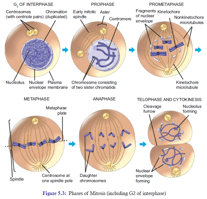
• The nuclear envelope breaks down at the end of prophase.
• The developing spindle now enters or invades the former nuclear area.
• The chromosomes have even become more condense.
• Kinetochores, a specialized multiprotein complex, bind to each centromere.
• Kinetochore microtubules extend from both the poles and bind to kinetochores of chromatids.
• Non-kinetochore microtubules originate from the two opposite poles and enter into thenuclear area where they overlap in the middle of the spindle.
3. Metaphase
• The centrosomes are now at opposite poles of the cell.
• The kinetochore microtubules from the two poles orient the chromosomes in such a way
that their centromeres become aligned at the metaphase plate, an imaginary plane that isequidistant between the two poles of the spindle apparatus.
4. Anaphase
• Once the centromeres are attached to the two sister chromatids. They start separating and
move towards the opposite poles. Consequently, the joined centromeres of sister chromatids
separate and give rise to two daughter chromosomes.
• The separation continues until the two poles of the cell have equivalent and completechromosomes.
5. Telophase
• The two sets of daughter chromosomes are assembled into two groups at opposite ends
of the cell.
• The chromosomes begin to uncoil and assume the elongated state characteristic of interphase.
• A nuclear envelope starts forming around each group of chromosomes.
• The spindle microtubules disappear; and the nucleolus or nucleoli reform.
• At this point, nuclear division is complete and the cell now has two identical nuclei.Cytokinesis
ACTIVITY 5.2
Aim: To observe permanent slides of plant root tip and animal cheek cells and outline the
differences between the plant cells and animal cells.
Materials Required:
1. Permanent (prepared) slides of animal cheek cells and plant root tips for mitosis.
2. Compound microscope.
Procedure:
1. Take the permanent slides.
2. Try to observe the stages of mitosis and meiosis from the given slides.
3. Draw a well-labelled diagram of the structures you have observed.
4. Outline the differences between how plant cells and animal cells divide.5. Record your observations in your notebook.
Discussion:
1. Firstly, discuss your result.2. Secondly, if there is any doubt, ask your biology teachers.
Cytokinesis is a division of cytoplasm. It compartmentalizes the two new nuclei into separate
daughter cells, completing mitosis and cell division (Figure 5.3). In animal cells, cytokinesis is
characterized by a constriction in the middle of the cell. The constriction continues until two
daughter cells are produced. In plant cells, a new cell membrane and cell wall are assembled
between the two new nuclei to form a cell plate. Cell wall material coats each side of the plate,and the result—two progeny cells.
Table 5.1: A table showing phases of cell cycle with one important event at each phase
ACTIVITY 5.3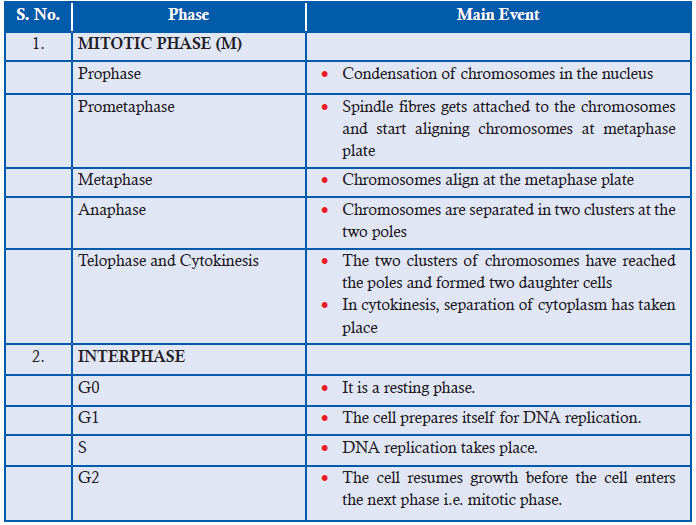
Aim: To Investigate how long onion root tip cells spend in each phase of the cell cycle and
present the findings in table form showing the stages of mitosis.
Materials Required:
1. Permanent slides of Onion root tips
2. Compound Microscopes
Procedure:
1. Fix the permanent slides of Onion root tips on the compound microscope.
2. Adjust the lens (10x, 40x etc.) to have a clear view of mitotically dividing onion root cells.3. Identify the mitotic stage of each cell by following the criteria given in the table below:
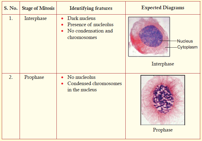
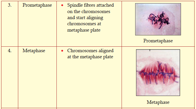
4. Identify and note down the number of cells at their specific stages. Note it down in your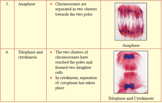
exercise book by copying the following table.
5. Onion takes a total time period of 12 hours (720 minutes) to complete mitosis.
Now with this information, find out the time period spent by each cell at each stage(i.e. interphase, prophase etc.) by using the given formula:
For example: Say, 500 cells were counted at interphase stage. And the total number of cells
counted was 1000. Then by applying the formula you can calculate how much time was
spent by cells at interphase in 12 hours mitosis cycle.
Time for interphase = 500/1000 × 720 minutes = 360 minutes
6. Calculate the time period spent by cells at different stages of mitosis. Note it down by copying
the following table in your exercise book. Then analyse the maximum and minimum timespend by cells in all the stages of mitosis.
Notes:
1. Normally, interphase takes the longest time period in the cell cycle.2. Compare your observations with your class mates.
5.2.2 Significance of Mitosis in Living Organisms
In the early development of an organism, the embryonic cells rapidly proliferate and differentiate
into specialized cells of adult tissues and organs. As cells differentiate from time to time, their
rate of proliferation usually decreases. As a result, most cells in adult animals are arrested at
the Go stage. Some cells at this phase may resume the cell cycle and proliferate when they
receive certain signals.
Some of the differentiated cells enter the Go resting phase and wait for the signal to resume
the cell cycle to repair injured tissue. There are numerous examples such as skin fibroblast,
endothelial cells, smooth muscle cells, and liver cells. Skin fibroblasts upon receiving growth
factor, they start secreting collagen and help in repairing cuts or wounds.
Most of the fully differentiated cells no longer possess the capability of cell division. Therefore,
they can be replaced by stem cells. Stem cells are undifferentiated biological cells that can
differentiate into specialized cells and can divide (through mitosis) to produce more stem cells.
The prominent role of stem cells can be seen in—blood cells (hematopoietic system), epithelial
cells of the skin, and epithelial cells lining the digestive tract (Figure 5.4). All of these cells
have short life spans, and they must be replaced continually by continual cell proliferation inadult animals.
The life span of blood cells ranges from less than one day to a few months. All of these cells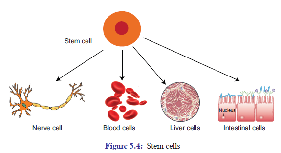
are derived from the same population of hematopoietic stem cells. In fact, there are more than
100 billion blood cells that are lost every day in humans. If there are no stem cells to replace the
loss of these cells, human beings will not be able to survive. Hence, these cells are continuallybeing replaced by cells produced from hematopoietic stem cells in the bone marrow.
The unrestrained, uncontrolled growth of cells in human beings results into a disease called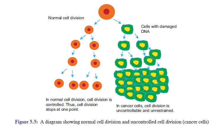
cancer (Figure 5.5). Cancer essentially is a disease of cell division. In other words, cancer occursdue to failure in controlling cell division.
Tumours
Cancer cells can be dangerous when it starts behaving abnormally in the body. The main problem
arises when a single cell in a tissue undergoes a process called transformation. It is a process
where a normal cell is converted into a cancer cell. Normally, the body’s immune system will
recognize the transformed cell as a foreign invading cell and, thus, destroys it. However, if the
transformed cell evades or escapes the destruction, it may proliferate and form a tumour—
a mass of abnormal cells. Tumours can be discussed in three subheadings:
(a) Benign tumour: This is a lump of the abnormal cells that remains at the original site.
Most benign tumours do not cause serious problems and can be completely removed by
surgery.
(b) Malignant tumour: These are abnormal cells that have become invasive enough to impair
with the functions of one or more organs. An individual with a malignant tumour is said
to have cancer.
(c) Metastasis: A few tumour cells may separate from the original tumour, enter blood vessels
and lymph vessels, and travel to other parts of the body. In the other parts of the body,
they may proliferate and form a new tumour. This spread of cancer cells to locations
distant from their original site is called metastasisAPPLICATION 5.1
1). Complete with appropriate terms:
(a). In ................. phase of the interphase DNA, replication occurs.
(b). Karyokinesis is the division of ................. .
(c). ................. is the resting phase of the cell cycle.
(d). It approximately takes ................. hours for one round of cell cycle in human beings.
(e). Cancer is the ................. growth of cells.2). The diagrams below show four animal cells at different stages of mitosis.
a) Name the structures A, B, C and D.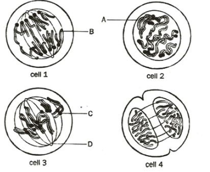
b) . (i) Name the stages of division shown by cells 1 and 3.
(ii) Use the numbers of each cell to arrange the stages in the correct sequence of
mitosis.3) . How does mitosis maintain genetic stability in an organism?
5.3 MEIOSIS AND ITS ROLE IN LIVING ORGANISMS
ACTIVITY 5.4
Through micrographs try to observe different stages of Meisois. State the arrangement ofchromosomes at every stage. Discuss your observations.
5.3.1 The Process of Meiosis
Meiosis is a reduction division where the number of chromosomes are reduced to half from diploid
parent cell to haploid daughter cells. It is divided into two stages: Meiosis I and Meiosis II. The
process of Meiosis alternates with an interphase, which is subdivided into G1, S, and G2 phases
(Figure 5.2). Meiosis I is further subdivided into prophase I, metaphase I, anaphase I, and
telophase I. In the same way, Meiosis II is also subdivided into prophase II, metaphase II,anaphase II, and telophase II.
Meiosis I
Prophase I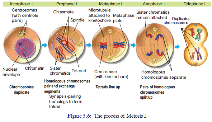
The DNA in prophase I coils tighter, and individual chromosomes first become visible under
the light microscope as a matrix of fine threads. Since the DNA has already replicated during
the S phase of interphase prior to meiosis I, each of the chromosomes actually consists of two
sister chromatids joined together at their centromeres.
Pairing or synapsis
In prophase I, homologous chromosomes become closely associated in synapsis. Synapsis
includes the formation of an elaborate structure called the synaptonemal complex, consisting
of homologous chromosomes paired closely along a lattice or zipper-like structure of proteins
between them (Figure 5.7). The components of synaptonemal complex include a meiosis-specific
form of cohesin, that helps the two homologous chromosomes to be closely associated along
their length.
Crossing over or recombination
Crossing over in meiosis I allows the homologous chromosomes to exchange their chromosomal
material. During this, the crossing over between sister chromatids is suppressed. Reciprocal
crossing over occurs only between non-sister chromatids and is controlled in such a way that
each chromosome arm usually has one or a few crossovers per meiosis. In human beings, thecrossovers are typically two to three in number.
Once the crossing over process is complete, the synaptonemal complex breaks down, and the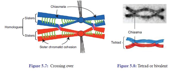
homologous chromosomes become less tightly associated. But the homologous chromosomes
remain attached at one particular point called chiasmata (chiasma-singular) (Figures 5.7
and 5.8). At this point, there are four chromatids of the two homologous chromosomes.
Two homologous chromosomes consist of two sister chromatids each. This structure of four
chromatids of the two homologous chromosomes attached at chiasmata is called as tetrad or
bivalent (Figure 5.8).
Some of the other events that occur along with synapsis are:
1. The nuclear envelope breaks down.
2. Two pairs of centrosomes migrate to opposite poles.3. Spindle fibres formation occurs.
Metaphase I
During metaphase I, the chiasmata have moved down on the paired homologous chromosomes
towards the ends. At this point, chiasmata are now called as terminal chiasmata. The terminal
chiasmata hold the homologous chromosomes together so that the homologous chromosomes
are now aligned at the equator of the cell. The kinetochores microtubules from the opposite
poles become attached to the kinetochore of homologous chromosomes.
The attachment of kinetochore microtubules at the monopolar centromere of each homologue
creates a tension on the homologous chromosomes, which are joined by sister chromatidcohesin at chiasmata.
Anaphase I
During anaphase, sister chromatid cohesion is released and the homologous chromosomes are
pulled apart to the opposite poles, but not the sister chromatids. Now when the spindle fibres
have fully contracted, each pole has a complete haploid set of chromosomes consisting of one
member of each homologous pair.
Telophase I
In telophase I, the chromosomes are segregated into two clusters at the two opposite poles.
Then the nuclear membrane reforms around each daughter nucleus. At the two poles, each
chromosome has sister chromatids attached to its centromere. And the interesting thing is that
the sister chromatids are no longer identical because of the crossing over that had taken place inprophase I.
Cytokinesis
Cytokinesis is the process of dividing the cytoplasm and its content into two equal cells
(Figure 5.7). Right after telophase I, cytokinesis may or may not occur. Meiosis I is followedby meiosis II, which occurs after an interval of variable length.
Meiosis II
Meiosis II is like mitotic division, which results into division of two equal cells without DNA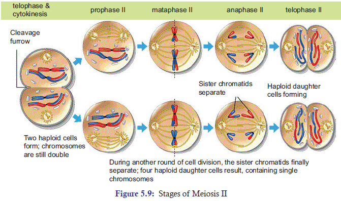
replication. Normally, the gap between meiosis I and meiosis II is interrupted by interphase.
But the interphase is very brief and it does not contain the S phase. Like mitosis cell division,
meiosis II is also subdivided into subphases. They are: (a) Prophase II (b) Metaphase II(c) Anaphase II (d) Telophase II (Figure 5.9).
Prophase II
Prophase II is brief. In prophase II, nuclear envelope breaks down and formation of new spindlefibres takes place.
Metaphase II
In metaphase II, the kinetochore microtubules extend themselves from the two poles and bind
to kinetochores of each sister chromatid. These kinetochore microtubules start pulling the
sister chromatids toward the two opposite poles. As a result, the sister chromatids are alignedat the metaphase plate.
Anaphase II
In anaphase II, as the spindle fibers contract, the cohesion complex joining the centromeres of
sister chromatids is destroyed or cleaved. As a result, the centromeres are split and the sisterchromatids are pulled towards the two opposite poles.
Telophase II
In telophase, the nuclear envelope re-forms around the four set of haploid daughter chromosomes.
Then cytokinesis follows resulting into complete four set of haploid daughter cells. These haploid
daughter cells may follow different fate depending upon the organisms. In animals, these haploid
daughter cells develop directly into gametes i.e. sperms and eggs. In plants, fungi, and manyprotists, they may divide mitotically to produce greater number of gametes.
5.3.2 Meiosis is a Reductional Division
Meiosis reduces the number of chromosomes sets from parental diploid chromosomes (2n) to
four haploid (1n) daughter cells. That is why meiosis is called a reduction division. In other
words, meiosis starts with diploid cell but ends with four haploid cells.
Simple explanation of Meiosis taking place in human beings is given below:
Parent cell = 46 chromosomes (Diploid)
Meiosis I = 2 cells (each chromosome with sister chromatids) = 46 × 2 = 92 chromosomes.Meiosis II = 4 haploid cells with unreplicated chromosomes = 92/4 = 23 chromosomes (Haploid)
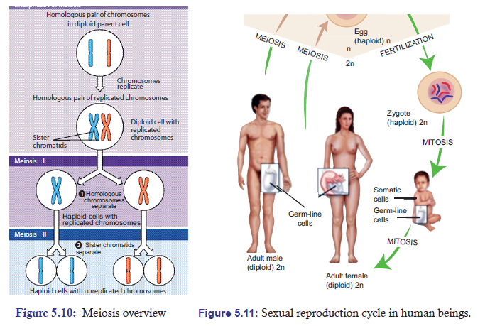
5.3.3 Significance of Meiosis
Cells Undergo Reduction Division Prior to Sexual Reproduction
Generally, a cycle of reproduction consists of meiosis and fertilization. Before sexual reproduction
occurs, gametes undergo meiosis and produce haploid cells. Thus during sexual reproduction,
one haploid (1n) gamete comes from the paternal side and another haploid (1n) gamete comes
from the maternal side; then, they both fuse to form a zygote, which is diploid (2n). The fusion
of gametes to form zygote or new cell is called as fertilization or syngamy (Figure 5.10).
If meiosis does not occur before sexual reproduction, the chromosome number would double
up with each fertilization. And after few generations, the number of chromosomes in each
cell would become impossibly large. For example in humans, in just 10 generations, the 46chromosomes would increase to about 47104 (46 × 210).
Role and Significance of Meiosis in Producing Gametes
Gametogenesis is a biological process by which diploid cells undergo cell division and
differentiation to form mature haploid gametes. It occurs through meiosis. In humans, the
male gamete (sperm) is produced by a process called spermatogenesis and the female gamete
(egg) is produced by a process called oogenesis through meiotic division.
Here gamete function takes place soon after meiosis but in plants it happens after gametophyte
formation sexual reproduction of plants starts with spore formation. Sporophyte is a diploids
generation of flowering plant where haploid spores are produced by meiosis which in turns
undergoes mitosis to form multi-celled haploid gametophytes. These haploid gametophyte
differentiate to produce gametes—sperm and egg cells. Similarly, embryo sac is formed by
reduction division. Each of the cells of embryo sac is haploid. Two of the nuclei fuse to producediploid nucleus.
The Role of Meiosis in Reproduction of Plants
Generally, plants reproducing sexually have life cycle consisting of two phases (Figure 5.11):
(a) A multicellular gametophyte or haploid stage: It is a haploid stage with n chromosomes.
It alternates with a multicellular sporophyte stage.
(b) A multicellular sporophyte or diploid stage: It is a diploid stage with 2n chromosomes,
made up of n pairs. A mature sporophyte produces spores by meiosis, a process which
reduces the number of chromosomes from 2n to 1n.
Alternation of generations (also known as mutagenesis) refers to the occurrence in the plant life
cycle of both a multicellular diploid organism (sporophyte) and a multicellular haploid organism
(gametophyte), each giving rise to the other. This is in contrast to animals, in which the only
multicellular phase is the diploid organism (such as the human man or woman), whereas the
haploid phase is a single egg or sperm cell.
In bryophytes (mosses and liverworts), the dominant generation is haploid, so that the
gametophyte comprises the main plant. On the contrary, in tracheophytes (vascular plants),the diploid generation is dominant and the sporophyte comprises the main plant.
Independent Assortment of Chromosomes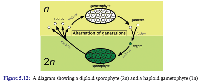
Specifically at metaphase I, each homologous pair of chromosomes positioned independently of
the other pairs. As a result, each homologous pair sorts out its maternal and paternal homologue
into daughter cells independently of every other pair. This act of separating homologous pairs
independently is called independent assortment. The random orientation of homologous pairs
of chromosomes due to independent assortment in meiosis I (metaphase) increases geneticvariation in organisms.
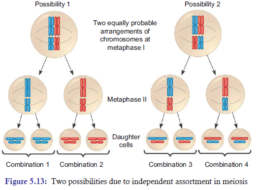
Crossing Over and Random Fertilization
During crossing over, DNA segments of the two parents-paternal and maternal are combined
into a single chromosome producing recombinant chromosomes, which are non-identical
with their sister chromatids. In humans, an average of one to three crossing over events occurs
per chromosome pair, depending on the position of their centromeres and on the size of the
chromosome. Thus, crossing over is an important event of meiosis that brings genetic variationin sexual life cycles.
Besides independent assortment and crossing over, the random fertilization during sexual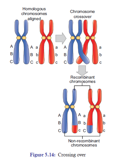
reproduction also increases genetic variation in organisms. During random fertilization, the
male gamete and female gamete fuse to form zygote. The most interesting thing is that this
zygote has the possibility of about 70 trillion diploid combinations. The number 70 trillion comes
from possible combinations of male and female gametes which are 223 × 223 = 70 trillions. The
possibility of this enormous number of combinations makes each and everyone of us uniquephysically and genetically.
Non-disjunction of Chromosomes
Proper separation of chromosomes during meiosis is essential for the normal growth in humans.
Any set of chromosomes that do not separate properly during meiosis results in improper
separation of chromosomes or non-disjunction, which is a serious issue in human genetics.
Non-disjunction is a condition in which the homologues or sister chromatids fail to separate
properly during meiosis. It can lead to the gain or loss of chromosome, a condition called asaneuploidy. Example: Down syndrome is an autosomal trisomy. It is also called as trisomy
21, where non-disjunction results in an embryo with three copies of chromosome 21 instead of
the usual two copies of chromosome 21 (Figure 5.15). It was first discovered by John LangdonDown. The chance of occurrence is one infant in every 800 live births.
The most common symptoms are: They are short. They may also have protruding, furrowed
tongues, which causes the mouth to remain partially open. They are mentally retarded. They
have a prominent epicanthic fold in the corner of each eye; and typical flat face and roundhead. Usually, there is a wide gap between the first and the second digits on their feet.
The origin of trisomics condition is through non-disjunction of chromosome 21 during
meiosis. Failure of paired homologues to separate during either anaphase I or II may lead to
gametes with 23 + 1 chromosome composition instead of the normal 23 gamete chromosome
composition. Therefore, instead of 46 normal chromosomes, Down syndrome patient willhave 47 chromosomes with three copies of chromosome 21 instead of the normal 2 copies.
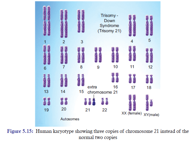
APPLICATION 5.2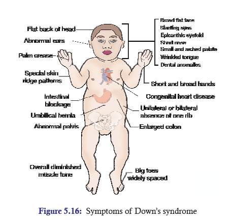
1. Complete with appropriate terms:
(a) Independent assortment of chromosomes takes place at .............. stage of Meiosis I.
(b) Chiasmata formation occurs at .............. .
(c) Meiosis is also known as .............. .
(d) Crossing over and random fertilization brings .............. in life cycles.
(e) Trisomy of chromosome number two leads to .............. syndrome.
2. The drawing below has been made from a photograph showing a cell undergoing mitosis.
a) In which stage of mitosis is the cell shown in this drawing?
b) Describe one piece of evidence, visible in the drawing, which could be used to confirm
that this cell is not in the first division of meiosis.
3. An organism has two pair of chromosomes (i.e., chromosome number = 4). Diagrammaticallyrepresent the chromosomal arrangement during different phases of meiosis-II.
5.4 COMPARISON OF MITOSIS AND MEIOSIS
ACTIVITY 5.5
Cells divided both by mitosis and meiosis contain nucleus which is a genetic material. Can cells
divided by mitosis act as carriers of genes to the next generation? Compare mitosis and meiosisin a tabular form. Research, debate and present your findings.
Table 5.2: Comparison between mitosis and meiosis
ACTIVITY 5.6
Carry out a research project to find out why cultured skin is grown in a medium of proteinssimilar to blood. Then write a journal entry to summarise the research.
5.5 SUMMARY
• Karyotype is a visual representation of the chromosomes within a single cell.
• Diploid cells contain two complete sets (2n) of chromosomes. Example, skin cells. Haploid
cells contain only one complete set (1n) of chromosome. Example, gametes (sperm and
egg). Homologous chromosomes are pairs of chromosomes that are similar in length, gene
position, and centromere location. Non-homologous chromosomes are chromosomes
that contain different genes.
• A typical cell cycle consists of mitotic and interphase phases. Interphase is subdivided
into G0, G1, S, G2 phases. S phase is the longest phase.
• Mitosis is subdivided into prophase, prometaphase, metaphase, anaphase, and telophase.
• DNA replication takes place at S phase of interphase prior to mitosis.
• Cytokinesis is a division of cytoplasm and usually follows the process of mitosis.
• Some differentiated cells are arrested at G0 phase; and they resume cell cycle when they
receive signal to divide and repair injured tissues. Example, skin fibroblasts.
• Some cells have stem cells that are undifferentiated biological cells which can differentiate
into specialized cells and continually replace the dying cells. Example, hematopoietic
(blood-forming) system.
• Unrestrained, uncontrolled growth of cells in cell cycle results into a disease called cancer.
• Meiosis is divided into meiosis I and meiosis II. Meiosis I is a reduction division and is
subdivided into prophase I, metaphase I, anaphase I, and telophase I. DNA replication
takes place at interphase I prior to meiosis I. Synaptonemal complex is formed during
prophase
I. Crossing over takes place in meiosis I.
• Meiosis II is just like mitosis. There is no DNA replication. It is divided into prophase II,
metaphase II, anaphase II, and telophase II.
• Meiosis is a reduction division. It reduces diploid chromosomes to four haploid daughter
cells. Reduction division usually happens before sexual reproduce where haploid gametes
(sperm and eggs) are formed for fertilization.
• The process of sperm formation is called spermatogenesis; while the process of egg
formation
is called oogenesis.
• Meiosis also reduces diploid plant into haploid gametes which eventually fuse to form
zygote.
• During meiosis, paternal and maternal homologues assort independently into four
daughter cells. This process adds genetic variation.
• Crossing over and random fertilization increase genetic variation in sexual life cycles.
• Improper separation of chromosomes during meiosis results into non-disjunction of
chromosomesthat are responsible for disease such as Down syndrome.
5.6 GLOSSARY
• Cancer: It’s an unrestrained, uncontrolled growth of cells in human beings. It is essentially
a disease of uncontrolled mitotic cell division.
• Chiasmata (chiasma-singular): During crossing over of genes, the point where
homologous chromosomes remain attached at one particular point is called chiasmata.
• Crossing over: The exchange of genetic material between homologous chromosomes
that occur during meiosis and contribute to genetic variability.
• Cytokinesis: It is a division of cytoplasm.
• Diploid cells: Cells which contain two complete sets (2n) of chromosomes (23 + 23 in
number) are called diploid cells. Example: somatic cells (Muscle cells).
• Down syndrome: It is an autosomal trisomy. It is also called as trisomy 21, where
non-disjunction results in an embryo with three copies of chromosome 21 instead of the
usual two copies of chromosome 21.
• Gametogenesis: It is a biological process by which diploid cells undergo cell division
and differentiation to form mature haploid gametes.
• Haploid cells: Cells that contain only one complete set (1n) of chromosome (23 in
number) are called as haploid cells. Example: sperm and ovum.
• Independent assortment: In meiosis I, specifically at metaphase I, each homologous pair
of chromosomes positioned independently of the other pairs at metaphase I.
• Karyotype: It is a visual representation of the chromosomes within a single cell where
the number of chromosomes, their arrangement, size and structure can be analysed.
• Meiosis: It’s a reduction division where the number of chromosomes is reduced to half
from diploid parent cell to haploid daughter cells.
• Mitosis: It is a process where a single cell divides into two identical daughter cells.
In other words, it’s a division of nucleus.
• Reduction division: It is a process of meiotic cell division where the number of
chromosome sets from parental diploid (2n) cells is reduced to haploid (1n) daughter cells.
• Stem cells: They are undifferentiated biological cells that can differentiate into specialized
cells (when stimulated by the right factor) and can divide to produce more stem cells.
Example: Hematopoietic system.
• Syngamy or fertilization: The fusion of gametes (one from mother and one from father)
to form a zygote is called as syngamy.
• Tetrad or bivalent: The structure of four chromatids of the two homologous chromosomesattached at chiasmata during the process of meiosis is called as tetrad.
END UNIT ASSESSMENT 5
Do all these exercises in your exercise book.
I. Choose whether the following statements are True (T) or False (F)
1. Cell cycle is a cyclical event of cell growth, mitosis, and cell division.
2. A typical cell spends most of its time in interphase.
3. Mitosis is a process where a single cell divides into three identical daughter cells.
4. Cytokinesis is a division of cytoplasm.
5. The process of mitosis is basically divided into 5 phases.
6. Meiosis is divided into three stages: Meiosis I, Meiosis II and Meiosis III.
7. The unrestrained, uncontrolled growth of cells in human beings results into a disease
called cancer.
8. Cancer occurs due to failure in controlling cell division.
9. Proper separation of chromosomes during meiosis is not essential for the normal
growth in humans.10. The life span of blood cells ranges from less than one day to a few months.
II. Multiple Choice Questions
1. Meiosis starts with diploid cell but ends with ......... haploid cells.
(a) one (b) two
(c) three (d) four
2. ........................... is a biological process by which diploid cells undergo cell division
and differentiation to form mature haploid gametes.
(a) Gametogenesis (b) Spermatogenesis
(c) Oogenesis (d) None of these
3. Generally, plants reproducing sexually have life cycle consisting of .......... phases.
(a) two (b) three
(c) four (d) five
4. In telophase, the nuclear envelope re-forms around the ........... set of haploid daughter
chromosomes.
(a) one (b) two
(c) three (d) four
5. The prominent role of stem cells can be seen in—
(a) blood cells (hematopoietic system)
(b) epithelial cells of the skin
(c) epithelial cells lining the digestive tract
(d) All of these
6. ........................is a condition in which the homologues or sister chromatids
fail to separate properly during meiosis.
(a) Disjunction (b) Non-disjunction(c) Down syndrome (d) None of these
7. Which of the events is not correct in prometaphase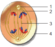
(a) Nuclear envelope breaks down
(b) Spindle fibres invade nuclear area
(c) Kinetochore binds to centromere
(d) Chromatids condense into discrete chromosome
8. Which of the event is correct in anaphase
(a) Sister chromatids separate and give rise to daughter chromosomes.
(b) Chromosomes are aligned at the metaphase plate.
(c) Cytokinesis starts occurring.
(d) Chromosomes begin to uncoil.
9. The total chromosome number at the end of meiosis I and II is
(a) 46 (b) 23(c) 44 (d) 92
10. One round of oogenesis produces
(a) One egg (b) Two eggs
(c) Three eggs (d) Four eggs
11. Which of the following results in genetic variation
(a) Independent assortment of chromosomes
(b) Crossing over
(c) Random fertilization
(d) All of these
12. In mitosis, which of the following occurs?
(a) Chiasmata formation (b) DNA replication
(c) Synapsis (d) None of these
13. Identify the cells which are divided by mitosis.
(a) Skin cells (b) Liver cells
(c) Blood cells (d) All of these
14. Identify the cells which are divided by meiosis.
(a) Intestinal cells (b) Stem cells
(c) Gametes (d) None of these
15. Which part of the plants is/are divided by mitosis?
(a) Stem (b) Flower
(c) Leaves (d) All of these
16. Identify the cells that are divided by meiosis in plants.
(a) Root tips (b) Pollen grains
(c) Ova (d) (b) and (c)
III. Long Answer Type Questions
1. Describe the main stages of cell cycle.
2. In your own words, explain what is meant by homologous pairs of chromosomes.
3. What do you mean by the terms haploid and diploid?
4. In your own words, describe the process of mitosis.
5. In your own words, describe the process of meiosis.
6. Outline the significance of mitosis in cell replacement and tissue repair by stem cells.
7. In your own words, explain how uncontrolled cell division can result in the formation
a tumour.
8. What is the need for reduction prior to fertilization in sexual reproduction?
9. In your own words, explain the importance of effective cell division.
10. Outline the role of meiosis in gametogenesis in humans and in the formation of
pollen grain and embryo sacs in flowering plants.
11. Explain how crossing over and random assortment of homologous chromosomes
during meiosis and random fusion of gametes at fertilization leads to genetic variation,
including the expression of rare recessive alleles.
12. (i) Identify the stage of cell division shown in the figure.
(ii) Label the structures marked as (1), (2), (3) and (4).
(iii) Which type of cell is involved in this division?
(iv) What will happen if the structure marked (3)
is not formed?
13. How can you correlate the spread of HIV virus with the process of Mitosis? Knowing
the viral disease and its spread, discuss in brief the stigma and discrimination facedby those affected by HIV and AIDS.
Unit 6 Protein Synthesis
Key Unit Competence
To be able to explain the relationship between a gene and the sequence of nucleotides in
DNA and to describe the process of protein synthesis in eukaryotes.
LEARNING OBJECTIVES
At the end of this unit, the learner will be able to:
• State the features of a genetic code.
• State that a gene is a sequence of nucleotides that form part of a DNA molecule that codes
for a specific polypeptide.
• Appreciate the importance of the genetic code in determining the structure of a protein.
• Describe how the information in DNA is used during transcription and translation to
construct polypeptides.
• Agree that the way DNA code for polypeptides is central to our understanding of how cells
and organisms function.
• Be aware that DNA is an extremely stable molecule that cells replicate with extreme accuracy
to minimise possibilities of DNA mutations.
• State the roles played by mRNA, tRNA and the ribosomes in the formation of the polypeptide.
• Appreciate the role of the genetic code in determining the characteristics of an individual.
• State that ribosomes provide surface area for the attachment of mRNA during polypeptide
synthesis.
• State that polysomes consists of up to 50 ribosomes on the same mRNA strand and that they
speed up polypeptide synthesis.
• Describe the way in which the nucleotide sequence codes for the amino acid sequence with
specific reference to HbA (normal) and HbS( sickle cell) alleles for b-globin poly peptides.
• State that gene mutation is a change in the sequence of nucleotides that may result in an
altered polypeptide.
• Construct a flow chart, in proper sequence, for the stages of transcription and translation.
• Using the evidence, predict the effect of change in genetic code on the structure of the protein
manufactured during protein synthesis.
• Carry out research to find and understand better about protein synthesis and on genetic diseases.
INTRODUCTORY ACTIVITY
You can refer to your notes of Bio S4 to answer the following questions:
1) Assort the list below into proteins and non- proteins:
Glucose – Enzymes – Cholesterol – Amylose – Keratin – Haemoglobin – Ascorbic
acid – Melanin – starch – Myosin
2) Why two ladies having the same diet can be one with long hairs and another with
short hairs?
6.1 GENETIC CODE
ACTIVITY 6.1
You have studied that codon is made up of three bases and DNAs in human body intermittently
open and close up during DNA replication and transcription, but how is it that genetic
information is passed on from generation to generation with minimal mistake? Research and
discuss your findings, and then list out the best answers. You may look into textbooks, videoclips, computer animations, Internet, etc for finding answers.
A gene is a sequence of nucleotides that forms part of a DNA molecule that codes for a specific
polypeptide.
The genetic code is the set of rules by which information encoded in genetic material (DNA
or RNA sequences) is translated into proteins (amino acid sequences) by living cells using
ribosome machinery. In other words, the genetic code is a set of rules that specify how the
nucleotides sequence (AUGC) of an m-RNA is translated into the amino acid sequence of a
polypeptide chain.
During translation, the process of making proteins, ribosome reads the sequence of an m-RNA
(nucleotide sequences) from 5’ end to 3’ end. Then it makes appropriate amino acids according
to the genetic information on the m-RNA. By nature in both eukaryotes and prokaryotes, the
5’ to 3’ nucleotide sequence of the coding DNA strand exactly corresponds or specifies thesame N-terminal to C-terminal amino acid sequence of the encoded polypeptide (Figure 6.1).
As studied in Unit 3, with four different nucleotides (A, C, G, U), a three-letter code (codon)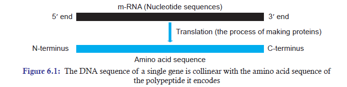
can give 64 different possible codons (i.e., 43 = 64) or (4 × 4 × 4 = 64). These 64 possible codons
are more than enough to code for the 20 amino acids found in living cells. The genetic code
allows an organism to translate the genetic information found in its chromosomes (m-RNA)into mature functional proteins.
6.1.1 The Characteristics of Genetic Code
The following are some characteristics of genetic code:
1. The Genetic Code is a Triplet Codon: A codon consists of a group of three nucleotides.
And each codon codes for a specific amino acid in a polypeptide chain with some exceptions.
2. The Genetic Code is Used without Comma: The three nucleotides in a codon are read
in a continuous fashion without any comma. Examples: AUG, UAG, UGA and UAA.
3. The Genetic Code is Non-overlapping: The codons in the m-RNA sequence are read
successively without overlapping.
4. The Genetic Code is Almost Universal: For many long years, it was thought that the
genetic code is universal, which led us into believing that all living organisms have the same
genetic code. However, recent studies have revealed that there are some organisms where
there is difference in genetic code (Table 6.1). That is the reason why it is appropriate
to use the phrase “almost universal” rather than the word “universal.” The examples of
organisms or organelles where genetic codes have different meanings:Table 6.1: Genetic Code
5. The Genetic Code is “Degenerate”: A codon is thought to code for a particular amino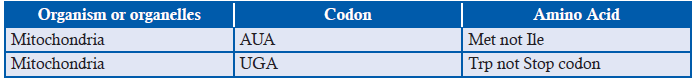
acid. That is one codon for one amino acid. But more than one codon can code for a
particular amino acid, with two exceptions of AUG and UGG. This multiple coding by
a single codon is called the degeneracy or redundancy of the code. Example: UUU and
UUC codons code for the same specific phenylalanine amino acid. In the same way, CAUand CAC codons code for the same specific histidine amino acid (Figure 6.2).
Figure 6.2: The Genetic Code: Out of the total 64 codons, 61 sense codons specify one of the 20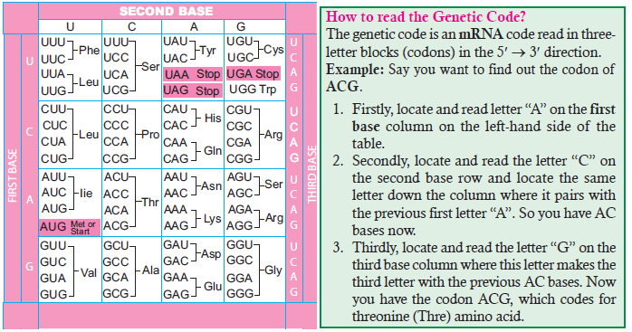
amino acids. The other three nonsense codons are Stop Codons and, therefore, do not specify anyacid. The sense codon AUG, which specifies Methionine, is a Start Codon
6. The Genetic Code has Start and Stop Codons: Out of 64 codons, only 61 codons are
called sense codons (Figure 6.2). The other three codons are called nonsense codons
or stop codons or chain-terminating codons. These three codons are UAG, UAA,
and UGA; they do not specify any amino acid, and there are no t-RNAs to carry the
appropriate anticodons. The AUG codon, which code for methionine, is most of the
time the start codon or initiation codon for protein synthesis in both eukaryotes and
prokaryotes.
7. Wobble Hypothesis: Francis Crick has pointed out that the complete set of 61 sense
codons can be read by fewer number than 61 t-RNAs. The simple reason being, the pairing
properties in the bases in the anticodons are wobble in nature. Here, the word “wobble”
simply means “fluctuating” or “unsteady.”
For example: The two different leucine codons (CUC, CUU) can be read by the sameleucine t-RNA molecule, contrary to regular base-pairing rules (Figure 6.3).
Figure 6.3: Example of base-pairing wobble. The same leucine t-RNA molecule (anticodon GAG)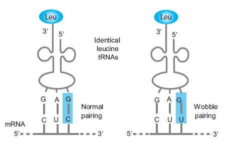
can read two different leucine codons (CUC, CUU)
6.1.2 Importance of the Genetic Code in Determining the Structure of a Protein
DNA is Extremely Stable and Replicates Accurately
According to central dogma concept, m-RNA is copied from DNA and m-RNA is then translated
to form proteins. Therefore, it is critical to maintain the integrity of DNA to accurately produce
the desired and correct amino acids (proteins).
DNA is the repository of genetic information gathered over millions of years and it is stored
in a stable form inside the cell. The stability of DNA is a property critical to the maintenance
of the integrity of the gene.
The stability of DNA can be explained and evidently supported by the fact that DNA has been
extracted from Egyptian mummies and extinct animals such as the woolly mammoth and it
can also be extracted from dried blood sample or from a single hair at a crime scene which is
old enough. DNA molecule is a stable structure and replicates accurately in order to avoid any
mutation or change in nucleotides sequences in DNA. The stability of DNA can be attributedto important factors — Hydrogen Bonds and Base Stacking.
Hydrogen Bonds
Hydrogen bond is the attractive force between the hydrogen attached to an electronegative
atom (O) of one molecule and an electronegative atom (N) of a different molecule
(Figure 6.4). In the structure of DNA, the strong electronegative atom is the oxygen (O) andNitrogen (N), while H atom has positive charge. In the structure of DNA (Figure 6.4), thymine
and adenine have two hydrogen bonds; while guanine and cytosine have three hydrogen bonds.
Hydrogen bonds play very important role in binding the bases of the opposite strands in the
DNA. Hydrogen bonds are very weak by themselves. But in a DNA sequence, there will bethousands of these H-bonds which make DNA very stable.
Figure 6.4: Hydrogen bonding and Base stacking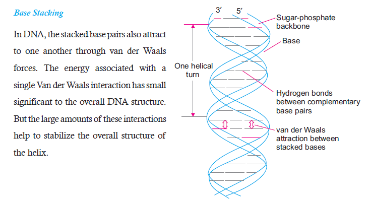
enabling stability of DNA
APPLICATION 6.1
1. Complete the sentence with correct word:
(a) A .............. is a sequence of nucleotide which codes for specific polypeptide.
(b) .............. allows an organism to translate genetic information into proteins.
(c) .............. is a start codon.
(d) .............. and .............. contributes to stability of DNA.
2. Describe briefly what is meant by Wobble Hypothesis3. Draw and label the structure of a t – RNA
6.2 TRANSCRIPTION
Transcription is the process of copying information from one strand of DNA into a singlestrandedRNA.
ACTIVITY 6.2
Discuss how DNA replication is different from the process of transcription. Make flow chart,
diagrams of bacterial and eukaryotic transcriptions. You can use for reference textbooks, videoclips, animation and Internet.
6.2.1 Transcription Unit in DNA
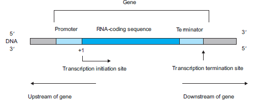
Figure 6.5: A transcription unit of DNA
Note:
- The nucleotide in the template strand at which transcription begins is designated with the
number +1.
- Downstream sequences are drawn, by convention, to the right of the transcription start site.
- Nucleotides that lie to the left of the transcription start site, are called the upstream sequences
and are identified by negative numbers.Note:
- The nucleotide in the template strand at which transcription begins is designated with the
number +1.
- Downstream sequences are drawn, by convention, to the right of the transcription start site.
- Nucleotides that lie to the left of the transcription start site, are called the upstream sequencesand are identified by negative numbers.
A transcription unit in DNA consists of three main regions (Figure 6.5):
(a) A promoter: A promoter is a region of DNA that initiates transcription of a particular
gene.
(b) RNA coding sequence: It is a DNA sequence that is transcribed by RNA polymerase
into RNA transcript (m-RNA).(c) A terminator: It is a DNA sequence which specifies termination of transcription.
6.2.2 RNA Polymerase
In bacteria, RNA polymerase is the only enzyme that is responsible for catalysing the process
of transcription. It is a DNA-dependent RNA polymerase, as it uses a DNA template strand
to synthesize a new RNA chain. During transcription, it synthesizes RNA in 5’ to 3’ direction
by using 3’ to 5’ strand of DNA as a template strand. The opposite 5’ to 3’ strand of DNA is
not used during transcription and it is called nontemplate strand.
RNA polymerase uses RNA precursors for synthesizing RNA chain. The RNA precursors are
ribonucleoside triphosphates ATP, GTP, CTP, and UTP. They are collectively known as NTPs
or Nucleoside triphosphate. The synthesis of RNA chain follows complementary base pairingrule i.e., A will pair with U; G will pair with C.
6.2.3 The Process of Transcription in Bacteria
The process of transcription is basically divided into three stages: (a) Initiation (b) Elongation
(c) Termination (Figure 6.6).
(a) Initiation: RNA polymerase accompanied by sigma (s) factor binds at the promoter.
Sigma factor ensures that RNA polymerase binds accurately and stably on the promoter.
Then RNA polymerase unwinds DNA in the promoter region to form open promoter
complex.
(b) Elongation: Once the initiation has commenced, RNA polymerase starts elongating or
adding NTPs one after the other using one of the strands of DNA as a template strand.
The nontemplate strand is not used for elongation of RNA. Elongation of the new RNA
takes place in 5’ to 3’ direction and follows complementary base pairing rule. For example:
If the DNA sequence in the DNA template is 3’-ATACTTGAACTAACTC-5’, then thesequence of newly synthesized RNA will be 5’-UAUGAACUUGAUUGAG-3’.
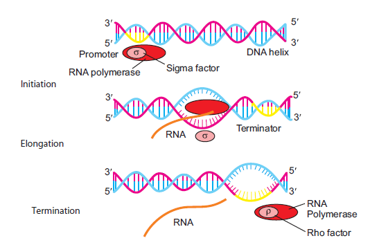
Figure 6.6: The process of transcription in bacteria
(c) Termination: Termination of transcription is signalled by terminator sequence located
downstream from the promoter. It can take place in two ways: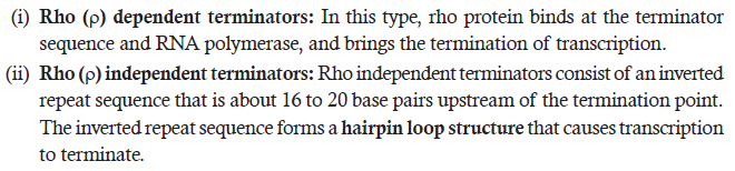
6.2.4 Transcription in Eukaryotes
Unlike the situation in prokaryotes, transcription in eukaryotes occurs within the nucleus and
mRNA moves out of the nucleus into the cytoplasm for translation. In eukaryotes, there are
two additional complexities:
1. There are at least three RNA polymerases in the nucleus (in addition to the RNA
polymerase found in the organelles). There are RNA polymerase I, II and III. The RNA
polymerase I transcribes rRNAs (285, 185 and 5.8S), whereas RNA polymerase III is
responsible for transcription of tRNA, RNA, 5s rRNA and sn RNAs (small nuclear
RNAs). The RNA polymerase II transcribes precursor of mRNA (pre-mRNA) or hnRNA
(heterogenous nuclear RNA). Thus, there is a division of labour in the functioning ofthe three types of RNA polymerase.
2. The second complexity is that the primary transcript contains two types of segments,
the non-coding introns or intervening sequences and the coding exons. The primary
eukaryotic mRNA transcript is much longer and is non-functional. Hence, it is subjected
to a process called ‘splicing’, where the introns are removed and exons are joined in a
definite order. Hn RNA (primary of RNA transcript) undergo two additional processing
called ‘capping’ and ‘tailing’. In capping an unusual nucleotide, methyl guanosine
triphosphate (mGppp) is added to the 5′ end of hn RNA. In ‘tailing’ adenylate residues
(200-300) are added (polyadenylation at 3′-end to hn RNA in a template independent
manner (i.e., without a template). The fully processed hnRNA is now called mRNA,that is transported out of the nucleus for translation (Figure 6.7).
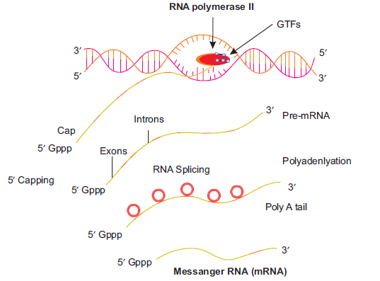
Figure 6.7: Transcription in eukaryotes
ACTIVITY 3
Read the process of translation carefully and know all the important steps. After finishing your
reading, put the steps of the process of translation in separate boxes in the form of flow chart.
Start the flow chart from the production of pre-m-RNA and end with the proteins (ultimateproduct). Compare your chart with that of your classmates.
APPLICATION 6.2
1. Complete the sentence with correct word:
(a) ................... is a DNA dependent polymers.
(b) The base pairs of eukaryotic gene are ............... with the bases of transcribed mRNA.
(c) mRNA coding information for more than one gene is called ...................
(d) ................... is a process of adding poly A tails to pre mRNA.
(e) ................... are removed by RNA splicing.2. What is the particularity of the Transcription in Eukaryotes?
6.3 TRANSLATION/PROTEIN SYNTHESIS
6.3.1 Role of Transfer RNA in the Formation of Polypeptide Chain
Though there are specific codons on m-RNA for specific amino acids, nucleic acids (m-RNA)
and proteins (amino acids) are written in two different languages. Therefore, there has to be
a mediator that can decode the message in m-RNA and direct the formation corresponding
proteins. This is where the role of t-RNA comes into play. The primary role of transfer RNA(t-RNA) is to decode (translate, like an interpreter) the codons on m-RNA and use the message
Figure 6.8: A diagram showing t-RNA molecule linking amino acid at its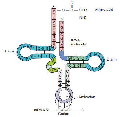
3’ end and codon on m-RNA at its anticodon site.
in codons to direct the process of synthesising polypeptide chain. Thus, t-RNA acts as an
adaptor or intermediaries. Since interpretation of the language between m-RNA and aminoacids is involved, the process of protein synthesis is called translation.
During translation, t-RNA links to a specific amino acid at its 3’ end giving rise to charged
aa-t-RNA, while the opposite end (anti-codon region) recognizes a particular codon in the
m-RNA (Figure 6.8). Depending upon the interaction between codons in m-RNA and specific
charged aa-t-RNAs, polypeptide chain (long amino acids) are synthesized during translation.
Transfer RNA is composed of 73–93 nucleotides, 10 of which are modified from the standard
4 nucleotides of RNA (A, G, C, and U) (Figure 6.8). Because of complementary base pairing,
the various t-RNAs become folded in a similar way to form a structure that can be drawn intwo dimensions as a cloverleaf.
6.3.2 The Role of Messenger RNA in the Formation of Polypeptide Chain
During transcription, the genetic information in DNA is copied and encoded in the intermediate
product called messenger RNA (m-RNA), which along with t-RNA will be used by ribosome
for protein synthesis/translation (Figure 6.9). Thus, the primary role of messenger RNA is to
carry the genetic information copied from DNA in the form of a series of codons (three-basecode), each of which specifies a particular amino acid.
Figure 6.9: (A) A simplistic diagram representing transcription and translation.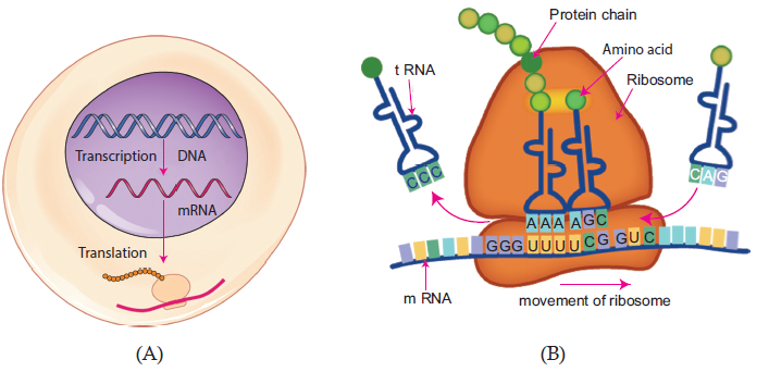
(B) A diagram showing m-RNA carrying genetic information copied from DNA in the form ofcodons. Examples: UUU, UCG codons are shown in the diagram
The series of codons in m-RNA code for specific amino acids. For example, as shown in
Figure 6.2, UUU codon will code for phenylalanine amino acid; similarly, UCG codon willcode for serine amino acid.
6.3.3 Role of Ribosomes in the Formation of Polypeptide Chain
Ribosomes are machines that carry our protein synthesis or translation. The main role of
ribosomes is to orient the m-RNA and amino acid carrying t-RNAs in such a position that the
genetic code can be read accurately and catalyse peptide bond formation.
Ribosomes are particles made up of ribosomal RNA (r-RNA) and proteins. In prokaryotes, they
are present in cytoplasm, while in eukaryotes they occur both free in the cytosol and bound to
membrane of the nuclear envelope. Mitochondria and chloroplast also have ribosomes.
Generally, a ribosome is composed of two dissociable subunits called the large and small subunits.
In prokaryotes (bacteria), ribosome has a sedimentation coefficient of 70S; it is made up by 30S
small subunit and 50S large subunit (Figure 6.10). In eukaryotes, ribosome has a sedimentationcoefficient of 80S; it is made up of 40S small unit and 60S large unit.
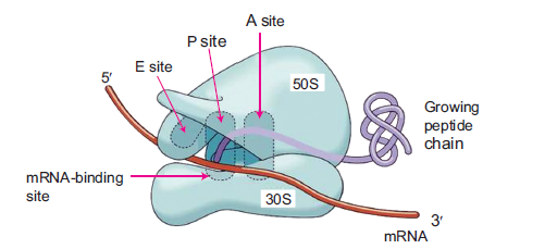
Figure 6.10: A bacterial ribosome
There are four important sites in the ribosome. These four sites are particularly important during
protein synthesis (Figure 6.10). These are:
(a) Messenger RNA-binding site: It is the site that binds m-RNA.
(b) A (aminoacyle) site: It is the site that binds each newly incoming t-RNA with its attached
amino acid.
(c) P (peptidyl) site: It is the site where the t-RNA carrying the growing polypeptide chain
resides.
(d) E (exit) site: It is the site from which t-RNAs leave the ribosome after they have dischargedtheir amino acids.
6.3.4 The Way DNA Codes for Polypeptides is Central to Understanding how Cells andOrganisms Function
The central dogma of molecular biology is an explanation of the flow of genetic information,from DNA to RNA, to make a functional protein within a biological system.
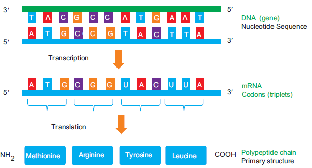
Figure 6.11: Central Dogma-information flow in a biological system
Once a DNA is transcribed into RNA (m-RNA), the genetic code (codon) in an m-RNA
specifies the amino acids that are assembled during protein synthesis to make polypeptides.
That is why the way DNA codes for polypeptides is central to our understanding of how cells
and organisms function.
More specifically, nucleotide sequence in a DNA molecule forms a gene. This particular gene
is then transcribed and translated into a specific polypeptide chain (proteins). In other words,
the nucleotide sequence in a DNA determines the sequence in which amino acids are linked
together when proteins are synthesized (Figure 6.11).
Furthermore, the properties and functions of proteins are determined by the structure of proteins.
The primary structure (simple sequence of amino acids) determines its three dimensional shape
and, therefore, its properties and functions. For example, the primary structure of an enzyme
determines the shape of its active site. And the shape of this active site will consequentlydetermine the substrate with which it can bind.
For proteins to become biological functional, they have to be expressed i.e., two molecules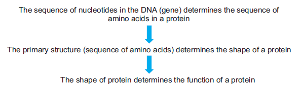
must bind to each other. For examples:–An antibody protein must bind to an antigen to trigger
an immune response; an enzyme protein must bind to a substrate to catalyze a reaction. The
binding of two molecules involves the two molecules to recognize each other and form a series
of non-covalent bonds. Recognition of two molecules for each other is termed “structuralcomplementarity.” It can be compared with a key fitting into a lock.
6.4 TRANSLATION IN BACTERIA
Translation is the production of protein molecules (polypeptides) by cellular ribosomes with
the help of information present on the m-RNA. The m-RNA and protein molecules are like
two languages written with different types of letters. The language by which the information
on m-RNA is written has to be translated into the language of amino acids in order to use it
to direct the sequential assembly of amino acids into a polypeptide chain. That is the reasonwhy protein synthesis is appropriately referred to as translation.
Charging of t-RNA
Prior to translation, each t-RNA molecule must be attached to the correct amino acid. Therefore,
the covalent linking of a specific amino acid to the 3’ end of the correct t-RNA by the enzyme
aminoacyl-t-RNA synthetase is called charging of t-RNA. An enzyme aminoacyl-t-RNA
synthetase catalyzes the linking of amino acids to their corresponding t-RNAs via an ester
bond, accompanied by the hydrolysis of ATP to AMP and pyrophosphate. This process is a
critical step in translation as it determines the accuracy of translation.Charging of t-RNA occurs in two steps:
1. ATP+ amino acid → aminoacyl-AMP + PPi
2. Aminoacyl-AMP + t-RNA → aminoacyl-t-RNA + AMP
6.4.1 The Process of Translation
The process of translation basically consists of three major stages: (1) Initiation, (2) Elongationand (3) Termination (Figure 6.12).
1. Initiation: It is the stage where m-RNA is bound to the ribosome and positioned itself
for proper translation. It can be further subdivided into three steps:
(a) Binding of initiation factors: The initiation factors along with GTP first bind to 30S
subunit.
(b) Binding of m-RNA and t-RNA: Now the m-RNA and the charged t-RNA with the
first amino acid bind to the 30S ribosomal subunit.
(c) Formation of 70S Subunit: The 30S ribosomal subunit now binds to a free 50S
ribosomal subunit forming the 70S initiation complex. During this step, all the
initiation factors are released.
2. Elongation: It is the stage where amino acids are sequentially joined together to form a
polypeptide chain via peptide bonds. The sequence of polypeptide chain is formed in an
order specified by the arrangement of codons in m-RNA. Elongation can be subdivided
into three steps:
(a) Binding of an aminoacyl-t-RNA: The binding of an aminoacyl-t-RNA to 70S
ribosome brings a new amino acid into a position on the ribosome that can be joined
to the polypeptide chain. In bacteria, normally the first incoming aminoacyl-t-RNA
is N-formylmethionine (fMet).
(b) Peptide bond formation: The newly incoming amino acid is linked to the growing
polypeptide chain by peptide bond formation.
(c) Translocation: It is a process in which the m-RNA is moved by a distance of threenucleotides (codon) to bring the next codon on the ribosome.
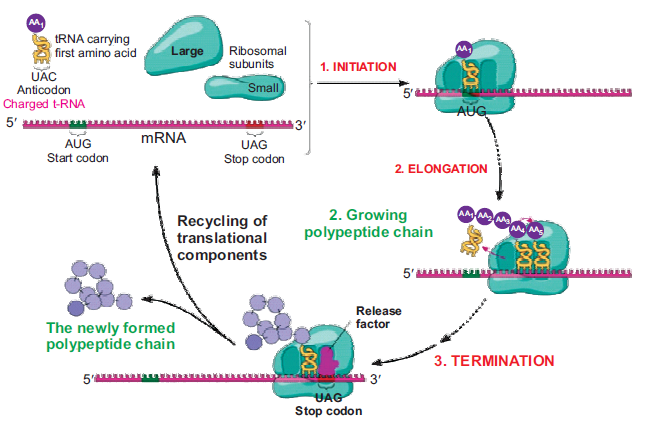
Figure 6.12: The process of Translation
3. Termination: It is the process of ending translation. At this stage, the newly formed
polypeptide chain and the m-RNA are released from the ribosome. Termination happens
when the ribosome comes across one of the stop codons (UAG, UAA, UGA) on the
m-RNA. The stop codons are not recognized by any t-RNA; rather, they are recognized by
release factors (RF). These release factors along with GTP bind on the stop codons and
initiate the termination process. RF1 recognizes UAA and UAG, while RF2 recognizesUAA and UGA.
Polyribosomes/Polysomes
Polyribosomes or polysomes are also known as ergosomes. It was first discovered and
characterized by Jonathan Warner, Paul Knopf, and Alex Rich in 1963. As the name
goes, ‘poly’ means many. Therefore, polyribosomes are complex of an m-RNA molecule
and multiple ribosomes that are simultaneously translating it.
Normally a single ribosome does not alone translate the m-RNA. But there are multiple
ribosomes, probably about 50 ribosomes, which come and bind on the m-RNA one after
the other as the preceding ribosomes move from the initiation codon (5’ end) towards 3’
end until it comes across terminating codons. Most importantly, these multiple ribosomes
that bind on m-RNA simultaneously carry on the translation process (Figure 6.13). As
a result, polyribosomes enable a large number of polypeptides to be produced fasterand efficiently from a single m-RNA compared to a single ribosome translating alone.
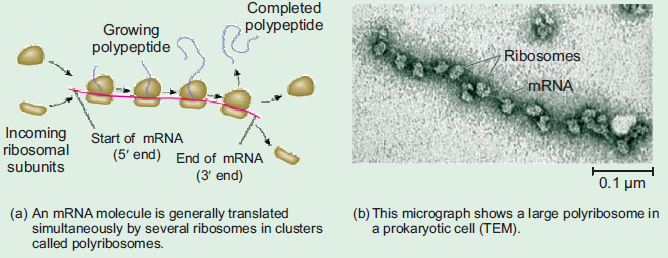
Figure 6.13: Polyribosomes
6.5 TRANSLATION IN EUKARYOTES
Translation in eukaryotes is much more complex than that of the translation in bacteria.
However, the basic process of bacterial translation remains the same in eukaryotes. Some of
the major differences between the bacterial and eukaryotic translations are:
1. Initiation:
(a) The first amino acid that binds on AUG start codon is methionine amino acid rather
than N-formylmethionine.
(b) It has 80S ribosomal initiation complex instead of 70S ribosomal initiation complex;
it is formed by small 40S ribosomal subunit and 60S ribosomal subunit.
(c) There are many more initiation factors (eIFs) in eukaryotes than in bacteria.
(d) A complex of 40S ribosomal subunit + charged Met-t-RNA + several eIFs + eIF
proteins move along the m-RNA and scan for the AUG start codon. This process is
called Scanning Model for initiation. Once the AUG start codon is located, the 60S
ribosomal subunit joints the complex to form 80S ribosomal initiation complex.
2. Elongation: Elongation in eukaryotes requires about nine eukaryotic elongation factors
(eEFs).
3. Termination: The process of termination is similar to that in bacteria. But in the case of
eukaryotes, a single release factor, called eukaryotic release factor 1 (e-RF1), recognizes
all the three stop codons (UAG, UAA, UGA). And the e-RF1 also stimulates the processof termination.
ACTIVITY 4
Watch video clips using the Internet on the process of protein.
Synthesis: Using chart papers of different colours, prepare a model of protein synthesis. You
should be very particular about shapes of structure made like with student of DNA, tRNA. You
may use the following internet links: -orAPPLICATION 6.3
1. Complete the sentence with correct word:
(a) ................... is a DNA dependent polymers.
(b) The base pairs of eukaryotic gene are ............... with the bases of transcribed mRNA.
(c) mRNA coding information for more than one gene is called ...................
(d) ................... is a process of adding poly A tails to pre mRNA.
(e) ................... are removed by RNA splicing.2. What is the particularity of the Transcription in Eukaryotes?
6.6 THE EFFECT OF CHANGE IN GENETIC CODE ON THE STRUCTURE OF
PROTEIN DURING PROTEIN SYNTHESIS
ACTIVITY 5
Make a minilab report to demonstrate how gene mutations affect protein synthesis using a
sequence of bases of one strand of an imaginary DNA molecule. You may use the examples
shown in the text below.
Research and present the findings in journal form on how genetic drugs can be used to stop the
expression of genetic diseases with specific reference to how they may interfere with activities
of nucleic acids in the nucleus and the cytoplasm of the cell. You may look upto research
papers, Internet and magazines
Mutations are changes in genetic codons caused by changes in nucleotide bases. Some mutations
do not have much effect. However, some mutations can have a huge effect on genetic code,
which can eventually affect the proteins they code for. The proteins produced in turn can havea profound effect on cellular and organismal function.
Mutations occur in two ways:
A. A base-pair substitution: It is a change from one base pair to another base pair in DNA.
B. Base-pair insertions or deletions: It is a change in which a base-pair is either incorrectlyinserted or deleted in a codon.
(A) A Base-Pair Substitution

This change in base pair brings changes in the m-RNA codon from one purine to the other
purine. In this case, the m-RNA codon is changed from 5’-AAA-3’ (lysine) to 5’-GAA-3’
(glutamic acid). This is missence mutation.Now look at the changes in DNA from
This change in base-pair in DNA results in change in m-RNA codon from 5′-AAA-3′ (lysine) to
5′-UAA-3′, which is a stop codon. This is a nonsense mutation. It causes premature termination
of polypeptide chain synthesis, thereby releasing shorter polypeptide fragments than the normal
length of polypeptide fragments during translation. These shorter fragments are often nonfunctional.
A silent mutation results from AT-to-GC transition mutation that changes the codon from
5′-AAA-3′ to 5′-AAG-3′. Both of these codons 5′-AAA-3′ to 5′-AAG-3′ specify the same amino
acid, lysine. It is worth mentioning that silent mutation often occurs by changes at the thirdwobble position of a codon. Refer wobble hypothesis in Genetic code.
(B) Base-Pair Insertions or Deletions
ACTIVITY 6
Consider a statement that is made up of the following words each having three letter like genetic
code.
RAM HAS RED CAP
If we insert a letter B in between HAS and RED and rearrange the statement, it would read as
follows:
RAM HAS BRE DCA P
Similarly, if we now insert two letters at the same place, say BE. Now it would read.
RAM HAS BIR EDC AP
Now we insert three letters together, say BIG. The statement would read.
RAM HAS BIG RED CAP
The same exercise can be repeated, by deleting the letters R, E and D, one by one and rearranging
the statement to make a triplet word.
RAM HAS EDC AP
RAM HAS DCA P
RAM HAS CAP
The conclusion from the above exercise is very obvious. Insertion or deletion of one or two
bases changes the reading frame from the point of insertion or deletion. Insertion or deletion of
three or its multiple bases insert or delete one or multiple codon hence one or multiple amino
acids and reading frame remains unaltered from that point onwards, mutations are referred toas frame-shift insertion or deletion mutations.
6.7 EFFECTS OF ALTERATION OF NUCLEOTIDE SEQUENCE
Change in Nucleotide (Mutation) Sequence Leads to Change in Polypeptides
Amino acids (proteins) are the ultimate product of the nucleotide sequence present in genes
(DNA). Thus, any change in the nucleotide sequence of a gene can result into producing
wrong or different polypeptide chain. In other words, gene mutation is a change in sequenceof nucleotides that results in change in the synthesis of polypeptide chains.
One of the best examples is Sickle-cell anaemia. In this disease, the nucleotide “T” in the DNA
sequence is replaced by “A” nucleotide. The minor substitution in the nucleotide sequence is
transcribed as a mutant codon on the m-RNA. And during translation, due to mutant codon
on the m-RNA, valine is synthesized instead of glutamic acid. Valine distorts red blood cellsand cause sickle-cell anaemia. You will be studying it in the next section.
Another example is Albinism. Albinism occurs due to mutation in the gene for tyrosinase, an
enzyme which converts tyrosine to DOPA (dihydroxyphenylalanine) (Figure 6.14). Melanin,
skin pigment, is derived from DOPA. Melanin absorbs light in the ultraviolet (UV) range and
protects the skin against harmful UV radiation from the sun. People with albinism produce
no melanin. Therefore, they have white skin, white hair, eyes with red iris, and they are verysensitive to light.
Figure 6.14: Mutation of tyrosinase gene results in albinism, lack of melanin pigment
Sickel Cell Anaemia
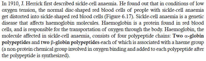
Cause
The mutation causing sickle cell anaemia is a single nucleotide substitution (A to T) in the
DNA of haemoglobin coding gene. The change in a single nucleotide is transcribed as a codon
for valine amino acid (GUG) on the m-RNA instead of glutamic acid (GAG) (Figure 6.15).
Eventually, due to change in the codon, valine amino acid is translated instead of glutamic
acid at the 6th position from N-terminus of the haemoglobin polypeptide chain (Figure 6.16).This defective form of haemoglobin in persons with sickle cell anaemia is referred to as HbS.
Figure 6.15: A single nucleotide substitution in haemoglobin gene resulting into replacement of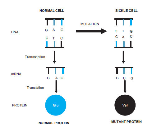
glutamic acid by valine amino acid
Figure 6.16: A diagram showing replacement of glutamine (Glu) by valine (Val) at 6th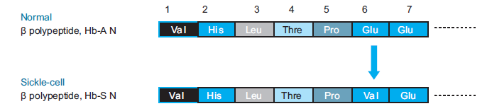
position from N-terminus in the sickled haemoglobin polypeptide
The amino acid valine makes the haemoglobin molecules stick together, forming long fibreswhich convert the normal disc-shape of red blood cells into sickle-shaped red blood cells.
Symptoms
The sickled red blood cells are fragile and break easily, resulting in the anaemia. Normal red
blood cells normally squeeze and pass through blood capillaries smoothly. However, sickled
cells are not flexible and therefore have the tendency to get clogged in capillaries (Figure 6.17).As a result, blood circulation is impaired and tissues become deprived of oxygen. Oxygen
deprivation occurs at the extremities, the heart, lungs, brain, kidneys, gastrointestinal tract,muscles, and joints.
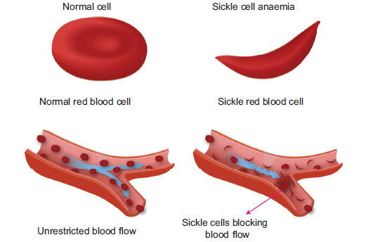
Figure 6.17: Difference between normal and sickle red blood cells
Sickle cell anaemia is an autosomal recessive disorder that affects 1 in 500 African-Americans,
and is one of the most common blood disorders and in the United States. By autosomal disorder,
it means that in order for full disease symptoms to manifest in an individual they must carry two
copies (homozygous genotype = SS, HbS & HbS) of the HbS gene (Figure 6.18). However, the
individuals who are heterozygous (genotype = AS, i.e., HbA and HbS) have what is referred
to as sickle cell trait, a phenotypically dominant trait.
Although heterozygous (AS) individuals are clinically normal, their red blood cells can sickle
under very low oxygen pressure. Their red blood cells may sickle when they are at high altitudesin airplanes with reduced cabin pressure.
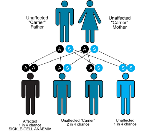
Figure 6.18: A diagram showing sickle-cell anaemia as autosomal recessive disorder
APPLICATION 6.3
1. Complete the sentence with correct word:
(i) ................. distorts red blood cells and cause sickle-cell anaemia.
(ii) ................. people are sensitive to light.
(iii) Mutations are of two types ................. and ................. .
(iv) Sickle cell anaemia is an ................. disorder.2. What are the symptoms and complications of sickle cell anemia?
6.8 SUMMARY
• Genetic Code is the set of rules by which information is encoded in genetic material
(DNA or RNA sequences) is translated into proteins (amino acid sequences) by living cells.
• A codon is made up of three nucleotides or triplets. Out of 64 codons, 61 codons are
sense codons and 3 codons are non-sense codons.
• Genetic code is almost universal; it shows degeneracy.
• It is through genetic code that the genetic information found in m-RNA is translated to
mature functional proteins.
• DNA molecule is a stable structure and replicates accurately in order to avoid any mutation
or change in nucleotide sequences in DNA.
• Transcription is the process of copying information from one strand of DNA into a
single stranded RNA (mRNA).
• A transcription unit in DNA is composed of a promoter, RNA coding sequence, and a
terminator.
• The process of transcription in bacteria includes —
♦ Initiation — a process of initiating transcription where a complex of RNA polymerase
with sigma factor binds at the promoter.
♦ Elongation — the process in which RNA polymerase synthesizes a complementary
RNA sequence of the DNA template strand.
♦ Termination — the process of ending transcription; and it can be carried out either
in rho dependent manner or rho independent manner.
• The process of transcription in eukaryotes involves —
♦ Initiation involves a complex of RNA polymerase II and general transcription factors.
♦ Elongation is similar to that of bacteria. But eukaryotic genes do not have terminator
sequences.
♦ The newly formed pre-m-RNA has to undergo RNA processing.
• Translation is the production of protein molecules (polypeptides) by cellular ribosomes
with the help of information present on the m-RNA.
• The covalent linking of a specific amino acid to the 3’ end of the correct t-RNA by
the enzyme aminoacyl-t-RNA synthetase is called charging of t-RNA.
• Translation in bacteria includes —
♦ Initiation is a stage where m-RNA is bound to the ribosome and positioned itself for
proper translation. It involves three steps.
♦ Elongation is a stage where amino acids are sequentially joined together to form a
polypeptide chain via peptide bonds.
♦ Termination is the process where the newly formed polypeptide chain and the m-RNA
are released from the ribosome.
♦ Polyribosomes are complex of an m-RNA molecule and multiple ribosomes that
are simultaneously translating it. It enables a large number of polypeptides to beproduced faster and efficiently.
• Translation in eukaryotes includes —
♦ Initiation differs from that of bacteria by: the first amino acid is methionine; it has
80S initiation complex; it locates start codon by scanning model for initiation.
♦ Elongation is characterized by the involvement of nine eukaryotic elongation factors.
♦ Termination codes are recognized only by e-RF1.
♦ Change in genetic code is known as mutation.
♦ Mutation is of two types: A base-pair substitution and base-pair insertions or deletions.
♦ Some mutations do not have much effect such as silent mutation. However, some
mutations can have a huge effect on genetic code such as frameshift mutation.
♦ Any change in the nucleotide sequence of a gene can result into producing wrong
or different polypeptide chain. The outcomes can be detrimental to the affected
organisms.♦ Example: Sickle cell anaemia; albinism.
6.9 GLOSSARY
• Albinism: It is a disease caused by alteration of nucleotide sequence. It occurs due to
mutation in the gene for tyrosinase, an enzyme which converts tyrosine to DOPA.
• Charging t-RNA: It is the process of attaching the correct amino acid to t-RNA with
the help of aminoacyl-t-RNA synthetase enzyme.
• Degeneracy: The coding of the same amino acid by multiple codons. Example:UUU
and UUC codons code for the same specific phenylalanine amino acid.
• Frameshift mutation: It is a gene mutation in which addition or deletion of one basepair
shifts the m-RNA’s downstream reading frame by one base so that incorrect amino
acids are added to the polypeptide chain after the mutation site.
• Missense mutation: It is a gene mutation in which change in base-pair (nucleotide
sequence) of DNA results in change in an m-RNA codon, which codes for different
amino acid.
• Polyribosomes: They are complex of an m-RNA molecule and multiple ribosomes that
are simultaneously translating it.
• Rho protein: It is a protein that binds to the terminator sequence and RNA polymerase
to end the process of transcription in bacteria.
• RNA processing: It is the process where pre-m-RNA undergoes modification to become
a functional m-RNA.
• Sickle cell anaemia: The mutation causing sickle cell anaemia is a single nucleotide
substitution (A to T) in the DNA of haemoglobin coding gene. The change in a single
nucleotide is transcribed as a codon for valine amino acid (GUG) on the m-RNA instead
of glutamic acid (GAG).
• Stop codon: It is a nonsense codon, meaning it does not specify any amino acid. It rather
stops translation.
• Transcription: It is the process of copying information from one strand of DNA into a
single-stranded RNA (m-RNA).
• Translation: It is the production of protein molecules (polypeptides) by cellular ribosomes
with the help of information present on the m-RNA.
• Translocation: It is a process in which the m-RNA is moved by a distance of three
nucleotides (codon) to bring the next codon on the ribosome (A site).
• Wobble hypothesis: The reading of two or more different codons by the same t-RNA
molecule. Example: The two different leucine codons (CUC, CUU) can be read by thesame leucine t-RNA molecule, contrary to regular base-pairing rules.
END UNIT ASSESSMENT 6
I. Choose whether the following statements are True (T) or False (F)
1. Genetic code is composed of A, C, G, and T nucleotides.
2. The main role of t-RNA is to decode the codons on m-RNA.
3. The main role of m-RNA is to carry genetic information in a series of codons.
4. Ribosomes are made up only by proteins.
5. TATA box is found 10 base pairs downstream from +1 start site.
6. RNA polymerase doesn’t need any factor to initiate transcription.
7. Hairpin loop structure is formed in rho dependent terminators.
8. Pre-m-RNA needs to undergo RNA processing to become functional m-RNA.
9. In prokaryotes, the first amino acid to bind at the P-site is N-formylmethionine.
II. Multiple Choice Questions
1. Sickle-cell anaemia is caused due to change in
(a) Nucleotide T by A in the DNA (b) Nucleotide A by T in the DNA.
(c) Nucleotide G by U in the DNA (d) Nucleotide U by G in the DNA.
2. Which of the following are the characteristics of genetic code?
(a) Triplet code (b) Almost Universal
(c) Nonoverlapping (d) All of these.
3. The wrong stop codon is
(a) UUA (b) AUU
(c) UAG (d) UGA
4. The word “wobble” means
(a) Jumping (b) Synthesis
(c) Unsteady (d) Stable
5. Which of the following is located in the upstream of a bacterial gene?
(a) Promoter (b) Terminator
(c) RNA-coding sequence (d) None of the above
6. RNA polymerase I doesn’t catalyze the synthesis of
(a) 28S molecule (b) 5S molecule
(c) 5.8S molecule (d) 18S molecule
7. Which of the following components is not involved in eukaryotic transcription?
(a) General transcription factors (b) RNA polymerase II
(c) Sigma factor (d) Enhancers
8. Which of the following are related to eukaryotes?
(a) RNA Processing (b) Introns & exons
(c) Poly (A) tail (d) All of these
9. Scanning model is related to
(a) Charging of t-RNA (b) Initiation
(c) Elongation (d) Termination
10. In sickle-cell anaemia, valine amino acid replaces
(a) Serine (b) Threonine
(c) Glutamic acid (d) Tyrosine
III. Long Answer Type Questions
1. In your own words, state features of a genetic code.
2. Describe the process of transcription in bacteria.
3. Using diagrams, compare the process of bacterial and eukaryotic transcriptions.
4. Describe the process of translation in bacteria.
5. State the roles of t-RNA, m-RNA, and ribosomes in the formation of polypeptides.
6. What is sickle-cell anaemia? Explain its cause and symptoms.
7. In Genetic code (Figure 6.2), CUU codes for leucine (leu) amino acid. If we change
the third letter of CUU i.e., U with C, A, G, which amino acid will the changedcodon code for during translation?
8. UUU codon codes for phenylalanine (Phe). If we change the third base “U” with
C, A, G, which amino acid will the changed codon code for during translation?
9. During translation, what will happen if there is mutation on a codon UAU (codes
for tyrosine) where the third letter “U” is replaced by either one of the bases A or Gor C?
10. State that ribosomes provide surface area for the attachment of mRNA during
polypeptide synthesis.
11. Construct a flow chart, in proper sequence, for the stages of transcription and
translation.
12. Using the evidence, predict the effect of change in genetic code on the structure of
the protein manufactured during protein synthesis.
13. Briefly describe the alteration of nucleotide sequence attacking the deadly AIDS.Also show how it can be an essential step towards poverty alleviation.
Unit 7 Autotrophic Nutrition
Key Unit Competence
To be able to describe the process of photosynthesis and explain the various environmentalfactors that influence the rate of photosynthesis.
LEARNING OBJECTIVES
At the end of this unit, the learner will be able to:
• State and explain the types of autotrophic nutrition.
• Explain the role of light in autotrophic nutrition.
• State the pigments involved in light absorption.
• Appreciate the importance of photosynthesis as an energy transfer process that produces
complex organic compounds using light energy absorbed by chloroplast pigments.
• Recall the structure of the leaf in relation to photosynthesis.
• Use their knowledge of plant cells and leaf structure from the section on cell structure while
studying photosynthesis.
• State the sites and stages of photosynthesis in chloroplasts.
• Describe the role of chloroplast pigments (chlorophyll a, chlorophyll b, carotene and
xanthophylls) in light absorption in the grana.
• Describe the relationship between the structure and function in the chloroplast, using
diagrams and electron micrographs.
• Interpret absorption and action spectra of chloroplast pigments.
• Carry out an investigation of limiting factors.
• outline the three main stages of the Calvin cycle.
• Describe and outline the conversion of the Calvin cycle intermediates to carbohydrates,
lipids and amino acids and their uses in the plant cell.
• Relate the anatomy and physiology of the leaves of C4 and CAM plants to high rates of
carbon fixation and low rates of transpiration.
• Explain the term limiting factor in relation to photosynthesis and the effects of the changes
in the limiting factors on the rate of photosynthesis.
• Apply knowledge and understanding of limiting factors to increase crop yields in protected
environments, such as glasshouses.
• Investigate the effect of light intensity or light wavelength on the rate of photosynthesis.
• Acknowledge that environmental factors influence the rate of photosynthesis and investigation
shows how they can be managed in protected environments used in crop production.• Differentiate between C4, CAM and C3 plants during carbon dioxide fixation.
INTRODUCTORY ACTIVITY
Rwanda has pledged to plant two million hectares of trees by 2020. Adopting a forest landscape
restoration (FLR) approach, the country has committed to the Bonn Challenge: a global
aspiration to restore 150 millioan hectares of the world’s deforested and degraded lands by
2020 and 350 million hectares by 2030.Can you advocate for planting more trees in Rwanda? Why?
ACTIVITY 1
The term ‘autotroph’ consists of two words ‘auto’ and ‘troph’. ‘Auto’ means ‘self ’ and ‘troph’
means ‘nutrition’. Search from the internet and using books about autotrophic nutrition. Makereport on the same and present it to the class.
All organisms require macromolecules like carbohydrates, proteins and fats for their growth and
development. Some organisms produce these organic compounds from inorganic sources on
their own. Such organisms are called autotrophs or producers and the process of synthesizing
complex compounds from simple inorganic sources is called autotrophic nutrition. While others
including humans are heterotrophs or consumers, which depend on autotrophs for source of
chemical energy. Green plants are autotrophs and require chlorophyll, sunlight, oxygen andminerals for preparing their own food.
7.1 TYPES OF AUTOTROPHIC NUTRITION
Chemoautotrophic: An autotrophic nutrition where organisms get energy from oxidation ofchemicals, mainly inorganic substances like hydrogen sulphide and ammonia.
Photoautotrophic: An autotrophic nutrition where organisms get energy from sunlight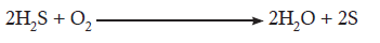
and convert it into usable form like sugars. Green plants and some bacteria like, green
sulphur bacteria can make their own food from simple inorganic substances by a processcalled photosynthesis.
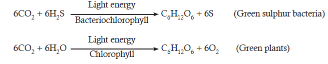
7.2 TESTS FOR STARCH AND FOR OXYGEN
ACTIVITY 2
Aim: To show that starch is important for photosynthesis in terrestrial plants.
Materials Required: Two potted plants, ethyl alcohol, iodine solution, sauce pan and burner.
Procedure: Take two potted plants. Keep one in dark and other in well illuminated condition. After
24 hrs, take leaves from each plant. Boil ethyl alcohol in a sauce pan and dip leaves for 30 s. Place
them in a beaker of ethyl alcohol until they turn white. Take leaves out and cover with iodine
solution.
Observation: Leaf taken from well illuminated condition plant turned bluish-black while other
tested negative.
Discussion: This shows that starch is present in terrestrial plants that carry out photosynthesis.
After performing the experiment, try to answer the following questions:
• What is the role of ethyl alcohol in experiment?• What made leaves turn blue-black?
ACTIVITY 3
Aim: To show that oxygen is required by aquatic plants.
Materials Required: Beaker, funnel, aquatic plant Elodea/Hydrilla and boiling tube.
Procedure: Take a few fresh twigs of aquatic plant Elodea/Hydrilla with one
end intact and put in a beaker full of water. Keep an inverted funnel on the
plant such that all plants are inside the funnel and their cut ends facing up. Now
take a boiling tube full of water and invert it on the funnel. Put the wholeapparatus in sunlight
Observation: Observe the air bubbles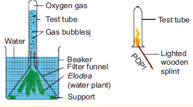
accumulating in the boiling tube.
Discussion: The air shows positive test for oxygen gas.
After performing the experiment, answer the following questions:
• What is the positive test for oxygen?• How is oxygen available for aquatic plants?
7.2.1 Importance of Autotrophic Nutrition
Photosynthesis is a process by which plants and other organisms, such as algae and bacteria
synthesize their own food using the energy of light for their growth and development. The food
produced by plants is in the form of carbohydrates. In preliminary studies, Julius von Sachs
proposed that glucose is the first product of photosynthesis. It is stored in chloroplasts within
plant cells. It provides energy in the form of food to organisms that feed on plants. It has been
rightly said “ALL FLESH IS GRASS”, as all organisms (herbivores, carnivores and omnivores)
are directly or indirectly dependent on plants as a source of energy. It is the means by which
solar energy is captured by plants for use by all organisms. In 1782, Jean Senebier proved that
green plants can produce oxygen in presence of light and carbon dioxide. It is the single most
important biological process that can replenish oxygen which is required for existence of all
other organisms. Have you ever thought about what will happen if there is no photosynthesis? This
unit focuses on the photosynthetic machinery, the reactions in this physiochemical process andthe factors affecting photosynthesis.
7.3 ADAPTATION OF PLANTS FOR PHOTOSYNTHESIS
Photosynthesis occurs not only in eukaryotic organisms such as green plants but also in
prokaryotic organisms like blue green algae and green sulphur bacteria.
In higher plants, photosynthesis occurs in the green part of the plant (Figure 7.1). Leaves are
adapted to carry photosynthesis efficiently. Most leaves are broad and flat to capture maximum
light. Also the bifacial nature of the leaf allows it to collect incident light on the upper surface
and diffuse light on the lower surface. The photosynthetic tissue is located between the upper
and lower epidermis. It consists of one to three layers of compactly arranged, elongated and
cylindrical palisade mesophyll cells, and loosely arranged, irregular and isodiametric spongy
mesophyll cells. In monocotyledonous leaf, there is no distinction of palisade and spongyparenchyma.
Figure 7.1: (A) Structure of leaf showing photosynthetic cells; (B) EM of chloroplast cells;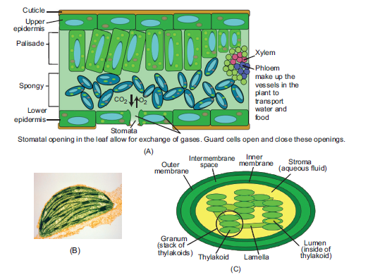
(C) Sectional view of chloroplast.
The mesophyll cells in leaves contain large number of chloroplasts that transform light energyinto ATP and NADPH which are then used to convert CO2 into sugars
7.4 STRUCTURE OF CHLOROPLAST–THE ORGANELLE FORPHOTOSYNTHESIS.
Chloroplast is the photosynthetic machinery. It is a double membrane organelle that contains
series of parallel membranes called thylakoids or lamellae, suspended in fluid like matrix called
stroma. The thylakoids are flattened discs arranged in stacks called grana. In a typical chloroplast
as many as 40-60 grana may be present and each granum may contain 2-100 thylakoids. The
stroma contains DNA, ribosomes, soluble proteins and enzymes, while pigments are confined
to thylakoids. Thylakoids have large surface for absorption of light and the space within them‘lumen’ allows rapid accumulation of protons.
7.4.1 Pigments of Chlorophyll
A pigment is a substance that absorbs light of different wavelengths. Pigments are involved
in absorption of light of certain wavelength. While some wavelengths are absorbed, other arereflected or scattered, which imparts them colour. The absorbed wavelength of light has the
correct energy to excite specific transitions of electrons in the pigments. Photosynthesis depends
on light absorption by pigments in leaves. However, it can be carried out in isolated chloroplastbut not in isolated pigments.
ACTIVITY 4
Aim: To perform chromatography to separate and identify chloroplast.
Materials Required: Whatmann’s filter paper No.1, mortar and pestle and
solvent.
Procedure: Cut a vertical strip (10 cm) × (2.5 cm) of Whatmann’s filter
paper No.1. Make it V-shaped at one end. Draw a horizontal line with a
pencil (not pen) about half an inch from the bottom.
Make leaf extract by crushing 20 g leaves in 20 mL acetone. With the help of capillary
tube load
a small drop of leaf extract at the
centre of pencil
mark and air dry. Repeat the previous
step
for 5-6 times. Insert paper strip in chromatographic
chamber pre-saturated with solvent
(1:9:: benzene:petroleum ether) such that only tip of paper is dipped into solvent. Do not dip the loaded
pigment into solvent. Allow it to run for few (1-2) hours. Take out strip and mark solvent front and
different coloured separated pigments.
Observation: Different bands of different colours are formed.
Discussion: Discuss and label the different bands formed on the filter paper. Also answer whythere are different bands formed.
Chlorophyll a is the major pigment involved in trapping light energy. It is the principal pigment
involved in photosynthesis. It is of universal occurrence. It is a large molecule composed of four
pyrrole rings with Mg at centre, and a long hydrocarbon phytol chain. It absorbs maximum
wavelengths of 430 nm and 660 nm.
Chlorophyll b constitutes one-fourth of the total chlorophyll content. It has a similar structure
as that of Chlorophyll a, except that the –CH3 group in chlorophyll a is replaced by –CHO
group in chlorophyll b. It absorbs maximum wavelengths of 460 nm and 680 nm.
Carotenes are tetraterpenes or polyunsaturated hydrocarbons containing 40 carbon atoms and
variable number of hydrogen atoms and no other elements. b-carotene is the common form
found abundantly in orange, yellow and green fruits and vegetables. Carotenes protect plant
against photo-oxidation.
Xanthophylls are yellow coloured pigments. They are structurally similar to carotenes, butcontain oxygen atoms. These are more common in young and etiolated leaves.
7.4.2 Absorption and Action Spectra
A plot showing absorption of light of different wavelengths of a pigment is called absorptionspectrum.
Figure 7.2: (a) Absorption spectrum of chloroplast pigments;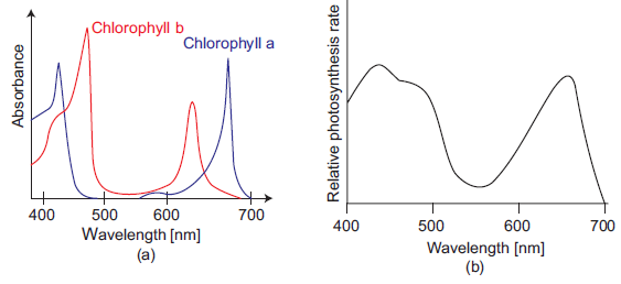
(b) Action spectrum of green plants.
Each pigment absorbs a specific wavelength. We can plot an absorption spectrum showing
the ability of pigments to absorb lights of different wavelengths. From Figure 7.2(a), it can be
concluded that chlorophyll a and b show absorption peaks at blue and red light. On the other
hand, action spectrum is the plot of graph depicting the rate of a light sensitive process at
different wavelength of light. The action spectrum of photosynthesis shows that most of the
photosynthesis also takes place in blue and red light. The absorption spectrum of a pigment
when compared with action spectrum of photosynthesis, gives the function of the pigment.
Therefore, it can be concluded that chlorophyll a is the chief photosynthetic pigment. The other
pigments like chlorophyll b, carotenes and xanthophylls are called accessory pigments and
form the antenna complex. They collect light of different wavelength and transfer it to reaction
centre (basic model of energy transfer). This is called Light Harvesting Complex (Figure 7.3).LHC is made up of hundreds of pigment molecules bound to proteins.
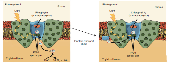
Figure 7.3: Light harvesting complex
In 1985, R Huber, H Michael and J Dissenhofer crystallized and worked on of light harvesting
complex of Rhodobacter, and got Nobel Prize in 1988. In green plants, pigments are organized
into two discrete photochemical complexes: Photosystem I (PSI) and Photosystem II (PSII),
named after their sequence of discovery. Each photosystem has all the pigments which help
in more efficient absorption of light. The chlorophyll a molecule forms the reaction centre.
In PSI, the reaction centre chlorophyll a molecule has absorption maxima at 700 nm, hence
called P700 and in PSII it has absorption maxima at 680 nm, called P680.
7.4.3 Role of Light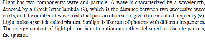
When a light photon is absorbed, an electron is excited from pigment molecule to a higher
energy level (triplet state). It remains there for 10–9 s and then fall to ground state. Sometimes,
it can emit the energy in the form of light and heat as it reaches the ground state. This process
is called fluorescence. When electron remains at triplet state for more than 10–9 s and then
comes back to ground state, the energy is lost in the form of heat and light. This happens evenafter the source is put off. Such a process is called phosphorescence.
1. Complete the sentences by correct terms:
(a) Chloroplast is a double membrane organelle that contains a parallel membrane called
................. .
(b) ................. is the yellow coloured pigment found in young and etiolated leaves.
(c) Most of the photosynthesis takes place at ................. and ................. light.
(d) PSII has absorption maxima at .................2. The graph below shows the strictum of two pigments in the chlorophyll
a.Is that absorption or action spectrum?
b. Identify the pigment X and Y.
7.5 MECHANISM OF PHOTOSYNTHESIS
ACTIVITY 5
Aim: Investigation to determine the effect of light intensity or light wavelength on the rate of
photosynthesis using a redox indicator.
Materials Required: Pestle and mortar, centrifuge, filter paper, test tube, sucrose buffer, DCPIP dye.
Procedure: Crush the interveinal portion of leaves in chilled sucrose buffer using pestle and
mortar. Filter it and centrifuge the filtrate at 200-300 rpm for 2-3 minutes. Collect the supernatant
and again centrifuge at higher speed above (5000 rpm). Collect pellet and dissolve in small amount
of buffer. Take a test tube and to each test tube add sucrose buffer, chloroplast extract, DCPIP dye
according to following table and keep in different light conditions for 10-20 minutes.Observation:
Discussion: Discuss the change in colour obtained. Also state the role of DCPIP in the experiment.
The process of photosynthesis takes place in two major phases: Light reaction and Dark
reaction. Both these phases are interdependent. Green plants convert light energy to chemical
energy. The light energy is used to transfer electron from water to reduce NADP, which in
reduced form, can further be used to fix carbon dioxide. Thus, carbon fixation does not
need light directly and is called dark reaction. Both light and dark reactions take place inchloroplasts.
Table 7.1
Light Reaction
Photochemical phase or Light reaction in which solar energy is trapped by chlorophyll and
stored in the form of chemical energy of ATP and as reducing power in NADPH2. The ATP
and NADPH2 together constitute the assimilatory power of the plant. Oxygen is evolved in
the light reaction by splitting of water (photolysis of water).
Mechanism of Light Reaction
The two photosystems (PS I and PS II) absorb different wavelengths of light. The light energy
absorbed anywhere in the harvesting zone of a photosystem is passed to its photocentre. When
the photocentre acquires a sufficient quantum of energy, it emits an electron. This electron with
high potential energy moves down to an electron transport chain, and results in the formation
of ATP. Thus, the primary function of the two photosystems which interact each other, is to
trap light energy and converts it to the chemical energy (ATP). This chemical energy stored inthe form of ATP is used by the living cells.
7.6 DARK REACTION/CARBON ASSIMILATION/CARBON FIXATION
7.6.1 Calvin Cycle
It is carbon assimilation process which utilizes assimilatory power generated from light reaction
to produce sugars. It occurs in stroma of chloroplasts. Melvin Calvin got Nobel Prize for his
outstanding work on carbon assimilation. Melvin Calvin, Andrew Benson and James Bassham
gave the Calvin cycle of dark reaction. They used autoradiography to detect path of cycle,
and chromatography to separate constituents. The first product that showed radioactivity
was a three carbon (3-C) compound Phosphoglyceric acid (PGA) and hence the cycle is alsocalled C-3 cycle.
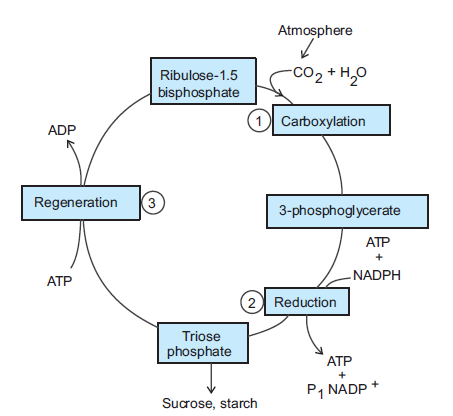
Figure 7.4: Calvin cycle
The process of carbon assimilation can be described under three stages: carboxylation, reduction
and regeneration (Figure 7.4).
Carboxylation: It is the process of fixation of carbon in stable organic intermediate,
phosphoglyceric acid. This reaction is catalyzed by an enzyme called RuBPcarboxylase-
oxygenase (RUBISCO). Rubisco-bis-phospahte (RuBP) is the initial acceptor or substratefor dark reaction.
Reduction or Glycolytic Reversal: It is the process involving reduction of carbon. It is
a multistep process that utilizes 12 ATP molecules and 12 NADPH for release of one
molecule of glucose. The glucose can further be converted into starch for storage or sucrosefor transport.
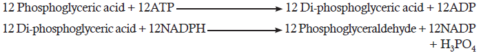
Regeneration: This process requires 6 ATP molecules to regenerate 6 molecules of RuBP,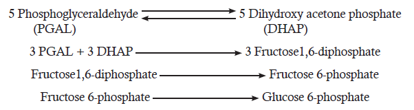
which is crucial for continuity of Calvin cycle.
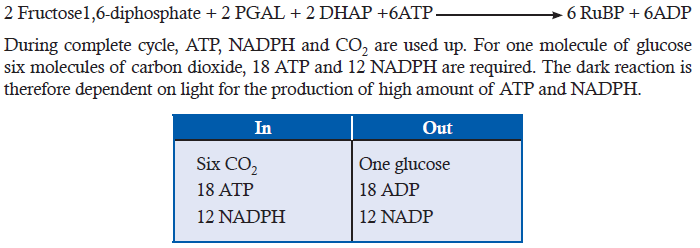
Another important requirement is high concentration of CO2. The efficiency of photosynthesis
declines at low concentration of CO2. This is because the enzyme RUBISO has low affinity
with carbon dioxide as compared to oxygen. At low CO2 concentration, RUBISCO catalyzes
the reaction between RuBP and oxygen. The oxygenation of RuBP in presence of light and
oxygen is called Photorespiration. It occurs in chloroplast, peroxisome and mitochondria.
It is a wasteful process as during this process carbon dioxide is released and efficiency of
photosynthesis decreases (Figure 7.5).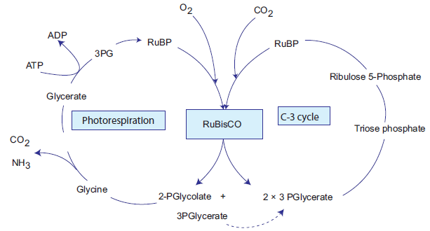
Figure 7.5: Photorespiration and C-3 cycle
7.6.2 Hatch and Slack Cycle
In some temperate plants such as wheat, rice, potato and bean only Calvin cycle occurs. Such plants
are called C-3 plants. While in some other plants dual carboxylation takes place: (1) carboxylation
of phosphoenol pyruvate (PEP) and (2) carboxylation of RuBP. Such plants are called C-4 plants
e.g. maize, sugar cane and sorghum. In these, the first product formed during carbon dioxide
fixation is a four carbon compound oxalo acetic acid (OAA). C-4 plants have special type of leaf
anatomy called Kranz Anatomy. They have special large cells around vascular bundles called
bundle sheath cells. These are characterized by having large number of chloroplasts, thick walls
and no intercellular spaces. The shape, size and arrangement of thylakoids in chloroplasts arealso different in bundle sheath cell as compared to mesophyll cell chloroplasts.
Table 7.2
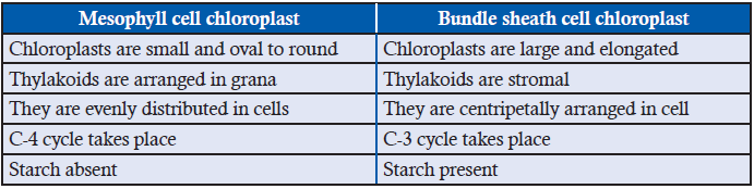
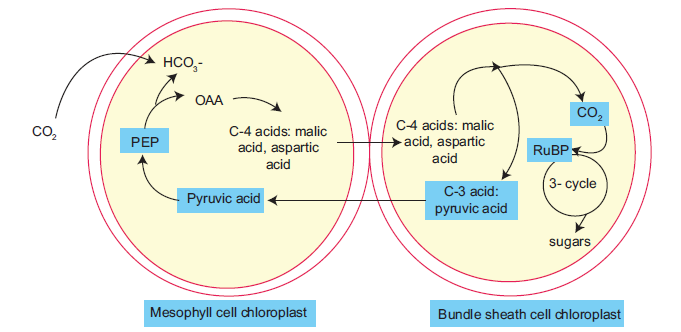
Figure 7.6: Hatch and slack pathway
The pathway followed by C-4 plants is called C-4 cycle or Hatch and Slack pathway. This was
discovered by Hatch and Slack in sugar cane. The primary CO2 acceptor is a 3-carbon molecule
phosphoenol pyruvate (PEP). The reaction is catalyzed by PEPcarboxylase or PEPcase in
mesophyll cell chloroplast. It forms 4-carbon compounds like OAA, malic acid or aspartate,
which are transported to the bundle sheath cells. In bundle sheath cells, these acids are broken
down to release CO2 and 3-carbon molecule. The 3-carbon molecule is transported back to
mesophyll cells and converted to PEP again, while CO2 enters into C-3 cycle to form sugars
(Figure 7.6). C-4 plants are more efficient than C-3 plants as in C-4 plants, photosynthesis canoccur at low concentration CO2 and photorespiration is negligible or absent.
Table 7.3
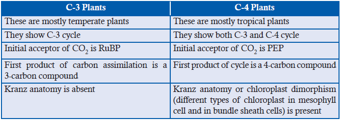
APPLICATION
1. Complete with appropriate terms:
(a) Photorespiration occurs in ........................, ..................... and ........................ .
(b) C4 plants have special type of leaf anatomy called ........................ .
(c) In C3 plants, initial acceptor of CO2 is ........................ .
(d) Hatch and Slack pathway occurs in ........................ and ........................ of chloroplast.2. Giving examples, write short note on C4 plants.
7.7 FACTORS AFFECTING PHOTOSYNTHESIS
Aim: To investigate the effect of carbon assimilation on the rate of photosynthesis.
Materials Required: Elodea plant, glass rod, sodium bicarbonate.
Procedure: Take a fresh, healthy twig of Elodea plant with one end intact and tie it gently to a
glass rod. Put the glass rod with plant in a boiling tube containing
water and add 1mg/mL sodium bicarbonate and keep it in
moderate light condition. Note the numbers of bubbles escaping
from cut end per minute. Again add same amount of sodium
bicarbonate and note the number of bubbles escaping from cut
end per minute. Do you find the number of bubbles increasing?
Repeat this step until bubbles escaping per minute do not
increase. Then take set up under high light intensity and note the
numbers of bubbles.
Observation: The bubbles evolved gradually.Discussion: Discuss what the bubbles are and why they evolved.
Carbon assimilation is directly related to the productivity of plants. It carries implications for the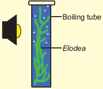
sustainability of the human population. The total carbon assimilation is known as Gross primary
productivity and the one available for increase in biomass is known as net primary productivity.
Net primary productivity is determined by deleting loss due to respiration by plants. It is the
biomass available for animals. By studying factors affecting photosynthesis, one can learn to
manage world resources in time. The rate of photosynthesis can be influenced by many factors
like number, size, orientation and age of leaf, sunlight, temperature, carbon dioxide and water.
However, when several factors can affect a process, the rate of reaction is governed by the factor
which is limiting. This is called Blackman’s (1905) law of limiting factor. For example, despite
the presence of green leaf, optimal carbon dioxide, the plant may not photosynthesize if light
intensity is very low. Thus, light behaves as limiting factor and controls the rate of photosynthesis(Figure 7.7). Hence, rate will be determined by the factor available at sub-optimal level.
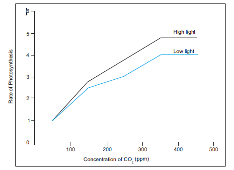
Figure 7.7: Graph depicting Blackmann’s law of limiting factor.
7.7.1 External Factors
ACTIVITY 7
Aim: To show effect of carbon dioxide on the rate of photosynthesis.
Materials Required: Elodea, beaker, NaHCO3, lamp.
Procedure: Place a pond weed Elodea upside in a test tube containing water at 25°C. Place the
tube in a beaker of fresh water. Place excess sodium bicarbonate (NaHCO3) in the water to
give a constant saturated solution of CO2. Place the lamp at a fixed distance from the plant.
Maintain the room temperature at 20°C. Count the number of oxygen bubbles given off by the
plant in a one minute period.
Observation: The bubbles are formed of oxygen.Discussion: Discuss why was NaHCO3 added to water.
CO2 concentration: Carbon dioxide is the inorganic substrate for photosynthesis. Increase inconcentration up to 0.05% in atmosphere can cause an increase in CO2 fixation. Carbon dioxide
is the major limiting factor, especially in C-3 plants; C-4 plants are more productive even at low
concentration of CO2. Nevertheless, both C-3 and C-4 plants show increase in rate of photosynthesis
at high CO2 concentration and high light intensities. The fact that C-3 plants respond to higher
CO2 concentration by showing increased rates of photosynthesis leading to higher productivity
has been used for some green house crops such as tomatoes and bell pepper. They are allowed togrow in carbon dioxide enriched atmosphere as in glasshouses leading to higher yields.
ACTIVITY 8
Aim: To show the effect of light intensity on photosynthesis in terrestrial plants.
Materials Required: Spinach, sodium bicarbonate solution, detergent, lamp.
Procedure: Take out uniform size discs from fresh leaves of spinach. Place discs in 0.2%
sodium bicarbonate solution and a drop of liquid detergent in a syringe. Plunge out air present
in between tissue, such that intercellular spaces are occupied by sodium bicarbonate and all leaf
discs sink to bottom. Then put leaf discs in the beaker containing water exposed to on lamp.
Count the number of leaves that then float on surface at regular interval of time. Similarly,
repeat the experiment with increasing light condition.
Observation: Count and note the number of leaves floating.Discussion: Discuss why the leaves floated.
ACTIVITY 9
Aim: To show the effect of light intensity on aquatic plants.
Materials Required: Elodea, glass rod, sodium bicarbonate.
Procedure: Take a fresh, healthy twig of Elodea plant with one end intact and tie it gently to a
glass rod. Put the glass rod with plant in a tube or jar containing water and a pinch of sodium
bicarbonate. Keep it in under a light source at a distance of 50 cm/ low light condition. Note
the numbers of bubbles escaping from cut end per minute. Place the apparatus at distance of
30 cm from the light source and count the number of bubbles evolving per minute. Similarly,
place the apparatus at variable distances from light source.
Observation: Count the number of bubbles evolving per minute.Discussion: Discuss any change in the number of bubbles.
Light: Light is an important factor to carry out photosynthesis. It is rarely a limiting factor
in nature as photosynthesis can occur even at low light intensities. There is a direct relation
between light and CO2 fixation. With increase in light intensity the rate of photosynthesisincreases. However, at higher light intensities, rate does not increase linearly but light
saturation occurs. At very high light intensity, there is breakdown of chlorophyll molecules
called photo-oxidation and the rate of photosynthesis decreases. The quality of light and
time of exposure also governs photosynthesis. Green plants show high rate of photosynthesisat red and blue light.
ACTIVITY 10
Aim: To show effect of temperature on photosynthesis.
Materials Required: Elodea plant, boiling tube, sodium bicarbonate.
Procedure: Take a fresh, healthy twig of Elodea plant with one end intact and tie it gently to a
glass rod. Put the glass rod with plant in a boiling tube containing water and a pinch of sodium
bicarbonate. Keep it in under moderate light condition. Note the temperature and numbers of
bubbles escaping from cut end per minute. Heat or cool the water in a boiling tube.
Observation: Count the number of bubbles at different temperatures.Discussion: Discuss the change in number of bubbles.
Temperature: The dark reactions are dependent on temperature as they are enzymatic. Rate
of photosynthesis is best at optimum temperature. Different plants have different temperature
optima that also depend on their habitats.
Water: Only about 1% of water absorbed by plants is used in photosynthesis. It is an
important factor for various metabolic processes in plant. Water may not have direct affect on
photosynthesis even though it is one of the reactants in light reaction. In water stress plants
wilt and their stomata close. Thus reducing availability of carbon dioxide and decreasing
the rate of photosynthesis. Water stress will also alter the hydration of enzymatic proteins,
affecting their activities.
Oxygen concentration: Atmospheric oxygen content affects photosynthesis directly or indirectly.
The decrease in rate of respiration at high oxygen concentration was first observed by O. Warburg
in 1920 in Chlorella. The phenomenon is called the Warburg effect.
Chemical pollutants: Plant growth has been adversely affected by accumulation of various
undesirable chemicals. Heavy metals such as lead, mercury, cadmium seem to be affecting
photosynthesis through stomata closure. Air pollutants like SO2, NO2 and O3 are also known
to affect photosynthesis at higher concentrations.
7.7.2 Internal Factors
Adaptation of leaf: Leaves are arranged on plants to minimize overlapping. The shape, size,age and orientation of leaf influences the absorption of light and thus effects photosynthesis.
Most leaves are broad for more absorption of light. The anatomy of the leaf is also highly
specialized for absorption of light. The epidermis is transparent and also acts as convex lens
to focus and intensify light reaching mesophyll cells for maximum absorption. The palisade
layer also helps in absorption of more light. Presence of hairs, salt glands and epicuticular waxincrease the reflection of light and thereby reducing the absorption.
Absorption of carbon dioxide is also dependent on leaf surface area and number of stomata.
Spongy parenchyma has large intercellular space so that carbon dioxide can easily diffuse.
Opening and closing of stomata is yet another factor that governs photosynthesis as the
exchange of gases is affected when stomata close. In some succulent plants such as Bryophyllum,
Kalanchoe, stomata open during night and close during day to reduce the rate of transpiration.
Such plants have special mechanism for photosynthesis called Crassulacean Acid Metabolism
(CAM), where CO2 fixation takes place in different time (day and night) as per availability
of carbon dioxide and light (Figure 7.8). CO2 is taken up by the plant during night time as
stomata open during night. It is fixed and converted to malic acid, which is stored in vacuole.
During day time when assimilatory power is available, malic acid is released from vacuole
and reverted back to pyruvate by releasing carbon dioxide. This CO2 then enters C-3 cycle
and assimilated. Pyruvate is stored in chloroplast in the form of starch and released duringnight time through glycolysis.
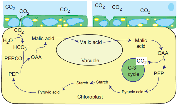
Figure 7.8: Crassulacean acid metabolism in CAM plants
Fill in the columns 2 and 3 in this table to highlight the differences between C3 and C4plants.
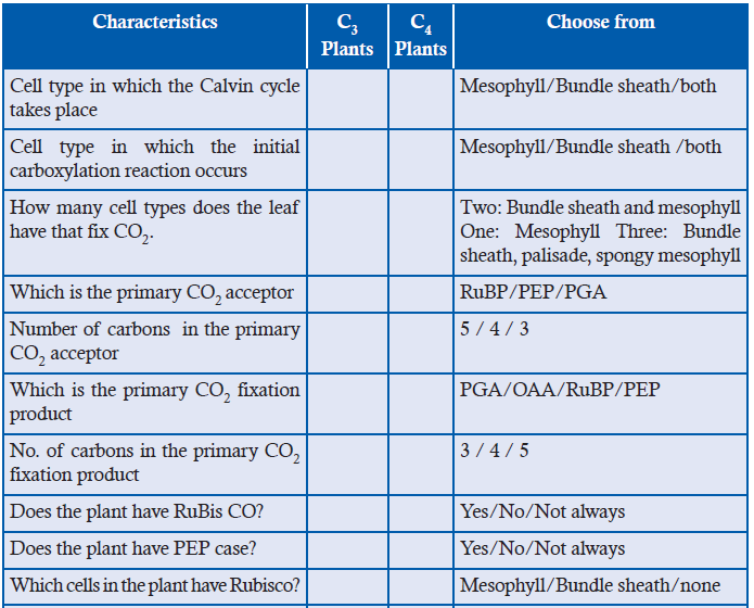
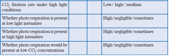
7.8 SUMMARY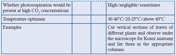
• Organisms that are autotrophic can make their own food from inorganic substances with
help of energy.
• Photosynthesis is the process where the source of energy is light. It is carried out by green
plants, algae and some bacteria.
• Photosynthesis takes place in green parts of a plant, mainly leaves. Within leaves,
chloroplasts in mesophyll cells are the site of photosynthesis.
• Photosynthesis has two stages: light reaction and dark reaction. Light reaction is a
photochemical reaction, in which light energy is absorbed by the pigments present in
antenna molecules of light harvesting complex. While, in dark reaction carbon is reduced
in the stroma of chloroplast.
• Chlorophyll, a molecule is the reaction centre which has two special forms PSI and PSII
with absorbance maxima at 700 nm and 680 nm, respectively.
• In temperate plants, C-3 cycle takes place with the help of enzyme RUBISCO. The C-3
cycle includes: carboxylation, reduction and regeneration. In some tropical plants, C-4
cycle takes place.
• C-4 cycle includes dual carboxylation that takes place in mesophyll cells chloroplast and
bundle sheath cell chloroplasts.
• Various environmental factors such as light, temperature, carbon dioxide concentration,
oxygen concentration and air pollutants are responsible for the plant productivity onaccount of photosynthesis.
7.9 GLOSSARY
• Absorption spectrum: Absorption spectrum is the graph plot that shows the measure
of absorption of radiation of different wavelengths.
• Action spectrum: Action spectrum is the graph plot that shows rate of physiological
activity at different wavelengths of light.
• ATP synthase: It is an important lipid binding protein. It is used to generate ATP from
ADP. It is present on thylakoid membrane.
• Autotrophs: An autotroph is an organism that produces complex organic compounds
such as carbohydrates, fats and proteins from simple inorganic substances.
• Chemoautotrophs: Chemoautotrophs are organisms that obtain energy by the oxidation
of electrons donors in their environment.
• Emerson enhancement effect: Increase in rate of photosynthesis when two wavelengths
of light: 680 nm and 700 nm are given simultaneously.
• Fluorescence: Emission of light of higher wavelength (usually red) by a substance or
system when exposed to light.
• Kranz anatomy: Occurrence of wreath of bundle sheath cells around vascular bundles
in leaves is called Kranz anatomy. The mesophyll cells and bundle sheath cells show
chloroplast dimorphism.
• Law of limiting factor: If a chemical process is affected by more than one factor, the
rate of process will be determined by the factor which is nearest to minimal value.
• Light Harvesting Complex: It is a complex of proteins subunits and photosynthetic
pigments. It collects light energy and transfers it to the reaction centre.
• Phosphorescence: Emission of light of higher wavelength (usually red) by a substance
or system even after light is put off.
• Photoautotrophs: Photoautotrophs are the organisms that carry out metabolism by using
light energy. They can carry out photosynthesis.
• Photon: The elementary particle of light.
• Photorespiration: The oxygenation of Ribulose bisphosphate in presence of light and
oxygen in green plants is called photorespiration.
• Photosynthesis: A process of synthesizing organic compounds from inorganic substances
in presence of light in green plants.
• Photosystems: Photosystems are functional and structural units of protein complexes
involved in photosynthesis. There are two types of photosystems : PSI and PSII.
• Pigment: A pigment is a material that changes the colour of reflected or transmitted
light as a result of wavelength selective absorption.
• Quantum: Quantum is the minimum amount of any physical entity involved in interaction.
A photon is a single quantum of visible light and referred as light quantum.
• Red drop phenomenon: Decrease in rate of photosynthesis beyond 680 nm of light.
• Redox potential: It is the measure of tendency of a chemical species to acquire electronsand thereby reduced. It is also called reduction potential.
END UNIT ASSESSMENT 7
I. Choose whether the given statements are True (T) or False (F)
1. Organisms that are heterotrophic can make their own food.
2. Photosynthesis has two stages—light reaction and dark reaction.
3. CAM cycle includes triple carboxylation.
4. Environmental factors improve crop yield.
5. Pigment is a material that changes colour of reflected or transmitted light.
6. Within leaves, chloroplasts are responsible for respiration.
II. Multiple Choice Questions
1. Green plants require which of the following for photosynthesis?
(a) Sunlight (b) CO2
(c) O2 (d) Water
2. C-4 cycle occurs in
(a) Wheat (b) Rice
(c) Sugar cane (d) All of the above
3. What is true about action spectrum?
(a) It can be carried out in isolated pigments
(b) It gives the function of pigments
(c) It is used to identify pigments
(d) It does not involve light
4. By looking at which internal structure, you can tell whether a plant is C-3 or C-4?
(a) Mesophyll cell (b) Bundle sheath cells
(c) Vascular bundles (d) Epidermal cells
5. How many ATP are required to produce 2 molecules of glucose?
(a) 12 (b) 24
(c) 18 (d) 36
6 Autotrophs are commonly called producers because they
(a) produce young plants
(b) produce CO2 from light energy
(c) produce sugars from chemical energy(d) produce water from light energy
III. Long Answer Type Questions
1. State and explain the types of autotrophic nutrition. Also explain the role of light in
autotrophic nutrition.
2. Analyse and appreciate the importance of photosynthesis as an energy transfer process.
3. State the role of chloroplast and structure of leaf in photosynthesis. Giving illustrative
diagrams, explain your answer.
4. State the pigments involved in light absorption. Throw light on absorption and action
spectra of chloroplast pigments.
5. Outline the three main stages of the Calvin cycle. State the uses of Calvin cycle
intermediaries in plant cell.
6. Summarize the limiting factors affecting photosynthesis. Also state how this can help
yield crop production.
7. Compare anatomy of C4 and CAM plants.
8. Differentiate between C4, CAM and C3 plants during carbon dioxide fixation.
9. Investigate the effect of light intensity or light wavelength on the rate of photosynthesis.
10. Describe the relationship between the structure and function in the chloroplast, using
diagrams and electron micrographs.
11. Acknowledge the importance of autotrophic nutrition in sustaining the balance of
life on Earth. Also state the ways to keep the environment sustained. Predict various
facts related to photosynthesis that state the importance of nutrition for all livingbeings.
Unit 8 Transport System in Plants
Key Unit Competence
To be able to describe the structure of the transport tissues in plants and the mechanismsby which substances are moved within the plant.
LEARNING OBJECTIVES
At the end of this unit, the learner will be able to:
• Recall that plants have two transport tissues: xylem and phloem.
• Appreciate the importance of transport systems in plants.
• Observe, draw and label, from prepared slides, plan diagrams of transverse sections of stems,
roots and leaves of herbaceous dicotyledonous plants to show the tissues in correct proportion.
• Draw and label, from prepared slides, the cells in roots, stems and leaves using transverse and
longitudinal sections.
• Recognise, from prepared slides, using the light microscope to draw and label the structure
of xylem vessel elements, phloem sieve tube elements and companion cells.
• Acknowledge that plants do not have systems for transporting oxygen and carbon dioxide.
Instead, these gases diffuse through air spaces within stems, roots and leaves.
• Show resilience when setting apparatus and making observations using microscopes and
solutions of different concentration to ensure improved reliability.
• Relate the structure of xylem vessel elements, phloem sieve tube elements and companion
cells to their functions.
• Explain the movement of water between plant cells, and between them and their environment,
in terms of water potential.
• Recall the term transpiration and understand that transpiration is an inevitable consequence
of gas exchange in plants.
• Experimentally investigate and explain the factors that affect transpiration rate using simple
potometers, leaf impressions, epidermal peels, and grids for determining surface area.
• Make annotated drawings, using prepared slides of cross-sections, to show how leaves of
xerophytes are adapted to reduce water loss by transpiration.
• Explain how hydrogen bonding is involved with the movement of water in the xylem by
cohesion-tension in transpiration pull and adhesion to cellulose cell walls.
• State that assimilates, such as sucrose and amino acids, move between sources and sinks in
phloem sieve tubes.
• Explain how transport systems in plants move substances from where they are absorbed or
produced to where they are stored or used.
• Show concern when selecting crop plants to reflect adaptations to environments e.g., where
they grow well, and when under water or not under water stress.
• Explain how sucrose is loaded into phloem sieve tubes by companion cells using proton
pumping and the co-transporter mechanism in the cell surface membranes.
• Explain mass flows in phloem sap down a hydrostatic pressure gradient from source to sink.• Carry out an investigation to demonstrate mass flow hypothesis.
INTRODUCTORY ACTIVITY
Have you ever thought about how plants get water and minerals from roots or how does
food prepared by leaves reach other parts of plants? What kind of transport system allows
the passage of water and food to various parts of plants? Research on these and discussyour findings in the class.
8.1 NEED FOR A TRANSPORT SYSTEM
ACTIVITY 1
Have you ever thought about how plants get water and minerals from roots or how does
food prepared by leaves reach other parts of plants? What kind of transport system allows
the passage of water and food to various parts of plants? Research on these and discuss
your findings in the class.
Water is the most abundant constituent of all living things on this earth. Plant tissues contain
more than 70 per cent water. Water content in cells affects many metabolic activities inside
the plant systems. Quantity of water in plant cells is maintained because these have evolved
various mechanisms to take up water from the soil through efficient conducting systems. The
water content in different plant parts is variable for example the amount of water in root cells
has been found to be different from that of fruits, stem and seed in sunflower plants. Differentplant species also have different water content expressed as percentage of fresh weight in their
plant parts. Plants suffer continuous water loss through the aerial parts by transpiration and
evaporation. However, these sustain continuous water loss from aerial parts by maintaining
an efficient transport system and uptake of water from the soil. It is interesting to study how
plants take up water from the soil and transport it to the aerial parts.
Plants have a unique mechanism for transport of water, nutrients and food. Water is taken up
by the roots and transported along with minerals to other parts of the plant. Along with water,
many nutrient elements that are essential for the growth of the plants are also taken up from
the soil. The food manufactured in leaves is similarly translocated from the source to the other
parts of the plant. The transport of food is also carried out by the conducting tissue. The unitdwells on various aspects of the transport in plants.
8.2 TRANSPORT TISSUES
To understand the movement of various substances in plants, one needs to recall what substances
are transported in plant systems and what structures are involved in the transport. Vascular
system in flowering plants or angiosperms is highly evolved. It is represented by complex
tissue system having xylem and phloem elements. There is a division of labour between thetwo tissue complexes.
Xylem is involved in uptake of water and minerals. Phloem is involved in uptake of food
material. These complex tissues are composed of various cell types that play very importantrole in the transport of water, mineral elements and photosynthates
8.3 TRANSPORT OF WATER AND MINERALS
8.3.1 Structure of Xylem Tissue
ACTIVITY 2
Aim: To investigate how plants transport water and minerals.
Requirements: A fresh green plant, a glass of water, natural food colour, a razor, slide and a
microscope.
Procedure:
Take a fresh green plant.
Give a cut at the basal end.
Put the cut segment in water with natural food colours.
Cut a transverse section of the stem and observe it under the microscope.
Discussion: What do you think the stem will look like?Could you see that some part of the stem appears coloured? Explain why.
Xylem is involved in uptake of water and mineral elements and phloem is involved in transport
of food material from source to the sink. The stem appeared coloured in the activity because
the water is rising through the specialized conducting tissues called the xylem.
Xylem: forms a continuous system running from the tips of the roots to the above ground
parts and also to the branches and leaves. It forms the long distance transport systems within
the plants. It is a complex tissue forming a part of vascular tissue. Xylem tissue is composed
of four types of cells: Tracheids, Vessels, Xylem fibres and Xylem parenchyma.
(a) Tracheids: Tracheids are elongated cells with blunt ends, present along the long axis of
the plant system. Tracheids are imperforate cells with bordered pits on their end walls.
They are arranged one above the other. These have broader lumen and lignified walls that
offer mechanical support to the plants. Sometimes an intermediate type of cell element
is also found in a vascular system known as fibre-tracheids.
(b) Vessels: Vessels are main transporting elements in xylem. These are long cylindrical tube
like structures made up of many cells called vessel members. These vessels are joined
end to end forming a continuous column. Sides of xylem vessels are lignified. These do
not have protoplasm and have perforations in their end walls.
(c) Xylem Parenchyma: These are thin walled cells that act as storage cells and their walls
are made up of cellulose. Radial conduction of water takes place by ray parenchymacells in thick tall trees.
(d) Xylem Fibres: These are cells with thick obliterated walls. These have narrow lumen
and their function is to attribute mechanical strength to the plants.
Xylem elements can be observed and studied well by using maceration technique. The slivers
of stem are cut and put into a maceration mixture. These are separated from the mixture,
washed, stained and mounted in glycerine and observed under microscope. Xylem elementsin macerated plant material as seen under microscope (Figures 8.1 and 8.2).
Figure 8.1: Diagram showing xylem elements fibres, parenchyma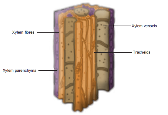
cells, tracheids and vessels (macerated material)
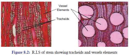
8.3.2 Absorption of Water Through Roots
ACTIVITY 3
Aim: To demonstrate the water moves through xylem vessels.
Requirements: A small potted plant of tomato, eosine solution, beaker, stand, microscope,
slide, razor and water.
Method:
Dig out a small tomato plant.
Cut the stem at the base 1 to 2 cm above roots under water.
Immerse the cut end in eosine solution contained in a beaker.
Fix the shoot erect with the help of a stand.
Keep it for a day or two without disturbing.
Cut the transverse sections of stem.
Observe it under the microscope.
• Could you see that xylem and tracheids in the section looks red?
• Can you say why?
Observations and results: Xylem and tracheids in the section turn red indicating water moves
up in the xylem.
Observing plants in different situations allows learners to make inferences about watermovement through the plant material.
Soil is the main source of water for the plants. The principal source of soil water is the water
that is stored in the spaces between the soil particles after precipitation or rainfall. From the
root hair cells the water enters into the epidermis, cortex and finally endodermis (Figure 8.3).
Endodermis is impregnated with fatty substances along the wall called casparian strips. These
strips form networks and these seal off the spaces between the endodermal cells. From theendodermis water enters into the vascular tissue.
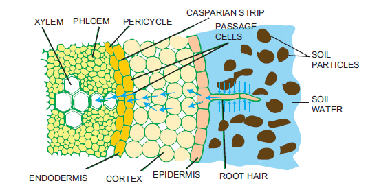
Figure 8.3: Diagram showing entry of water in the root system
The movement of water into the roots can take place by various pathways.
The first pathway is referred to as apoplast. It means that water is moving along the interconnecting
cell walls and spaces between the walls. Water moves through apoplast because of capillary action
or free diffusion along the gradient. It is also called the non-living continuum (Figure 8.4). The other
pathway is the symplast, in which water moves across the root hair membrane and through thecells themselves. Plasmodesmata act as channels to transport water between the cells (Figure 8.4).
Figure 8.5: Diagram showing the movement of water through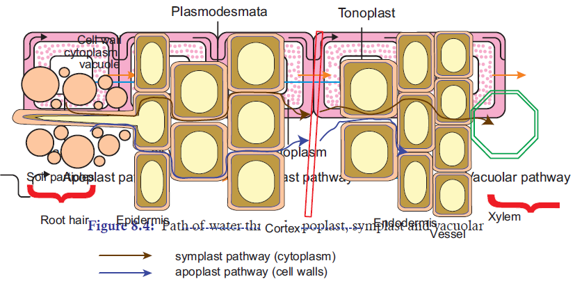
various pathways and through various cell layers in the roots
In natural conditions, the apoplastic and symplastic pathway do not separate and are operating
simultaneously within the system (Figure 8.5). The absorption of water through roots is affected
by various abiotic and biotic factors. When the soil temperature is high, movement is fast. Low
temperature reduces water uptake in plants. The branching pattern of the roots also affects
the uptake of water.
Water is a polar molecule. When two water molecules approach one another, the slightly
negative charged oxygen atom of one forms a hydrogen bond with a slightly positively charged
hydrogen atom in the other. This attractive force, along with other intermolecular forces, is one
of the principle factors responsible for the occurrence of surface tension in liquid water. It alsoallows plants to draw water from the root through the xylem to the leaf.
8.3.3 Anatomy of Root
In most of the herbaceous plants, the roots show various layers of cells through which the water
travels inside the root systems. The outer layer that is protective is termed as epidermis and is
followed by ground tissue consisting of multiple layers of cortex, endodermis and pericycle.
Two to six exarch xylem bundles are found. Phloem tissue is alternate with the xylem tissue(Figure 8.6).
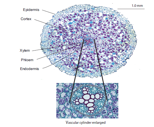
Figure 8.6: Anatomy of ranunculus root
8.3.4 Rise of Water/Ascent of Sap
Cohesive and adhesive forces are important for the transport of water from the roots to the
leaves in plants. Various processes are operating inside the plant system and cells to facilitate
the movement of water from soil to roots and from one cell to another. Absorption of water by
root hairs from the soil and movement of water from one living cell to another within the plant
is brought about by osmosis. The most important factor that affects the uptake is the mineralconcentration of salts in soil.
Before we discuss other things, we should understand that solutions are classified on the basis of
mineral concentrations present in them. On the basis of mineral concentration in these, various
types of solutions are Hypotonic solutions, Hypertonic solutions and Isotonic solutions. Youhave already studied about them in Unit 2.
Water absorption in plants takes place with the expenditure or energy or without use of energy.
The two processes involved are: passive and active absorption.
Passive absorption: Root does not play an active role in water uptake. The root system merely
acts as a physical absorbing system. Energy is not spent for absorption. Passive absorptionaccounts for most of the water absorbed by plants.
Active absorption: Water is absorbed as a result of activity of the root. Root hairs take in
minerals by expenditure of energy. Then water moves from low solute concentration to highsolute concentration across the membrane.
APPLICATION 8.1
1. Complete sentences with appropriate terms:
(a) Xylem tissue is composed of four types of cells:
...................., ...................., .................... and .................... .
(b) Plants .................... water from soil and .................... it to aerial parts.
(c) Two pathways regulating uptake of water from roots are ................. and .................. .2. The diagram below represents a transversal section of a young stem of a dicotyledonous:
a) Name the parts A, B, C.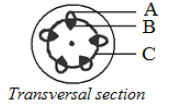
b) Give any one reason which proves that it is:
(i) A structure of a stem but not that of a root(ii) A primary structure but not a secondary structure.
8.3.5 Mechanism of Uptake of Water and Mineral Salts
It is quite clear that various mineral elements are present in the water and these are carried along
with water to the aerial parts of the plants. This is possible because water is a polar solvent and
many mineral ions are highly soluble in it. Many viewpoints have been put forward to help inunderstanding of water and mineral ions in the plants.
Root Pressure
As various ions from the soil are actively transported into
the vascular tissues of the roots, water follows (its potential
gradient) and increases the pressure inside the xylem. This
positive pressure is called root pressure, and can be responsible
for pushing up water to small heights in the stem. How can we
see that root pressure exists? Choose a small soft-stemmed plant
and on a day, when there is plenty of atmospheric moisture,
cut the stem horizontally near the base with a sharp blade,
early in the morning. You will soon see the drops of solutionooze out of the cut stem; this comes out due to the positive
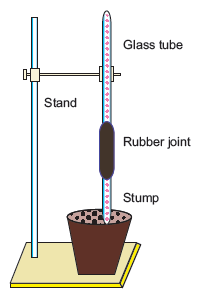
Figure 8.7: Root pressure
root pressure. If you fix a rubber tube to the cut stem as a sleeve you can actually collect and
measure the rate of exudation, and also determine the composition of the exudates. Effects
of root pressure is also observable at night and early morning when evaporation is low, and
excess water collects in the form of droplets around special openings of veins near the tip
of grass blades, and leaves of many herbaceous parts. Such water loss in its liquid phase is
known as guttation (Figure 8.7).
Root pressure can, at best, only provide a modest push in the overall process of water
transport. It obviously does not play a major role in water movement up tall trees. The greatest
contribution of root pressure may be to re-establish the continuous chains of water molecules
in the xylem which often break under the enormous tensions created by transpiration. Root
pressure does not account for the majority of water transport; most plants meet their needby transpiration pull.
Transpiration Pull
Despite the absence of a heart or a circulatory system in plants, the flow of water upward
through the xylem in plants can achieve fairly high rates, up to 15 metres per hour. How is this
movement accomplished? A longstanding question is, whether water is ‘pushed’ or ‘pulled’
through the plant. Most researchers agree that water is mainly ‘pulled’ through the plant, and
that the driving force for this process is transpiration from the leaves. This is referred to as the
cohesion-tension-transpiration pull model of water transport (Figure 8.8). But, what generatesthis transpirational pull?
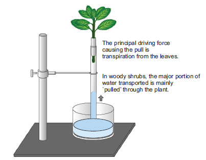
Figure 8.8: Transpiration pull
Water is transient in plants. Less than 1 per cent of the water reaching the leaves is used in
photosynthesis and plant growth. Most of it is lost through the stomata in the leaves. This water
loss is known as transpiration.
You have studied transpiration in an earlier class by enclosing a healthy plant in polythene bag
and observing the droplets of water formed inside the bag. You could also study water lossfrom a leaf using cobalt chloride paper, which turns colour on absorbing water.
1 MPa = 10 atmospheres or 14.5 pounds/inch2.
The root hairs are not developed in some of conifer plants; thus, water is absorbed with
the help of mycorrhizal associations. Mycorrhizal fungi also called vesicular arbuscular
mycorrhizae (VAM) also play an important role in absorption of water. Mycellia absorb
water and minerals and transfers to the roots. These fungal mycelia obtain their food from
the roots.
Velamen is a specialized tissue present on the outer side of the cortex found in epiphytes suchas orchid that absorb water through the hanging roots.
ACTIVITY 4
Aim: To observe various parts of a leaf.
Requirements: A fresh leaf (Bryophyllum), foreceps, needles, watch glass, slides, a razor blade
and compound microscope.
Methods: Cut a vertical section of the leaf.
Stain with a fast green and safranin.
Mount in glycerine.
Observe it under a microscope. What do you observe?
Discussion: Do you see some small openings in the lower layer of the leaf ? What are these
openings called?
Do you see any green coloured pigments in the leaf ?What are these pigments called?
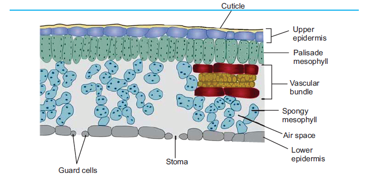
Figure 8.9: Verticle Section of the leaf showing various parts
The outer layer of the leaf is epidermis surrounded by palisade parenchyma cells. The epidermis
has stomata composed of guard cells. Stomata can be present on both the leaf surfaces. In
many plants, stomata are present only on the lower leaf surface. The epidermis is protective in
function. The palisade layer is made up of columnar epithelial cells joined end-to-end having
many chloroplasts. Below the palisade layer are round spongy parenchyma cells that have
conspicuous intercellular spaces. The conducting tissue system consists of tissues present near or
at the centre of midrib region. The xylem is composed of vessels and trachieds and phloem has
fibres and parenchyma. Larger vascular bundles are surrounded by bundle sheaths (Figure 8.7).
Plants do not have systems for transporting oxygen and carbon dioxide. Instead, these gasesdiffuse through air space within stems, roots and leaves.
8.4 TRANSPIRATION IN PLANTS
ACTIVITY 5
Aim: To demonstrate the phenomenon of transpiration by bell jar method.
Requirement: A potted plant, glass plate, bell jar, oilcloth, grease and thread.
Methodology: Take a watered healthy plant. Cover the soil
by cloth to avoid evaporation.
Place the pot on a glass plate and cover with a bell jar.
Leave the apparatus for some time and observe.
What do you see at the inner side of the bell jar? Where do
these come from?
Results: Small drops of water start appearing on the inner side
of bell jar due to condensation of water vapour transpiredfrom the plant.
Transpiration is a physical process responsible for uptake of water in the form of water vapours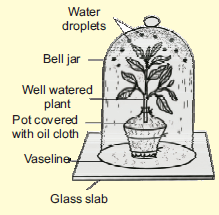
from the plants. Most of the transpiration takes place from the leaves through stomata, cuticles
and lenticels. Transpiration through stomata is called stomatal transpiration. It accounts to
90-95% of the total transpiration. Small quantity of water is also lost through the cuticle. Stomatal
opening and closing affects the rate of transpiration in plants. Changes in turgor pressure of
the guard cells cause stomata to open or close. Both the upper and lower leaf surfaces have a
flattened layer of cells called epidermis. Epidermis is covered by a waxy layer called cuticle. In
many plants, the lower epidermis has a pair of bean shaped cells called guard cells which along
with the subsidiary cells and other guard cells form stomatal complex. Guard cells in dicots are
kidney shaped and in monocots are dumbbell shaped. Guard cells have thickenings in inner
walls. The guard cells together form a stomatal pore or aperture. Of the total water taken up
by the plant most of it is lost in the form of water vapour. This type is cuticular transpiration.
Stomata are also meant for gaseous exchange of oxygen and carbon-dioxide, but transpiration
also occurs when they are open (Figure 8.10). There is a trade off during the gas-exchange that
is important for respiration and photosynthesis in the plant systems.
About less than 0.5% is lost through the lenticels, tissues on stem and fruits. This is calledlenticular transpiration.
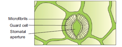
Figure 8.10: Transpiration occurs through stomatal aperture
8.4.1 Factors Affecting Transpiration
The absorption of water from the roots is affected by many physico-chemical properties of
soil such as soil temperature, soil air, amount of water available in the soil and concentration
of mineral salts in the soil (Figure 8.11). Atmospheric humidity, temperature, wind velocity,
light and water availability in the soil affect the rate of transpiration in the plants. Study of
various temperature treatments on plants can be studied by using simple potometers.
In increased light intensity stomata open wider to allow more carbon dioxide into the leaf forphotosynthesis.
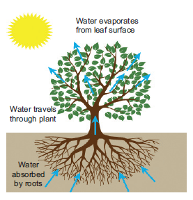
Figure 8.11: Diagram showing process of transpiration on a sunny day
With the increase in wind velocity, there is also increase in the rate of transpiration as water
evaporates fast.
Temperature affects transpiration indirectly through its effect on water vapour present in the air.
An increase in temperature brings about decrease in relative humidity of the air thus increasingrate of transpiration.
ACTIVITY 6
Aim: To study the effect of different light intensities on the rate of transpiration using a
potometer.
Requirements: Twig of Dracaena. Potometer, Luxmeter, Table lamp.
Procedure: Place a twig of plant in one end of the potometer and seal it air-tight. Fill the
entire apparatus with water so that there are no air spaces in between. The plant is exposedto different light intensities (Figure 8.12).
Do you see any changes in the level of water at the other end of the tube? Explain why.
Results: With the increase in light intensity, the level of the water drops indicating increase inrate of transpiration.
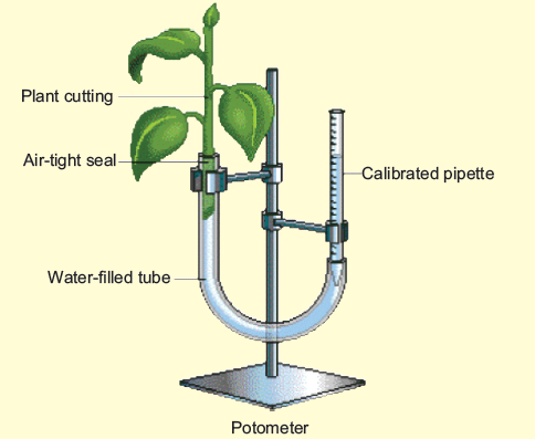
Figure 8.12: Study the effect of light intensities on rate of transpiration using potometer
8.4.2 Significance of Transpiration
Transpiration helps the plants in many ways. It has been considered as a necessary evil. This
is because plants lose lot of water due to the process but it is vital for many other physiological
processes. The reasons why this process is advantageous to plants are:
1. It maintains and regulates temperature by evaporative cooling.
2. It helps in absorption and transportation of mineral ions in plants.
3. It provides water to keep cells turgid in order to support the plant.4. It makes water available to leaf cells for photosynthesis.
8.4.3 Adaptations of Xerophytes to Reduce Water Loss
Many plants show various morphological features that help them survive in regions with low
water availability. The morphological variations are observed in root, stem, branching pattern,types of leaves and other parameters.
These variations are manifestations of changes taking place in the plants at various other levels
such as anatomical and biochemical level. These variations are termed as adaptations and help
plants to survive in a particular environment. Plants that grow in environments that have plenty of
water have stomata on both upper and lower epidermal cells of the leaves. These have isobilateral
leaves, aerenchyma in stems and undeveloped vascular bundles.
However, the plants growing in low water availability show various xeromorphic and xerophytic
characters. Xerophytic plants exhibit a variety of specialized adaptations to survive in such
conditions. Xerophytes may use water from their own storage, allocate water specifically to sites
of new tissue growth, or lose less water to the atmosphere and so convert a greater proportion
of water in the soil to growth.
Xerophytic adaptation of reducing water loss by transpiration:
1. Xerophytes have thick waxy cuticle which reduces evaporation as it acts as a barrier.
The shiny surface also reflects heat and so lowers temperatures reducing water loss.
2. They have rolled leaves or leaves reduced to spines to reduce water loss.
3. Stomata are present in pits. They are sunken. They open at night to reduce the
amount of water lost by transpiration.
4. Roots are deep and/or spreading to maximize the absorption of underground water.
5. They exhibit crassulacean acid metabolism, i.e., CAM Physiology.
6. They have fleshy stems or leaves—some cells in stems or leaves have very largevacuoles that act as water storage areas. These stems are also called succulent stems.
APPLICATION 8.2
Complete with appropriate terms:
(i) The association of Mycorrhizal fungi is ……………….. .
(ii) ……………….. is the loss of water from plants.
(iii) ……………….. is used to study the rate of transpiration.
(iv) Xerophytes have ……………….. stomata.(v) Xerophytes exhibit ……………….. metabolism.
8.5 TRANSPORT OF FOOD
Process of the movement of food synthesized during photosynthesis from the leaves to the
roots and other parts of a plant through the phloem is called translocation. This is carried outby another conducting tissue phloem.
8.5.1 Structure of Phloem
An analysis of the phloem exudate obtained by making an incision into the phloem tissue
provides evidence that photoassimilates are transported through phloem.
The phloem collects photoassimilates in green leaves, distributes them in the plant and supplies
it to the other heterotrophic plant organs. Phloem is composed of various specialized cells called
sieve tubes, companion cells, phloem fibres, and phloem parenchyma (Figure 8.13).
1. Sieve Tubes: Sieve tubes are series of cells joined end to end. The cross walls between
successive sieve elements become perforated forming sieve plates. The cell walls are
thin. Although the cell contents are living, the nucleus disintegrates and disappears. The
lumen is filled with a slimy sap which is composed mainly of protein. The function of
sieve tubes is transport of organic compounds.
2. Companion Cells: These are specialized parenchyma cells which always appear with the
sieve tube elements. They are also elongated, thin-walled and have distinct nucleus in the
cytoplasm of the companion cell. Their function is to regulate the metabolic activities
of the sieve tube elements.
3. Phloem Fibres: These cells are elongated and tapering. They have thickened walls.
Phloem fibres give mechanical strength to the plants.
4. Phloem Parenchyma: These are living cells with thin walls. Phloem parenchyma storescompounds such as starch.
Figure 8.13: (a) and (b) A Structure of phloem: RLS of the stem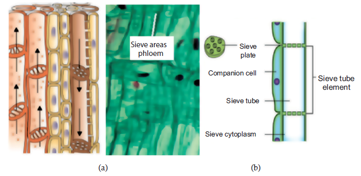
showing various parts of phloem
8.5.2 Movement of Sugar in Plants
As sugar is synthesized in the leaves by the process of
photosynthesis its high concentration inside the phloem cells
of the leaf creates a diffusion gradient by which more water
is transported into the cells. Translocation takes place in the
sieve tubes, with the help of adjacent companion cells. Food
is translocated in the form of sucrose. The movement of water
and dissolved minerals in xylem is always upward from soil to
leaves against the gravitational pull. However, the movement
of food can be upward as well as downward depending upon
the needs of the plants (Figure 8.14).
Figure 8.14: Phloem Transport can be bidirectional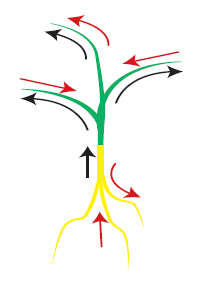
represented by red arrows
8.5.3 Mechanism of Uptake of Food in Plants
As explained earlier, leaves manufacture food by the process of photosynthesis and transport
it to other parts of the plants. Part of plants where food is formed more than requirement is
known as source. Leaves act as a source and where these are stored and sent is the sink. Roots
act as sinks for food. Movement of food takes place from source to the sink. Leaves prepare
food in the form of glucose that is converted into sucrose. Sucrose enters into the phloem
at the expense of energy in the form of ATP. It is noteworthy that in plants, roots, fruits
and other organs also act as storage organs for food and from here the food is translocated
to other parts. So, the direction of movement of the phloem can be both upwards and
downwards and hence the movement is bidirectional. Sugars, hormones and amino-acids are
also transported or translocated through phloem. Transport of food involves 3 steps: Phloem
loading, translocation and phloem unloading. Sucrose and other carbohydrates are actively
loaded into the sieve tubes at the source by a chemiosmotic mechanism. It requires ATP.
• ATP supplies energy to pump protons out of the sieve tube members into the apoplast.
• Creates proton gradient.
• The gradient drives the uptake of sucrose into the symplast through channels by the
cotransport of protons back into the sieve tube members.
Osmotic concentration of phloem increases due to presence of sucrose. Therefore, water enters
into sieve tubes by osmosis, due to which the hydrostatic pressure is created in phloem. High
pressure in the phloem allows the movement of food to all parts of the plants having low
pressure in their tissues. The pressure-flow hypothesis proposed by Munch is the simplest model and
continues to earn widespread support among plant physiologists. The pressure-flow mechanism
is based on the mass transfer of solute from source to sink along a hydrostatic (turgor) pressure
gradient. Translocation of solute in the phloem is closely linked to the flow of water in the
transpiration stream and a continuous recirculation of water in the plant.
Theory proposes that food molecules flow under pressure through the phloem. The food is
mixed with water. The pressure is created by the difference in water concentration of the
solution in the phloem and the relatively pure water in the nearby xylem. Sugars manufactured
in mesophyll cells are driven by energy into the companion cells and sieve tube elements
of the phloem. After accumulation into the phloem, water enters in cells by osmosis. A
pressure is built up in sieve tubes called turgor pressure. Due to this pressure, sugars are
removed by the cortex of both stem and root and consumed or converted into storage
products such as starch. Starch does not exert any osmotic effect. Hence, osmotic pressure
of phloem cells decreases. Thus, the pressure gradient created between leaves and shoot androot drives the contents of the phloem up and down through the sieve tubes (Figure 8.15).
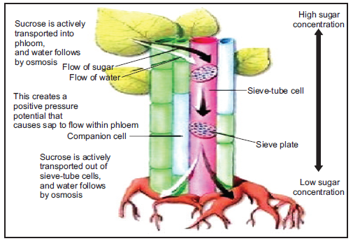
Figure 8.15: Illustrated account of the phloem transport in plants
Assimilates including sucrose, amino acids and nutrients are transferred into sieve elements
of fully expanded leaves against significant concentration and electrochemical gradients. This
process is referred to as phloem loading. The cellular pathways of phloem loading, and hence
transport mechanisms and controls, vary between plant species. Longitudinal transport of
assimilates through sieve elements is achieved by mass flow and is termed phloem translocation.
Mass flow is driven by a pressure gradient generated osmotically at either end of the phloem
pathway, with a high concentration of solutes at the source end and a lower concentration at
the sink end. At the sink, assimilates exit the sieve elements and move into recipient sink cells
where they are used in growth or storage processes. Movement from sieve elements to recipient
sink cells is called phloem unloading. The cellular pathway of phloem unloading, and hencetransport mechanisms and controls, vary depending upon sink function.
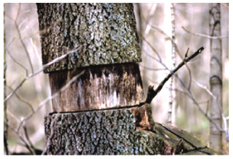
Figure 8.16: (a) Girdling in trees
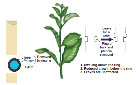
Figure 8.16: (b) Picture showing the role of phloem in translocation of food
A simple experiment, called girdling, was used to identify the tissues through which food is
transported (Figure 8.16(a)). On the trunk of a tree a ring of bark up to a depth of the phloem
layer, can be carefully removed. In the absence of downstream movement of food, the portion
of the bark above the ring on the stem becomes swollen after a few weeks (Figure 8.16(b)). This
simple experiment shows that phloem is the tissue responsible for translocation of food : andthat transport takes place in one direction, i.e., towards the roots.
To Investigate Mass Flow Hypothesis
• In mass flow, Munch’s model demonstrates that fluid flows from the region of high
hydrostatic pressure to the region of low hydrostatic pressure.
• As fluid flows, it carries the whole mass of different substances.
• In osmometer A, concentrated sucrose solution (leaf) has lower water potential. Water
flows into it from a high water potential region (xylem vessel) to a low water potential
region (leaf cells) by osmosis.
• This create high hydrostatic pressure in A and forces sucrose solution to enter into the
connecting tube (sieve tube) and pass to B (root cell).
• As the flow of mass from osmometer A to osmometer B continues, the sucrose solution
is pushed along and finally appears in B.
• In B, contain water/dilute sugar solution, water moves out from a higher water potential
region by the hydrostatic pressure gradient produced and redistributed through connecting
tube (xylem vessels) between the two containers.
• Mass flow continues until the concentration of sugar solution in A and B are equal (balanced).
• In nature, equilibrium is not reached because solutes are constantly synthesized at sourceA and utilized at the sink B.
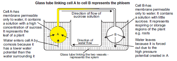
Figure 8.17: Demonstration of mass flowhypathesis
APPLICATION 8.3
Complete with the appropriate terms:
(i) …………… is the process to show food flows from leaves to roots.
(ii) Phloem transports phloem sap from …………. to ………………… .
(iii) Phloem …………….. gives mechanical strength to cells.(iv) Movement from sileve elements to recipient sink cells is called ……….
8.6 SUMMARY
• Water is an important solvent and acts as a reagent in various chemical reactions in the plants.
• It helps to maintain turgidity of cells and is important for growth of plants as it serves
as a raw material for photosynthesis.
• Transport of water is an important process in plants and has been well understood.
• Several physical phenomena such as imbibition, diffusion, osmosis, turgor and water
potential facilitate uptake of water in plants.
• Forces of cohesion and adhesion also play an important role in transport of the water upstream.
• Water enters the plants through active or passive absorption process. The upward
movement of water through stem is called the ascent of sap.
• Practically most of the water absorbed by plants is lost into the atmosphere through the
process of transpiration.
• A variety of internal and external stimuli govern the rate of transpiration in plants.
• Atmospheric humidity, temperature, light, wind velocity, leaf area, leaf structure and
availability of water affect the process.
• Plants also take up inorganic nutrients from the soil with water. The sugars synthesized in
leaves are translocated downwards, upwards and to lateral organs mostly through phloem.
• Experiments have been conducted to demonstrate the movement of food through phloem.
Besides sugars that are end products of photosynthesis, amino acids are also transportedthrough phloem.
8.7 GLOSSARY
• Active transport: Transport of ions or molecules across a cell membrane against a
concentration gradient.
• Adhesion: Attraction of water molecules to polar surfaces is called adhesion.
• Apoplast: Intercellular spaces, excluding the protoplasts is called apoplast.
• Aquaporins: Protein channels for transport across membranes.
• Casparian strips: A band like structure in endodermis of root cells that contain suberin
and lignin.
• Cohesion: Mutual attraction between water molecules is called cohesion.
• Cuticle: A three layered structure present on the epidermis that prevents movement of
gases and water to move into or out of the plants.
• Diffusion: Movement of substance from high concentration to low concentration.
• Lignin: An aromatic polymer that rigidifies may secondary cell walls.
• Lenticels: Pores on woody stems and roots for gaseous exchange.
• Osmosis: Movement of water from area of low to area of high solute concentration.
• Phloem: the photosynthate-conducting tissue of plants.
• Plasmodesmata: Connection between protoplasts of adjacent cells through cell walls.
• Plasmolysis: Shrinkage of cytoplasm under the influence of hypotonic solution.
• Root pressure: Hydrostatic pressure created inside the roots due to absorption of water.
• Root: The portion of a plant axis produced by the root apical meristem.
• Stem: The portion of a plant axis produced by the shoot apical meristem.
• Leaf: A lateral appendage of the stem produced by the shoot apical meristem.
• Sieve element: a conducting cell in the phloem.
• Surface tension: Any liquid has a tendency to occupy the least possible surface area.
This property is called surface tension.
• Symplast: The continuous system of protoplasts connected by plasmodesmata.
• Tensile strength: It is a measure of maximum force per unit area that would be needed
to break a continuous column of water.
• Tracheid: A conducting cell of the xylem characterized by an elongated shape and
lignified secondary cell wall.
• Turgor Pressure: Hydrostatic pressure developed inside cell vacuole that presses cytoplasm
against the cell wall.
• Vascular tissue system: Tissues derived from the procambium or vascular cambium that
transports water and photosynthates.
• Vessels: Tracheary element with perforation plates.
• Water potential: Chemical potential of water in relation to pure water.
• Xylem: The water-conducting tissue of plants.
END UNIT ASSESSMENT 8
I. Multiple Choice Questions
1. Much of the transpiration takes place through
(a) stomata (b) lenticels (c) cuticle (d) epidermis
2. The roots absorb water through
(a) epidermal hairs (b) root hairs (c) root xylem (d) root phloem
3. The ascent of sap in plants takes place due to
(a) root pressure (b) transpiration pull
(c) osmosis (d) both (a) and (b)
4. Stomata open and close due to
(a) presence of valves (b) hormonal control
(c) turgor pressure of guard cells (d) concentration gradient of the gases
5. The food is transported in the phloem in the form of
(a) glucose (b) sucrose (c) amino acids (d) fats
6. The movement of particles from the region of their higher concentration to the region
of their lower concentration is called
(a) osmosis (b) diffusion (c) active transport (d) ascent of sap
7. Plant transport system does not transport
(a) CO2 (b) organic salts (c) water (d) plant hormones
8. The strongest force to pull water up the xylem and into the leaf is
(a) capillary action (b) root pressure(c) transpiration pull (d) active transport
9. The loss of water in the form of vapours by the leaves and stem of a plant is called
(a) translocation (b) osmosis
(c) active transport (d) transpiration
10. The transport of sugar from the leaf to the rest of the plant is called
(a) translocation (b) osmosis(c) active transport (d) transpiration
II. Long Answer Type Questions
1. Name the two transport tissues present in the plant.
2. What are the factors affecting the rate of diffusion?
3. Explain why pure water has the maximum water potential.
4. Differentiate between the following:
(i) Diffusion and Osmosis
(ii) Active and Passive Transport
(iii) Osmosis and Diffusion
(iv) Transpiration and Evaporation
5. Discuss the factors responsible for ascent of xylem sap in plants.
6. How is turgor pressure created in the sieve elements?
7. What is the difference between apoplast and symplast?
8. Explain in detail the absorption of water through root hairs up to the xylem.
9. Discuss the structure of phloem and its components in plants.
10. Explain how food prepared in the leaves reaches the other parts of the plant.
11. Explain the hypothesis proposed by Munch regarding translocation of food in plants.
12. Appreciate the importance of transport systems in plants.
13. Draw and label, from prepared slides, the cells in roots, stems and leaves using
transverse and longitudinal sections.
14. There are a million processes that account for life on Earth. All these processes play
a major role in balancing the climate of Earth. Investigate in the same regard the role
of transport in plants in regulating the environment of surroundings. Also necessitate
the presence of plants or a wholesome regulation of atmosphere.
15. Draw this picture in your exercise book. It shows various internal parts of a leaf.These are marked us A, B, C, D, E and F. Identify and name these parts.
Unit 9 Gas Exchange in Animals
Key Unit Competence
To be able to describe structures of gas exchange in different groups of animals.
LEARNING OBJECTIVES
At the end of this unit, the learner will be able to:
• Describe the tracheal system of insects and relate to its function.
• Describe the structure of the gills in relation to function.
• Explain the significance of counter current flow in bony fish.
• Dissect an insect, fish and a small mammal to study gaseous exchange organs.
• Relate the structure of gas exchange organs to function.
• Differentiate between the gaseous exchange in bony fish and that in cartilaginous fish.
• Describe the mode of gaseous exchange in amphibians.
• Describe the structure of the human gas exchange system.
• Appreciate the similarities and differences in gas exchange surfaces of animals.
• Interpret a graph of human lung volumes measured with a spirometer.
• Calculate the volume of air in the lungs and in the alveoli.
• Describe the distribution of tissues within the trachea, bronchi, bronchioles and alveoli and
relate each tissue to its function.
• Explain the mechanism of ventilation in humans.
• Explain the process of gas exchange in alveoli with emphasis on diffusion.
• Describe the role of the brain in controlling gas exchange in humans.
• Appreciate the role of the brain in controlling gas exchange.
• Define terms related to the lung capacities (tidal, reserve volume, vital capacity, residual
volume, and dead air space).
• Describe how a spirometer can be used to measure vital capacity, tidal volume, breathingrates, and oxygen uptake.
INTRODUCTORY ACTIVITYObserve the following images and answer the following
1. According to your observation what is happening to each living organism
2. What are the types of gas the dog is giving out?
3. You are inhaling and exhaling as shown on the figure b. Search and find the description ofthe pathway through which that air is passing in your body
9.1 INTRODUCTION
ACTIVITY 1
Place your hands on your chest: You can feel the chest moving up and down. You know it is dueto breathing. How do you breathe? What happens during breathing? Discuss.
Air breathing animals (aerobic) require a continuous supply of oxygen for various metabolic
activities. They also require continuous removal of carbon dioxide formed as a by-product
of these metabolic activities. This process of gas exchange is vital for their survival. This
continuous ‘exchange’ of oxygen and carbon dioxide with the animal and the environment is
known as gas exchange. For a surface to be able to exchange gases in living system, it should
be moist, have large surface area, and highly vascular i.e., richly supplied with blood vessels.
Exchange of gases through the biological membrane occurs by a process known as diffusion.
Diffusion is movement of gas molecules from a region of higher concentration to a region oflower concentration.
9.2 GASEOUS EXCHANGE IN INSECTS
Insects have a specialised system of ‘tubes’ called the tracheal system for exchange ofgases (Figure 9.1). This system consists of a vast network of cuticular (i.e., made of chitin, a
(a) The respiratory system of an insect consists of branched internal tubes. The largest tubes, called
tracheae, connect to external openings spaced along the insect’s body surface. Air sacs formed
from enlarged portions of the tracheae are found near organs that require a large supply of oxygen.
(b) Rings of chitin keep the tracheae open, allowing air to enter and pass info smaller tubes called
tracheoles. The branched tracheoles deliver air directly to cells throughout the body. Tracheoles
have closed ends filled with fluid (blue-gray). When the animal is active and using more O2, most
of the fluid is withdrawn into the body. This increases the surface area of air-filled tracheoles incontact with cells.
(c) The TEM above shows cross sections of tracheoles in a tiny piece of insect flight muscle. Each ofthe numerous mitochondria in the muscle cells lies with about 2.5 μm of a tracheole.
Figure 9.1: Tracheal system in insect (Grasshopper). Note that the fine tubes called bronchiolesinnervating at cellular level. (Source: Campbell Biology, 2011)
long-chain polymer of an N-acetyl glucosamine) tubes penetrating to almost each individual cells
of the body. This system serves two functions: it brings air into the body, and also distributes it
to the cells. This pattern of tracheal system is very much similar to the system of blood vesselsin higher animals.
Air enters the tracheal system of the animal through special openings called spiracles. These
are present mostly on the lateral side of the animal. These are usually guarded by valves,
operated by muscles and sometimes provided with filters. Tracheal tubes are invaginations
(infoldings) of the body surface. Thus, their walls are similar in structure and composition to the
general body surface (integuments) of the animals. Sometimes larger tracheas have thickenings
called taenidia. These are spiral cuticular layers which give strength and elasticity. The tubes
become progressively smaller and thinner to form tracheoles or air capillaries (Figure 9.2). The
smaller tubes may have incomplete taenidial support. They have a diameter of less than 1 μm
(1 μm = 1×10⁻⁶ m). Tracheoles are the most important physiological unit of this gas exchange
system. It is because they make numerous close contacts with the individual cells for gas
exchange to take place. They sink into cell’s plasma membrane bringing oxygen very close to
the mitochondria of the cells.
Figure 9.2: Detail structure of the tracheal system in insects
9.2.1 Mechanism of Ventilation in Insects
Normally there is no active ventilation in most tracheates (i.e., animals possessing trachea).
Many of the tracheates (like onychophora, myriapoda, and insect larvae and pupae) depend on
simple diffusion of gases in the air tubes. But ventilation and control of direction and volume
of the air flowing through the system is present in adult insects. This is because adult insects
are larger and so have higher metabolic rate which demands more oxygen. The spiracles and
air sacs help the insect in ventilation and creating unidirectional flow of air. In grasshopper,
thoraxic spiracles are used for inspiration while abdominal spiracles are used for expiration. This
creates a unidirectional flow of air. Air sacs greatly increase the efficiency of ventilation. These
are balloon-like structures of the trachea with a variety of size and shape. Active ventilation
is brought about by rhythmic contraction and relaxation of body walls. This forces the air
movement in and out of the tracheal system. Dorso-ventral flattening of abdomen is observed
in grasshopper and beetles.
ACTIVITY 2
Aim: To dissect an insect (cockroach or grasshopper) and study its tracheal system.
Materials Required: Cockroach, dissecting microscope, surgical scissors, chloroform,
forceps, scalpels, pins, dissecting tray etc.
Procedure:
1. Obtain a live cockroach (Periplaneta americana) and anaesthesize it with chloroform.
2. Locate the position of spiracles on thorax and abdomen and record their position by making
a rough sketch on the record book.
3. Pin the animal on the dorsal side with the ventral surface facing upwards on a dissecting tray.
4. Carefully remove the abdominal sterna (exoskeleton covering of the abdomen) without
disturbing the internal tissues.
5. Remove the fat bodies and reproductive organs carefully to expose the tracheal system.
Observation:
The tracheal system should be easily identified by its silvery appearance due to entrapped air in
it. Can you locate the taenidia? Label the different parts of the tracheal system. Notice that in
grasshopper, thoraxic spiracles are used for inspiration while abdominal spiracles are used forexpiration.
9.3 GASEOUS EXCHANGE IN FISH: GILLS
Gills are typical respiratory organs of aquatic animals, including fishes. Gills range in shape and
size. It may be finger-like projections or simple epithelial extensions. Gills are more developed
in fishes. Fish gills consist of thousands of highly specialised gill lamellae enclosed in a gill
cavity. The gill cavity is covered by an operculum and continuously ventilated by flowing water.
Respiration through gills is also known as branchial respiration. All gill surfaces are provided
with a dense network of thin capillary vessels and supported by skeletal elements called the
branchial arches.
Types of gills
Gills can be of two types:
External gills: These gills are exposed to the environment and not enclosed within a pouch or
cavity. They are found in the larvae of many vertebrates, including lungfishes, actinopterygians,
and amphibians.
Internal gills: Gills are covered and protected laterally by soft skin folds, like the interbranchial
septum in cartilaginous fishes, or by a firm operculum in many bony fishes. They are found
within pharyngeal gills slits or pouches of most cartilaginous and bony fishes.
In cartilaginous fishes, the gills are found on the lateral side of the branchial arch (Figure 9.3).
Gills are usually five pairs in number. They are located in vertical, anterioposteriorly compressed
branchial chambers or gill pouches. Each branchial pouch is separated from each other by a
stout interbranchial septum. This septum is made up of fibro-muscular tissue with blood vessels.
A branchial pouch communicates to exterior with the help of narrow external branchial
aperture or gill slits. Each gill has a central partition called the interbranchial septum. Within
this septum, a stiff structure called gill ray gives support to the gills. This septum is covered
on each face by primary lamellae or gill filaments. Gill filaments are series of raised thin,
highly vascular horizontal lamellar folds of the interbranchial septum. The primary lamellae
are again made up of standing rows of secondary lamellae. Water flows across their sides toirrigate the gills.
Figure 9.3: Structure of gills in shark. (Source: Kardong, Vertebrates:
Comparative anatomy, Function, Evolution, 2012)\
When gill lamellae are present on both anterior and posterior sides of a septum, it is called
a holobranch or complete gill. However, when lamellae is present on only one face, it is
called a hemibranch (Figure 9.4). Facing plates of lamellae on adjacent gills constitute a
respiratory unit. A branchial pouch therefore consists of posterior hemibranch of one gill andanterior hemibranch of the succeeding gill.
The pharyngeal structural region in bony fishes is almost similar to that of cartilaginous fishes.
The gill/branchial chamber on each side is covered by a fold of integument called the operculum
(gill covering). It is supported by four opercular bones. The operculum protects the branchial
arches and its gill lamellae and also helps in gill ventilation. There are five pair of gill pouches
and four pairs of holobranchs or complete gills. In cross section, each gill is V-shaped and
composed of primary lamellae (gill filaments) that are subdivided into secondary lamellae andsupported on a branchial arch (Figure 9.5).
ACTIVITY 3
Aim: To observe and study the structure of gills in freshwater fish.
Materials Required: Fish (Tilapia), dissecting microscope, surgical scissors, chloroform,
forceps, scalpels, pins, dissecting tray, etc.
Procedure:
1. Obtain a live fish Tilapia from a local fish market and anaesthesize it with chloroform.
2. Locate the gill on the lateral side of the head by lifting the operculum. Cut out the operculum
and expose the red coloured gills.
3. Dissect out the gills and observe it under the microscope.
4. Draw a detail sketch structure of the gill and it lamellae and label it different parts with the
help of your teacher.Observation: Can you see the gill lamellae and the gill rakers?
9.3.1 Mechanism of Gill Ventilation
ACTIVITY 4
Aim: To observe and study the ventilation mechanism in fish.
Materials Required: Fish in an aquarium, notebook and pencil timer, etc.
Procedure:
Observe the movement of the mouth and operculum of a fish in the aquarium. Note the number
of times the mouth and operculum opens in a minute and record it in your notebook.Observation: Record the rate at which ventilation occurs in the fish.
Ventilation rate is much higher in aquatic animals than air breathing animals. This is because
water has lower oxygen and greater density than air. So, more ventilation is required for oxygen
uptake. This is achieved in gills by having a unidirectional flow of water. Ventilation of fish
gills is achieved by rhythmic movement of various muscles. This generates a continuous
current of water through the gills. The muscular pump of the buccal cavity actively drives
water across the internal gills bringing about ventilation. First the mouth is opened, buccal
floor drops and the pharyngeal floor lowered at the same time. This creates a vacuum inside
pharyngeal cavity. At the same time, external branchial openings are closed. This results in
water rushing into the pharyngeal cavity through the mouth. Now mouth gets closed and the
external aperture opens. This makes water flow out through the branchial apertures. As water
passes through the gills, it washes the gill lamellae. Exchange of gases takes place betweenthe blood flowing in the gill lamellae and the water current.
Figure 9.4: Gill ventilation in sharks. Lateral (a, c) and frontal view (b, d). Relative positive and
negative pressures indicated by + and –, respectively (Source: Kardong, Vertebrates: Comparativeanatomy, Function, Evolution, 2012)
Figure 9.5: Mechanism of gill ventilation in Tilapia
Difference between gaseous exchange of cartilaginous and bony fish
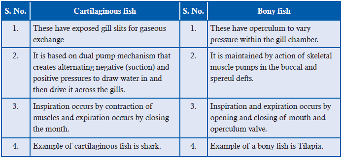
9.4 THE COUNTER CURRENT MECHANISM
ACTIVITY 5
To study counter current flow and parallel flow.
Use the internet or textbooks to make a chart diagram of countercurrent and concurrent flow
of fluids. Using arrows of different colours indicate the direction of flow of fluid in both the
flow system. Download videos/animations from the internet depicting the countercurrent flow
and exchange of gases through this system. Ask each of the students to give an advantage to
each of countercurrent flow over concurrent flow.
The high efficiency of fish gills, especially teleost in gas exchange is due to the presence of a
counter current flow of blood and water through the system. In ‘counter current’ system, twochannels in close proximity carry fluids in opposite directions (Figure 9.6). In such a system,
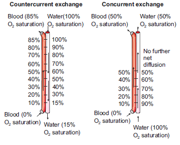
Figure 9.6: Current and counter current fluid flow.
equilibria will be established in the concentration of any permeable materials under two conditions
i.e., if the channels walls are freely permeable to the particular materials and if the channels are
long enough. The flow of blood in the gill lamellae and nearby water is a countercurrent type.
In most fish gills, the blood in the secondary lamellae flows in one direction and water flows
in the opposite direction. This establishes a countercurrent exchange system. As we can see in
Figure 9.7, the counter current system maintains a continuous gradient of oxygen concentrationbetween blood and water which is not found in case of concurrent exchange system.
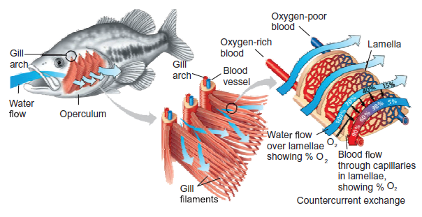
Figure 9.7: Countercurrent mechanism of gas exchange in bony fishes
This countercurrent flow maximizes difference in oxygen (and carbon dioxide) concentration
between water and blood. Countercurrent exchange arrangement results in blood always
being exposed to water with a higher oxygen concentration. A diffusion gradient is, therefore,
maintained across the surface of gill. Blood in gill lamellae capillaries contains less oxygen
and more carbon dioxide as it comes from different tissues after metabolism. However, the
water ventilating the gills has a greater concentration of oxygen compared to that of blood.
Hence, oxygen diffuses readily from water to blood in capillaries of gill lamellae continuously
till equilibrium is maintained. Due to the presence of a countercurrent exchanger system, a
continuous difference in the concentration of the gases is maintained all throughout the lengthof the gill lamellae, and therefore, a continuous efficient gas exchanger system is obtained.
9.4.1 Significance of Countercurrent Mechanism in Bony Fishes
• A larger difference in PO2 (i.e., partial pressure of O2; the pressure of a specific gas in a
mixture is called its partial pressure) can be maintained across the exchange surface. The
larger the difference, the more the exchange of gases; thus, allowing more transfer of gas.
• The system is so efficient that in some teleost 85% of oxygen may be extracted from water
passing over the gills using this system.
• This type of exchanger is also found in temperature control system of cold arctic animals,
in air bladders of fish and even in the kidneys of vertebrates.
• A few fish have some warm tissues. For example, Tuna have warm muscles, eyes, andbrains. This is only possible because of a countercurrent blood supply to selected tissues.
APPLICATION 9.1
1. Complete with appropriate terms:
(i) Two gases involved in gas exchange are ..................... and .....................
(ii) The high efficiency of tedeost gills is due to .....................
(iii) ..................... fish have exposed gill slits.
(iv) Taenidia are thickening of larger trachea in .....................
(v) Active ventilation in insects is brought about by ..................... and .....................
2. Explain why large , active organisms need special surfaces for exchange3. Describe and explain the features that make an exchange surface efficient
9.5 GASEOUS EXCHANGE IN AMPHIBIANS
ACTIVITY 6
Aim: To observe a live frog or toad in a glass tank and discuss its gas exchange surfaces.
Materials Required: A live frog/toad, aquarium/glass tank, notebook and pen etc.
Procedure:
1. Obtain a live frog and put it in an aquarium or glass tank slowly. Take care to handle the
animal gently and the animal should not be harmed.
2. Now observe carefully how it keeps itself ventilated and frequently comes to the surface,
etc.
3. While on land observe carefully the movement of the buccal chamber for ventilation.
4. Touch the surface of the frog and examine the skin whether it is dry or wet.
Observation:
Did you observe the wet slimy condition of the skin of the frog? Try to explain why is it so? Also
note the continuous ventilation of the lungs when in land by alternate lowering and raising ofthe buccal chamber.
Amphibians use the moist skin, gills or the lungs for gas exchange (Figure 9.8). Gas exchange
occurring through the skin is known as cutaneous respiration. In some larval Salamanders and
adult, external gills are also used for respiration. Modern amphibians rely heavily on cutaneous
respiration. Sometimes, they develop accessory skin structures to increase the surface areaavailable for gas exchange.
Figure 9.8: Cutaneous respiratory structures in some amphibians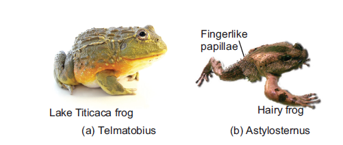
In salamanders of the family Plethodontidae, adults do not have lungs and gills. They depend
entirely on cutaneous respiration for metabolism. Lake Titicaca frog, Telmatobius culeus, has
prominent loose skinfolds on its back and limbs for cutaneous respiration. In the male hairy frog,
Astylosternus robustus, numerous papillae appear on its sides and hindlimbs during the breeding
season, forming supplementary respiratory organ (Figure 9.9). The amphibian skin is thin,
moist, and rich supplied with capillaries making it best suited for gas exchange by diffusion.
In aquatic amphibians, pharyngeal slits often persist with internal gills. Feathery external gills
are often present, especially among larval amphibians.
Amphibian larvae like Salamander larvae typically have both internal and external gills. Pumping
action of the throat irrigates the internal gills with a unidirectional stream of water across
their surfaces. Feathery external gills are held out in the passing current of water. In modern
amphibians, ventilation depends not on ribs but on pumping movements of the throat to irrigate
gills or fill lungs.
Most adult amphibians have lungs for breathing air (Figure 9.10). Normally, the respiratory
surface within the lungs on the anterior region is more developed than the posterior along
the inner walls. The inner surface of lungs forms partitions and divides to increase the
surface area for gas exchange. Such a surface is called septal. The interconnecting septa
divide the internal wall into compartments called faveoli. These faveoli open into the central
chamber within each lung. Faveoli differ from the alveoli of mammalian lungs. Alveoli
are found at the end of a highly branched tracheal system but faveoli are not. Faveoli are
internal subdivisions of the lung wall that open into a common central chamber. Inspired
air travels along the trachea into the central lumen of the lung and from here diffuses into
the surrounding faveoli. Capillaries located within the thin septal walls of the faveoli takeup oxygen and give up carbon dioxide.
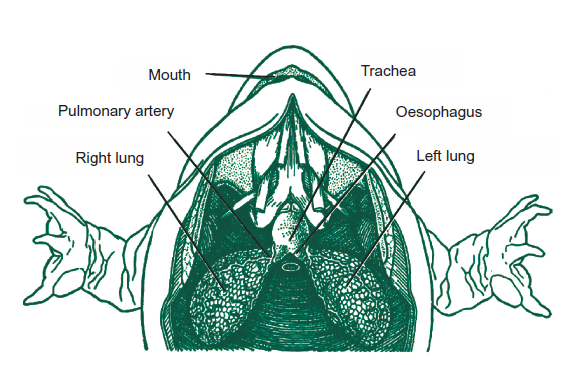
Figure 9.9: Structure of lungs in frog (amphibians).
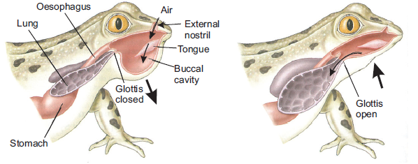
Figure 9.10: Mechanism of ventilation in frog (amphibians)
APPLICATION 9.2
1. Complete with appropriate terms;
(i) Gas exchange occurring through skin is called .............. respiration.
(ii) Amphibians develop .............. to increase surface area for gaseous exchange.
(iii) Partitions of inner lungs in Amphibians are known as ..............2. State 3 ways in which the structure of the lungs allows efficient gas exchange
9.6 GASEOUS EXCHANGE IN HUMANS
Higher vertebrates including humans have specialized organs called lungs for gas exchange.
The process of gas exchange in the body, called respiration, has three basic steps:
1. Pulmonary ventilation or breathing is the inhalation (inflow) and exhalation (outflow) of
air and involves the exchange of air between the atmosphere and the alveoli of the lungs.
2. External (pulmonary) respiration is the exchange of gases between the alveoli of the
lungs and the blood in pulmonary capillaries across the respiratory membrane. In this
process, pulmonary capillary blood gains O2 and loses CO2.
3. Internal (tissue) respiration is the exchange of gases between blood in systemic
capillaries and tissue cells. In this step, the blood loses O2 and gains CO2. Within cells,
the metabolic reactions that consume O2 and give off CO2 during the production of
ATP are termed cellular respiration.9.6.1 Structure of Gaseous Exchange in Humans\
ACTIVITY 7
Make a computer model/simulation of the human respiratory system. This can also be
downloaded from the internet. Use the internet to search videos, graphics and simulation or
animations showing the different parts and surfaces of the gas exchanges system in humans.
Also study the process of gas exchange and the mechanism of ventilation. Now, a smallpresentation on the same topic. You can also clay models of the respiratory system in humans.
Air is inhaled through the nose into the pharynx (throat). Pharynx is a common passage for
both air and food. The pharynx branches into two tubes, the oesophagus or food pipe and
the larynx. The larynx is part of the airways and it houses the vocal cords. The nose, mouth,
pharynx, and larynx are also called the upper airways. The larynx opens into a long tube,
the trachea. The trachea then branches into two bronchi, the right primary bronchus enters
the right lung and the left primary bronchus enters the left lung. The walls of the trachea and
bronchi contain cartilage, which supports them and gives them their characteristic cylindrical
shape. The right primary bronchus is more vertical, shorter, and wider than the left. Within
each lung, the bronchi branch continuously into narrower, shorter, and more numerous tubes,more than 20 generations of branching (Figure 9.11a).
The primary bronchi divide to form smaller bronchi which are known as the secondary (lobar)
bronchi, one for each lobe of the lung. The secondary bronchi continue to branch, forming still
smaller bronchi called tertiary (segmental) bronchi. Tertiary bronchi divide to form smaller
bronchioles. Bronchioles are without cartilage. Alveoli (explained later) first begin to appear in
them attached to their walls. Alveoli normally form grapelike clusters terminally. The airways
are surrounded by smooth muscle whose contraction or relaxation can alter airway radius.
Bronchioles in turn branch repeatedly, and the smallest ones branch into even smaller tubes
called terminal bronchioles. This extensive branching from the trachea resembles an inverted
tree and is sometimes commonly referred to as the bronchial tree.
The lung is a paired cone-shaped organ in the thoracic cavity (Figure 9.11(a)). The lungs
extend from the diaphragm to just slightly superior to the clavicles (collarbone). They are
guarded by the ribs anteriorly and posteriorly. The mid region of left lung also has concavity
called the cardiac notch, in which the heart lies. This makes the left lung about 10% smaller
than the right lung. Each lung is divided into several lobes; three lobes in right and two in
left lungs. Tiny air containing sacs called alveoli (singular, alveolus) arranged like bunch of
grapes at the end of each bronchioles are the respiratory unit of the lungs (Figure 9.11 (b)).
Alveoli are approximately 300 million in number in an adult and are the actual sites for gasexchange.
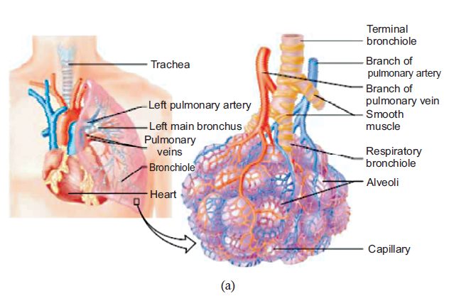
Figure 9.11: (a) Detail structure of human respiratory system: the lungs and the alveoli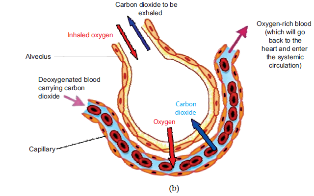
(Source: Vander et. al., Human Physiology, 2001) (b) gas exchange in the alveoli
Each lung is enclosed and protected by a double-layered serous membrane called the pleural
membrane. It consists of two layers: the outer parietal pleura and the deeper visceral pleura.
The space between the two is called the pleural cavity and contains a small amount of lubricating
fluid secreted by the membranes. The important function of this pleural fluid is to reduce friction
between the membranes during breathing movement.
9.6.2 Functions of Tissues within the Gas Exchange System
The respiratory system consists of four main layers (Figure 9.12):
(i) The respiratory mucosa (epithelium and supporting lamina propria)
(ii) Submuscosa
(iii) Cartilage and/or muscle layer(iv) Adventitia
Figure 9.12: Diagrammatic representation of cross section of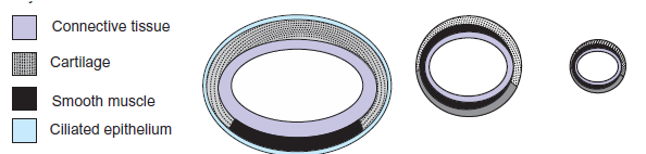
airways showing distribution of tissues (not to scale)
Trachea
The trachea is a wide flexible tube. The respiratory mucosa and submucosa are adapted to warm
and moisten the air, and to trap particles in mucous. It consists of pseudostratifed columnar,
ciliated epithelium with mucous secreting goblet cells. It has twenty C-shaped rings of hyaline
tracheal cartilage which supports the trachea and keeps its lumen open. The gaps between the
rings of cartilage are filled by a bundle of smooth muscle (trachealis muscle) and fibroelastic
tissue. These structures together gives flexibility for ventilation. Adventitia is the outermost
fibroelastic connective tissue layer.
The respiratory mucosa is made up of the epithelium and supporting lamina propria. The
epithelium is tall columnar pseudostratified with cilia and goblet cells. Lamina propia lies
underneath the epithelium. It contains elastin and has a supporting role. Blood vessels warm
the air. The sub-mucosa contains mixed sero-mucous glands. The watery secretions from the
serous glands humidify the inspired air. The mucous, together with mucous from the goblet
cells traps particles from the air which are transported upwards towards the pharynx by the
cilia on the epithlium. This helps to keep the lungs free of particles and bacteria. There are lots
of seromucous glands in the submucosa layer.
The epithelial surfaces of the airways upto the end of the respiratory bronchioles have cilia that
constantly beat toward the pharynx. They also contain mucous secreting glands (Figure 9.13).
This mucous keeps the lungs clear of particulate matter and the many bacteria that enter the
body on dust particles. Macrophage present in the airways and alveoli also protect againstinfection.
Bronchi
Bronchi have the same basic structure as trachea. A few differences are respiratory epithelium
are less tall than that of trachea and contains fewer goblet cells. Lamina propia has more elastic
tissue. Muscularis mucosae begin to appear in lamina propia and submucosa. There are fewer
submucosal glands and cartilage is in plates. There is less cartilage in the tertiary bronchi, Itdoes not completely encircle the lumen.
Bronchioles
The tertiary bronchii branch into bronchioles. They have a diameter of 1mm or less, and the
wall structure changes. There is no cartilage and no glands. The ring of smooth muscle is
arranged in discrete bundles with a variety of organisations. The epithelium is made up of
ciliated columnar cells in larger bronchioles, or nonciliated in smaller bronchioles. There are
no goblet cells, but there are cells called Clara cells. These are secretory cells and they secreteone of the components of surfactant.
Terminal Bronchioles
The final branches of the bronchioles are called terminal bronchioles. These have a layer smooth
muscle surrounding their lumens. Stimulation of the vagus nerve (parasympathetic) causes the
smooth muscle to contract, and reduce the diameter of the terminal bronchioles. Small sacs are
found extending from the walls of the terminal bronchi called respiratory bronchioles. These
are lined by a ciliated cuboidal epithelium, and some non-ciliated cells called clara cells. Therespiratory bronchii have a few single alveoli off their walls.
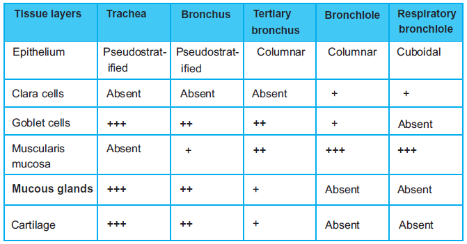
Figure 9.13: Table showing different tissue layers of the gas exchange system
Alveoli
The alveoli are the sites of gas exchange with the blood. The wall of the air-facing surface(s)
are lined by type I alveolar cells which is a one cell thick, continuous layer of flat epithelial
cells. Type II alveolar cells are thicker specialized cells producing a detergent-like substance
called surfactant and they are interspersed between type I cells. In some of the alveolar walls,
pores are present which permit the flow of air between alveoli.
The alveolar walls contain capillaries and a very small interstitial space, made of interstitial
fluid and a loose meshwork of connective tissue. However, the interstitial space is absent
altogether at most places and the basement membranes of the alveolar-surface epithelium and
the capillary-wall endothelium fuse. As a result, the blood within an alveolar-wall capillary
is separated from the air within the alveolus by an extremely thin barrier around 0.2 μm. The
branching of bronchioles and the vast number of alveoli collectively increases the respiratory
surface area to as much as 80 square metres. The extensive surface area of alveoli in contact
with capillaries and the thin barrier results in the rapid exchange of large quantities of oxygen
and carbon dioxide by diffusion.
APPLICATION 9.3
1.Complete with appropriate terms:
(i)Tiny air containing sacs found in the human gaseous exchange system are................ .
(ii) ................ is a wide flexible tube in human gas exchange
(iii) Respiratory mucosa in humans is made up of ................ and ................
(iv) Alveoli are lined up by ................ and ................2.Explain why the barrier to diffusion must be as thin as possible
9.6.3 Mechanism of Ventilation (Breathing)
Inspiration (inhalation or breathing in) is the movement of air from the external environment
through the airways into the alveoli during breathing. Expiration (exhalation) is movement inthe opposite direction. An inspiration and an expiration constitute a respiratory cycle.
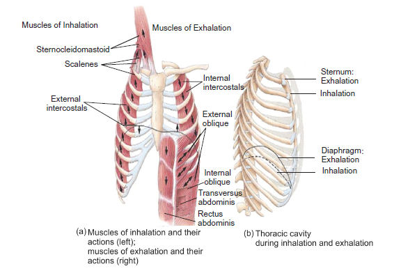
Figure 9.14: Structure and mechanism of lung ventilation in human
Inhalation: Air will move into the lungs when air pressure inside the lungs is less than that
of outside (atmospheres). Expansion of the lungs increases the volume and so the pressureinside the lungs decreases. Expansion of the lungs during normal quiet inhalation is achieved
by contraction of the diaphragm and external intercostals which are the main muscles of
inhalation (Figure 9.14). The diaphragm is the dome-shaped skeletal muscle that forms the
floor of the thoracic cavity. Contraction of the diaphragm causes it to flatten, lowering its
dome. This increases the vertical diameter of the thoracic cavity. Around 75% of air enters
the lungs by this action. Also contraction of the external intercostals elevates the ribs resulting
in an increase in the volume of the chest cavity. About 25% of the air that enters the lungs
during normal quiet breathing is due to this action. As the volume of the lungs increases and
the pressure inside the lungs (alveolar or intra-pulmonic pressure) decreases and atmosphericair rushes into the lungs.
Exhalation: On the other hand if the volume of the lungs decreases, pressure inside the lungs
increases. As a result, air rushes out of the lungs resulting in exhalation or expiration. However,
normal exhalation during quiet breathing, unlike inhalation, is a passive process because no
muscular contractions are involved. Exhalation results from elastic recoil of the chest wall
and lungs. Elastic recoil is the natural tendency of the chest wall and the lungs to spring back
after they have been stretched. The inspiratory muscles relax with the start of exhalation.
Diaphragm and external intercostal muscles also relax resulting in decrease in volume of the
lungs, causing air to move out of the lungs. Interestingly, exhalation becomes an active process
(requiring energy supply) only during the time of forced exhalation (for example during heavy
exercise etc). During these times, the muscles of exhalation are the abdominals and internalintercostals muscles which contract to increase pressure in the abdominal region and thorax.
Gas Exchange in Alveoli
Alveoli are the respiratory units of lungs. The alveolar and capillary walls together form
the respiratory membrane. The exchange of gases in the alveoli and between the air
spaces in the lungs and the blood takes place by diffusion across this respiratory membrane
(Figure 9.15).
The pressure of a specific gas (x) in a mixture is called its partial pressure (Px). The difference
in partial pressures determines the movement of O2 and CO2 between the atmosphere and
lungs, between the lungs and blood, and between the blood and body cells. Gas diffuses across
a permeable membrane from an area where its partial pressure is higher to the area where its
partial pressure is low and the rate of diffusion is directly proportional to the difference inpartial pressure.
Figure 9.15: Detail structural components of an alveolus: (a) section through alveolus and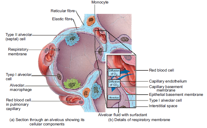
(b) details of respiratory membrane
As stated earlier, external respiration or pulmonary gas exchange is the diffusion of O2 from
air in the alveoli of the lungs to blood in pulmonary capillaries and the diffusion of CO2 in the
opposite direction. In this process, blood picks up O2 from alveolar air and unloads CO2 into
alveolar air as it flows through pulmonary capillaries. In a resting person, PO2 is 105 mmHg
in the alveolar air which is higher than that of blood in pulmonary capillaries, where it is only
40 mmHg. This results in diffusion of O2 from alveolar air into pulmonary capillaries.
However, CO2 diffuses in the opposite direction because the PCO2 of deoxygenated blood is
45 mmHg in a resting person, and the PCO2 of alveolar air is 40 mmHg. Hence, carbon dioxide
diffuses from deoxygenated blood into the alveoli until the PCO2 of the blood decreases to40 mmHg.
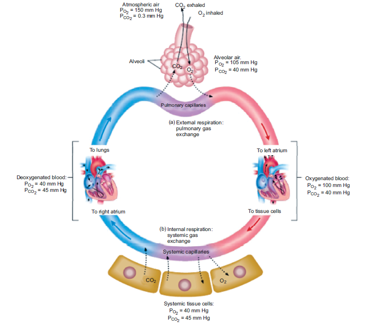
Figure 9.16: Exchange of gases in alveoli of humans
As a result of this diffusion, the capillary blood PO2 rises while its PCO2 falls. This process of
diffusion continues as long as there is difference in partial pressure of the two gases between
the two sides. An equilibrium is reached well before the end of the capillaries because blood
flow in the capillaries is slow and gas exchange is rapid. Oxygenated blood now leaves the
pulmonary capillaries to return to the heart from where it is pumped into the systemic arteries.
The exchange of O2 and CO2 between systemic capillaries and tissue cells is called internalrespiration or systemic gas exchange (Figure 9.16).
9.6.4 Lung Volume and Capacities
Tidal volume: It is the volume of air entering the lungs during a single inspiration during
normal quiet breathing. It is about 500 ml. It is approximately equal to the volume leaving onthe subsequent expiration.
Inspiratory reserve volume: The maximal amount of air that can be increased above the resting
tidal volume during deepest/forced inspiration is termed the inspiratory reserve volume. It is
about 3000 ml in average adult males which is sixfold greater than resting tidal volume and1900 ml in average adult females.
Expiratory reserve volume: The 500 ml of air inspired with each resting breath adds to and
mixes with the much larger volume of air already in the lungs, and then 500 ml of the total is
expired. However, through maximal active contraction of the expiratory muscles i.e., forced
expiration, it is possible to expire much more of the air remaining after the resting tidal volume
has been expired; this additional expired volume is termed the expiratory reserve volume(about 1500 ml).
Residual volume: Even after a maximal active expiration, approximately 1000 ml of air
still remains in the lungs. This is because the subatmospheric intrapleural pressure keeps the
alveoli slightly inflated, and some air also remains in the non-collapsible airways. This volume,
which cannot be measured by spirometry, is called the residual volume and amounts to about1200 ml in males and 1100 ml in females.
Vital capacity: It is the maximal volume of air that a person can expire after a maximal
inspiration. It is a useful clinical measurement for detecting various respiratory system related
conditions. It is the sum of inspiratory reserve volume, tidal volume, and expiratory reservevolume (4800 ml in males and 3100 ml in females).
Inspiratory capacity is the sum of tidal volume and inspiratory reserve volume
(500 ml + 3100 ml = 3600 ml in males and 500 ml + 1900 ml = 2400 ml in females).
Total lung capacity is the sum of vital capacity and residual volume
(4800 ml + 1200 ml = 6000 ml in males and 3100 ml + 1100 ml + 4200 ml in females).
However, in a normal adult only 70% (=350 ml) of tidal volume reaches the respiratory zone
because of the presence of anatomical dead space. Dead Space refers to the conducting airways
which have a volume of about 150 ml. Exchanges of gases with the blood does not occur in
this 150 ml of the airways. It occurs only in the alveoli. Since these airways do not permit gasexchange with the blood, the space within them is termed the anatomic dead space. Thus, the
volume of fresh air entering the alveoli during each inspiration equals the tidal volume minus
the volume of air in the anatomic dead space.
Alveolar ventilation: The total volume of fresh air entering the alveoli per minute is called the
alveolar ventilation which is given by,
Alveolar ventilation (ml/min) = (Tidal volume – Dead space) × Respiratory rate
(ml/breath) (ml/breath) (breath/min)
= (500 – 150) ml/breath × 12 breath/min= 350 × 12 = 4200 ml/min.
ACTIVITY 8
Calculate pulmonary ventilation (PV) and alveolar ventilation (AV) from the data provided.
(i) Tidal volume = 550 ml, Dead space = 185 ml, Respiratory rate = 17/min, inspiratory
reserve volume = 2500 ml, tidal volume = 550, and expiratory reserve volume = 1450.
(ii) Tidal volume = 600 ml, Dead space = 195 ml, Respiratory rate = 15/min, inspiratory
reserve volume = 2800 ml, tidal volume = 600, and expiratory reserve volume = 1350.
(iii) Tidal volume = 550 ml, Dead space = 175 ml, Respiratory rate = 20/min, inspiratory
reserve volume = 2500 ml, tidal volume = 500, and expiratory reserve volume = 1500.
Hint: What is formula for calculation of pulmonary ventilation (PV) and alveolar ventilation(AV)?
Importance of Lung Capacities
These pulmonary function tests are useful diagnostic tools:
• An examination of ventilation function of lungs is necessary for evaluation of functional
properties of human respiratory system.
• It is used for estimation of defects in respiratory system and also for consideration of fitness
load in sports medicine.
• Various respiratory disorders may be diagnosed by comparison of actual and predictednormal values for a patient’s gender, height, and age.
APPLICATION 9.4
1. Complete with appropriate terms:
(i). Maximum volume of air that a person can expire after a maximal inspiration is called
................ .
(ii). Diffusion of O2 from air to expillaries and CO2 in opposite direction is called ...............
(iii). During .................. volume of lungs increases in humans.2. Describe how a steep diffusion gradient is achieved in lungs
Spirometry
The spirometer is an apparatus for measuring inspired and expired volumes during breathingand the respiratory rate (Figure 9.17). The record is called a spirogram (Figure 9.18).
Figure 9.17: (a) A basic model of a spirometer; and
(b) a patient taking readings on a modern spirometer
Use of Spirometer to Measure Ventilation Rate
The lung volumes and capacities can be measured by routine spirometry. A typical spirometer
is a tube like instrument with an open end called the mouthpiece. The spirometer (Figure 9.17a)
consists usually of a water-filled tank with a bell shaped floating device. A tube connects the
air space within the spirometer with the airways of the person whose lung volumes is being
measured. A counterweight is placed on the bell. The position of the bell indicates how much
air is in the spirometer and is calibrated in volume units. A person under the test blows air into
it after deep breath. Usually, the airway through nose is shut or blocked using a clip so that
air can only enter or leave through the mouth. Inhalation is recorded as an upward deflection,
and exhalation is recorded as a downward deflection. The bell on the spirometer rises when
the person blows into the device (expiration), and falls during inspiration. If the spirometer
is equipped with a recording device (spirograph), it can also be used for graphic measurement
of the total ventilation per unit time. Based upon the reading indicated corresponding to each
breathing in or out, an expert physician can diagnose the health of the person’s lungs and detect
disorder if any. Nowadays, the instrument is integrated with a computer system to accuratelymonitor the readings and give instant results.
Figure 9.18: Spirogram of lung volumes and capacities in a healthy man and woman
(within parentheses). The spirogram is read from the right(i.e., start of record) to the left (i.e., end of record)
ACTIVITY 9
Aim: To design a model of the spirometer based on its main features.
Materials Required:
1. 2–3 litre empty soda/cold drink bottle with cap
2. One or two feet long piece of plastic tubing
3. One measuring cup with units in millilitres
4. One bucket or pan that can hold more than 3 litres of water5. One permanent marker
Procedure:
1. Marking measuring lines on the bottle: Add 500 ml of water to the soda/cold drink
bottle using the measuring cup and mark a line with the marker at the top of the water
level. Repeat this until the bottle is full. When the bottle is full, put the cap on the
bottle.
2. Add sufficient water to the bucket or pan to submerge the soda bottle.
3. Invert the soda bottle and submerge it in the bucket, and remove the cap under the water.
4. Open the bottle underwater to prevent any unwanted air from entering the bottle.
5. Place one end of the tubing into the soda bottle in the water, and leave the other end outside
of the water.
6. Use the tubing to blow into, for determining lung capacity.
7. Remember to place the bottle in the water upside down before removing the cap.
8. Don’t forget to insert one end of the hose in the bottle after you open the cap
underwater9. Before you exhale into the tubing, your spirometer should resemble the picture below.
10. Using the spirometer to obtain the readings
(i) One student holds the bottle to keep it from flipping over. Another student inhales
normally and then exhales the air normally into the tubing connected to the
spirometer. Note: Do not blow out all the “extra” air in your lungs.
(ii) Note the amount of air you exhaled, remembering that each line on the bottle
represents a half litre, starting from the top down.
(iii) Record this volume, it is your “tidal volume.” The tidal volume is the amount of air
that you normally breathe in and out.
(iv) Refill the bottle with water and reinsert the tubing. One student holds the bottle while
another take a few normal breaths initially. This is to get a good reading in the next
step. Then inhale as much air as you can and exhale this air into the end of thetubing outside of the water.
(v) Again note the amount of air you exhaled by looking at the lines on the soda bottle.
(vi) This volume is your “inspiratory reserve.” The inspiratory reserve is the amount of air
that your lungs can hold in.
(vii) Refill the bottle with water and reinsert the tubing. One student holds the bottle while
the other takes a few normal breaths to get himself back to a normal breath. Then
exhale as much air as you can into the end of the tubing outside of the water.
(viii) Note the amount of air you exhaled by looking at the lines on the soda bottle.
(ix) This is your “expiratory reserve.” The expiratory reserve is the amount of air that your
lungs can blow out after a normal breath.
Observations:
The vital capacity is the greatest change in volume that can occur in the lungs.
Inspiratory Reserve + Expiratory Reserve + Tidal Volume = Vital Capacity
ACTIVITY 10
Aim: To use the illustrations of spirometer trace to define tidal volume, inspiratory reserve
volume, expiratory reserve volume, vital capacity and residual volume.
Materials Required: Notebook, pen, pencil etc.
Procedure:
1. First write down the definitions of tidal volume, inspiratory
reserve volume, expiratory reserve volume, vital capacity
and residual volume.
2. Using a colour pencil/pen note try to locate the tidal
volume in the spirometer trace provided.
3. Perform step 2 above for inspiratory reserve volume,
expiratory reserve volume, vital capacity and residual
volume.
4. Give a logical explanation for your labelling.
5. Show the labelled spirometer trace to your teacher and explain your results. Ask for correctionsif any.
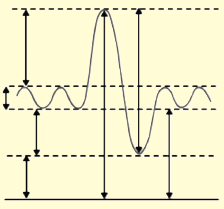
9.7 NERVOUS CONTROL OF BREATHING
ACTIVITY 11
Inform the class about the function or role of a particular part of the brain controlling the
process of respiration in humans viz. the medullary rhythmicity area and the pneumotaxic area
in the pons.
Breathing depends entirely upon cyclical respiratory muscle excitation of the diaphragm and
the intercostal muscles by their motor nerves as a result of nerve impulses transmitted to them
from centres in the brain. When a burst of action potentials is initiated in the nerves to the
inspiratory muscles, these muscle contracts and inspiration occurs. When these action potentials
stop, the inspiratory muscles relax, and expiration occurs as the elastic lungs recoil. Similarly,
in situations when expiration is facilitated by contraction of expiratory muscles, the nerves to
these muscles, begin firing during expiration. This neural activity is primarily controlled byneurons in the medulla oblongata.
The respiratory centre is the cluster of neurons located bilaterally in the medulla oblongata
and pons of the brain stem. It can be divided into three areas on the basis of their functions(Figure 9.19):
1. The medullary rhythmicity area in the medulla oblongata:
• Controls the basic rhythm of respiration.
• There are inspiratory and expiratory areas.
• Nerve impulses generated in the inspiratory area establish the basic rhythm of
breathing during quiet breathing by causing contraction of external intercostal muscle.
• The neurons of the expiratory area remain inactive during quiet breathing. However,
during forceful breathing nerve impulses from the inspiratory area activate the
expiratory area.
• Impulses from the expiratory area cause contraction of the internal intercostal and
abdominal muscles, which decrease the size of the thoracic cavity and causes forceful
exhalation.
2. The pneumotaxic area in the pons:
• Transmits inhibitory impulses to the inspiratory area.
• The major effect of these nerve impulses is to help turn off the inspiratory area before
the lungs become too full of air.
• In other words, the impulses shorten the duration of inhalation. When the pneumotaxicarea is more active, breathing rate is more rapid.
3. The apneustic area in the lower pons:
• This area sends stimulatory impulses to the inspiratory area that activate it and prolong
inhalation.
• The result is a long, deep inhalation.• When the pneumotaxic area is active, it overrides signals from the apneustic area.
Figure 9.19: Respiratory centre in the human brain
In addition to the above, there are ‘Pulmonary stretch receptors’ in the smooth-muscle layer
of the airway. They respond to stretch stimulus on this muscles. Whenever there is large lung
inflation, they are activated. Electric signals in the afferent nerve fibres from the stretch receptors
travel to the brain and inhibit the medullary inspiratory neurons. This phenomenon is known as
the Hering-Breur inflation reflex. Thus, inspiration is terminated by feedback from the lungs.
However, this pulmonary stretch-receptor reflex plays a role in setting respiratory rhythm onlyunder conditions of very large tidal volumes, for example in rigorous exercise
APPLICATION 9.5
1.Complete with appropriate terms:
(i)................ is used to measure inspired and expired volumes of air.
(ii) Respiratory centre is a cluster of neurons located bilaterally in ................ and ................
of brain stem.
(iii) ................ in brain prolongs inhalation.2. Explain the importance of the cartilage found in the trachea and bronchi
9.8 SUMMARY
• Aerobic animals require a continuous supply of oxygen for metabolic processes and also
removal of metabolic waste (CO2) from its body.
• This is achieved by developing a complex system of gas exchange in every animal.
• Gases exchange takes place by the process of diffusion where it moves from a place of
higher concentration to a place of lower concentration.
• Small invertebrates like insects have a vast network of ‘tubes’ made of chitin called the
tracheal system spread all over their body which is used for exchange of gases.
• The tubes or trachea branches and interbranches to form fine tubes called tracheoles
innervating tissues at cellular level. Air enters and leaves through openings called spiracles.
• In aquatic animals like fish and some amphibian larvae exchange of gases takes place
through special structures called gills.
• Gills can be external or internal depending on its location in the body. Gills are highly
vascular, thin and always ventilated with water.
• A holobranch or complete gill refers to a branchial arch and the lamellae on both
anterior and posterior faces of its septum. A gill arch with lamellae on only one face is
a hemibranch.
• Ventilation of gills in fish is achieved by the coordinated action of the buccal cavity and
the operculum or gill cover.
• Countercurrent mechanism of gas exchange is present in gills of teleost. It is a very
efficient mechanism of gas exchange and almost 85% of oxygen is extracted from water.
• Amphibians can respire through skin (cutaneous respiration), gills or the lungs.
• Exchange of gas in the skin, gills or the lungs takes place by diffusion of gas (O2) from
air or water to the blood capillaries in the skin or the septal walls of faveoli.
• In humans, exchange of gases takes place through the lungs. The lungs are elastic
structures. The lungs, the airways leading to them, and the chest structures responsible
for movement of air into and out of the lungs.
• The conducting zone of the airways consists of the trachea, bronchi, and terminal
bronchioles. The respiratory zone of the airways consists of the alveoli, which are the
sites of gas exchange.
• The alveoli are lined mostly by type I cells along with some type II cells, which produce
surfactant.
• The lungs are covered by pleura and between the two pleural layers is an extremely thin
layer of intrapleural fluid.
• During inspiration, the contractions of the diaphragm and inspiratory intercostal muscles
increase the volume of the thoracic cage causing atmospheric air to rush into the lungs.
• During expiration, the inspiratory muscles cease contracting, allowing the elastic recoil
of the chest wall and lungs to return them to their original between-breath size resulting
in the air moving out of the lungs through the nose.
• The vital capacity is the sum of resting tidal volume, inspiratory reserve volume, and
expiratory reserve volume.
• Gases diffuse from a region of higher partial pressure to a region of lower partial pressure.
Exchange of gases in lungs and tissues takes place though the process of diffusion because
of the differences in partial pressures of gases.
• There is net diffusion of oxygen from alveoli to blood and of carbon dioxide from blood
to alveoli when systemic venous blood flows through the pulmonary capillaries.
• In certain conditions like when the alveolus capillary surface area is decreased or when
the alveolar walls thicken inadequate gas exchange between alveoli and pulmonary
capillaries may occur
9.9 GLOSSARY
• Alveoli: The plural of alveolus. The alveoli are firy air sacs within the human lungs
where exchange of gases takes place.
• Breathing: The process of taking air in and expelling it from lungs through the nose or
mouth.
• Bronchi: The plural of bronchus. It is any of the major air passages of lungs which
diverge from windpipe.
• Counter current mechanism: Maintenance of equilibria in the concentration of any
permeable materials under two conditions.
• Cutaneous respiration: The process of respiration through skin.
• Exhalation: It is the process or act of exhaling out air, generally CO2.
• Gills: These are the paired respiratory organ of fish and some amphibians, by which
oxygen is extracted from water.
• Inhalation: It is the process or act of inhaling in air, generally O2.
• Paveoli: A small pit or cavity resembling a cell of a honey comb alveola.
• Spirometer: An apparatus for measuring inspired and expired volumes of air during
breathing.
• Trachea: A large membranous tube of cartilage extending from larynx to bronchial tubes
and conveying air to and air from the lungs.• Ventilation: The bodily process of inhalation and exhalation.
END UNIT ASSESSMENT 9
Do all these exercises in your exercise book.
I. Choose whether the following statements are True (T) or False (F)
1. Insects have a specialised system of ‘tubes’ called the tracheal system for exchange
of gases.
2. There is active ventilation in most treacheates (i.e., animals possessing trachea).
3. Fish gills consist of thousands of highly specialised gill lamellae enclosed in a gill
cavity.
4. Amphibians use the moist skin, gills or the lungs for gas exchange.
5. Modern amphibians do not rely heavily on cutaneous respiration.
6. Most adult amphibians have lungs for breathing air.
7. Internal (tissue) respiration is the exchange of gases between blood in systemic
capillaries and tissue cells.
8. Alveoli are the structure for gas exchange in humans.
9. The apparatus for measuring inspired and expired volumes during breathing is a
spirometer.
10. The sum of inspiratory reserve volume, tidal volume, and expiratory reserve volumeis called residual volume.
II. Long Answer Type Questions
1. Describe the tracheal system of insects and relate to its function.
2. Describe the structure of the gills in relation to its function.
3. In your own words, explain the significance of counter current flow in bony fish.
4. Describe the mode of gaseous exchange in amphibians.
5. Describe the structure of the human gas exchange system.
6. Describe the distribution of tissues within the trachea, bronchi, bronchioles and
alveoli and relate each tissue to its function.
7. Explain the mechanism of ventilation in humans.
8. Explain the process of gas exchange in alveoli with emphasis on diffusion.
9. Describe the role of the brain in controlling gas exchange in humans.
10. Define the following terms related to the lung capacities:
(i) Tidal volume (ii) Reserve volume
(iii) Vital capacity (iv) Residual volume
(v) Dead air space
11. Describe how a spirometer can be used to measure vital capacity, tidal volume,
breathing rates, and oxygen uptake.
12. Calculate vital capacity and alveolar ventilation from the data provided.
Tidal volume = 550 ml, Dead space = 185 ml, Respiratory rate = 17/min, inspiratory
reserve volume = 2500 ml, tidal volume = 550, and expiratory reserve volume = 1450.
13. What contribution does exchange of gases make on global warming? Discuss your
answer with relevant data. Also throw light on the dialect “Global warming: a mythor truth.”
Unit 10 Smoking and Related Diseases
Key Unit Competence
To be able to describe the effects of tobacco smoking on the gas exchange system.
LEARNING OBJECTIVES
At the end of this unit, the learner will be able to:
• state the features of a genetic code.
• describe the effects of tar and carcinogens in tobacco smoke on the gas exchange system.
• describe the signs and symptoms of lung cancer and chronic obstructive pulmonary diseases
(COPD).
• describe the effects of nicotine and carbon monoxide on the cardiovascular system.
• explain how tobacco smoking contributes to atherosclerosis and coronary heart disease.
• interpret photographs to differentiate healthy lungs from infected lungs.
• interpret data linking cigarette smoking to disease and early death.
• observe and interpret research statistics linking to tobacco smoking.
• evaluate the epidemiological and experimental evidence linking cigarette smoking to disease
and early death.• influence the campaign against cigarette smoking.
INTRODUCTORY ACTIVITY
Observe the following image and answer the following

Suggest the possible consequences undergone by that man who smokes
10.1 CIGARETTE SMOKING
ACTIVITY 1
To observe the effect of tobacco on animals.
Collect some tobacco leaves or cigarette butts and boil them with water.
Allow the solution to cool. Now filter it with a stainer. Pour the solution into
a large squirt bottle. Spray the solution on a plant infested with aphids. Wait
for a while. Do you find the aphids stay on the plant? What makes them
leave the plant? This is the tobacco that forces the pests to leave the plant.
Tobacco leaves contain toxins which are harmful to human.
ACTIVITY 2
To observe the effect of cigarette smoking.
Put some cotton bolls inside a flexible plastic bottle. Wrap
some modelling clay around the cigarette as shown. Fit
the cigarette on the mouth of the bottle with the filter end
inside. Light the cigarette end outside the bottle. Squeeze
and release the bottle to simulate smoking. When the
cigarette is almost finished, remove it from the bottle.
Take out the cotton bolls on a petri dish. Touch the bolls
with your finger. Do you find some black coloured tar onthe bolls? Where does this tar come from?
This is how cigarette smoke harms the lungs. Cigarette smoking is very harmful for health,
and it nearly affects every organ of the body, causes many diseases, and reduces the health of
smokers in general.
Cigarette smoking is the leading preventable cause of death in the United States.
• Smoking causes more deaths each year than the following causes combined:
♦ Human immunodeficiency virus (HIV)
♦ Illegal drug use
♦ Alcohol use
♦ Motor vehicle injuries
♦ Firearm-related incidents
• Smoking causes about 90% (or 9 out of 10) of all lung cancer deaths in men and women.
More women die from lung cancer each year than from breast cancer.
• About 80% (or 8 out of 10) of all deaths from chronic obstructive pulmonary disease
(COPD) are caused by smoking.• Cigarette smoking increases risk for death from all causes in men and women.
Tobacco Control
ACTIVITY 3
Discuss the steps to control the use of tobacco. Everyone knows the detrimental effects oftobacco but still it is used. Write a journal stating why the youth need to control tobacco.
Tobacco control is a field of international public health science, policy and practice to reduce
the tobacco use which causes high morbidity and mortality due to tobacco smoke. Tobacco
control is a priority area for the World Health Organization (WHO), through the Framework
Convention on Tobacco Control. It has started to protect present and future generations from the
devastating health, social, environmental and economic consequences of tobacco consumption
and exposure to tobacco smoke. Progress was initially notable at a state or national level,
particularly the pioneering smoke-free public places legislation introduced in New York City
in 2002 and the Republic of Ireland in 2004, and the UK effort for tobacco control in 2004.
WHO produced an internationally-applicable and now widely recognised summary of the
essential elements of tobacco control strategy, publicised as the mnemonic MPOWER tobacco
control strategy. The 6 components are:
Monitor tobacco use and prevention policies
Protect people from tobacco smoke
Offer help to quit tobacco use
Warn about the dangers of tobacco
Enforce bans on tobacco advertising, promotion and sponsorshipRaise taxes on tobacco
10.2 DANGEROUS CHEMICALS IN TOBACCO SMOKE
Smokers inhale about 7,000 chemicals in cigarette smoke. Many of these chemicals come from
burning tobacco leaf. Some of these compounds are chemically active and trigger profound
and damaging changes in the body. Tobacco smoke contains over 60 known cancer-causing
chemicals. Smoking harms nearly every organ in the body, causing many diseases and reducing
health in general.
The most damaging components of tobacco smoke are:
1. Tar: Tar is the collective term describing toxins produced by smoking cigarettes and the
coating they place on the lungs. Tar is sticky and brown, and stains teeth, fingernails andlung tissue. Tar contains the carcinogen benzo(a)pyrene. When inhaled, these toxins form
a particulate matter that coats lungs much the same way that soot from log fires coats
chimneys. But unlike chimneys, which are made of stone or brick, human lungs are made
of thin, delicate tissue not intended for toxic smoke intake.
2. Nicotine is the addictive drug in tobacco smoke that causes smokers to continue
to smoke and affects the brain activity. Addicted smokers need enough nicotine over
a day to ‘feel normal’—to satisfy cravings or control their mood.
How Nicotine Affects the Brain
• Brain is made up of billions of nerve cells. They communicate by releasing chemical
messengers called neurotransmitters. Each neurotransmitter is like a key that fits
into a special “lock,” called a receptor, located on the surface of nerve cells. When aneurotransmitter finds its receptor, it activates the receptor’s nerve cell.
• The nicotine molecule is shaped like a neurotransmitter called acetylcholine. Acetylcholine
and its receptors are involved in many functions, including muscle movement, breathing,
heart rate, learning, and memory. They also cause the release of other neurotransmitters
and hormones that affect your mood, appetite, memory, and more. When nicotine
gets into the brain, it attaches to acetylcholine receptors and mimics the actions of
acetylcholine. Nicotine also activates areas of the brain that are involved in producing
feelings of pleasure and reward. Recently, scientists discovered that nicotine raises the
levels of a neurotransmitter called dopamine in the parts of the brain that produce feelings
of pleasure and reward. Dopamine, which is sometimes called the pleasure molecule, is
the same neurotransmitter that is involved in addictions to other drugs such as cocaine
and heroin. Researchers now believe that this change in dopamine may play a key role in alladdictions.
3. Carbon monoxide (CO): This odourless gas is fatal in large doses because it takes the place
of oxygen in the blood. It is also called ‘Silent killer’. Each red blood cell contains a protein
called haemoglobin that transports oxygen molecules around the body. However, carbonmonoxide binds to haemoglobin better than oxygen. In response, the body makes more
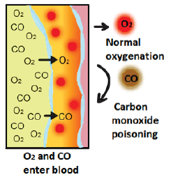
Figure 10.2: Carbon monoxide poisoning
red blood cells to carry the oxygen it needs, but it makes the blood thicker. This means
that when the body demands more oxygen during exercise, less oxygen reaches the brain,heart, muscles and other organs.
How it Affects to the Body
• Carbon monoxide poisoning occurs when carbon monoxide builds up in your bloodstream.
When too much carbon monoxide is in the air, your body replaces the oxygen in the red
blood cells with carbon monoxide. This prevents oxygen from reaching your tissues and
organs. This leads to serious tissue damage, or even death.
Carbon monoxide poisoning can be especially dangerous for people who are sleeping orintoxicated. People may have irreversible brain damage or even be killed before anyone realizes
Figure 10.3: Typical symptoms of carbon monoxide poisoning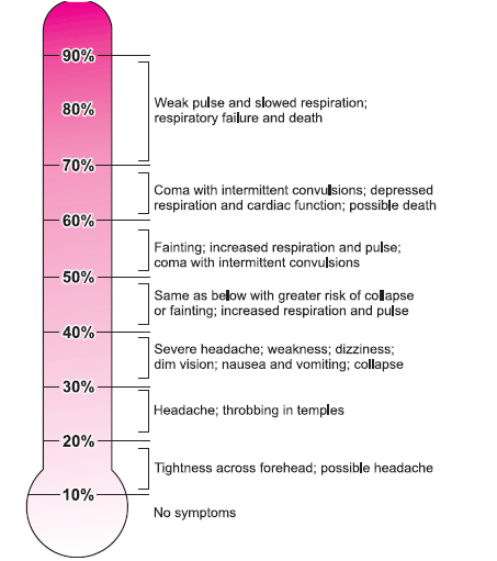
(increasing percentage of CO in blood).
• Hydrogen cyanide – The lungs contain tiny hairs (cilia) that help to clean the lungs by
moving foreign substances out. Hydrogen cyanide stops this lung clearance system from
working properly, which means the poisonous chemicals in tobacco smoke can build up
inside the lungs. Other chemicals in smoke that damage the lungs include hydrocarbons,
nitrous oxides, organic acids, phenols and oxidising agents.
• Oxidizing chemicals – These highly reactive chemicals (which include free radicals) can
damage the heart muscles and blood vessels. They react with cholesterol, leading to the
build-up of fatty material on artery walls. Their actions lead to heart disease, stroke and
blood vessel disease.
• Metals – Tobacco smoke contains dangerous metals including arsenic, cadmium and lead.
Several of these metals are carcinogenic.
• Radioactive compounds – Tobacco smoke contains radioactive compounds that are knownto be carcinogenic.
APPLICATION 10.1
1. Complete with appropriate terms:
(i)................... is also called Silent killer.
(ii) ................... is the addictive drug found in cigarette.
(iii) Tar contains carcinogen ...........................
(iv) Radiation compounds present in tobacco smoke are ............................
(v) Carbon monoxide poisoning .................... percentage of CO in flood.
2. Name the three main substances in tobacco smoke that can be harmful
3. Explain how the deposition of tar can lead to smoker’s cough4. Explain why smokers are more than non-smokers to suffer from infections in the lungs
10.3 LUNG CANCER AND SMOKING
Lung cancer is strongly correlated with cigarette smoking, with about 90% of lung cancers
arising as a result of tobacco use. The risk of lung cancer increases with the number of cigarettes
smoked over time. If someone smokes one pack of cigarettes per day, he/she has a risk for the
development of lung cancer that is 25 times higher than a non-smoker. Among those who smoke
two or more packs of cigarettes per day, one in seven will die of lung cancer. But even though
the risk is higher; the more you smoke, there is no safe level of exposure to tobacco smoke.
Passive smoking, or the inhalation of tobacco smoke from other smokers sharing living or
working quarters, is also an established risk factor for the development of lung cancer. Research
has shown that non-smokers who reside with a smoker have a 24% increase in risk for developinglung cancer when compared with other non-smokers.
Lung cancer is the uncontrolled growth of abnormal cells that start off in one or both lungs;
usually in the cells that line the air passages. The abnormal cells do not develop into healthylung tissue, they divide rapidly and form tumours.
As tumours become larger and more numerous, they undermine the lung’s ability to provide
the bloodstream with oxygen. Tumours that remain in one place and do not appear to spreadare known as “benign tumours”.
Malignant tumours, the more dangerous ones, spread to other parts of the body either through
the bloodstream or the lymphatic system. Metastasis refers to cancer spreading beyond its siteof origin to other parts of the body. When cancer spreads, it is much harder to treat successfully.
Primary lung cancer originates in the lungs, while secondary lung cancer starts somewhere
else in the body, metastasizes, and reaches the lungs. They are considered different types ofcancers and are not treated in the same way.
According to the World Health Organization (WHO), 7.6 million deaths globally each year
are caused by cancer; cancer represents 13% of all global deaths and lung cancer is by far thenumber one cancer killer.
10.3.1 Signs and Symptoms of Lung Cancer
The most common symptoms of lung cancer are:
• A cough that does not go away or gets worse
• Chest pain that is often worse with deep breathing, coughing, or laughing
• Hoarseness
• Weight loss and loss of appetite
• Coughing up blood or rust-coloured sputum (spit or phlegm)
• Shortness of breath
• Feeling tired or weak
• Infections such as bronchitis and pneumonia that don’t go away or keep coming back
• New onset of wheezing
If lung cancer spreads to distant organs, it may cause:
• Bone pain (like pain in the back or hips)
• Nervous system changes (such as headache, weakness or numbness of an arm or leg,
dizziness, balance problems, or seizures), from cancer spread to the brain or spinal cord
• Yellowing of the skin and eyes (jaundice), from cancer spread to the liver
• Lumps near the surface of the body, due to cancer spreading to the skin or to lymph nodes(collections of immune system cells), such as those in the neck or above the collarbone.
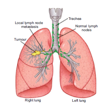
Figure 10.4: Tumour formation in lung (right) and healthy lung (left)
10.3.2 Effect of Lung Cancer on the Lung
Tobacco smoke contains over 4,000 chemical compounds, many of which have been shown
to be cancer-causing, or carcinogenic. The two primary carcinogens in tobacco smoke arechemicals known as nitrosamines and polycyclic aromatic hydrocarbons.
10.4 CHRONIC OBSTRUCTIVE PULMONARY DISEASES
(COPD) AND SMOKING
Chronic Obstructive Pulmonary Disease (COPD) is a chronic inflammatory lung disease that
causes obstructed airflow from the lungs. COPD is always caused by smoking. Over time,
breathing tobacco smoke irritates the airways and destroys the stretchy fibres in the lungs. It
usually takes many years for the lung damage to start causing symptoms, so COPD is most
common in people who are older than 60. Other things that may put you at risk include breathing
chemical fumes, dust, or air pollution over a long period of time. Secondhand smoke also may
damage the lungs.
The main symptoms are:
• A long-lasting (chronic) cough.
• Breathing difficulty, especially during physical activities.
• Cough
• Sputum production.
• Wheezing.
• Blueness of the lips or fingernail beds (cyanosis).
• Frequent respiratory infections.• Lack of energy.
As COPD gets worse, you may be short of breath even when you do simple things like get
dressed or fix a meal. It gets harder to eat or exercise, and breathing takes much more energy.
People often lose weight and get weaker. People with COPD are at increased risk of developingheart disease, lung cancer and a variety of other conditions.
Emphysema and Chronic bronchitis are the two most common conditions that contribute toCOPD. It causes airway obstruction in the lungs.
People with COPD are also likely to experience episodes called exacerbations, during which their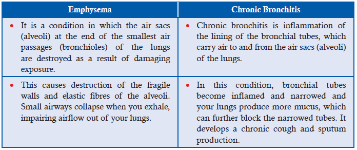
symptoms become worse than usual day-to-day variation and persist for at least several days.
The main cause of COPD in developed countries is tobacco smoking. In the developing world,
COPD often occurs in people exposed to fumes from burning fuel for cooking and heating in
poorly ventilated homes.
Only about 25 per cent of chronic smokers develop clinically apparent COPD, although
up to half have subtle evidence of COPD. Some smokers develop less common lung
conditions. They may be misdiagnosed as having COPD until a more thorough evaluationis performed.
Risk factors for COPD include:
• Exposure to tobacco smoke. The most significant risk factor for COPD is long-term cigarette
smoking. The Pipe smokers, cigar smokers and marijuana smokers are at risk, as are people
exposed to large amounts of secondhand smoke.
• People with asthma who smoke. The combination of asthma, a chronic airway disease,
and smoking increases the risk of COPD even more.
• Occupational exposure to dusts and chemicals. Long-term exposure to chemical fumes,
vapours and dusts in the workplace can irritate and inflame your lungs.
• Age. COPD develops slowly over years, so most people are at least 35 to 40 years old when
symptoms begin.
• Genetics. As noted above, the uncommon genetic disorder alpha-1-antitrypsin deficiency is
the cause of some cases of COPD. Other genetic factors are likely to make certain smokersmore susceptible to the disease.
Complications of COPD include:
• Respiratory infections: People with COPD are more susceptible to cold, the flu and
pneumonia. Any respiratory infection can make it much more difficult to breathe and
could cause further damage to lung tissue.
• Heart problems: COPD increases the risk of heart disease, including heart attack.
• Lung cancer: Smokers with chronic bronchitis have a greater risk of developing lung cancer
than those smokers who don’t have chronic bronchitis.
• High blood pressure: COPD may cause high blood pressure in the arteries that bring blood
to your lungs (pulmonary hypertension).
• Depression: Difficulty in breathing and dealing with serious illness can contribute todevelopment of depression.
10.5 SMOKING CONTRIBUTES TO CARDIOVASCULAR DISEASE
Athrosclerosis (Artherosclerosis) and Coronary Heart Disease (CHD):
1. Atherosclerosis (or arteriosclerotic vascular disease) is a condition where the arteries
become narrowed and hardened due to an excessive build up of plaque around the artery
wall (Figure 10.5). The disease disrupts the flow of blood around the body, posing serious
cardiovascular complications. The plaque clogs up the artery, disrupting the flow of blood
around the body. This potentially causes blood clots that can result in life-threatening
conditions such as heart attack, stroke and other cardiovascular diseases which are the
usual causes of heart attacks, strokes, and peripheral vascular disease — what together
are called “cardiovascular disease.” Carbon monoxide exposure has been implicated inthe process of atherosderosis.
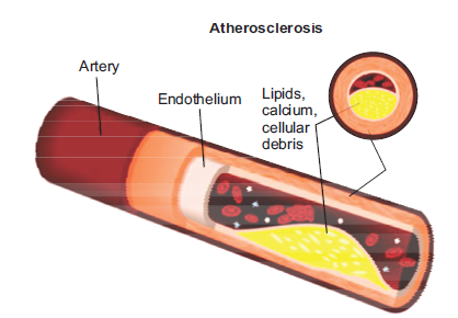
Figure 10.5: Showing plaque formation around the artery wall in Atherosclerosis
2. Coronary Heart disease, where platelets: components in the blood—stick together along
with proteins to form clots which can then get stuck in the plaque in the walls of arteries
and cause heart attacks (Figure 10.6). The most common symptoms of coronary artery
disease are angina (say “ANN-juh-nuh” or “ann-JY-nuh”) and shortness of breath when
exercising or doing other vigorous activity. Women are somewhat more likely than mento have other symptoms like nausea and back or jaw pain.
10.5.1 Effect of Carbon Monoxide and Nicotine in Cardiovascular Diseases
You have already studied that both carbon monoxide and Nicotine along with other carcinogens
affect the brain and heart. They too increase the risk of developing cardio vascular diseases,which includes coronary heart disease and stroke.
• The carbon monoxide in tobacco smoke reduces the amount of oxygen in blood. This meansthe heart has to pump harder to supply the body with the oxygen it needs.
• The nicotine in cigarettes stimulates body to produce adrenaline, which makes the heartbeatfaster and raises the blood pressure, making the heart work harder.
Figure 10.6: The process of blocking arteries by plaque in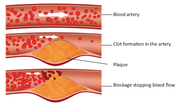
coronary heart disease
10.6 SMOKING AND HEALTH RISKS
ACTIVITY 4
Research on the effects of smoking on gas exchanges system and its health risks. You may takehelp of magazines, books and the internet. Present your findings in class.
Smokers are more likely than non-smokers to develop heart disease, stroke, and lung cancer.
• Smoking is estimated to increase the risk—
♦ For coronary heart disease by 2 to 4 times
♦ For stroke by 2 to 4 times
♦ Of men developing lung cancer by 25 times
♦ Of women developing lung cancer by 25.7 times
• Smoking causes diminished overall health, increased absenteeism from work, and increasedhealth care utilization and cost.
10.6.1 Smoking and Cardiovascular Disease
Smokers are at greater risk for diseases that affect the heart and blood vessels (cardiovascular
disease).
• Even people who smoke fewer than five cigarettes a day can have early signs of cardiovascular
disease.
• Smoking damages blood vessels and can make them thicken and grow narrower. This makes
your heart beat faster and your blood pressure go up. Clots can also form.
• A stroke occurs when a clot blocks the blood flow to part of your brain or when a blood
vessel in or around your brain bursts.
• Blockages caused by smoking can also reduce blood flow to your legs and skin.10.6.2 Smoking and Respiratory Disease
Immediate Effects of Smoking on the Breathing System
Smoking is bad for health from the very first cigarette, because some of the chemicals in the
smoke have an immediate effect on the body.
• Carbon monoxide from the smoke is taken into the blood instead of oxygen. The cells get
less oxygen for respiration, especially during exercise. This is particularly damaging during
pregnancy because a developing baby can be starved of oxygen.
• The cilia in your trachea and bronchi are anaesthetised so they no longer move mucus
and pathogens away from the lung and lungs become more likely to get infections of the
breathing system. Long-term smokers may also develop a ‘smoker’s cough’ as your bodytries to get rid of the mucus which builds up in the lungs.
• Irritation of the trachea (windpipe) and larynx (voice box)
The effects of tobacco smoke on the respiratory system include:
• Reduced lung function and breathlessness due to swelling and narrowing of the lung airways
and excess mucus in the lung passages
• Impairment of the lungs’ clearance system, leading to the build-up of poisonous substances,
which results in lung irritation and damage
• Increased risk of lung infection and symptoms such as coughing and wheezing
• Permanent damage to the air sacs of the lungs.
Smoking can cause lung disease by damaging your airways and the small air sacs (alveoli)
found in your lungs.
• Lung diseases caused by smoking include COPD, which includes emphysema and chronic
bronchitis.
• Cigarette smoking causes most cases of lung cancer.
• In condition of asthma, tobacco smoke can trigger an attack or make an attack worse.• Smokers are 12 to 13 times more likely to die from COPD than non-smokers.
10.6.3 Smoking and Other Health Risks
Smoking harms nearly every organ of the body and affects a person’s overall health.
• Smoking can make it harder for a woman to become pregnant and can affect her baby’s
health before and after birth. Smoking increases risks for:
♦ Preterm (early) delivery.
♦ Stillbirth (death of the baby before birth).
♦ Low birth weight.
♦ Sudden infant death syndrome (known as SIDS or crib death).
♦ Ectopic pregnancy.
• Smoking can also affect men’s sperm, which can reduce fertility and also increase risks for
birth defects and miscarriage.
• Smoking can affect bone health.
♦ Women past childbearing years who smoke have weaker bones than women who never
smoked, and are at greater risk for broken bones.
♦ Smoking affects the health of your teeths and gums and can cause tooth loss.
♦ Smoking can increase your risk for cataracts (clouding of the eye’s lens that makes it
hard for you to see) and age-related macular degeneration (damage to a small spot nearthe centre of the retina, the part of the eye needed for central vision).
Evidence Linking Cigarette Smoking to Disease and Early Death
Cigarette smoking began en masse in the beginning of the twentieth century, and doctors
started noticing a huge increase in cases of lung cancer from 1930 onwards, and by 1950s it
was declared an epidemic. For comparison, in 1912 there were 374 lung cancer cases, and nowthere are over 35,000 deaths a year, an increase of nearly 100 times.
The correlation between lung cancer and cigarette smoking is plain in the chart—it shows the
20 year ‘lag’ between the rise of cigarettes and the rise of lung cancer. Epidemiological data
links smoking and cancer, and up to 50% of smokers may die of smoking-related diseases(Figure 10.7).
One third of cancer deaths are as a result of cigarette smoking, and a quarter of smokers die
of lung cancer. Chronic obstructive pulmonary disease is very rare in non-smokers, less than
10% of victims are non-smokers, and less than 2% of people with emphysema are non-smokers.
One fifth of smokers suffer from emphysema, and as a result, deaths from pneumonia andinfluenza are twice as high amongst smokers.
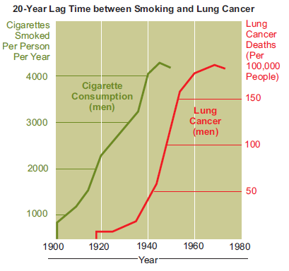
Figure 10.7: Lag time between smoking and lung cancer
Cigarette smoke contains over 4,000 chemicals, including 43 known cancer-causing (carcinogenic)
compounds and 400 other toxins. These cigarette ingredients include nicotine, tar, and carbon
monoxide, as well as formaldehyde, ammonia, hydrogen cyanide, arsenic,and DDT. Nicotine is highly addictive
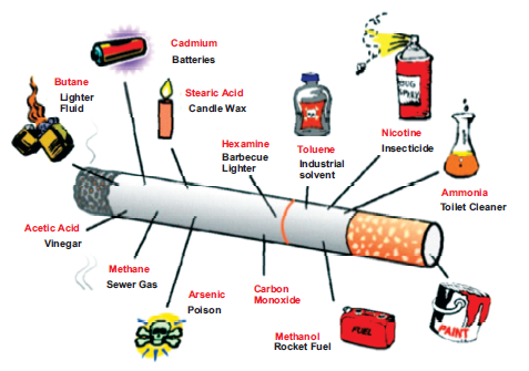
Figure 10.8: Cigarette ingredients
Anti-smoking Campaigns
We will continue to run ‘smoke-free’ campaigns to encourage people to change their behaviour.
The campaigns are aimed at:
• Making people aware of the health dangers of smoking.
• Stopping young people from taking up smoking.
• Encouraging smokers to try and quit, and to do so in the most effective way.
• Encouraging people to stop smoking in their homes and family cars—emphasising how itaffects children
10.7 SUMMARY
• Smoking harms nearly every bodily organ and organ system in the body and diminishes
person’s health and smokers are more likely than non-smokers to develop heart disease,
stroke, and lung cancer.
• Smoking is a leading cause of lung cancer and death from cancer.
• It causes stroke and coronary heart disease, which are among the leading causes of death
in the United States.
• Atherosclerosis and coronary heart disease results in damaging of blood vessels and
make them thick and grow narrow, when a clot blocks the blood flow to part of brain or
when a blood vessel in or around your brain bursts, it causes stroke.
• Chronic Obstructive Pulmonary Disease (COPD) is a chronic inflammatory lung disease
caused by smoking which damage the airways and the small air sacs (alveoli) found in the
lungs. Emphysema and Chronic bronchitis condition also contributed to COPD which
obstruct the airways of the lung.
• People with COPD are at higher risk of developing heart disease, lung cancer and a variety of
other conditions. They suffer from breathing difficulty, suddenly lose weight and feel low energy.
• Cigarette smoking and tobacco smoking contain number of harmful and carcinogenic
chemicals like:
• Nicotine — not carcinogenic but highly addictive. Smokers find it very hard to quit
because they are hooked on nicotine. Nicotine is an extremely fast-acting drug. It reaches
the brain within 15 seconds of being inhaled. Nicotine is used as a highly controlled
insecticide. Exposure to sufficient amounts can lead to vomiting, seizures, depression of
the CNS (central nervous system), and growth retardation.
• Carbon Monoxide — a poisonous gas with no smell or taste. The body finds it hard to
differentiate carbon monoxide from oxygen and absorbs it into the bloodstream. Carbon
monoxide decreases muscle and heart function, it causes fatigue, weakness, and dizziness.
It is especially toxic for babies still in the womb, infants and individuals with heart or
lung disease
• Tar — Tar’ describes the particulate matter which, generated by burning tobacco, forms a
component of cigarette smoke. Each particle is composed of a large variety of organic and
inorganic chemicals consisting primarily of nitrogen, oxygen, hydrogen, carbon dioxide,
carbon monoxide, and a wide range of volatile and semi-volatile organic chemicals. In its
condensate form, tar is a sticky brown substance that is the main cause of lung and throatcancer in smokers. Tar can also cause unsightly yellow-brown stains on fingers and teeth..
10.8 GLOSSARY
• Atherosclerosis: A condition where the arteries become narrowed and hardened due to
an excessive build up of plaque around the artery wall.
• Benign tumour: Tumour that remains in one place and does not appear to spread.
• COPD: Chronic Obstructive Pulmonary Disease is a chronic inflammatory lung disease
caused by smoking which damage the airways and the small air sacs (alveoli) found in
the lungs.
• Cardiovascular disease: It includes all types of heart and blood vessel diseases.
• Malignant tumour: Tumour that spreads to other parts of the body either through the
bloodstream or the lymphatic system.
• Tar: A sticky, dark brown substance that can accumulate and coat the airways andlungs.
END UNIT ASSESSMENT 10
I. Choose whether the following statements are True (T) or False (F)
1. Smoke from cigarettes can make non-smokers sick.
2. Smoking can affect a person’s ability to smell and taste food.
3. Secondhand smoke kills about 3,000 non-smokers each year from lung disease.
4. It takes about ten seconds for nicotine absorbed into the bloodstream to reach the
brain.
5. Smoking is a difficult habit to quit.
6. Nicotine, the chemical found in cigarettes, is an addictive drug.
7. A smoker is twice as likely to have a heart attack as a non-smoker is.
8. Cigarette brands that are heavily advertised on TV, in magazines, on billboards, and
on T-shirts are the brands more teens buy.
9. One out of every ten smokers will die of a smoking-related sickness.10. More germs get into your lungs when you smoke.
II. Multiple Choice Questions
1. Smoking causes
(a) Lung cancer (b) Heart disease
(c) Respiratory disease (d) All of above
2. Atherosclerosis is a condition where,
(a) Body needs more energy
(b) Lungs get filled with mucus
(c) Plaque clogs up the artery, disrupting the flow of blood around the body
(d) Brain becomes dead
3. Chronic Obstructive lung disease occurs due to
(a) Chronic bronchitis (b) Emphysema
(c) None of these (d) Both of these
4. COPD stands for
(a) Cuticular Obstetric Pelvic Disease
(b) Critical Obstructive Pituarity Disorder
(c) Chronic Obstructive Pulmonary Disease
(d) Chronic Obesity Personal Decision
5. Emphysema is caused due to
(a) Bursting of alveoli (b) A decrease in the surface area of gas exchange
(c) Both A and B (d) None of above
6. Gas exchanging system cannot be damaged by
(a) Carcinogens (b) CO
(c) Nicotine (d) Any of these
7. The blood is not oxygenated well enough due to
(a) Asthma (b) Emphysema
(c) Chronic Bronchitis (d) All of above
8. Common causes of Chronic Obstructive Pulmonary Disease (COPD) does not include
(a) Smoke from the factory furnace (b) Vehicle pollution
(c) Industrial pollution (d) Water pollution
9. Chronic Obstructive Pulmonary Disease (COPD) does not include
(a) Asthma (b) Chronic Bronchitis
(c) Emphysema (d) Retinoblastoma
10. The smoker’s cough is
(a) A sign of recovery
(b) An attempt to move the mucus up the airways
(c) An attempt to move the air down the airways(d) A mixture of bacteria and BCs
III. Long Answer Type Questions
1. What are the dangerous components of tobacco smoke?
2. In your words, describe the effects of tar and carcinogens in tobacco smoke on the
gas exchange system.
3. How can you control the use of tobacco in your society? Explain briefly.
4. Describe the signs and symptoms of lung cancer and chronic obstructive pulmonary
diseases (COPD).
5. Describe the effects of nicotine and carbon monoxide on the cardiovascular system.
6. Explain how tobacco smoking contributes to atherosclerosis and coronary heart
disease.
7. Interpret data linking cigarette smoking to disease and early death.
8. Comment on smoking gender disparities prevalent in the community. Also state the
effect of smoking on a pregnant woman. How does it affect the infant?
9. Correlate smoking to behavioural economics and health behavioural changes. Support
your answer with examples.
10. Differentiate between:
(a) Emphysema and chronic bronchitis
(b) Atherosclerosis and coronary heart disease11. Look at the pictures and answer the questions that follow:
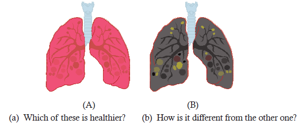
Unit 11 General Principles of Homeostasis
Key Unit Competence
To be able to explain general principles of homeostatic mechanisms.
LEARNING OBJECTIVES
At the end of this unit, the learner will be able to:
• Explain the significance of a constant internal environment.
• State the factors that must be kept constant in the internal environment of the body.
• Carry out research on homeostasis and deduce the findings.
• Appreciate the importance of maintaining a constant internal environment.
• Relate organisms’ ways of life to their environmental conditions.
• Discuss the role of the negative feedback mechanism.
• Explain the feedback mechanism in relation to the endocrine and nervous system.
• Identify the main internal and external causes of change in the internal environment.
• Describe the formation, composition and movement of tissue fluid in relation to blood and
lymphs.
• Appreciate the adaptations of animals to different environmental conditions in relation to homeostasis.INTRODUCTORY ACTIVITY
The picture below shows human who is swimming underwater of Muhazi Lake. Observecarefully this picture and answer the question that follows
Explain why this human can swim underwater for long time?
11.1 SIGNIFICANCE OF CONSTANT INTERNAL ENVIRONMENT
ACTIVITY 11.1
Have you ever thought about how your body maintains the same body temperature even if it is too hot or cold outside? Do you know about homeostasis and why it is required? Use library and Internet sources to collect information about homeostasis and various factors that must be kept constant in the human body. Learn, write and draw diagrams of homeostatic mechanisms and present your findings in the class.
All living organisms have an ability to maintain stable internal conditions. It requires continuous adjustments to the changes occuring in both internal and external environments. This selfregulating property of living beings to maintain a constant internal environment is termed as ‘homeostasis’ (‘homeo’, “similar,” and ‘stasis’, “stable”). Homeostasis is a key concept in the understanding of biological mechanisms that play an important role in survival of individual cells, to an entire body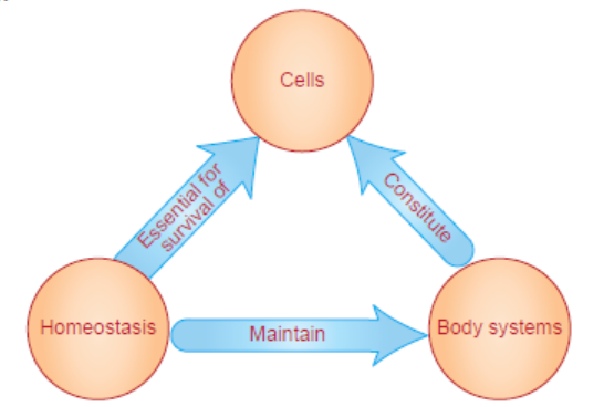
Figure 11.1: Homeostasis
A living body ensures stable internal conditions in order to survive, grow and develop. The homeostatic mechanisms are inevitable for proper functioning of the body. Many systems of the body operate together to maintain a steady state. The cells, tissues, and organs may perform very different functions; however, all the cells in the body are similar in their metabolic needs. Homeostasis continuously provides the necessary ingredients of survival, for example, oxygen and nutrients to cells and thus, to complete the body. The metabolic activities and cellular processes can continue even though the external environment fluctuates substantially. Therefore, the regulation of an optimal internal environment enables an organism to live in wider range of environmental conditions (Figure 11.1).Automatic systems such as thermostats or air conditioners maintain constant internal (room) conditions.
ACTIVITY 2
Have you ever read or heard about some persons having diabetes or high blood pressure. Do you
know why such conditions occurred in these people while others have no problems? Discuss
and find out the appropriate answers.Note: Refer to negative feedback mechanism to learn about high blood pressure.
Homeostasis can be considered as dynamic equilibrium rather than a constant, unchanging
state. The body mechanisms maintain various fluctuating physical and chemical variables
within tolerable limits. These important variables include temperature, glucose, pH, water,
ions, respiratory gases and osmotic pressure of body fluids among others.
Some principal homeostatic mechanisms in humans to be kept constant are as follows:
• The maintenance of a steady body temperature involves mechanisms such as sweating or
shivering. These mechanisms occur whenever the internal body temperature becomes high
or low.
• The human body constantly works to maintain a normal glucose level in blood. When
glucose levels are high, a hormone called insulin is released by beta cells of the pancreas.
Insulin stimulates the conversion of glucose as insoluble glycogen by the body cells. This
lowers the glucose concentration in the blood. A condition called as diabetes occurs due to
the deficiency of insulin in the body, due to which glucose level of blood increases. When
the blood glucose levels are low, another hormone known as glucagon is released by the
alpha cells of pancreas. Glucagon breaks down stored glycogen in the form of glucose. The
addition of glucose in blood returns the body glucose levels to normal.
• Whenever the water content of the blood and lymph fluid gets low, it is restored initially by
extracting water from the cells. Also, the throat and mouth become dry. These symptoms
of thirst motivate humans to drink water.
• When high amount of salt and ions are present in the body, the kidneys produce concentrated
urine. This process removes extra amount of salt and ions while conserving water, and return
the body to normal metabolic range. In contrast, when the salt and ions concentration is
low in the human body, kidneys produce dilute urine and conserve salt and ions.
• A change in breathing and heart rate occurs in humans due to various activities like exercise.
As a result, the amount of carbon dioxide produced and oxygen demand in the body
increases. The heart rate increases so that the blood flows rapidly to the tissues to fulfill
the oxygen requirement and remove the carbon dioxide from the cells. Also, the speed and
depth of breathing increases. The body works to normalize breathing and heart rate whenactivity stops
• The pH of the blood is regulated at 7.365 (a measure of alkalinity and acidity). The tolerable
lower and upper limit for a human body is about pH 7.0 and pH 7.8, respectively. To prevent
a change in the pH, all body fluids, including cell cytoplasm are buffered (buffer is a chemicalor a combination of chemicals) absorbing either hydrogen ions (H+) or hydroxide ions.
All the body systems in humans are interdependent and function invariably to keepfluctuating factors within tolerable limits.
11.1.2 Components of Homeostatic Mechanisms
All homeostatic control mechanisms have three interdependent components for regulation
of any type of change or variable. The receptor is the sensing component that monitors
and responds to changes in the environment. When the receptor senses a stimulus, it sends
information to a “control/integration centre”, the component that sets the range at which
a variable is maintained. The control centre determines an appropriate response to the
stimulus. The control centre then sends signals to an effector, which can be muscles, organs,
or other structures that receive signals from the control centre. After receiving the signal, a
change occurs to correct the deviation by opposing or enhancing the stimulus. This ongoing
process continuously works to restore and maintain homeostasis. For example, in order to
regulate body temperature, thermo-receptors are present in the skin which communicates
information to the brain, the control centre. The control centre commands the effectors i.e.,blood vessels and sweat glands to function accordingly (Figure 11.2).
Figure 11.2: Components of homeostatic mechanisms
11.2 NEGATIVE AND POSITIVE FEEDBACK REGULATION
ACTIVITY 3
Have you ever thought how the body brings itself to the normal state after any change
in its system/s? How the signal that brings any change in body is regulated either stopped
or amplified? With the help of charts, discuss the feedback mechanism(s) and their role inmaintaining homeostasis in the body.
Feedback regulation is a type of response to a stimulus which determines the effect of original
stimulus. When a change of variable occurs, there are two main types of feedback mechanisms
to which the system reacts: Negative feedback is a response in which the system functions in
such a way as to reverse the direction of change. This response system tends to keep things
constant allowing the maintenance of homeostasis. The regulations of body temperature, pH,
ionic balance and blood pressure are the most common type of negative feedback systems.
Positive feedback is the response to amplify the change in the variable. Blood clotting and events
in childbirth are the types of positive feedback system. Both types of feedback mechanisms are
equally important for the proper functioning of a body. If any or both these mechanisms areaffected or altered somehow, it can lead to many complications.
11.2.1 Negative Feedback Mechanisms
Negative feedback consists of reducing the output or activity of any system or organ back to
its normal range of functioning. This change either raises or lowers the variable to its normal
set point automatically by counteracting. Here, negative means ‘opposite, not bad’. This can
be understood by the controlling process that regulates blood pressure. Blood pressure is the
measure of the force of blood pushing against blood vessel walls. The heart pumps blood into
the arteries (blood vessels), which carry the blood throughout the body. Whenever the blood
pressure increases, the blood vessels can sense the resistance of blood flow against the walls.
The blood vessels act as the receptors and relay the change to the brain. The brain acts as
control centre and transmits the signal to the heart and blood vessels, both of which act as the
effectors. The heart rate would decrease as the blood vessels increase in diameter, known as
vasodilation. This change would cause the blood pressure to fall back to its normal range. The
opposite would happen when blood pressure decreases, leading to vasoconstriction (decreasein diameter of blood vessel) (Figure 11.3).
Several factors/conditions interfere with the normal process of regulation of blood pressure.
Smoking, obesity, high salt concentration in diet, alcohol consumption, stress, hormonal
disorders affect the heart and blood vessels. This leads to high blood pressure or hypertension
which causes the heart to work harder to pump blood in the body. This can further damage theheart, blood vessels and other organs.
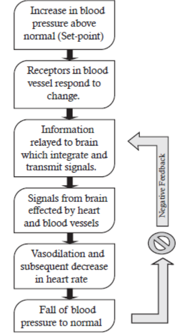
Figure 11.3: Negative feedback mechanism
Negative feedback mechanisms are most common in living organisms, working in a specificmanner sequence.
11.2.2 Nervous and Endocrine Control Mechanisms
In the human body, all the organs and organ systems are controlled by nervous and endocrine
systems. The nervous system controls the activities of body parts by reacting quickly to external
and internal stimuli. The endocrine system regulates those activities slowly but its effects are
long lasting. The hypothalamus is a part of the brain (nervous control center) located just
above the brain stem and consists of a group of neurons that forms the primary link betweenthe nervous system and the endocrine system.
This small part of the brain is responsible for regulating many key body processes, including
internal body temperature, hunger, thirst, blood pressure, and daily body rhythms (Figure 11.4(a)).
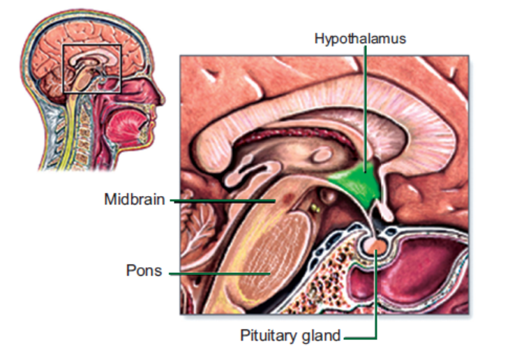
Figure 11.4: (a) Hypothalamus and Pituitary gland
Nervous system consists of receptive nerve cells which transmit the signal to the brain, which
in turn, command the effector nerve cells, muscles and glands to respond. For instance,
humans maintain a constant body temperature, usually about 37.4°C. It increases during
the day by about 0.8°C and decreases slightly during sleeping. The core body temperature is
usually about 0.7-1.0°C higher than skin or axillary temperature. A change in temperature
is sensed by receptors found in the skin, veins, abdominal organs and hypothalamus. The
receptors in the skin provide the sensation of cold and transmit this information to brain.
The brain process and commands for the vasoconstriction of blood vessels in the skin and
limb. This drops the surface temperature, providing an insulating layer (fat cell) between the
core temperature and the external environment. The major adjustment in cold is shivering
to increase the metabolic heat production. On the contrary, if the body temperature rises,
blood flow to the skin increases, maximizing the potential for heat loss by radiation andevaporation (Figure 11.4 (b)).
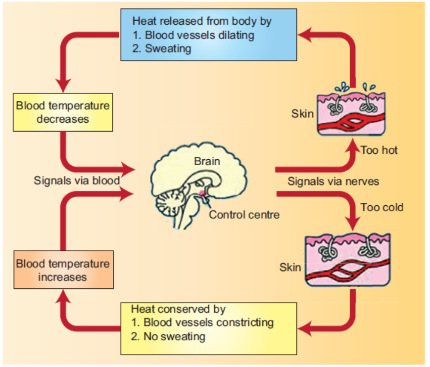
Figure 11.4: (b) Homeostatic regulation of body temperature
The endocrine system consists of glands which secrete hormones into the bloodstream. Each
hormone has an effect on one or more target tissues. In this way, it regulates the metabolism
and development of most body cells and its systems through feedback mechanisms, mostly
negative. For example, when blood calcium becomes too low, calcium-sensing receptors in
the parathyroid gland become activated. This results in the release of Parathyroid Hormone
(PTH), which acts to increase blood calcium by release from the bones. This hormone also
causes calcium to be re-absorbed from urine and the gastrointestinal tract. Calcitonin, released
from the thyroid gland functions in reverse manner, i.e., decreasing calcium levels in the bloodby causing more calcium to be fixed in bones (Figure 11.5).
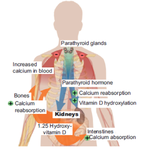
Figure 11.5: Calcium regulation
Both the nervous and endocrine system of the human body co-ordinate to ensure a balance
between fluid gained and fluid loss. The ADH (Anti-diuretic Hormone) or vasopressin is the
principal compound that controls water balance by decreasing water output by the kidney.
Vasopressin is formed in the hypothalamus and get stored in the posterior pituitary (a part of
endocrine system). If the body becomes fluid-deficient, osmoreceptors (monitor blood plasma
osmolality) in the hypothalamus signals for the release of vasopressin from posterior pituitary. An
increase in the secretion of vasopressin causes retention of fluid by the kidneys and subsequent
reduction in urine output. Conversely, if fluid levels are excessive, release of vasopressin is
suppressed resulting in less retention of fluid and resulting increase in the volume of urineproduced.
APPLICATION 11.1
1.Complete with appropriate terms:
(i)The tolerable pH level in human body ranges from ................ to ...................... .
(ii) .................... provides necessary ingredients for survival.
(iii) Endocrine system consists of ..................... which secretes hormones.
(iv) ............................ is a response in which the system functions in a way to reverse
the direction of change.
(v) ......................... and ................... are mechanisms to maintain body temperature.2. Name the heat regulation centre in the brain.
3. What is the function of
a) Detectors
b) Comparators
c) Effectors
4.What type of feedback system is involved in bringing the temperature of an overheated
person back to normal
5.Explain briefly the advantage of having two corrective mechanisms that regulates blood
glucose concentration, rather than one
6. A person fasted overnight and then shallowed 75 g of glucose. The graph shows the resultingchanges in the concentrations of insulin and glucose in the blood
a. Explain the relationship between the concentrations of glucose and insulin in the blood in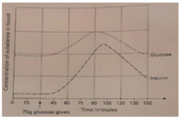
the first 30 minutes after the glucose was swallowed
b. Use information from the graph to explain what is meant by term negative feedback
c. Explain why the concentration of glucagon in the blood rises during exercise while thatof insulin falls
11.3 CAUSES OF CHANGES IN HOMEOSTASIS ENVIRONMENT
Homeostasis is maintained through a series of control mechanisms. When homeostatic process
is interrupted, the body can correct or worsen the problem, based on certain influences. Thereare internal and external causes influencing the body’s ability to maintain homeostatic balance.
11.3.1 Internal Causes: Heredity
Genetic/Reproductive: A variety of diseases and disorders occur due to the change in the
structure and function of genes. For example, cancer can be genetically inherited or can be
induced due to a gene mutation from an external source such as UV radiation or harmful drugs.
Another disorder, Type 1 diabetes, occurs due to the lack or inadequate production of insulinby the pancreas to respond to changes in a person’s blood glucose level.
11.3.2 External Causes: Lifestyle
Nutrition: A diet lacking specific vitamin or mineral leads to cellular malfunction. A menstruating
woman with iron deficiency will become anaemic. As iron is required for haemoglobin, an
oxygen transport protein present in red blood cells, the blood of an anaemic woman will havereduced oxygen-carrying capacity.
Physical Activity: Physical activity is essential for proper functioning of our cells and bodies.
Adequate rest and exercise are examples of activities that influence homeostasis. Lack of sleep
causes ailments such as irregular cardiacrhythms, fatigue, anxiety and headaches.
Overweight and obesity are related to poor nutrition and lack of physical activity that greatly
affects many organ systems and their homeostatic mechanisms. It increases a person’s risk ofdeveloping heart disease, Type 2 diabetes, and certain forms of cancer.
Mental Health: Both the physical and mental health is inseparable. Negative stress (also calleddistress) leads to thoughts and emotions harmful for homeostatic mechanisms in the body.
11.3.3 Environmental Exposure
Many substances act as toxins, including pollutants, pesticides, natural and synthetic drugs,
plants and animal products interfering at cellular levels. Modern medicines practice can also
be potentially harmful in case of wrong or over dosage. For instance, drug overdose affects
the central nervous system, disrupts breathing and heartbeat in the human body. It can further
result in coma, brain damage, and even death. Therefore, alterations or interruption of beneficial
pathways, whether caused by an internal or external factor will result in harmful change in
homeostasis. Therefore, adequate positive health influences are to be taken into considerationin order to maintain homeostasis.
11.4 FORMATION, COMPOSITION AND MOVEMENT OF TISSUE FLUID
ACTIVITY 4
Aim: To observe and analyze the pressure flow of liquid coming out from the perforated rubber
tubes.
Materials Required:
1. A rubber tube (1 cm width and 50 cm in length)
2. Two beakers3. One bucket.
Procedure:
1. Take a rubber tube of about 1 cm width and 50 cm in length and make two set of tiny holes
on at a distance of 10 cm before both ends.
2. Attach tube to a water tap tightly. Try to adjust the tubular part with holes towards downside
so as to collect the liquid in beakers or trays.
3. At the other end, put a collecting vessel such as a bucket. Adjust the rubber tube in horizontal
position and open the tap for 10-20 seconds and collect the water coming out of rubber holes
in beakers placed under the holes in the tube.
4. Measure the collected liquid in two beakers separately by glass cylinders. Note the readings
and compare the volume of water collected in the beakers.
Discussion:
1. Do you find any difference in volume of water collected in beakers?
2. Where do you think high and low pressure end on rubber tubes?3. Does the high pressure end expel more water than low pressure end?
The blood supplies nutrients and essential metabolites to the cells of a tissue and collects back
the waste products. This exchange of respective constituents between the blood and tissue cells
occurs through interstitial fluid or tissue fluid formed by the blood. The fluid occupies the spaces
between the cells known as tissue spaces. It is the main component of the extracellular fluid,
which also includes plasma and transcellular fluid. On an average, a person has about 10 litresof interstitial fluid making 16% of the total body weight.
11.4.1 Formation
The formation of the tissue fluid is based on the difference in pressure of flowing (Starling’s law)
of blood through capillaries. A hydrostatic pressure is produced at the arterial end of blood
capillaries which is generated by the heart. This results in expulsion of water and other solutes
(known as plasma) from capillaries except blood proteins (like serum albumin). This retention
of solutes in capillaries creates water potential. The osmotic pressure (water moves from a
region of high to low concentration) tends to drives water back into the capillaries in an attempt
to reach equilibrium. At the arterial end, the hydrostatic pressure is greater than the osmotic
pressure, so the net movement favours water along with solutes being passed into the tissue
fluid. At the venous end, the osmotic pressure is greater, so the net movement favours tissue
fluid being passed back into the capillary. The equilibrium is never attained because of the
difference in the direction of the flow of blood and the solutes imbalance created by the netmovement of water (Figure 11.6).
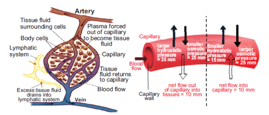
Figure 11.6: Formation of interstitial fluid from blood
11.4.2 Composition
As the blood and the surrounding cells continually add and remove substances from the interstitial
fluid, its composition continually changes. Water and solutes can pass between the interstitial
fluid and blood via diffusion across gaps in capillary walls called intercellular clefts; thus,
the blood and interstitial fluid are in dynamic equilibrium with each other. Generally, tissue
fluid consists of a water solvent containing sugars, salts, fatty acids, amino acids, coenzymes,
hormones, neurotransmitters, as well as metabolic waste products from the cells.
Not all of the contents of the blood pass into the tissue, which means that tissue fluid and
blood are not the same. Red blood cells, platelets, and plasma proteins cannot pass through
the walls of the capillaries. The resulting mixture that does pass through is, in essence, blood
plasma without the plasma proteins. Tissue fluid also contains some types of white blood cells,which help to combat infection.
11.4.3 Movement
To prevent a buildup of tissue fluid surrounding the cells in the tissue, the lymphatic system
plays an important role in its transport. Tissue fluid can pass into the surrounding lymph
vessels where it is then considered as lymph. The lymphatic system returns protein and excess
interstitial fluid to the blood circulation. Thus, it is transported through the lymph vessels to
lymph nodes and ultimately with blood in the venous system, and tends to accumulate morecells (particularly, lymphocytes) and proteins.
The formation, composition and movement of tissue fluid are important processes for thedevelopment of immunity in the human body.
11.5 ADAPTATIONS OF ORGANISMS TO ENVIRONMENTAL CONDITIONS
ACTIVITY 5
A field trip to a zoo/wildlife sanctuary/national park can be organized under the guidance of a
teacher. The students/learners should observe, understand and discuss the various environmental
conditions in which animals live and survive. Different groups of animals which live on land,
water and other places can be studied for the adaptations in their habitat. Learners should write
their observations about the different features/characteristics of animals, timing of appearance,
season, place/location as well as behaviour of different animals. A report should be submitted
about the field trip describing the observations along with the diagrams/images/photographstaken and computer aided material.
Every organism has certain features or characteristics which enables it to live successfully in
its particular habitat. These features are called adaptations, and the organism is said to be
adapted to its habitat. Organisms living in various habitats need different adaptations in order
to maintain homeostasis. The animals adapt to such changes in their environment which
threatens their chances of survival. The main threats are temperature, lack of water and food.
Besides the environmental threats, many animals also need to be able to defend themselves
from predators and pathogens.
Different organisms have adapted to the great diversity of habitats and distinct conditions in
the environment. Although, the adaptations are many and varied, they can be categorized intomainly three types: Structural, physiological and behavioural.
11.5.1 Structural Adaptations
Structural (or morphological) adaptations are the physical features of the organism. It includes
shapes or body covering as well as its internal organisation. Microscopic organisms which
includes protozoans and bacteria employ encystment (a state of suspended form, separated
by the outside world by a solid cell wall) to surpass hostile conditions for long periods of time,
even millions of years. Larger animals like polar bears are well adapted for survival in the cold
climate of the Arctic region. They have a white appearance to camouflage from prey on the
snow and ice. Also, polar bear have thick layers of fat and fur, for insulation against the coldand a greasy coat which sheds water after swimming.
Figure 11.7: Polar bear in cold climate
Dolphins are fish-like mammals which have streamlined shape and fins instead of legs. They
also have blowholes on the tops of their heads for breathing, rather than their mouth and nose.
Desert animals like camels have many adaptations that allow them to live successfully in hot
and dry conditions. They have long eyelashes and nostrils that can close and open to prevent
entry of sand. Thick eyebrows shield the eyes from the desert sun. Camels store fat in the
hump which can be metabolised for energy. A camel can go a week or more without water,
and they can last for several months without food. Their huge feet help them walk on sandwithout sinking into it.
Figure 11.8: Camel’s adaptations in desert environment: (a) Nostrils and (b) Hump
Similarly, the long necks of giraffes allow them to feed among treetops
and spot predators. Also, they have tough and long tongues (upto 18
inches) enabling to pull leaves from branches without being hurt bythe thorns. Spotted coat camouflages giraffes among the trees.
In tropical areas, natural radiators are an efficient way of lowering
the body’s temperature: for instance, the ears of the elephant and the
rabbit are full of blood vessels, helping the animal cool its body in theheat. Rabbits living in Arctic areas have smaller ears.
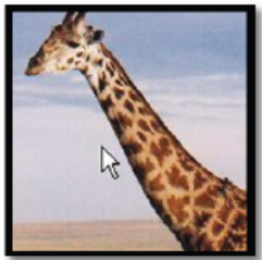
Figure 11.9: Giraffe
11.5.2 Physiological Adaptations
Physiological adaptations are related to the working of an organism’s metabolism. These
adaptations enable the organism to regulate their bodily functions, such as breathing and
temperature, and perform special functions like excreting chemicals as a defence mechanism
(Sea stars). Chameleon (a reptile) changes colour or body markings in order to blend
into its surroundings. Marine mammals such as whales are endothermic/warm blooded
(able to maintain a constant body temperature). They cope with the temperature changes during
migration over large distances and can spend time in arctic, tropical and temperate waters. In
contrast, Arctic fish (cold-blooded animals) lives easily in temperatures lower to sub-zero level.
Such temperatures results in the formation of ice crystals in the organism’s cells that may cause
irreversible damage and ultimately, death. However, arctic fishes living in the same freezing
waters survive due to an antifreeze protein in the blood that prevents ice crystals formation intheir cells and maintains metabolic functions.
11.5.3 Behavioural Adaptations
Behavioural adaptations are learned that help organisms to survive. The whales produce
sounds that allow them to communicate, navigate and hunt prey. Bears hibernate or ‘sleep’
through the coldest part of the year. Bryozoans are water dwelling small individual animals
found in colonies in high numbers on the continental shelf in New Zealand. These animals
band together for collecting food and survive predation. Penguins are the flightless birds
found in the oceans around Antarctica. During extreme winter, Emperor penguins show
social behaviour by huddling together in groups comprising several thousand penguins tostay warm.
APPLICATION 11.2
1.Complete with appropriate terms:
(i) Micro organisms employ .................... to surpass hostile conditions for a long time.
(ii) .................... is a type of mammal having fins and a streamlined body.
(iii) ................... and ..................... influences/tends to disturb homeostatic mechanisms.
(iv) RBCs, platelets and plasma cannot pass through ........................... .
(v) .................... fishes have an antifreeze protein in the blood to prevent formation of ice
crystals.
2.The control of blood glucose concentration involves a negative feedback mechanism.
a) What are the stimuli, receptors and effectors in this control mechanism?
b) Explain how negative feedback is involved in this homeostatic mechanism.
3. Explain why blood contains many proteins that are not found in the tissue fluid or lymph
4. What produces the hydrostatic pressure in the blood
5.Describe how fluid can pass through the capillary wall from the plasma to the tissue fluid
6. a) Describe the immediate environment of a typical cell within the body of a mammal.
b) Explain why it is important that the internal environment of a mammal is carefully
regulated.
c)Explain how the following are involved in maintaining the internal environment: stimuli,
receptors, central control, coordination systems and effectors.
d)Explain the meaning of the terms homeostasis and negative feedback.e) Distinguish between the input and the output in a homeostatic control mechanism.
11.6 SUMMARY
• Homeostasis is the ability of a living body to maintain a relatively stable internal environment.
• Homeostasis is an important characteristic of living things requiring continuous
adjustments due to the changes occurring in internal and external environment.
• Variables that must be kept constant and regulated to the normal level (set points) in a
body are temperature, glucose, pH, water, ions, respiratory gases and osmotic pressure
of body fluids.
• Homeostatic control mechanisms have three interdependent components: receptor,
integration centre and effectors.
• Negative feedback occurs when the response to a stimulus reduces the original stimulus.
• Positive feedback occurs when the response to a stimulus increases the original stimulus.
• The nervous system controls the activities of body parts quickly to external and internal
stimuli.
• The endocrine system regulates body activities slowly with long lasting effects.
• The hypothalamus is a part of brain and links the nervous system and endocrine system.
• The homeostatic mechanisms are altered or interrupted based on internal (genetic) and
external (lifestyle choices and environmental exposures) factors.
• Interstitial/tissue fluid is formed from blood plasma and it surrounds and bathes the
cells in tissue spaces.
• Tissue fluid provides nutrients and removes waste products from the cells of the body.
• Tissue fluid is formed due to the pressure difference in flow of the blood through the
blood capillaries.
• Tissue fluid contains sugars, salts, fatty acids, amino acids, coenzymes, ions, hormones,
neurotransmitters, as well as metabolic waste products from the cells in a water medium.
• Tissue fluid moves from tissue spaces to lymph vessels (lymph), to lymph nodes and
finally returns to the blood.
• Adaptation is a feature/characteristic of an animal which enables it to survive in its habitat.
• Different organisms have adapted to distinct habitats and environmental conditions.
• Three categories of adaptations are structural, physiological and behavioural. Each typeof adaptation has its own survival value.
11.7 GLOSSARY
• Adaptation: A feature or characteristic of an organism which helps in its survival in a
particular habitat.
• Control centre or integration centre: Receives and processes information from the
receptor.
• Effector: Responds to the command of the control centre by either opposing or enhancing
the change.
• Homeostasis: A balancing act which maintains a particular internal condition.
• Receptor: Receives information about a change in its environment.
• Negative feedback: A response system which reverses the direction of change.• Positive feedback: A response system which amplifies the change in the variable.
END UNIT ASSESSMENT 11
I. Choose whether the given statements are True (T) or False (F)
1. A living body ensures stable internal conditions in order to survive, grow and develop.
2. The property of living beings to maintain a constant internal environment is termed
as ‘homeostasis’.
3. Blood pressure is the measure of the force of blood pushing against blood vessel
walls.
4. The endocrine system controls the activities of body parts by reacting quickly to
external and internal stimuli.
5. Organisms living in various habitats need different adaptations in order to maintain
homeostasis.
6. Behavioural adaptations are related to the working of an organism’s metabolism.
7. The chemical messengers secreted from the endocrine system affect on specific target organs.
8. The thick fur layer on the skin of polar bears protects them from sunlight.
9. Whales migrating to oceanic waters can maintain a constant body temperature.
10. Colonial animals, if being separated, can survive easily in their habitats.11. Camel’s hump is an adaptation to store water in desert climate.
II. Multiple Choice Questions
1. An example of positive feedback mechanism is:
(a) maintaining stable blood glucose levels
(b) the production of milk in a nursing mother
(c) maintaining a stable body temperature
(d) all of the above
2. Which hormone increases the glucose level in the blood?
(a) glucose (b) glucagon
(c) insulin (d) glycogen
3. Which is the proper sequence of events in maintaining homeostasis?
(a) Signal, Receptor, Stimulus, Response
(b) Stimulus, Response, Signal, Receptor
(c) Receptor, Stimulus, Signal, Response
(d) Stimulus, Receptor, Signal, Response
4. Which is an example of negative feedback?
(a) maintaining stable blood glucose levels
(b) the production of milk in a nursing mother
(c) contractions of the uterus during childbirth
(d) all of the above
5. The principal systems controlling and regulating body activities are:
(a) nervous and respiratory system (b) endocrine and digestive system
(c) nervous and endocrine system (d) respiratory and digestive system
6. Which of the following statements best describes homeostasis?
(a) keeping the body in a fixed and unaltered state
(b) dynamic equilibrium
(c) maintaining a relatively constant internal environment(d) altering the external environment to accommodate the body’s needs
7. Tissue fluid is different from blood in terms of:
(a) red blood cells (b) platelets
(c) plasma proteins (d) all of the above
8. The human body’s “thermostat” is found in:
(a) nervous system (b) integumentary system
(c) endocrine system (d) urinary system
9. Which statement is not correct about calcium homeostasis?
(a) parathyroid hormone increases blood calcium level
(b) calcitonin decreases calcium levels in blood
(c) both hormones (PTH and calcitonin) are secreted by same gland
(d) calcium regulation is an example of negative feedback mechanism
10. Which statement is correct about vasopressin?
(a) an increase in secretion of vasopressin decreases fluid volume in the body
(b) a decrease in secretion of vasopressin increases fluid volume in the body
(c) an increase in secretion of vasopressin increases fluid volume in the body(d) none of the above
III. Long Answer Type Questions
1. Giving suitable examples, in your own words, explain the significance of a constant
internal environment.
2. State the factors that must be kept constant in the internal environment of the body.
3. Discuss the role of the negative feedback mechanism.
4. Explain the feedback mechanism in relation to the endocrine and nervous system.
5. What are the main internal and external causes of change in the internal environment?
6. Describe the formation, composition and movement of tissue fluid in relation to
blood and lymphs.
7. Giving examples, relate organisms’ ways of life to their environmental conditions.
8. How do pollutants alter homeostasis of an organism? What role does homeostasisplay in environmental protection?
Unit 12 Regulation of Glucose
Key Unit Competence
To be able to explain the mechanism of the regulation of blood glucose level.
LEARNING OBJECTIVES
At the end of this unit, the learner will be able to:
• Describe the role of hormones in sugar regulation.
• Describe the detailed structure of a liver lobule and the Islet of Langerhans.
• Explain the negative feedback mechanism in the process of blood glucose control.
• Discuss the causes and effects of blood sugar imbalances in the body.
• Appreciate the importance of a controlled diet for diabetics.
• Assist diabetics and people having hypertension in coping with their situation.
• Relate the structure of the liver and the pancreas to their functions.
• Relate the microstructure of the liver and the pancreas to sugar regulation.
• Describe the functions of the liver and pancreas in the regulation of glucose in the body.
• Describe the three main stages of cell signalling in control of blood glucose by adrenaline
as follows:
♦ Hormone-receptor interaction at the cell surface.
♦ Formation of cyclic AMP that binds to kinase protein.
♦ An enzyme cascade involving activation of enzymes by phosphorylation to amplify the
signal.
• Make research using internet or articles on the role of adrenaline in the control of blood
sugar.
• Explain the principles of the operation of dip sticks and biosensors for quantitative
measurements of glucose in the blood and urine.• Explain how urine analysis is used in diagnosis with reference to glucose, protein and ketones.
INTRODUCTORY ACTIVITY
The human body maintains constant different substances in the blood, a process called
homeostasis. The figures below show different organs involved in the regulation of bloodglucose level in the body
Observe the illustations X and Y above and answer to the questions that follow
a)What are the parts represented by letters A,B and C on the illustration X ?
b) All the organs shown in the illustration X are involved in the digestion of food.
What are the functions of A and B in the digestion
c) What are the organs involved in the regulation of blood level on the illustration X?
In which way does each organ state help in this regulation
d) The illustration Y shows the regulation of blood glucose level. What does the letters
A,,B and C show in this regulation
e) Alpha and beta cells are responsible for producing the hormones that are involved in
the regulation of blood glucose level. Which organ on the illustration Y produces these
hormones
f) Compare the mechanism of working of organs A and B in the regulation of bloodglucose level
12.1 IMPORTANCE OF GLUCOSE
ACTIVITY 1
Visit a nearby hospital to see the patients.
You will notice many patients lying on the bed with a bottle
hanging from a stand by the side of their bed as shown in the
picture.
Ask the doctor, what is inside the bottle and why is it given to the
patients?
Record the answer.
Note: In the hospital, patients are generally given glucose by
drip instead of food because, the drip directly deposits the
glucose into the blood stream and hence the body cells get it in matter of minutes, but if it is
given through food, the glucose’s energy would reach the body parts only after the digestionprocess and this will take a longer time.
Glucose is one of the most important carbohydrates molecules in our body. Body requires
glucose to carry out some of its most important functions. Glucose is synthesized in green
plants, from carbon dioxide, CO2 and water, H2O with the help of energy from sunlight. This
process is known as photosynthesis. The reverse of the photosynthesis reaction i.e., breakdown
of glucose in the presence of oxygen to form carbon dioxide and water releasing the energy, is
the main source of power for all the living organisms. The excess glucose in plants is stored inthe form of starch which serves as foods for various animals.
12.1.1 Glucose as Energy Source
Almost 80 per cent of carbohydrates in our food are converted to glucose during digestion in the
alimentary canal. Fructose and galactose is the other main product of carbohydrates digestion.
After absorption from the alimentary tract, fructose and galactose are converted into glucose
in the liver. And therefore, glucose constitutes more than 95 per cent of all the carbohydrates
circulating in the blood.
Body cells require glucose continuously for its various metabolic activities. These cells directly
absorbed glucose from the blood. Once inside the cells, glucose combines with a phosphate moiety
to form Glucose-6-phosphate with the help of enzyme glucokinase in liver and hexokinase in
most other cells. This phosphorylation reaction is irreversible and helps to retain the glucose
inside the cells. However, in liver cells, renal tubular epithelial cells and intestinal epithelialcells, an enzyme glucose phosphatase converts the glucose-6-phosphate back to glucose.
Complete oxidation of one molecule of glucose into carbon dioxide and water inside the cells
produce as many as 38 molecules of ATP (2 from glycolysis, 2 from Krebs cycle and 34 fromoxidative phosphorylation).
12.1.2 Glucose as Structural Component
As we discussed above, glucose is the main source of energy in all the living cells. Besides
being regarded as the universal fuel, glucose also acts as the source of carbon for all the
carbon containing compounds of the body. For example, ribose, a pentose monosaccharides
used in the synthesis of nucleotides and nucleic acids is synthesized from glucose through
Pentose Phosphate Pathway (PPP). Other important compounds include, glycoprotein, a
protein with oligosaccharide chains (glycans) covalently attached to their polypeptide side
chain; proteoglycans, a special class of glycoproteins which contain about 95% polysaccharide
(glycosaminoglycans) and 5% protein and various other polysaccharides like cellulose, chitin,
glycogen etc.
Glycoproteins are integral membrane proteins which play an important role in the cell-cell
interactions. For example, antigen-antibody interaction in blood-type compatibility is mediated
by glycoproteins which determine the blood type of the individual. Proteoglycans, on the otherhand are major components of extracellular matrices.
12.1.3 Glycogen
Glycogen is a homopolymer of glucose in which each molecule is linked to each other by
1 → 4 glycosidic bond and branching through 1 → 6 linkages. It is synthesized in the liver
cells. The process of biosynthesis of glycogen from glucose with the help of enzyme glycogen
synthase is known as glycogenesis. In response to decrease in blood glucose concentration,
glycogen breaks down to glucose-1-phosphate and glucose in the liver and muscle. This
process is known as glycogenolysis. The enzyme glycogen phosphorylase removes the
glucose residue sequentially from the glycogen to yield glucose-6-phosphate. Other enzymes,
glucan transferase and glucosidase help in breaking the branch forming α (1→ 6) glycosidic
bonds of the glycogen molecule. The overall final product of the glycogenolysis is glucose-
6-phosphate which is converted back to glucose by glucose phosphatase and release intothe blood stream.
12.2 ROLE OF LIVER AND PANCREAS IN GLUCOSE REGULATION
ACTIVITY 2
Discuss functions of various organs of our body.
Note down the organ and its functions which helps in the regulation of blood glucose level.Find the particular organ(s) which has/have the major role in glucose metabolism.
Our body maintains a narrow range of glucose concentration in the blood between
70 mg/dL to 130 mg/dL which may increase upto 180 mg/dL after a meal containing high
amount of carbohydrates. The hormones responsible for the regulation of blood sugar level—
insulin and glucagon are secreted by the pancreas. The excess glucose in our blood is converted
into glycogen in the liver. Therefore, pancreas and liver play a vital role in the regulation ofblood sugar concentration.
12.2.1 Role of Liver in Glucose Regulation
The liver is the largest internal solid organ in the body second to the skin as the largest organ
overall. It performs various functions in our body, including synthesis and storage of proteins
and fats, carbohydrates metabolism, formation and secretion of bile, detoxification and excretion
of potentially harmful compounds. Liver contains two main cell types: Kupffer cells andHepatocytes.
1. Kupffer cells are a type of macrophage that capture and break down old, worn out red
blood cells passing through liver sinusoids.
2. Hepatocytes are cuboidal epithelial cells that line the sinusoids and make up the majority
of cells in the liver. Hepatocytes perform most of the liver’s functions—metabolism,
storage, digestion, and bile production.
Hepatocytes cells contain various enzymes which help in the regulation of blood glucose.These are:
1. Glycogen synthase; responsible for glycogen biosynthesis (Glycogenesis). When the
concentration of glucose in the blood increases beyond the normal value, the excessglucose is converted to glycogen in the liver with the help of enzyme glycogen synthase.
2. Glycogen phosphorylase; responsible for breaking down of glycogen (Glycogenolysis).
When the blood glucose level drops, the enzyme glycogen phosphorylase convert glycogen
to glucose-6-phosphate. Other two enzymes, glucan transferase and glucosidase also helpin glycogenolysis.
3. Glucose phosphatase; responsible for conversion of glucose-6-phosphate to glucose in
the liver. Glucose is then released into the blood stream, thereby increasing the bloodglucose level
12.2.2 Role of Pancreas in Glucose Regulation
Pancreas is the most important endocrine organ for the regulation of blood glucose. It
secretes insulin and glucagon, the two main hormones responsible for the regulation of
blood glucose.
1. Insulin: When the blood glucose concentration increases rapidly, for example after a
meal with high carbohydrates content, pancreas secretes insulin hormone into the blood
stream. Insulin binds to its receptors and increases the rate of glucose uptake, storage
and utilization by almost all tissues of the body resulting in lowering of blood glucose
level. Besides, insulin also stimulates glycogenesis, lipid and proteins biosynthesis which
helps in decreasing blood glucose concentration.
2. Glucagon: In response to decrease in blood glucose concentration, pancreas secretes
glucagon which activates the enzyme glycogen phosphorylase responsible for degradation
of glycogen to glucose-6-phosphate. Glucose-6-phosphate is then dephosphorylated to
form glucose and finally released into the blood stream thereby increasing the blood
glucose level. Glucagon also stimulates gluconeogenesis i.e., biosynthesis of glucosefrom non-carbohydrate compounds like pyruvate and amino acids.
12.3 DETAILED STRUCTURE OF LIVER LOBULE AND ISLET OFLANGERHANS
ACTIVITY 3
Observe permanent slides of transverse section of liver and pancreas under the light microscope
in low magnification followed by higher magnification.
Observe the liver lobules and islet of Langerhans and draw your observation on your record.
Discuss the structure and relate the observed structure with the function of liver and
pancreas.
Note: Care should be taken while focussing the slide under the microscope. Focussing shouldbe done starting from the lower magnification to avoid any unwanted damage to the lens.
12.3.1 Liver and Liver Lobules
The liver is a roughly triangular in shape and extends across the entire abdominal cavity under
the diaphragm. Most of the liver’s mass is located on the right hypochondrium (i.e., upper
part of the abdomen) as well as part of the abdomen (Figure 12.3). The liver is made of very
soft, pinkish-brown tissues encapsulated by a connective tissue capsule. This capsule is further
covered and reinforced by the peritoneum of the abdominal cavity, which protects and holds
the liver.
The liver consists of 4 distinct lobes—the left, right, caudate, and quadrate lobes. The
Falciform ligament divides the liver into two main lobes, right and left. The larger right lobe
is again sub-divided into three lobes, the right lobe proper, the caudate lobe and the quadrate
lobe (Figure 12.1). Each liver lobe is made up of about 100,000 small hexagonal functional
units known as lobules. A typical liver lobule comprises rows of liver cells, hepatocytes,
radiating out from a central vein. The six angles of the hexagon are occupied by a portal
triad comprising a hepatic portal vein, a hepatic artery and a bile duct. The portal veins
and arteries are connected to the central vein through a network of capillary-like tubes
called sinusoids (Figure 12.2). Blood flows out of the sinusoids into the central vein and istransported out of the liver.
Figure 12.1: Anatomy of Liver—(A) Anterior view (B) Posterior view
Figure 12.2: Transverse section of Liver showing liver lobules and
diagrammatic representation of a typical liver lobule
12.3.2 Pancreas
The pancreas is an elongated, tapered organ, located in the abdominal region, behind the
stomach and next to the duodenum—the first part of the small intestine (Figure 12.3). The
right side of the organ, called the head, is the widest part of the organ and lies in the curve of
the duodenum. The tapered left side which extends slightly upward is the body of the pancreas.The tail of the pancreas ends near the spleen.
Figure 12.3: Diagrammatic representation of the location of pancreas and liver in the body
Structure and Function of Pancreas
Pancreas has two main functional components:
1. The Exocrine cells, the acini—Cells that release digestive enzymes into the gut via the
pancreatic duct. These enzymes include trypsin and chymotrypsin to digest proteins;amylase for the digestion of carbohydrates; and lipase to break down fats. The pancreatic
duct joins the common bile duct to form the ampulla of Vater in the duodenum. The
pancreatic juices and bile (from gallbladder) released into the duodenum help the bodyto digest fats, carbohydrates as well as proteins.
2. The Endocrine pancreas—Highly vascularized groups of cells known as the Islets of
Langerhans within the exocrine tissue constitute the endocrine pancreas (Figure 12.4).
The human pancreas has 1–2 millions islets of Langerhans. It contains four different
types of cells which are distinguished from one another by their morphology and stainingcharacteristics;
(i) Alpha cells: Which secrete glucagon, constitute about 25 per cent of all the cells of
islet of langerhans.
(ii) Beta cells: The most abundant of the islet cells constitute about 60% of the cells.
They release insulin and amylin hormones with unknown function, secreted in
parallel to the insulin.
(iii) Delta cells: Constitute about 10 per cent of total cells and secrete somatostatin which
regulates both the alpha and beta cells.
(iv) F cells or PP cells: Are present in small number and secrete a polypeptide known
as pancreatic polypeptide which inhibits the digestive enzymes produced by theexocrine pancreas.
Figure 12.4: Transverse section of Pancreas showing the acini and Islet of Langerhans and
Diagrammatic representation of an Islet of Langerhans
APPLICATION 12.1
1.Complete the sentence with appropriate terms:
(i)................... is a homopolymer of glucose synthesized in liver cells.
(ii) Complete oxidation of one molecule of glucose yields ..................... molecules of ATP.
(iii) The liver consists of four distinct lobes ....................., ......................, ................., and
...................... .
(iv) The endocrine pancreas consists of highly vascularized groups of cells called
.......................... .
2.What is the main cause of type I diabetes?
3.What is the main treatment for type I diabetes4 What type of diet is most closely associated with the development of type II diabetes?
12.4 HOMEOSTATIC CONTROL OF BLOOD GLUCOSE CONCENTRATION BYINSULIN AND GLUCAGON
ACTIVITY 4
Discuss the mechanism of glucose regulation. Prepare a PowerPoint presentation usinginternet available resources. Show your presentation to the class.
Insulin and glucagon are the major hormones responsible for the regulation of blood glucose.
Both insulin and glucagon are secreted by the pancreas, and are referred to as pancreaticendocrine hormones.
12.4.1 Insulin
Insulin was first discovered in 1922 by Banting and Best. Although there is always a low level
of insulin secreted by beta cells of pancreas, the amount secreted into the blood increases as the
blood glucose level rises. In the blood, it circulates entirely in an unbound form with plasma
half-life of about 6 minutes. Only a small portion of the insulin binds with the insulin receptors
of the target cells while the rest is degraded by the enzyme insulinase, mainly in liver and to alesser extends in kidney and muscles.
Function of Insulin
Binding of insulin to the receptors stimulates the rate of glucose uptake, storage and utilization
by almost all tissues of the body mainly in muscles, adipose tissue and liver. Other important
functions of insulin include:
1. Insulin promotes glycogenesis by activating enzyme glycogen synthase.
2. Insulin inactivates liver phosphorylase, the key enzyme of glycogenolysis.
3. Insulin promotes lipid synthesis by increasing the conversion of excess glucose into fatty
acids in the liver. These fatty acids are transported as triglycerides to the adipose tissue
where it is deposited as fat.
4. Insulin inhibits the enzymes responsible for gluconeogenesis in liver.
5. Insulin promotes protein synthesis by increasing the rate of transcription and translation.
It also stimulates transport of many amino acids into the cells.6. Insulin inhibits breakdown of lipids and proteins.
Regulation of Insulin Secretion
The secretion of insulin by beta cells of islet of Langerhans depends on the following factors:
1. Blood glucose level: Increased in the blood glucose level stimulates the insulin secretion
while decreased in the blood glucose concentration inhibits the secretion.
2. Blood fatty acids and amino acids concentration: Insulin secretion is also stimulated
by increased in the concentration of blood’s fatty acids and amino acids concentration
and inhibited when its concentration decreased.
3. Gastrointestinal hormones: Insulin secretion increases moderately in response to several
gastrointestinal hormones—gastrin, secretin, cholecystokinin and gastric inhibitory peptide.
These hormones are released after the person takes meal and the increased in insulin secretion
can be regarded as preparation for the glucose and amino acids uptake by cells.
4. Other hormones: Other hormones that are associated with the increase in the insulinsecretion are glucagon, growth hormone, cortisol, progesterone and estrogen.
12.4.2 Glucagon
Glucagon is secreted by the alpha cells of the pancreatic islets in response to low blood glucose
levels and to events whereby the body needs additional glucose, such as in response to vigorousexercise.
Functions of Glucagon
The effect of glucagon in regulating blood glucose level is exactly opposite to insulin.
1. The most important function of glucagon is activation of glycogen phosphorylase enzyme
responsible for degradation of glycogen to glucose-6-phosphates. The glucose-6-phosphate
is then dephosphorylated to glucose and finally released into the blood stream resulting
in increase in blood glucose concentration.
2. Glucagon also stimulates the increase in rate of amino acid uptake and its conversion
into glucose, i.e., gluconeogenesis.
3. Glucagon activates adipose cell lipase enzyme which stimulates lipids metabolism.
4. Glucagon also inhibits the storage of triglycerides in the liver by preventing the liver from
removing fatty acids from the blood.
5. Glucagon also enhances the strength of the heart; increases blood flow in some tissues,especially the kidneys; enhances bile secretion; and inhibits gastric acid secretion.
Regulation of Glucagon Secretion
Glucagon secretion increases with the decrease in the concentration of blood glucose level while
the increasing concentration of glucose inhibits its secretion. Other factors which stimulate
glucagon secretion are, increase in the concentration of amino acids in blood and vigorousphysical exercise
12.4.3 Negative Feedback Mechanism
Negative feedback is an important regulatory mechanism for physiological function in all
living cells. It occurs when a reaction is inhibited by increased concentration of the product.
Regulation of blood glucose level is an excellent example of homeostatic control throughnegative feedback mechanism (Figure 12.5).
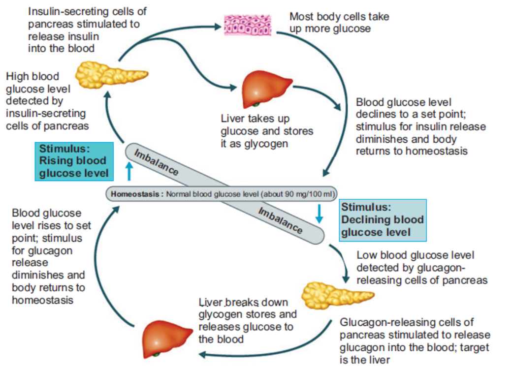
Figure 12.5: Negative feedback regulation of blood glucose level by insulin and glucagon
Response to an Increase in Blood Glucose
When there is increase in blood glucose level, the beta cells of the pancreatic islets of langerhans
increase the release of insulin into the blood. Insulin binds to receptors on the cell membrane
and stimulates the cells to increase glucose absorption. This led to a decrease in blood glucose
level. Besides, insulin also stimulates glycogenesis and glycolysis while inhibiting glycogenolysis,gluconeogenesis, lipolysis etc. which all contributes in reducing blood glucose levels.
Response to a Decrease in Blood Glucose
Decreased in blood glucose level stimulates the alpha cells of pancreas islets to increase the
secretion of glucagon. Glucagon activates enzyme glycogen phosphorylase in the liver and
muscle cells which start glycogenolysis. It also promotes gluconeogenesis, lipid metabolism etc.The overall effect of glucagon is an increase in the concentration of blood glucose.
12.5 INTERACTION OF GLUCOSE CONTROL MECHANISMS BYOTHER HORMONES
ACTIVITY 5
Using the internet, find research papers on the role of adrenaline in regulating blood glucose
level.
Make a PowerPoint presentation of the research paper.Show the presentation to the class.
12.5.1 Hormones Involved in Glucose Regulation
Other than insulin and glucagon, there are many hormones which contribute to the regulation
of blood glucose level (Figure 12.6). They are:
1. Somatostatin: It is secreted by delta cells of pancreatic islet of langerhans in response
to many factors related to ingestion of food like increased concentration of glucose,
amino acids, fatty acids and several gastrointestinal hormones released from the upper
gastrointestinal tract. Somatostatin acts locally within the islets of Langerhans and
inhibits the secretion of both insulin and glucagon. It also reduces the motility of the
stomach, duodenum, and gallbladder and decreases the secretion and absorption in the
gastrointestinal tract. Hence, lowers overall blood glucose level.
2. Epinephrine: Commonly known as Adrenaline, it is secreted by the medulla of the
adrenal glands in response to strong emotions such as fear or anger. It causes increases
in the heart rate, muscle strength, blood pressure and sugar metabolism. In response, it
enhances the process of glycogenolysis, increasing the overall blood glucose concentration.
3. Cortisol: It is also known as stress hormone and is secreted by the adrenal cortex of the
adrenal gland in response to stress. Cortisol enhances gluconeogenesis and increases the
concentration of glucose in the blood.
4. Adrenocorticotropic Hormone (ACTH): In response to various stresses, hypothalamus
secretes corticotropin-releasing hormone which stimulates anterior pituitary to secreteACTH. It stimulates the the adrenal cortex to release the cortisol hormones.
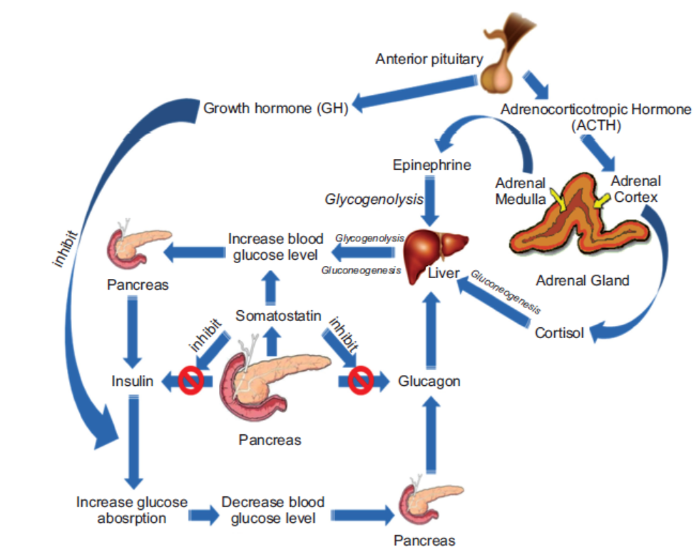
Figure 12.6: Hormonal regulation of blood glucose level
5. Growth hormone (GH): It is another anterior pituitary hormone which antagonizes
the action of insulin by inhibiting the glucose uptake by cells and increasing the blood
glucose level.
6. Gastrointestinal hormones: The hormones released by gastrointestinal tract such as
gastrin, secretin, cholecystokinin and gastric inhibitory peptide etc. increase the digestion
and absorption of nutrients in the gastrointestinal tracts. These hormones stimulate thepancreas to secrete insulin in anticipation of the increase in blood glucose level.
12.5.2 Mechanism of Hormonal Regulation
Our body maintains certain variables like temperature, pH etc. within a safe range so that it
does not cause any harm to the body and the internal environment remains stable and relatively
constant. This is known as homeostasis. Hormones are chemical messenger that are directlyreleased into the blood stream. They play a very important role in maintaining the homeostasis.
Steps of Hormonal Signalling
Hormonal signal transduction is a complex process which involves the following steps:
1. Hormones are the first synthesis in particular cells of an organ and stored for secretion
in response to certain stimulus.
2. When the organ receives the stimulus; hormones are secreted directly into the blood
stream.
3. Blood carries the hormone to the target cell(s).
4. The hormone is recognized by the specific receptor in the cell membrane or by the
intracellular receptor protein.
5. The hormonal signal is relayed and amplified through a series of signal transductionprocess in the target cells which lead to cellular response.
Cell Signalling Mechanism Involves in Glucose Regulation—Glycogenolysis
As mentioned earlier, several hormones affect glycogenolysis. Among these, glucagon and
epinephrine are the most important hormones which trigger the breakdown of glycogen.
Epinephrine is released in response to rigorous muscular activity from the adrenal medulla
and stimulates the breakdown of glycogen in muscle to a larger extent than in the liver. The
liver cells are more responsive towards glucagon. Both epinephrine and glucagon can bind
to common receptor and through a series of other enzymes; activate the enzyme glycogen
phosphorylase necessary to initiate glycogenolysis.
The whole process of signal transduction epinephrine and glucagon can be divided into threemain steps (Figure 12.7).
1. Hormone-receptor interaction at the cell surface
The hormone epinephrine and glucagon act as a ligand and bind to specific receptors
known as 7TM, found in the plasma membranes of muscle and liver cells. The bindingactivate the alpha-subunit of the Gs protein.
Figure 12.7: Hormonal signal transduction pathway in the activation of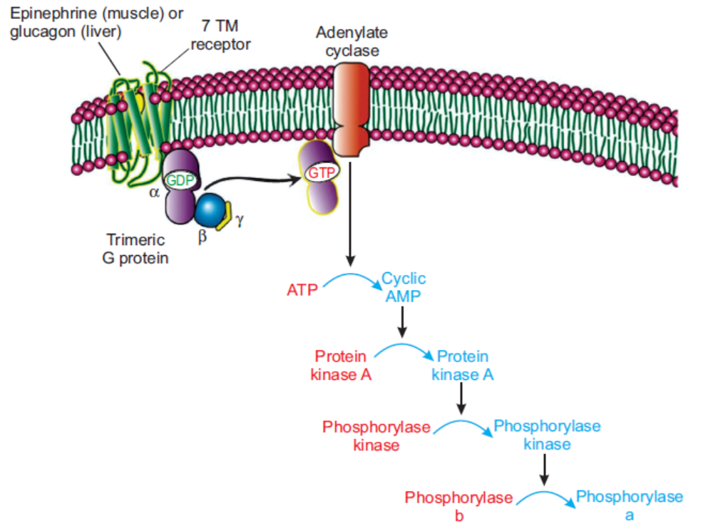
glycogen phosphorylase enzyme during glycogenolysis
2. Formation of cyclic AMP that binds to kinase protein
The activated alpha-subunits of Gs protein activate the adenylate cyclase, a transmembrane
protein. Adenylate cyclase catalyzes the formation cyclic AMP (cAMP) which acts as
the second messenger in the signal transduction pathways.
3. Activation of an enzyme cascade by phosphorylation and amplification of the signal
As level of cyclic AMP increases in the cytoplasm, it binds to the regulatory subunits
of protein kinase A and activates protein kinase A. The activated protein kinase A
phosphorylates and activates another enzyme phosphorylase kinase which finally activates
glycogen phosphorylase and initiates the glycogenolysis.
Once activated, cyclic AMP cascade can highly amplify the effects of hormones through the
number of enzymes. Therefore, binding of a small number of hormone to cell-surface receptorscan lead to the release of a very large number of sugar molecules.
APPLICATION 12.2
1. Complete with appropriate terms:
(i) Insulin was first discovered in 1922 by ...................... .
(ii) Glucagen is secreted by ....................... cells of pancreatic islets.
(iii) Epinephrine commonly known as ....................... .
(iv) Hormone .................... enhances glycogenolysis and increases concentration of glucose
in the blood.
(v) ............................. catalyzes the formation of cycle AMP.
(vi) ............................ activates glucagon phosphorylase and initiates the glycogenolysis.
2. An investigation was carried out to determine the response of pancreatic cells to an increase
in the glucose concentration of the blood. A person who had been told not to eat or drink
anything other than water for 12 hours then took a drink of a glucose solution. Blood samples
were taken from the person at one hour intervals for five hours, and the concentration of
glucose, insulin and glucagon in the blood were determined. The results are shown in thegraph below:
a) Explain why the person was told not to eat or drink anything other than water for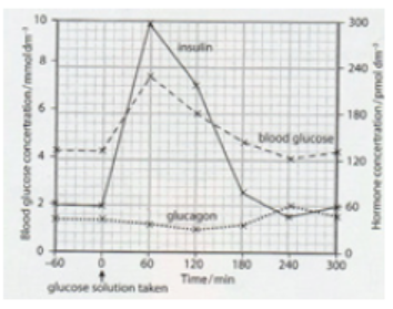
12 hours before having the glucose drink.
b) Use the information in the figure to describe the response of the pancreatic cells to
an increase in the glucose concentration.
c) Outline the role of insulin when the glucose concentration in the blood increases.
d) Suggest how the results will change if the investigation continued longer than fivehours without the person taking any food.
12.6 CAUSE OF BLOOD SUGAR IMBALANCES IN THE BODY
ACTIVITY 6
Draw the diagram of the negative feedback mechanism of glucose regulation on the chart
paper.
Illustrate the effect of various hormones on each step.Highlight the important steps which can cause or affect the blood sugar balance.
Our body obtains glucose from various food sources or synthesis in the liver and muscles from
other compounds like pyruvate, lactate, glycerol, and glucogenic amino acids. The blood carriesglucose to all the cells in the body where it is metabolized to produce energy.
Blood sugar levels keep on fluctuating throughout the day increasing after meals and decreasing
in between the meals. When the blood glucose level rises beyond the normal value, the condition
is known as hyperglycaemia. On the other hand, hypoglycaemia or low blood sugar is thecondition in which the blood glucose level is below normal (~70 mg/dL).
12.6.1 Hyperglycaemia
High blood glucose level can be caused due to various reasons like:
1. Carbohydrates: Eating food containing too much carbohydrates. The body of a person
cannot process high levels of carbohydrates fast enough to convert it into energy.
2. Insulin control: The pancreas of the individual are unable to produce enough insulin.
3. Stress: Stress stimulates the secretion of certain hormones like cortisol and epinephrine
etc., which increases the blood glucose level.
4. Low levels of exercise: Daily exercise is a critical contributor to regulating blood sugar
levels.
5. Infection, illness, or surgery: With illness, blood sugar levels tend to rise quickly over
several hours.
6. Other medications: Certain drugs, especially steroids, can affect blood sugar levels.
A high blood sugar level can be a symptom of diabetes. If hyperglycaemia persists for several
hours, it can lead to dehydration. Other symptoms of hyperglycaemia include dry mouth, thirst,
frequent urination, blurry vision, dry, itchy skin, fatigue or drowsiness, weight loss, increased
appetite, difficulty breathing, dizziness upon standing, rapid weight loss, increased drowsinessand confusion, unconsciousness or coma.
12.6.2 Hypoglycemia
Hypoglycaemia is generally defined as a serum glucose level below 70 mg/dL. Symptoms
typically appear when the blood glucose levels reaches below 60 mg/dL and levels below 50
mg/dL can be fatal.
Common causes of low blood sugar include the following:
1. Overmedication with insulin or antidiabetic pills
2. Use of alcohol
3. Skipped meals
4. Severe infection
5. Adrenal insufficiency
6. Kidney failure
7. Liver failure, etc.
Common symptoms of hypoglycemia include trembling, clammy skin, palpitations (pounding
or fast heart beats), anxiety, sweating, hunger, and irritability. If the brain remains deprived
of glucose for longer period, a later set of symptoms can follows like difficulty in thinking,
confusion, headache, seizures, and coma. And ultimately, after significant coma or loss ofconsciousness, death can occur.
12.7 DIABETES MELLITUS
ACTIVITY 7
Using the internet, study the different symptoms and causes of diabetes mellitus.Prepare a PowerPoint presentation and present to the class.
Diabetes mellitus (commonly referred to as diabetes) is a chronic condition associated with
abnormally high levels of sugar in the blood due to impaired carbohydrate, fat, and protein
metabolism. It can be due to absence or insufficient production of insulin by the pancreas, or
inability of the body to properly use insulin. Hence, there are two types of diabetes mellitus –
Type I causes by lack of insulin secretion and Type II, caused by reduced sensitivity of targetcells to insulin.
12.7.1 Type I Diabetes
It is known as insulin dependent diabetes mellitus (IDDM) and cause due to insufficient
insulin production by the beta cells of pancreatic islet of langerhans or due to absence of the
beta cells itself. Since the pancreas makes very little or no insulin at all, glucose cannot get
into the body’s cells and remain in the blood leading to hyperglycemia. The concentration of
blood glucose level can be as high as 300 – 1,200 mg/dL. The symptoms of Type I diabetesincludes:
1. Loss of glucose in urine; due to increase in blood glucose, concentration goes beyond
180 mg/dL.
2. Dehydration; due to osmotic loss of water from cells and inability to reabsorb water in
kidney.
3. Tissue injury; due to damages blood vessels in many tissues.
4. Metabolic acidosis; due to increased fat metabolism.5. Depletion of body’s protein; due to increase protein metabolism.
Treatment of Type I Diabetes
Persons with Type I diabetes require treatment to keep blood sugar levels within a target range
which includes:
1. Taking insulin from external source everyday either through injections or using an insulin
pump.
2. Monitoring blood sugar levels several times a day.
3. Eating a healthy diet that spreads carbohydrates throughout the day.
4. Regular physical activity or exercise. Exercise helps the body to use insulin more efficiently.
It may also lower your risk for heart and blood vessel disease.
5. Not smoking.6. Not drinking alcohol if you are at risk for periods of low blood sugar.
12.7.2 Type II Diabetes
Also known as non-insulin dependent diabetes mellitus (NIDDM), it is caused due to the
inability of cells to take up glucose from the blood. It can be either due to defective insulin
receptors over cell surfaces or abnormality of blood plasma protein, amylin. Due to decrease
sensitivity of cells to insulin, a condition known as insulin resistance, the beta cells secrete
large amount of insulin into the blood stream resulting in increase concentration of insulin in
blood. This condition is known as hyperinsulinemia. Type II diabetes are more common and
account for almost 80–90 per cent of the total diabetes mellitus cases.
The symptoms of type II diabetes include:
1. Obesity, especially accumulation of abdominal fat;
2. Fasting hyperglycemia;
3. Lipid abnormalities such as increased blood triglycerides and decreased blood highdensity
lipoprotein-cholesterol; and4. Hypertension.
Treatment of Type II Diabetes
There’s no cure for diabetes, so the treatment aims to keep the blood glucose levels as normal as
possible and to control the symptoms and prevent health problems developing later in life. In type II
diabetes, the pancreas is still working but our body develops insulin resistance and is unable to
effectively convert glucose into energy leaving too much glucose in the blood. Therefore, Type
II diabetes can be managed through lifestyle modification including:
1. Healthy diet as eating well helps manage our blood glucose levels and body weight.
2. Regular exercise helps the insulin work more effectively, lowers your blood pressure and
reduces the risk of heart disease.
3. Regular monitoring of blood glucose levels to test whether the treatment being followedis adequately controlling blood glucose levels or we need to adjust the treatment.
12.7.3 Importance of Controlled Diet in Diabetes
Controlled diet is very important for diabetic patients because blood sugar is mostly affected
by the food one eats. The glycaemic index of a food measures how the food affects the blood
glucose level. The higher the glycemic index of the food, the greater the potential of increasing
blood glucose. Therefore, in order to control glucose levels in the blood, it is important that
diabetic primarily chooses low glycaemic index carbohydrates like dried beans and legumes
such as lentils and pintos, non-starchy vegetables, fruits, whole grain bread and cereals, sweet
potatoes etc. Foods like white bread, white rice, cornflakes, white potatoes, popcorn, pineapple,
and melons are high glycaemic index foods and should be eaten moderately.
Because people with diabetes are at risk of high blood pressure, it makes sense to also choose
foods that are heart healthy (i.e., lean, low-fat) and the ones that are low in salt. Increasing
the amount of fibre in diet and reducing fat intake, particularly saturated fat, can help prevent
diabetes or manage the diabetic condition from developing any complications. Therefore, one
should:
1. Increase the consumption of high-fibre foods, such as wholegrain bread and cereals,
beans and lentils, and fruits and vegetables.
2. Choose foods that are low in fat for example, replace butter, ghee and coconut oil with
low-fat spreads and vegetable oil.
3. Choose skimmed and semi-skimmed milk, and low-fat yoghurts.
4. Eat fish and lean meat rather than fatty or processed meat, such as sausages and burgers.
5. Grill, bake, poach or steam food instead of frying or roasting it.
6. Avoid high-fat foods, such as mayonnaise, chips, crisps, pasties, poppadoms and samosas.
7. Eat fruit, unsalted nuts and low-fat yoghurts as snacks instead of cakes, biscuits, bombaymix or crisps etc.
12.7.4 Coping with Situation of Diabetics and Hypertension
Blood pressure is the measure of the force of blood pushing against blood vessel walls. The
heart pumps blood into the arteries, which carry the blood throughout the body. The normal
plod pressure is less than 120 (systolic) over 80 (diastolic). High blood pressure, also called
hypertension, is dangerous because it makes the heart work harder to pump blood out to the
body and contributes to hardening of the arteries, or atherosclerosis, to stroke, kidney disease,
and to the development of heart failure. Diabetics are more likely to develop high blood pressure
and other heart and circulation related problems, because diabetes damages arteries and makes
them targets for hardening (atherosclerosis). Obesity is another main factor which is responsible
for hypertension.
When it comes to preventing diabetes complications, normal blood pressure is as important as
good control of blood glucose levels. Therefore, to treat and help prevent high blood pressure,
one should control their blood glucose, stop smoking, eat healthy, maintain a healthy bodyweight, limit alcohol and salt consumption and exercise regularly.
Assisting Hypertension Patients
The following first aid tips are recommended when a person experiences hypertension:
1. Make the patient to lie on the bed and rest adequately. Tell the patient to take deep breaths
and reassure them.
2. Do not allow them to walk about, accompany the patient if it is really needed.
3. If the patient is in a stressful situation, try to comfort and reduce anxiety, as anxiety
alone can increase blood pressure.
4. If the patient is vomiting or having seizures, turn to lateral side to prevent aspiration.
5. Keep monitoring breathing, pulse rate, blood pressure, level of consciousness and for
any other dangerous signs ( e.g., paralysis of body in stroke, convulsions, etc.)
6. Do not give anything by mouth to eat/drink if there is suspicion of stroke.
7. If the patient’s nose is bleeding, administer appropriate first aid.
8. Give drinks that are high in potassium to help balance the amount of sodium (salt) inthe body to lower blood pressure levels.
12.8 MONITORING OF BLOOD GLUCOSE LEVELS
ACTIVITY 8
Aim: To perform blood glucose quantification test.
Materials Required:
1. Glucose meter
2. Test strips
3. Lancets (small needles used to prick the skin) and lancet device
that
holds the lancet.
Procedure:
1. Wash your hands with soap and water and dry them properly.
2. Prepare the blood glucose meter with the test strip according to the manufacturer’s instructions.
3. Use the lancet device to prick the side of your fingertip with a lancet.
4. Place a drop of blood onto the correct part of the test strip.
5. The strip will draw up the blood into the meter and show a digital reading of the blood
glucose level within seconds.
6. Note the reading.
7. Use a clean cotton ball to apply pressure to the fingertip for a few moments until the bleeding
stops.
8. Similarly, measure the blood glucose level of your friends.
9. Compare your blood glucose level with that of your friends.
Discussion:
In general, a fasting blood glucose reading (taken before a meal) should be between 72 mg/dL
to 126 mg/dL. And a blood glucose reading 2 hours after a meal should be between 90 mg/dL
to 180 mg/dL.
Precautions:
1. Make sure the lancelet is properly sterilized.2. Insert the test strip properly.
Blood glucose monitoring is a way of testing the concentration of glucose in the blood (glycaemia).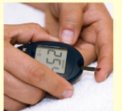
As mentioned earlier, the concentration of blood glucose kept on fluctuating throughout the
day. Under certain physiological disorders, especially when the person is suffering from diabetesmellitus, the blood glucose concentration can increase well above the normal concentration.
Most people with type II diabetes need to monitor their blood sugar levels at home. A blood
glucose test is generally performed by piercing the skin (typically, on the finger) to draw blood,
then applying the blood to a chemically active disposable ‘test-strip’ as described (see activity 8)or to a biosensor.
Dipstick Test
A dipstick or the reagent strips is a narrow strip of plastic with small pads attached to it.
Each pad contains specific reagents for a different reaction, thus allowing for the simultaneous
determination of several compounds. The blood glucose test use enzymes glucose oxidase and
hexokinase which are specific to glucose, embedded on a test strip or a dipstick. When the
blood sample is applied onto the strip, the enzymes catalyzed glucose specific reaction whichchanges the colour. The chemical reaction involves in the glucose oxidase test is as follows:
Numbers of chromogen like potassium iodide, tetramethylbenzine, O-tolidinehydrochloride,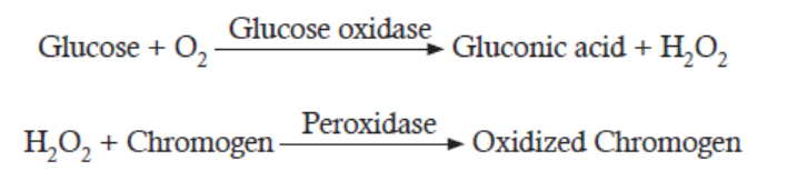
4-aminoantipyrine etc. are used in the dipstick. The colour reaction of the dipsticks is kinetic
and will continue to react after the prescribed time. Therefore, reading taken after the prescribedtime can give false result.
Biosensors
A biosensor is a device which is composed of two elements; a bio-receptor that is an immobilized
sensitive biological element (e.g. enzyme, DNA probe, antibody) recognizing the analyte (e.g.
enzyme substrate, complementary DNA, antigen) and a transducer, used to convert biochemical
signal resulting from the interaction of the analyte with the bioreceptor into an electronic
signal. The intensity of generated signal is directly or inversely proportional to the analyte
concentration. For example, the glucose biosensor is based on the fact that the immobilized
Glucose oxidase enzyme which catalyzes the oxidation of β-D-glucose by molecular oxygen
producing gluconic acid and hydrogen peroxide. An electrochemical transducer converts thisreaction into electronic signal which appears on the screen of the glucose meter.
Continuous Glucose Monitoring
Continuous glucose monitoring systems (CGMS) use a glucose sensor inserted under the
skin in the form of a small needle. The signal from the sensor is transmitted wirelessly and the
result is recorded in a small recording device. The monitor of the device updates and displays
the blood sugar level every few minutes. The glucose sensor needs to be removed and replaced
at least once per week.
Advantages of continuous glucose monitoring:
1. The monitor displays blood sugar level every few minutes, allowing one to see whether
the level is increasing, decreasing, or is stable.
2. The receiver can also be set to alarm if the blood sugar level is above or below a pre-set
level.
3. The blood sugar results from the continuous monitor can be downloaded to a computer,
allowing you to check blood sugar trends over time.
The only disadvantage of continuous monitor other than the cost is its inaccuracy compared
to more traditional accurate dipstick method. Therefore, most experts recommend continuous
glucose monitoring along with several finger sticks daily to calibrate the CGMS device and toverify that the sensor readings are accurate.
Roles of Adrenaline in the Control of Blood Sugar Level
Adrenaline, a natural stimulant created in the kidney’s adrenal gland, travels through the
bloodstream and controls functions of the autonomous nervous system, including the secretion
of saliva and sweat, heart rate and pupil dilation. The substance also plays a key role in the
human flight-or-flight response.
The “fight or flight” hormone that gives us a quick boost of extra energy to cope with danger
— including the danger of low blood glucose. When blood glucose levels drop too low, the
adrenal glands secrete epinephrine (also called adrenaline), causing the liver to convert stored
glycogen to glucose and release it, raising blood glucose levels. Epinephrine also causes many
of the symptoms associated with low blood glucose, including rapid heart rate, sweating, and
shakiness. The epinephrine response spurs the liver to correct low blood glucose or at least raiseblood glucose levels long enough for a person to consume carbohydrate.
12.9 DETECTION OF GLUCOSE IN URINE
ACTIVITY 9
Aim: To test the presence of glucose in the urine sample using Benedict’s test.
Materials Required:
1. Benedict’s solution (100 g of anhydrous sodium carbonate, 173 g of sodium citrate and
17.3 g of copper (II) sulfate pentahydrate in 1L dH2O prepared freshly; not more than 3
months old),
2. Urine sample
3. Dropper,
4. Test-tube,
5. Test-tube holder.
Procedure:
1. Take 5 ml (one teaspoon) of Benedict’s solution in the test-tube.
2. Holding the test-tube with the holder, heat it over a spirit lamp till the Benedict’s Solution
boils without overflowing.
3. Drop 8 to 10 drops of urine into the boiling Benedict’s solution.
4. After again boiling the mixture, let it cool down.
• Do you see any change in the colour of the mixture?
• Why does it happen?
Discussion:
1. Note down the colour of the mixture after cooling.
2. The colour of the mixture serves as a guide to the amount of sugar in the urine:
• Blue: sugar absent;
• Green: 0.5% sugar;
• Yellow: 1% sugar;
• Orange: 1.5% sugar;
• Red brown/Red ppt.: 2 % or more sugar.
Precautions:
1. Care should be taken while heating the Benedict’s solution.
2. Use disposable gloves while handling the urine sample.
3. Result should be noted only when the solution cold to the room temperature.Note: Coloured water sample can be used instead of the urine sample to avoid the cases of infection.
ACTIVITY 10
Aim: To determine the urine glucose concentration using coloured water (simulated urine).
Materials Required:
1. Coloured water (Blue, Green, Yellow, Orange, Red Brown)
2. Test tubes
Procedure:
1. Take a test tube containing coloured water.
2. Write down approximate concentration of glucose depending on the colour of the water.
3. Explain your result.
Discussion:
1. The colour of the water serves as a guide to the amount of sugar in the simulated urine:
• Blue: sugar absent;
• Green: 0.5% sugar;
• Yellow: 1% sugar;
• Orange: 1.5% sugar;• Red brown/Red ppt.: 2% or more sugar.
Urine analysis can be used to test pH, protein, glucose, ketones, occult blood, bilirubin,
urobilinogen, nitrite, leukocyte esterase etc. in the urine sample. Simple test for glucose in
urine can be used to diagnose diabetes mellitus. Generally, healthy person do not loss glucose
in their urine whereas a person with diabetes mellitus loses small to large quantities of glucosein their urine.
Detection of Glucose in Urine
The presence of glucose in the urine is called glycosuria (or glucosuria). The urine analysis
of glucose is based on enzyme glucose oxidase which is impregnated in a dipstick (reactiondescribed in previous section).
Detection of Protein in Urine
The glomerular filtrate of a normal kidney contains little amount of low–molecular weight
protein. Most of these proteins get reabsorbed in the tubules with less than 150 mg being
excreted through urine per day. Therefore, the abnormal increase in the amounts of proteinin the urine, Proteinuria, can be an important indicator of renal diseases. There are certain
physiologic conditions such as exercise and fever that can lead to increased protein excretionin the urine in the absence of renal disease.
Proteinuria is a symptom of chronic kidney disease (CKD), which can be due to diabetes, high
blood pressure, and diseases that cause inflammation in the kidneys. Therefore, urine analysis
for protein is part of a routine medical assessment for everyone. If CKD is not checked in time,
it can lead to end-stage renal disease (ESRD), when the kidneys completely stop functioning.
A person with ESRD requires a kidney transplant or regular blood-cleansing treatments calleddialysis to further sustain.
The tests for proteinuria are based either on the “protein error of indicators” principle (ability
of protein to alter the colour of some acid-base indicators without altering the pH) or on the
ability of protein to be precipitated by acid or heat. According to “protein error of indicators”
principle, a protein-free solution of tetra bromophenol blue at pH 3 is yellow in colour and
its colour changes from yellow to blue (or green) when the pH increases from pH 3 to pH 4.
However, in the presence of protein (albumin), the colour changes occur between pH 2 and 3
i.e., an “error” occurs in the behaviour of the indicator. The method is more sensitive to albuminthan to other proteins, whereas the heat and acid tests are sensitive to all proteins.
The test result may show false-positive results in a highly buffered alkaline urine, which may
result from alkaline medication or stale urine. Also, if the dipstick is left in the urine for too
long, the buffer could be washed out of the reagent resulting in increase pH and the strip may
turn blue or green even if protein is not present. On the other hand, false-negative results
can occur in dilute urines or when the urine contains proteins other than albumin in higherconcentrations.
Detection of Ketones in Urine
As discussed earlier, ketones, or ketone bodies are formed during lipid metabolism. One of
the intermediate products of fatty acid breakdown is acetyl CoA. If the lipid metabolism and
carbohydrate metabolism are in balanced, Acetyl-CoA enters the citric acid cycle (Krebs cycle)
where it reacts with oxaloacetate to form citrate. When carbohydrate is not available in the cells,
all available oxaloacetate get converted to glucose and so none is available for condensation
with Acetyl- CoA. As such, Acetyl-CoA cannot enter the Krebs cycle and is diverted to form
ketone bodies.
The ketone bodies are acetoacetic acid (diacetic acid), hydroxybutyric acid and acetone.
Acetyl-CoA is first converted to acetoacetic acid which later gets converted to other two ketonesthrough the following reaction:
Hydroxybutyric acid is formed by reversible reduction, and acetone is formed by a slow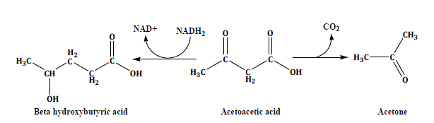
spontaneous decarboxylation. Acetoacetic acid and hydroxybutyric acid are normal fuels of
respiration and are important sources of energy. In fact, the heart muscle and the renal cortex
prefer to use acetoacetate instead of glucose. The odour of acetone may be detected in the breath
of an individual who has a high level of ketones in the blood because acetone is eliminatedthrough lungs.
Laboratory tests for ketones include reagent test-strip methods and tablet-based tests such as
Acetest. The test strip or dipstick contains the reagents sodium nitroprusside and an alkaline
buffer, which react with diacetic acid in urine to form a maroon colour, as in the followingreaction:
False-positive results may occur when the urine sample is highly pigmented, contains large
amounts of levodopa metabolites, have high specific gravity and a low pH or contain sulfhydrylgroups
APPLICATION 12.3
1.Complete with appropriate terms:
(i) Hyperglycaemia is the condition when the blood glucose level is below ....................... .
(ii) .................... is caused due to insufficient insulin production by beta cells.
(iii) A ........................... diet is important to regulate glucose level in body.
(iv) The test strip or dipstick contains the reagents ..................... and .......................... .
(v) The presence of glucose in urine is called ......................... .
2.a) Name the process by which glucose enters and leaves cells.
b) Explain why:
i. Muscle cells do not have receptors for insulin.
ii. There are second messengers for insulin and glucagon.
iii. Insulin and glucagon have different second messengers.
3.a. Explain why insulin cannot be taken by mouth.
b. Suggest how people with type 1 diabetes can monitor the effectiveness of the insulin
that they take.c. Suggest how people with type 2 diabetes can control their blood glucose
12.10 SUMMARY
• Glucose is the universal fuel which provides energy to all the living cells.
• Glucose is converted to glucose-6-phosphate with the help of enzyme glucokinase in
liver and hexokinase in most cells, which help the cells to retain glucose.
• One molecule of glucose yields 38 molecules of ATP.
• Polysaccharides of glucose viz. cellulose, chitin are important structural components of
plants and animal cells.
• Excess glucose in the body is stored in the form of glycogen through glycogenesis.
• When the blood glucose level falls, glycogen breaks down into glucose through glycogenolysis.
• Pancreas and liver are the main organs responsible for regulation of blood glucose level.
• Pancreas secretes insulin and glucagon hormones responsible for glucose regulation.
• Exocrine pancreas release digestive enzymes including trypsin, chymotrypsin, amylase
and lipase.
• The islet of langerhans is the endocrine component of pancreas and secretes insulin
(by beta cells), glucagon (by alpha cells), somatostatin (by delta cells) and pancreatic
polypeptide (by F cells).
• Liver is a vital organ of our body which performs various functions ranging from protein
synthesis to detoxification of drugs.
• Liver lobes are compost of small units, known as liver lobules.
• Insulin stimulates rapid glucose uptake by cells, promote glycogenesis while inhibiting
glycogenolysis and gluconeogenesis resulting in decreasing blood glucose level.
• Glucagon activates enzyme glycogen phosphorylase and initiates glycogenolysis in the
liver and muscle cells.
• In liver cells, glucagon increases the rate of amino acid uptake and converts it into glucose.
• Blood glucose level regulation by insulin and glucagon is an excellent example of negative
feedback mechanism where the effect of one hormone stimulates the other hormone
and vice versa.
• Hormonal regulation of glucose is a three step process which involves hormone-receptor
interaction followed by activation of second messenger, cAMP and a series of enzyme
cascade.
• Hyperglycemia is a condition when the blood glucose level rises higher than the normal
level.
• Hypoglycaemia is the condition when the serum glucose level is below 70 mg/dL.
• Diabetes mellitus is a chronic condition associated with abnormally high levels of glucose
in the blood.
• The concentration of insulin increase in the blood (hyperinsulinemia) as more and more
insulin is secreted by beta cells in response to decrease sensitivity by cells. This condition
is known as insulin resistance.
• Blood glucose monitoring is a very important exercise to keep the glucose level checked
and avoids various harmful consequences of high blood sugar due to diabetes mellitus.
• Urine analysis of glucose, ketone bodies and protein, blood glucose test, glucose tolerancetest and acetone breath test.
12.11 GLOSSARY
• Glycosuria: Presence of glucose in urine.
• Glycolysis: Splitting of glucose molecule into two molecules of pyruvic acid.
• Glycogenesis: Biosynthesis of glycogen.
• Glycogenolysis: Breaking down of glycogen.
• Gluconeogenesis: Formation of glucose from non-carbohydrate compounds.
• Hyperglycemia: Condition of high blood glucose concentration.
• Hypoglycemia: Condition of low blood glucose concentration below 70 mg/dL.• Hyperinsulinemia: High concentration of insulin in blood.
END UNIT ASSESSMENT 12
I. State whether the following statements are True (T) or False (F)
1. Excess glucose in the body is stored in the form of glycogen.
2. Trypsin is an enzyme used for carbohydrate digestion.
3. Bile salt is secreted by exocrine liver.
4. Glucagon is secreted by pancreas in response to high blood glucose concentration.
5. Insulin administration is recommended for people with type II diabetes mellitus.
6. Type I diabetes mellitus is due to insufficient secretion of insulin by beta cells.
7. Ketone bodies are formed when our body have excessive fat metabolism.
8. Hyperinsulinaemia is associated with type II diabetes mellitus.
9. Glycogenolysis is the breakdown of glucose to form pyruvate.10. The binding of hormones to receptors activates cAMP.
II. Multiple Choice Questions
1. Which of the following monosaccharides is not a product of carbohydrate metabolism
in our body?
(a) Glucose (b) Fructose
(c) Ribose (d) Galactose
2. One molecule of glucose yield …………………… molecules of ATP.
(a) 34 (b) 36
(c) 38 (d) 40
3. Which of the following is not a part of the portal triad?
(a) Central vein (b) Hepatic artery
(c) Hepatic portal vein (d) Bile duct.
4. Somatostatin is secreted by
(a) Alpha cells (b) Beta cells
(c) Delta cells (d) F cells
5. The process of formation of glucose from non-carbohydrates source in the body is
known as
(a) Glycogenesis (b) Gluconeogenesis
(c) Glycolysis (d) Glycogenolysis
6. Which of the following hormones is responsible for decreasing blood glucose level?
(a) Glucagon (b) Insulin
(c) Somatostatin (d) Adrenaline
7. Which of the following compounds is not a ketone body?
(a) Acetoacetic acid (b) Hydroxybutyric acid
(c) Acetone (d) Citric acid
8. In the digestive tract, fructose is absorbed inside the epithelial cells of intestine through
(a) GLUT1 (b) GLUT3
(c) GLUT5 (d) SGLUT2
9. The enzyme used in the dipstick for testing concentration of glucose is
(a) Glucose oxidase (b) Glycogen phosphorylase
(c) Glucose phosphatase (d) Glucosidase
10. Hypoglycemia is the condition when the blood glucose level reach
(a) Above 140 mg/dL (b) Below 140 mg/dL(c) Above 70 mg/dL (d) Below 70 mg/dL
III. Long Answer Type Questions
1. Glucose is the most important carbohydrate in our body. Justify the statement.
2. Describe the negative feedback mechanism with an example.
3. Describe the functions of liver and pancreas in regulating blood glucose level.
4. What are the causes of sugar imbalance in our body?
5. What is diabetes mellitus? Discuss its cause and effect.
6. Discuss in brief the importance of analysing urine in diagnosis diabetes mellitus.
7. How does glucose regulation effect aging? What possible effects can regulation of
glucose have on health of an organism?
8. In your own words, describe three main stages of cell signalling in control of bloodglucose by adrenaline.
Unit 13 Regulation of Temperature
Key Unit Competence
To be able to explain the importance and ways by which organisms regulate body temperature.
LEARNING OBJECTIVES
At the end of this unit, the learner will be able to:
• Explain the importance of temperature regulation.
• Describe the morphological, physiological and behavioural adaptations to temperature
changes in the environment.
• Interpret data related to the effects of temperature on animal behaviour.
• Describe the responses to cold and hot conditions by endothermic and ectothermic animals.
• Interpret and list the adaptive features shown by plants inhabiting extreme cold and hot
environments.
• Acknowledge the importance of maintaining fairly constant temperatures for efficient
metabolism.
• Explain the role of the brain and thermo receptors in temperature regulation.
• Research using the internet the role of brain in temperature regulation.
• Describe the different processes in which plants minimise overheating.• Design and investigate the effect of temperature.
INTRODUCTORY ACTIVITY
Observe the photos below and answer the questions that follow
a). Show 2 main differences between individual A and individual E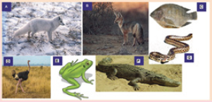
b). How is individual C different from individual D?
c). The individual A is adapted to live in cold environments. Analyze it carefully to identify
any two characteristics that this animal hasd). Which among the animals on the photo adapted to live in hot climates? Justify your answer
13.1 IMPORTANCE OF TEMPERATURE REGULATION
ACTIVITY 1
Aim: To investigate the effect of temperature on enzyme activity.
Materials Required:
1. Water bath
2. Ice
3. Test tubes
4. Thermometers
5. Bunsen burners
6. Cornstarch
7. Distilled water
Procedure:
1. Pour some water into the water baths and set the temperature at 37°C and 60°C.
2. Make a starch solution by adding 1 g of cornstarch to 10 ml of distilled water. Pour the
mixture into 50 ml of boiling water and stir until the solution becomes transparent.
3. Prepare amylase solution by adding 2 ml of saliva to 12 ml of water.
4. Take three test tubes and label as Ice, 37°C and 60°C.
5. Add 4 ml of the starch solution and 4 ml of amylase solution in the three test tubes.
6. Immediately place one test tube in the ice, one in the water bath at 37°C and other at 60°C.
7. Incubate the test tubes for 15 minutes.
8. Take 4 drops of samples from each test tube on a glass plates.
9. Add 1 drop of iodine to each sample.
10. Note the time taken for the iodine to turn yellow from blue.
Discussion:
Amylase is an enzyme that hydrolyzes starch into its components i.e., glucose. Iodine turns blue
when it comes into contact with starch, but it stays yellow in the presence of glucose. Therefore,
faster the iodine turns yellow from its blue colour, the faster amylase works on the starch.
Note the time taken for different samples and discuss the result with your teacher.
Precautions:
1. The temperature of the water baths should be properly set.
2. Starch solution should be homogenous.3. Time should be noted carefully.
Besides water, our body consists of many inorganic and organic compounds including proteins,
lipids, carbohydrates etc. Among these, proteins are the most important compounds and are
regarded as “workhorse” molecules of life, taking part in essentially every structure and activity
of life. Proteins make up about 75 per cent of the dry weight of our bodies and serve fourimportant functions;
1. They are nutritious.
2. They also form the structural components of our body including skin, hair etc. They are
building materials for living cells, appearing in the structures inside the cell and within
the cell membrane.
3. As haemoglobin, Hb they carry oxygen to all the body organs and
4. They functions as biological catalysts as enzymes facilitating and controlling various
chemical reactions of our body.
Protein molecules are often very large and are made up of hundreds to thousands of amino
acid units. They are of varying shape and size. For examples, keratins, a protein in hair and
collagen in tendons and ligaments linear chains of amino acids. Other proteins called globular
proteins, fold up into specific shapes and often more than one globular unit are bound together.
Enzymes are globular proteins. Though large, enzymes typically have a small working region,
known as active site which acts as the binding site of ligands. The shape of globular proteins
is held together by many forces, including highly resistant strong covalent bonds. However,
there are also many weak forces, like hydrogen bonds, which are susceptible to pH, osmolarityand temperature changes.
Since the function of enzymes is attributed to its shape, small changes in the shape can
greatly reduce its function. Every enzyme has an optimal temperature at which it works best
and this temperature is approximately the normal body temperature of the body. Therefore,
in order to ensure the optimal function of the enzymes within, the core body temperature
need to be maintained more or less constant. If the body temperature falls below the normal
value, the enzymes catalyzed reactions of the animal will be slowed. Similarly, too much
rise in body temperature might result in enzyme denaturation and hence reduced catalytic
activities.
Rise in body temperature also reduces the oxygen carrying capacity of haemoglobin. Increasing
temperature weakens and denatures the bond between oxygen and haemoglobin which in turn
decreases the concentration of the oxyhaemoglobin. This can lead to hypoxia – a condition inwhich tissues receive insufficient oxygen supply from the blood.
13.2 MORPHOLOGICAL, PHYSIOLOGICAL AND BEHAVIOURALADAPTATION TO TEMPERATURE CHANGES IN THE ENVIRONMENT
ACTIVITY 2
Use internet to find the pictures of animals living in different temperature conditions like arctic,
snow covered mountains, forest, deserts, sea etc.Use the following link: https://www.youtube.
com/results?search_query=animal+habitat&sp=EgIwAQ%253D%253D
Print the pictures and paste it in your scrapbook.
Write down different morphological, physiological and behavioural adaptations of the
animal which help it to live in a particular environment. Discuss various adaptations of yourcollections.
From deepest corner of the sea to high mountains, living organisms have colonized almost
everywhere. However, they are not distributed evenly with different species found in different
areas. Many abiotic factors including temperature, humidity, soil chemistry, pH, salinity, oxygen
levels etc., influence the availability of species in certain area. Each species has certain set of
environmental conditions within which it can best survive and reproduce to which they are
best adapted. This is known as limits of tolerance (i.e., the upper and lower limits to the range
of particular environmental factors within which an organism can survive). No organism can
survive if the environmental factor is below its lower limits of tolerance or above the higher
limits. Therefore, organisms having a wide range of tolerance are usually distributed widely,while those with a narrow range have a more restricted distribution. For examples, euryhaline
fishes (like salmon) can survive wide range of salt concentration and therefore are found both
in freshwater and salt water environment while stenohaline fishes are found only in saltwateror freshwater.
Temperature is one of the most important factors which directly or indirectly influence the
distribution of organisms to a large extent. For example, polar bears can survive very well in
low temperatures ranges, but would die from overheating in the tropics. On the other hand, a
giraffe does very well in the heat of the African savanna, but would quickly freeze to death in
the Arctic. Compared to ectotherms or cold blooded animals, endotherms due to their ability to
generate their own body heat, are generally more widely distributed. Besides, all the organisms
have varying degree of morphological, physiological or behavioural adaptations that helps themto survive the extreme temperature conditions of their habitat.
13.2.1 Effect of Temperature
As discussed above, all the living organisms have a particular range of temperature within which
they can best survive and reproduce. Temperature below or above this temperature ranges are
harmful to the organism in various ways. Some of well known effects of temperature on livingorganisms are given below.
1. Effect of temperature on cells: If the temperature is too cold, the cell proteins could be
destroyed due to the formation of ice, or as the water is lost, the cytoplasm can become
highly concentrated. Conversely, extreme heat can coagulate cell proteins.
2. Effect on metabolism: Most of metabolic activities of microbes, plants and animals are
regulated by enzymes and the functions of enzymes are greatly affected by temperature.
Therefore, increase or decrease in the body temperature will greatly affect the various
metabolic activities. For example, the activity of liver arginase enzyme upon arginine
increases gradually with increase in the temperature from 17°C to 48°C. With the increase
in temperature beyond 48°C, the enzymatic activity decreased sharply.
3. Effect on reproduction: Changes in temperature affect both the maturation of gonads
i.e., gametogenesis and fecundity of animals. For example, some animal species can breed
throughout the year, some only in summer or in winter, while some species have two
breeding periods, spring and autumn. Therefore, temperature determines the breeding
seasons of most organisms. Also, it was observed that female chrotogonus trachypterus
an acridid insect lays highest number of eggs per female at of 30°C and decreases with
increase in temperature from 30°C to 35°C.
4. Effect on sex ratio: In certain animals like copepod Maerocyclops albidu, rises in
temperature significantly increases the number of male offspring. Similarly in plague flea,
Xenopsylla cheopis, males’ population outnumbered females when the mean temperature
is between 21°C to 25°C. However, further decreases in temperature reverse the conditions
with the considerable increases in female population.
5. Effect on growth and development: In general growth and development of eggs and
larvae is more rapid in warm temperatures. For example, Trout eggs develop four times
faster at 15°C than at 5°C. On the other hand, seeds of many plants will not germinate
and the eggs and pupae of some insects will not hatch until chilled.
6. Effect on colouration: Animals generally have a darker pigmentation in warm and
humid climates than those found in cool and dry climates. This phenomenon is known
as Gioger rule. In the frog Hyla and the horned toad Phrynosoma, low temperatures
have been known to induce darkening. Some prawn turn light coloured with increasing
temperature.
7. Effect on morphology: Temperatures have profound effects on the size of animals and
various body parts. Endotherms generally attain a larger body size (reduced surface-mass
ratio) in colder temperatures than in warmer temperatures. As such the colder regions
harbour larger species. Conversely, the poikilotherms (ectotherms) tend to be smaller in
colder regions. We will discuss the various morphological modifications due to extreme
climates in the later sections.
8. Effect on animal behaviour: Temperature certainly has profound effect on the behavioural
pattern of animals. The advantage gained by certain cold blooded animals through
thermotaxis or orientation towards a source of heat are quite interesting. Ticks locate
their warm blood hosts by a turning reaction to the heat of their bodies. Certain snakes
such as rattle snakes, copper heads, and pit vipers are able to detect mammals and birds
by their body heat which remains slightly warmer than the surroundings.
9. Effect on animal distribution: Since the optimum temperature for many organisms
varies, temperature imposes a restriction on the distribution of species. The diversity ofanimals and plants gradually decrease as we move from equator towards the pole.
13.2.2 Morphological Adaptations
1. Body size and shape: Ectotherms or cold-blooded animals whose body temperature
depends on the temperature of external environments are usually smaller in size compared
to endotherms or warm blooded animals. For instance, compare the size of elephant,
blue whales and crocodiles or snakes. Within the same species, individuals living in thecolder climates tend to be larger than those living in warmer climates. This is known as
Bergmann’s rule. For example, whitetail deer in the southern part of the United States
have a smaller body size than whitetail deer in the for norhtern states (Figure 13.1 (A))the far northern states (Figure 13.1 (B)).
Figure 13.1: Bergmann’s rule: Body size and temperature. White-tailed deer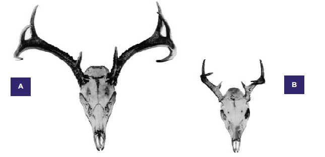
(Odocoileus virginianus) extend from Canada through Central America into South America north of
the Amazon River basin. There is a strong size gradient, with the largest
animals in the temperate north (A) and the smallest in the warm neotropics (B). Antler growth is
positively allometric with respect to skull size: the smaller animalhas disproportionately small antlers
2. Body Extremities: According to Allen’s rule, animals living in the colder climates have
more rounded and compact form. This is achieved by reducing the size of the body
extremities i.e., ears, limbs, tails etc. On the other hand, animals living in the warmer
climates have longer body extremities. For instance, compare the size of the ear of Arctic
fox with that of the Desert fox (Figure 13.2). Longer body extremities increase the surface
to volume ratio of the desert fox which enable them to lose heat more easily.
Most cold-blooded organisms have either an elongated or a flat body shape. For example,
fishes, snakes, lizards, and worms have long and slender body form which ensures rapidheat up and cool down processes.
Figure 13.2: Allen’s rule: Body extremities and temperature. (A) Arctic fox (Alopax lagopus) with its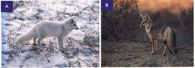
short tail, ear and legs and (B) Desert fox (Vulpes chama) with longer tail, ear and legs
Both Bergmann’s rule and Allen’s rule depend on simple principle that “the ratio of surface
area to volume of an object is inversely proportional to the volume of the object”. In other words,
the smaller an animal is, the higher the surface area-to-volume ratio. Higher surface
area-to-volume ratio ensures these animals to lose heat relatively quickly and cool down
faster, so they are more likely to be found in warmer climates. Larger animals, on the
other hand, have lower surface area-to-volume ratios and lose heat more slowly, so andthey are more likely to be found in colder climates.
3. Insulation: All the marine mammals have a thick insulating layer of fat known as
Blubber, just beneath the skin. It covers the entire body of animals such as seals, whales,
and walruses (except for their fins, flippers, and flukes) and serves to stores energy,
insulates heat, and increases buoyancy. Thickness of blubber can range from a couple
of inches in dolphins and smaller whales, to 4.3 inches in polar bears to more than
12 inches in some bigger whales. To insulate the body, blood vessels in blubber constrict
in cold water. Constriction of the blood vessels reduces the flow of blood to the skin and
minimizes the heat loss. In such animals, skin surface temperature is nearly identical
to the surrounding water, though at a depth of around 50 mm beneath the skin, thetemperature is the same as their core temperature.
Some marine mammals, such as polar bears and sea otters, have a thick fur coat, as well
as blubber, to insulate them. The blubber insulates in water while fur insulates in air or
terrestrial environment. The feathers of the birds also function in insulating the bodyfrom cold temperature.
13.2.3 Physiological Adaptations
1. Evaporation: In a colder region, i.e., when the surrounding environment of the animal is
cooler than the body temperature, conduction and radiation are the main ways an animal
will dissipate heat. However, in warmer region, the air temperature is often higher than
the animal’s body temperatures, so the only physiological thermoregulatory mechanism
available is evaporation. Animals use three evaporative cooling techniques that includesweating, panting, and saliva spreading.
(a) Sweating: It is the loss of water through sweat glands found in the skin of mammals.
The number of sweat glands can vary from none in whales, few in dogs to numerous
in humans. Most small mammals do not sweat because they would lose too much
body mass if they did. For example, in a hot desert the amount of water a mouse
would lose through sweating to maintain a constant body temperature would be
more than 20% of its body weight per hour, which could be lethal for the animal.
Therefore, smaller mammals use other techniques to cool down their body. On the
other hand, sweating is an important thermoregulatory mechanism for primates
including humans. An adult human can loss as much as 10–12 litres of water per
day through sweating.
(b) Panting: It is rapid, shallow respiration that cools an animal by increased evaporation
from the respiratory surfaces. It is a common thermoregulatory technique used by
small animals like dogs and rodents to loss heat.
(c) Saliva spreading: It is a means of thermoregulation used by marsupials. Under
extreme heat, saliva will drip from animal’s mouth and is then wiped on its fore and
hind legs. This technique induces the cooling effect of evaporation by wetting the fur.
However, since the animal cannot spread saliva while moving, they need to adapt
other evaporative techniques during such situations.
2. Counter current mechanism: As mentioned above, in addition to its role in the transport
of oxygen and food, circulatory system of our body is responsible for distribution of
heat throughout the body. This is true in case of both endotherms and ectotherms.
In endotherms, most of the body heat is generated in brain, liver, heart and skeletal
muscles. This heat is transported to other parts of the body through blood. On the other
hand, in ectotherms, the circulatory system help in transporting heat from skin to others
body parts. The counter current heat exchanger is generally located in body extremitieslike limbs, neck, gills, which are directly in contact to the external environment.
In colder region, when the warm blood flows through the arteries, the blood gives up
some of its heat to the colder blood returning from the extremities in the veins running
parallel to the arteries. Such veins are located in the deeper side of the body and carry
the warm blood to the heart and most of the body heat is retained. Such mechanism
can operate with remarkable efficiency. For instance, a seagull can maintain a normal
temperature in its torso while standing with its unprotected feet in freezing water(Figure 13.3).
When the external temperature is higher than the body temperature and heat loss is not
a problem, most of the venous blood from the extremities returns through veins located
near the surface. If the core body temperature becomes too high, the blood supply to
the surface and extremities of the body is increased enabling heat to be released to thesurroundings.
Figure 13.3: Counter current heat exchange mechanism. As warm blood travels from the heart to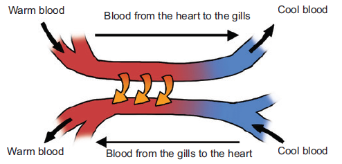
the gills, an exchange of heat takes place with the colder blood returning from the gills
3. Hyperthermia: Hyperthermia is a condition of having the body temperature greatly above
the normal. Although all the endotherms can maintain a constant body temperature, some
are able to raise their body temperature as a way to decrease the amount of water and
energy used for thermoregulation. For example, camels and gazelles can increase their
body temperature by 5–7°C during the day when the animal is dehydrated. Hyperthermia
helps in saving water by letting their body temperature increase instead of using evaporative
cooling to keep it at a constant temperature.
4. Water retention: Human body obtains about 60 per cent of the water they need from
ingested liquid, 30 per cent from ingested food, and 10 per cent from metabolism. While
rodent adapted to arid conditions obtains approximately 90 per cent from metabolismand 10 per cent from ingested food. The predaceous marsupial Mulgara can go its whole
life without ingesting water but by obtaining water from the food they eat and from
metabolism. The fawn hopping mouse eats seed, small insects, and green leaves for
moisture, and Kowaris eat insects and small mammals to obtain water. These animals
have specialized kidneys with extra microscopic tubules to extract most of the water from
their urine and return it to the blood stream. And much of the moisture that would be
exhaled in breathing is recaptured in the nasal cavities by specialized organs.
Many desert dwelling insects tap plant fluids such as nectar or sap from stems, while
others extract water from the plant parts they eat, such as leaves and fruit. The abundance
of insects permits insectivorous birds, bats and lizards to thrive in the desert. Elf owls
survive on katydids and scorpions. Pronghorns can survive on the water in cholla fruits.
Kit foxes can satisfy their water needs with the water in their diet of kangaroo rats, mice,and rabbits, along with small amounts of vegetable material.
5. Excretion: As mentioned above, desert dwelling mammals and birds have specialized
kidneys with long loops of Henle compared to animals that live in aquatic environments
and less arid regions. A longer tubules help in reabsorbing most of the water from their
urine and return it to the blood stream. As a result, the urine becomes highly concentrated.
In these animals, most of the water in the faeces gets reabsorbed in the alimentary
canals and colon. Camels produce dryer faeces than other ruminants. For example, sheep
produce faeces with 45 per cent water after 5 days of water deprivation, while camels
produce faeces with 38 per cent water even after 10 days of water deprivation. The ability
to excrete concentrated urine and dry faeces is an important adaptation to arid conditions.Desert rodents can have urine five times as concentrated as that of humans.
13.2.4 Behavioural Adaptations
Behavioural adaptations are used to reduce the amount of heat gained or lost by animals, and,
thereby, reducing the amount of energy and water to maintain the body temperature. Ectodermsor cold blooded animals rely on their behaviour to maintain a favourable body temperature.
1. Nocturnality: It is the simplest form of behavioural adaptation characterized by activity
during the night and sleeping during the day. As such, nocturnal animals avoid direct
exposure to heat of the day, thereby preventing loss of water needed for evaporative
cooling. The night temperatures are generally 15–20°C colder than the daytime, so require
much less energy and water to regulate body temperature. Most of the desert animals
like quoll, bilby, and the spinifex hopping mouse, are nocturnal. Other large animals likelions prefer to hunt at night are to conserve water.
Crepuscular animals are those animals that are mainly active during twilight (i.e., the
period before dawn and that after dusk). Examples include hamsters, rabbits, jaguars,
ocelots, red pandas, bears, deer, moose, spotted hyenas etc. Many moths, beetles, flies,
and other insects are also crepuscular in habit. These crepuscular animals take advantage
of the slightly cooler mornings and evenings to escape the daytime heat, and to evaporateless water
2. Microhabitat: Among the diurnal animals (animals which are mainly active during the
day and rest during night), the use of microhabitat like burrows, shade is another type of
behavioural adaptation to avoid the daytime heat. Fossorial animals (digging animals),
such as mulgaras, spent much of their time below ground eating stored food. Lizards
and snakes seek a sunny spot in the morning to warm up their body temperatures morequickly and remain in shade when the temperature rises.
3. Migration: It is the physical movement of animals over a long distance from one area to
another. It is found in all major animal groups, including birds, mammals, fish, reptiles,
amphibians, insects, and crustaceans. Many factors like climate, food, the season of the
year or mating could lead to migration. It helps the animals in avoiding the extreme
environmental conditions by moving to more favourable places. For example, many
migratory birds like arctic tern (Sterna paradisaea) migrate north-south, with species
feeding and breeding in high northern latitudes in the summer, and moving some hundreds
of miles south during the winter to escape the extreme cold of north. Monarch butterflies
spend the summer in Canada and the Northern America and migrate as far south asMexico for the winter.
4. Hibernation and Aestivation: Warm blooded animals which do not migrate generally
survive the extreme cold condition of winter by sleeping. Hibernation is the state of
dormancy during the cold conditions, i.e., winter. During hibernation, body temperature
drops, breathing and heart rate slows, and most of the body’s metabolic functions are put
on hold in a state of quasi-suspended animation. This allows them to conserve energy,and survive the winter with little or no food.
Many insects spend the winter in different stages of their lives in a dormant state. Such
phenomenon is known as diapause. During diapauses, insect’s heartbeat, breathing
and temperature drop. Some insects spend the winter as worm-like larvae, while others
spend as pupae. Some adult insects die after laying their eggs in the fall and eggs hatch
into new insects in the spring when the food supply and temperature become favourable.
Aestivation or summer dormancy on the other hand, is a state of animal dormancy,characterized by inactivity and a lowered metabolic rate, in response to high temperatures
and arid conditions. It allows an animal to survive the scarcity of water or food as
aestivating animal can live longer off its energy reserves due to the lowered metabolism,
and reduced water loss though lowered breathing rates. Lung fishes, toad, salamander,
desert tortoise, swamp turtles are some of the other non-mammalian animals whichundergo aestivation.
5. Social behaviour: Among all the adaptations, living together is one of the most important
adaptations of the animal kingdom. Animals can derive a lot of benefit from spending
time with other members of the same species like finding food, defence against predators
and care for their young. For example, emperor penguins can survive the harsh Antarctica
winter huddling together in groups that may comprise several thousand penguins.
Huddling greatly reduces the surface area of the group compared to individuals and a
great deal of warmth and body fat is conserved. Many social mammals, including manyrodents, pigs and primates survive extreme cold by huddling together in groups.
6. Locomotion: Different types of locomotion require varying amount of energy. Many
mammals like kangaroo, hares hop, which is an energy efficient type of locomotion.
When animals go from walking to running, there is an increasing energy cost; however,
once kangaroos start moving, there is no additional energy cost. This is because when a
kangaroo lands, energy is stored in the tendons of its hind legs which is used to powerthe next hop.
APPLICATION 13.1
1. Complete with correct terms:
(i) ................... is a dormant state experienced by many insects during winters.
(ii) Gioger rule states that animals have ................... colouration in warm climate.
(iii) Individuals of some species living in colder climates tend to be larger than those in
warmer climates exhibiting ................... rule.
(iv) Polar bears and sea otters have ................... and ................... for insulation.
(v) Camels and gazelles save water by letting their body temperature .................
2. Why does a mammal have to eat much more food than a reptile of equivalent size?
3. List the three main ways in which heat can be transferred from the environment to an animal4. Suggest why a person who has dry skin in a hot environment might be in danger
13.3 RESPONSE TO COLD AND HOT CONDITIONS BY
ENDOTHERMIC AND ECTOTHERMIC ANIMALS
ACTIVITY 3
Select an animal to study the temperature regulation mechanism.
Study different morphological, physiological and behavioural adaptation of the animals in
different temperature.Make a PowerPoint presentation and present it in the class.
13.3.1 Endotherms’ Response to Temperature Changes
Endothermic organism can maintain relatively high body temperatures within a narrow
range. Since most of the body heat is produced as a result of various metabolic activities,
thermoregulation in endotherms depends on food and water availability. For example, bear
undergoes hibernation during the winter because there is no sufficient food during the cold
season. On the other hand, in arid environment like deserts, many deserts animals are nocturnalto avoid the extreme daytime heat to avoid loss of water through evaporation.
Response to Hot Temperature
When the body temperature increases in response to the external temperature, the body’stemperature control system uses three important mechanisms to reduce the body heat. These are:
1. Vasodilation: The blood vessels in skin become intensely dilated due to the inhibition
of the sympathetic centres in the posterior hypothalamus that cause vasoconstriction.
Vasodilation increases the rate blood flow to the skin and as a result, the amount of heattransfer from the core of the body increases tremendously.
2. Sweating: As discussed in the previous section, sweating is an important adaptation
to lose body heat through evaporative cooling. An increase in 1°C in body temperaturecauses enough sweating to remove ten times the basal rate of body heat production.
3. Decrease in heat production: As mentioned above, metabolic activities of the body are
the main source of body heat. The mechanisms that cause excess heat production, such
as shivering and chemical thermogenesis, are strongly inhibited when exposed to hottemperature
Response to Cold Temperature
In response to cold temperature, the temperatures control system performs exactly oppositemechanism to that performs in hot temperature. These are:
1. Vasoconstriction: The blood vessels in the skin constrict under the influence of posteriorhypothalamic sympathetic centres which reduce the blood flow to the skin.
2. Piloerection: Piloerection means hairs “standing on end”. Sympathetic stimulation causes
the arrector pili muscles attached to the hair follicles to contract, which brings the hairs to
an upright stance. The upright projection of the hairs allows them to entrap a thick layer
of air next to the skin which acts as insulator, so that transfer of heat to the surroundingsis greatly depressed.
3. Increase in heat production (thermogenesis): Endothermic metabolic rates are several
times higher than those of ectotherms. The metabolic heat production of endotherms is
regulated in response to fluctuations in the environment temperature. This phenomenon
is known as adaptive thermogenesis or facultative thermogenesis. It can be defined as
“Heat production by metabolic processes in response to environmental temperature with the purpose
of protecting the organism from cold exposure and buffering body temperature from environmental
temperature fluctuations”. Under cold temperature stress, heat production by the metabolic
activities increased tremendously by promoting shivering, sympathetic excitation of heat
production, and thyroxine secretion. These mechanisms will be discussed later. Extremeshivering can increase the temperature four to five times the normal production.
13.3.2 Ectotherms’ Response to Temperature Changes
Ectotherms cannot maintain stable body temperature and their body temperature relies on the
external temperature. They depend more on energy assimilation rather than utilizing it for
temperature regulation. Therefore, ectotherms regulate their body temperature behaviourally
and by cardiovascular modulation of heating and cooling rates. At the same time, metabolism
and other essential rate functions are regulated so that reaction rates remain relatively constant
even when body temperatures vary. This process is known as acclimatization or temperature
compensation. For example, many fish adjust metabolic capacities to compensate for seasonal
variation in water temperature with the result that metabolic performance remains relatively
stable throughout the year. Reptiles often regulate their body temperature to different levels
in different seasons to minimize the behavioural cost of thermoregulation. At the same time,
tissue metabolic capacities are adjusted to counteract thermodynamically-induced changes inrate functions.
Response to Hot Temperature
When the external temperature increases, ectotherms protect their bodies from overheatingusing various mechanisms. These are:
1. Use of microhabitat: Under extreme heat conditions, many ectotherms like lizards and
snakes prefer to stay in shade, either beneath the rocks, crevices or underground burrows.Amphibians and fishes enter cold water when their body temperature increases.
2. Acclimatization: If a salamander living at 10°C is exposed to 20°C, its metabolic rate
increases rapidly. But if the exposure to the higher temperature lasts for several days,
the animal experiences a compensating decrease in the metabolic rate. This decrease
in the metabolic rate is due to acclimatization. The higher metabolic rate is due to the
increase in the enzymes activity with temperature. However, with prolonged exposure to
the condition, the metabolic rates decrease to prevent excessive energy loss. Ectotherms
also exhibit acclimatization of temperature tolerance range with animal acclimated to
high temperature are able to tolerate higher temperature than those exposed only to low
temperature. Similarly, cold acclimated animals have better tolerance to low temperaturethan high temperature acclimated animal.
Response to Cold Temperature
Ectotherms response to cold temperature is exactly opposite to the response shown whenexposed to hot temperature. That is:
1. Basking to sun: When the body temperature of the ectotherms becomes colder than the
normal, the animals either bask to sunlight to warm up the body or move to a warmer
place. Under extreme cold conditions, all the metabolic activities may cease and theanimals enter the state of torpor (reduced metabolic activities).
2. Cold Acclimatization: Decrease in the temperature result in reduced metabolic rate.
Therefore as a compensatory measures to meet the require body metabolism, the cold
acclimatization of ectotherms is characterized by increase in concentration of various
metabolic enzymes. There is also significant increase in the mitochondria and capillaries
concentration in the skeletal muscle. This increase the ATP production through aerobic
respiration in these tissues. Therefore, in those animals which have prolonged exposure
to cold temperature, there may be increase in the locomotion, though the basal rates ofmetabolism remain below the warm acclimatized animals.
13.4 THE ROLE OF THE BRAIN: HYPOTHALAMUS ANDTHERMORECEPTORS IN TEMPERATURE REGULATION
ACTIVITY 4
Study the role of hypothalamus and different thermoreceptors in thermoregulation.
Make a PowerPoint presentation and present it in front of the class. Discuss yourpresentation with your teacher and seek suggestions for any improvements.
So far we have discussed that on the basis of types of thermoregulation, all the living organisms
can be classified into two groups – ectotherms and endotherms. Endotherms can regulate their
body temperature within a narrow range through various physiological mechanisms while
ectotherms being depended on external temperature mostly rely on their behaviour to maintain
body temperature. But how do these animals sense and counter the changing temperature oftheir body will be discussed in the section.
13.4.1 Thermoreceptors
A thermoreceptor is a sensory receptor which is basically the receptive portion of a sensory neuron
that converts the absolute and relative changes in temperature, primarily within the innocuous range
to nerves impulses. Thermoreception is the sense by which an organism perceives the temperature
of the external and internal environment from the information supply by thermoreceptors.
In vertebrates, most of the thermoreceptors are found in skins which are actually free nerve
endings. Deep body thermoreceptors are also found mainly in the spinal cord, in the abdominal
viscera, and in or around the great veins in the upper abdomen and thorax region.
Mammals have at least two types of thermoreceptors – the warm receptors, those that detect
heat or temperatures above normal body temperature and cold receptors, those that detect
cold or temperatures below body temperature. The warm receptors are generally unmyelinated
nerves fibres, while cold receptors have thinly myelinated axons and hence faster conduction
velocity. Incerasing body temperature results in an increase in the action potential discharge rate
of warm receptors while cooling results in decrease. On the other hand, cold receptors’ firing
rate increases during cooling and decreases during warming. Another types of receptor called
nociceptors, detect pain due to extreme cold or heat which is beyond certain threshold limits.
A specialized form of thermoception known as distance thermoreception is found in some
snakes like pit viper and boa, use a specialized type of thermoreceptor which can sense the
infrared radiation emitted by hot objects. The snake’s face has a pair of holes, or pits, lined
with temperature sensors. These sensors indirectly detect infrared radiation by its heating effect
on the skin inside the pit which helps them to locate their warm blooded prey. The commonvampire bat may also have specialized infrared sensors on its nose.
13.4.2 Hypothalamus
The hypothalamus is a very small, but extremely important part of the brain that acts as the link
between the endocrine and nervous systems of the body. The hypothalamus plays a significant
role in the endocrine system and is responsible for maintaining the body’s homeostasis by
stimulating or inhibiting many key processes, including heartbeat rate and blood pressure,
body temperature, fluid and electrolyte balance, appetite and body weight, glandular secretions
of the stomach and intestines, production of substances that influence the pituitary gland torelease hormones and sleep cycles.
13.4.3 Thermoregulation—Role of Hypothalamus
Thermoregulation is carried out almost entirely by nervous feedback mechanisms, and
almost all these operate through temperature-regulating centres located in the hypothalamus
(Figure 13.4). The hypothalamus contains large numbers of heat-sensitive as well as coldsensitive
neurons which acts as thermoreceptor, sensing the temperature of the brain. The
posterior hypothalamus region contain the thermoregulatory centre which integrate the signals
from of all the thermoreceptors found in skin, deep organs and skeletal muscles, as well as
from the anterior hypothalamus and control the heat-producing and heat-conserving reactionsof the body.
Cooling Mechanism
When the body temperature increases beyond the set-point, the anterior hypothalamus is
heated. The posterior hypothalamus senses the heat and inhibits the adrenergic activity of the
sympathetic nervous system, which control vasoconstriction and metabolic rate. This causes
cutaneous vasodilation and increase heat loss through skin. It also reduces the body metabolic
rate resulting in decreasing heat production through metabolic reactions. Under intense heating,
the cholinergic sympathetic fibres innervating the sweat glands release acetylcholine, stimulating
the secretion of sweat. Many behavioural responses to heat, such as lethargy, resting in shade,lying down with limbs spread out, etc., decreases heat production and increases heat loss.
Heating Mechanism
When the body temperature falls below the set-point, the body regulating mechanism tries to
reduce heat loss and increase heat production. The immediate response to cold is vasoconstriction
throughout the skin. The result is vasoconstriction of the skin blood vessels, reducing the blood
flow and subsequent heat loss through skin. Sympathetic stimulation also causes piloerectionand reduces the heat loss from the body by trapping heat within the body hair.
The primary motor centre for shivering is excited by the cold signals from skin and spinal cord
which cause shivering of the skeletal muscles. Intense shivering can increase the body heat
production four to five times normal. Cooling the anterior hypothalamic due to decrease in body
temperature stimulates hypothalamus to increases the production of the neurosecretory hormone
thyrotropin-releasing hormone. This hormone in turn stimulates the anterior pituitary gland,
to secrete thyroid-stimulating hormone. Thyroid-stimulating hormone then stimulates thyroid
glands to increased output of thyroxine. The increased thyroxine level in the blood increasesthe rate of cellular metabolism throughout the body and hence increases heat production.
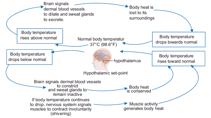
Figure 13.4: Neural feedback mechanism for regulation of body temperature
13.5 EFFECT OF TEMPERATURE CONDITIONS ON ANIMAL BEHAVIOUR
ACTIVITY 5
Aim: To investigate the effect of temperature conditions on animal behaviour.
Materials Required:
1. A long piece of metal at least 1 cm thick
2. Hot plates
3. Ice
4. Crickets/Cockroach
5. Transparent plastic pipe (at least 6 cm in diameter).
Procedure:
1. Cut the transparent plastic pipe longitudinally into two equal halves.
2. Place one end of the metal rod on a hot plate and the other end in ice to form a
continuous thermal gradient.
3. Over top the metal rod, place the long half cylinder clear plastic pipe and seal the ends with
cotton.
4. Release some 5–10 crickets/cockroachcs into the tunnel.
5. Observe the behaviour of the animals inside the tunnels.
6. Remove the hot plates and ice from the ends of the metal rods.
7. Observe the change in the behaviour of the animals.
Discussion:
1. Observe whether the animals seek out a preferred temperature or do they remain
dispersed.
2. Note down the temperature of the point of the tunnels where the animals aggregate.3. Discuss your result in the class.
Temperature generally influences the behavioural pattern of animals. In temperate waters, the
influence of temperature on the behaviour of wood borers is profound. For example, in the winter
months in general, both Martesia and Teredo occur in smaller numbers in comparison with Bankia
campanulaia whose intensity of attack is highest during the winter months. Further, the advantage
gained by certain cold blooded animals through thermotaxis or orientation towards a source of
heat are quite interesting. Ticks locate their warm blood hosts by a turning reaction to the heat
of their bodies. Certain snakes such as rattle snake, copper heads, and pit vipers are able to detectmammals and birds by their body heat which remains slightly warmer than the surroundings.
Even in the dark, these snakes strike on their prey with an unnerving accuracy, due to heat
radiation coming from the prey. The arrival of cold weather in temperate zones causes thesnakes to coil up and huddle together.
However, changes in temperature conditions affect the normal adaptational behaviour of
animals. For example, animal species that hibernate throughout winters end their hibernation
sooner than normal due to intense climatic changes. This disruption from normal hibernation
could mean life or death for these species. Migration patterns of many animal species have
also been affected due to temperature change. One of the reasons for change in migration
patterns is loss of habitat at either end of the migratory route. Another reason for change is
that some animals are travelling farther towards higher altitudes in search of colder climates,
invading the territory of already established species. Many animals cover long distances to
reach warmer climates for breeding purposes. However, the devastation of migratory route
or loss of habitat has forced these species to either change or not migrate at all. The same ishappening for aquatic animals.
13.6 TEMPERATURE CONTROLS IN PLANTS
ACTIVITY 6
Select a plant grown in extreme cold and hot environments.
Study the plant grown in hot climate and the plant grown in cold climate.
Point out various adaptive features of the plants.Make a PowerPoint presentation and present it in the class.
Like all the other living organisms, plants depend on enzymes catalyzed chemical reactions for
their growth and development. For example, plants synthesize their own food from water and
carbon dioxide using sunlight through photosynthesis. The process of photosynthesis involves
a series of complex enzyme system and other proteins. Therefore, along with carbon dioxide,
water, light, nutrients and humidity, temperature is also one of the limiting factors for growth
and development of plants.
Unlike animals, plants remain fixed in a particular site and absorb heat from the sunlight. The
excess heat from the body is released to the surrounding through radiation and evaporation.
The process of evaporation of water from the leaves and stem of plants to the surrounding
environment is known as transpiration. It occurs through stomata, small opening located on
the underside of the leaves. The stomata are specialized cells in the leaves which can open or
close, limiting the amount of water vapour that can evaporate. Higher temperature causes the
opening of stomata whereas colder temperature causes the opening to close. The opening of
the stomata and hence the transpiration rate of plants depends on environmental conditions
such as light, temperature, the level of atmospheric CO2 and relative humidity. Higher relative
humidity leads to more opening, while higher CO2 levels lead to closing of stomata. Under
high environmental temperature, the plant body gets heat up. In order to cool down, the plant
increases its transpiration rate. The evaporative loss of water from the plant’s body lowers thetemperature.
Besides transpiration, many plants have different adaptations that help them survive in extreme
temperature conditions ranging from hot and arid deserts to cold and snow covered mountains.
These adaptations make it difficult for the plant to survive in a different place other than the
one they are adapted to. This explains why certain plants are found in one area, but not in
another. For example, cactus plants, adapted to desert conditions can’t survive in the Arctic.These adaptations will be discussed later in this unit.
13.6.1 Effect of Temperature Changes on Plants
The most obvious effect of temperature on plants is changes in the rate of photosynthesis
and respiration. Both processes increase with rise in the temperature upto a certain limits.
However, increase in temperature beyond the limits, the rate of respiration exceeds the rate ofphotosynthesis and the plants productivity decreases.
Another important effect of temperature is during the process of germination of seeds. Like
most other processes it also depends on various factors including air, water, light, and, of course,
temperature. In many plant species, germination is triggered by either a high or low temperature
period that destroys germination inhibitors. This allows the plant to measure the end of winter
season for spring germination or end of summer for fall germination. For example, winter
adapted plant seeds remain dormant until they experience cooler temperatures. Temperature of
4°C is cool enough to end dormancy for most cool dormant seeds, but some groups, especially
within the family Ranunculaceae and others, need conditions cooler than –5°C. On the other
hand, some plants like Fire poppy ( ) seeds will only germinate after hot
temperatures during a forest fire which cracks their seed coats. The fire does not cause direct
germination, rather weakens the seed coat to allow hydration of the embryo.
Pollination is another phenological stage of plants sensitive to temperature extremes across
all species. Since pollination is carried out by pollinators like honey bees, butterflies etc., anyfactors including temperature that affect these pollinators will certainly affect the process.
Heat Adapted Plants
In extremely hot and dry desert region with annual rainfall averages less than 10 inches per year,
and there is a lot of direct sunlight shining on the plants, the main strategy for the survival of
the plants is to avoid extensive water loss through transpiration. Therefore, in such region many
plants called succulents, like cactus can store water in their stems or leaves. Some plants are
leafless or have small seasonal leaves that only grow after rains. These leafless plants conduct
photosynthesis in their green stems. Leaves are often modified into spines to discourage animals
from eating plants for water. Also waxy coating on stems and leaves help reduce water loss.
Other plants have very long root systems that spread out wide or go deep into the ground toabsorb water.
On the other hand, in hot and humid tropical rainforest, the abundance of water can cause
problems such as promoting the growth of bacteria and fungi which could be harmful to
plants. Heavy rainfall also increases the risk of flooding, soil erosion, and rapid leaching of
nutrients from the soil. Plants grow rapidly and quickly use up any organic material left from
decomposing plants and animals. The tropical rainforest is very thick, and not much sunlight
is able to penetrate to the forest floor. However, the plants at the top of the rainforest in the
canopy must be able to survive the intense sunlight. Therefore, the plants in the tropical rainforest
usually have large leaves with drip tips and waxy surfaces allow water to run off easily. Someplants grow on other plants to reach the sunlight.
Similarly, in aquatic plants adapted for life in water, the leaves are very large, fleshy and waxy
coated. Increase surface area allows plants to lose excess water while the shiny wax coating
discourages the growth of microbes. The roots and stems are highly reduced since water,nutrients, and dissolved gases are absorbed from the water directly through the leaves.
Cold Adapted Plants
In extremely cold region like tundra which is characterized by a permanently frozen sub-layer of
soil called permafrost, the drainage is poor and evaporation slow. With the region receiving very
little precipitation, about 4 to 10 inches per year usually in the form of snow or ice, plant life is
dominated by small, low growing mosses, grasses, and sedges. Plants are darker in colour, some
even red which helps them absorb solar heat. Some plants are covered with hair which helpskeep them warm while others grow in clumps to protect one another from the wind and cold.
In a slightly warmer temperate forest, with temperature varies from hot in the summer to
below freezing point in the winter, many trees are deciduous that is they drop their leaves in
the autumn to avoid cold winter, and grow new ones in spring. These trees have thin, broad,
light-weight leaves that can capture a lot of sunlight to make a lot of food during the warm
weather and when the weather gets cooler, the broad leaves cause too much water loss and can
be weighed down by too much snow, so the tree drops its leaves. They usually have thick barkto protect against cold winters.
APPLICATION 13.2
1.Complete with appropriate terms:
(i) Piloerection helps in ........................ .
(ii) ........................ can regulate their body within a narrow range.
(iii) ........................ receptors detect pain due to extreme cold or heat.
(iv) ........................ is responsible for maintaining homeostasis.
(v) Plants in tropical forests have ........................, and ........................ .(vi) Plants in cold regions shed their leaves to ........................ .
2.a) Describe the immediate environment of a typical cell within the body of a mammal.
b) Explain why it is important that the internal environment of a mammal is carefully regulated.
c) Explain how the following are involved in maintaining the internal environment: stimuli,
receptors, central control, coordination systems and effectors.
d)Explain the meaning of the terms homeostasis and negative feedback.
e) Distinguish between the input and the output in a homeostatic control mechanism3.The diagram shows the way in which temperature is regulated in body of a mammal
a) Which part of the brain is represented by box X?
b) i. How does the heat loss center control the effectors which lower the body temperature?ii. Explain how blood vessels can act as effectors and lower the body temperature?
13.7 SUMMARY
• Endotherms or warm-blooded animals are those animals that actively maintain a stable
body temperature by generating heat.
• Ectotherms or cold-blooded animals are those animals whose body temperature depends
on their surrounding environment.
• Ectotherms can conserve more energy while endotherms use their energy to maintain
body temperature, hence remain active even in wide temperature changes.
• All the enzymes have an optimum range of temperature beyond which they cease to function.
• Temperature is one of the most important factors which directly or indirectly influence
the distribution of organisms to a large extend.
• Temperature above or below the limits of tolerance can have various effects on animal’s
body including cells, metabolism, reproduction etc.
• Bergmann’s rule states that animals living in the colder climates tend to be larger than
those living in warmer climates.
• According to Allen’s rule, animals living in the colder climates have more rounded and
compact form which is achieved by reducing the size of the body extremities i.e., ears,
limbs, tails etc.
• The counter current heat exchanger is generally located in body extremities like limbs,
neck, gills, which are directly in contact to the external environment and helps to conserve
or loss body heat.
• Desert dwelling mammals and birds have specialized kidneys with long loops of Henle
compared to animals that live in aquatic environments and less arid regions.
• Hibernation is the state of dormancy during the cold conditions, i.e., winter.
• Aestivation or summer dormancy is a state of animal dormancy, characterized by inactivity
and a lowered metabolic rate, in response to high temperatures and arid conditions.
• Torpor is the state of decreased physiological activity in an animal, usually by a reduced
body temperature and metabolic rate.
• Thermogenesis or mechanisms of heat production, such as shivering and chemical
thermogenesis, are strongly inhibited when exposed to hot temperature.
• Ectotherms depends more on their behaviour to regulate their body temperature.
• Ectoderms can adjust metabolism and other essential rate functions so that reaction rates
remain relatively constant even when body temperatures vary. This process is known as
acclimatization or temperature compensation.
• A thermoreceptor is a sensory receptor which is basically the receptive portion of a sensory
neuron that converts the absolute and relative changes in temperature to nerves impulses.
• The hypothalamus is a small, but extremely important part of the brain that acts as the
link between the endocrine and nervous systems of the body.
• The primary motor centre for shivering is excited by the cold signals from skin and spinal
cord and depress by heat.
• All animals have a preferred range of temperature conditions at which it functions most
optimally.
• Changes in temperature conditions affect the normal behavioural adaptations of the
animals.
• Plants also depends on enzymes catalyzed chemical reactions for their growth and
development.
• The process of evaporation of water from the leaves and stem of plants to the
surrounding environment is known as transpiration.
• The stomata are specialized cells in the leaves which can open or close, limiting the
amount of water vapour that can evaporate.
• Temperature affects the photosynthesis, respiration, germination as well as pollination
of plants.
• Plants adapted to hot and dry climate have reduced leaves and longer roots.
• The large waxy coated leaves of plants in tropical rainforest are waterproof and help in
losing water more easily.
• Small, low growing mosses, grasses, and sedges are the characteristics of extremely coldregion like tundra.
13.8 GLOSSARY
• Adaptive thermogenesis: Heat production by metabolic processes in response to
environmental temperature.
• Aestivation: State of animal dormancy, characterized by inactivity and a lowered
metabolic rate, in response to high temperatures and arid conditions.
• Hibernation: State of dormancy during the cold conditions, i.e., winter.
• Hyperthermia: Condition when the body temperature is higher than normal.
• Hypothermia: Condition of low body temperature.
• Torpor: State of decreased physiological activity in an animal, usually by a reduced body
temperature and metabolic rate.
• Transpiration: The process of evaporation of water from the leaves and stem of plantsto the surrounding environment.
END UNIT ASSESSMENT 13
I. State whether the following statements are True (T) or False (F)
1. All the living organisms have a particular range of temperature within which they
can best survive and reproduce.
2. Nocturnality is the simplest form of behavioural adaptation characterized by activity
during the day and sleeping during the night.
3. Crepuscular animals take advantage of the slightly cooler mornings and evenings to
escape the daytime heat, and to evaporate less water.
4. Body temperature of Ectotherms rely on the external temperature.
5. Thermoregulation in endotherms depends on food and water availability.
6. Invertebrates, most of the thermo receptors are found in skins.
7. Changes in temperature conditions do not affect the normal adaptation behaviour
of animals.
8. Hibernation is the state of dormancy during the cold conditions, i.e., winter.
9. Most of the heat in our body is generated through metabolic activity.10. Larger animals can easily lose their body heat to their surrounding environment.
II. Multiple Choice Questions
1. The physical movement of animals over a long distance from one area to another is
known as ........................... .
(a) Hibernation (b) Aestivation
(c) Migration (d) None of these
2. Large animals like lions prefer to hunt at night to ........................... .
(a) conserve water (b) avoid direct exposure to heat
(c) utilise less energy (d) All of these
3. With rise in the temperature, the plant’s rate of .......................... increases.
(a) respiration (b) photosynthesis
(c) both (a) and (b) (d) None of these
4. Temperature of .......................... depends on the temperature of external environments.
(a) Ectotherms (b) Endotherms
(c) Both (a) and (b) (d) None of these
5. The state of reduced metabolic rate on a daily basis is an example of
(a) Hibernation (b) Aestivation
(c) Torpor (d) None of the above
6. Reptiles are
(a) Ectotherms (b) Endotherms
(c) Homeotherms (d) Heterotherms
7. Animals living in warmer climates have longer ears according to
(a) Bergmann’s Rule (b) Allen’s Rule
(c) Gloger’s rule (d) Jordon’s rule
8. The process of increasing body temperature in response to the environmental
temperature is known as
(a) Acclimatization (b) Adaptive thermogenesis
(c) Piloerection (d) Insulation
9. The waxy coating of leaves of aquatic plants helps to
(a) Conserve water (b) Increase transpiration rate
(c) Avoid growth of microbes (d) Float in water
10. The rate of transpiration of plants depends on environmental conditions such as
(a) Temperature (b) Level of atmospheric CO2
(c) Relative humidity (d) All the above
11. Increased thyroxin level in our blood increase the rate of
(a) Metabolism (b) Excretion(c) Muscle contraction (d) Food assimilation
III. Long Answer Type Questions
1. In your own words, explain the importance of temperature regulation.
2. Describe the morphological, physiological and behavioural adaptations to temperature
changes in the environment.
3. Giving suitable examples, describe the responses to cold and hot conditions by
endothermic and ectothermic animals.
4. Explain the role of the brain and thermo receptors in temperature regulation.
5. Describe the different processes in which plants minimise overheating.
6. In your own words, explain the importance of maintaining fairly constant temperatures
for efficient metabolism.
7. List few adaptive features shown by plants inhabiting extreme cold and hot
environments.
8. Why is thermoregulation assessed with health and disorder? How is thermoregulationcorrelated to the environment? What changes can help organism’s survival?
Unit 14 Behaviour and Responses in Mammals
Key Unit Competence
To be able to explain the different forms of behaviour and responses and their importancein the survival of organisers.
LEARNING OBJECTIVES
At the end of this unit, the learner will be able to:
• State the different types of behaviour.
• Recall that the nervous system is responsible for coordinating behaviour.
• Explain the different types of behaviour in terms of stimulus, receptor, nerves and effectors.
• Explain how the types of behaviour result from sequential responses.
• Apply knowledge of reflex actions to describe the components of a reflex arc and explain the
different reflex behaviours.
• Distinguish between simple reflex actions and a fixed action pattern.
• Give examples of imprinting and understand its significance.
• Explain the value of habituation.
• Define the terms: conditioning, habituation, survival, courtship behaviour and migration.
• Analyse the forms of conditioning.
• Analyse the contribution of innate behaviour and learned behaviour to an animal’s overall
behaviour and survival.
• Distinguish between classical and operant conditioning.
• Analyse the significance of latent learning.
• Relate learning and response to survival in the environment.
• Discuss the advantages and disadvantages to organisms living in societies.
• Describe how birds and mammals maintain their territory.
• Explain the significance of behavioural rhythms.
• Distinguish between migration and dispersion.
• Discuss the advantages of bird migration.
• Appreciate the importance of animal welfare.
• Value the causes and effects of bird and other animal migration.
• show concern for the behaviour of animals in societies.
• acknowledge the need for a territory by some animals for their continued survival.• show concern for the importance of conditioned reflex in relation to survival.
INTRODUCTORY ACTIVITY
1. At some point or another, almost everyone has watched a string of ants march across a
sidewalk or kitchen floor as the figure below indicates. Ant behavior such as this is pretty
fascinating.But why do ants crawl ?, or technically walk in a line?
2. Observe and analyse carefully the photo below that shows the newly hatched ducks follow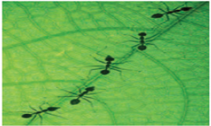
their mother everywhere it goes
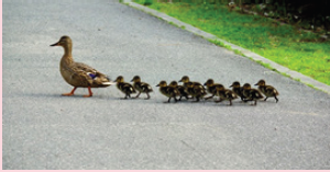
a. What do you think about this phenomenon?
b. What is the most likely advantage to the phenomenon illustrated above?
14.1 INTRODUCTION
ACTIVITY 1
Watch videos of animals from the internet or on television. Try to observe their behaviours and
responses. Discuss and make an elaborative report on it. ue the following internet link: https://www.youtube.com/results?search_query=animal+behavious&sp=EgIwAQ%253D%253D
Behaviour can be defined more precisely as an internally directed system of adaptive activities
that facilitate survival and reproduction. A stimulus is an environmental change that directly
influences the activity of an organism. Behaviour is a result of sensory and motor integration
in an organism i.e., nervous system includes sensory cells that detect changes in environment.
Nerve cells transmit and integrate information, chemical messengers transmit information into
body and muscle cells translate information into action.
Orientation behaviours are coordinated movements (walking, flying, swimming, etc.) that
occur in response to an external stimulus. These behaviours have adaptive value for survival
by helping the organism to locate (or avoid) the source of a stimulus. The simplest behaviours
involve input from only a single sensory receptor whereas more advanced behaviours requirebilateral input from a pair of receptors.
14.2 TYPES OF BEHAVIOUR
Behavioural activities are divided into two groups: Innate and Learned
14.2.1 Innate Behaviour (Simple Response)
Innate behaviour, also known as inherited behaviour, is genetically programmed. Individuals
inherit a suite of behaviours just as they inherit physical traits such as body colour and wing
venation. In general, innate behaviours will always be:
1. Heritable — encoded in DNA and passed from generation to generation
2. Intrinsic — present in animals raised in isolation from others
3. Stereotypic — performed in the same way each time by each individual
4. Inflexible — not modified by development or experience
5. Consummate — fully developed or expressed at first performance
Since innate behaviour is encoded in DNA, it is subject to genetic change through mutation,
recombination, and natural selection. Just like physical traits, innate behaviours are phylogenetic
adaptations that have an evolutionary history.
In general, innate behaviours are viewed as “programmed” responses to external stimuli. Theyusually fit into one of the following categories:
A. Automatic Innate Behaviour
ACTIVITY 2
Take some moist soil with woodlice and place it on paper. Place earthworm near the light
source.
Note the activity of woodlice and earthworm.
Discuss how animals orient in both activities of taxis and kinesis.Also state and cite any such behaviour.
The most basic unit of innate behaviour is a simple reflex arc. This is a neural pathway that
may involve as few as two neurons: a sensory neuron detects a stimulus and is linked with
a motor neuron that sets off a response in an effector cell (such as a muscle or a gland cell).
More commonly, reflex arcs also include an association neuron spliced between the sensory and
motor neurons. The association neuron also synapses with other neurons to relay information
to the brain and other parts of the body.Examples of automatic innate behaviour are:
1. Reflex arc: When you touch a hot object, you quickly pull your hand away using the
withdrawal reflex (Figure 14.1). Reflex action is different from fixed action pattern
Firstly, reflex action is a simple motor action, stereotype and repeatable but fixed
action is complex motor act, involving a specific temporal sequence of component acts.
Secondly, reflex is elicited by a sensory stimulus and the strength of the motor action
being graded with intensity of the stimulus while fixed action pattern are generated
internally or elicited by a sensory stimulus. This stimulus acts as a trigger, causing release
of coordinated motor act. Action may be graded in intensity and it may be contingent
on the type of sensory stimulus but maintain its basic pattern. Most insects have
simple “startle” reflexes triggered by small disturbances as well as more comprehensive“escape” reflexes triggered by larger disturbances.
Figure 14.1: Showing the reflex action pattern
While Fixed Action Pattern (FAP) is a sequence of coordinated movements that are performed
together as a “unit” without interruption. Each FAP is triggered by a unique stimulus variously
known as a sign stimulus, a key stimulus, or a releaser. A praying mantis striking at prey is
a typical example. The releaser for this FAP is any movement by a small (prey-sized) object
within striking distance. Once initiated, the mantis cannot change direction in mid-strike or
abort the mission if the prey escapes. Other common examples of FAPs include courtship
displays, hunting or food gathering, nest-building activities, and attack or escape movements.
Unlike simple reflexes, FAPs may involve a whole-body response and often require a thresholdlevel of internal readiness (drive).
2. Fight-or-flight response: It mobilizes the body for greater activity. Your body is being
prepared to fight or run from danger (Figure 14.2). It is controlled by hormones and thenervous system.
3. Taxis: It is a movement directly toward (positive) or away from (negative) a stimulus.
A klinotaxis involves side-to-side motions of the head or body with successive
comparison of stimulus intensity as the animal moves forward. A tropotaxis requires
bilateral input from paired sensory receptors such that the signal is equalized in both
receptors. Stimulus intensity increases with movement toward the source and decreases
with movement away from the source. For example: Movement of cockroaches awayfrom a light source.
4. Kinesis: It is a change in the speed of movement (orthokinesis) or a change in the rate of
turning (klinokinesis) which is directly proportional to the intensity of a stimulus. Input
from only a single sensory receptor is necessary. A kinesis is non-directed orientation,that is, the animal exhibits a “random walk”.
Example: Locomotion of woodlice in relation to humidity.
With increased humidity there is an increase in the percentage
time that the woodlice will remain stationary.
B. Instinct Innate Behaviour
An instinct is a complex pattern of innate behaviour. Instinctive behaviour begins when the
animal recognizes a stimulus and continues until all parts of the behaviour have been performed.
Examples of instinctive behaviour include Courtship, Territoriality and Migration. We willdiscuss these in detail later.
ACTIVITY 3
Make a list of innate and learned behaviour seen in animals. Note the points related to bothbehavioural types. Make a report on the same.
14.2.2 Learning
Learning, or learned behaviour, takes place when behaviour changes through practice or
experience.
Learning allows an animal to adapt to change, an ability that is important for animals with
long life spans (Figure 14.3). In general, learned behaviours will always be:
1. Non-heritable — acquired only through observation or experience
2. Extrinsic — absent in animals raised in isolation from others
3. Permutable — pattern or sequence may change over time
4. Adaptable — capable of modification to suit the changing conditions5. Progressive — subject to improvement or refinement through practice
Figure 14.3: Learning behaviour: A trainer is teaching the actions to dogs.
A. Habituation
A simple form of learning in which an organism decreases or ceases its response to a stimulus
after repeated presentations. It is progressive decrease of the amplitude or frequency of a motorresponse to repeated sensory stimulation that is not caused by sensory receptor adaptation or
motor fatigue. Habituation provides an important mechanism for filtering sensory information, as
it allows filtering out irrelevant stimuli and thereby focusing on important stimuli, a prerequisite
for many cognitive tasks.Example: Horses or cows disregarding noisy cars and scare crow habituation to crows.
Figure 14.4: Crows have habituated to the scare crow
B. Imprinting
It is a form of learning in which an animal, at a specific critical time of its life, forms a social
attachment to another animal. During this brief interval, the animal acquires an indelible
memory of certain salient stimuli in its “home” environment (taste of the host plant, smell of
the nest site, etc.). This memory is retained throughout life and recalled later when needed.Example: Relation between mother and new born (Figure 14.5).
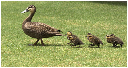
Figure 14.5: Ducklings showing the imprinting behaviour
Behavioural imprinting acts as an instinct for survival in newborns. The offspring must
immediately recognize its parent, because threatening events, such as the attack by a predator
or by other adults could occur just after hatching. Thus, imprinting is very reliable to induce
the formation of a strong social bond between offspring and parent, even if it is the wrong
one. Birds learn the characteristics of their siblings, which later on will influence their matingpreferences as adults.
14.3 CONDITIONING AND LATENT LEARNING
Learning that a particular stimulus or a particular response is linked to a reward or punishment
is called conditioning.14.3.1 Classical Conditioning
ACTIVITY 4
Discuss about the Pavlov’s experiment.
Write down the interpretation of the experiment in your exercise book.Cite some examples of classical conditioning.
It is a learning process in which an innate response to a potent stimulus comes to be elicited in
response to a previously neutral stimulus; this is achieved by repeated pairings of the neutral
stimulus with the potent stimulus. Eventually, the animal learns to respond to the stimulus
even in the absence of a reward or punishment. The scientist Ivan Pavlov conducted a famous
experiment on classical conditioning in which he trained a dog to salivate at the sound of a bell.Example: Ivan Pavlov’s classical conditioning experiment (Figure 14.6).
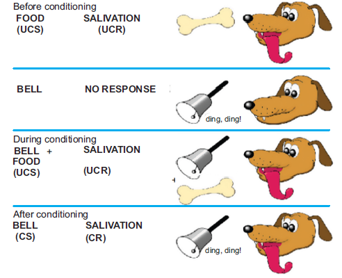
Figure 14.6: Pavlov’s experiment showing the classical conditioning
14.3.2 Operant Conditioning (Learning by Trial and Error)
An animal learns to associate one of its own behavioural acts with a positive or negative effect.
The animal tends to repeat the response if it is rewarded, but avoids the response if it is harmed.
For example, predators quickly learn to associate certain kinds of prey with painful experiences.
A coyote may learn the hard way not to attack a porcupine nose-first. Learning by trial and error
often reinforces behaviours that are important to survival. In this an animal receives a reward for
making a particular response. Motivation is an internal need that causes an animal to act, and
is necessary for learning to take place.
Examples: Learning to ride a bike or birds using different materials to build a nest untilit is just right (Figure 14.7).
Figure 14.7: Riding a bicycle showing operant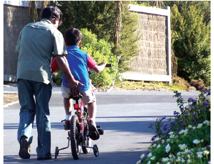
conditioning behaviour
14.3.3 Insight Learning
It is most complex type of learning in which an animal uses previous experience to respond
to a new situation. It involves the ability to analyze problems and to test possible solutions.
Insight is not technically a form of learning. Furthermore, insight may itself be based on trialand-
error experience with related problems.
Example: A chimpanzee was placed in a room with several boxes on the floor and a banana
hung high above its head. The chimp eventually “sized up” the situation and then stacked theboxes in order to reach the food (Figure 14.8).
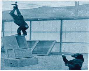
Figure 14.8: Chimpanzee showing Insight learning behaviour.
14.3.4 Latent Learning
ACTIVITY 5
Collect information about the latent learning behaviour. Write down the points related to latent
learning.Discuss the significance of latent learning.
It is a form of learning that is not immediately expressed in an overt response; it occurs without
any obvious reinforcement of the behaviour or associations that are learned. Latent learning
implies that learning can take place without any behavioural changes being immediately present.
This means that learning can be completely cognitive and not instilled through behavioural
modification alone. This cognitive emphasis on learning was important in the development of
cognitive psychology. Latent learning can be a form of observational learning (i.e., learning
through observing the behaviour of others), though it can also occur independently of anyobservation.
Example: A rat completes a maze several times, without an incentive. The rat learns the maze
very slowly (Figure 14.9). When food is placed at the end of the maze, the rat completes the
maze very quickly, demonstrating that latent learning had occurred and a cognitive map ofthe maze was informed.
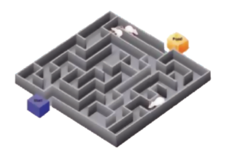
Figure 14.9: Rats are showing latent learning behaviour
APPLICATION 14.1
1.Complete with approprite terms:
(i) .......... is a sequence of coordinated moments that are performed together as a unit.
(ii) Animal exhibiting a random walk is an example of animal showing ............... or
nondirected orientation.
(iii) Ducklings show ...................... behaviour.
(iv) .............. is a learning that a particular response in linked to a reward or punishment.
(v) ...................... learning involves previous experience to respond to a new situation.
(vi) Birds show ..................... behaviour while building nests with different materials until
it is right.
2.List the main components of a reflex arc
3. Name the type of behavior that produces a movement towards a directional light source4. How does innate behavior differ from learning?
14.4 SOCIAL BEHAVIOUR
Social behaviour is any action directed by an individual towards a member of its species.
It includes competitive behaviour such as fighting, threat and submission and co-operativeinteractions like parental care and mating. All mammals show social behaviour.
14.4.1 Social Communication
It is defined as the passage of information from one individual to another and usually results
in the modification of the recipient’s behaviour or physiological state. A social signal is a
behaviour which has evolved to convey information to a non-specific receiver with the object
of modifying its behaviour for the benefit of the signaller.
Types of social signal
1. Discrete
2. Graded
Examples: For discrete social signal: alarm call of a ground squirrel, a chemical signalling
oestrus or the territorial song of a gibbon.For graded signal: aggressive vocalizations and threats displays.
14.4.2 Altruism
When an individual expends energy or runs risks in helping another, its behaviour is termed as
altruistic. Altruistic behaviour lowers the fitness of the altruist, while increasing that of the recipient.
Three theories have been suggested in attempts to accommodate altruism within current
evolutionary theory.
1. Group selection: In this group of people who support and help one another may have
an advantage over the groups whose members are selfish. Major drawback is that it is not
a stable strategy because some selfish individuals in an altruistic group would inevitably
increase their own fitness at the expense of altruistic group members.
2. Kin selection: Proposed by Hamilton, the evolutionary strategy which favours the
reproductive success of an organism’s relatives, even at a cost to the organism’s own
survival and reproduction. Kin altruism is an altruistic behaviour whose evolution is driven
by kin selection. Kin selection is an instance of inclusive fitness, which combines the
number of offspring produced with the number an individual can produce by supporting
others, such as siblings.
Example: In humans, it depends how closely related they are to the recipient.
Vervet monkeys use allo-mothering, where related females such as older sisters or grandmothers
often care for young-ones, according to their relatedness.
14.4.3 Reciprocate (Reciprocal Altruism)
It is a behaviour whereby an organism acts in a manner that temporarily reduces its fitness
while increasing another organism’s fitness, with the expectation that the other organism willact in a similar manner at a later time.
Conditions for reciprocal altruism:
1. The behaviour must reduce a donor’s fitness relative to a selfish alternative;
2. The fitness of the recipient must be elevated relative to non-recipients;
3. The performance of the behaviour must not depend on the receipt of an immediate benefit;
Example: In primates, Vervet monkey shows that among unrelated individuals, grooming
induces higher chance of attending to each other’s calls for aid. However, Vervet monkeys alsodisplay grooming behaviours within group members, displaying alliances.
Advantages of living in social groups
1. Animals are more successful in finding food if they search as a group. Foraging in a
group makes it easier to capture a prey. Example: Dolphins are known to surround a
school of fish and to take turns darting into the centre to eat the fish that are trapped in
the middle. Many carnivores will band together when they try to capture large prey.
2. Animals live in social groups to get protection. Example: One baboon might not be able
to fight off a leopard; a troop of baboons often are able to do so.
3. More individuals cooperating together, some can serve as sentries looking for danger
while the other group members are eating or sleeping. Example: Prairie dogs normally
have some individuals acting as sentries, which makes it nearly impossible to sneak up
on a prairie dog town.
4. Many prey species travel in groups because their movements are highly coordinated.
Example: Schools of fish and flocks of shorebirds. This behaviour creates confusion for
the predator.
5. Some animals form social groups to make travel easier. Example: Canada geese and
other bird species typically fly in a V formation in order to reduce wind resistance.6. Some animals congregate in close proximity in cold weather in an effort to stay warm.
Disadvantages of living in social groups
1. Increased sickness and disease: Animals living in groups face higher risks of infection
than others.
2. Increased vulnerability to predators: Animals living in social groups get protection but
they also have difficulty seeking hide during attack.
3. Increased competition for food.4. Increased competition for mates.
14.5 COURTSHIPS, TERRITORIALITY AND DOMINANCE HIERARCHIES
14.5.1 Courtship
It is the behaviour that males and females of a species carry out before mating. It communicates
to each of the potential mates that the other is not a threat. It also reveals information to each
animal that the species, gender, and physical condition of the other are suitable for mating.Courtship allows one or both sexes to select a mate from several candidates.
Examples:
(a) Rabbits: Female usually secretes an airborne hormone scent, called pheromone. Once
the male detects this scent, courtship behaviour could begin. The male and female sniff
each other, possibly to make sure of each other’s sex and mating receptivity. One bunny
then dashes off with the other in hot pursuit. Once they stop playing hard to get, the
bunnies stomp their feet and may do a little “dance” by kicking their feet in the air as
they run.
(b) Ferrets: Courtship and mating in ferrets can be a bit noisy, prolonged and even ferocious
event. Ferrets respond to the length of daylight and their natural breeding season is from
March to August. When the female goes into estrus (heat) and is receptive, the male
grabs her by the back of the neck. The male continues to bite the nape of the neck and
the ferrets are pressed together. Mating occurs and may last for around one hour.14.5.2 Territoriality
ACTIVITY 6
Research about the animal that lives in territories.
Study how they maintain their territories.Write down the points about them in your exercise book.
Territoriality refers to the exclusive use of fixed space, which entails obtaining, defending, or
advertising occupancy of the space. Animals divide geographical area around them into four
broad regions:
1. Total range (entire area covered)
2. Home range (large area for all activities – feeding, sex and roaming is done)
3. Territory (small area within home ranges, driving away intruders and visited in days4. Core area (within territory but much smaller)
Sizes of Territories
This varies from species to species depending on body size, group size, and habitat and food
requirement. Size of territory depends on size and diet of animals.
Larger species have larger territories e.g., wildebeest, zebras. Predators have larger territories
than plant eaters. Territories are smaller when food is found in abundance and distribution is
not spread far. Territorial animals patrol their outer limits.
Functions of Territoriality
1. Well shaped aggregation of local population.
2. Well defined area for parental care.
3. Limitation of breeding population and control beyond carrying capacity.
4. Adequate food.
5. Reserve of unmated males and females for prompt replacement.
6. Reduction in rate of contracting parasites or diseases.
7. Helps intending against predators and share resources.
8. Collectively defence nests and young from predators.
9. Porters separate after breeding for short periods.
Example: Wolves maintain territories in which they hunt and live. These areas are aggressively
defended from other group members. The male cougar has a large territory that may overlap
with the territories of several females but is defended against other males. Responding to scentmarks, the inhabitants of the overlapping ranges also avoid each other, except for breeding.
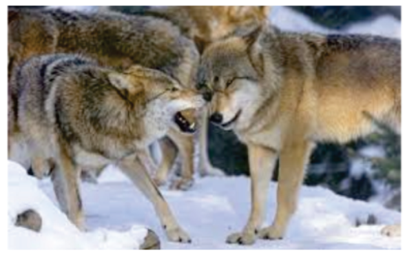
Figure 14.10: Wolves showing the territoriality behaviour
How they Mark their Territory
• Scent marking: Scent marking, also known as territorial marking or spraying when this
involves urination, is a behaviour used by animals to identify their territory. Strong-smelling
substances are present in urine, faeces, or secreted from scent glands located on various
areas of the body. These scents contain pheromones or proteins that produce odours. These
odours not only for communication, but can also mark the presence of territory.
For example, leopards and jaguars mark their territory by rubbing themselves against
vegetation. New World monkeys use urine washing to communicate.
In Blue wildebeest scent markings are secreted from two glands, the pre-orbital gland and
a scent gland in the hoof.
• Visual: Ring-tailed lemurs hold their distinctive tails high in the air during territorial scent
marking. They also engage in “stink fights” with intruding males.
To mark the territory, visual sign-posts may be short-term or long-term. Short-term
communication includes colouration or behaviour of the animal, which can only be
communicated when the resident is present. But in case of long-term visual signals, faecal
matter is deposited on the vegetation or ground.
Some animals have prominent “badges” or visual displays to mark their territory with scent
marking or auditory signals.
The ring-tailed lemur (Lemur catta) marks its territory with urine scent. When it is urinating
for marking purposes, it holds its extremely distinctive tail high in the air adding a visual
component to the advertisement; when it is urinating for eliminative purposes; its tail is
only slightly raised.
After leaving urination mark, some animals scrape or dig the ground nearby, thereby leaving
a visual advertisement of the territory. This includes domestic dogs.
• Auditory: Many animals use vocalizations to mark their territory. These are short-term
signals transmitted only when the animal is present, but can travel long distances and over
varied habitats.
Examples of wolves marking their territories to other packs through a combination of scent
marking and howling. Under certain conditions, wolf howls can be heard over areas of up
to 130 km2 (50 sq mi). When howling together, wolves harmonize rather than chorus on
the same note, thus creating the illusion of there being more wolves than there actually are.
Wolves from different geographic locations may howl in different fashions– the howls of
European wolves are much more protracted and melodious than those of North Americanwolves, whose howls are louder and have a stronger emphasis on the first syllable.
Ways to Defend their Territories
(a) Ritualized aggression: Animals use a range of behaviours to intimidate intruders and
defend their territories, but without engaging in fights which are expensive in terms
of energy and the risk of injury. This is ritualized aggression. Such defence frequently
involves a graded series of behaviours or displays that include threatening gestures such
as vocalizations, spreading of wings or gill covers, lifting and presentation of claws, head
bobbing, tail and body-beating, and finally, direct attack.Examples:
(i) Domestic cats (Felis catus) are very territorial and will defend this with ritualizedbody (Figure 14.11).
Figure 14.11: Two domestic cats posturing during ritualised aggression over a territory
(ii) Spider monkeys (genus Ateles) defend their territory by screams, barks, rattling or
dropping branches, and urinating and defecating on intruders below.
(iii) Male ring-tailed lemurs have scent glands on their wrists, chests, and in the genital
area. During encounters with rival males, they may perform ritualised aggression
by having a “stink fight”.
(b) Aggressive behaviour threatens other animals: Aggressive behaviour is used to intimidateanother animal of the same species (Figure 14.12).
Figure 14.12: Aggressive behaviour of dog
14.5.3 Dominance Hierarchies
It is defined as a form of animal social structure in which a linear or nearly linear ranking
exists, with each animal dominant over those below it and submissive to those above it in thehierarchy. It is present in mammals like baboons, wolves, etc.
Types of Dominance Hierarchies
Linear Hierarchy
This kind of dominance hierarchy can be depicted with the help of a hierarchy pyramid. Here,
individuals on a particular level use their power to dominate on other individuals that are in
lower order or level but at the same time, they tend to be submissive to individuals in orders
above them. Such individuals can be seen to get influenced by social interactions and they alsotend to have a much better access to food and other facilities.
Despotic Hierarchy
In this, one single individual is dominant on the rest of the group or clan. The orders and
instructions given by the pack leader or the troupe leader are followed submissively by the rest
of the group members. There is no fight for superiority between the followers.
Effects of Dominance Hierarchy
Individuals in higher order have a better and prior access to food. Individuals in the lower order
get the left over feed after the dominant individual has had its feed. This alpha position also
brings better mating opportunities thereby increasing the chances of reproductive success and
a healthier offspring. In case of species, where a single female mates with multiple males, themales naturally tend to be more aggressive to gain the dominant or alpha position.
An important aspect connected to dominance hierarchy is that of territorial advantage in favour
of dominant individuals. This territorial advantage is important from point of view of nesting
place, mating locations and ample supply of food.
In case of weakening or death of a dominant individual, the alpha position is assumed by
one of the individuals of the immediate next order to the alpha position after a reasonable
tussle between competing individuals. Once the dominant individual is selected, the aggression
gradually subsides and the rest of the members turn submissive.
Examples: Pack Animals
Animals that move around in packs such as wolves, wild dogs, hyenas, etc. Wolves or wild
dogs that are in dominant position have a habit of marking their territories or dens by spraying
their urine at prominent locations around their territory. This is an instant signal for other pack
members that this territory is off limits for them. Similarly, such marking helps keep prey animals
away from the dominant individual’s territory. It is easy to identify the dominant male when
the pack is defending itself from a rival pack. At such times, the dominant male is usually the
one with long puckered up ears and a straight vertical tail. Amongst the hyenas however, thealpha position is always assumed by females.
APPLICATION 14.2
1.Complete with appropriate terms:
(i) Animals living in groups face ................ risk of infection.
(ii) Courtship and mating in ferrets is ................, ................ and ................ .
(iii) Territorial marking is also known ................ .
(iv) Aggressive behaviour ................ other animals.
(v) ................ hierarchy involves one single individual dominance.
2. What are the main distinctions between classical conditioning and operant conditioning
3.Explain briefly the difference between the way a dog may solve a detour problem and theway a chimpanzee usually solves the same type of problem
14.6 BEHAVIOURAL RHYTHMS AND BIOLOGICAL CLOCKS
ACTIVITY 7
Observe all the activities which are regulated by biological clocks.
You may watch downloaded videos of animals exhibiting such behaviour.Write down and discuss them in your exercise book.\
14.6.1 Behavioural Rhythms
They are periodic biological fluctuation in an organism that corresponds to, and is in response
to, periodic environmental change. These rhythms are the repeating patterns of biochemical,
physiological, and behavioural processes. They are found in most living things, including plants,
animals and many microorganisms.
These rhythms allow animals of different species to share the same food sources without
direct competition because some animals are active only during hours of darkness (i.e., they
are nocturnal) while others are active only during the day (diurnal). The advantage to having
a built-in method of responding to light and darkness, rather than relying on actual changes in
light as a cue, is that, in effect, the organism is prevented from “sleeping late” and missing the
optimal time of day for foraging.
Most common biological rhythm is the circadian rhythm (circa- about plus dian- day). The
circadian rhythm is a rest-activity cycle that is centered on light, meaning when a preset amount
of light occurs, an animal will be active; and at another time the animal will rest. Humans are
active when there is a lot of light, which is usually during the day and rest when there is less
light, usually at night. These circadian rhythms control the core human body temperature,
sleep-wake cycle and secretion of hormones.
Hibernation and migration are the examples of biological rhythms.
Examples: Ground squirrels gather rations and pack on fat reserves in the fall in preparation
for cold winters spent underground.
Moose reproductive cycles match the birth of fawns in the spring to the rich emergence of
forage at that time.
Human core body temperature cycles with a low during the middle of their sleep cycle andhighs around lunch time and early evening.
14.6.2 Biological Clocks
Biological Clocks are self-sustaining oscillators which will continue to a period of free-running
cycling even in the absence of external cues. Biological Clocks exhibit a high degree of inheritance,
independence of temperature and social conditions, strong resistance to pharmacological andchemical disruption, may even be expressed at the level of single cells.
When an animal that functions according to such a clock is rapidly translocated to a geographic
point where the environmental cycle is no longer synchronous with the animal’s cycle, the clock
continues for a time to function synchronously with the original environmental cycle. Humans
similarly transported over great distances often experience fatigue and lowered efficiency forseveral days, a phenomenon known as “jet lag,” or jet syndrome (Figure 14.13).
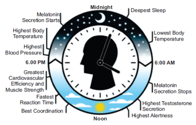
Figure 14.13: Biological clocks of human being
14.7 ANIMAL MIGRATION
ACTIVITY 8
Research about the migratory birds.Using the following internet link: https://www.google.
com/search?as_st=y&tbm=isch&hl=en&as_q=animal+migration&as_epq=&as_oq=&as_eq
=&imgsz=&imgar=&imgc=&imgcolor=&imgtype=&cr=&as_sitesearch=&safe=images&as_
filetype=&tbs=sur%3Afmc
Note the activities for which migratory birds do migrate.Write down the advantages of the migration.
It is termed as periodic movements of animals away from and back to their place of origin. It is
done annually. Animals migrate to other places with more suitable conditions of temperature,
food, more favourable living or breeding places and hibernation.Example: African antelopes migrate seasonally to avoid drought. Fur seals and many whales
make ocean voyages of thousands of miles to their breeding grounds, the former coming ashore
on islands. Little brown bat live on trees in warm weather, but in cold weather they migrate tocaves for warmer conditions.
14.7.1 Causes of Migration
• External pressures like temperature, drought, food shortage.
For example, most of the mule deer of Yellow stone Park, migrate between summer and
winter pastures, but those living near hot springs, where grazing is available all year, do not.
• Physiological and environmental changes.
Example: Birds migrate due to cycle of enlargement of the reproductive organs in spring
and their reduction in fall. Variation in day length is the chief external stimulus for this
cycle: light received by the eye affects production of a hormone by the anterior pituitarygland, which stimulates growth of the reproductive organs.
14.7.2 Advantages of Migration
• Animals remain in favourable conditions e.g., avoid cold/extremes.
• Parents and offspring grow larger and therefore have a high survival rate, they leave more
offspring.
• The population has a constant supply of food.
• Migration may lead to the colonization of a new area.
• Reduces diseases as the disease doesn’t always have a host in the area.
• Reduces effect of predation habitats that have abundant food sources year-round also attract
a greater number of predators that can threaten the nests.
• Birds that migrate to different habitats can avoid that onslaught of predators, giving their
young-ones a better chance of reaching maturity.
• Because many different populations often meet at the “breeding grounds”, migrationincreases genetic diversity as they often breed with individuals from a different population.
14.7.3 Effects of Migration
• Migration increases diversity in the gene pool of the population.
• Migration increases competition for resources, habitat and breeding places.
• Migratory animals acting as vectors for disease, nutrients and energy, and other materials
such as seeds across habitat or ecosystem boundaries.
Migration and Dispersal are different from each other. Migration is the movement of large
number of species from one place to another like bird migration. While dispersal is the spreading
of individuals away from others, often parents or siblings, which are left behind in originalareas, for example: mammals move away from their social groups.
APPLICATION 14.3
1.Complete with appropriate terms
(i) Migration increases ....................... in the gene pool.
(ii) ....................... are self-sustaining oscillators.
iii) ......................., and ....................... are examples of biological rhythms.
2. How is the territory of, for example, a bird usually established and maintained?
3. Robins have individual territories during the autumn and early winter, but from early in the
new year pairs of birds begin to share the same territory, which they maintain throughout
the spring and summer
a) Describe and explain the advantages of territorial behavior, with reference to the behavior
of the robin throughout the year
b) The territories are usually defended by song and displays, which often involve exhibiting
the red breast as much as possible to any intruding robin. Fighting is sometimes involved,
especially when the territory is being established. Suggest why robins usually defend theirterritories by song and display rather than fighting
14.8 SUMMARY
Activities that facilitate survival and reproduction.
• Genetically programmed behaviours like physical traits such as body colour and wing
venation.
• The most basic unit of innate behaviour is a simple reflex arc.
• Animal recognizes a stimulus and continues until all parts of the behaviour have been
performed.
• Courtship behaviour: Males and females of a species carry out this behaviour before
mating.
• Territoriality: Exclusive use of fixed space, which entails obtaining, defending, or
advertising occupancy of the space. Mark territories using pheromones, visual and
auditory signals.
• Ritualized aggression: To intimidate intruders and defend their territories, but without
engaging in fights which are expensive in terms of energy and the risk of injury.
• Dominance hierarchy: Linear or nearly linear ranking exists, with each animal dominant
over those below it and submissive to those above it in the hierarchy.
• Biological Rhythms are the repeating patterns of biochemical, physiological, and
behavioural processes.
• Migration is movements of animals away from and back to their place of origin.
• Behaviour changes occur through practice or experience relates to learning.
• Habituation is a form of learning in which an organism decreases or ceases to respond to
a stimulus after repeated presentations.
• Imprinting is a permanent attachment.
• Conditioning is a particular stimulus or a particular response linked to a reward or
punishment.
• Latent learning is a form of learning that is not immediately expressed in an overt response.
• Social Behaviour is the action directed by an individual towards a member of its species.
It includes competitive behaviour such as fighting, threat and submission and co-operative
interactions like parental care and mating.
• Group selection: People who support and help one another may have an advantage over
groups whose members are selfish.
• Kin selection: Favours the reproductive success of an organism’s relatives, even at a cost
to the organism’s own survival and reproduction.14.9 GLOSSARY
• Behaviour can be defined more precisely as an internally directed system of adaptive
activities that facilitate survival and reproduction.
• Biological clocks are periodic biological fluctuation in an organism that corresponds to,
and is in response to, periodic environmental change.
• Conditioning is a particular stimulus or a particular response is linked to a reward or
punishment.
• Habituation is progressive decrease of the amplitude or frequency of a motor response to
repated sensory stimulation that is not caused by sensory receptor adaptation or motor
fatigue.
• Kinesis is a change in the speed of movement (orthokinesis) or a change in the rate of
turning (klinokinesis) which is directly proportional to the intensity of a stimulus.
• Reciprocate is a behaviour whereby an organism acts in a manner that temporarily reduces
its fitness while increasing another organism’s fitness, with the expectation that the other
organism will act in a similar manner at a later time.
• Reflex action is different from fixed action pattern. Firstly, reflex action is a simple motor
action, stereotype and repeatable but fixed action is complex motor act, involving a
specific temporal sequence of component acts.
• Taxis is a movement directly toward (positive) or away from (negative) a stimulus.
END UNIT ASSESSMENT 14
I. State whether the following statements are True (T) or False (F)
1. Behaviour is precisely an external response directed internally.
2. Riding a bicycle is an innate behavioural activity.
3. In classical condition, animal learns to respond to the stimulus even in the absence
of a reward or punishment.
4. Innate behaviour is also known as inherited behaviour.
5. Kinesis is no change in the speed of movement in presence of a stimulus.
6. Animals hibernate for food and breeding.
7. Touching something hot and pulling your hand back is a learned behaviour.
8. To mark their territories, animals use pheromones.9. Latent learning is a form of learning which is immediately expressed.
II. Multiple Choice Questions
1. A trainer is teaching actions to dogs. It is
(a) learning (b) imprinting
(c) kinesis (d) courtship
2. What happens in a situation of fear?
(a) heart rate decreases (b) blood pressure decreases
(c) secretion of non-adrenaline (d) heart rate increases
3. Which behaviour activity is done before mating?
(a) learning (b) habituation
(c) courtship (d) imprinting
4. Territorial behaviour of animal provides:
(a) shelter (b) food
(c) breeding (d) all of the above
5. Periodic movement of animals from one place to another is called
(a) migration (b) imprinting
(c) conditioning (d) taxis
6. Which is an example of imprinting behaviour?
(a) secretion of saliva (b) riding bicycle
(c) newborn baby and mother (d) sacrificing life for others
7. In which behaviour are scent markings and visual signalling observed?
(a) courtship (b) territorial
(c) migration (d) habituation
8. Which of the following belongs to altruism?
(a) Hamilton’s rule (b) Pavlov’s experiment
(c) reflex arc (d) biological clock
9. Habituation behaviour is
(a) period before mating
(b) periodic movement from one place to other
(c) structure in which a linear or nearly linear ranking exists(d) organism decreases or ceases to respond to a stimulus after repeated presentations
III. Long Answer Type Questions
1. Explain the different types of behaviour giving examples.
2. State the role of nervous system in coordinating behaviour.
3. Explain how the types of behaviour result from sequential responses.
4. Explain different reflex behaviours describing the components of a reflex arc.
5. Differentiate between simple reflex actions and a fixed action pattern.
6. Describe giving examples the forms of conditioning.
7. Explain the following stating their significance.
(a) imprinting
(b) conditioning
(c) habituation
(d) survival
(e) courtship
(f) behaviour
(g) migration
8. Analyse and appreciate the importance of animal welfare. Also state behaviour of
animals in society. Why do animals make territories?
9. Discuss the causes and effects of bird and other animal migration.
10. Differentiate the following:
(a) classical and operant conditioning
(b) migration and dispersion
11. Analyse the significance of latent learning. Relate learning and response to survival
in the environment.
12. Explain the role of behavioural rhythms.
13. We all behave differently on different issues prevailing. What responses can effect a
mental state of a diseased person? Surround your answer with logistics from AIDS
patients. Also, advise on what treatment and behavioural response could generatepliable recovery for such patients.
Unit 15 Immune System, Vaccination and Antibiotics
Key Unit Competence
To be able to describe the immune system and apply the knowledge gained in familiar andunfamiliar contexts.
LEARNING OBJECTIVES
At the end of this unit, the learner will be able to:
• State the origin and describe the mode of action of phagocytes.
• Describe the modes of action of B-lymphocytes and T-lymphocytes.
• Recognise phagocytes and lymphocytes under a light microscope.
• Explain the meaning of the term immune response, making reference to the terms antigen,
self and non-self.
• Support and promote national immunisation days.
• Explain the role of memory cells in long-term immunity.
• Explain the role of antibodies in allergies.
• Relate the molecular structure of antibodies to their functions.
• Interpret the differences between cellular responses and humoral responses.
• Distinguish between active and passive, natural and artificial immunity and explain how
vaccination can control disease.
• Distinguish between generalised and localised allergic reactions.
• Discuss causes, symptoms and treatment of asthma and hay fever.
• Support and have sympathy for asthmatic patients.
• Discuss the reasons why vaccination programmes have eradicated smallpox but not measles,
TB, malaria or cholera.
• Define antibiotic as a substance produced by one microorganism that is capable of destroying
or inhibiting the growth of another microorganism.
• Explain how antibiotics work.
• Explain the reasons for antibiotic resistance.
• Carry out research and be able to present findings on the reasons for antibiotic resistance inthe treatments of infections.
INTRODUCTORY ACTIVITY
The year 2020 will remain unforgettable because of the outbreak of the pandemic Covid-19.
WHO declared covid-19 pandemic disease. Wearing mask, using hydro alcoholic sanitizer to
wash hand and social distance were among the best preventive measures.
Aged people and chronicle disease patients were warned to take care.The picture below was taken during a youth meeting during the Covid-19 lockdown.
Youth meeting during Covid-19 lockdow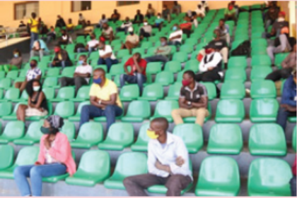
Who declared covid-19 pandemic disease?
Why Aged people and chronicle disease patients were warned to take care?Why Covid-19 is considered human threat?
Introduction
For a very long time, people held the view that something in the thin air or foul air arising
out of decomposing matter can cause illness. It was the pioneering work of scientists Louis
Pasteur and Robert Koch who laid the foundation of germ theory of disease, showing for
the first time that micro-organisms are responsible for causing various illnesses. Thus, in this
unit, we study how our body fights with these micro-organisms to keep us healthy; how our
immune systems are built; what is the role of vaccinations for building our immunity; and
how in the event of extreme illness one resorts to the use of antibiotics to control infections.
For example the year 2020 will remain unforgettable because of the outbreak of the pandemic
Covid-19.WHO declared covid-19 pandemic disease. Wearing mask, using hydro alcoholic
sanitizer to wash hand and social distance were among the best preventive measures.Aged
people and chronicle disease patients were warned to take care.
Who declared covid-19 pandemic disease?
Why aged people and chronicle disease patients were warned to take more care?Why Covid-19 is considered as human threat?
15.1 IMMUNE SYSTEM
ACTIVITY 1
Observe the prepared slides of blood smear under microscope, first under
10 X and then under
40 X magnification. Draw what all you observe and try to answer the following:
(i) Do you see cells? (ii) Are they all similar? (iii) What are the differences between different
types of cells? (iv) Which cells are most numerous? (v) What are they called? (vi) Which cellsare less numerous? (vii) Are these cells all alike? (viii) What does the stain indicate?
We are exposed to micro-organisms all the time, as they are present in large numbers in the air
we breathe, the food we eat and the water we drink. It is our defence system that keeps us free
of disease and constitutes our immune system. Immune system has two main components:Innate and Acquired.
15.1.1 Innate Immunity
Innate immunity is present at birth and depends
on:
(i) Anatomical and Physiological barriers,
(ii) White blood cells (mainly phagocytes)(iii) Some soluble mediators
Figure 15.1: Shows two neutrophils, one
monocyte and one lymphocyte among alarge number of RBCs
Anatomical and Physiological Barriers
Anatomical barriers consist of skin on the outside and mucous membranes lining gastrointestinal
tract, respiratory tract and genitourinary tract, while acidity in the stomach constitutes
Physiological barriers.
Skin is the largest organ of our body and presents a physical barrier for pathogens to prevent
their entry into the body. Some openings into the body are needed for the purpose of feeding,
breathing and reproduction. They are also the potential sites of entry of pathogens from
the environment. Therefore, the entire digestive tract, respiratory tract and genito-urinary
tract are lined by mucous membrane which does not allow pathogens to gain entry into the
tissues. High acidity within the stomach also helps to kill some pathogens ingested alongwith food.
White Blood Cells
White blood cells are the chief components giving us protection against infection.
When a blood smear is prepared, two types of cells can be identified: small, very numerous,
without nucleus called red blood cells (RBC) because they contain red pigment, haemoglobin
for oxygen transport and large, less numerous, with darkly staining nucleus called white blood
cells (WBC), because they do not contain red pigment. Among the WBCs, different cell types
can be distinguished:
(i) Phagocytes, which include neutrophils (having single, multi-lobed, nucleus) and monocytes
(having kidney-shaped nucleus) with a moderate amount of cytoplasm, and
(ii) Lymphocytes, with a very large, darkly staining nucleus occupying the entire volume of
the cell, with very little cytoplasm.
All blood cells arise within bone-marrow of long bones. RBCs and most WBCs complete their
development within the bone-marrow, while T-lymphocytes migrate to thymus for further
maturation and development. Monocytes circulate in blood for varying periods of time and
then migrate to tissues and differentiate into macrophages. As the most important function of
the immune system is to kill the pathogenic organisms to prevent the occurrence of disease, avery crucial attribute of immune system is to distinguish self from non-self.
In the absence of such a mechanism, the immune system might target one’s own tissues.
Identification of self from non-self is perfect by innate immune system, where-in receptors on
phagocytes recognize molecules present on microbial organisms as non-self. But lymphocytes,
which constitute adaptive immunity, may sometimes, target one’s own tissues, leading to the
generation of auto-immune diseases, e.g., rheumatoid arthritis, multiple sclerosis, systemiclupus erythematosus.
Mode of Action of Phagocytes
Phagocytes, which include both neutrophils and macrophages, play an important role in innate
immunity. They can identify foreign invading pathogens, discriminate them from cells of the
body, and internalize them by throwing pseudopodia around them. Once within the phagocytes,
pathogens are digested by a number of hydrolytic enzymes, thus freeing the body of disease-causinggerms. Phagocytes also help in removing old, dead cells as well as cancerous cells (Figure 15.2).
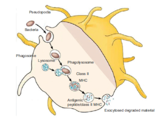
Figure 15.2: Showing process of phagocytosis by a macrophage
Soluble Mediators
Among soluble mediators of innate immunity are predominantly complement proteins which
can identify foreign microbial organisms and punch holes in their membranes, thus, effectively
killing them.
As soon as there is invasion of the body by any microbial organisms, innate branch of the
immune system immediately comes into operation as it is present from the time of birth.
However, if the infection persists, leading to the appearance of disease symptoms, it recruits
elements of second line of defence, the Specific or Adaptive immune system. Though both branches
of immune system help in eliminating the infectious agents, there are important differences in
the way these two systems work (see Table15.1).
Immune Response: Response produced by the body upon invasion of a foreign substance,
especially infectious microbes and toxins produced by them and is protective in nature.
Antigen: Any substance, that is foreign to the body, upon entering the body, evokes the generation
of immune response is called antigen. Infectious agents such as bacteria and viruses present alarge number of antigens to which the body responds by mounting an immune response.
15.1.2 Adaptive or Acquired Immune System
Adaptive or acquired immune system is not present at the time of birth but acquired during one’s
life-time. However, once acquired, its memory persists in the body in the form of memory cells
for a very long period of time. Generation of memory cells upon first exposure to infectious agent
is seen in primary response. Primary response leads to the generation of activated lymphocytes
of the B- or T- type as well as memory cells. This response is not only weak in intensity butalso takes a long time to initiate.
However, upon second exposure to the same infectious agent, the immune response generated is
faster and greater in intensity due to the already existing memory cells, and is called secondary
response (Table 15.2). It is the genesis of heightened immune response upon second exposure
that laid the foundation of all active vaccination programmes.Table 15.1: Comparing Innate and Adaptive Immunity
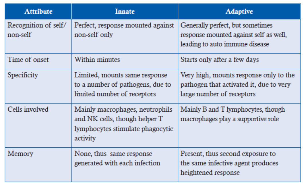
Table 15.2: Comparing primary and secondary immune response
Components of the Acquired or Adaptive Immunity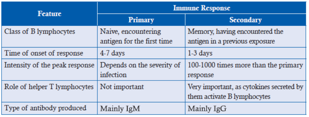
(i) Humoral Immunity: It is mediated by B-lymphocytes.(ii) Cell-mediated Immunity: It is achieved by T-lymphocytes.
Humoral Immunity: B lymphocytes
Humoral immunity was discovered by Emil Behring and Shibasaburo Kitasato in 1890.
This proved to be a landmark experiment and it earned von Behring Nobel Prize in Medicine.
This experiment showed two important things: one, that following infection or immunization,
substances appeared in serum that have the capacity to protect against the infective agent; this
laid the foundation of humoral branch of immunity; second, that immunity could be transferred
from immunized to non-immunized organism; this laid the foundation of strategy of passive
immunization. Generation of humoral response involves:
(a) Activation of B-lymphocytes
(b) Conversion of B-lymphocytes into plasma cells
(c) Secretion of antibodies(d) Functions performed by antibodies
B-lymphocytes, 6 micrometre in size, having a darkly staining, large nucleus, and very little
cytoplasm, bear receptors on their surface which recognize and bind antigens on microbial
organisms. Binding of receptors leads to activation of B-lymphocytes, which undergo a number
of mitotic divisions producing two kinds of cells, effecter cells called plasma cells and memory
cells. Memory cells are long lived, retain the same receptors as the original B-lymphocyte and
can get activated upon second exposure to the same infectious agent to give rise to a heightenedresponse (Figure 15.3).
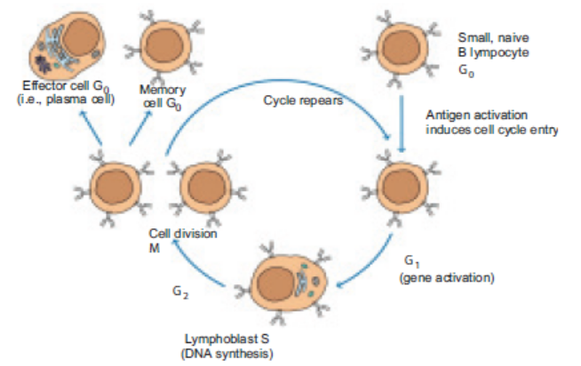
Figure 15.3: Activation of B-lymphocyte
Effecter cells or plasma cells, about 15 micrometre in size, with a large amount of cytoplasm
having Golgi apparatus and endoplasmic reticulum, represent the end stage B-lymphocytes,
which do not further divide but actively secrete antibody molecules at a high rate.Antibodies are, proteins, called immunoglobulins, designated as Ig.
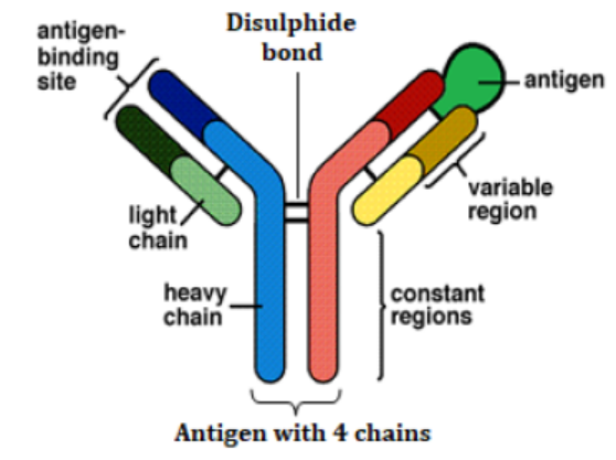
Fig15.4. Bonding between antibody and antigen
The diagram shows the basic structure of an antibody, which consists of four chains, two light and
two heavy joined by disulphide bonds, light chain and heavy chain at one end, together constitute
antigen binding site whereas constant region of two heavy chains performs the biological functions
as described below. Different classes of antibodies differ in the number of such monomers and the
numbers of disulphide bonds present in one molecule and hence are capable of performing different
functions. Example: IgM is a pentamer composed of five monomers
The five primary classes of immunoglobulins are IgG, IgM, IgA, IgD and IgE. These are
distinguished by the type of heavy chain found in the molecule. IgG molecules have heavy
chains known as gamma-chains; IgMs have mu-chains; IgAs have alpha-chains; IgEs have
epsilon-chains; and IgDs have delta-chains.. These antibodies are capable of recognising over
a million different antigens and confer protection in a number of ways. Functions performedby antibodies are:
1. Antibodies bind to toxins produced by bacteria that cause infection like diphtheria or
tetanus, effectively nullifying them.
2. By attaching to microbial pathogens, antibodies promote their clearance by phagocytes
manifold.
3. Antibodies form a covering on bacteria and viruses, not allowing them to gain entry into
tissues. Bacteria and viruses, thus having been coated, are eliminated by beating of cilia
present on the epithelial cells in the respiratory tract or by peristalsis of the gastrointestinal
tract.
Cell-Mediated Immune Response: T Lymphocyte
Though antibody molecules are highly specific and confer high degree of protection to the
body against toxins and microbes present in blood and extra-cellular fluids, they are not able
to neutralise those pathogens which live within the cells e.g., viruses, malarial parasite, some
bacteria such as Salmonella, Mycobaterium etc. Therefore, in order to protect the body from
intra-cellular infectious organisms as well as to eliminate cancerous cells, body mounts cellmediated
immune response.
Generation of cell-mediated immune response:
1. Recognition and binding of antigens by T-lymphocytes, macrophages, neutrophils, and
natural killer cells (NK). These cells differ in the way they bind antigens. T-lymphocytes
recognize and bind antigens only in association with another set of proteins called major
histocompatibility complex (MHC). Macrophages, neutrophils and NK cells can bind
to antibody-tagged cells.
2. T lymphocytes are of two types, helper T lymphocytes and Tc cells. Helper T lymphocytes
play important role in both humoral and cell mediated immunity by secreting important
cytokines.
3. Activation of the above mentioned cells (appearance of granules in T-lymphocytes and
macrophages which are normally agranulocytes). T-lymphocytes get transformed into
cytotoxic T lymphocytes, or CTL.
4. Secretion of cytotoxic chemicals/cytokines, perforins, granzymes, interferon gamma
and tumour necrosis factor in the vicinity of cells carrying intracellular pathogens.5. Cytotoxic chemicals/cytokines cause target cell destruction.
Table 15.3: Comparing Attributes of Humoral and Cellular Immunity
APPLICATION 15.1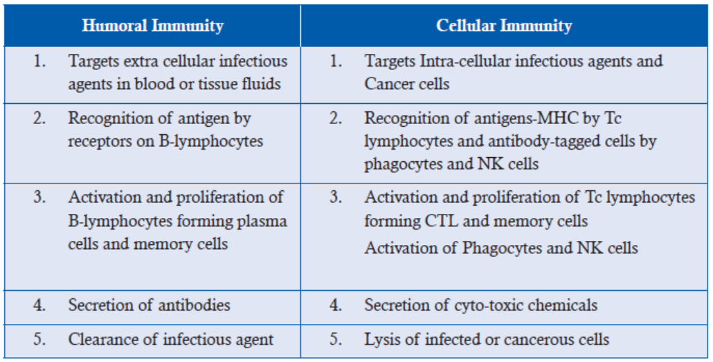
1.Complete with appropriate terms:
(i) ............... is the type of antibody produced by primary immune response.
(ii) Cell mediated immunity is achieved by ......................
(iii) Antibody .................... is highly mobile and capable of reaching skin to neutralize
bacteria.
(iv) ................ immunity leads to auto-immune disease.
2.What is the difference between T helper and T killer cells?
3. What is the role of the memory cells?
4.Explain how herd vaccination could be used to protect us from bird flu
5. Explain the difference between antigens and antibodies
6. a) Explain what is meant by clonal selection during the immune response
b) How are antibodies made specific to the antigen?
c) Apart from specificity, describe how the structure of an antibody molecule is suited to its
function
7. a) What is the difference between passive immunity and active immunity?
b) Explain what is meant by an attenuated pathogenc) Describe how an attenuated pathogen could be used to create artificial active immunity
ACTIVITY 2
Using different colours, learners can make flow diagram of immune response following
infection by a toxin/bacteria in the extracellular fluid and also a flow chart of immune response
following infection by bacteria such as Mycobacteria.
In these flow charts, draw different cell types participating in the immune response, and show
all the steps which ultimately lead to clearence of the pathogen.
Discuss the following questions to clarify major immune mechanisms of the body.
(i) How does humoral branch of immunity recognize the pathogen?
(ii) How does cell mediated branch of immunity recognize the pathogen?
(iii) How are the two branches interconnected?(iv) How are these two branches connected with innate immunity?
15.2 VACCINATION
ACTIVITY 3
Collect data of various diseases affecting a population such as smallpox, measles, malaria and
tuberculosis. You can collect data from health centres, or hospitals. Make a point of collecting
data before and after the vaccination provided in the same population.
Discuss the findings of different groups to assess whether vaccination programme has been
equally successful for all the diseases or not.Discuss why vaccination strategy has not worked against certain diseases.
While looking for answers for the above mentioned questions, you would have realized that
in the past 40 years immunization against a number of diseases has saved millions of lives.
World Polio Day is celebrated every year on 24th October, World Tuberculosis Day falls on
24th March every year, and World Immunization Week is observed by WHO from 24th to
30th April every year to have campaigns for awareness of the importance of vaccination in
eradicating these debilitating diseases as well as to provide vaccinations worldwide.
Success of immunization programmes is evident by the facts such as: Small pox has been
globally eradicated; Africa has not had a case of wild poliovirus since August 2014; India has
been declared free of maternal and neonatal tatanus; during 2000–2014, measles vaccination
prevented nearly 17.1 million deaths (decreaing deaths by 79%); deaths caused by tuberculosis
have come down only marginally.
The pioneering work of Jenner and Pasteur laid the foundations of immunization
programmes. They were able to confer protection to individuals against infectious diseases
by exposing them to infectious agents. This initiated the process of active immunization
in the medical field and saved millions of lives. However, the works of Kitasato showed
that immunity can be transferred between two individuals, and this laid the foundation
of passive immunization programmes. Though both these programmes confer protection
against infectious agents, there are important differences between the two (as shown inTable 15.4).
Table 15.4: Comparing Active and Passive Immunization
ACTIVITY 4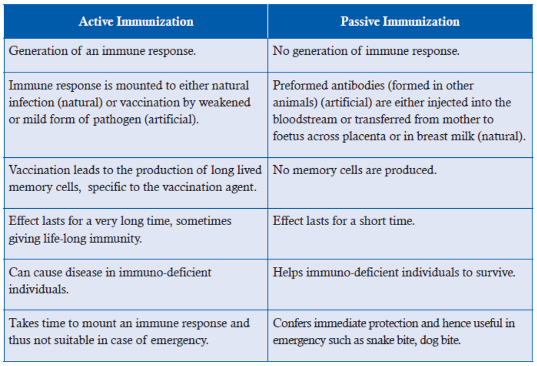
Find out National Immunization Days for tuberculosis, measles, and cholera. Search medical
journals to find out symptoms of these diseases and discuss in the class as to what role you canplay to promote awareness campaigns.
Though smallpox has been successfully eradicated, eradication of others such as measles,
tuberculosis, cholera and malaria has not been so successful. Success of smallpox vaccine
was due mainly to the fact that pox virus did not mutate and the same vaccine could be used
everywhere and the vaccine was highly effective.
On the other hand, though measles vaccination has decreased death rates drastically, its total
eradication has not been achieved so far due to several reasons. The disease is highly infectious,
and spreads very fast. As long as it is present in one area, unvaccinated children in any country
are at risk. For measles, boosters are required, difficult to achieve in poor countries, Parents’
decision not to vaccinate their children due to fear or other misconceived notions has also madethe vaccination programme less effective.
Effective vaccine against cholera has not been available for two major reasons: (a) Immunity
conferred by the vaccine is not long-lasting; (b) Cholera is a toxin-mediated disease while
protective immune mechanism is antibacterial rather than antitoxic. Oral cholera vaccines
have become available recently.
Tuberculosis is a major killer, causing 2 to 3 million deaths annually. According to WHO
reports, nearly one-third of the world’s population is currently infected with TB. Today, the
only approved tuberculosis vaccine is Bacilli Calmette-Guerin (BCG) which was started
in 1921. Though it is quite effective in infants and young children, in adults, its efficacy is
variable. Many boosters are also being developed, MVA85A, being the most advanced boost
available. BCG vaccine has not been modified since 1921 and that may also be one reason
why it is not so effective. That bacteria may have changed through evolution is suggested
by their evolution of resistance to a number of known antibiotics. A lot of effort is being
devoted, but proving difficult as the bacterium lives within the cells and lack of suitable animal
model for developing and testing human tuberculosis vaccine is posing a big challenge. In
Africa, coinfections of human immunodeficiency virus and TB have led to increases in theincidence rate of TB.
Malarial parasite lives intracellularly and mutates very often to change surface antigens andthus proved very difficult organism for effective vaccine development.
15.3 ALLERGIES
ACTIVITY 5
Watch one video of a person with watery eyes, running nose, sneezing; another with an
asthmatic person with breathing problems and using inhaler; another with a person stung by a
bee, showing swelling and redness.
Collect labels on certain food items prepared from shellfish/peanuts/walnuts/showing warning
to individuals.
See labels on penicillin vials, as a warning to be used only after initial testing.
After having seen these, try to answer the following:
(a) Why should some persons show watery eyes, running nose or asthmatic symptoms when
there is nothing alarming in the environment?
(b) Why should common food items be a cause of worry?(c) Why can’t we use penicillin just as we use any other antibiotic?
Allergic reactions are seen when humoral or cell-mediated immune responses to non-parasitic,
non-pathogenic substances lead to extensive damage of tissues in individuals prone to
allergies or hypersensitive reactions. Hypersensitive responses are classified into four types
(Figure 15.5).
1. IgE-mediated (Type I)
2. IgG or IgM-mediated (Type II)
3. Immune complexes (Type III)4. Cell-mediated (Type IV)
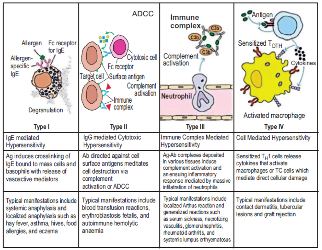
Figure 15.5: Showing different types of hypersensitive responses
IgE-mediated (Type I) Hypersensitivity: First exposure to allergen leads to the formation of
IgE type of antibodies which bind to mast cells present in large numbers throughout the body.
Second exposure to the same allergen causes cross-linking of IgE molecules on the already
sensitized mast cells, leading to their degranulation and release of substances stored in their
granules such as histamine, heparin, proteases, etc. Principal effects seen are vasodilation and
smooth muscle contraction (Figure 15.6). Vasodilation decreases blood pressure and contractionof smooth muscles of bronchioles affects respiration.
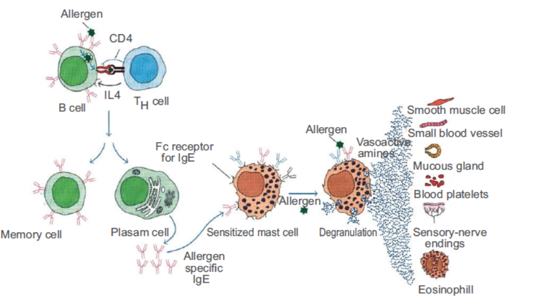
Figure 15.6: Showing type I hypersensitive reaction
Generalized allergic reaction, called anaphylaxis, starts when an allergen enters directly into
the bloodstream or is absorbed from the gut or skin. It can be caused by venom from bee, wasp,
ant stings; drugs such as penicillin, antitoxins. This should be treated quickly; otherwise it is
shock-like and often fatal.
In localized allergic reaction, the damage is limited to a specific target tissue or organ. Examples
include allergic rhinitis (hay fever), asthma, dermatitis, food allergies.
Hay fever occurs upon inhaling certain allergens in the air leading to watery eyes, running nose,
sneezing and coughing, involving mainly upper respiratory tract.
Asthma involves lower respiratory tract when histamine released from mast cells causes
contraction of bronchioles. Mucus accumulates in the air sacs, causing respiratory problems
and the characteristic wheezing sound. It can prove fatal if left untreated for too long.
Allergens, generally responsible for this reaction, are pollens, dust, fumes, insect products,
or viral antigens.
Epinephrine helps in generalized reaction by relaxing the smooth muscles for respiration to
restore and reducing vascular permeability so that blood pressure can normalize improvingcardiac output. Antihistamines are used to relieve the symptoms of asthma and hay fever.
Table 15.5: Distinction between Generalized and Localized Allergic Reactions
IgG or IgM-mediated (Type II) Hypersensitivity occurs when antigen-antibody complex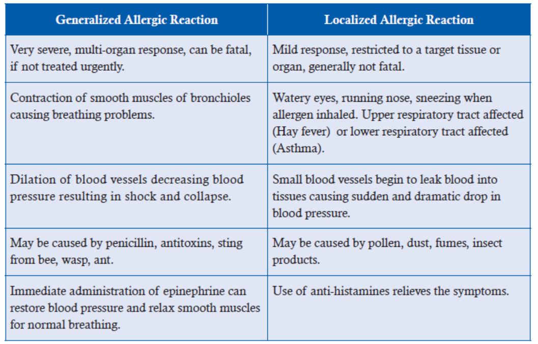
activates complement proteins which can cause rupture of cells. This is seen in blood transfusion
reactions.
Immune complexes (Type III) deposited in tissues lead to complement activation and
inflammation. This is seen after insect bites as swelling and reddening locally or in serum
sickness as fever and shivering after receiving anti-tetanus anti-toxin.
Cell-mediated (Type IV) Hypersensitivity is mediated by helper T lymphocytes and macrophagesas in contact dermatitis and graft rejection.
Important Message
Having read this section on hypersensitivity, can you visualize how you would go about helpingone of your classmates showing such symptoms
15.4 ANTIBIOTICS
ACTIVITY 6
Collect data on diseases and their antibiotics. Make a table to present your research. Once you
are done, discuss the following questions:
(i) How bacteria have acquired resistance against the antibiotic over the years.
(ii) Comment on the efficacy of antibiotics in treating a certain disease when the antibiotic for
the disease was first discovered.
Note: Some of the diseases which can be investigated are: Dysentry, Tuberculosis, Pneumonia,Diphtheria, Cholera, Measles, Mumps, Smallpox.
The word ‘antibiotics’ is derived from the Greek word “anti” meaning against and “bios”
life. Antibiotics are a class of chemicals produced by bacteria or fungi in order to inhibit the
growth of other organisms in their vicinity so that competition for limited resources can be
minimized. The first antibiotic, penicillin, was discovered by Alexander Fleming in 1928. Since
then antibiotics have found great use in medicine. Though they are not effective against viruses,
they are used to treat a number of bacterial infections.
A large number of antibiotics have been discovered from a variety of organisms, broadly
belonging to two categories: bactericidal, which kill the bacteria and bacteriostatic, which slow
down their growth and reproduction. Bactericidal antibiotics prevent the formation of cell wall
while bacteriostatic antibiotics interfere with some aspect of bacterial metabolism, affecting
either protein or RNA synthesis or DNA replication. They must work together with the immune
system to remove microorganisms from the body. High concentrations may also be bactericidal.
Table 15.6 shows a few commonly used antibiotics, source organisms producing them and theiractivity. Figure 15.7 shows mode of action of some antibiotics.
Table 15.6: Types of Antibiotics
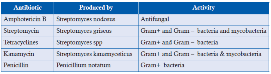
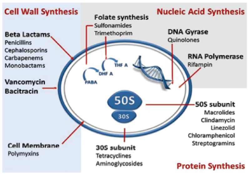
Figure 15.7: Mode of action of antibiotics
As the use of antibiotics has increased, it has also led to the evolution of resistance in a number
of microbial pathogens with the result that earlier antibiotics are no longer effective in treating
a disease. Resistance may have developed by a number of mechanisms e.g.,
1. Production of enzyme beta lactamase that breaks down beta- lactam ring of antibiotics
such as penicillin and cephalosporin.
2. Mutation in a gene leading to the formation of an altered protein which does not bind
penicillin.
3. Altered cell wall permeability confers resistance to tetracyclines, quinolones, penicillin.
4. Creating a barrier of biofilm, where bacteria are not attacked by the host’s immune system
as seen in Salmonella.
5. A gene can produce a product that can pump out the antibiotic as in Staphylococcus
against erythromycin.
6. Some bacteria show alteration in ribosome structure so that protein synthesis is notaffected.
APPLICATION 15.2
1.Complete with appropriate terms:
(i) ................. leads to production of long lived memory cells.
(ii) Cholera is a ................. mediated disease.
(iii) .................... decreases blood pressure and contraction of muscles of bronchiols.
(iv) ............... occurs upon inhaling certain allergens in the air leading to sneezing and
coughing.
(v) Penicillin is produced by ....................................
2. Two people took part in a study to find out the effectiveness of two types of immunization.
Person A received an injection of antibodies against tetanus and person B receive a tetanus
vaccination. Over the next few weeks, the blood from these two people was analyzed forthe presence of antibodies to tetanus. The results are shown in the figure 1A and figure 1B.
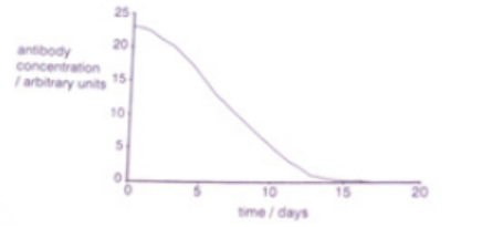
Figure 1A
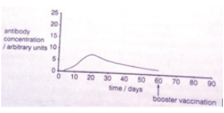
Figure 1B
a) Name the types of immunity shown by figures 1A and 1B.
b) Explain why the antibody concentration in person A,
i. Decreased during the study period
ii. Did not increase
c) Sketch on a copy of figure 1B what you would expect to happen to the antibody concentration
if person B received a booster vaccination at day 60.
d) Explain why in this investigation, the experimenters had to measure the concentration ofantibodies to tetanus rather than the concentration of all antibodies in the blood of A and B.
ACTIVITY 7
Using internet sources and medical journals, try to find out the following:
(i) Mortality caused by tuberculosis, measles, small pox and cholera.
(ii) How has medical science helped in reducing death rate caused by these diseases?
(iii) Has it been completely successful in this effort?
(iv) What role does nutrition play in fighting these diseases?
(v) Role of water and sanitation services in controlling spread of disease.
15.5 SUMMARY
• Immune system has the capacity to kill cells, it is very important for it to make a distinction
between self and non-self.
• Whenever there is a failure in distinguishing self from non-self, auto-immune diseases
develop such as multiple sclerosis, rheumatoid arthritis.
• Mounting of a successful immune response depends on a number of cells and chemical
mediators, defect in any component can lead to immunodeficiency state such as absence
of mature T lymphocytes in Di George syndrome.
• Immune system has two main parts, innate and adaptive.
• Two branches of the immune system collaborate with each other to make a highly
effective immune response.
• Innate system is present at birth, comes into operation immediately upon infection,
relies on barriers such as skin and mucous membranes, phagocytes and NK cells,
and lacks memory.
• Adaptive system is acquired by 6 months after birth, takes time to mount an immune
response, relies on B and T lymphocytes and has memory.
• Adaptive or acquired immunity is of two types, humoral and cell mediated.
• Humoral immunity generates specific antibodies against pathogens present in blood.
• Cell mediated immunity generates cyto-toxic chemicals to lyse infected cells.
• Antibodies neutralize toxins and help in eliminating microbial organisms.
• Antibodies are of five types IgM, IgG, IgA, IgE and IgD.
• There are two types of T lymphocytes: helper T lymphocytes and Tc lymphocytes.
• T lymphocytes recognize antigen of the pathogen in association with MHC.
• Helper T lymphocytes actively secrete a number of cytokines which affect the activity of
macrophages, B lymphocytes and Tc cells converting the latter into CTL.
• CTLs, macrophages and NK cells secrete cytotoxic chemicals onto cells having intracellular
pathogen causing cell death.
• Vaccinations can be Active or Passive.
• Active vaccination is achieved either by natural infection or by immunization with live
attenuated or killed infectious organisms.
• Passive immunization is achieved by introduction of preformed antibodies.
• Generation of memory cells during first encounter with the infectious agent produces a
secondary response with a high intensity.
• Vaccination programmes for a number of diseases have been very successful, though for
some diseases, effective vaccines are not available yet.
• Although active vaccines have helped eradicate a number of diseases, they pose a serious
threat in immuno deficient individuals.
• Passive immunization is chosen in cases of emergency or immunodeficiency.
• Allergies, also termed hypersensitive responses, which are potentially damaging to
the tissues, are produced by the body to seemingly non-pathogenic substances in the
environment.
• Allergies are broadly classified into four types: class I mediated by IgE; class II mediated
by IgG or IgM; class III by activation of complement by immune complexes; class IV
cell mediated (esp. macrophages ) hypersensitivity.
• Type I allergies are seen in hay fever, asthma, food allergies; type II allergies are seen in
blood transfusion reactions; type III allergies seen after insect bite or serum sickness
after anti-tetanus injection; type IV allergies seen in contact dermatitis, graft rejection.
• When the hypersensitive reactions are limited to specific target tissue or organ, it is called
localized reaction as seen in hay fever, asthma. However, when a large number of organs
become simultaneously affected upon entry of allergen directly into the bloodstream
absorbed from the gut, it results in generalized reaction with fatal consequences.
Symptoms and treatment
• Type I hypersensitive reactions are mediated by heparin released by mast cells and
epinephrine can reverse these effects in generalized reactions while antihistamines are
used for localized reactions.
• In all other cases of allergies involving drug, food item, blood transfusion, anti-toxin
antiserum,
the best strategy is to immediately discontinue the use of such agents.
• Antibiotics are drugs used extensively by doctors to treat bacterial infections. Mortality
rate due to bacterial infections has declined drastically since the use of antibiotics started.
• A number of soil microbes produce a large variety of chemicals to inhibit other organisms
growing in their surroundings and provide a rich source of antibiotics.
• Antibiotics either kill bacteria by inhibiting their cell wall synthesis or slowing their
growth by affecting DNA replication or RNA/ protein synthesis.
• Different antibiotics show different specificities, some antibiotics working against a
number of bacteria, while others being more specific.
• Prolonged and overuse of antibiotics has led to evolution of different mechanisms by
which bacteria have become resistant to commonly used antibiotics such as development
of enzyme beta lactamase to break down beta lactam ring, actively move the drug outof the cell (efflux), change its cell wall permeability etc.
15.6 GLOSSARY
• Acquired immunity: It is the immunity that our body gains over time, similar to how an
individual gains knowledge over time.
• Antibiotics: Substance that destroys or inhibits the growth of other microorganisms.
• Antigen: A substance that your immune system reacts against.
• Cellular immunity: It is an immune response that does not involve antibiotics but rather
in values the activation of phagocytes, antigen specific T-lymphocytes.
• Humoral immunity: Immunity involving the transformation of B-lymphocytes into
plasma cells that produce and antibiotics to a specific antigen.
• Immune response: The reaction of the cells and fluids of the body to the presence of a
substance which is not recognised as a constituent of the body itself.
• Immunity: It is the defence mechanism of our body.
• Innate immunity: Immunity that is naturally present and is not due to prior mustization
to an antigen form.
• Vaccination: Injection of a killed microbe in order to stimulate the immune systemagainst the microbe, thereby preventing disease.
END UNIT ASSESSMENT 15
I. Choose whether the given statements are True (T) or False (F)
1. Innate immunity is present at birth.
2. Breast milk confers protection to newborn by providing IgE type of antibodies.
3. Antibodies can work by promoting phagocytosis of microbial agents.
4. Antibiotics help a patient in mounting an effective immune response.
5. Treating tuberculosis is becoming difficult because Mycobacterium tuberculosis has
become resistant to a number of antibiotics.
6. Hay fever is a generalized allergic reaction caused by release of active mediators from
mast cells.
7. Secondary immune response appears much faster because of the presence of memory
cells persisting from previous infection.
8. Vaccination against snakebite is an example of passive immunization.
9. Allergies are of two types—innate and adaptive.
10. Beta-lactam antibiotics kill bacteria by blocking synthesis of their cell walls.
II. Multiple Choice Questions1. Humoral immunity is carried out by the
(a) B lymphocytes (b) T lymphocytes
(c) Phagocytes (d) T lymphocytes, phagocytes and NK cells
2. Antibodies transferred across placenta to the developing embryo are
(a) IgM (b) IgG
(c) IgA (d) IgE
3. Plasma cells represent
(a) B lymphocytes which are actively secreting antibodies.
(b) T lymphocytes which are actively secreting cytokines
(c) Monocytes which have entered tissues
(d) CTL which are secreting perforins.
4. Mast cell degranulation leads to the release of
(a) IgE, which causes vasodilatation and bronchoconstriction
(b) IgE, which causes vasoconstriction and bronchodilation.
(c) Histamine, which causes vasodilation and bronchoconstriction
(d) Histamine, which causes vasoconstriction and bronchodilation
5. Tetracycline helps in treating respiratory tract infections by
(a) preventing protein synthesis in influenza viruses
(b) preventing protein synthesis in bacteria
(c) preventing DNA repair in viruses
(d) preventing cell wall synthesis in bacteria
6. Children born with Di George syndrome lack mature
(a) B lymphocytes (b) T lymphocytes
(c) Macrophages (d) NK cells
7. Faulty recognition of self-tissues as non-self leads to the development of(a) AIDS (b) Rheumatoid arthritis
(c) Hay fever (d) Polio
8. Live attenuated vaccine types have the disadvantage over the inactivated vaccine types
as
(a) They require booster shots
(b) They do not confer life-long immunity
(c) They may mutate to virulent form
(d) They do not stimulate the immune system strongly.
9. Immune response is
(a) the defence mechanism of our body.
(b) reaction of the cells and fluids of the body.
(c) a substance that destroys or inhibits the growth of other microorganisms
(d) None of the above
10. Nowadays, many antibiotics don’t seem to work because
(a) body starts degrading the antibiotics very rapidly
(b) of increased levels of pollution
(c) bacteria are rapidly evolving resistance to antibiotics already in use
(d) pharmaceutical companies are not making good medicines now.
III. Long Answer Type Questions
1. With an illustrative diagram, state the origin and describe the mode of action of
phagocytes.
2. Analyse and relate the molecular structure of antibodies to their functions. Also,
state the role of antibodies in allergies.
3. Explain the following:
(a) Phagocytes
(b) Lymphocytes
(c) Immune response
4. Compare giving diagrams the modes of action of B-lymphocyte and T-lymphocyte.
5. With examples, explain the role of memory cells in long-term immunity.
6. Differentiate between the following:
(a) Active and passive immunity
(b) Generalised and localised allergic reactions
7. Discuss causes, symptoms and treatment of asthma and hey fever. Also suggest the
ways to encourage such patients.
8. State why vaccination programmes are able to eradicate smallpox but not measles,
TB, malaria or cholera.
9. Define antibiotic. State how it works. Also, explain the reasons for antibiotic
resistance.
10. Interpret the differences between cellular responses and humoral responses.
11. Carry out research and be able to present findings on the reasons for antibiotic
resistance in the treatment of infections.
12. There is a lot of research for curing diseases around the globe. Discuss the plausible
research going on for HIV AIDS. Also state the bodies immune response condition
while tackling HIV.
Unit 16 Human Reproductive System and Gametogenesis
Key Unit Competence
To be able to relate the structures of the human reproductive system to their functions anddescribe gamete formation.
LEARNING OBJECTIVES
At the end of this unit, the learner will be able to:
• Describe the structure of human male and female reproductive systems.
• State where female and male gametes are produced.
• Describe the histology of mammalian ovary and testis.
• Relate the histology of the testis and ovary to their functions.
• Outline gametogenesis in a male and a female human as a process involving mitosis, growth,
meiosis and maturation.
• Explain how spermatozoa are produced.
• Explain how oocytes are produced.
• Explain the significance of gametogenesis.
• Analyse and interpret chart diagrams of spermatogenesis and oogenesis.
• Prepare slides well to study the structure of gametes.
• Research about gametes and their formation and deduce their findings.
• Appreciate the significance of the process of gametogenesis at puberty as a key characteristic
of sexual maturity.
• Acknowledge the relevance of meiosis during gametogenesis as an essential tool inmaintaining the diploid condition after fertilisation.
ACTIVITY 1
Have you ever wondered how is the generation transferred from one to another? Look into
internet for matter related to the process of reproduction and reproductive organs. Make areport on it and present to the class.
INTRODUCTORY ACTIVITY
Observe the photo below and answer the questions that follow
a. The woman on the left side is pregnant. What do you think about the origin of the fetus
in the womb of the pregnant woman?
b. Use the photo on the right side to make notes on the dembryonic development
c. Use the photo and try to identify the parts of the female reproductive system and their
functions
d. The child on the photo is a boy. Can you identify the parts of the male reproductive
system and their functions
e. What do you think are the causes to the changes observed on the woman on the rightside of the photo
16.1 REPRODUCTION IN HUMANS
ACTIVITY 2
Aim: To dissect and identify structures of male and female reproductive system of rat.
Theory: The sex of the rat can be determined by looking for external testes and teats. Large
testes are visible at the ventro-posterior side of the male rat and 6 pairs of teats ventrally in
females. The major male reproductive organs of the male rat are the testes (singular: testis)
which are located in the scrotal sac, along with internally placed duct systems and associated
glands. Female reproductive system consists of a pair of ovary, duct system and glands.
Procedure:
• Obtain and sacrifice a rat. Place it in your dissecting tray with ventral side facing up so as to
observe the external features and determine the sex of the rate you have taken.
• Examine the anal opening just posterior to the tail. Insert the scissor blades through the anus
along the midline lifting the skin carefully so as not to damage the underlying organs.
In male rat, cut through the scrotum carefully to reveal the testis and also observe other
internal structures—epididymis, vas deferens, seminal vesicles, prostate gland and
seminiferous tubules.
In female rat, locate the urethral and vaginal openings on the ventro-posterior side of the
body, under the tail, just posterior to the last pair of teats. Also, observe other reproductiveorgans—uterus, ovaries, oviducts and vagina.
Precautions:
1. Be careful not to cut too deeply. Keep the tip of your scissors pointed upwards.
2. Always wear gloves and safety goggles when handling the rat to protect your hands and eye
from chemical splattering or debris.
3. Once an incision is made, allow the rat to be drained off the fluid.
4. Handling and dissection of animals should be done in the presence of experts, followingethical guidelines.
Human beings reproduce by sexual means where the male and female involve in sexual
intercourse, resulting in fertilization. During sexual intercourse, the interaction between the
male and female reproductive systems results in fertilization of the woman’s ovum by the
man’s sperm. The ovum and sperm are specialized reproductive cells called gametes, generated
by a process called gametogenesis (i.e., spermatogenesis in males and oogenesis in females).
The gametes are haploid in nature and it is when these two cells merge into one zygote cell
that genetic recombination occurs and diploid condition is achieved back. After a gestation
period, i.e., nine months in humans, childbirth takes place. In the present unit, we will discuss
the histological and anatomical details of organs involved in male and female reproductive
systems, the process of formation of gametes i.e., spermatogenesis and oogenesis, structure ofgametes i.e., spermatozoan and ovum in details.
16.2 MALE REPRODUCTIVE SYSTEM
The male reproductive system consists of the male sex organs, a system of various ducts and
accessory sex glands.
Figure 16.1: The overview of components of male reproductive system
16.2.1 Male Sex Organs
Scrotum: Scrotum is a bag like structure which is an out pouching of abdominal wall. Externally,
it appears as a single pouch separated with a median ridge called raphe. Internally, it is divided
into two sacs each of which contains a testis.
Figure 16.2: Scrotum from anterior view
The temperature of testes is maintained 2°C–3°C below the core body temperature which
provides favourable environment for spermatogenesis. This lowering of temperature is regulated
by scrotal muscle reflexes and venous pampiniform plexus present around the testicular artery.
Testes: Present inside the scrotal sacs are the oval shaped male sex organs, called testes. A
human testis is about 4.5 cm long and 2.5 cm broad with 3 cm antero-posterior diameter,
weighing approximately 10-15 grams. The testes are surrounded by serous sheath called tunica
vaginalis, anteriorly and laterally. Behind tunica vaginalis is present a thick, white, fibrous
capsule called tunica albugenia. Interior to it lies the tunica vasculosa, which is rich in vascular
supply. Posteriorly, the tunica albuginea thickens greatly and is projected into the interior of
the testis as the Corpus Highmori or mediastinum testis. The testes are held in position by
mesenteries called mesorchium.
The ducts, blood & lymphatic vessels and nerves enter or leave the testis through the mediastinum.
The connective tissue septa, called septula testis (singular- septum of testis) radiate from
mediastinum into the testis. These septulae subdivide the interior of the testis into a number of
pyramidal lobules called the testicular lobules. Each testicular lobule further contains several
sperm producing convoluted tubules known as the seminiferous tubules (semin = seed; fer =to carry).
Figure 16.3: (A) Entire testis section at low magnification. (B) Histological sections of testis
observed at different magnifications of objective lens. (C) Diagrammatic view of testis histology
The seminiferous tubules contain specialized epithelial cells, the spermatogenic cells and the
supporting cells, also known as the sertoli cells. Sertoli cells provide support and nourishment,
help in cell-to-cell communication, secrete inhibin and androgen binding protein etc. These are
surrounded and supported by intertubular connective tissue which is rich in blood vessels and
groups of epithelial cells, known as the Leydig cells (also called the interstitial cells or interstitialendocrinocytes). The leydig cells produce androgen and the male sex hormone, testosterone.
16.2.2 Male Reproductive Ducts
The ducts in male reproductive system help in storage and transportation of spermatozoa.
The spermatozoa along with testicular fluid after their release into the lumen of seminiferoustubules (spermiation process) are propelled towards the rete testis. Rete testis is composed
of various channels through which spermatozoa travel to efferent ducts, i.e., vasa efferentia
(also called ductuli efferentes). Vasa efferentia further joins with epididymis. Epididymis
is divided into three parts: head (caput), body (corpus) and tail (cauda) epididymis. The
cauda epididymis is attached to scrotal sacs by a connective tissue called gubernaculum.
Vasa efferentia joins epididymis from head region and vas deferens leaves the epididymis
from tail region. The epididymis performs functions such as storage of spermatozoa,
disposal of aged and abnormal spermatozoa and carries out maturational changes by
secretion and absorption. Spermatozoa then pass through a single tube known as the vas
deferens, also called ductus deferens or ductus epididymis. It joins with duct of seminal
vesicle to form the ejaculatory duct. Both the ejaculatory ducts from either side further
penetrate into prostate gland and empty into urethra. Urethra finally opens to the exterior
through penis. Penis is both the excretory and reproductive functions as it passes both the
urine and spermatozoa. Penis is divided into root, body i.e., shaft and glans penis. The
shaft is composed of three columns of erectile tissues i.e. pair of corpora cavernosa on
dorsal side and single corpus spongiosum on ventral side. Each column is surrounded by
dense connective tissue called tunica albuginea. The root is the attached proximal portion
and consists of bulb of penis. Glans penis is the distal, sensitive, cone shaped, hairless
structure that contains sensory receptors for sexual stimulation. Prepuce or foreskin is the
loose skin fold that covers the glans penis as sheath. Surgical removal of prepuce is calledcircumcision.
Figure 16.4: Diagrammatic view of Male Reproductive Ducts
Figure 16.5: Anatomy of Penis, the Male Reproductive Organ
16.2.3 Accessory Male Reproductive Glands
These include a pair of seminal vesicles, a prostate and a pair of bulbourethral glands.
(i) The seminal vesicles are one pair of sac like structures near the base of the bladder. Their
ducts join the vasa deferfintia to form the ejaculatory ducts. They produce an alkaline
secretion which forms 70% of the volume of semen. The secretion of the seminal
vesicle contains spermatozoa activating substances such as fructose, citrate, inositol,
prostaglandins and several proteins. Fructose is a source of energy for the sperm.
Prostaglandins stimulate uterine contractions and thus, may help the sperm to move
towards the female’s oviducts, where fertilization takes place. Some proteins of the
secretion help coagulation of semen after ejaculation. Alkalinity of the seminal fluid
helps to neutralize the acidic environment of the male urethra as well as that of female
reproductive tract which would otherwise inactivate and kill sperms.
(ii) The prostate gland is of a size of a golf ball and surrounds the proximal portion of urethra.
It produces a milky, slightly acidic secretion (pH = 6.5) which form 25% of the volume
of semen. The secretion of the prostate gland nourishes the spermatozoa and aids intheir motality.
(iii) The bulbourethral glands or cowper’s glands are present on either side of the proximal
portion of urethra. They secrete mucus that helps in the lubrication of penis and linning
of urethra.
The secretions of the above glands constitute seminal plasma which is rich in fructose, calcium
and certain enzymes. Fructose which is produced by the seminal vesicles, is not present anywhere
else in the body, provides a forensic test, for rape. The presence of fructose in the female’s genital
tract confirms sexual intercourse.
Semen is the mixture of spermatozoa and seminal plasma (the secretions of the seminal vesicles,
prostate gland and cowper’s glands). It is ejected from the penis during ejaculation. A single
ejaculate may contain 200 to 300 million spermatozoa (sperms). Semen is slightly alkaline
(pH 7.35 to 7.50). Its alkalinity helps to neutralize the acidity of urethra left from the passageof urine and protects the sperms from the acidity of vagina.
Figure 16.6: Sagittal view of human male reproductive system
Testosterone is the principal hormone of the testes which is synthesized in the Leydig cells
from cholesterol and is also formed in the adrenal cortex. The major functions performed by
testosterone in male reproductive system are:
• Gonadotropin regulation
• Spermatogenesis
• Sexual differentiation
• Wolffian stimulation
• Sexual maturation at puberty
Inhibin is another hormone of testicular origin that inhibits FSH secretion. Estradiol and estroneare also synthesized in testis in minor quantities.
APPLICATION 16.1
1. Complete with appropriate terms:
(i) Male sex organs include ...................... and ...................... .
(ii) The seminiferous tubules contain specialized cells, the spermatogenic and supporting
cells called ...................... .
(iii) ...................... joins epididymis from head region and ...................... leaves the
epididymis from tail region.
(iv) ............... provide alkaline environment that protects the passing spermatids
2. What is the function of the prostate gland
3. In which ovarian structures do oocytes develop
4. List the sequence of structures through which a sperm passes from its site of production to
the site of fertilization of an ovum5.How do egg cells move from the ovary to the uterus
16.3 FEMALE REPRODUCTIVE SYSTEM
The human female reproductive system consists of the primary sex organs or the gonads
(ovaries), the genital ducts (oviducts or the uterine/fallopian tubes, uterus, cervix and vagina)
and the external genitalia, along with a pair of mammary glands.
Ovaries (singular: Ovary, Latin: Ovarium, literally meaning ‘egg’ or ‘nut’) — the primary sexorgans in females are egg-shaped, paired structures, located in the upper pelvic cavity, one on
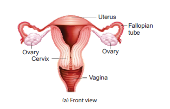
Figure 16.7: An overview of human female reproductive system
either side of the uterus in front of the ureter, embedded in the connective tissue matrix called
ovarian fossa. A single ovary is about 2 to 4 cm long, 2 cm wide and 1.5 cm thick and weighs
about 15 grams. Each ovary is held in place by three ligaments: mesovarium or the broadligament, suspensory ligament and the uteroovarian ligament.
Figure 16.8: The relation between various structures of human female reproductive system.
Ovarian ligaments can be seen clearly
Histology of the ovary: A typical human ovary is composed of connective tissue called stroma,
wrapped by germinal epithelium which is further layered by tunica albuginea. The stroma
is divided into two zones: an outer dense cortex and a less dense inner medulla. The medulla
consists of loose connective tissue, blood vessels, lymphatics, smooth muscle fibres and nerves.
The cortex consists of rounded structures called ovarian or the Graafian follicles, at various
stages of development. Each follicle consists of a large ovum surrounded by several layers of
follicular or granulosa cells.
A fully grown ovarian or the Graafian follicle typically consists of:
• an oocyte (15-30 μm wide) with a nucleus called the germinal vesicle, bounded by vitelline
membrane which is further surrounded by zona pellucida.
• surrounding the zona pellucida is present membrane granulosa, consisting of granulosa
cells or the follicular cells. The granulosa cells lying in close vicinity of the oocyte may
become elongated to form the corona radiata.
• membrana granulosa is further covered on the outside by theca interna and theca externa.
• a cavity called follicular antrum/cavity filled with a fluid, the liquor folliculi.
• the oocyte anchors to the wall of the follicle by a thin layer of follicle cells called cumulusoophorus, which nourishes the oocyte.
Figure 16.9: Diagrammatic view of the cross section of a human ovary.
Ovarian follicles can be seen at various stages of development
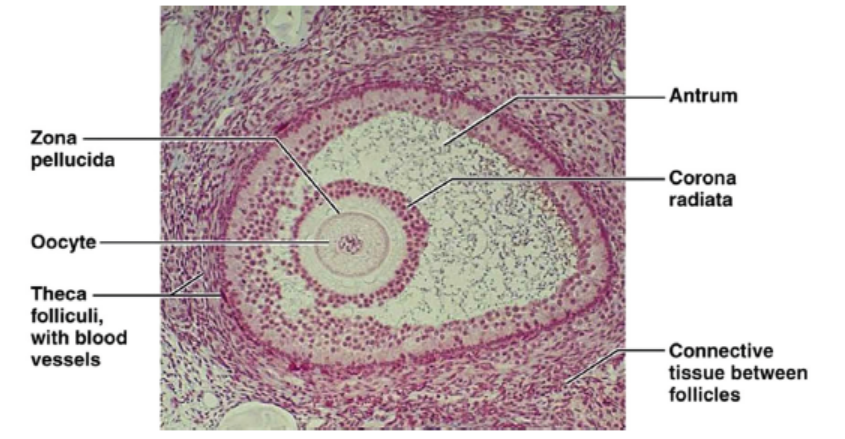
Figure 16.10: Structure of a mature Graafian follicle
Ovaries perform two important functions:
(a) Oogenesis: production of female gamete (ovum, pl. ova).(b) Production of hormones: some of the follicular cells produce the hormone estrogen, while
the follicle is developing and progesterone and relaxin while the follicle is degenerating
as corpus luteum. Estrogen stimulates the growth and functions of female sex organs
and development of secondary sexual characteristics. Progesterone prepares the uterine
lining to receive embryo and maintains it during pregnancy. It also stimulates the growth
of mammary glands. Relaxin helps in the relaxation of pelvic ligaments and softeningand widening of the cervix during delivery of the baby (parturition).
ACTIVITY 3
Aim: To study the histology of human testis and ovary using prepared slides and micrographs.
Theory: A typical testis is composed of various cell types i.e., sertoli cells, leydig cells,
fibroblasts,
primary and secondary spermatocytes, spermatids and basal lamina etc. A typical
human ovary is composed of connective tissue consisting of rounded follicles, at various stages
of development. Each follicle consists of a large ovum surrounded by several layers of follicular
or granulosa cells. Germinal epithelium is the outermost boundary of the ovary.
Procedure:
Observe a permanent slide of transverse section (T.S.) of human ovary and testis under lowpower and then under high power microscope and locate various cell types.
On the basis of your observations;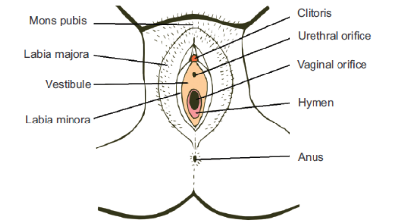
1. Label the cell types in following micrograph of testis and ovary.2. Complete the table given below based on histological analysis ofovary:
16.3.1 Female Reproductive Ducts
Oviducts: Also known as the uterine ducts or the fallopian ducts, lined with ciliated epithelia,
they function to transfer the ovum from the ovary to the uterus and serve as the site of fertilization
of the male and female gametes. Each oviduct, 10-12 cm long, extends from the margins of
the ovary to the uterus and can be divided into four continuous regions as:
• Infundibulum — the part closest to ovary. It has finger-like projections called fimbriae that
drape over the ovary and serve to receive the ovum released by the ovary during ovulation.
• Ampulla — the widest and major part of the tube, the site of fertlization.
• Isthmus — the narrower part, that links to the uterine wall.
• Interstitial or the intramural or the uterine part—that lies within the uterine wall.
Uterus: This is an ‘inverted pear’-shaped, muscular, hollow (uterine cavity lies within),
hormone-responsive organ that serves to house, nourish and protect the growing foetus till
birth. Anatomically, the uterus consists of three parts:
• Fundus—the dome shaped part above the openings of the uterine parts of the fallopian tubes.
• Corpus uteri or the ‘body’—the main centrally expanded portion.
• Cervix—the inferior narrow portion that opens into the vagina. A narrow, constricted
region, about 1 cm long called isthmus joins the uterus with the cervix. The cervical canal
or the cavity of the cervix communicates with the uterus internally by an aperture called
internal os and with the vagina by external os.
Cervix and vagina together form the birth canal. During childbirth, it dilates widely to allow the
baby to pass through. The wall of the uterus is composed of tissue layer called endometrium.
The endometrium is shed during menstruation and is regenerated by the basal layer after eachmenstruation.
Vagina: It is a distensible, muscular tube, about 10 cm long, which extends from vulva (external
genitalia) to the uterus. The vaginal opening on the vulva is termed the vaginal orifice. The
orifice is partially covered by a membrane called hymen that ruptures during the first act of
intercourse. However, it may get ripped off during some strenuous activities like sports or due
to some disease. Vagina serves as a receptacle for the male copulatory organ during sexual
intercourse, provides a passageway for the menstrual flow and forms part of the birth canalduring childbirth.
16.3.2 External Genitalia (Vulva)
The female external genitalia include mons pubis, labia majora, labia minora, hymen and clitoris.
Mons pubis is a cushion of fatty tissue covered by skin and pubic hair. The labia majora are fleshy
folds of tissue which extend down from the mons pubis and surround the vaginal opening. The
labia minora are smaller folds which lie under the labia majora. Anteriorly, labia minora merge
together to form a small eractile organ called clitoris. It is Homologous to the male’s glans penis.
Posteriorly, the labia minora are fused together to form the fourchette. They also containnumerous sebaceous glands.

Figure 16.11: The external genitalia in human female
16.3.3 Female Reproductive Glands
The glands associated with vestibular region are broadly of two types:
• Glands of Skene or Paraurethral glands (lesser vestibular glands): Those are numerous
minute glands present around the urethral orifice. They secrete mucus and are homologousto the male prostate glands.
• Bartholin’s glands (greater vestibular glands): These are located on each side of vagina
beside the vaginal opening and produce a mucus secretion to lubricate the vestibule that aids
in sexual intercourse. They are considered homologous to the bulbourethral glands in males.
• Mammary glands or Breasts: These are modified sweat glands that produce milk. Though
characteristic of all female mammals, they are also present in a rudimentary form in males.
In females also, they remain underdeveloped until puberty. Each breast is covered with
skin and bears a central protruding nipple surrounded by a pigmented area called areola.
Internally, each breast consists of the glandular tissue, the fibrous connective tissue and the
fatty or the adipose tissue:
o The glandular tissue of each breast is divided into 15-20 mammary lobes. Each
lobe is further divided into a number of lobules containing clusters of cells called
alveoli. The cells of alveoli secrete milk, which is stored in the cavities (lumens) of
alveoli. The alveoli open into mammary tubules. The tubules of each lobe join to
form a lactiferous or mammary duct, which opens on the nipple. Just before their
opening, the lactiferous duct swells to form the mammary ampulla or lactiferous
sinus, which acts as a reservoir for milk during lactation. In non-pregnant and
non-nursing women, the glandular structure of the breast is largely underdevelopedand the duct system is rudimentary.
Figure 16.12: (a) Front view human breast with right half showing internal structure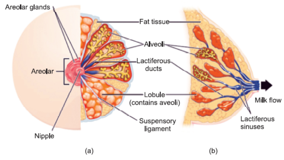
(b) Sagittal section of human breast
Human milk consists of water, organic and inorganic components. The main constituents
include fat (droplets), casein (milk protein), lactose (milk sugar), mineral salts (sodium, calcium,
potassium, phosphorus, etc.) and vitamins. Human milk is poor in iron and vitamin C content.
With childbirth, the anterior lobe of pituitary secretes the hormone prolactin, which stimulates
the production of milk. Another hormone oxytosin secreted by the posterior lobe of pituitary
stimulates the release of milk, though the psychic state and nutrition of the mother also governs
milk production.The first milk produced by each breast after childbirth is termed the colostrum.
It is thick, yellowish fluid rich in proteins and antibodies, that provides passive immunity to
the breastfed baby. Colostrum also helps the newborn’s digestive system to grow and function
properly.
16.3.4 Menstrual Cycle
Menstrual cycle is the reproductive cycle involving cyclic in the ovary and uterus that occurs in
the female primates, e.g., humans, monkeys, apes, etc. This cycle is responsible for maturation of
the ovum and its release from the ovary (ovulation) and preparation of the uterus for pregnancy.
The menstrual cycle is regulated by hormones. A typical menstrual cycle in human female is
about 28 days long with 3–5 days plus or minus variation. The onset of this cycle (known as
menarche) generally starts at the age 12–15 (puberty) in girls and ends (known as menopause)
by the age of 45–50 in humans.
One cycle is completed in three phases: the uterine cycle is divided into menstrual phase,
proliferative phase, and secretory phase and correspondingly the ovarian cycle consists of thefollicular phase, ovulation, and luteal phase
1. Menstrual phase or bleeding period or the menses or period (days 1–4): Low levels
of hormones progesterone and estrogen make the blood vessels of the endometrium
constrict. This causes the endometrial lining to shed along with blood and unfertilized
ovum.
At the same time, under the influence of Follicle stimulating hormone (FSH) secreted
by the anterior lobe of pituitary, ovaries are beginning follicular phase i.e., growth and
proliferation of one of the follicles to become Graafian follicle or tertiary follicle.
2. Proliferative phase (days 5-14): Gonadotrophic releasing factor (GnRF) secreted by
the hypothalamus stimulates the release of FSH and Luteinizing hormone (LH). High
levels of FSH stimulate the ovarian follicle to secrete estrogens. The combination of FSH
and LH, and estrogen, has a positive feedback effect on anterior pituitary, causing therelease of more and more FSH and LH and thus, more estrogen.
Estrogens cause the endometrial lining of the uterus to proliferate, rapid cell multiplication
occurs with development of uterine glands and blood vessels. Ovary is still undergoing
follicular phase.
Towards the end of proliferative phase, high levels of estrogen trigger a sudden
increase in the levels of LH. This LH surge, as it is called, lasts for 24-48 hours, causes
the rupture of ovarian follicle and release of ovum from the ovary into the oviduct(= ovulation phase of the ovarian cycle).
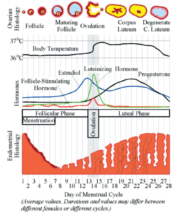
Figure 16.13: Diagrammatic presentation of various events during a menstrual cycle
3. Secretory phase (days 14-28): The final phase of the uterine cycle corresponds to the
luteal phase of the ovarian cycle that lasts for about 10 days. The Luteinizing hormone(LH) secreted by the anterior lobe of pituitary stimulates the development of corpus
luteum from degenerating cells of the ovarian follicle after ovulation. Corpus luteum
secretes large amounts of progesterone and some estrogen.
The combination of estrogen and progesterone inhibits the release of FSH and LH from
pituitary (negative feedback). Progesterone prepares the uterus to receive, implant and
nourish a fertilized egg during the second half of the menstrual cycle.
If implantation does not occur, the endometrium breaks down and menstruation occurs.
Corpus luteum degresses and production of progesterone is lowered. This releases theinhibition of FSH and LH, thus initiating the next menstrual cycle.
Corpus Luteum
Corpus luteum (Latin means “yellow body”; plural: corpora lutea) is a temporary endocrine
structure that develops from degenerating cells of the ovarian follicle after the release of ovum,
under the influence of LH. It produces large amounts of progesterone.
If the oocyte is fertilized, the corpus luteum continues to proliferate and increases hormone
production. By the end of the third month of pregnancy, luteal cells occupy a large part of the
ovary and keep releasing progesterone. However, by the end of the fourth month, they regress
slowly.
If the oocyte is not fertilized, the corpus luteum degenerates in 10–12 days after ovulation.It is visible only in the form of a white scar, the corpus albicans, on the outside of the ovary.
APPLICATION 16.2
1.Complete with appropriate terms:
(i)...................... stimulates the release of milk.
(ii) Ovary is held in place by three ligaments called .............., .............., and ................. .
(iii) Follicular cells produce ...................... , ...................... and ...................... .
(iv) ...................... acts for reservs for milk during lactation.
2. The diagram shows a section through part of a human testis
a) Giving a reason for your answer in each case, what percentage of each of the following
types of cell would you expect to contain a Y chromosome
i. type A cells
ii. type C cells?
b) Many of the type B cells will undergo mitosis. Explain the importance of mitosis in a
mature testis
c) Give two ways in which cell division results in the type C cells being genetically differentfrom one another
16.4 GAMETOGENESIS
Gametogenesis is the process of formation of haploid gametes from undifferentiated, diploid
germ cells in the gonads for sexual reproduction. Male and female sex cells or gametes i.e.,
sperms and ova are formed respectively in the male and female gonads (testes and ovaries).
Types and phases of gametogenesis: Formation of male gametes (sperms) is termed
spermatogenesis and that of female gametes (ova, singular: ovum) is referred to as oogenesis.
Both the processes undergo three basic phases, common to both:
1. Proliferative or the multiplication phase
2. Growth phase3. Maturation or differentiation phase
ACTIVITY 4
Aim: To understand the process of gametogenesis and haploid nature of gametes.
Theory: Gametes are haploid cells that are formed from diploid germ cells through the process
of gametogenesis. The significance of developing haploid gametes lies in the fact that after
fertilization, the developing zygote attains the diploid status back. In this way, the developing
embryo gets the single copy of all the chromosomes from each parent.
Procedure: Based on the chart diagrams of spermatogenesis and oogenesis shown below,compute the number of chromosomes at each stage, assuming 2n = 46.
16.4.1 Spermatogenesis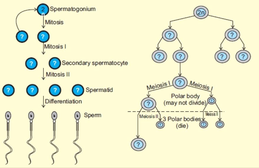
The process of formation of haploid male gametes or spermatozoa from diploid reproductive
cells in males is called spermatogenesis. The complete process is broadly divided into two parts:(i) Formation of spermatids and (ii) Spermiogenesis or spermatoleosis.
Formation of Spermatids
The process of formation of spermatids is further divided into three stages as:
(a) Multiplication phase: The primordial germ cells or sperm mother cells differentiate from
germinal epithelium of testis and increase in size with prominent nuclei. These cells
divide repeatedly by mitosis (i.e., equational division) and produce a number of diploid
daughter cells, known as spermatogonia. Thus, in this stage, multiplication of germ cells
takes place mitotically.
(b) Growth phase: In this phase, spermatogonia increase in size by accumulating food
reserves and are now called primary spermatocytes.
(c) Maturation phase: The primary spermatocytes (which are diploid) undergo first
maturation division which is meiotic division (or reductional division) to produce two
haploid secondary spermatocytes. These haploid secondary spermatocytes divide further
by mitosis to give rise to four haploid spermatids. This mitotic division is called secondmaturation division.
The spermatids so produced are non-motile rounded structures that metamorphose into functional
and motile spermatozoa through a process known as spermiogenesis or spermatoleosis.
The spermatozoa from testis are incapable of fertilizing an ovum. They undergo several
morphological, physiological and biochemical changes as they move through the epididymis
to attain this structural and physiological maturity. The epididymis i) provides a favourable
environment to spermatozoa in acquiring fertilizing ability and ii) stores them until they are
ejaculated or move down to the vas deferens.
The morphological changes include structural remodelling of acrosome and formation of
disulfide linkages. The physiological and biochemical changes include increase in net negative
charge on spermatozoa, change in pattern of motility, change in content of sialic acid, increase
in specific activity and reflection power, resistance to pH and temperature and changes inmetabolic patterns.
Spermiogenesis
A series of changes in spermiogenesis that transform a non-motile spermatid into motile,
functional spermatozoa are listed below:
• The nucleus shrinks and flattens by losing water. Only DNA is left in the nucleus, making
cells very light that aids to its motility.
• The two centrioles of a centrosome form proximal and distal centrioles. The proximal
centriole lies at the posterior end of nucleus and the distal centriole gives rise to axial
filament of the flagellum and acts as a basal granule.
• The mitochondria gather around axial filament and gradually unite to form spiral sheath
or nebenkern. It acts as power house of the sperm and provides energy.
• The golgi bodies form the covering over nucleus called acrosome. During acrosome
formation, one or more vacuoles start enlarging with a small, dense body called
pro-acrosomal granule which further enlarges to form acrosomal granule. The vacuole loses
its liquid content and forms the cap of spermatozoan. The remaining part of golgi apparatus
is reduced and discarded from sperm.
During all these steps, head of the developing sperm remains embedded in sertoli
cells for nourishment. At the end, fully formed spermatozoan shows distinct head,middle piece and tail region.
Figure 16.14: (a) Process of spermatogenesis showing chromosome numbers at various stages and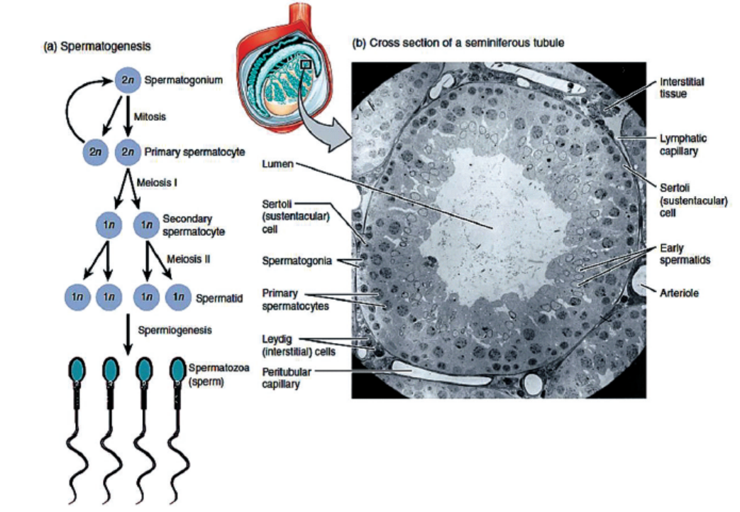
the cross-section of a seminiferous tubule showing histological arrangement of various cell types
(b) Stages in the formation of spermatozoan from spermatid and acrosome formation fromgolgi apparatus during spermiogenesis.
Structure of Spermatozoa
The sperms are microscopic and motile cell. Each sperm is composed of four parts—a head, a
neck, a middle piece and a tail. A plasma membrane covers the whole body of sperm.
(i) Head is the enlarged end of the sperm, containing an elongated haploid nucleus. The
anterior of the nucleus is covered by a cap-like structure called acrosome. The acrosome
contains enzymes sperm lyftins or hyaluronidases, which are used to contact and
penetrate the ovum at the time of fertilization.
(ii) Neck is very short and is present between the head and middle piece. It contains the
proximal centriole towards the nucleus which plays a role in the first cleavage of thezygote and the distal centriole which gives rise to the axial filament of the sperm.
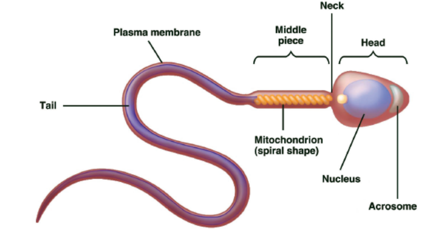
Figure 16.15: Structure of a sperm
(iii) Middle piece possesses numerous mitochondria which produce energy for the movement
of the sperm. At the end of the middle piece, there is a ring centriole (annulus) with
unknown function.
(iv) Tail is several times longer than the head. It consists of an axial filament surrounded by
a thin layer of cytoplasm. The tail provides motality to the sperm, which is essential forfertilization.
The male ejaculates about 200 to 300 million sperms during a coitus. For a normal fertility,
at least 60 per cent sperms of the ejaculate must have normal shape and size, and at least 40
per cent of the normal sperms must show vigorous motility. Sperms remain alive and retain
their ability to fertilize an ovum from 24 to 48 hours after having been released in the femalegenital tract.
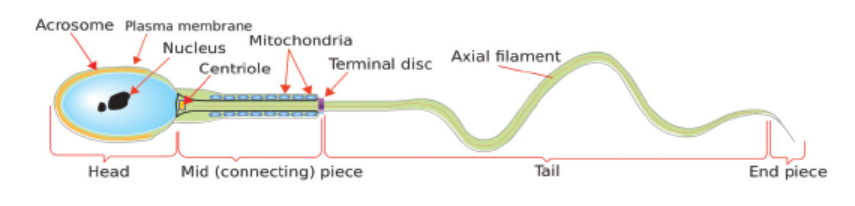
16.4.2 Oogenesis
The process of oogenesis occurs in the ovaries. The three phases of proliferation, growth and
maturation occur in discontinuous steps.
(a) Proliferative or multiplication phase: During early foetal development, certain cells
within the germinal epithelium of the ovary become enlarged. These cells proliferate by
mitosis, producing undifferentiated germ cells called egg mother cells or oogonia (2n).The oogonia divide mitotically to produce groups of oogonia, termed follicles.
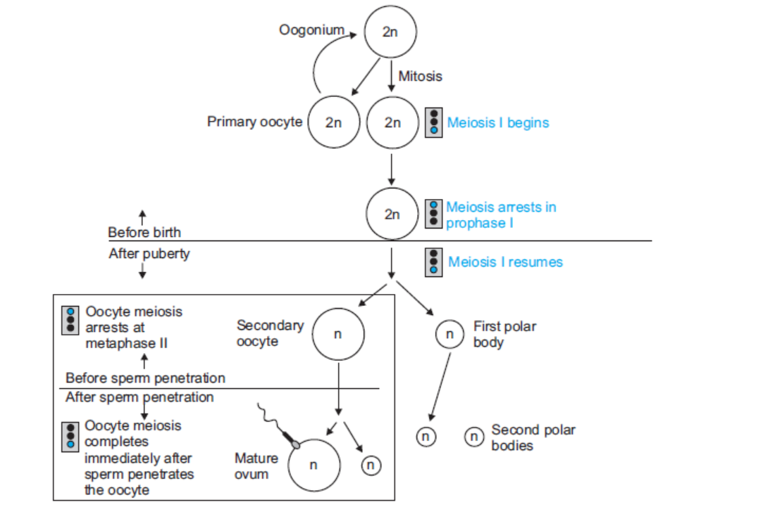
Figure 16.17: Schematic representation of oogenesis
(b) Growth and differentiation phase: During this long phase, which may last upto years,
one cell in a follicle prepares for the formation of ovum. It starts meiotic division but
gets arrested at prophase-I stage and is called primary oocyte. The remaining cells of
the follicle lose the potential to become primary oocyte and are known as the follicular
cells or granulosa cells. These follicular cells serve to protect and nourish the primary
oocyte. The complete follicle with a primary oocyte surrounded by a layer of follicular
cells is called the primary or the ovarian follicle.
(c) Maturation phase: At puberty, only one of the primary oocytes resumes division per
menstrual cycle, alternately in each ovary. The tertiary follicle matures into a Graafian
follicle, within which the primary oocyte divides to form two very unequal cells - a large
secondary oocyteand a very small 1st polar body or polocyte
may further be divided into two polar bodies. However, the secondary oocyte again gets
arrested at metaphase stage of meiosis-II and is released from the ovary during ovulation.
It waits in the oviduct for the sperm to arrive. If fertilization occurs, sperm entry into
the secondary oocyte marks the resumption of meiosis. The 2nd maturation division
(meiosis-II) again divides the secondary oocyte into two unequal daughter cells—a large
ootid and a very small 2nd polar body. The ootid undergoes maturation into a functional
haploid ovum. A thin vitelline membrane develops outside the plasma membrane of theovum that protects and nourishes the latter.
Figure 16.18: Maturation of a follicle shown in a clockwise direction proceeding from the primordial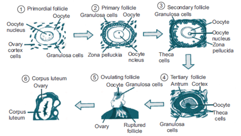
follicles. FSH stimulates the growth of a tertiary follicle, and LH stimulates the production of estrogen
by granulosa and theca cells. Once the follicle is mature, it ruptures and releases the oocyte. Cellsremaining in the follicle then develop into the corpus luteum.
Thus, from one oogonium (egg mother cell), one ovum and three polar bodies are formed.
The ovum is the functional female gamete while the polar bodies take no part in reproduction
and soon degenerate. The formation of polar bodies only helps the egg to get rid of one set of
chromosomes and still enables the ovum to retain most of the cytoplasm and food for the future
embryo. In case fertilization does not occur, the secondary oocyte undergoes degeneration andis driven out of the body.
Structure of Ovum
An ovum is a spherical, non-motile cell, in the secondary oocyte stage of oogenesis, where the
second maturation division is yet to occur. Human ovum is extremely small in size i.e., 0.15
mm in diameter, polar and microlecithal. The large nucleus is called germinal vesicle or later
the female pronucleus. The nucleolus is called the germinal spot and cytoplasm is known as
ooplasm. The peripheral layer of ooplasm, known as cortex, is more viscous and contains
cytoskeletal structures like microtubules and microfilaments, pigment granules and cortical
granules of mucopolysaccharides. The inner part of cytoplasm, called the endoplasm is withcell-organelles, informosomes, tRNAs, histones, enzymes etc. The ovum is covered over by a
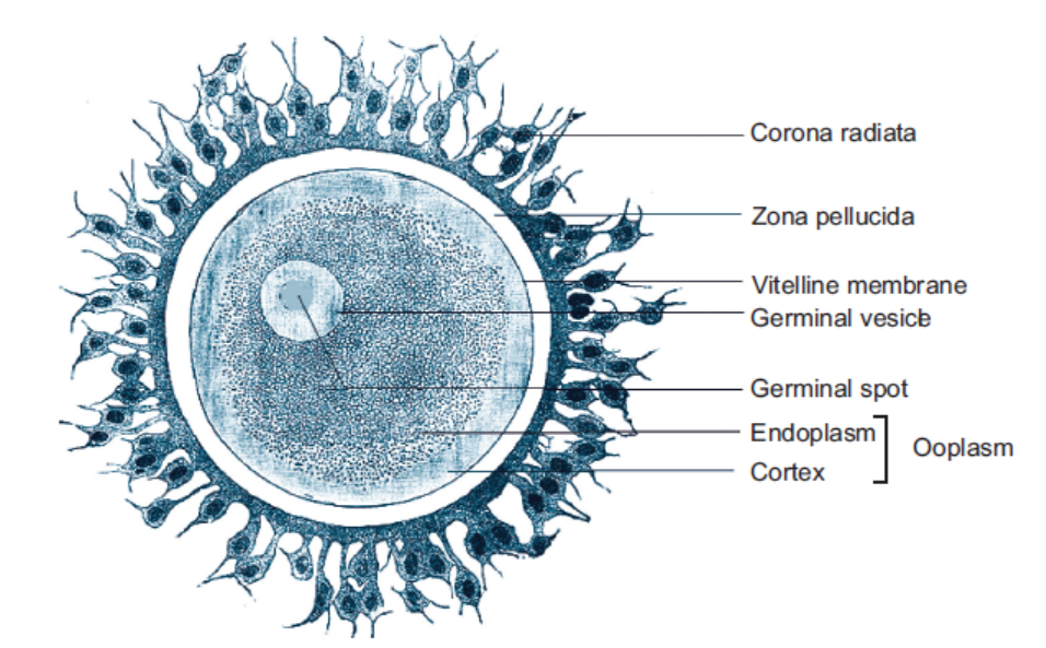
Figure 16.19: Structure of a mature human ovum with corona radiata surrounding it
thin, transparent vitelline membrane which is further covered over by zona pellucida. There isa narrow space between these two membranes known as perivitelline space. During discharge
of ovum from the Graafian follicle, several layers of follicular cells adhere to the outer surfaceof zonapellucida and are arranged radially forming corona radiata.
Differences between spermatogenesis and oogenesis
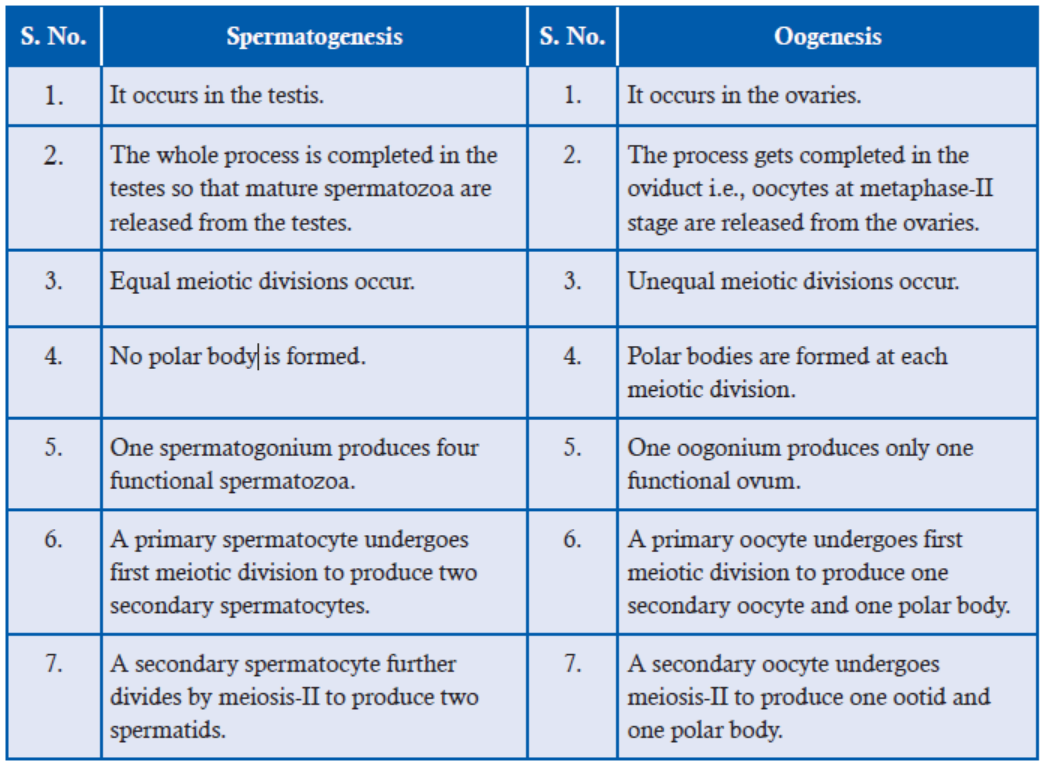
ACTIVITY 5
Study the chart diagrams of spermatogenesis and oogenesis. Identify various stages and writedown the points of similarities and dissimilarities between the two processes.
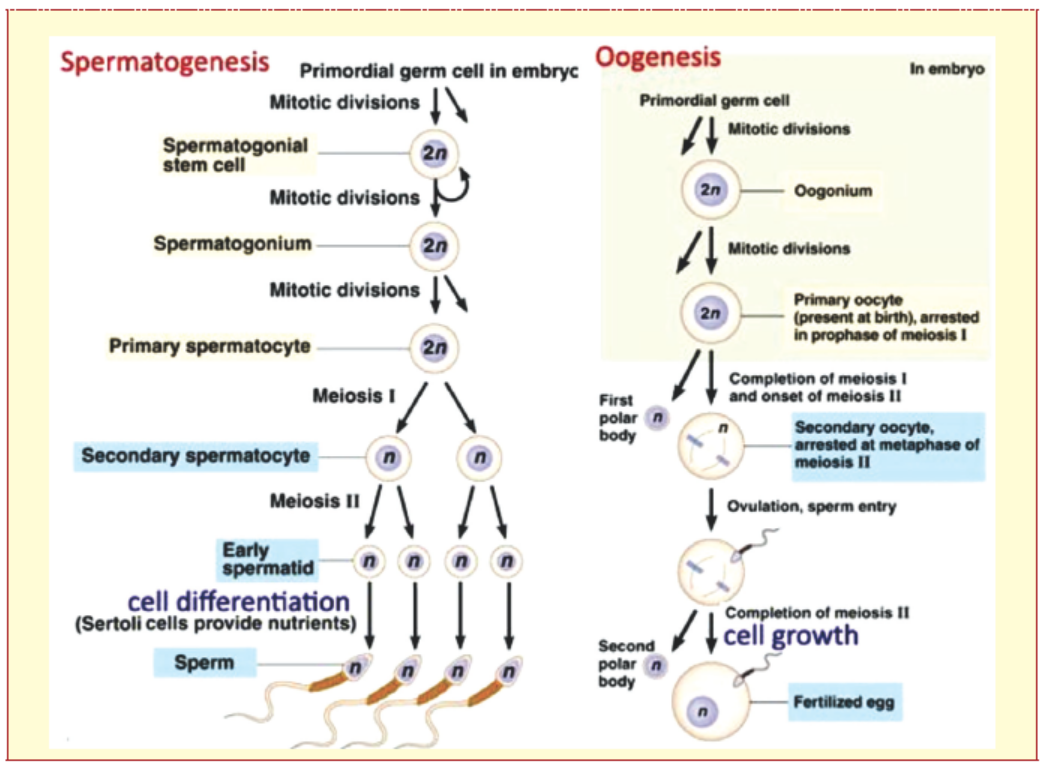
Differences between sperm and ovum
ACTIVITY 6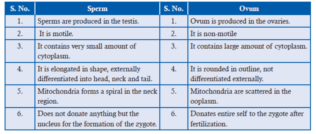
On the basis of your observations, draw the structure of a human spermatozoan and an ovum,labelling the following parts along with the functions of each:
APPLICATION 16.3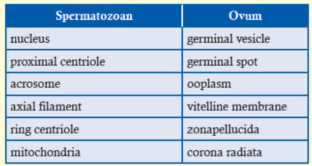
1.Complete with appropriate terms:
(i) The three basic steps of gametogenesis includes ..............., ............... and ............... .
(ii) One spermatogonium produces ............... and one oogonium produces ............... .
(iii) ............... cells provide nutrition in spermatogenesis.
(iv) The tertiary follicle matures into ............... .2. The diagram shows some stages in the formation of a mammalian egg cell
a.How many chromosomes will there be in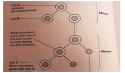
i. cell B
ii. cell C
b.Suggest one advantage in the way in which the cytoplasm divides during meiosis
c. Describe and explain two ways in which the events of meiosis cause the egg cells to begenetically different from one another
16.5 ONSET OF PUBERTY IN HUMANS
Males attain sexual maturity, or puberty as it is called, at the age of 11–13 years. At puberty,
the primary sex organs (=gonads) mature and secondary (=external) sex organs mature and
secondary sexual characters appear viz. development of pubic, chest and underarm hairs, growth
of larynx resulting in deepening of voice, masculine pattern of fat distribution, thick secretion
from skin oil glands, bone and muscle development, etc., are some of the prominent male
secondary sexual characters. Testosterone controls the onset of puberty in males. Hypothalamus
starts producing GnRH, which stimulates the anterior lobe of pituitary to secrete luteinizing
hormone (LH) and follicle stimulating hormone (FSH).
Females attain puberty at the age of 12–15 years. At this age, pituitary gland starts producing
follicle-stimulating hormone (FSH). The FSH induces the development of ovaries, which
produce the hormone estrogens, chiefly estradiol. Estrogen is responsible for the development of
secondary sex organs and secondary sexual characters in females, which include development
of breasts and external genitalia, pigmentation of the areola, growth of pubic hair, axillary
hair (in the armpits), widening of the pelvis and deposition of fat in thighs, buttocks and face.Puberty marks the onset of menstruation or menarche in females.
Differences between primary and secondary sex organs
16.6 SUMMARY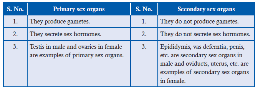
• Human male reproductive system carries out the functions of spermatogenesis i.e.,
formation of functionally active, motile sperms along with seminal plasma.
• The system comprises male sex organs i.e., testes (paired organ) and scrotum, a series
of ducts that help in transportation and maturation of spermatozoa and the accessory
glands which secrete the essential components of semen plasma.
• The temperature of testes is maintained 2°C –3°C below the core body temperature which
provides favourable environment of spermatogenesis.
• The male reproductive ducts include vasa efferentia, vasa defrentia, epididymis, ejaculatory
ducts and urethra primarily.
• The accessory glands of reproduction in human reproductive system are seminal vesicles,
prostate gland, urethral (Littre’s) glands and bulbourethral (Cowper’s) glands.
• Human female reproductive system is designated to carry out the functions of ovulation,
carry male and female gametes, fertilization, gestation and childbirth.
• Human female reproductive system consists of female sex organs- ovaries, duct system
- oviducts or the uterine/fallopian tubes, uterus, cervix, vagina, the external genitalia –
vulva and a pair of mammary glands.
• Ovaries produce female gametes- ova. They contain ovarian follicles at various stages of
development. An ovarian follicle consists of ovum surrounded by granulosa cells and
other layers for protection and nutrition.
• Oviducts transfer ovum from ovary to the uterus and serve as the site of fertilization.
Each oviduct is divided into an infundibulum, ampulla, isthmus and uterine parts.
• Uterus is a hollow organ that serves as the site of implantation and nourishment of the
embryo till birth. It consists of three parts – fundus, body and cervix. The inner lining
is called endometrium.
• Cervix and vagina form the birth canal.
• External genitalia or vulva consists of mons pubis, clitoris, labia majora, labia minora
and perineum.
• Glands include lesser vestibular and greater vestibular glands and a pair of mammary
glands. Mammary glands function in the production of milk for the young one.
• Menstrual cycle consists of three phases: menstrual, proliferative and secretory in the
uterus, corresponding to follicular, ovulation and luteal phases of ovarian cycle.
• The onset of menstrual cycle at puberty is termed menarche and end is called menopause.
• The menstrual cycle is governed by hormones.
• The process of formation of haploid male gametes or spermatozoa from diploid
reproductive cells in males is called spermatogenesis.
• The complete process of spermatogenesis is broadly divided into two parts:
(i) Formation of spermatids and (ii) Spermiogenesis or spermatoleosis.
• Spermatid formation is further divided into three phases as multiplication, growth and
maturation phase.
• Spermiogenesis is the process of series of changes to transform a non-motile spermatid
into motile, functional spermatozoa.
• Oogenesis is the formation of haploid ovum from diploid undifferentiated germ cells in
the ovary.
• Oogenesis is completed in three phases of discontinuous steps.
• Proliferative or multiplication phase involves proliferation of oogonial cells by mitosis.
• Growth phase is all about growth and differentiation of primary oocyte and development
of mature ovarian follicle.
• Maturation phase provides time for the oocyte to undergo two meiotic divisions toproduce functional haploid ovum.
16.7 GLOSSARY
• Castration: The surgical removal of testis is called castration.
• Coitus: The sexual act of transferring sperms of male to vagina of female.
• Cryptorchidism: Abnormality wherein the testes do not descend down from the abdomen
to scrotum is called cryptorchidism (crypt = hidden; orchid = testis).
• Ectopic pregnancy: A pregnancy occurring in the fallopian tube or outside of the uterine
lining.
• Ejaculation: The process of propulsion of semen out of the urethra at the time of orgasm.
• Emission: Emission is the phenomenon of movement of sperms from the testes into the
urethra along with secretions from the various accessory glands of reproduction, where they
mix to form semen.
• Erection: The accumulation of blood in penile erectile tissue leading to temporary
swelling and elongation of penis is called erection. This accumulation of blood is because
of dilation of arteries and compression of veins of penis.
• Gynecomastia: The condition when mammary glands become functional in males.
• Hypermastia: Presence of more than the normal number of breasts.
• Hysterectomy: Surgical removal of the uterus is known as hysterectomy.
• Impotence or erectile dysfunction: Impotence is the failure to achieve and/or maintain
the erection for coitus.
• Orgasm: Orgasm is a pleasurable feeling of physiological and psychological release
associated with the culmination of sexual stimulation. Emission and ejaculation of semen
accompany orgasm in males.
• Tubectomy: Surgical procedure for sterilization in which a woman’s fallopian tubes are
clamped and blocked, or severed and sealed.
• Vasectomy: Bilateral ligation of vas deferens as a contraceptive measure in males iscalled vasectomy.
END UNIT ASSESSMENT 16
I. Choose whether the following statements are True (T) or False (F)
1. Germinal vesicle is the mitochondria of the ovum.
2. Urethral gland in human males is also called gland of Littre.
3. The proliferative phase involves two meiotic divisions of the oocyte.
4. Clitoris is considered homologous to penis of males.5. Testosterone is synthesized in sertoli cells.
II. Fill in the blanks
1. Cowper’s gland in human male is also called ............................. .
2. Testes are present in the sac called ..............................
3. ....................... hormone helps in the relaxation of pelvic ligaments during childbirth.
4. The site of fertilization in human female is ............................. .
5. Corpus luteum secretes large amounts of hormone ............................... .
6. The onset of menstrual cycle at puberty is termed ............................... .
7. The caudaepididymis is attached to scrotal sacs by a connective tissue called
............................. .
8. The hormone responsible for most of the secondary sex characters in human maleis ............................. .
III. Long Answer Type Questions
1. Give an account of various processes involved in oogenesis in human females.
2. Draw a well labelled diagram of human spermatozoan.
3. Give a detailed account of male reproductive accessory glands and discuss their role
in male reproductive system.
4. Describe the histological structure of human testis.
5. With the help of suitable diagrams, explain the structure of human female reproductive
system in detail.
6. Describe the internal anatomy of a typical human ovary.
7. Explain the steps leading to the formation of corpus luteus? What is its significance?
8. State where male and female gametes are produced.
9. Explain the significance of gametogenesis.
10. (i) Identify the structure shown in figure.
(ii) Name the parts marked 1–8 in the figure.
(iii) Write the functions of the parts marked 7 and 8.
(iv) Name the chemical substance present in the part marked 2.(v) What is the importance of the chemical substance present in the part marked 2?
11. Genetic disparity has since ages disturbed the masses on Earth. Reproduction is a natural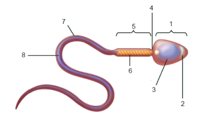
process but still the work for male and females is defined in the society. Assess your
understanding to support gender equality. Also, list the names of organizations supportingthe cause in Rwanda.
Unit 17 Genetics
Key Unit Competence
To be able to explain the role of genes in inheritance and how genetic disorders occur.
LEARNING OBJECTIVES
At the end of this unit, the learner will be able to:
• Explain the terms gene, locus, allele, dominant, recessive, co-dominant, linkage, test cross,
F1 and F2, phenotype, genotype, homozygous and heterozygous.
• Explain how to conduct a test cross.
• Explain why monohybrid ratios of 1:2:1 occur.
• Describe an example of inheritance involving multiple alleles.
• Explain the effect of lethal genes on phenotype ratios.
• Analyse various patterns of inheritance.
• Appreciate the roles of genes in determining the phenotype and patterns of inheritance.
• Use genetic diagrams to solve problems involving monohybrid and dihybrid crosses, including
those involving autosomal linkage, sex linkage, and codominance, multiple alleles and gene
interactions. (The term epistasis does not need to be used: knowledge of the expected ratio
for various types of epistasis is not required. The focus is on problem-solving).
• Use the complete and accurate format to show a genetic cross and the results of a simple
monohybrid cross.
• Use genetic diagrams to solve problems involving test crosses.
• Use the chi-squared test to test the significance of the differences between observed and
expected results (the formula for the chi-squared test will be provided.).
• Demonstrate monohybrid and dihybrid inheritance.
• Give a genetic explanation of Mendelian dihybrid inheritance.
• Explain the use of test crosses to determine unknown genotypes in studies of dihybrid
inheritance.
• Explain the significance of recombination.
• Explain how sex is determined in humans and the role of sex related Y genes in determining sex.
• Describe how non-disjunction can affect the distribution of sex chromosomes in gametes
and offspring.
• Explain why linked genes do not show independent assortment.
• Explain how crossover values can be used to make a chromosome map.• Interpret Pedigree charts.
INTRODUCTORY ACTIVITY
Read the passage below and answer the questions that follow
We all are aware of the fact that we look similar to our parents ( grand –parents) and siblings
in our appearance such as eye color, hair texture, skin color etc.
We are also aware of the fact that certain diseases run in the family such as albinism, hemophilia
etc, which indicates that certain characters on traits are passed on from parents to their offspring
a. Why do you resemble your parents or siblings?
b. Which structure do you think controls the transmission of information from parentsto their offsring
17.1 CONCEPTS OF INHERITANCE
ACTIVITY 1
You must be familiar with the statements such as:
“You look similar to your mum but your skin colour is like that of your dad.” Or
“Your eye colour is same as that of your grandfather”.
Discuss such example for traits where you resemble to your family in traits such as hair colour,eye colour, skin colour etc.
We all are aware of the fact that we resemble to our parents (grand-parents) and siblings in
our appearance such as eye colour, hair texture, skin colour etc. We are also aware of the fact
that certain diseases run in the family such as diabetes, hemophilia etc. which indicates that
certain characters or traits are passed on from parents to their offspring. This phenomenon isknown as Heredity or Inheritance.
An organism produced by sexual reproduction has two parents and inherits certain traits from
father and certain traits from mother. It leads to variation in organism. So heredity and variation
is characteristics of sexually reproducing organism. The study of heredity and variations in
biology is referred to as Genetics.17.2 MENDEL’S LAWS OF INHERITANCE
ACTIVITY 2
A farmer came to complain to the seed seller in this way: “ I always buy the red bean seeds from
your shop but this time, look, they produce red and some few yellow colored been. What did
you put in the seed you gave me?”
How can you help to solve such a conflict?Is there any pattern by which traits pass on from one generation to another?
17.2.1 Mendel’s Experiments
In 1856, Gregor Mendel conducted experiments in garden pea (Pistum sativum) in the limited
space of a monastery garden. Garden pea plant has both male (pollen-producing part) and
female parts (pollen-receiving part). Since both the male and female parts are on the same
plant, it has tendency to undergo self-fertilization. Because of self-fertilization, the tall plants
always give rise to tall plants and dwarf plants always produce dwarf plants. Such true breeding
varieties are known as pure lines. Furthermore, he was lucky to get pure lines in garden pea.
He then carefully conducted hybridization experiments between two parent plants expressing
contrasting form of single trait. He also made sure that self-fertilization didn’t happen by removing
male parts from one parent (say tall plants) before female part got matured. In his initial experiments
(Figure 17.1), he carefully transferred pollen from male parent (say dwarf plant) to tall parent’s
female part and analyzed transmission of one particular trait (stem height) in all progenies of
the first generation (also known as F1 generation where F symbolizes the Latin word “filial”
meaning progeny and 1 represents first). Furthermore, he followed the transmission of same trait
(stem height) in second (F2) and third generation (F3) progenies as well which were naturally
produced by self-fertilizing power among first generation plants and second generation plants.
He maintained the quantitative records of all his experiments.
Since Mendel focused on one trait at a particular time, a cross between parents which differsin contrasting form of single trait is known as Monohybrid cross or inheritance.
Gregor Mendel: Father of Genetics Figure 17.1: A hypothetical experimental plan by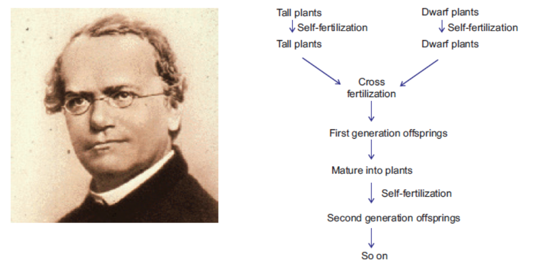
(Source: http://www.biography.com/ ; Mendel to follow the inheritance of particular trait orpeople/gregor-mendel-39282) monohybrid inheritance (for example – stem height)
17.2.2 Mendel’s Observation
Mendel carried out experiments to follow the pattern of inheritance of particular trait in several
generations. On crossing tall plants (which provided the female part) verse dwarf plants (whichprovided pollen), he observed that (Figure 17.2).
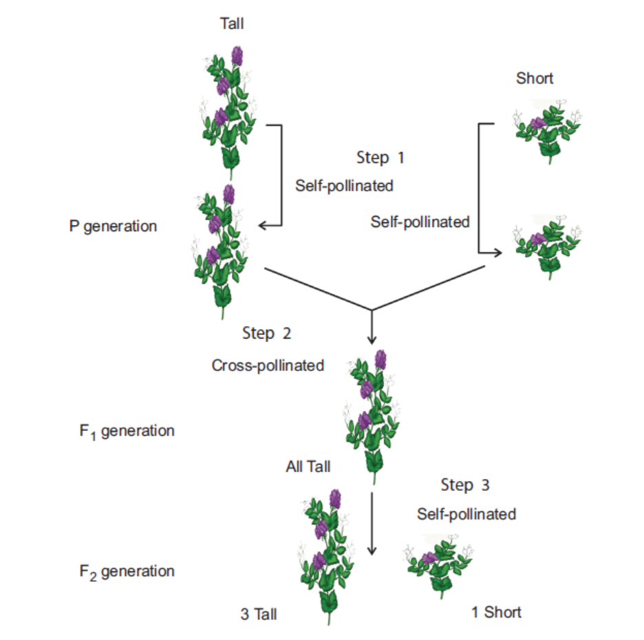
Figure 17.2: The experimental observation from a cross between tall and dwarf plants
• First generation progenies were always tall.
• Second generation progenies (also known as F2 generation) include tall plants as well as
dwarf plants almost in ratio of 3 (tall plants) : 1 (dwarf plants).
Mendel then performed the reciprocal cross (A similar cross where tall plants provided male
parts whereas dwarf plants represented female plants). Mendel observed similar results.
On performing similar cross-fertilizing experiments with parent plants showing other
contrasting set of traits such as seed colour, seed shape, seed coat colour, pod colour, pod
shape and flower position/arrangement (figure 17.3), he observed similar observation and
concluded that:
• First generation progenies were always showing one form of trait expressed in one of the
parent plants.
• Second-generation progenies include the plants showing both contrasting forms of traits,almost in ratio of 3:1.
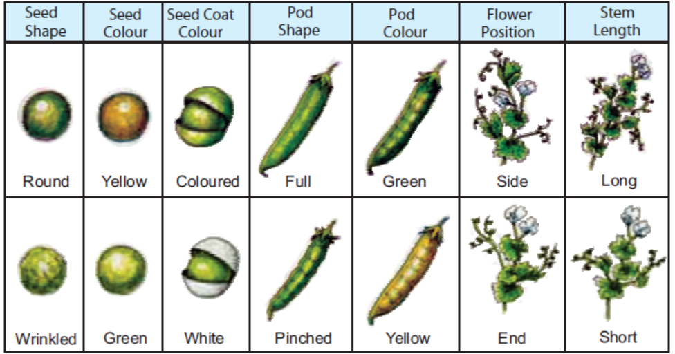
Figure 17.3: A seven pairs of contrasting traits in garden pea, the inheritance pattern was followed
On self-fertilization of F2 plants for various contrasting traits (for example: for stem height),
Mendel observed the following points:
• Dwarf F2 plants always yielded dwarf plants only.
• All F2 tall plants were not genetically same. The one-third tall plants produced tall plants
only but two-third tall plants yielded both tall plants and dwarf plants in the ratio of 3 : 1.
It means phenotypic ratio is 3 : 1 but genetically the ratio is 1 : 2 : 1.
The results of Mendel´s experiment were published in the monograph – “Experiments in PlantHybridization” in 1866.
17.2.3 Mendel’s Postulates: Principles of Inheritance
Based on consistency of his results in transmission of seven contrasting traits, he derived
postulates which later became principles of inheritance.
• There are two factors (Unit factor in pairs) for each trait. In pure lines of plants, both the
factors for particular trait (stem height) are alike. For example, if Factor “T” donates height,
there are two factors for each trait. The tall plants have TT and dwarf plants have tt.
• At the time of gamete formation, the factors for particular trait randomly segregate
with equal likelihood. Each gamete contain single factor, therefore the gamete is always
pure for the trait. Later on, it becomes popular as “Mendel’s principle of segregation”.
For example: all the gametes from tall plants have single factor “T” and dwarf plants
have “t” (Figure 17.4).
• After fertilization, when gametes from parents randomly fuse, factors for a particular
trait also unite together. For example, in a cross between tall and dwarf plants, gamete
from tall plant with factor “T” fuses with gamete from dwarf plant with factor “t” to
form “Tt” organism.
Figure 17.4: Mendel’s conclusion from hybridization experiments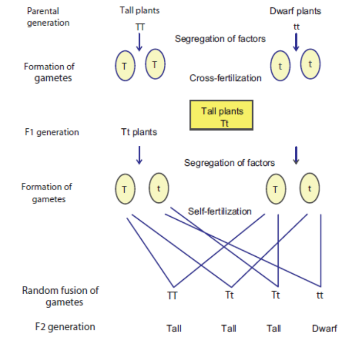
(shown for trait : stem height)
• In F1 generation, only one of the parental traits is expressed, it indicates that out of two
forms of factors for the single trait, one factor is dominant. For example: Tt organisms are
tall, so the factor for tallness “T” is dominant over the factor “t” for dwarfness. The factor
which remains under-expressed in presence of dominant factor is known as recessive factor.
• In the F1 populations, the factors again randomly segregate in the gametes. So a ‘Tt’
organism will produce two type of gametes either having ‘T’ or‘t’ form for particular
trait. During self-fertilization, there is random fusion of gamete and formation of TT,
Tt, Tt or tt. Hence, three out of four F2 progeny becomes tall whereas one out of four
progeny are small (figure 4). The tall and dwarf varieties are obtained in the ratio of
3 : 1. The F2 ratio (3 : 1) obtained is known as monohybrid ratio.
• Mendel concluded that the F2 ratio (3 : 1) indicates the morphological pattern of trait
(for example in case of stem height, it indicates tall verse short), so it is also known as
phenotypic ratio (Phenotype: morphological appearance). Genetically, it is further dividedinto 1 : 2 : 1 (Genotypic ratio) as Pure dominant: hybrid: pure recessive.
17.2.4 Reasons Behind Mendel’s Success
His experiments were highly successful and he was able to discover the pattern of inheritance.
The reasons for his success are as follows:
• His choice of experimental organism i.e. garden pea, was extremely good as it generates a
large number of generations in a very short duration.
• Pollination of garden pea leads to large number of offsprings due to which the inheritance
pattern could be followed in the progeny.
• Furthermore, Mendel studied one trait at a time thereby allowing him to deduce useful
interpretations.• Lastly, his mathematical background played an important role in deducing the results.
ACTIVITY 3
Problem A
Aim: A plant with round seeds along with wrinkled seeds is crossed. What will be the
genotype and phenotype of F1 generation and F2 generation if trait follows Mendelianpattern of Inheritance?
Procedure: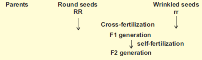
• Observe the given diagram carefully.
• Draw the punnett square showing the gametes from both the parents on each side.
• Note down the genotype and phenotype of F1 generation.
• Again draw the punnett square showing the gametes of F1 parents.• Note down the genotype and phenotype of F2 generation.
Problem B
Aim: Demonstrate monohybrid inheritance using beads of two different colours.
Materials Required:
Class notebook
Two colour beads (red and yellow)
Procedure for monohybrid inheritance:
• Open pouch containing beads of two different colours.
• Count total number of beads.
• Divide total number of beads with 4 (T/4 = z)
• Differentiate beads according to their colour and count them respectively.
• Divide each number obtained in the previous step with value “z” and make a ratio.
• Discuss whether the observed ratio is in accordance with the Mendelian monohybridratios.
17.3 DEFINITION OF GENETIC TERMS
Gene: Gene is the entity/unit which has the information for particular trait. For example:
in garden pea, gene for stem height has information for height whether it would be long
or small.
Locus: The position of gene on chromosome constitutes its loci/locus.
Allele: The alternate forms of genes are known as Alleles. A pair of alleles for each trait is
present in the zygote of an organism. For example: in garden pea, true breeding tall parent
plants have two similar alleles (TT).
Dominant Allele: In individual, out of two alleles for the particular trait, only one allele is
expressed. The expressed allele is known as dominant. For example, allele (T) for tallness is
expressed in F1 individuals (Tt), dominant allele. Dominant allele is generally referred by
capital alphabet.
Recessive Allele: In individual, out of two alleles for the particular trait one allele is underexpressed.
The under-expressed allele is known as recessive. For example, allele (t) for shortness
is not expressed in F1 individuals (Tt), recessive allele. Recessive allele is generally referred bysmall alphabet.
Co-dominant: It’s a phenomenon when both alleles present in an individual, are equally
expressed. For example, in humans, Blood cells express both the alleles M and N (alternate
form of gene encoding Red blood cell membrane protein) when present together.
Linkage: The genes are said to be linked when present on the same chromosome and inherited
together as unit.
F1: F symbolized filial, which means “progeny” in Latin. F1 is the filial generation first,
produced by cross between parent individuals.
F2: F2 is the filial generation second, produced by cross between F1 individuals.
Phenotype: The morphological appearance for particular trait constitutes its phenotype. For
example: In the cross between tall and dwarf parent plants, F1 plants are tall. Tallness is their
phenotype. In F2 plants, tall and dwarf plants are obtained in ration of 3 : 1, it is phenotypic ratio.
Genotype: The combination of allele for a particular trait in an individual constitutes its
genotype. For example: In the cross between tall and dwarf parent plants, F1 plants are Tt. “Tt”
constitute their genotype for the trait stem height. Similarly, F2 plants are tall and dwarf. But
genotype of all tall F2 plants is not same, one third are pure (TT) while two third are hybrid
(Tt). So genotypically F2 ratio is 1 : 2 : 1.
Homozygous: When in an individual, two alleles for a particular trait are alike, then individual
is considered homozygous for the particular trait. For example, parent plants tall and dwarf
plants are homozygous for stem height.
Heterozygous: When in an individual, two alleles for a particular trait are different then individual
is considered heterozygous for the particular trait. For example, F1 plants are genotypically
“Tt”. They are heterozygous for stem height.
Monohybrid Cross: ‘It is a cross between two individuals of a species which is made to study the
inheritance of a single pair of factors or genes of a trait.’ A ratio among the offspring of F2 generation
of a monohybrid cross is called a ‘monohybrid ratio.’ It is usually 3 : 1 (phenotypic ratio) or
1 : 2 : 1 (genotypic ratio), in which 1/4 individuals carry the recessive trait, 1/4 pure dominantand 1/2 have impure dominant trait (Figure 17.5).
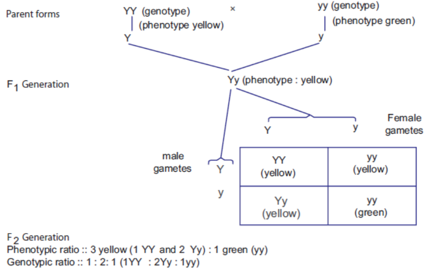
Figure 17.5: Mendel’s monohybrid experiment on garden pea (Pisum sativum)
Test Cross: It is cross between hybrid forms (dominant phenotype) with other parent with
recessive form of particular trait (homozygous recessive). It is generally used to identify
the genotype of hybrid form. The progenies are observed. If all progeny demonstrates only
dominant form of trait thereby indicating that unknown genotype must be homozygous
for the particular trait. Or If F1 progeny shows both dominant and recessive form of
trait in the ratio of 1 : 1 indicating that unknown genotype must be heterozygous for
the particular trait.
There can be two possible genotypes of an unknown dominant phenotype as illustrated below.
Possibility 1. If the unknown is homozygous yellow (YY), then crossing with green recessive(yy) gives all yellow offspring (i.e., all Yy) as shown below:
Possibility 2. If the unknown is heterozygous yellow (Yy), then crossing with green recessive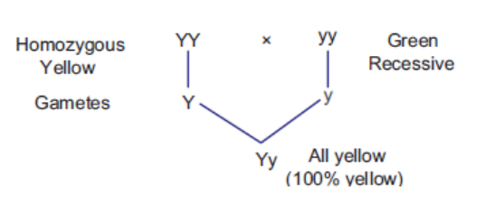
results in 50% yellow (Yy) and 50% green (yy) progeny as shown below:
In case of a double heterozygous, i.e, heterozygous yellow and round (Yy Rr) crossed with
double recessive, i.e, recessive green and wrinkled (yy rr) the ratio will be 1 : 1 : 1 : 1.
Back Cross: The mating of the hybrid form with one of its parent or with individual genetically
similar to parent is known as back cross. When the back cross is carried out for several generations,
the hybrid becomes genetically closer to that parent. The back cross is commonly used in
horticulture and animal breeding.
Chi-Square Test: The statistical test used to analysis genetic data especially obtained
from breeding experiments. When breeding experiments are carried out, the progeny
number is counted and based on their phenotype, the ratio is calculated. But there is
always a difference in observed value from expected value. However, this might happen by
chance or there can be a real difference behind this deviation. Hence, chi-square analysis
test is used to find out whether deviations are by chance or there is indeed difference inobserved value.
Probability of Deviation
Using chi-square analysis, the probability or p value is calculated. Probability of deviation
indicates whether differences in observed value from expected value are by chance or not. If
probability is very high (equal to or more than 0.05 or 5%) then probability of deviation by
chance is high and there is no significant difference in observed and expected value. However,
if probability of deviation by chance is less (less than 0.05 or 5%) then observed value issignificantly different from expected value.
Calculation of p ValueFor its calculation, chi-square and degree of freedom is needed.
How to Use Chi-square Analysis by Example
Just consider a monohybrid cross for a trait pod colour in garden pea was carried out. In F2
generation, out of 582 plants, 428 green and 152 yellow were observed. We want to know
whether this cross is following Mendelian monohybrid ratio 3:1. According to expected ratio,
plant with green phenotype should be 437 (3/4 of total number) and yellow phenotype should be
145 (1/4 of total number). Now we can see there is difference in observed value from expected
value, whether this deviation is by chance or significantly different, and chi-square analysis canbe used according to formula in Table 17.1.
Table 17.1: Calculation of chi-square value from the given data
Degree of freedom is always one number less than total number of classes. Degree of freedom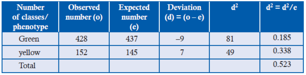
represents the quantum of independency that is there with the given classes. If there are
two classes, then only one class has freedom to fall in any criteria other class has no choice.
(for example : provided there are pairs of socks, there are two classes but freedom is there with
one socks which can be part of left or right foot, there is no option for second socks). Hence,degree of freedom (D) can be represented as:
D = n – 1(Where n is total number of classes)
Probability of deviation calculation can be calculated by using probability table (Table 17.2)
wherein left column degree of class (D) is written and on right row (top most) Probabilities
are mentioned. At degree of freedom 1 (black arrow), summation of chi-square 0.523 value
(red box) shows p value as 0.30–0.50 (blue oval) or 30 to 50%. It indicates that deviation is by
chance. There is no significant difference between observed and expected value. Hence, thegiven cross follows Mendelian ratio (3 : 1).
Table 17.2: Probability table for chi-square analysis
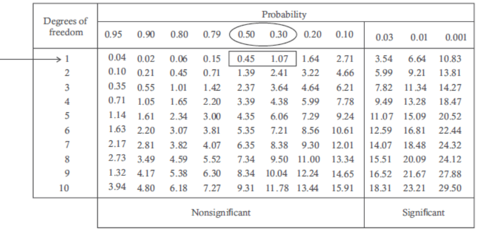
ACTIVITY 4
Aim: Use genetic diagram below to solve problems involving test crosses
On test cross, out of 500 progeny 245 plants are observed with blue flowers and 255 plants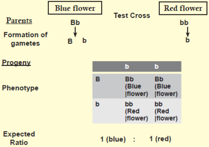
are observed with red flowers. But according to test cross ratio, total 500 progeny should be
expected as 250 blue flowers and 250 red flowers.
With the help of a chi-squared analysis test the significance of differences between observedand expected results.
• Using Chi-square formula, calculate the value for chi-square followed by its summation.
• Find the degree of freedom.
• Find the value of probability of deviation using probability table.• Discuss your results in group.
17.4 INDEPENDENT ASSORTMENT AND SEGREGATION
What do you think would have happened if Mendel would have tried inheritance pattern oftwo traits? Do inheritance of one trait is dependent on the inheritance pattern of another trait?
The cross was made between the double dominant plants (round seed shape with yellow seed
colour) with double recessive parent (wrinkled seed shape with green seed colour) and the
following points were observed:
• All round yellow seeds were observed in F1 generation indicating dominant factor for a gene
was expressed in the same manner as in monohybrid cross.
• On self-fertilization of F1 plants, F2 seeds were obtained and segregated in the ratio of
9 : 3 : 3 : 1 based on their phenotype.
In addition to parental phenotype combination, two new phenotype combinations/
recombinants (wrinkled and yellow and round and green seeds) were observed. Mendel
hypothesized that the factors for different traits separate and assort independently in the
gametes (factor for seed shape can assort with any seed colour factor and vice versa) then F1
plants should produce four types of gametes.
So male and female F1 plant gametes can fuse randomly and combine in 16 possible ways
which can be simply represented by a simple square popularly known as Punnett’s square*
(Figure 17.6).
Mendel observed similar results when he analyzed results of dihybrid cross for the other pair
of traits as well.
• The dihybrid results did not contradict monohybrid results, the round seeds and wrinkled
mseeds as well as yellow and green seeds were in ratio of 3 : 1. He hypothesized dihybrid crossevent as two independent monohybrid cross events.
(*Punnett’s square or checker-board: square-shaped presentation used to predict result of a particular
cross or breeding experiment in which gametes from each parent are placed on the top and left side
of the square. This diagram is used to predict of genotype of the individual when gametes fromparents randomly fuse. It is named after Reginald C. Punnett, who devised the approach.)
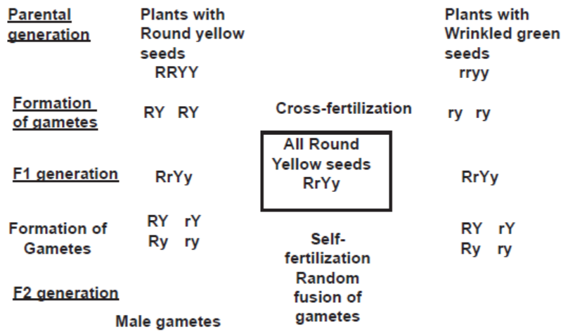
Figure 17.6: A dihybrid cross between plants with dominant round yellow seeds with plants with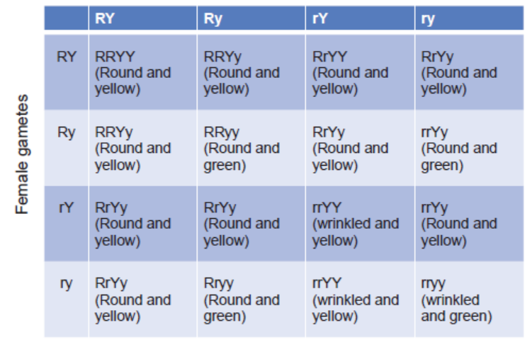
recessive traits wrinkled and green seeds through two generations
Random fusion of F1 gametes is represented with the help of Punnett’s square. The four
phenotypes—two parental (written in black) and two recombinants (written in purple and red)are obtained in the ratio of 9:3:3:1.)
17.4.2 Law of Independent Assortment
From the result of dihybrid cross experiments, Mendel gave the following postulates:
• The dominant allele of a particular gene is expressed in the presence of alleles of other
genes for different traits.
• On self-fertilization F1 plants, F2 plants were observed in the phenotypic ratio 9:3:3:1
(Dihybrid ratio). He concluded that factors for different traits assort segregate and assort
independently in the gamete. This is popularly known as Law of Independent Assortment.
17.4.3 Significance of Test Crosses in Dihybrid Inheritance
Test cross can be used to differentiate genotype of dihybrid organisms (whether it is homozygous
and heterozygous for the traits) if phenotypically same for a traits.
For example: plants with similar phenotype rounded seed shape and yellow seed colour can
have different genotype RRYY or RrYy. So the genotype of such plants can be identified by
test cross. So the plant with unknown genotype is crossed with plant with recessive form of
both the traits. There are two possibilities.
1. If progeny plants are observed in phenotypic dihybrid test ratio 1 (round and yellow):1
(round and green):1 (wrinkled and yellow):1 (wrinkled and green), then the parent plantmust have heterozygous genotype for both the traits (Figure 17.7).
Expected ratio for dihybrid test cross
Figure 17.7: Dihybrid test cross ratio when plant has dominant heterozygous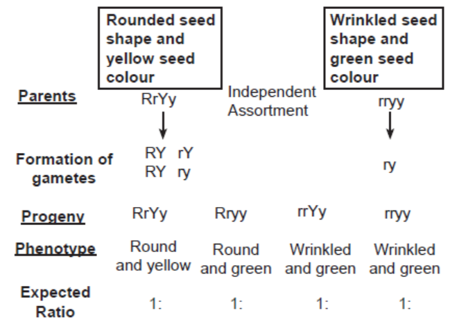
genotype for two traits
2. If after the cross all the plants are formed with dominant phenotype i.e., round seed
shape and yellow seed colour, it indicates that given parent plant must have homozygousgenotype for both the traits.
ACTIVITY 5
Aim: To demonstrate dihybrid inheritance using beads of two different colour and two
different sizes.
Materials Required:
Class notebook
Two colour beads (red and yellow)
Two size beads (Large and small)
Procedure for dihybrid inheritance:
• Open pouch containing beads of two different colours and different sizes.
• Count total number of beads.
• Divide total number of beads with 16 (T/16 = h)
• Differentiate beads according to their colour and size and count them respectively.
• Divide each number obtained in the previous step with value “h” and make a ratio.Discuss whether the observed ratio is in accordance with the Mendelian dihybrid ratios.
17.4.4 The Chromosome Theory of Inheritance
According to Mendel‘s law of inheritance, there are two factors for each character, each factor
segregates and assorts independently in the gametes without getting influenced by presence
of other factors for different traits. Similarly, same rules are followed for chromosomes during
cell division and cell biologists have confirmed that similar to Mendel’s factors.
• Chromosomes are also found in pairs,
• Segregate and assort independently in the gametes at the time of meiotic cell division.
This indicates that Mendel’s factors or genes are located on the chromosomes and it is chromosomes
which segregate and independently assort in the gametes. This is most popularly known aschromosome theory of inheritance which was independently given by Sutton and Boveri in 1902.
APPLICATION 17.1
1. Complete with appropriate terms:
(i) A cross of F1 with the recessive parent is ............................. .
(ii) ............................. is the father of genetics.
(iii) Mendel selected ............................. varieties of garden pea.
(iv) F2 generation shows 9 : 3 : 3 : 1 ratio in ......................
2. One of the genes for color vision in cats is sex-linked. The allele A gives orange fur, whereas
the allele B gives black. The two alleles are codominant, and when both are present the cat
has patches of orange and black, which is known as tortoiseshell.
a) Explain why male cats cannot be tortoiseshell.
b) Draw a genetic diagram to show the expected genotypes and phenotypes of the offspring
from a cross between an orange male and a tortoiseshell female cat.
3. In a species of animal, it is known that the allele for black eyes is dominant to the allele for
red eyes, and that the allele for long fur is dominant to the allele of short fur.
a) What are the possible genotypes for an animal with black eyes and long fur?b) How could you find out which genotype this animal had?
17.5 INHERITANCE: VARIATIONS FROM MENDEL’S PATTERN
Do you find all traits in accordance with Mendalian pattern?
Is there always two alleles for a trait in a population, if no discuss some examples.
Is one allele is always dominant to another?
Do product of all alleles are equally beneficial?
Are all our traits independent of inheritance of other traits? Think of metabolic pathways where
product of one reaction is substrate for next event and so on. Here the enzymes of reaction willnot be dependent on another enzyme of reaction of same metabolic pathways.
17.5.1 Co-dominance
When in heterozygote, both alleles of gene for particular trait are expressed, the phenomenon
is known as co-dominance. The best example of co-dominance is the MN blood group antigens
which are found in Red Blood Cells in Human. In Homozygous MM and NN individuals,
antigen M and Antigen N are expressed but in heterozygous MN individuals both antigen
M and N are co-expressed on the surface of RBC. When two heterozygous individuals are
mated, Progeny are obtained in phenotypic ratio of 1 (MM) : 2 (MN) : 1(NN) instead of 3 : 1monohybrid Mendalian ratio (Figure 17.8).
Figure 17.8: Cross between heterozygous MN individual – demonstrating co-dominance and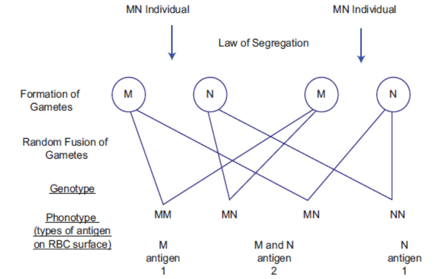
characteristic phenotypic ratio 1 : 2 : 1
17.5.2 Multiple Alleles
It is well established that there are two alleles for a single gene. However, sometimes in a
population there can be more than two alleles for a certain gene which can be illustrated by the
ABO blood group system. In ABO blood group system, there are three alleles IA, IB and IO in
the population. The alleles decide the type of glycoprotein found on the surface of erythrocytes
(red blood cells). There are four blood types phenotype as depicted in table 17.3. The Individual
with blood group A express A type of glycoprotein while the individual with blood group B
express B type of glycoprotein. The individual with blood group AB expresses both types ofglycoprotein while O type individual contains neither A or B.
Alleles IA and IB are dominant to IO so A type individuals can have IA IA or IA IO genotype.
Similarly, B type can have IB IB or IB IO genotype. Alleles IA and IB are co-dominant so when
present together in AB individuals are expressed together. Alleles IO is recessive so O type
individuals are recessive homozygous IO IO. ABO Blood type example demonstrates uniquecombination of multiple alleles as well as co-dominance.
Table 17.3: The ABO blood type in human population
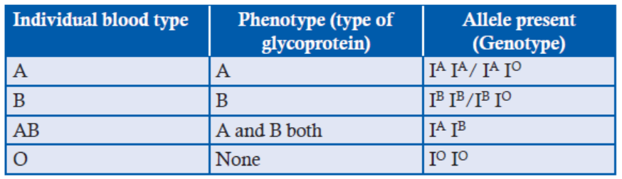
ACTIVITY 6
Aim: Consider two co-dominant alleles P and R. Draw a genetic diagram showing a cross
between two heterozygous individual (PR)? Discuss genotypic and phenotypic ratio in
progeny.
Procedure:
• Make a genetic diagram showing two heterozygous parents.
• Draw their gametes and with the help of Punnett square, make their progeny and give their
genotype and phenotypic ration.• Discuss it among yourselves and with class teacher.
17.5.3 Lethal Alleles
Sometimes genes have serious effect on development, physiology of the organism in such a way that
organism is unable to survive. Such genes are known as lethal genes. The particular allele responsible
for death of the organism is known as lethal alleles. Lethal allele can be dominant or recessive.
For example: The dominant allele C in chicken has serious effect on development of the
organism and results in following phenotype:
• Aberrant form “creepers” in Heterozygous individual (Cc)
• Completely “lethal” in homozygous dominant (CC).
When two heterozygous creeper individuals are mated, progeny are obtained in phenotypicratio of 2 (Creeper): 1(Normal) instead of 3 : 1 monohybrid Mendelian ratio (Figure 17.9).
Figure 17.9: Cross between two creepers chickens — demonstrating lethality and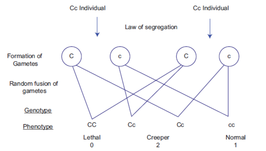
characteristic ratio 2 : 1
ACTIVITY 7
Aim: In mice, dominant allele Y is for yellow coat colour while recessive allele y is for agouti
coat colour.
(a) The yellow coat colour mice were self-bred. The yellow and agouti mice are produced in
the ratio of 2 : 1. What could be the reason for change in Mendelian monohybrid ratio?
(b) What is the genotype of mice having yellow coat colour?
Materials Required:
Notebook
Lethal allele information
Procedure:
• Read about lethal allele.
• Try to solve the genetic problem.• Discuss it among yourself.
17.5.4 Incomplete Dominance
Sometimes the allele is not completely dominant or completely recessive. In such heterozygotes
individuals, intermediate phenotype is observed. Such phenomenon is known as incomplete
dominance or semi-dominance or intermediate inheritance. For example: in Snapdragon
(Antirrhinum majus) a cross between plants with red and white flower variety produces all
plants with pink flower or intermediate phenotype. When two heterozygous plants with pink
flowers are mated, Progeny are obtained in phenotypic ratio of 1 (red) : 2 (pink) : 1 (white)instead of 3 : 1 monohybrid Mendelian ratio (Figure 17.10).
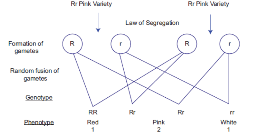
Figure 17.10: Cross between two heterozygous plants (pink variety) in snapdragon —demonstrating incomplete dominance and characteristic phenotypic ratio 1 : 2 : 1
17.5.5 Gene Interaction
There are cases when product of one gene influences the product/phenotypic expression of
another gene and this phenomenon is known as gene interaction (meaning: standing on/to mask).
One of the examples illustrating this is flower colour in sweet pea (Lathyrus odoratus), where
P dominant allele determines formation of the pigment anthocyanin (responsible for purple
flower colour). However, plants with PP and Pp will have purple flower only when another
allele of different gene C is present in dominant form. So product of gene C influences
phenotypic expression of gene P. The reason for this phenomenon can be explained as follows.
It is observed that gene C encodes for enzyme that synthesizes intermediate compound x
from precursor molecule. The intermediate compound is converted to coloured pigment via
product/enzyme of gene P.
Therefore, at least one allele of both gene C and P should be dominant to have colour flower. Such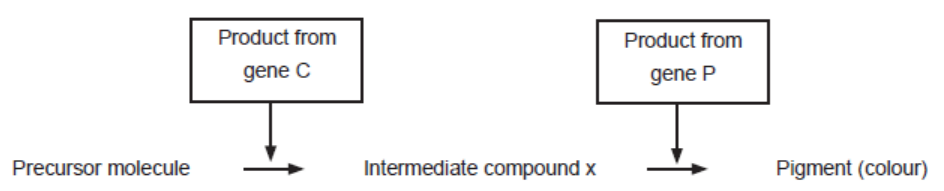
interaction where allele for one gene complements expression of allele of another gene is knownas complementary gene interaction. The respective genes are known as complementary gene.
ACTIVITY 8
Aim: In a given reaction, can you find gene interaction? If yes, what are the genes? If gene
“H” is present in homozygous recessive form and “A” gene is present in homozygous dominant
form, would you expect product A?
Procedure:
• Observe the reaction carefully.
• Note the number of gene that is required for the formation of product A.
• Considering the given condition in the question, try to answer to the best of your knowledge.• Discuss among yourself and with your class teacher.
17.6 LINKAGE AND CROSSING OVER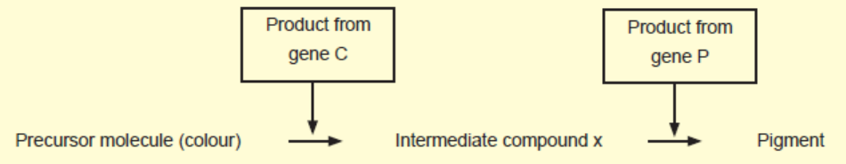
According to chromosomes theory of Inheritance, it is the chromosomes which segregate and
assort independently in the gametes. So the question arises as to then what happens to genes
located on same chromosome? Do they always remain together or linked (exception to law of
independent assortment)? Or, do they segregate and assort independently, if yes what couldbe the mechanism?
17.6.1 Linkage
There are cases when genes (present on the same chromosome) for different traits do not show
independent assortment, inherit together and behave as if genes are linked; the phenomenon
is known as linkage.
For example: two genes for trait flower colour and pollen grain texture in sweet pea (Lathyrusodoratus) where blue flower colour (B) allele is dominant over red flower colour (b) and long
pollen (L) is dominant over round pollen (l). A test cross was carried out between heterozygous
plant with double homozygous recessive plant (bbII), the observed phenotype had higher
frequency of parental phenotype (87.4%) and lower frequency of recombinants phenotype
(12.6%) in contrast to expected dihybrid test ratio (figure 17.6). It indicated that genes do not
assort independently and appear as if they are linked. However, occasionally they may separate
therefore resulting in lower frequency of recombinants.
Such genes are identified as linked when present on the same chromosome and do not assort
independently and tends to form parental phenotype but occasionally they may separate resultingin low recombinants frequency. This phenomenon is known as linkage.
Table 17.4: Observed dihybrid test cross frequency exception to lawof independent assortment
17.6.2 Linkage Crossing-Over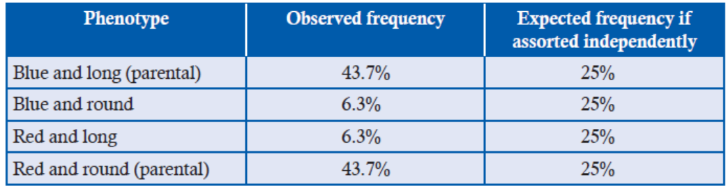
Now the question arises what could be the possible mechanism for the separation of the genes
located on the same chromosomes. The answer is crossing-over or recombination. Crossing-over
is the physical exchange of chromosome parts between non-sister chromatids of the homologous
chromosomes during meiosis division. The chiasma formation (observed by Janssens in 1909)
clearly provides the site at which non-sister chromatids of paired homologous chromosomes
cross over (Figure 17.12). The cross-over event between two gene loci in non-sister chromatidsis responsible for formation of recombinant chromatids and their separation (Figure 17.11).
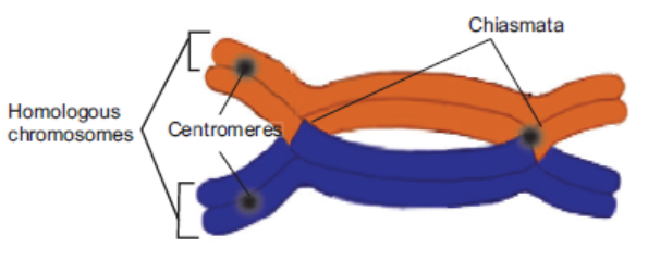
Figure 17.11: Micrograph demonstrating chiasmata formation in homologous chromosomes.
Here two paired homologous chromosomes (each with two sister chromatids) with centromeres
and gene loci are shown. Two alleles of a gene A (A and a) and two alleles of gene B (B and b)
occupy same position in homologous chromosomes (Figure 17.12). The crossing over between
two non-sister chromatids involves breakage of non-sister chromatids and reunion of broken
parts. The chromatids which participate in crossing over generate recombinants chromatids.
In the recombinants, the alleles on the same chromatid get separated and combine with allelesof non-sister chromatid.
Significance of Recombination/crossing-over
• The major significance is generation of variations. Due to crossing over, genes even on the
same chromosome can be assorted differently. It leads to variations in the progeny. The
variations are very useful in nature as it provides raw material on which natural selection
can act.
• The frequency of crossing over becomes higher with increase in physical distance between
gene loci. So recombinant frequency between two genes can be used to determine distancebetween genes, hence it helps to create chromosome map.
17.6.3 Chromosome Map
A chromosome map is the linear arrangement of genes in the chromosome with their relative
distance. It is based on fact that the frequency of crossing over between two genes is directly
proportional to the physical distance between the two. Therefore, the distance between genes
is indicated by percentage of crossing over (recombinant frequency). The distance unit is map
unit or 1cM (centi-Morgan, in honour of T. H. Morgan), so when 1% recombinant frequency
is observed between two genes, the genes are said to be 1 map unit apart.
T. H. Morgan and Sturtevant carried out extensive work in drosophila in finding recombinationfrequency between different genes and based on recombination data, they generated chromosome map.
Figure 17.12: Separation of genes A and B located on the same chromosome via crossing over—two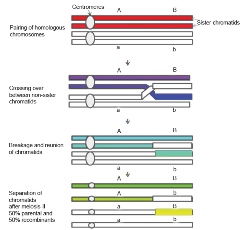
chromatids retain parental arrangement of the alleles while two are recombinant
A and a alleles of gene A while B and b are alleles of gene B. In recombinant chromatids allele
“A” combine with “b” allele while “a” combine with “b”.
For example: if recombination frequency between two genes A and B is 3% or 3 map unit. If
distance between B and C is 10 map unit and distance between A and C is 7 map unit, then
we can make chromosome map based on the information of recombination frequency. Thereare three possibilities:
But based on the information, possibility B represents the actual order of genes.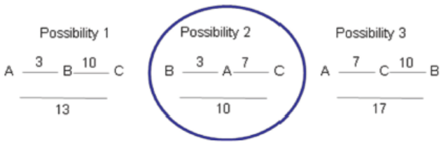
At the same time, we should remember even when genes are very far, there are 100% chances of
crossing over but recombinant frequency will not exceed more than 50% only because with increaseddistance, double or multiple cross events happens which cancels the effect of single cross over.
APPLICATION 17.2
1. Complete with appropriate terms:
( i) .............. is the phenomenon in which genes for different traits do not show independent
assortment.
(ii) Complete the table. Identify a, b and c.
(iii) A plant with genotype AABbcC is selfed F2 phenotypic ratio would be ............. (iv)
How many types of gametes are found in F1 progeny of cross AABBCC and aabbcc
2. Pure-breeding Drosophila with straight wings and grey bodies were crossed with pure-breeding
curled-wing, ebony bodied flies. All of the offspring were straight-winged and grey-bodied.
Female offspring were then test-crossed with curled-wing, ebony-bodied males, giving the
following results.
Straight-wing, grey body 113
Straight-wing, ebony body 30
Curled-wing, grey body 29
Curled-wing, ebony body 115
a) State the ratio of phenotypes expected in a dihybrid test cross such as this.
b) Explain the discrepancy between the expected result and the results given.c) Calculate the crossover value.
(iii) A plant with genotype AABbcC is selfed F2 phenotypic ratio would be .............
(iv) How many types of gametes are found in F1 progeny of cross AABBCC and aabbcc.
17.7 SEX DETERMINATION
Mostly, the organisms that produce their progeny using sexual reproduction have two sexes,
male and female. Occasionally, there are hermaphrodites which have characteristics of both
sexes. Sex determination is the biological system which initially determines sex of the organismwhile development.
17.7.1 System for Sex Determination
Based on whether genes play an important role in sex determination, there are two types of systems:
(a) Genetic sex determination in which chromosomes (especially sex chromosomes) play
an important role in determining sex of the individual.
For example: mammals
(b) Non-genetic sex determination in which other environmental factors such as diet,
temperature etc., play an important role in sex determination.For example: Certain reptiles
17.7.2 Sex Determination in Humans
In humans and other placental mammals, male and female differ in their chromosome
complement. Generally, there are two types of chromosomes, autosomes and sex chromosomes.
Generally in one sex (mostly female), both the sex-chromosomes are alike/homomorphic (XX)
and in other sex (male), there are two different/heteromorphic sex chromosomes (XY).
As the females are homomorphic (44 autosomes and XX, they produce single type of
ovum, containing 22 autosomes and one X chromosome while males are heteromorphic
(44 autosomes and XY) and therefore, they produce two types of sperm, one containing 22
autosomes and an X chromosome while other with 22 autosomes and a Y chromosome.
It is the Y chromosome which determines the sex of an individual. Y chromosome has Testisdetermining
factor (TDF) gene which produces testis determining factor which causes primordial
gonadal tissue in developing foetus to differentiate into testis. In the absence of TDF, tissue
differentiates into ovaries. So, the
• Individuals with Y chromosome are genetically male.
• Individuals without Y chromosome are genetically female.
Thus, the sex in human is determined at the moment of conception or fertilization of male (sperms)
and female gamete (ovum). If ovum gets fertilized by sperm containing an X-chromosome,then resulting zygote will have two XX chromosomes and will develop into female.
But if ovum gets fertilized by sperm containing a Y-chromosome, then resulting zygote will
have two XY chromosomes and will develop into male. So biologically, father is responsiblefor sex of the child.
17.7.3 Evidence for Role of Y Chromosomes in Sex Determination in Humans
The early evidence for the role of Y chromosome in sex determination is provided by certain
individuals with chromosome number abnormality i.e. turner syndrome and Klinefelter
syndrome which are caused by non-disjunction of sex chromosomes in meiosis.
• Turner syndrome: Here individuals have 45 chromosomes in contrast to normal complement
of 46 chromosomes. The turner individuals have chromosome complement 45 (XO) and are
sterile females. They tend to have short height; fail to develop secondary sexual characters
and immature internal sex organs.
It indicates that the presence of two X chromosomes is not important at least in female sex
determination; obviously, it is essential for proper development of female.
• Klinefelter syndrome: Here individuals have 47 chromosomes in contrast to normal
complement of 46 chromosomes. The Klinefelter individuals have chromosome complement
47 (XXY) and sterile male. They tend to have underdeveloped testis, taller than the average
male, breast development.
It indicates that despite the presence of two XX chromosomes, the sex of the individual is male.So presence of Y chromosome determines maleness.
17.7.4 Non-disjunction of Sex Chromosomes
Chromosome non-disjunction: it is the failure of the homologous chromosomes to separate
at anaphase at the time of cell division. The phenomenon was first observed by C. Bridges in
drosophila.
It involves autosomes or sex chromosomes.
Generally, two sex chromosome synapse at the time of meiosis and segregate equally in the
gametes, so gametes have single sex chromosomes.
If synapsed sex chromosomes fail to separate, then one type of gamete receives both sex
chromosomes while other receives none.
For example: If non-disjunction of sex chromosomes happens in egg formation, then one egg
will receive both X chromosomes while other receives none in contrast to equal distribution
of sex chromosomes. The fusion of the egg with normal sperm with single chromosome X or
Y leads to individuals with XXX (super-female), XXY (Klinefelter syndrome), XO (Turnersyndrome) and YO (lethal phenotype) (Figure 17.13).
Figure 17.13: Non-disjunction of sex chromosomes at meiosis—leads to abnormal gametes and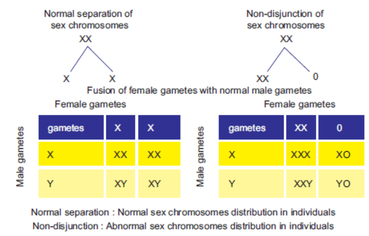
abnormal phenotype in humans.
17.8 SEX LINKAGE
Have you ever wondered that some variations are associated with particular sex of the individual?
For example, the diseases like colourblindness, Haemophila etc., are more common in male as
compared to female. Is mutation sex associated?
There are certain genetic traits, the expression of which depends upon sex of the individual
or inheritance of sex chromosomes. The transmission of such traits (or alleles responsible for
traits) is tied up or linked with the sex chromosomes; inheritance pattern of such genes is known
as sex-linked inheritance. The phenomenon is called as sex linkage.
Sex linkage was first demonstrated in 1910 by Morgan while working with white-eye
(mutant) Drosophila. He carried several breeding analysis with white-eyed male drosophila
and red-eye female drosophila. The F1 flies (male and female) are all red-eyed. On mating
F1 male and female, he found F2 flies with red-eye and white eye in the ratio of 3 : 1 in
accordance with Mendelian monohybrid ratio thereby concluding that white-eye colour is
recessive character. In Mendel’s cross, expression of recessive trait in F2 is not associated
with sex of the individual. Strangely, he observed that all F2 white-eye flies were male justlike their grandfather (Table 17.5).
Table 17.5: Summary of Morgan breeding experiment of cross between white eye male andred eye female
Morgan explained that the recessive allele must have been associated with X chromosome.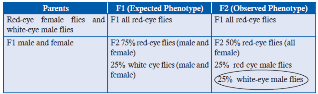
Males have one X chromosome only unlike female which carries two X chromosome so they
are hemizygous (only one allele for a gene) for X-linked genes.
In this experiment, white-eye male parent must have transferred its X chromosome to all F1
females and Y chromosome to all sons. F1 females are heterozygous carrying one normal
X chromosome with normal eye colour i.e., red and other X chromosome with white eye gene
while F1 males had one normal X chromosome from their mother, thus had red-eyed and
Y chromosome from father.
The F2 males obtained their X chromosomes from heterozygous F1 mothers. The half of the F2
males received normal X chromosome while other half received X chromosome with recessiveallele hence developed white-eyes (Figure 17.14).
Figure 17.14: A cross between F1 red-eye male and female representing sex-linked inheritance of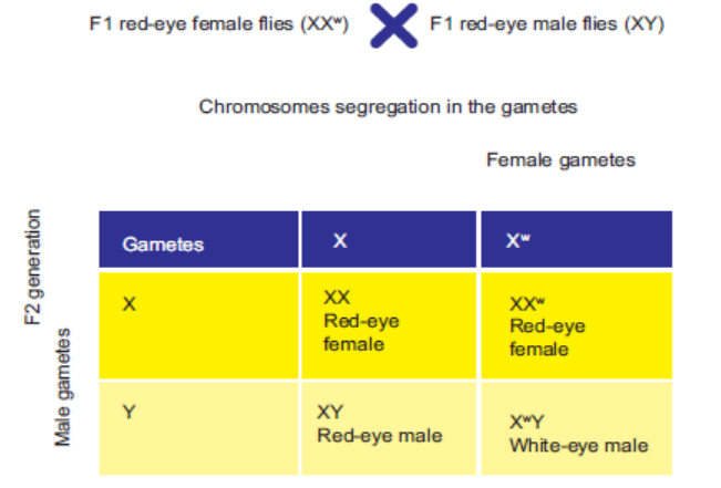
trait to F2 generation
Here, the characters are transmitted from male parent to grandson through carriers (heterozygous)
F1 daughter. Trait is recessive and X-chromosome linked. For this work, Mendel was awardedNobel Prize in 1933
17.8.1 Types of Sex Linkage
There are two types of sex-linked inheritance:
(1) Genes located on X chromosomes demonstrate X-linked inheritance. It is of two types’
X-linked recessive inheritance and X-linked dominant inheritance.
X-linked recessive inheritance, gene causing a mutant phenotype (variant phenotype)
is recessive. It is more common in male. As male has single X chromosome only, they
are pure for X-linked genes (hemizygous). While for female to express X-linked recessive
trait, both the X chromosome should carry recessive allele. Here, criss-cross inheritance
pattern is seen when recessive trait from male are transmitted through their daughter to
their grandson. For example:
• Hemophilia A in human, here individuals lack a clotting factor; thus, a minor cut
may cause excessive bleeding. It follows X-linked recessive inheritance.
X-linked dominant inheritance: Here, the gene causing for a mutant phenotype (variant
phenotype) is dominant. It is less common than X-linked recessive trait. Only a few
X-linked dominant traits have been identified. For example:
• X-linked hypophosphatemia is X-linked dominant trait that can cause bone deformity
in human.
(2) Genes located on Y-chromosomes demonstrate Y-linked inheritance.
Here, genes are transmitted according to inheritance of Y chromosomes. All males receive
Y chromosome from their father, so here Y-linked genes (hence their information) are
directly passed from father to son. This type of inheritance is also known as Holandric
(“wholly male”) inheritance.
It never appears in female. Y chromosome has very few genes. A few traits are Y-linked.
For example:• Hairy ears trait in human in which bristly hairs grow from ear.
ACTIVITY 9
Aim: If a colourblind man (XcY) marries a normal homozygous female (XX), what are the
chances of their progeny being affected with the disease?
Procedure:
• Make gametes from male and female.
• Draw Punnett square showing the random fusion of gametes to get F1 generation.• Observe the results and discuss among yourselves and class teacher.
17.9 GENETIC DISORDER
In humans, how can we study inheritance pattern of different genetic disorders? So here, we have
to study the history of families of person suffering from particular genetic disease by making a
tree or chart. Also, we can predict the chance of transmission of disease to future generation.
Genetic disorders are the diseases which are caused by abnormalities in genetic information
of the organisms. Genetic diseases are quite rare in population and their frequency varies from1 : 1000 to 100,000.
Types of Genetic Disease
Single gene disorder: caused by abnormalities in single gene so that its product becomes either
non-functional or abnormal. For example: haemophilia.
Polygenic genetic disorder: caused by abnormalities in more than one gene. For example:
cancer, diabetes etc.
Chromosomal genetic disorder: caused by change in the structure (deletion, duplication) or
number of chromosomes (chromosomes becomes high or less 47, 45 etc). For example: Turner’ssyndrome, klinefelter’s syndrome.
17.9.1 Single-gene Genetic Disorder
There are two types:
1. Autosomal-linked disorder: in this case, the affected gene is located on the autosomes
and it can be dominant and recessive.
In autosomal dominant, the affected gene allele is dominant in its expression. Only one
allele is sufficient to cause the disease in affected person. Affected person will have 50%
chance to pass it to offspring if he or she marries a normal person and it inherits in everygeneration in affected person’s family.
For example: Huntington’s disease is a neurodegenerative genetic disease that affects
muscle coordination.
In autosomal recessive, affected gene allele is recessive. Both copies of allele must be
recessive for a person to be affected by the disease. An affected person usually has
unaffected parents who each carry a single copy of the mutated gene.
For example: Albinism disease which is characterized by the complete or partial absence
of the pigment in the skin, hairs and eyes.
2. Sex-chromosome linked disorder: here the affected gene is located on the sex chromosome.
Inheritance of this genetic disorder depends upon sex of the affected person.
In X-chromosome dominant, the affected gene is dominant and present on the
X-chromosomes. The female with X-linked disease (XX*) when marries a normal man
(XY), has 50% chance of having an affected offspring.
The male with X-linked disease (X*Y) when marries a normal woman (XX), has all
affected daughters (XX*) but sons will be unaffected.
For example: X-linked hypophosphatemia which causes bone deformity.
In X-chromosome recessive, the affected is recessive and is present on the X-chromosome.
It is more common in males. The male with X-chromosome linked genetic disease will
have normal sons and daughter but pass trait to half of their grandson.
The female with one mutant allele and one normal allele (XX*) will have 50% chance of having
affected sons but 100% chance of unaffected daughters.
For example: Haemophilia
Y-linked genetic disease: It is caused by mutation in the gene located on the Y chromosome.
It only appears in male. The disease is always transferred from father to son.
For example: hairy pinna
17.9.2 Pedigree Analysis: Studies of Inheritance of Genetic Diseases in Humans
The inheritance pattern of different genetic diseases can be studied by pedigree analysis. It
involves collection of information about the family’s history for a particular genetic trait. Thenthe expression of trait is represented into a family tree (also known as Pedigree tree).
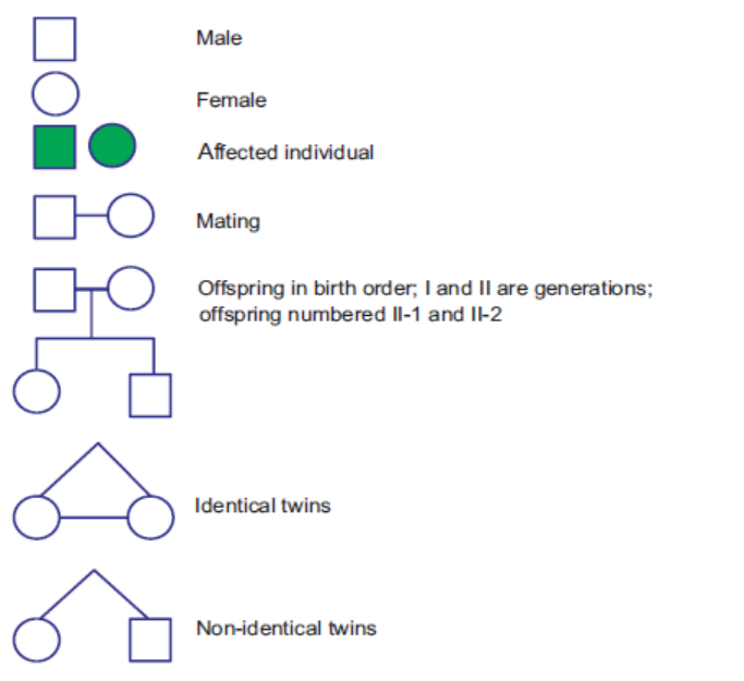
Figure 17.15: The symbols used in human pedigree analysis
In the tree, there are different symbols (Figure 17.15). The females are represented by circles
and males by squares. A horizontal line between circle and squares represent marriage. The
vertical line in middle of horizontal line represents their offsprings. The offsprings are written
from left to right in the order of birth. The solid symbols represent the particular genetic diseasestudies while open symbols represent unaffected individuals.
Hypothetical Pedigree for Different Genetic Traits
Based on the transmission of genetic trait, the different pedigree can be made.
(a) Recessive pedigree: if an affected person has unaffected parents, it indicates that
nature of affected gene is recessive. It can be autosomal or sex-linked.
If it appears randomly in male and female, then it is autosomal (Figure 17.16 (a)).
If affected person is mostly males, then it might be sex-linked recessive genetic trait(Figure 17.16 (b))
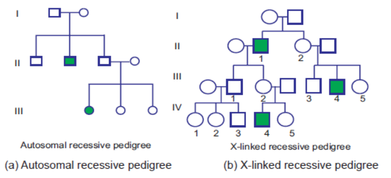
Figure 17.16: Recessive pedigree
(b) Dominant pedigree: if an affected person has affected parents and traits appears
almost in every generation, it indicates that nature of affected gene is dominant. It
can be autosomal or sex-linked.
If it appears randomly in male and female, then it is autosomal (Figure 17.17 (a)).
If trait is passed on every generation but differentially transmitted based on sex of
the affected person, then trait is X-linked dominant (Figure 17.17 (b)).
An affected father will pass the disease to all of his daughters. An affected motherwill pass the trait equally to her sons and daughters.
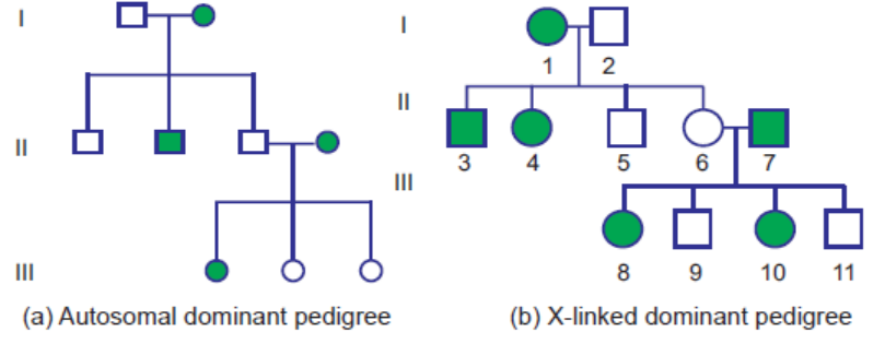
Figure 17.17: Dominant pedigree
17.9.3 Significance of Pedigree Analysis
(a) Pedigree analysis is very useful to find out nature of genetic trait in family.
(b) It is useful to study the inheritance pattern of traits in organisms in which there is
long generation time, especially in humans.
(c) Pedigree analysis is used by genetic counsellors to advise couples about the possibilityof having genetically defective children when a defect runs in their family.
APPLICATION 17.3
1.Choose the correct statement or complete with appropriate terms: (i)
(i)
The following pedigree shows: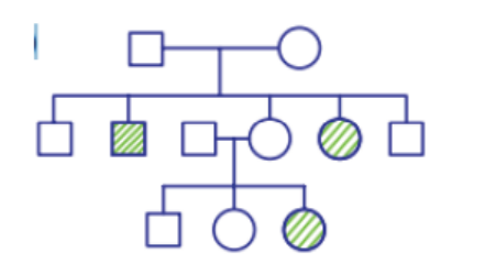
(a) Inheritance of a recessive sex-linked disease like haemophilia.
(b) Inheritance of a sex-linked inborn error of metabolism like phenylketonuria.
(c) Inheritance of a condition like phenylketonuria and autosomal recessive trait.
(d) The pedigree chart is wrong.
(ii) ........... disease affects muscle coordination. It is a neurogenetic digenerative disease.
(iii) In turner’s syndrome ............... .
(iv) ..................... is the failure of homologous chromosomes to seggregate at anaphase.
2.
a) State and explain the second law of Mendel.
b) Manx cats do not have tails. When a Manx cat is mated with a normal long tailed
cat, approximately half the offspring are long tailed and approximately half are
Manx. When two Manx cats are mated, the ratio of phenotypes of the offspring
is 2 Manx to 1 long tailed cat.
i. What does this suggest of the Manx condition in cats?
ii. Show the inheritance of Manx condition when two Manx cats are matedACTIVITY 10

17.10 SUMMARY
• Inheritance/heredity is the phenomenon of transmission of traits from parents to offspring.
• Mendel conducted breeding experiments in garden pea to study inheritance pattern of
several traits. He found that the first generation progeny always exhibited one of the parentaltrait (dominant) while the second generation progeny exhibited both the forms of trait
dominant and recessive in 3 : 1 ratio, popularly known as phenotypic monohybrid ratio.He postulated that there are two factors for each trait, the factors segregate (principle of
segregation) at the time of gamete formation and reunite in zygote.
• Mendel’s factors are now known as gene, two alternate forms are known as alleles. Mendel
also devised a test cross which helps us to differentiate pure dominant form from the
hybrid dominant form. Chi-square test is used to analysis genetic data when observed
data has deviation from the expected value. We can find out whether there is significant
deviation from the expected value or not.
• Mendel carried out dihybrid cross where parent plants differ in contrasting form of two
traits. He found that F1 plants exhibited dominant form of both traits and F2 plants
exhibited four types of phenotypes (two parental types and two new recombinants) in
the dihybrid ratio of 9 : 3 : 3 : 1. He concluded that factors of different traits segregate
and assort independently in the gametes (principle of independent assortment). Mendel’sfactors or genes are found to be located on chromosomes.
• In population there can be more than two alleles (multiple allele), although a particular
individual will have two alleles. Sometimes both the alleles are equally expressed (codominance)
as in ABO blood type. There can be incomplete dominance where one form
is not completely dominant. Also, sometimes allele is lethal which leads to the death of
the individual. Also, one gene may influence the expression of another gene. It leads tovariation in Mendelian ratio.
• The genes located on the same chromosomes, inherit together the phenomenon is called
as linkage. The genes located on the same chromosome occasionally assort independently
via the physical phenomenon known as crossing over. The frequency of crossing over
increases as distance between gene loci on the chromosome increases, so the frequency
of recombinant will vary. It also helps in finding the distance between gene loci ( chromosomemap).
• Sex determination in humans is determined by the presence of Y chromosome. The
“Y” chromosome plays an important role in determining maleness. It has gene testis
determining factor (TDF) which initiates the sequence of events required to differentiateprimordial gonadial tissue into testis.
• The genes located on sex chromosome (X and Y chromosome), demonstrate sex linkage.The inheritance of such traits depends on the sex of the individual.
• The genetic diseases may happen due to changes in genes or chromosomes on which
genes are located. The inheritance of genetic disease caused by genes can be traced
by pedigree analysis which involves collection of information about the expression ofparticular genetic trait in the family’s history.
17.11 GLOSSARY
• Alleles: There are two genes which occur on the same locus in homologous chromosomes
and centrails due expression of a trait or character in an individual.
• Character: It is a feature of an individual for example, stem height, flower colour, seed
shape etc.
• Dominant gene: Gene which always expresses itself in presence of its contrasting allele.
• F1 generation: It is generation of hybrids produced from a cross between two true breeding
potental forms.
• F2 generation: It is a generation of individuals, which arises as a result of inbreeding of
individuals of F1 generation.
• Gene: A unit of heredity.
• Genotype: It is the genetic constitution of an organism.
• Recessive gene: A gene which fails to express itself in presence of its contrasting dominant
allele.
• Phenotype: It is expressed on observable characteristics of an organisms.
• Trait: It is an inherited character and its detectable variant. For example, tall and dwarfvariant of stem height.
END UNIT ASSESSMENT 17
Do all these exercises in your exercise book.I. Choose whether the following statements are True (T) or False (F)
1. Heredity is the transfer of traits from one parent to offsprings.
2. In the monohybrid cross between tall and dwarf plants, second generation plants
were all tall.
3. In codominance, both the alleles of gene are equally expressed.
4. The genes located on same chromosomes are always linked and never assort
independently.
5. In humans, XXY individual will be male.
6. In recessive genetic diseases, affected person can have one affected and another
unaffected homozygous parent.
7. The frequency of crossing over increases as distance between the linked genes
decreases.
8. The sex linkage phenomenon was first observed in Drosophila.
9. In pedigree analysis, the dominant traits are observed every generation.10. Non-disjunction of chromosomes can happen at meiosis and mitosis.
II. Multiple Choice Questions
1. Which of the following reasons are true for Mendel’s success?
(a) He studied many traits at one time
(b) He worked with pure lines
(c) His model organisms were Homo sapiens
(d) He was a chemist
2. Offsprings produced during first progeny are also known as:
(a) F1 generation (b) F2 generation
(c) F3 generation (d) P generation
3. Mendel was successful in his experiments because garden pea:
(a) Produces large number of offsprings
(b) Has long reproduction cycle
(c) Does not show self-pollination
(d) Has difficulty to grow
4. A gamete has ........ number of alleles for a particular gene:
(a) 0 (b) 1
(c) 2 (d) 3
5. The Phenotypic Mendelian monohybrid ratio can be described as:
(a) 3 : 1 (b) 1 : 2 : 1
(c) 2 : 1 b (d) 2 : 2
6. What is Semi-dominance
(a) When both the traits are expressed
(b) When both traits are partially expressed
(c) When both traits are not expressed
(d) When only one trait is expressed
7. A cross between two plants with two pair of contrasting traits is commonly known as:
(a) Monohybrid cross (b) Dihybrid cross
(c) Trihybrid cross (d) Tetra-hybrid cross
8. Test cross is a cross between
(a) F1 hybrid with any of the parent
(b) F1 hybrid with recessive parent
(c) F1 hybrid with other individual similar to parent
(d) None of the above
9. Genetic makeup of Klinefelter syndrome is
(a) 44 autosomes + XXY (b) 44 autosomes + XXX
(c) 44 autosomes + XXXX (d) 44 autosomes + XY
10. Huntington’s disease in humans is the example of:
(a) Autosomal dominant genetic disease
(b) Autosomal recessive genetic disease
(c) X-linked dominant genetic disease
(d) X-linked recessive genetic disease
III. Long Answers Type Questions
1. Explain the terms gene, locus, allele, dominant, recessive, co-dominant, linkage, test
cross, F1 and F2, phenotype, genotype, homozygous and heterozygous.
2. Explain how to conduct a test cross.
3. Explain why monohybrid ratios of 1:2:1 occur.
4. Describe an example of inheritance involving multiple alleles.
5. Explain the effect of lethal genes on phenotype ratios.
6. Give a genetic explanation of Mendelian dihybrid inheritance.
7. Explain the use of test crosses to determine unknown genotypes in studies of dihybrid
inheritance.
8. Explain the significance of recombination.
9. Explain how the sex is determined in humans and the role of sex related Y genes in
determining sex.
10. Describe how the non-disjunction can affect the distribution of sex chromosomes in
gametes and offspring.
11. Explain why the linked genes do not show independent assortment.
12. Explain how crossover values can be used to make a chromosome map.
13. How can genetic studies by supportive for environment protection? State the role of
different genetic aspects to list and cure diseases since ages. What relevance can be
cited to support genetics as an important branch of biology?
14. A man claims to be the father of a child who is blood group AB. The man is blood
group O and the mother of the child is blood group A. State with reasons whether
the man could be the father of the child.
15. Red-green colour blindness is sex-linked recessive condition. The gene of colour
blindness is carried on the X-chromosome. The figure below shows a family tree.Workout the genotypes of the individuals labelled A – E.
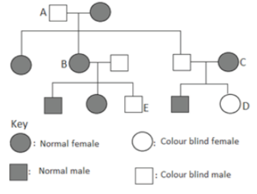
Unit 18 Mutations
Key Unit Competence
To be able to describe the types, causes and effects of mutation in organisms.
LEARNING OBJECTIVES
At the end of this unit, the learner will be able to:
• Define mutation.
• Describe types of mutation and causes of mutations.
• Explain the significance of mutations.
• Make a chart illustrating and summarising different kinds of gene and chromosomal
mutations.
• Distinguish between gene and chromosomal mutation.
• Use a thin clay log composed of different colours to represent different chromosomes.
• Manipulate the clay to show how an inversion can occur.
• Explain that gene mutation occurs by substitution, deletion, inversion and insertion of base
pairs in DNA. Outline how such mutations may affect the phenotype.
• Outline the effects of mutant alleles on the phenotype in the following human conditions:
albinism, sickle cell anaemia, haemophilia and Huntington’s disease.
• Explain the relationship between genes, enzymes and phenotypes with respect to the gene for
tyrosinase involved in the production of melanin.
• Explain how a change in the base sequence of the gene for haemoglobin results in abnormal
haemoglobin and sickle-shaped red blood cells.
• Explain that the environment may affect the phenotype.• Use internet to search simulations of mutations and deduce the findings.
INTRODUCTORY ACTIVITY
After the 1945 Hiroshima and Nagasak atomic bomb, the survivors and their offspring developed
many health problems such as blood cancer (Leukemia), thyroid cancer, polydactyly etc.
How the atomic is related to health problem?Why these health problems of 1945 have effects on children borne many years later?
18.1 MUTATIONS: INTRODUCTION
ACTIVITY 1
After the 1945 Hiroshima and Nagasak atomic bomb, the survivors and their offspring developed
many health problems such as blood cancer (Leukemia), thyroid cancer, polydactyly etc.
How the atomic is related to health problem?Why these health problems of 1945 have effects on children borne many years later?
Nothing was known about mutations before 19th century. Darwin first noticed some sudden
changes in the organisms which he called as “sports”. De Vries (1901) observed several, sudden
changes in Oenothera lamarckiana, and called them mutations. Several sudden mutations were
observed, for example, Ancon sheep (Figure 18.1) is a short legged variety which appearedsuddenly in 1791 and hornless cattle developed from horned cattle in 1889.
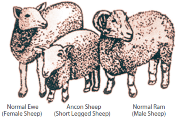
Figure 18.1: Ancon sheep with its normal parents
With the work of G. Mendel, inheritance of characters was established and subsequent work
by several researchers firmly established that DNA constitutes the genetic material of any
individual and it is very faithfully replicated and passed on to the offspring to conserve the
parental characters in all subsequent generations. However, once in a while, the process becomes
erratic for various reasons and alterations are seen in the DNA. Mutation is the sudden change
in the genetic material of an individual.
In this unit, you will study the different types of mutations, how are they produced, their effect
on phenotype and what role environment plays in the production of a phenotype. Finally,
we will discuss as to why a detailed study of mutations is helping us not only to understandevolutionary process better, but also find the ways of treating cancer.
18.2 TYPES OF MUTATIONS
ACTIVITY 2
Have you noticed the variations which run in the families? It means it can pass from one
generation to another generation.Discuss more such examples.
Mutations can broadly be categorized as somatic and germ-line, depending on whether mutation
occurs in a somatic cell or gamete. If mutation occurs in a somatic cell in a particular tissue,
it may not affect the functioning of the tissue as the tissue may be composed of hundreds of
normal cells. However, if a mutation occurs in a gamete, upon fertilization the zygote andhence all cells of offspring will carry the mutation.
Mutations can also be classified as shown in the following chart.
Although all mutations occur in genetic material altering the structure of DNA, consequences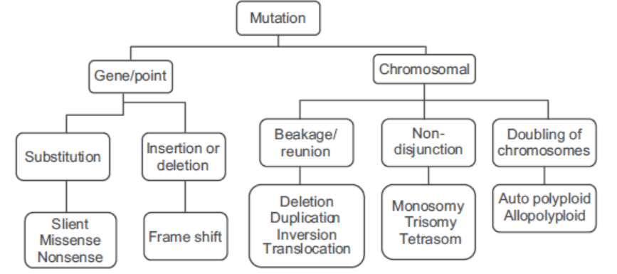
of different types of mutations are very different. Table 18.1 compares the attributes of genemutations and chromosomal mutations.
ACTIVITY 3
Aim: To discuss the types and significance of mutations with the help of computer simulations.
Materials Required:
DNA sequences of different human proteins
Genetic code table
Genetic code table
Types of amino acid table
Notebook
Tetrahedron dice with names of each nucleotide (A, C, G and T).
Procedure:
1. First transcribe the given DNA sequence into mRNA sequence.
2. Translate mRNA sequence into amino acids with the help of genetic code table.
3. Note down the sequence of amino acid for protein.
4. Now randomly decide any nucleotide to change of given DNA sequence. (Choose by your
favourite number, your birthday date..... completely random). Now roll the tetrahedron dice
and look for the nucleotide. Replace it with new nucleotide. Consider it mutation 1.
5. Note down nucleotide and replace the original nucleotide with new nucleotide. There can bedifferent possibility. Make a table as given below and record your observation.
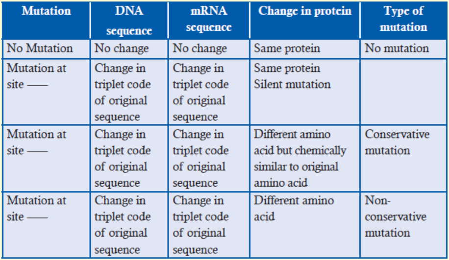
6. Record your observation for mutation 1.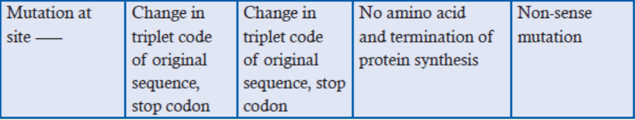
7. Repeat it thrice (for three mutation) from step 4 to 6, randomly choose any nucleotide to
mutate and record your observation.
8. Discuss the types of mutations in the class.
For deletion mutation
9. Now randomly decide any nucleotide to delete in given DNA sequence. Suppose we mutate
nucleotide number 10.
Note down DNA sequence. Consider it deletion mutation.
10. Transcribe the new sequence into mRNA sequence.
11. Translate the mRNA sequence into protein.
12. Note down its effect.
For Insertion mutation
13. Now randomly decide any new nucleotide to insert at random site in given DNA sequence.
Note down DNA sequence. Consider it deletion mutation.
14. Transcribe the new sequence into mRNA sequence.
15. Translate the mRNA sequence into protein.16. Note down its effect.
18.2.1 GENE OR POINT MUTATIONS
Gene or point mutations involve single nucleotides and can occur by one of the following
mechanisms (Figure 18.2):
Substitution is the replacement of one base by another. One purine replaced by another purine
or pyrimidine replaced by another pyrimidine is called transition. However, pyrimidine replacing
purine or purine replacing pyrimidine is called transversion.
Silent mutation, when the triplet codon continues to code for the same amino acid because
genetic code is degenerated, or the amino acid substituted has similar chemical property causing
no change in the function of the protein or the change has occurred in non-coding regionof DNA.
Missense mutation, when substitution of a base produces a codon that causes incorporation
of a different amino acid. If the amino acid added is chemically similar to the original amino
acid, it is called conservative missense mutation but if the amino acid added is chemically
dissimilar, it is called non-conservative missense mutation.
Nonsense mutation, when substitution of a base leads to the formation of a stop codon,
terminating protein synthesises at that point. Polypeptide, thus formed, is incomplete andhence non-functional.
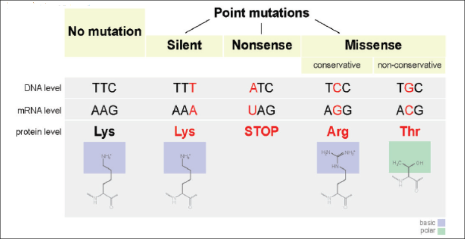
Figure 18.2: Consequences of point mutation
Frame-shift mutation here insertion or deletion of bases alters the reading frame of the genetic
code which is comma-less, causing the different sequences of amino acids being coded from
the point of mutation onwards. This type of mutation is called frame-shift mutation and this
has far reaching consequences on protein function. Effect of this mutation in not confined to
just one amino acid replacing another but the entire sequence of amino acids in the proteingets altered (Figure 18.3) and the protein becomes totally non-functional.
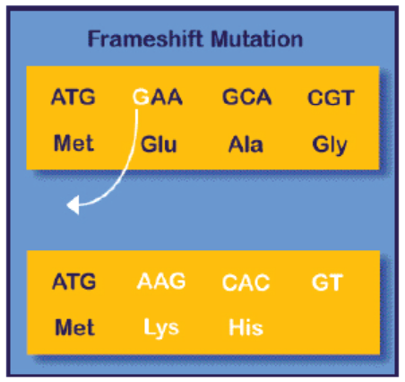
Figure 18.3: Frame-shift mutation
18.2.2 Chromosomal Mutations
ACTIVITY 4
Aim: To manipulate a thin clay log composed of different colours to represent different genes
in order to show how an inversion can occur.
Materials Required:
Thin clay log composed of different colours
Notebook
Colour markers
Scale
Procedure:
1. Just consider the thin log as single chromosomes, assemble five different colours to representas gene such as A, B, C, D and E.
3. Reunion the broken parts in inverse manner.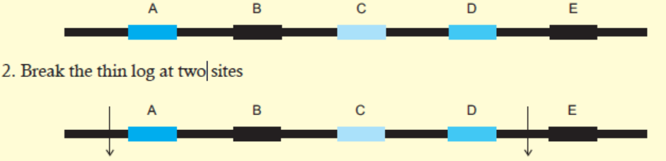
4. Observe the order of genes after reunion of fragments in inverse manner.
5. Compare it with original order of genes.
6. Discuss your observation in the class.7. Enumerate the effect of inversion in phenotype of organism.
Whenever breaks occur in chromosomes, their structures change. If a chromosome or set of
chromosomes shows more than one break followed by reunion, chromosomal rearrangements
are formed. If a break occurs in a chromosome followed by loss of the fragment, it is called
deletion, resulting in loss of genetic information. On the other hand, if a segment occurs more
than once, it results in gain of genetic information and is called duplication. If a chromosome
breaks at two points and fuses again but in reverse order, there is no loss or gain of genetic
information but it alters the sequence of genes in the chromosome and is called inversion.
If breaks occur in non-homologous chromosomes, and the broken fragment from one joins
another non-homologous chromosome, it results in translocation, altering linkage relationships(Figure 18.4).
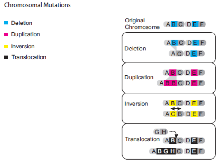
Figure 18.4: Structural changes in chromosome
Whenever the number of chromosomes gets changed, it results in Numerical changes in
chromosomes. It happens due to non-disjunction, failure of homologous chromosomes to
segregate at anaphase, leads to monosomy (2n – 1) and trisomy (2n + 1). Sometimes fertilization
of ovum by two sperms can produce triploidy and fertilization of diploid gametes or doublingof chromosomes can produce tetraploidy
18.2.3 Differences between Gene and Chromosomal Mutations
ACTIVITY 5
Aim: To discuss the differences between gene and chromosomal mutation and one possible
effect on an organism.
Materials Required:
Red colour clay Beads, log made up of clay, notebook
Procedure:
1. In log, make 5 beads of red colour clay and number it 1 to 5.
2. Now consider the beads as gene and log as chromosome.
3. First in the log, try to change any bead. Either break it by deleting some part of clay material
from it or paste some extra clay to it or replace the clay with same and different colour clay.4. Note down your observation according to the following table:
5. Now take the log, do the following changes: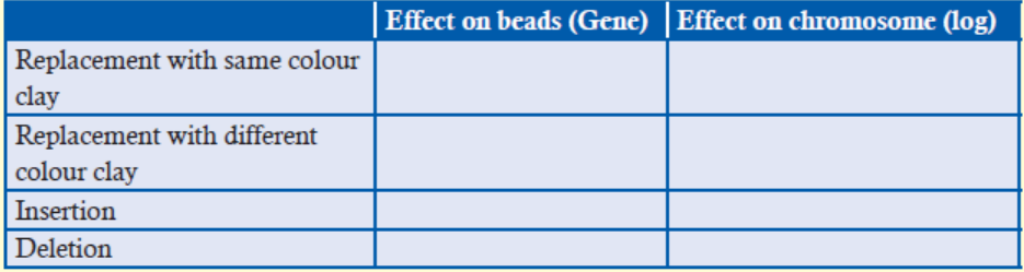
(a) cut the log.
(b) cut the log at two sites and paste it in opposite orientation.
(c) Add the clay beads of same colour.
(d) Add the clay beads of different colours.(e) Note down your observation according to the following table:
6. Discuss the effect of gene and chromosome mutation on the phenotype of the organism with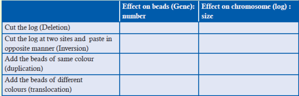
the help of example.
Table 18.1: Gene mutation vs chromosome mutation
18.3 CAUSES OF MUTATIONS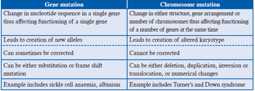
Have you ever wondered for the causes of variation? Sometimes we say its spontaneous or
sometimes we say don’t stand in sunlight for so long, or Nuclear weapons or World War II
has prolonged mutagenic effect on the victims or don’t take particular medicine, it might be
mutagenic. So what could be the causes of mutation? Discuss among your friends.
(i) Random mutations can occur spontaneously due to chance as:
(a) DNA replication errors:
• Normally each base exists in its more stable keto form and is responsible for the
normal Watson-Crick base pairing of T with A and C with G. However, under
certain physiological conditions, rare imino and enol forms (tautomers) of the
bases are present, leading to altered base pairing affinities.
• If by chance, there is looping out of DNA from the template strand, it may be
missed by DNA polymerase, resulting in deletion mutation. Similarly, if additional
untemplated base is synthesised by DNA polymerase, addition mutation results.
(b) Spontaneous chemical changes include depurination and deamination:
• When bond breaks between the base and the deoxyribose sugar, purine is removed
from the DNA, resulting in an apurinic site. Thousands of purines are lost in each
mammalian cell cycle. If these apurinic sites are not repaired, DNA polymerase
will not be able to add a complementary base and will dissociate from the DNA.
• Removal of amino group from a base is called deamination. Deamination of
cytosine produces uracil. As uracil is not a normal base for DNA, repair system
can correct the change. However, if not corrected, adenine will pair up with uracil,
ultimately, causing a change from C-G to T-A, a transition mutation.
DNA also contains small amounts of 5-methylcytosine (5mC) in place of normal
base cytosine. Deamination of 5mC produces thymine, a normal base in DNA and
hence not corrected. Therefore, 5mC results in C-G to T-A transitions.
(ii) Induced mutation happens due to mutagens (agents that induce mutations). It can bephysical mutagens or chemical mutagens (Figure 18.5).
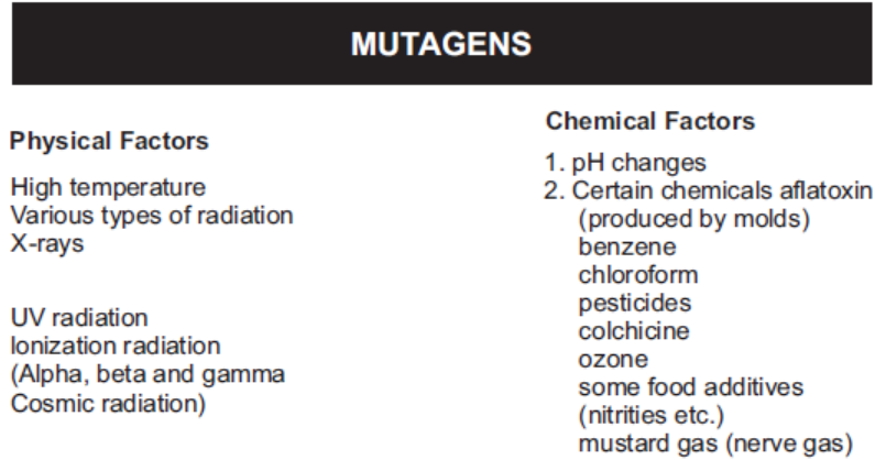
Figure 18.5: Physical and chemical mutagens
(a) Radiation: H. J. Muller was the first to show, in 1927, that mutation can be induced by
X ray treatment. High energy rays collide with atoms and cause the release of electrons,
leaving positively charged-free radicals or ions. These ions, in turn, collide with other
molecules, causing release of further electrons. Thus, as the rays pass through the tissue,
they leave a core of ions along its entire track. This process of ionization can occur
by background radiation or be induced by machine-produced X rays, protons, and
neutrons, as well as by alpha, beta, and gamma rays released by radioactive isotopes of
the elements. Ultraviolet rays, though have less energy, can raise electrons in the outer
orbitals to higher energy level called excitation. When molecules contain atoms either
in ionic state or excited state, they become chemically less stable and thus, more prone
to change, making radiation as powerful mutagens. Energy of X rays can also cause
physical breaks in chromosomes, thus resulting in the loss of chromosome segments or
changes in chromosome structure (deletion, duplication, inversion, translocation).
Mutational effect of ultraviolet (UV) radiation was demonstrated by Edgar Altenburg in
1928. UV rays are strongly absorbed by pyrimidines, especially thymine, leading to the
formation of thymine dimmers. Thymine dimers interfere with DNA replication andDNA repair mechanism, causing mutation in DNA.
The damage by radiation has been shown in Figure 18.6.
Since radiation affects large segments of chromosome at the same time, a number of charactersget altered simultaneously, but molecular details cannot be studied.
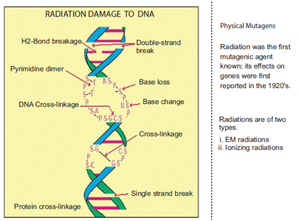
Figure 18.6: Damage to DNA by physical mutagens – radiations
(b) Chemical: C. Auerbach first discovered the mutagenic effects of mustard gas and related
compounds during World War II. Bhopal gas tragedy in India in December 1984 resulted
in the death of 2500–6000 individuals, affecting adversely 200,000 people. Tragedy
occurred with the release of methyl iso cyanate (MIC) in the form of gas and more than
21 chemicals in the MIC storage tank. A number of tests provided evidence that MIC is
capable of inducing chromosomal damage.
People working in nickel and asbestos refineries, rubber industry, leather industry, coal tars,
wood dust are routinely exposed to a number of mutagenic and potentially carcinogenicagents.
Chemical mutagens are of two types:
(a) That are mutagenic to both replicating and non-replicating DNA, e.g., alkylatingagents (Figure 18.7) and nitrous acid (Figure 18.8).
Figure 18.7: Alkylating agents (EMS) change bases such that their Hydrogen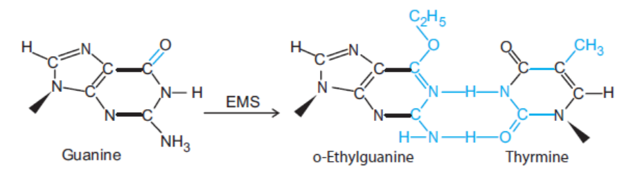
bonding pattern alters. For example, guanine gets changed to ethylguanine and pairswith thymine instead of cytosine.
Mutagenesis by Nitrous Acid (HNO2)
Figure 18.8: Nitrous acid leads to deamination of cytosine and adenine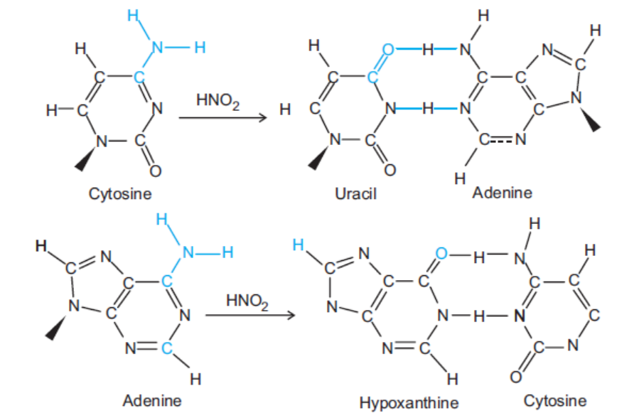
which get change into uracil and hypoxanthine.
(b) That’s mutagenic only to replicating DNA, base analogs and acridine dyes.
Base analogs are structurally very similar to normal bases of nucleic acids and thus, can beincorporated mistakenly in place of the normal ones. However, once incorporated, they alter base
pairing affinities, e.g., 5-bromouracil (Figure 18.9), a thymine analog, undergoes a tautomeric
shift and pairs with guanine. During DNA replication, if 5-bromouracil is present in its enol
form, it will be added opposite guanine in the template strand causing AT to GC transition(Figure 18.10).
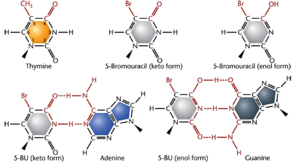
Figure 18.9: 5-bromouracil–Base analog of thymine and its tautomeric forms (keto and enol)
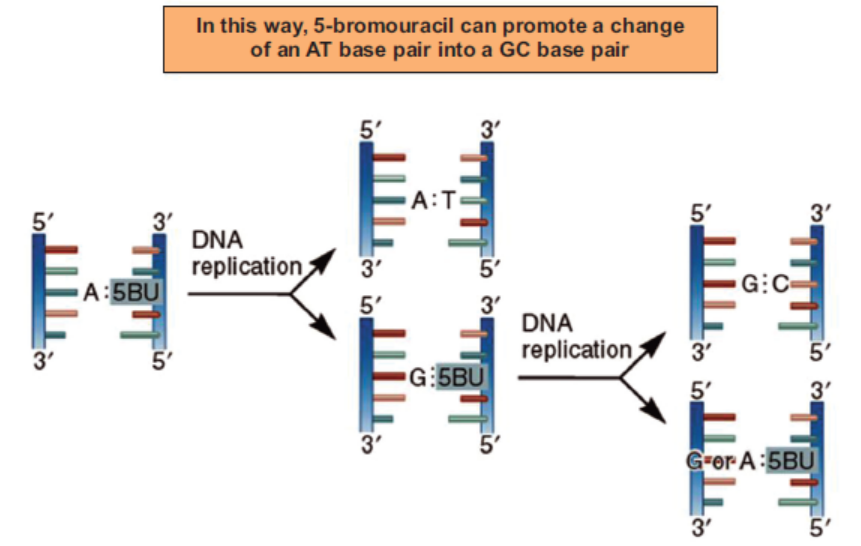
Figure 18.10: 5-bromouracil causes mutation while DNA replication
Acridine dyes intercalate DNA sequences (Figure 18.11) and stabilize DNA looping which
might be missed by DNA polymerase resulting in deletion or addition of genetic material
causing frame shift mutation.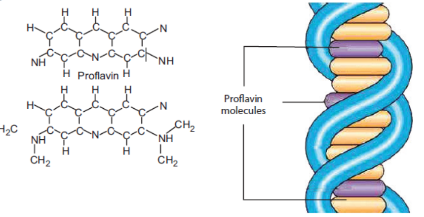
Figure 18.11: Acridine dyes – intercalating agent causing frameshift mutation
APPLICATION 18.1
1.Complete the sentence with appropriate terms:
(i) Substitution of one base by another may lead to ................................... or ..................
mutation.
(ii) Now disjunction of chromosomes can lead to ................. or ................. .
(iii) Induced mutation happens due to ................. and ................. .
(iv) If a chromosomes breaks and fuses again in reverse order, it is called ..............
2.
Suggest why:
a) A mutation in which one nucleotide of a triplet code is altered often makes no difference to
the protein molecule coded by the DNA.
b) The addition or deletion of three nucleotides in the DNA sequence of a gene often has less
effect on the encoded protein than the addition or deletion of a single nucleotide.3. In most people, the first amino acids in their β-globin polypeptide chains are:
The DNA triplet for the sixth amino acid (Glu) in most people is CTT. In some people this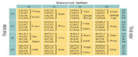
DNA triplet is CAT.
a) What type of mutation is the change from CTT to CAT?
b) Use the genetic code below to identify the amino acid in the β-globin polypeptide chains
of people with this mutation.c) State the consequences for a person of having two copies of the mutated gene.
18.4 EFFECTS OF MUTATIONS ON PHENOTYPES
Spontaneous or induced mutagens cause changes in genotype which influences the phenotype.
The phenotype can be physiological, morphological, biochemical, anatomical etc. So let’s think
of effect of mutation on phenotype.
A gene represents the smallest unit that can code for protein. Gene is made up of DNA consisting
of four nucleotides present in a particular sequence, which, when read in triplet codons, code
for a particular amino acid sequence of a protein. Proteins play a number of important roles in
the body, such as enzymes, hormones, structural etc. Whenever nucleotide sequence in DNA
changes, it can lead to alteration in amino acid sequence affecting the function of the protein.
For example:
Albinism is caused by an autosomal recessive mutation. Tyrosine is converted to DOPA by
the enzyme tyrosinase (Figure 18.12) and DOPA is converted to melanin, the pigment whichgives colour to the skin.
Figure 18.12: A biochemical reaction for the production of skin pigment melanin (left). The mutation in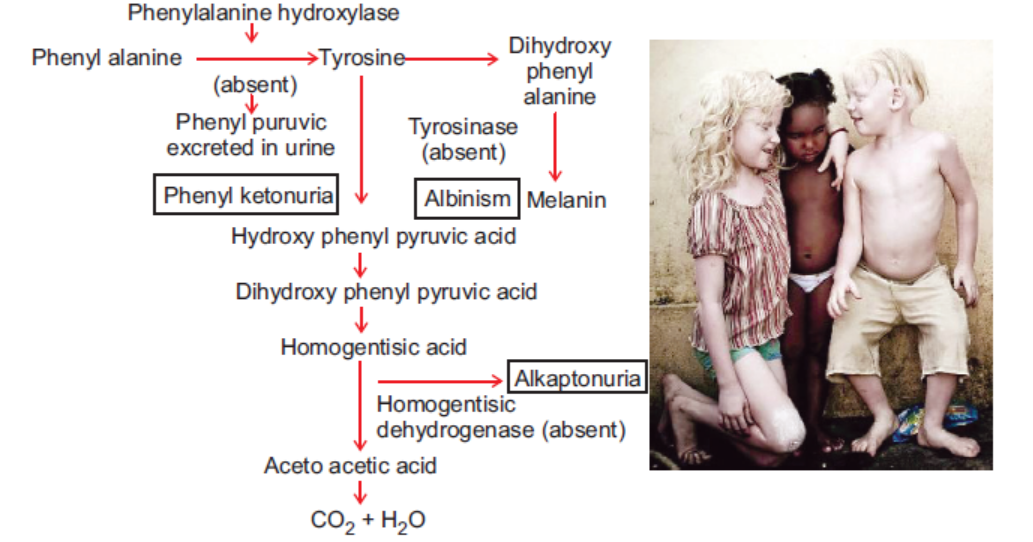
gene responsible for the formation of tyrosinase leads to change in phenotype resulting in albinism (right)where affected person have white skin, white hairs and red eyes.
Melanin absorbs light in the ultraviolet (UV) range and protects the skin against UV radiation
from the sun. If a mutation occurs in the gene responsible for production of tyrosinase, tyrosine
cannot be converted to DOPA and melanin cannot be produced. Therefore, people with sucha mutation have white skin, white hair and red eyes and are very sensitive to light.
ACTIVITY 6
Aim: Use charts and illustrations to show how sickle cell anaemia is inherited and outline the
features of the offspring with or without sickle cell anaemia.
Materials Required:
Charts (pedigree tree) and literature about sickle cell anaemia
Notebook
Procedure:
1. Read charts and literature about sickle cell anaemia.2. For inheritance, try to look for sickle cell anaemic patients’ family history (pedigree tree).
3. Observe the following points:
• Whether trait is seen in every generation or it skips generation Whether the affected
person has unaffected parents or affected parents
• Whether the trait is limited to particular sex or randomly happens in both the sexes.
4. Note down the inheritance pattern (recessive/dominant and autosomal/X-chromosome/
Y-chromosome linked).
5. Discuss your observations in the class.
6. Based on effect of mutation on phenotype, tabulate effect of sickle cell anaemia on theaffected offspring in comparison with offspring without disease.
7. Discuss your observations in the class.
Sickle-cell anaemia is a disease which is caused due to synthesis of abnormal haemoglobin,
the protein present in red blood cells for transporting oxygen. This disease was first studied by
J. Herrick, who found that red blood cells in patients suffering from the disease have the
following characteristics:
• Lose their characteristic disc shape, become sickle-shaped whenever oxygen tension becomes
low (Figure 18.13),
• Rupture very easily thus causing anaemia.
It has also been found that sickle cells don’t easily squeeze through the capillaries (Figure 18.14)
as they are not flexible. This leads to blockage of capillaries, not letting blood flow into tissuesdepriving them of oxygen and ultimately causing tissue damage.
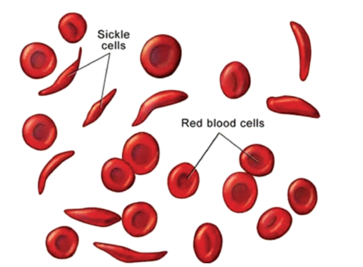
Figure 18.13: Red blood cells become sickle shaped in sickle cell anaemia
Thus, people suffering from sickle cell anaemia can have a number of health problems like
heart failure, pneumonia, paralysis, kidney failure, abdominal pain, etc. Survival rate of such
patients is very low. Disease occurs in a milder form and is known as sickle cell trait whereinpatients show some symptoms in areas of low oxygen tension but do survive.
Figure 18.14: Sickle shaped red blood cells are not able to squeeze through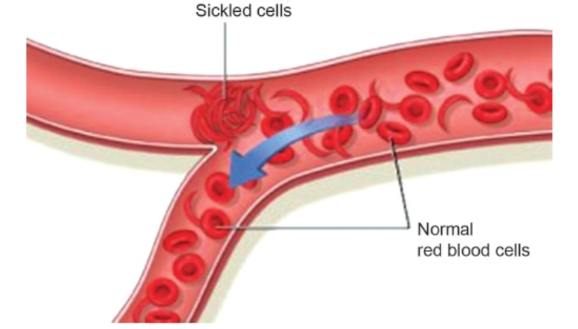
the capillaries leading to blockage in capillaries
Work of L. Pauling showed that normal people made one type of protein haemoglobin, while
people suffering from sickle cell anaemia had another type of haemoglobin and people with
sickle cell trait had 1:1 mixture of two types of haemoglobins. Thus, it was hypothesized that
people with sickle cell trait were heterozygous, carrying two different alleles and making two
types of haemoglobins, Hb-A and Hb-S; normal people were homozygous and making one
type of haemoglobin, Hb-A and people with sickle cell anaemia were homozygous, making
one type of haemoglobin, Hb-S.
Haemoglobin consists of four polypeptide chains, two alpha and two beta, each of which is
associated with a heme group to bind oxygen. V. M. Ingram, on comparing the amino acid
sequence of Hb-A and Hb-S, found that while beta polypeptide of Hb-A had glutamic acid
(with a negative electric charge) at the sixth position, beta polypeptide of Hb-S had valine (with
no electric charge) at the same position. This substitution of amino acids (Figure 18.15) causesthe beta polypeptides to fold up in a different way causing sickling of red blood cells.
Figure 18.15: Substitution of single nucleotide in beta S chain (shown in red) resulting in change in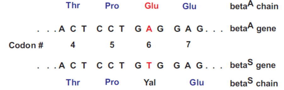
6th amino acid and different type of hemoglobin Hb-S
Haemophilia normally, we find that after minor injury or prick, bleeding automatically stops
after a brief period. Excessive bleeding is prevented by the presence of clotting factors which
work in a cascade-like fashion. However, there are individuals who continue to bleed for long
periods of time even with minor bruises and may also show spontaneous bleeding. This bleeding
disorder is called haemophilia. Haemophilia is of two types: haemophilia A and haemophilia
B. Though both types occur due to a defect in blood clotting process, the two are a result of
mutations in different genes. Haemophilia A (also called classical haemophilia) is more common,
occurring with a frequency of 1 in 4000 males, and is due to deficiency of blood clotting factor
VIII. Haemophilia B (also known as Christmas disease) is less common, occurring with a
frequency of 1 in 20,000 males, and is due to deficiency of blood clotting factor IX. As the geneF8, coding for factor VIII and gene F9, coding for factor IX are present on X chromosome, a
single copy of either of the mutant genes can cause this disorder in males whereas females will
show the disorder only when homozygous for the mutant alleles. This accounts for the higherfrequency of the disorder seen in males in the population.
Huntington disease: All individuals have Huntington gene which codes for huntingtin protein.
Although it is synthesized by all cells, its critical function is seen in the brain where it interacts
with other proteins in the nerve cells. Addition of CAG repeats in Huntington gene in excess
of the normal number increases the number of glutamines in the protein, causing misfolding of
the protein and a mutant phenotype. This protein accumulates in nerve cells, causing extensive
damage. Symptoms include involuntary movements and progressive central nervous system
degeneration. Although Huntington disease is found to be due to autosomal, dominant allele,
expression of this allele begins only by the age of thirty years by which time the parents havealready passed on the gene to their offspring.
18.5 EFFECT OF ENVIRONMENT ON THE EXPRESSION OF PHENOTYPE
Have you ever thought if identical twins get separated at the time of birth and reared separately
in different regions of earth with varied environment, will they be phenotypically identical?
Discuss among your friends and teachers.
It is not always true that phenotype is completely reflected by genotype. Although our phenotype
is governed by our genotype, environment also plays a very important role. It is the close
interaction between genotype and environment that determines the phenotype shown by any
individual. This can be appreciated by the following examples.
(a) A person who has normal genes for making haemoglobin but lacks sufficient iron in
the diet develops anaemia. Phenotype of this individual can be reversed by including
sufficient iron in the diet.
(b) Individual with normal genes can make adequate amounts of thyroid hormone, thyroxine;
yet, in the absence of sufficient dietary iodine, he may develop hypothyroidism.
(c) Surrounding temperature can have an important influence on phenotype of individuals
by affecting kinetic energy of reacting substances. Plant evening primrose shows red
flowers when grown at 23°C and white flowers when grown at 18°C. Siamese cats and
Himalayan rabbits (Figure 18.16) show white fur on all parts except nose, ears and paws,
as the wild type enzyme responsible for pigment production is functional at the lower
temperature present in extremities, but it loses its catalytic activity at the slightly highertemperature found in the rest of the body.
Figure 18.16: Siamese cats and Himalayan rabbits
(d) Individuals who are born with a deficiency of phenylalanine hydroxylase enzyme needed
to convert phenylalanine to tyrosine, concentration of phenylalanine builds up in the
body, especially in the brain causing neurological damage. Phenylalanine free diet allows
them to lead a near normal life, without showing the effects of mutation.
(e) Every day, we are exposed to a large number of chemicals in our environment such as
food additives, colouring agents in food items, textile dyes, cosmetics, pesticides, industrial
compounds and so on. Some of these chemicals have mutagenic effects, and can causegenetic diseases.
18.6 SIGNIFICANCE OF MUTATIONS
Although the term ‘mutation’ was not used by Mendel, he was able to deduce that genetic
characters are controlled by unit factors that exist in pairs in individual organisms and if two
unlike unit factors exist in the same individual, one unit factor is dominant to the other, which
is called recessive. Later studies revealed the true nature of these unit factors which are now
called genes. As seen in the sections above, mutations have played very important roles as
discussed below.
(i) Role in disease: As studied in earlier sections, mutations have been responsible for a
number of diseases such as sickle cell anaemia, haemophilia, Huntington disease, and
albinism. Each individual has tumour suppressor genes and mutation in any of thesegenes can lead to the development of tumours.
(ii) Role in evolution:
(a) Mutations play the most important role of creating new alleles. If there were no
different alleles, all individuals would be homozygous at all loci. Presence of different
alleles in individuals of a population is responsible for the diversity seen in any
population. For example, blood group alleles IA, IB and IO. So mutation can bring
about change in genetic constitution of an organism. So mutations bring genetic
polymorphism in population which may or may not lead to evolution.
(b) Furthermore, it has been observed that certain African countries show higher incidence
of sickle cell allele as compared to other regions. Sickle cell allele somehow confers
protection against malaria and hence occurs with higher frequency in those regions
where malaria is prevalent. Individuals homozygous for sickle cell allele do not survive
as oxygen transport to tissues is affected and individuals homozygous for the normal
allele may suffer from malaria. Hence, mutant allele in this case happens to confer
an advantage in the heterozygous condition.
(c) Mutations have another very important consequence. Rapid rate of mutation in
bacteria and viruses has helped them evolve resistance not only to our immune
system but also to various antibiotics. Thus, treatment against diseases caused by
these microbial organisms is becoming increasingly difficult.
(iii) Role in genetic research: Humans have around 20,000 genes. Although, scientists know
the functions of a number of genes, vast majority of the genes have still not been assigned
function. To study the function of a gene, researchers induce mutations in specific genes
and look for possible effects. Thus, induced mutagenesis is helping us gain insight into
genetics of cell cycle control points and hence the cells becoming cancerous. Cytogenetic
studies have revealed a high degree of correlation between chromosomal rearrangements
and leukaemias.
(iv) Mutations play an important role in agriculture as well by providing diversity of alleles
which may confer stress resistance, yield and regional adaptability.(ii) Role in evolution:
(a) Mutations play the most important role of creating new alleles. If there were no
different alleles, all individuals would be homozygous at all loci. Presence of different
alleles in individuals of a population is responsible for the diversity seen in any
population. For example, blood group alleles IA, IB and IO. So mutation can bring
about change in genetic constitution of an organism. So mutations bring genetic
polymorphism in population which may or may not lead to evolution.
(b) Furthermore, it has been observed that certain African countries show higher incidence
of sickle cell allele as compared to other regions. Sickle cell allele somehow confers
protection against malaria and hence occurs with higher frequency in those regions
where malaria is prevalent. Individuals homozygous for sickle cell allele do not survive
as oxygen transport to tissues is affected and individuals homozygous for the normal
allele may suffer from malaria. Hence, mutant allele in this case happens to confer
an advantage in the heterozygous condition.
(c) Mutations have another very important consequence. Rapid rate of mutation in
bacteria and viruses has helped them evolve resistance not only to our immune
system but also to various antibiotics. Thus, treatment against diseases caused by
these microbial organisms is becoming increasingly difficult.
(iii) Role in genetic research: Humans have around 20,000 genes. Although, scientists know
the functions of a number of genes, vast majority of the genes have still not been assigned
function. To study the function of a gene, researchers induce mutations in specific genes
and look for possible effects. Thus, induced mutagenesis is helping us gain insight into
genetics of cell cycle control points and hence the cells becoming cancerous. Cytogenetic
studies have revealed a high degree of correlation between chromosomal rearrangements
and leukaemias.
(iv) Mutations play an important role in agriculture as well by providing diversity of alleleswhich may confer stress resistance, yield and regional adaptability.
APPLICATION 18.2
1. Complete with appropriate terms:
(i) Tell the cause of the following diseases
(a) Sickle cell anaemia
(b) Huntington disease
(c) Dawn’s syndrome
(d) Albinism
(ii) .................... have helped develop resistance in virus and bacteria.2. Can a mutation in a somatic cell be lethal?
18.7 SUMMARY
I. Mutation, types and their effects on phenotype
• Mutation is any permanent change in the genetic material of an organism. Somatic
mutations are not passed to offspring whereas germinal mutations are heritable.
• Point mutations refer to changes occurring in single nucleotides.
• Substitution of one base by another may lead to silent (no change), missense (altered
codon) or nonsense (stop codon). Change is confined to single codon.
• Altered codons lead to the incorporation of a different amino acid altering the function
of the protein. Examples are seen in diseases like sickle cell anaemia, haemophilia, and
albinism.
• Insertion or deletion of a base changes the frame of reading the genetic code, resulting
in frame shift mutations. Genetic code being comma-less, addition or deletion of one
base alters all subsequent codons, coding for a sequence of amino acids which are totally
different from the original sequence.
• Chromosomal mutations refer to changes in structure or number of chromosomes.
• Chromosome breakages can cause deletion (loss of genetic material), duplication (gain
of genetic material), inversion (fusion of broken segments in opposite orientation)
or translocation (fusion of a part of one chromosome to another, non-homologous
chromosome).
• Non-disjunction of chromosomes can lead to monosomy (2n – 1) or trisomy (2n + 1).
Example of monosomy is Turner’s syndrome and example of trisomy is Down’s
syndrome.
• Addition of haploid sets to the chromosome complement of a cell can change the ploidy
level creating triploid, tetraploid individuals. This can occur whenever two sperms happen
to fertilise a single ovum, creating a triploid situation or a diploid egg is fertilised by a
diploid sperm (all chromosomes fail to disjoin). This has given rise to newer varieties of
plants such as seedless bananas.
II. Causes of mutations
• Chance effects like mistakes during DNA replication, hydrolysis.
• Ionizing radiation for example X rays, protons, neutrons and alpha, beta and gamma rays
emitted by radioactive elements have high energy and can penetrate the tissues causing
damage to DNA in a number of ways depending on the dose.
• Non-ionizing radiation for example UV rays don’t penetrate tissues because of low energy
but are strongly absorbed by nitrogenous bases, esp. thymine, causing the formation of
thymine dimers. This makes them highly mutagenic and excessive exposure to solar
radiation can lead to development of skin cancers.
• Chemical mutagens belong to two classes: one which produces mutations in replicating
and non-replicating DNA for example alkylating agents, nitrous acid and the other
produces mutations only in replicating DNA for example acridine dyes, base analogs.
• These chemicals generally produce mutations by altering base pair affinities.
• A number of common chemicals routinely encountered in environment such as bromine,
pesticides, food additives, etc., may work the same way and thus potentially carcinogenic.
• A number of these chemicals have found in treating cancers.
III. Effects of environment on the phenotype
• Phenotype of any organism is the result of interaction of genotype and environment.
• Interaction may be due to the effect of temperature on enzyme activity.
• Effect of mutant allele can be minimized by modifying the environment for example,
children born with phenylketonuria may lead a near normal life if fed on diet free of
phenylalanine.
IV. Significance of mutation
• Majority of new mutations are generally deleterious, resulting in disease; some mutationsare adaptive for example, sickle cell allele conferring protection against malaria.
18.8 GLOSSARY
• Chromosome mutation: In values charge in chromosome structure due to leakage of
chromosome.
• Mutagens: Any chemical or physical agent that came mutations.
• Mutations: It is a permanent alteration of the nucleotide sequence gene mutation. Involvesof geneme of an organism single nucleotide change.
END UNIT ASSESSMENT 18
Do all these exercises in your exercise book.
I. Choose whether the given statements are True (T) or False (F)
1. Mutations can broadly be categorized as somatic and germ-line, depending on whether
mutation occurs in a somatic cell or gamete.
2. When breaks occur in chromosomes, their structures do not change.
3. Induced mutation happens due to mutagens (agents that induce mutations).
4. Removal of amino group from a base is called deamination.
5. Albinism is caused by an autosomal recessive mutation.
6. Haemophilia A and Haemophilia B are a result of mutations in different genes.
7. There is no interaction between genotype and environment that determines the
phenotype shown by any individual.
8. Sickle cell anaemia is due to a dominant sex-linked allele.
9. Mutagens are DNA sequences which get changed due to radiations and chemicals.10. Mutation has important role in bacterial resistance to antibiotic
II. Multiple Choice Questions
1. A point mutation that changes a codon specifying an amino acid into a stop codon
is called
(a) missense mutation (b) nonsense mutation
(c) frame shift mutation (d) silent mutation
2. Sickle cell anaemia results because of
(a) deletion mutation (b) insertion mutation
(c) substitution mutation (d) chromosomal mutation
3. In mutational event, when adenine is replaced by guanine, it is a case of
(a) transition (b) transcription
(c) transversion (d) frame shift mutation
4. Which of the following is not ionising radiation
(a) X rays (c) UV rays
(b) cosmic rays (d) alpha rays
5. Which of the following chemicals can affect non-replicating DNA?
(a) nitrous acid (b) Acridine dyes
(c) bromouracil (d) None of the above
6. Phenotype of individual depends upon
(a) environment only (b) genotype only
(c) environment and genotype (d) mutagens
7. Which is the type of chromosome structure mutation?
(a) Aneuploidy (b) Polyploidy
(c) Trisomy (d) duplication
8. Which is the example for gene mutation?
(a) Turner syndrome (b) Klinefelter syndrome
(c) Haemophilia (d) Down syndrome
9. Thymine dimers are caused by
(a) X-Rays (b) Gamma rays
(c) alpha or beta particles (d) UV rays
10. A mutation that causes the change in one aminoacid with chemically similar amino
acid is known as
(a) Non-conservative mutation (b) Conservative mutation(c) Non-sense mutation (d) Silent mutation
III. Long Answer Type Questions
1. In your own words, explain what is mutation.
2. Describe the types of mutation and causes of mutations.
3. Explain the significance of mutations.
4. Explain that gene mutation occurs by substitution, deletion, inversion and insertion
of base pairs in DNA. Outline how such mutations may affect the phenotype.
5. Explain that the environment may affect the phenotype.
6. Outline the effects of mutant alleles on the phenotype in the following human
conditions: albinism, sickle cell anaemia, haemophilia and Huntington’s disease.
7. Explain the relationship between genes, enzymes and phenotypes with respect to the
gene for tyrosinase involved in the production of melanin.
8. Explain how a change in the base sequence of the gene for haemoglobin results in
abnormal haemoglobin and sickle-shaped red blood cells.
9. Distinguish between gene and chromosomal mutation.
10. Mutation plays a significant role in evolution. State the role. Also, evolution is the
biggest theory supporting environment sustenance. Guide the role of mutation indeveloping a sustained environment and continue the diversity in organisms.
REFERENCES
•Dr. B. S. Chauhan, Environmental Studies, University Science Press, New Delhi.
•Wilfred D. Stein, W. R. Lieb, Transport and Diffusion Across Cell Membranes, Academic Press Inc. (London) Ltd.
•Daniel L. Kaplan, The Initiation of DNA Replication in Eukaryotes, Springer International Publishing.
•Michael Robert, Michael Jonathan Reiss, Grace Monger; Advanced Biology, Nelson Publishers, UK.
•C. Ladd Prosser, Environmental and Metabolic Animal Physiology, Wiley-Liss Inc.
•Daniel D. Chiras, Human Biology, Jones and Bartlett Publisher, Sudbury.
•Beckett B. S. (1986). Biology A Modern Introduction. Oxford University Press, GCSE Edition.
•Kent, M (2000). Advanced Biology. A New Mainstreamtext for the New Specifications. Oxford University Press, New York.
•McKean DG (1984). Introduction to Biology. Third tropical edition.
•MINEDUC, Advanced Level Biology Curriculum. Kigali: NCDC.
•https://en.wikibooks.org/wiki/A-level_Biology/Biology_Foundation/nuclear_division
•http://www.biology-pages.info/D/Diffusion.html
• http://www.biology-pages.info/D/DNAReplication.html
•http://www.biotopics.co.uk/genes/dna.html
•http://www.biology-pages.info/C/Codons.html
•http://byjus.com/biology/photosynthesis-autotrophic-nutrition/
•http://www.livescience.com/26579-immune-system.html
•http://www.mcwdn.org/body/reproductive.html•http://www.youtube.com/watch? v=8mvkcH 15220
INDEX