Unit 5 Cell and Nuclear Division
Key Unit Competence
To be able to describe the stages of the cell cycle and explain the significance of cell andnuclear division in organisms.
LEARNING OBJECTIVES
At the end of this unit, the learner will be able to:
• Describe the main stages of the cell cycle, including: interphase (growth and DNA replication),
mitosis and cytokinesis.
• Explain what is meant by homologous pairs of chromosomes.
• Explain the meaning of the terms haploid and diploid.
• Describe the process of mitosis and meiosis.
• Outline the significance of mitosis in cell replacement and tissue repair by stem cells.
• State that uncontrolled cell division can result in the formation of a tumour.
• Define meiosis as reduction division in which the chromosome number is halved from
diploid to haploid.
• Explain the need for reduction prior to fertilisation in sexual reproduction.
• Outline the role of meiosis in gametogenesis in humans and in the formation of pollen grain
and embryo sacs in flowering plants.
• Explain how crossing over and random assortment of homologous chromosomes during
meiosis and random fusion of gametes at fertilization leads to genetic variation, including
the expression of rare recessive alleles.
• Interpret data related to time for different cell cycles to identify tissues from which the cells
came.
• Apply knowledge of mitosis to predict which set of cells came from and which part of the
plant and where other cells have come from.
• Make a table showing the phases of the cell cycle mentioning one important event that
occurs at each phase.
• Compare mitosis and meiosis.
• Appreciate the importance of effective cell division.• Show concern to individuals with physical disabilities like Down’s syndrome.
INTRODUCTORY ACTIVITY
A teenage girl got undesired pregnancy when she could not believe. She said: I am 12 years
old. I never got any menstrual period before meeting the boy who pregnant me. Three
month later my stomach was swollen but after pregnancy testing, the doctor told me, you
are pregnant, and your baby is growing well by cell divisions.
1. Can you explain how the baby can grow and yet the cells divide?
2. What types of cell division took place there in this case study? Support youropinion.
5.1 HAPLOID AND DIPLOID CONDITIONS OF THE CELL CYCLE
ACTIVITY 5.1
We know that human beings have 46 chromosomes in total. If it is so, will the number of
chromosomes increase when gametes from father and mother fuse together during fertilizationto form a zygote. Discuss and come out with valid points to support your arguments.
Cell cycle is one of the most important biological events in organisms. Every actively dividing
cell undergoes cell cycle. The most prominent events in cell cycle consist of nuclear division
(karyokinesis) and cytoplasm division (cytokinesis). During cell cycle, chromosomes undergomany changes such as replication, uncoiling, condensation, pairing, cross over, and separation.
A karyotype is a visual representation of the chromosomes within a single cell where thenumber of chromosomes, their arrangement, size and structure can be analysed (Figure 5.1).
The chromosome pairs, one from each parent, which is similar in length, gene position, and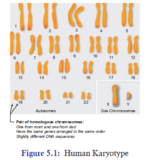
centromere location, is called as a homologous chromosome. In humans, for example, the
23 chromosome pairs are homologous chromosomes. On the contrary, the chromosomes
that contain different genes and do not pair during meiosis are called as non-homologous
chromosomes. For example: In the human karyotype, chromosome 1 and chromosome 2 arenon-homologous.
Chromosomes are of two types: (a) autosomes (b) sex chromosomes. Out of 23 pairs of
chromosomes, one pair of chromosomes is sex chromosome. And chromosomes other than
sex chromosome are called autosomes. The cells that are involved in reproduction are called
gametes (Sperm and eggs). The cells that are not involved in gamete formation are calledsomatic cells (Muscle cells, liver cells).
The cells which contain two complete sets (2n) of chromosomes are called diploid cells. These
cells are formed by the fusion of two haploid gametes, one comes from the female parent and
the other comes from the male parent. For example, all the somatic cells are diploid cells. In
contrast, cells that contain only one complete set (1n) of chromosome are called as haploid
cells. Example is gamete cells in humans. Two separate haploid gametes, one from male parent
(sperm, 1n) and another from female parent (ovum, 1n), come together and fuse to form a
zygote, which is a diploid (2n).
5.2 MITOSIS AND ROLE OF MITOSIS IN LIVING ORGANISMS
Cell cycle is a cyclical event of cell growth, mitosis, and cell division. All somatic cells of an
organism’s body divide by mitosis. The cell cycle basically consists of two phases: MitoticPhase and Interphase.
1. Mitotic (M) Phase: It includes mitosis and cytokinesis. Mitosis is the division of nucleus
while cytokinesis is the division of cytoplasm. As result of mitotic phase, one cell divides
into completely two identical daughter cells.
2. Interphase: A typical cell spends most of its time in interphase. It accounts for about
90% of the whole cell cycle. Interphase involves growth and DNA replication processes.
It is further divided into (Figure 5.2):
(a) G0: It is a resting phase and it can be temporary or permanent.
(b) G1-Phase: It is also called the first growth phase or post mitotic gap phase.
During this phase, the cell grows in size and there is an active synthesis of RNA and
proteins. In this phase, the cell carries out its physiological functions and prepares
the machinery needed for the cell to proceed to the next stage. A large number
of nucleotides, amino acids for histone synthesis and energy rich compounds are
formed. Cell organelles also increase in number. However, it shows no change inits DNA content.
(c) S-Phase: It is also called synthetic phase. In this phase, DNA molecule of each
chromosome replicates by the synthesis of a new DNA on the template of the
existing DNA. Thus, the DNA content doubles and duplicate set of genes are
formed. Along with DNAs, chromatin fibres also replicate. As chromatin fibres
are elongated chromosomes, each chromosome comes to have two chromatin
threads or sister chromatids which remain attached at a common point called
centromere. The cell thus retains the original diploid (2n) chromosome number but
now has a duplicate set of genes. S-phase is also called invisible phase of mitosis,
since in this phase chromosome prepares themselves for equitable distribution
later on. The centriole also divides into two centriole pairs in the cells containing
the same.
(d) G2-phase: It is also called the second growth phase or premitotic gap phase. The
synthesis of RNA and protein continues in this phase and the cell prepares itself to
go into the mitotic phase. The phase produces macromolecules for multiplication
of cell organelles, spindle formation and cell growth. During G2-phase, a cell
contains double DNA content, i.e., 1C to 2C for haploid cells and 2C to 4C for
diploid cells.
Data Related to Time for Different Cell Cycles
The time consumed by each stage in the cell cycle varies from organism to organism. In human
beings, one round of cell cycle takes 24 hours. The relative time division is (Figure 5.2):
(a) G1 phase takes about 5-6 hours.
(b) S phase takes about 10-12 hours.
(c) G2 phase takes about 4-6 hours.(d) M phase takes about less than one hour.
G1 phase is of the most variable length in the cell cycle. Normally, S phase is the longest
phase. Cells such as muscle and nerve cells remain in the resting state permanently. On the
contrary, cells such as liver cells can resume G1 phase in response to growth factor released
during injury.
5.2.1 The Process of Mitosis
Mitosis is one of the phases of cell cycle, which normally last only about less than an hour.
It is a process where a single cell divides into two identical daughter cells. And it is normally
followed by cytokinesis but not always. The process of mitosis is basically divided into 5 phases:
1. Prophase 2. Prometaphase 3. Metaphase 4. Anaphase 5. Telophase
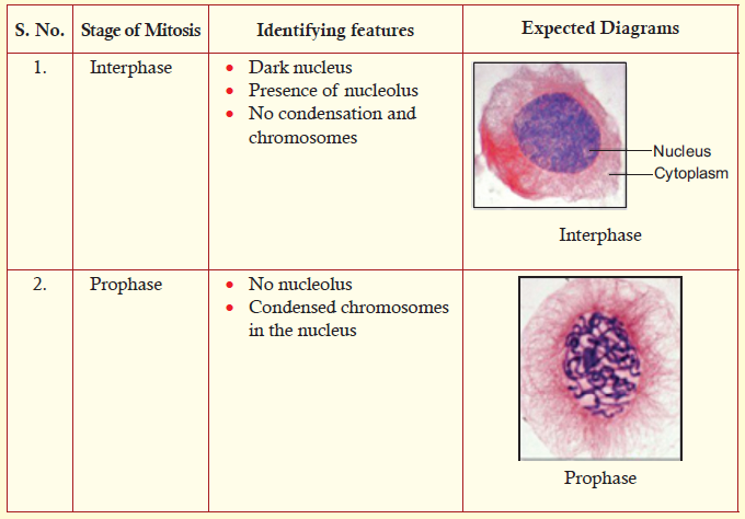
1. Prophase
• The chromosome fibres become more tightly coiled.
• The chromatids condense into discrete chromosome.
• Each chromosome can be seen to consist of two sister chromatids.
• Nucleolus shrinks and eventually disappears in most species.
• Two pairs of centrioles are seen. The mitotic spindle assembles outside the nucleus.
• The radial arrays of shorter microtubules are called as asters.
• In most animal cells, the centrioles are the focal points for spindle assembly. Higher plantsdo not have centrioles, though they do have a mitotic spindle.
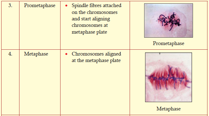
2. Prometaphase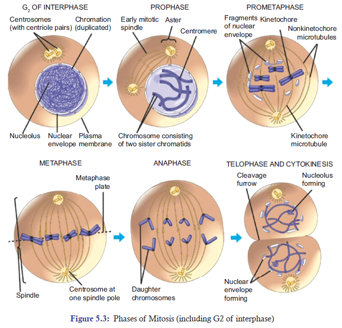
• The nuclear envelope breaks down at the end of prophase.
• The developing spindle now enters or invades the former nuclear area.
• The chromosomes have even become more condense.
• Kinetochores, a specialized multiprotein complex, bind to each centromere.
• Kinetochore microtubules extend from both the poles and bind to kinetochores of chromatids.
• Non-kinetochore microtubules originate from the two opposite poles and enter into thenuclear area where they overlap in the middle of the spindle.
3. Metaphase
• The centrosomes are now at opposite poles of the cell.
• The kinetochore microtubules from the two poles orient the chromosomes in such a way
that their centromeres become aligned at the metaphase plate, an imaginary plane that isequidistant between the two poles of the spindle apparatus.
4. Anaphase
• Once the centromeres are attached to the two sister chromatids. They start separating and
move towards the opposite poles. Consequently, the joined centromeres of sister chromatids
separate and give rise to two daughter chromosomes.
• The separation continues until the two poles of the cell have equivalent and completechromosomes.
5. Telophase
• The two sets of daughter chromosomes are assembled into two groups at opposite ends
of the cell.
• The chromosomes begin to uncoil and assume the elongated state characteristic of interphase.
• A nuclear envelope starts forming around each group of chromosomes.
• The spindle microtubules disappear; and the nucleolus or nucleoli reform.
• At this point, nuclear division is complete and the cell now has two identical nuclei.Cytokinesis
ACTIVITY 5.2
Aim: To observe permanent slides of plant root tip and animal cheek cells and outline the
differences between the plant cells and animal cells.
Materials Required:
1. Permanent (prepared) slides of animal cheek cells and plant root tips for mitosis.
2. Compound microscope.
Procedure:
1. Take the permanent slides.
2. Try to observe the stages of mitosis and meiosis from the given slides.
3. Draw a well-labelled diagram of the structures you have observed.
4. Outline the differences between how plant cells and animal cells divide.5. Record your observations in your notebook.
Discussion:
1. Firstly, discuss your result.2. Secondly, if there is any doubt, ask your biology teachers.
Cytokinesis is a division of cytoplasm. It compartmentalizes the two new nuclei into separate
daughter cells, completing mitosis and cell division (Figure 5.3). In animal cells, cytokinesis is
characterized by a constriction in the middle of the cell. The constriction continues until two
daughter cells are produced. In plant cells, a new cell membrane and cell wall are assembled
between the two new nuclei to form a cell plate. Cell wall material coats each side of the plate,and the result—two progeny cells.
Table 5.1: A table showing phases of cell cycle with one important event at each phase
ACTIVITY 5.3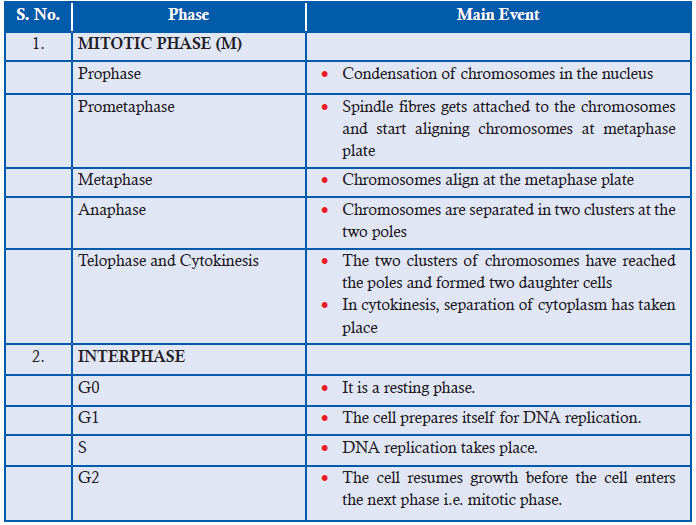
Aim: To Investigate how long onion root tip cells spend in each phase of the cell cycle and
present the findings in table form showing the stages of mitosis.
Materials Required:
1. Permanent slides of Onion root tips
2. Compound Microscopes
Procedure:
1. Fix the permanent slides of Onion root tips on the compound microscope.
2. Adjust the lens (10x, 40x etc.) to have a clear view of mitotically dividing onion root cells.3. Identify the mitotic stage of each cell by following the criteria given in the table below:
4. Identify and note down the number of cells at their specific stages. Note it down in your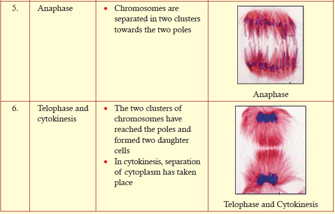
exercise book by copying the following table.
5. Onion takes a total time period of 12 hours (720 minutes) to complete mitosis.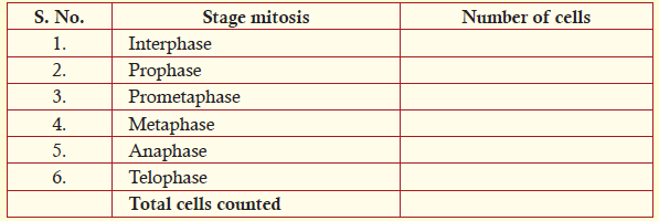
Now with this information, find out the time period spent by each cell at each stage(i.e. interphase, prophase etc.) by using the given formula:
For example: Say, 500 cells were counted at interphase stage. And the total number of cells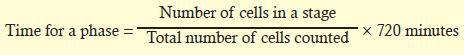
counted was 1000. Then by applying the formula you can calculate how much time was
spent by cells at interphase in 12 hours mitosis cycle.
Time for interphase = 500/1000 × 720 minutes = 360 minutes
6. Calculate the time period spent by cells at different stages of mitosis. Note it down by copying
the following table in your exercise book. Then analyse the maximum and minimum timespend by cells in all the stages of mitosis.
Notes: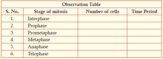
1. Normally, interphase takes the longest time period in the cell cycle.2. Compare your observations with your class mates.
5.2.2 Significance of Mitosis in Living Organisms
In the early development of an organism, the embryonic cells rapidly proliferate and differentiate
into specialized cells of adult tissues and organs. As cells differentiate from time to time, their
rate of proliferation usually decreases. As a result, most cells in adult animals are arrested at
the Go stage. Some cells at this phase may resume the cell cycle and proliferate when they
receive certain signals.
Some of the differentiated cells enter the Go resting phase and wait for the signal to resume
the cell cycle to repair injured tissue. There are numerous examples such as skin fibroblast,
endothelial cells, smooth muscle cells, and liver cells. Skin fibroblasts upon receiving growth
factor, they start secreting collagen and help in repairing cuts or wounds.
Most of the fully differentiated cells no longer possess the capability of cell division. Therefore,
they can be replaced by stem cells. Stem cells are undifferentiated biological cells that can
differentiate into specialized cells and can divide (through mitosis) to produce more stem cells.
The prominent role of stem cells can be seen in—blood cells (hematopoietic system), epithelial
cells of the skin, and epithelial cells lining the digestive tract (Figure 5.4). All of these cells
have short life spans, and they must be replaced continually by continual cell proliferation inadult animals.
The life span of blood cells ranges from less than one day to a few months. All of these cells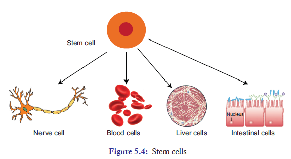
are derived from the same population of hematopoietic stem cells. In fact, there are more than
100 billion blood cells that are lost every day in humans. If there are no stem cells to replace the
loss of these cells, human beings will not be able to survive. Hence, these cells are continuallybeing replaced by cells produced from hematopoietic stem cells in the bone marrow.
The unrestrained, uncontrolled growth of cells in human beings results into a disease called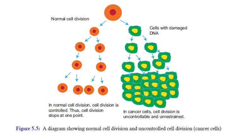
cancer (Figure 5.5). Cancer essentially is a disease of cell division. In other words, cancer occursdue to failure in controlling cell division.
Tumours
Cancer cells can be dangerous when it starts behaving abnormally in the body. The main problem
arises when a single cell in a tissue undergoes a process called transformation. It is a process
where a normal cell is converted into a cancer cell. Normally, the body’s immune system will
recognize the transformed cell as a foreign invading cell and, thus, destroys it. However, if the
transformed cell evades or escapes the destruction, it may proliferate and form a tumour—
a mass of abnormal cells. Tumours can be discussed in three subheadings:
(a) Benign tumour: This is a lump of the abnormal cells that remains at the original site.
Most benign tumours do not cause serious problems and can be completely removed by
surgery.
(b) Malignant tumour: These are abnormal cells that have become invasive enough to impair
with the functions of one or more organs. An individual with a malignant tumour is said
to have cancer.
(c) Metastasis: A few tumour cells may separate from the original tumour, enter blood vessels
and lymph vessels, and travel to other parts of the body. In the other parts of the body,
they may proliferate and form a new tumour. This spread of cancer cells to locations
distant from their original site is called metastasisAPPLICATION 5.1
1). Complete with appropriate terms:
(a). In ................. phase of the interphase DNA, replication occurs.
(b). Karyokinesis is the division of ................. .
(c). ................. is the resting phase of the cell cycle.
(d). It approximately takes ................. hours for one round of cell cycle in human beings.
(e). Cancer is the ................. growth of cells.2). The diagrams below show four animal cells at different stages of mitosis.
a) Name the structures A, B, C and D.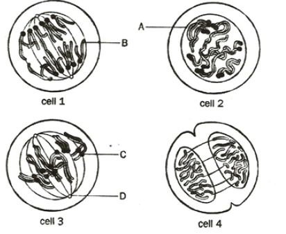
b) . (i) Name the stages of division shown by cells 1 and 3.
(ii) Use the numbers of each cell to arrange the stages in the correct sequence of
mitosis.3) . How does mitosis maintain genetic stability in an organism?
5.3 MEIOSIS AND ITS ROLE IN LIVING ORGANISMS
ACTIVITY 5.4
Through micrographs try to observe different stages of Meisois. State the arrangement ofchromosomes at every stage. Discuss your observations.
5.3.1 The Process of Meiosis
Meiosis is a reduction division where the number of chromosomes are reduced to half from diploid
parent cell to haploid daughter cells. It is divided into two stages: Meiosis I and Meiosis II. The
process of Meiosis alternates with an interphase, which is subdivided into G1, S, and G2 phases
(Figure 5.2). Meiosis I is further subdivided into prophase I, metaphase I, anaphase I, and
telophase I. In the same way, Meiosis II is also subdivided into prophase II, metaphase II,anaphase II, and telophase II.
Meiosis I
Prophase I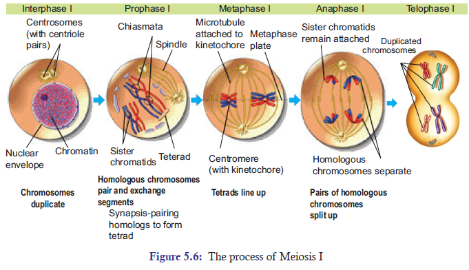
The DNA in prophase I coils tighter, and individual chromosomes first become visible under
the light microscope as a matrix of fine threads. Since the DNA has already replicated during
the S phase of interphase prior to meiosis I, each of the chromosomes actually consists of two
sister chromatids joined together at their centromeres.
Pairing or synapsis
In prophase I, homologous chromosomes become closely associated in synapsis. Synapsis
includes the formation of an elaborate structure called the synaptonemal complex, consisting
of homologous chromosomes paired closely along a lattice or zipper-like structure of proteins
between them (Figure 5.7). The components of synaptonemal complex include a meiosis-specific
form of cohesin, that helps the two homologous chromosomes to be closely associated along
their length.
Crossing over or recombination
Crossing over in meiosis I allows the homologous chromosomes to exchange their chromosomal
material. During this, the crossing over between sister chromatids is suppressed. Reciprocal
crossing over occurs only between non-sister chromatids and is controlled in such a way that
each chromosome arm usually has one or a few crossovers per meiosis. In human beings, thecrossovers are typically two to three in number.
Once the crossing over process is complete, the synaptonemal complex breaks down, and the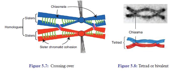
homologous chromosomes become less tightly associated. But the homologous chromosomes
remain attached at one particular point called chiasmata (chiasma-singular) (Figures 5.7
and 5.8). At this point, there are four chromatids of the two homologous chromosomes.
Two homologous chromosomes consist of two sister chromatids each. This structure of four
chromatids of the two homologous chromosomes attached at chiasmata is called as tetrad or
bivalent (Figure 5.8).
Some of the other events that occur along with synapsis are:
1. The nuclear envelope breaks down.
2. Two pairs of centrosomes migrate to opposite poles.3. Spindle fibres formation occurs.
Metaphase I
During metaphase I, the chiasmata have moved down on the paired homologous chromosomes
towards the ends. At this point, chiasmata are now called as terminal chiasmata. The terminal
chiasmata hold the homologous chromosomes together so that the homologous chromosomes
are now aligned at the equator of the cell. The kinetochores microtubules from the opposite
poles become attached to the kinetochore of homologous chromosomes.
The attachment of kinetochore microtubules at the monopolar centromere of each homologue
creates a tension on the homologous chromosomes, which are joined by sister chromatidcohesin at chiasmata.
Anaphase I
During anaphase, sister chromatid cohesion is released and the homologous chromosomes are
pulled apart to the opposite poles, but not the sister chromatids. Now when the spindle fibres
have fully contracted, each pole has a complete haploid set of chromosomes consisting of one
member of each homologous pair.
Telophase I
In telophase I, the chromosomes are segregated into two clusters at the two opposite poles.
Then the nuclear membrane reforms around each daughter nucleus. At the two poles, each
chromosome has sister chromatids attached to its centromere. And the interesting thing is that
the sister chromatids are no longer identical because of the crossing over that had taken place inprophase I.
Cytokinesis
Cytokinesis is the process of dividing the cytoplasm and its content into two equal cells
(Figure 5.7). Right after telophase I, cytokinesis may or may not occur. Meiosis I is followedby meiosis II, which occurs after an interval of variable length.
Meiosis II
Meiosis II is like mitotic division, which results into division of two equal cells without DNA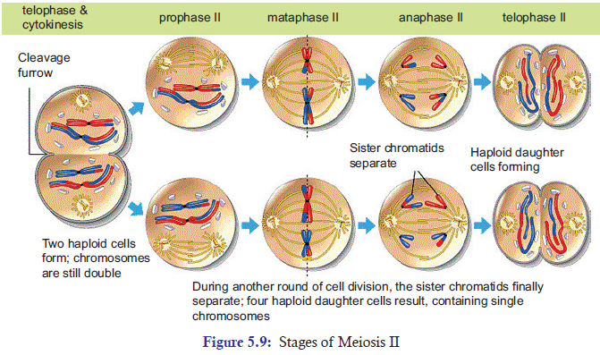
replication. Normally, the gap between meiosis I and meiosis II is interrupted by interphase.
But the interphase is very brief and it does not contain the S phase. Like mitosis cell division,
meiosis II is also subdivided into subphases. They are: (a) Prophase II (b) Metaphase II(c) Anaphase II (d) Telophase II (Figure 5.9).
Prophase II
Prophase II is brief. In prophase II, nuclear envelope breaks down and formation of new spindlefibres takes place.
Metaphase II
In metaphase II, the kinetochore microtubules extend themselves from the two poles and bind
to kinetochores of each sister chromatid. These kinetochore microtubules start pulling the
sister chromatids toward the two opposite poles. As a result, the sister chromatids are alignedat the metaphase plate.
Anaphase II
In anaphase II, as the spindle fibers contract, the cohesion complex joining the centromeres of
sister chromatids is destroyed or cleaved. As a result, the centromeres are split and the sisterchromatids are pulled towards the two opposite poles.
Telophase II
In telophase, the nuclear envelope re-forms around the four set of haploid daughter chromosomes.
Then cytokinesis follows resulting into complete four set of haploid daughter cells. These haploid
daughter cells may follow different fate depending upon the organisms. In animals, these haploid
daughter cells develop directly into gametes i.e. sperms and eggs. In plants, fungi, and manyprotists, they may divide mitotically to produce greater number of gametes.
5.3.2 Meiosis is a Reductional Division
Meiosis reduces the number of chromosomes sets from parental diploid chromosomes (2n) to
four haploid (1n) daughter cells. That is why meiosis is called a reduction division. In other
words, meiosis starts with diploid cell but ends with four haploid cells.
Simple explanation of Meiosis taking place in human beings is given below:
Parent cell = 46 chromosomes (Diploid)
Meiosis I = 2 cells (each chromosome with sister chromatids) = 46 × 2 = 92 chromosomes.Meiosis II = 4 haploid cells with unreplicated chromosomes = 92/4 = 23 chromosomes (Haploid)
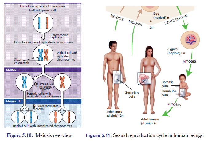
5.3.3 Significance of Meiosis
Cells Undergo Reduction Division Prior to Sexual Reproduction
Generally, a cycle of reproduction consists of meiosis and fertilization. Before sexual reproduction
occurs, gametes undergo meiosis and produce haploid cells. Thus during sexual reproduction,
one haploid (1n) gamete comes from the paternal side and another haploid (1n) gamete comes
from the maternal side; then, they both fuse to form a zygote, which is diploid (2n). The fusion
of gametes to form zygote or new cell is called as fertilization or syngamy (Figure 5.10).
If meiosis does not occur before sexual reproduction, the chromosome number would double
up with each fertilization. And after few generations, the number of chromosomes in each
cell would become impossibly large. For example in humans, in just 10 generations, the 46chromosomes would increase to about 47104 (46 × 210).
Role and Significance of Meiosis in Producing Gametes
Gametogenesis is a biological process by which diploid cells undergo cell division and
differentiation to form mature haploid gametes. It occurs through meiosis. In humans, the
male gamete (sperm) is produced by a process called spermatogenesis and the female gamete
(egg) is produced by a process called oogenesis through meiotic division.
Here gamete function takes place soon after meiosis but in plants it happens after gametophyte
formation sexual reproduction of plants starts with spore formation. Sporophyte is a diploids
generation of flowering plant where haploid spores are produced by meiosis which in turns
undergoes mitosis to form multi-celled haploid gametophytes. These haploid gametophyte
differentiate to produce gametes—sperm and egg cells. Similarly, embryo sac is formed by
reduction division. Each of the cells of embryo sac is haploid. Two of the nuclei fuse to producediploid nucleus.
The Role of Meiosis in Reproduction of Plants
Generally, plants reproducing sexually have life cycle consisting of two phases (Figure 5.11):
(a) A multicellular gametophyte or haploid stage: It is a haploid stage with n chromosomes.
It alternates with a multicellular sporophyte stage.
(b) A multicellular sporophyte or diploid stage: It is a diploid stage with 2n chromosomes,
made up of n pairs. A mature sporophyte produces spores by meiosis, a process which
reduces the number of chromosomes from 2n to 1n.
Alternation of generations (also known as mutagenesis) refers to the occurrence in the plant life
cycle of both a multicellular diploid organism (sporophyte) and a multicellular haploid organism
(gametophyte), each giving rise to the other. This is in contrast to animals, in which the only
multicellular phase is the diploid organism (such as the human man or woman), whereas the
haploid phase is a single egg or sperm cell.
In bryophytes (mosses and liverworts), the dominant generation is haploid, so that the
gametophyte comprises the main plant. On the contrary, in tracheophytes (vascular plants),the diploid generation is dominant and the sporophyte comprises the main plant.
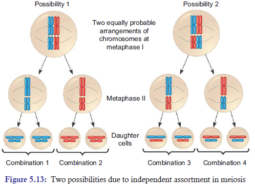
Independent Assortment of Chromosomes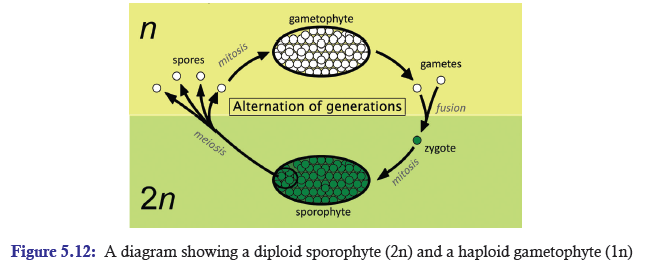
Specifically at metaphase I, each homologous pair of chromosomes positioned independently of
the other pairs. As a result, each homologous pair sorts out its maternal and paternal homologue
into daughter cells independently of every other pair. This act of separating homologous pairs
independently is called independent assortment. The random orientation of homologous pairs
of chromosomes due to independent assortment in meiosis I (metaphase) increases geneticvariation in organisms.
Crossing Over and Random Fertilization
During crossing over, DNA segments of the two parents-paternal and maternal are combined
into a single chromosome producing recombinant chromosomes, which are non-identical
with their sister chromatids. In humans, an average of one to three crossing over events occurs
per chromosome pair, depending on the position of their centromeres and on the size of the
chromosome. Thus, crossing over is an important event of meiosis that brings genetic variationin sexual life cycles.
Besides independent assortment and crossing over, the random fertilization during sexual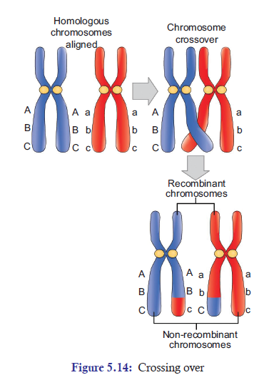
reproduction also increases genetic variation in organisms. During random fertilization, the
male gamete and female gamete fuse to form zygote. The most interesting thing is that this
zygote has the possibility of about 70 trillion diploid combinations. The number 70 trillion comes
from possible combinations of male and female gametes which are 223 × 223 = 70 trillions. The
possibility of this enormous number of combinations makes each and everyone of us uniquephysically and genetically.
Non-disjunction of Chromosomes
Proper separation of chromosomes during meiosis is essential for the normal growth in humans.
Any set of chromosomes that do not separate properly during meiosis results in improper
separation of chromosomes or non-disjunction, which is a serious issue in human genetics.
Non-disjunction is a condition in which the homologues or sister chromatids fail to separate
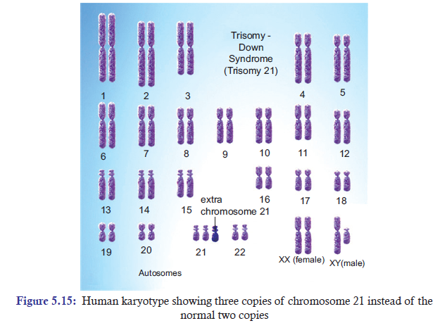
properly during meiosis. It can lead to the gain or loss of chromosome, a condition called asaneuploidy. Example: Down syndrome is an autosomal trisomy. It is also called as trisomy
21, where non-disjunction results in an embryo with three copies of chromosome 21 instead of
the usual two copies of chromosome 21 (Figure 5.15). It was first discovered by John LangdonDown. The chance of occurrence is one infant in every 800 live births.
The most common symptoms are: They are short. They may also have protruding, furrowed
tongues, which causes the mouth to remain partially open. They are mentally retarded. They
have a prominent epicanthic fold in the corner of each eye; and typical flat face and roundhead. Usually, there is a wide gap between the first and the second digits on their feet.
The origin of trisomics condition is through non-disjunction of chromosome 21 during
meiosis. Failure of paired homologues to separate during either anaphase I or II may lead to
gametes with 23 + 1 chromosome composition instead of the normal 23 gamete chromosome
composition. Therefore, instead of 46 normal chromosomes, Down syndrome patient willhave 47 chromosomes with three copies of chromosome 21 instead of the normal 2 copies.
APPLICATION 5.2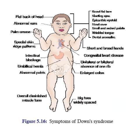
1. Complete with appropriate terms:
(a) Independent assortment of chromosomes takes place at .............. stage of Meiosis I.
(b) Chiasmata formation occurs at .............. .
(c) Meiosis is also known as .............. .
(d) Crossing over and random fertilization brings .............. in life cycles.
(e) Trisomy of chromosome number two leads to .............. syndrome.
2. The drawing below has been made from a photograph showing a cell undergoing mitosis.
a) In which stage of mitosis is the cell shown in this drawing?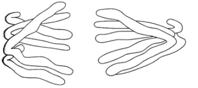
b) Describe one piece of evidence, visible in the drawing, which could be used to confirm
that this cell is not in the first division of meiosis.
3. An organism has two pair of chromosomes (i.e., chromosome number = 4). Diagrammaticallyrepresent the chromosomal arrangement during different phases of meiosis-II.
5.4 COMPARISON OF MITOSIS AND MEIOSIS
ACTIVITY 5.5
Cells divided both by mitosis and meiosis contain nucleus which is a genetic material. Can cells
divided by mitosis act as carriers of genes to the next generation? Compare mitosis and meiosisin a tabular form. Research, debate and present your findings.
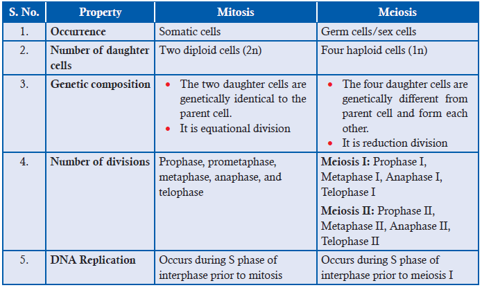
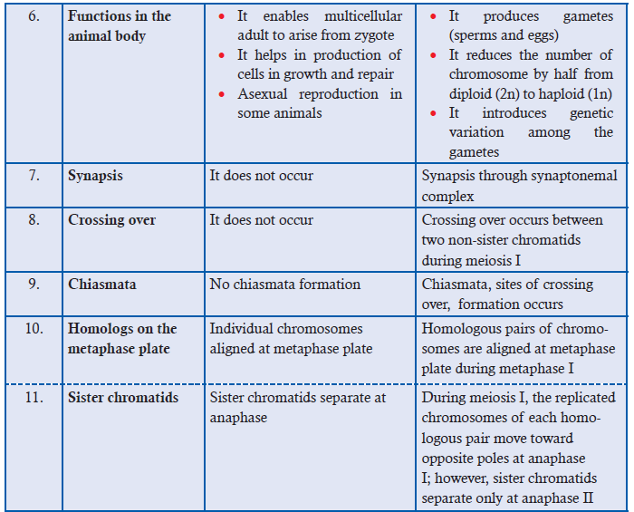
Table 5.2: Comparison between mitosis and meiosis
ACTIVITY 5.6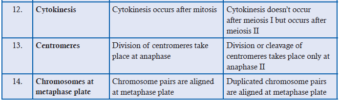
Carry out a research project to find out why cultured skin is grown in a medium of proteinssimilar to blood. Then write a journal entry to summarise the research.
5.5 SUMMARY
• Karyotype is a visual representation of the chromosomes within a single cell.
• Diploid cells contain two complete sets (2n) of chromosomes. Example, skin cells. Haploid
cells contain only one complete set (1n) of chromosome. Example, gametes (sperm and
egg). Homologous chromosomes are pairs of chromosomes that are similar in length, gene
position, and centromere location. Non-homologous chromosomes are chromosomes
that contain different genes.
• A typical cell cycle consists of mitotic and interphase phases. Interphase is subdivided
into G0, G1, S, G2 phases. S phase is the longest phase.
• Mitosis is subdivided into prophase, prometaphase, metaphase, anaphase, and telophase.
• DNA replication takes place at S phase of interphase prior to mitosis.
• Cytokinesis is a division of cytoplasm and usually follows the process of mitosis.
• Some differentiated cells are arrested at G0 phase; and they resume cell cycle when they
receive signal to divide and repair injured tissues. Example, skin fibroblasts.
• Some cells have stem cells that are undifferentiated biological cells which can differentiate
into specialized cells and continually replace the dying cells. Example, hematopoietic
(blood-forming) system.
• Unrestrained, uncontrolled growth of cells in cell cycle results into a disease called cancer.
• Meiosis is divided into meiosis I and meiosis II. Meiosis I is a reduction division and is
subdivided into prophase I, metaphase I, anaphase I, and telophase I. DNA replication
takes place at interphase I prior to meiosis I. Synaptonemal complex is formed during
prophase
I. Crossing over takes place in meiosis I.
• Meiosis II is just like mitosis. There is no DNA replication. It is divided into prophase II,
metaphase II, anaphase II, and telophase II.
• Meiosis is a reduction division. It reduces diploid chromosomes to four haploid daughter
cells. Reduction division usually happens before sexual reproduce where haploid gametes
(sperm and eggs) are formed for fertilization.
• The process of sperm formation is called spermatogenesis; while the process of egg
formation
is called oogenesis.
• Meiosis also reduces diploid plant into haploid gametes which eventually fuse to form
zygote.
• During meiosis, paternal and maternal homologues assort independently into four
daughter cells. This process adds genetic variation.
• Crossing over and random fertilization increase genetic variation in sexual life cycles.
• Improper separation of chromosomes during meiosis results into non-disjunction of
chromosomesthat are responsible for disease such as Down syndrome.
5.6 GLOSSARY
• Cancer: It’s an unrestrained, uncontrolled growth of cells in human beings. It is essentially
a disease of uncontrolled mitotic cell division.
• Chiasmata (chiasma-singular): During crossing over of genes, the point where
homologous chromosomes remain attached at one particular point is called chiasmata.
• Crossing over: The exchange of genetic material between homologous chromosomes
that occur during meiosis and contribute to genetic variability.
• Cytokinesis: It is a division of cytoplasm.
• Diploid cells: Cells which contain two complete sets (2n) of chromosomes (23 + 23 in
number) are called diploid cells. Example: somatic cells (Muscle cells).
• Down syndrome: It is an autosomal trisomy. It is also called as trisomy 21, where
non-disjunction results in an embryo with three copies of chromosome 21 instead of the
usual two copies of chromosome 21.
• Gametogenesis: It is a biological process by which diploid cells undergo cell division
and differentiation to form mature haploid gametes.
• Haploid cells: Cells that contain only one complete set (1n) of chromosome (23 in
number) are called as haploid cells. Example: sperm and ovum.
• Independent assortment: In meiosis I, specifically at metaphase I, each homologous pair
of chromosomes positioned independently of the other pairs at metaphase I.
• Karyotype: It is a visual representation of the chromosomes within a single cell where
the number of chromosomes, their arrangement, size and structure can be analysed.
• Meiosis: It’s a reduction division where the number of chromosomes is reduced to half
from diploid parent cell to haploid daughter cells.
• Mitosis: It is a process where a single cell divides into two identical daughter cells.
In other words, it’s a division of nucleus.
• Reduction division: It is a process of meiotic cell division where the number of
chromosome sets from parental diploid (2n) cells is reduced to haploid (1n) daughter cells.
• Stem cells: They are undifferentiated biological cells that can differentiate into specialized
cells (when stimulated by the right factor) and can divide to produce more stem cells.
Example: Hematopoietic system.
• Syngamy or fertilization: The fusion of gametes (one from mother and one from father)
to form a zygote is called as syngamy.
• Tetrad or bivalent: The structure of four chromatids of the two homologous chromosomesattached at chiasmata during the process of meiosis is called as tetrad.
END UNIT ASSESSMENT 5
Do all these exercises in your exercise book.
I. Choose whether the following statements are True (T) or False (F)
1. Cell cycle is a cyclical event of cell growth, mitosis, and cell division.
2. A typical cell spends most of its time in interphase.
3. Mitosis is a process where a single cell divides into three identical daughter cells.
4. Cytokinesis is a division of cytoplasm.
5. The process of mitosis is basically divided into 5 phases.
6. Meiosis is divided into three stages: Meiosis I, Meiosis II and Meiosis III.
7. The unrestrained, uncontrolled growth of cells in human beings results into a disease
called cancer.
8. Cancer occurs due to failure in controlling cell division.
9. Proper separation of chromosomes during meiosis is not essential for the normal
growth in humans.10. The life span of blood cells ranges from less than one day to a few months.
II. Multiple Choice Questions
1. Meiosis starts with diploid cell but ends with ......... haploid cells.
(a) one (b) two
(c) three (d) four
2. ........................... is a biological process by which diploid cells undergo cell division
and differentiation to form mature haploid gametes.
(a) Gametogenesis (b) Spermatogenesis
(c) Oogenesis (d) None of these
3. Generally, plants reproducing sexually have life cycle consisting of .......... phases.
(a) two (b) three
(c) four (d) five
4. In telophase, the nuclear envelope re-forms around the ........... set of haploid daughter
chromosomes.
(a) one (b) two
(c) three (d) four
5. The prominent role of stem cells can be seen in—
(a) blood cells (hematopoietic system)
(b) epithelial cells of the skin
(c) epithelial cells lining the digestive tract
(d) All of these
6. ........................is a condition in which the homologues or sister chromatids
fail to separate properly during meiosis.
(a) Disjunction (b) Non-disjunction(c) Down syndrome (d) None of these
7. Which of the events is not correct in prometaphase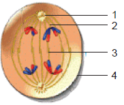
(a) Nuclear envelope breaks down
(b) Spindle fibres invade nuclear area
(c) Kinetochore binds to centromere
(d) Chromatids condense into discrete chromosome
8. Which of the event is correct in anaphase
(a) Sister chromatids separate and give rise to daughter chromosomes.
(b) Chromosomes are aligned at the metaphase plate.
(c) Cytokinesis starts occurring.
(d) Chromosomes begin to uncoil.
9. The total chromosome number at the end of meiosis I and II is
(a) 46 (b) 23(c) 44 (d) 92
10. One round of oogenesis produces
(a) One egg (b) Two eggs
(c) Three eggs (d) Four eggs
11. Which of the following results in genetic variation
(a) Independent assortment of chromosomes
(b) Crossing over
(c) Random fertilization
(d) All of these
12. In mitosis, which of the following occurs?
(a) Chiasmata formation (b) DNA replication
(c) Synapsis (d) None of these
13. Identify the cells which are divided by mitosis.
(a) Skin cells (b) Liver cells
(c) Blood cells (d) All of these
14. Identify the cells which are divided by meiosis.
(a) Intestinal cells (b) Stem cells
(c) Gametes (d) None of these
15. Which part of the plants is/are divided by mitosis?
(a) Stem (b) Flower
(c) Leaves (d) All of these
16. Identify the cells that are divided by meiosis in plants.
(a) Root tips (b) Pollen grains
(c) Ova (d) (b) and (c)
III. Long Answer Type Questions
1. Describe the main stages of cell cycle.
2. In your own words, explain what is meant by homologous pairs of chromosomes.
3. What do you mean by the terms haploid and diploid?
4. In your own words, describe the process of mitosis.
5. In your own words, describe the process of meiosis.
6. Outline the significance of mitosis in cell replacement and tissue repair by stem cells.
7. In your own words, explain how uncontrolled cell division can result in the formation
a tumour.
8. What is the need for reduction prior to fertilization in sexual reproduction?
9. In your own words, explain the importance of effective cell division.
10. Outline the role of meiosis in gametogenesis in humans and in the formation of
pollen grain and embryo sacs in flowering plants.
11. Explain how crossing over and random assortment of homologous chromosomes
during meiosis and random fusion of gametes at fertilization leads to genetic variation,
including the expression of rare recessive alleles.
12. (i) Identify the stage of cell division shown in the figure.
(ii) Label the structures marked as (1), (2), (3) and (4).
(iii) Which type of cell is involved in this division?
(iv) What will happen if the structure marked (3)
is not formed?
13. How can you correlate the spread of HIV virus with the process of Mitosis? Knowing
the viral disease and its spread, discuss in brief the stigma and discrimination facedby those affected by HIV and AIDS.