Unit 2: Optical instruments
Key unit Competence
Describe and use optical instruments.
My goals
By the end of this unit, I will be able to:
* explain an optical instrument.
* explain the physical features of a human eye.
* describe the image formation by the eye.
* identify the physical features of a simple and compound microscope.
* explain the applications of simple and compound microscopes.
* differentiate between simple and compound microscopes.
* explain the operation of a lens camera and its application.
* explain the operation of a slide projector and its applications.
* describe the physical features of a telescope.
* list different types of telescopes.
* demonstrate the operation of telescopes.
* differentiate between telescopes and microscopes.
* identify the physical features of prism binoculars.
INTRODUCTORY ACTIVITY
When a patient goes to hospital having a headache and fever, a doctor may
require a blood test for malaria. When a sample of blood is taken, it is not
possible to check whether a patient has malaria or not. But a laboratory
technician may need to test the blood using some instrument and decidewhether the patient has malaria or not.
Questions:
(i) Which instrument do you think may be used to test malaria from
blood sample?
(ii) In summary, discuss how that instrument function.(iii) What other instrument do you think can be used for such purpose?
Introduction
Once the rules for predicting how rays travel through lenses have been
discussed; a fantastic range of practical devices began to appear which aided
the development of the modern world. The simple magnifying glass became
the basis for telescopes, microscopes and spectacles. These devices were
modified to improve the projection of images and with the discovery and
development of light-sensitive chemicals, gave birth to modern photographyand cinematography.
Definition of an optical instrument
Activity 2.1
In our daily activities and development, we observe different things in
environment or in universe. Sometimes, some objects cannot be easily
observed using our naked eyes. We need to see these very small things at
big distance.
(i) What do you think we use to observe those distant or very small
bodies?
(ii) Discuss the properties used by those instruments?
(iii) Name at least four instruments that people use to observe distant orvery small objects.
We use our eyes to see and view different objects. The eye cannot be used
to view clearly these objects at night, and some distant objects or hidden
objects. Objects which cannot be viewed by the eye can be focused using
other instruments. All the instruments used to aid vision are called Opticalinstruments.
Man has always shown interest in observing things in a more detailed manner.
In your early secondary, you looked at the uses of mirrors. We have also learnt
in unit 1 of this book that lenses are used to focus objects. When the lenses
or mirrors or both are arranged in a way, the arrangement can be used to
observe objects in a more detailed manner. The arrangement makes what we
call a compound optical instrument. The compound instruments include acompound microscope, telescopes, prism binoculars etc.
THE HUMAN EYE
The eye is a biological instrument used to see objects at different distances. It
uses a convex lens system to form a small, inverted, real image of an objectinfront of it.
Structure of the eye
Activity 2
(i) In groups of two, look at one another’s eye.
(ii) Observe critically its external shape.
(iii) Observe it carefully and note its behaviour as one tries to see some
objects in class.Notice that the eye ball is round and fleshy.
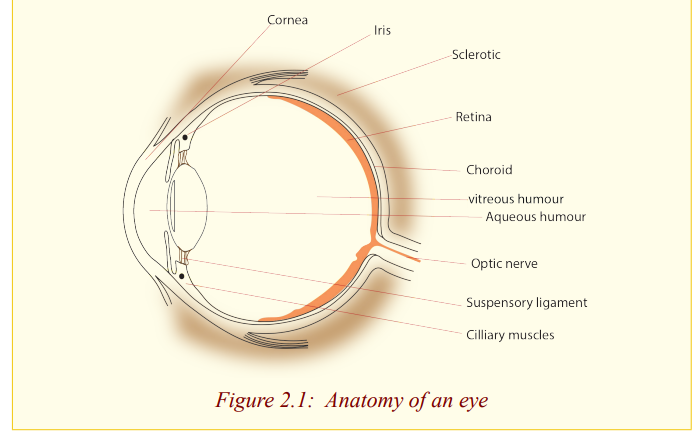
Functions of the parts of the eye
The cornea: It is made out of a fairly dense, jelly like material which provides
protection for the eye, and seals in the aqueous humour. It also
provides most of the power of the eye (59 Dioptres), having about46 Dioptres. So it provides most of the bending of light rays.
The aqueous humour: This is a waterly liquid that helps to keep the cornea ina rounded shape, similar to that of a lens.
The iris: This controls the amount of light entering the eye. The amount of
light that enters the eye is one of the factors determining how
focused an image is on the retina. The brighter the light the eye is
exposed to, the smaller the iris’ opening will be. The brighter the
light the eye is exposed to, the smaller the iris’ opening will be.
The iris is the coloured part of the eye as seen from the outside.
The iris opening or a gap through which light passes is called apupil.
The lens: This is used to focus an image on the retina. It controls the
bending of light rays by change of its shape, a process calledaccommodation, which is done by the ciliary muscles.
The ciliary muscles: These control the thickness of the lens during focusing.
By contracting or squeezing the lens, they make it thicker and
vice versa. Because the power of the lens is directly related to
its thickness, the ciliary muscles change the power of the lens by
their movement.
The retina: This is the light sensitive part of the eye and it is where images
are formed. It contains millions of tiny cells which are sensitive
to light. The cells send signals along the optic nerve to the brain.
So the retina is the screen of the eye and the image is formed by
successive refraction at the surfaces between air, the cornea, the
aqueous humour, the lens and vitreous humour. The retina is
black, which prevents any light rays that hit it from reflections
and thereby changing the image.
The vitreous humour: This is a jerry like substance that helps the eye to
keep its round shape. It is very close in optical density to the lensmaterial.
The yellow spot: This is a small area on the retina where the sharpest image,
that is, the finest detail can be seen.
The optic nerve: This is the nerve that transmits images received by the retina
to the brain for interpretation. The part of the eye where the optic
nerve joins the retina is called the blind spot because no imagescan be observed at at this point.
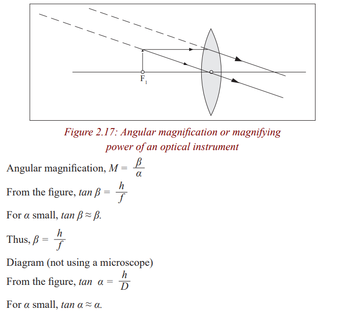
Angular magnification or magnifying power of an opticalinstrument
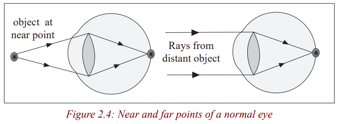
Accommodation of the eye
Accommodation of the eye is the ability of the eye to see near and distant
objects. The eye is capable of focusing objects at different distances by
automatic adjustment of the thickness of the eye lens which is done by the
ciliary muscles. To focus a distant object, the eye lens is made thinner, so less
powerful, and the rays from the object are brought to focus on the retina by
the eye lens. In this case, the ciliary muscles are relaxed and pull the lens. For
nearer objects, the eye lens must be made thicker and hence more powerful so
that the rays from the near object can be brought to a focus on the retina. Inthis case, the ciliary muscles tighten and squeeze the lens.
Near point and far point of the eye
Activity 5
(i) Hold a book at an arm’s length and move it closer to find the nearest
distance that you can focus the words clearly without straining your
eyes.
(ii) Approximate the distance between your eyes and the book.(iii) What does this distance represent?
The near point of the eye is the nearest point that can be focused by the un
aided eye. It is a closest distance that the ‘normal’ human eye can observe
clearly; without any strain to the eye. It is called the least distance of distinctvision. The near point of a normal eye is 25 cm.
Activity 6
(i) Look at the trees around your school.
(ii) Now, try to look at objects far from the school.
(iii) Are you able to focus the distant objects?
(iv) Measure this distance from the object to your eye.(v) Write down your observation in the notebook.
Notice that you can not be able to measure this distance. The distance from a
distant object to the eye is the far point of the eye. The far point of the eye isinfinity. The far point is the farthest point that can be focused by the eye.
The distance of 25 cm from the eye is called distance of most distinct vision or
least distance for distinct vision. The range of accommodation of the normal
eye is thus from 25 cm to infinity. This range is based upon the average human
eye which has an age of 40 years. Young persons have a much wider range butthe average 70 year – old has a reduced range.
People with normal vision can focus both near and distant objects.
Defects of vision and their correction
Activity 7
(i) Have you seen before some people putting on eye glasses?(ii) What do you think these glasses(spectacles) are used for.
People put on eye glasses for different reasons. Some people wear them in
order to read a text, some put them on to see near objects if their eyes cannot
be able to do so while others put them on so as to focus distant objects; otherswear them for fan like sun goggles
Short-sightedness (myopia)
Activity 8
(i) Hold a book at an arm’s length and move the lens so that the prints
are read without the eye getting strained.
(ii) Now, try to read the words on a chalkboard a distance from the book.(iii) Are you able to focus both near and distant objects?
People with normal vision can focus clearly near and distant objects. Those
who only focus near objects are said to be short-sighted, meaning that theysee nearer
Short-sightedness is the defect whereby a person can see near objects clearly
but cannot focus distant objects. His far point is nearer than infinity. This is
because the eyeball is too long or the lens is too strong so that rays of lightfrom a distance object are focused in front of the retina.
The rays are focused in front of the retina because the focal length of the eye lens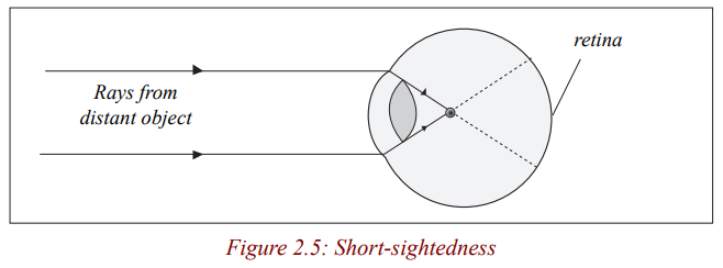
is too short for the length of the eye ball. This defect can be corrected by wearing
a concave (diverging) spectacle lens. The rays of light from a distant object are
diverged so that they appear to come from a point near, and so they are focused bythe eye.
Rays from object at infinity appear to come from a near point F and converge to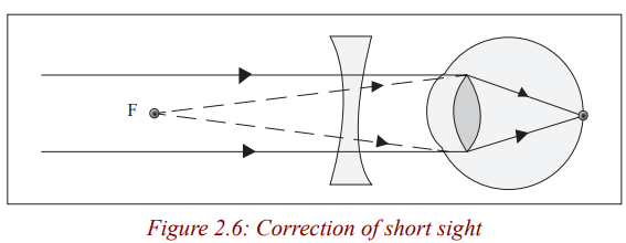
the retina.
Long-sightedness (hypermetropia)
This is where a person is able to see distant objects clearly but cannot focus near
objects. This is because either his eye ball is too short or the eye lens is too weak
(thin) so that rays of light from a close object are focused behind the retina.This eye’s near point is further than 25 cm.
The image of the near object is focused behind the retina because the focal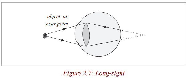
length of the eye lens is too long for the length of the eye ball. This defect can
be corrected by wearing a convex lens spectacle. The rays of light from a near
object are converged so that the rays appear to come from a point far, and soare focused by the eye.
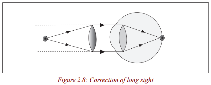
Rays from a near object O appear to come from a distant object.
Presbyopia
Activity 9
(i) How many of you still have their grandparents?
(ii) Have you ever tried to observe how grand parents observe objects?
(iii) Discuss with your neighbour and write in your notebook results ofyour discussion.
When people grow older, their eye lens become stiff and it becomes hard for
the ciliary muscles to adjust it. Such people have a defect called Presbyopia.
Presbyopia is the stiffening of the eye lens such that it is less capable of being
adjusted by the ciliary muscles. This means that the eye lens becomes less
flexible and loses its power (ability) to accommodate for objects at different
distances. This defect is corrected by wearing bifocals spectacles whose lenses
have a top part for looking at distant objects and a bottom part for close ones.
These bifocal spectacles have a diverging top part to correct for distant visionand converging lower part for reading.
Astigmatism
This is the defect that occurs if the curvature of the cornea varies in different
directions so that rays in different planes from an object are focused in
different positions by the eye and the image is distorted. A person suffering
from astigmatism sees one set of lines more sharply than others. This defect is
corrected by wearing corrected lenses. These help to bend the incoming raysto correct for irregular refraction.
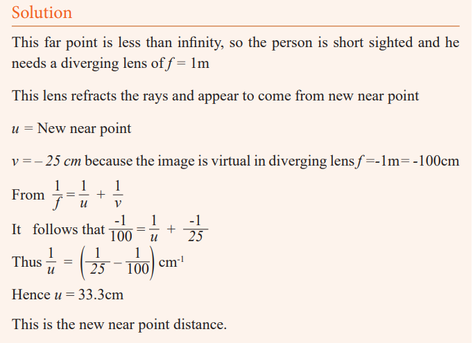
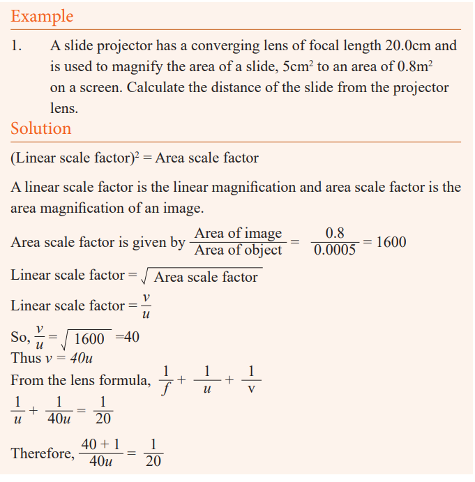
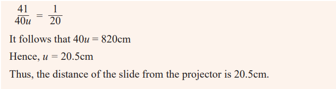
Example
The far point of the defective eye is 1m. What lens is needed to correct
this lens. With this lens, at what distance from the eye is its near point, ifthe near point is 25cm without the lens?
Formation of an image by the eye
Light enters the eye through the transparent cornea, passes through the
lens and is focused on the retina. The retina is sensitive to light and sends
messages to the brain for interpretation. Although the image is inverted, thebrain interpretes it correctly.
Visual Angle
Activity 3
(i) Go outside class and view the trees around.
(ii) Are the trees of the same height?
Notice that some trees at a distance, look shorter than the nearby trees
when it is not the case? Why do you think it is so?Discuss and write down in your notebook about your observation.
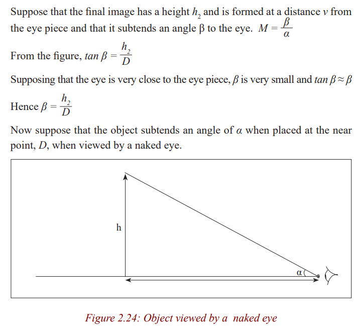
The height of an object depends on the angle of elevation of its top from the
eye. The larger the angle, the taller the objects. This angle is called the visualangle.
The visual angle is the angle subtended at the eye by an object.
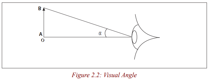
This angle decreases when the distance D increases and increases when the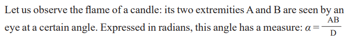
distance D decreases. It also increases when the length AB increases anddecreases when AB decreases. We call it visual angle of the object.
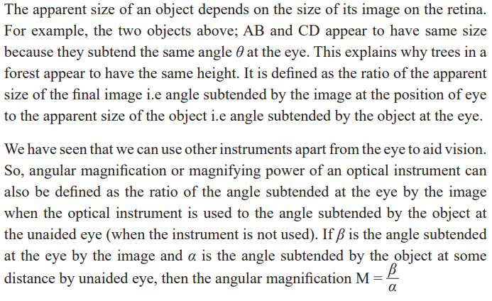
Lead the learners to define the visual angle of an object as the angle between
two rays of light from extremities of the object and penetrating into the eye ofan observer.
Activity 4
Objects that subtend the same angle at the eye appear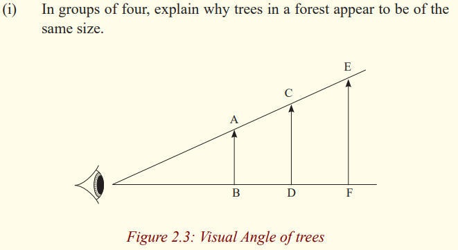
to be of the same size as viewed by the eye.
Application activity 2.1
1. Name the part of the eye
a) which controls how much light enters it,
b) on which the image is formed,
c) which changes the focal length of the crystalline lens.
2. A farsighted person has a near point of 100 cm. Reading glasses
must have what lens power so that this person can read a
newspaper at distance of 25 cm? Assume the lens is very
close to the eye.
3. A nearsighted eye has a near and far point of 12 cm and 17 cm,
respectively.
a) What lens power is needed for this person to see distant objects
clearly, and
b) What then will be the near point?Assume that the lens is 2.0 cm from the eye (typical for eyeglasses).
A lens camera
Activity 10
(i) Make a paper box and carefully use a pin to make a tiny hole in the
centre of the bottom of the paper box.
(ii) Place a piece of wax paper on the open end of the box. Hold the
paper in place with the rubber band.
(iii) Turn off the room lights. Point the end of the box with a hole in a
bright window.
(iv) Look at the image formed on the wax paper.
Which kind of image have you seen? Is it upside down or right side up. Isit smaller or larger than the actual object? What type of image is it?
The image is upside down. The pin hole helps you to see the image of theobject. This device is called a pin hole camera.
Activity 11
(i) When you were going to register for Rwanda National Examinations,
you took some photographs.(ii) What device did the person that took your photograph use?
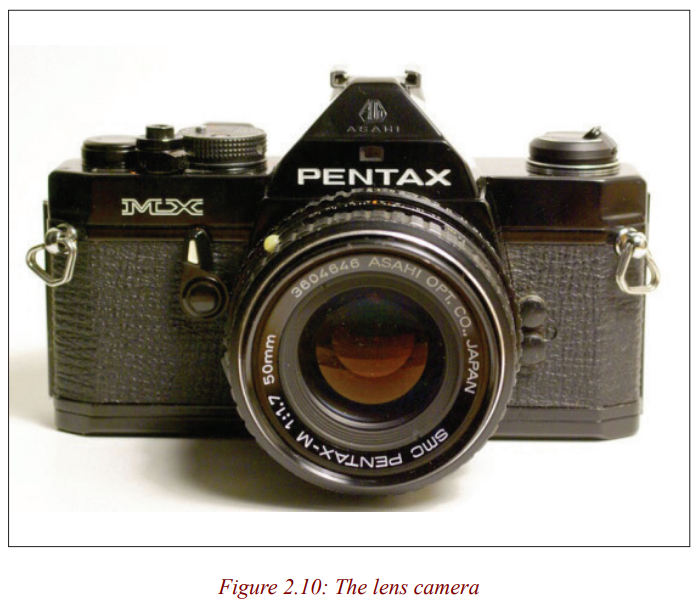
In our daily lives, we take photographs. We use a lens camera to take these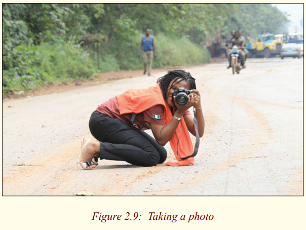
photographs.
Activity 12
(i) Enlarge the hole in the pinhole camera above at the front of the box
and hold convex lens over the hole.
(ii) Adjust the position of the lens for either near or far objects to make
a sharp image on the screen.
(iii) Is the image erect or inverted? If the objects are coloured, is theimage coloured?
Notice that the image formed is inverted and coloured if the object is coloured.
By placing a lens above the hole, you are making a lens camera from a pinhole camera.
Formation of images by a lens camera
Activity 13
(i) Draw a ray diagram for the formation of an image of an object
placed at a point beyond 2F of a thin converging lens.
(ii) State the nature and size of the image.Is the image bigger or smaller?
We have already seen that when an object is beyond 2F of a thin converginglens, the image formed is smaller than the object.
A camera consists of a light- tight box with a convex (converging) lens at
one end and the film at the other end. It uses the convex lens to form a small,inverted, real image on the film at the back.
The lens focuses light from the object onto a light sensitive film. It is moved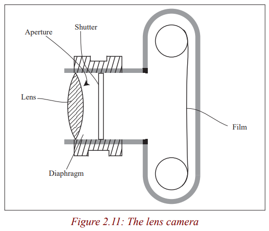
to and fro so that a sharp image is formed on the film. In many
cameras, this happens automatically. In cheaper cameras, the lens
is fixed and the photographer moves forwards and backwards tofocus the object.
The diaphragm is a set of sliding plates between the lens and the film. It
controls the aperture (diameter) of a hole through which light
passes.
In bright light, a small aperture is used to cut down the amount
of light reaching the film and in dim light, a large hole is needed.
Very large apertures give blurred images because of aberrations
so the aperture has to be reduced to obtain clear images.
In many cameras, the amount of light passing through the lens
can be altered by an aperture control or stop of variable width.
This size of the hole is marked in f – numbers i.e 1.4, 2, 2.8, 4,
5.6, 8, 11, 16, 22, 32. The smaller the f-number, the larger the
aperture. An f-number of 4 means the diameter d of the aperture
is ¼ the focal length, f of the lens. To widen the aperture, the f
number should therefore be decreased.
The aperture also controls the depth of field of the lens camera.
The depth of field is a range of distances in which the camera can
focus objects simultaneously. This depth of field is increased byreducing the aperture.
This large depth of field ensures a large depth of focus. The
depth of focus is the tiny distance the film plane can be moved
to or from the lens without defocusing the image. A large depth
of focus means that both near and far objects appear to be in
focus at the same time which is obtained by a small hole in thediaphragm.
The shutter controls the exposure time of the film. It opens and closes quicklyto let a small amount of light into the camera.
The exposure time affects the sharpness of the image. When the exposure
time is short, the image is clear (sharp) but when it is long theimage becomes blurred.
The film. This is where the image is formed. It is kept in darkness until the
shutter is opened. It is coated with light sensitive chemicals
which are changed by the different shades and colours in the
image. When the film is processed, these changes are fixed andthe developed film is used to print the photograph.
Note that a diminished image is always formed on the film and that the image
of distant object is formed on a film at distance f from the lens. For near
objects, the lens is moved further away from the film (closer to the object) to
obtain a clear image. In this case, the film is at a distance greater than f of the
lens. Digital cameras are similar to film cameras except that the light does not
form an image on photographic film. The image in a digital camera is formedon a charge-coupled device (CCD).
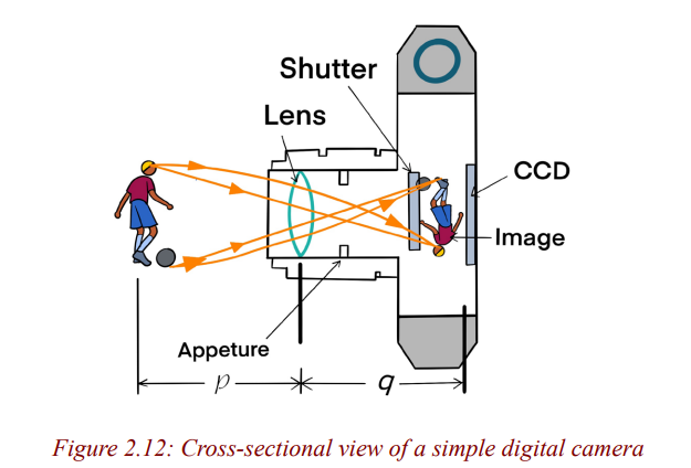
The CCD is the light-sensitive component of the camera. In a nondigital
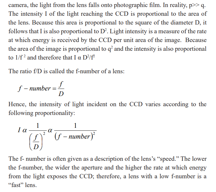
Application activity 2.2
1. A camera gives a clear image of a distant landscape when the lens
is 8 cm from the film. What adjustment is required to get a
good photograph of a map placed 72 cm from the lens?
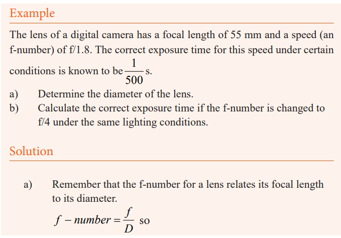
2. The lens of a certain 35 mm camera (where 35 mm is the width of
the film strip) has a focal length of 55 mm and a speed (an
f-number) of f/1.8. The correct exposure time for this speed
under certain conditions is known to be (1/500) s.
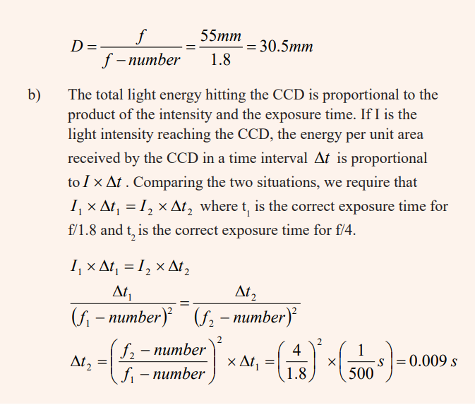
a) Determine the diameter of the lens.
b) Calculate the correct exposure time if the f-number ischanged to f/4 under the same lighting conditions.
The slide projector
Activity 14
(i) Have you ever seen an instrument called a slide projector?
(ii) What is that instrument used for?
(iii) Have you ever watched a cinema where the pictures are seen on the
white wall?
(iv) What device were they using to throw the pictures on the screen
(wall or white cloth)?
(v) Where do you think the pictures came from?(vi) Are the images small or large?
The pictures are thrown on the screen using a slide projector.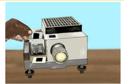
A projector is a device used to throw on a screen a magnified image of a filmor a transparent slide. It produces a magnified real image of an object.
A slide projector is an opto-mechanical device for showing photograhic slides.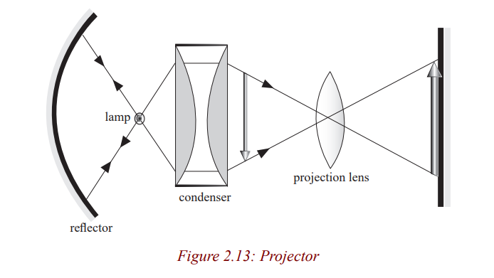
It consists of an illumination system and a projection lens. The illumination
system consists of a lamp, concave reflector and the condenser. The illuminant
is either a carbon electric arc or a quartz lamp to give a small but very highintensity source of light in order to make the image brighter.
The lamp is situated at the centre of curvature of the mirror so that the rays are
reflected back along their original path. The concave mirror reflects back light
which would otherwise be wasted at the back of the projector housing. The
condenser consisting of two Plano concave lenses collects light which would
otherwise spread out and be wasted, and concentrates it on to the film (slide)so that it is very bright and evenly illuminated.
The light is then scattered by the film and focused by a convex projection lens
on to the film. The projection lens is mounted in the sliding tube so that it ismoved to and fro to focus a sharp image on the screen.
Application activity 2.3
1. A colour slide has a picture area 2.4 cm x 3.6 cm. Find the focal
length of the projection lens which will be needed to throw an image1.2m x 1.8m on a screen 5m from the lens.
2. A projector projects an image of area 1 m2
onto a screen placed 5m
from the lens. If the area of the slide is 4 cm2
, calculate;
(i) The focal length of the projection lens.(ii) The distance of the slide from the lens
Activity 15
Make a projector on the bench using a ray box lamp, a single convex lens (focal
length about 5 cm) for the condenser; a slide; a convex lens (focal length 5cm or
10cm) as the projection lens and a sheet of paper for the screen.
Is the image inverted?By how much is it magnified?
Note that if the film is placed just after the lamp, the object would be poorly
illuminated. So to give a bright picture, a condenser is included. The film O
is placed between F and 2F of the projection lens so that the image I is real,
inverted and magnified. The film is put in the projector while it is upside downso that the picture on the screen is upright.
Microscope
Simple Microscope (Magnifying Glass)
Activity 16
(i) Hold a hand lens at above the word Rwanda at a distance of about
4cm from the word.
(ii) Move the lens farther away slowly from the word while observing
the word through the lens.
(iii) What changes do you notice after observing?
(iv) Share ideas with your neighbour and write your observation inyour notebook.
The word Rwanda becomes larger and larger and finally disappears. This word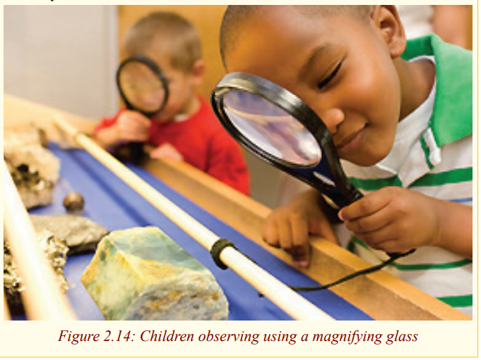
gets larger because of the lens. We say that it is being magnified by the lens.
Activity 17
(i) Place your hand on a table and hold a hand lens above it and do the
same as in activity 16.(ii) What do you notice?
Notice that the hair (fur) and other small holes on the skin are seen clearly.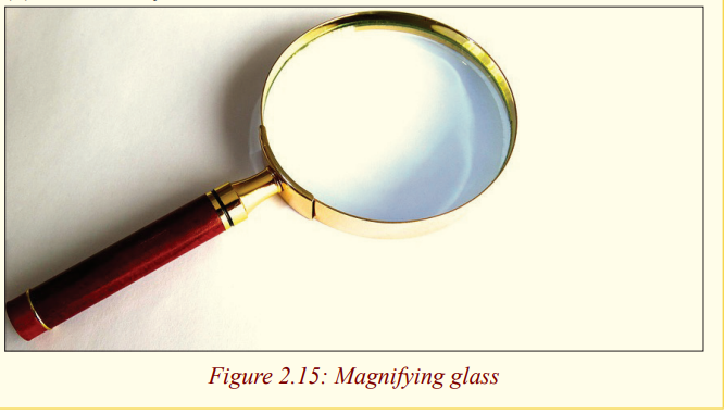
These parts of the skin are made bigger by the glass lens and this enables one
to see them clearly. This lens which magnifies images is called a magnifyingglass or a simple microscope.
A magnifying glass consists of a thin converging lens and It is used to view
very small organisms or parts of organisms which cannot be easily seen by thenaked eye.
Formation of images by a magnifying glass
Activity 18
Using the knowledge from thin lenses, draw a ray diagram to show the
formation of an image by a magnifying glass.State the characteristics of the image formed.
We have already seen in unit 1 that when an object is between the lens and its
principal focus, the image formed is magnified and upright. So, a magnifying
glass forms a virtual, upright, magnified image of an object placed betweenthe lens and its principal focus.
Activity 19
Making a simple microscope
(i) Use a pin or a nail to make a hole about 2 mm in diameter in a piece
of a kitchen foil or glass.
(ii) Carefully let a drop of water fall on the hole so that it stays there
and acts as a tiny lens with short focal length.(iii) Use it to observe prints on a piece of paper.
Simple microscope (magnifying glass) in normal adjustment.
The magnification of a magnifying glass depends upon where it is placed
between the user’s eye and the object being viewed and the total distancebetween them.
Activity 20
(i) Carefully place a magnifying glass above some prints on a piece of
paper and adjust it until they are seen clearly.
(ii) Make sure that you don’t feel any strain in the eye while you are
observing.(iii) What do you think is the position of the image from the eye?
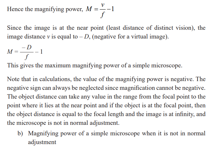
The image is at the least distance of vision since the eyes are not strained andthe magnifying glass is said to be in normal adjustment.
A microscope is in normal adjustment if the final image is formed at the nearpoint, and it is not in normal adjustment if the final image is at infinity.
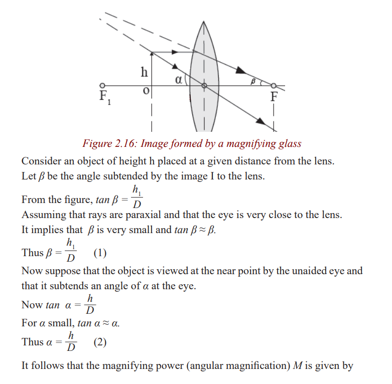
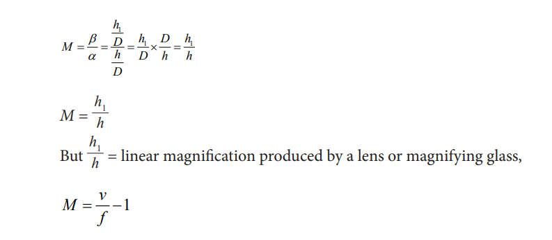
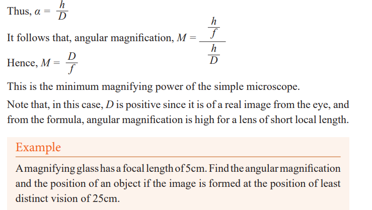
Magnifying power of a simple microscope
We have already seen that the size of the image depends on the angle subtended
by the object on the eye called the visual angle. Thus, the magnifying powerdepends on the visual angle.
It is defined as the ratio of the angle subtended by the image to the lens to the
angle subtended by the object at the near point to the eye.a) Magnifying power of a simple microscope in normal adjustment
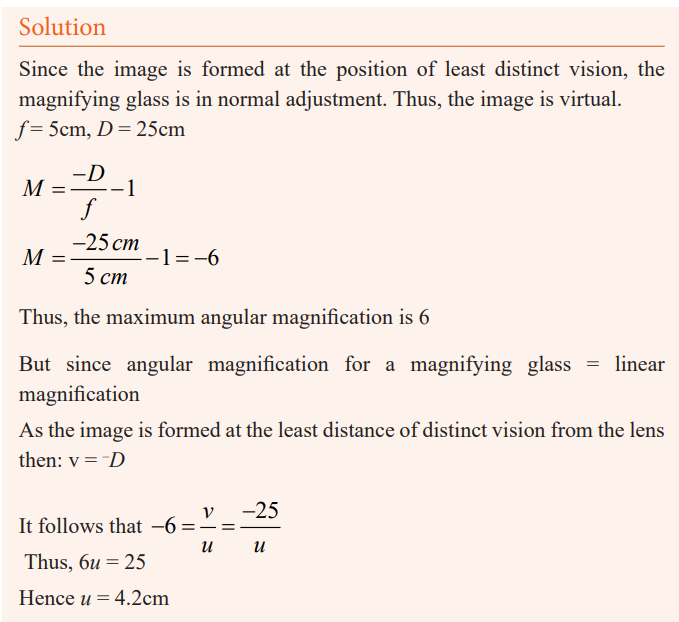
Application activity 2.4
1. Find the angular magnification produced by a simple microscope of
focal length 5cm when used not in normal adjustment.
2. Explain why angular magnification of a simple microscope is high
for a lens of short local length.
3. Why the image formed by magnifying glass is free from chromaticabberation.
Activity 21
In groups of five, discuss why the image formed in a magnifying glass is
almost free of chromatic abbreviation.
When an object is viewed through the magnifying glass, various coloured
images corresponding to IR, IV for red and violet rays are formed but each
image subtends the same angle at the eye close to the lens and therefore these
colours overlap. The overlap of these colours makes a virtual image seen in amagnifying glass free of a chromatic abberation.
Group Activity 22
Provided a magnifying glass, go outside and pick different kinds of leaves.
Examine, with the use of a magnifying glass, the structures of the leaves.Discuss in details the structural characteristics of each leaf
Group Activity 23
You are provided with dirty water in a glass container.
Use the magnifying glass provided and view some living organisms in it.Record what you see.
Activity 24
(i) Observe critically and describe the activity being done in the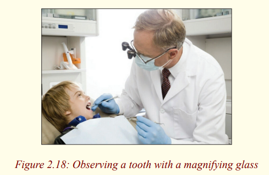
photograph.(ii) State other uses of a magnifying glass.
Uses of magnifying glass: Magnifying glasses have many different uses.
Some people use it for fun activities such as starting fires, or use the lens to
help them read. You can start a fire with a magnifying glass when the sun rays
are concentrated on the lens. Some retail stores sell reading glasses with the
double convex lens. In everyday life, magnifying glasses can be used to do a
variety of things. The most common use for magnifying glasses would be howscientists use them, they use magnifying glasses to study tiny germs
The compound microscope
Activity 25
Have you ever heard or seen an instrument called a compound
microscope?What is it used for?
The compound microscope is used to detect small objects; is probably the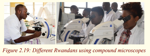
most well-known and well-used research tool in biology.
Activity 26
Observe the above pictures carefully and discuss places where a compound
microscope is used in daily life.In daily life, microscopes are used in hospitals, in biology laboratories, etc.
Activity 27
(i) You are provided with two lenses of focal lengths 5cm and 10cmtogether with a half meter ruler and some plasticine.
(iii) Move the object to and fro until it appears in focus.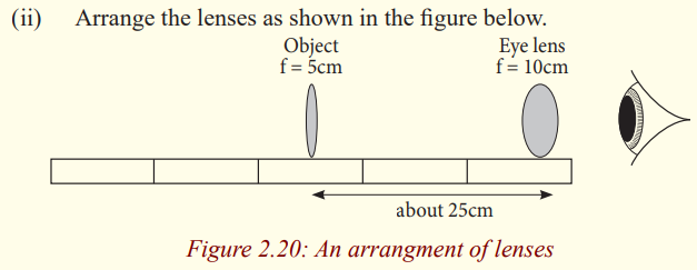
What do you notice about the image? Is it distorted? Is it coloureddifferently in any way?`
By arranging the lenses as above, you have actually made a compound
microscope. We have already seen how a single lens (magnifying glass) can
be used to magnify objects. However, to give a higher magnifying power, twolenses are needed. This arrangement of lenses makes a compound microscope.
It produces a magnified inverted image of an object.
A compound microscope is used to view very small organisms that cannot beseen using our naked eyes for example micro organisms.
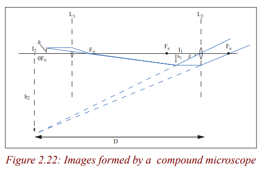
A compound microscope consists of two convex lenses of short focal lengths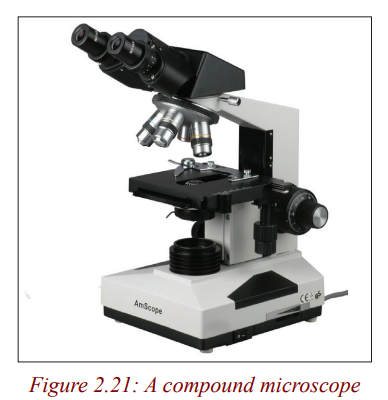
referred to as the objective and the eye piece. The objective is nearest to the
object and the eye piece is nearest to the eye of the observer. The object to be
viewed is placed just outside the focal point (at a distance just greater than the
focal length) of the objective lens. This objective lens forms a real, magnified,
inverted image at a point inside the principal focus of the eye piece. This
image acts as an object for the eye piece and it produces a magnified virtual
image. So the viewer, looking through the eye piece sees a magnified virtualimage of a picture formed by the objective i.e of the real image.
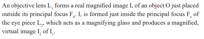
Image formation in a compound microscope
Compound microscope in normal adjustment (normal use)
Activity 28
You are provided with a bird's feather; observe it critically using a
compound microscope and draw it in a fine detail.Make sure you observe the features when your eyes are relaxed.
When the eyes are relaxed, the image is at the near point and the compound
microscope is said to be in normal adjustment. The compound microscope is
in normal adjustment when the final image is formed at the near point (leastdistance of distinct vision), D of the eye.
Angular magnification (magnifying power) of a compound
microscope
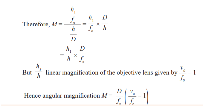
The magnifying power of a compound microscope is the ratio of the angle
subtended by the final image to the eye when the microscope is used to theangle subtended by the object the unaided eye.
Angular magnification of a compound microscope in normal use
We have already seen that when a microscope is in normal use, the image I2is formed at the least distance of distinct vision, D from the eye. Thus v = D.
Consider an object of height h at a given distance slightly greater than the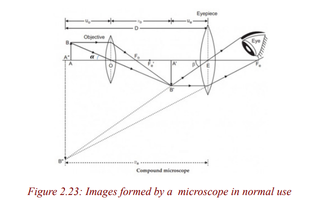
focal length of the objective lens.

Example
A compound microscope has an eye piece of focal length 2.50cm and an
objective of focal length 1.60cm. If the distance between the objective
and eye piece is 22.1cm, calculate the magnifying power produced whenthe final image is at infinity.
Activity 29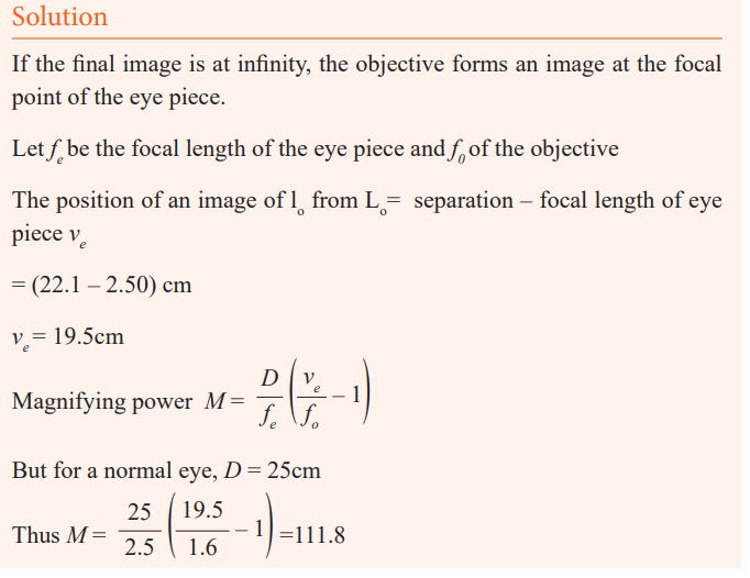
Viewing specimens
The purpose of this exercise is to view micro organisms found in pond
water while learning to operate a microscope.
Equipment
* Microscope
* Jar of pond water
* Slide
* Coverslip* Dropper
Procedure
1. Collect a jar of pond water containing micro organisms. To ensure
that you capture the largest number of micro organisms, do not
simply scoop a jar of water from the centre of a pond. Instead, fill the
jar partway with pond water and then squeeze water into the containerfrom water plants or pond scum.
3. Set up the microscope.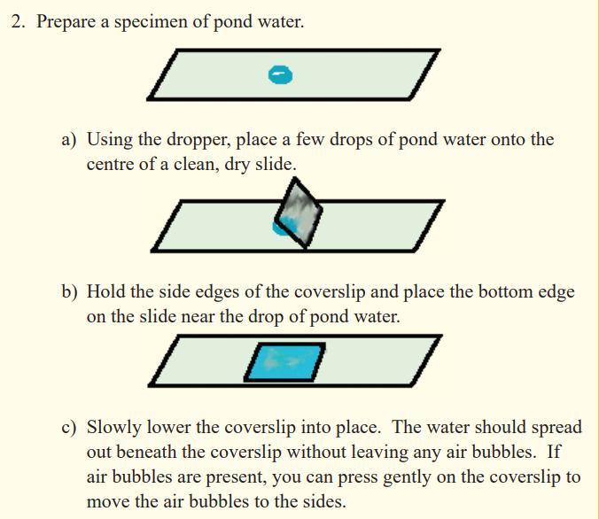
a) Remove the dust cover from the microscope.
b) Plug in the microscope.c) Turn on the microscope’s light source.
4. View the specimen with the low-power objective. Move the slide
around on the stage using your fingers or the control knobs until youfind a micro organism.
5. View the micro organism with the high-power objective.
6. Sketch a picture of the micro organism.
7. Repeat steps 4, 5, and 6 until you have sketched atleast five differentmicro organisms.
8. Turn off the microscope.
a) Carefully, lower the objective to its lowest position by turning
the coarse’ adjustment knob.
b) Turn off the light source.
c) Remove your slide. Clean the slide and cover slip with water.
d) Unplug the microscope and store it under a dust cloth.
Application activity 2.5
A compound microscope consists of a 10× eyepiece and 50× objective
17.0 cm apart. Determine (a) the overall magnification, (b) the focal length
of each lens, (c) the position of the object when the final image is in focuswith the eye relaxed. Assume a normal eye, so N = 25 cm.
Telescopes
Activity 30
You have heard in your early secondary that there are some heavenly and
distant earthly bodies that cannot be seen by our naked eyes. How did thepeople know that there exist such bodies?
Which instrument do you think is used to see these bodies and to observewhat takes place on these bodies?
Why do you think it is difficult to see distant objects using our eyes?
Telescopes are instruments used to view distant objects such as stars and other
heavenly bodies. Distant objects are difficult to see because light from them
has spread out by the time it reaches the eyes, and since our eyes are too smallto gather much light.
There are two kinds of telescopes; refracting telescopes and reflectingtelescopes.
Refracting telescopes
Activity 31
(i) Hold a convex lens of focal length 5cm close to your eye.
(ii) Hold another lens of focal length 20cm at an arm’s length.
(iii) Use the lens combination to view distant objects.
(iv) Adjust the distance of the farther lens until the image is clear (take
care not to drop the lenses).What type of image do you see?
The above lens combination is a refracting telescope. It is called a refracting
telescope because it forms an image of the object by refracting light. Therefore,
Refracting telescopes use lenses and they form images by refraction of light.Below are different kinds of refracting telescopes.
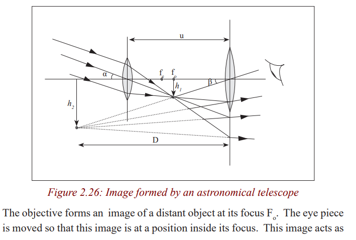
Astronomical telescope
The telescope made in the above activity is called an astronomical telescope.
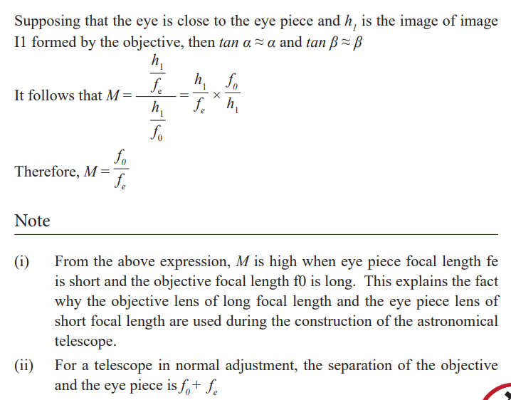
It consists of two convex lenses, the objective lens of long focal length and aneye piece lens of short focal length.
An astronomical telescope in normal adjustment
Activity 32
Using a telescope made in activity (30) above, view a distant object by
moving the lenses so that the eyes are relaxed.What do you think is the position of the image?
When the eyes are relaxed, the image is at infinity and the telescope is in
normal adjustment. Therefore, an astronomical telescope is in normaladjustment when the final image is formed at infinity.
The rays of light coming from a distant object form a parallel beam of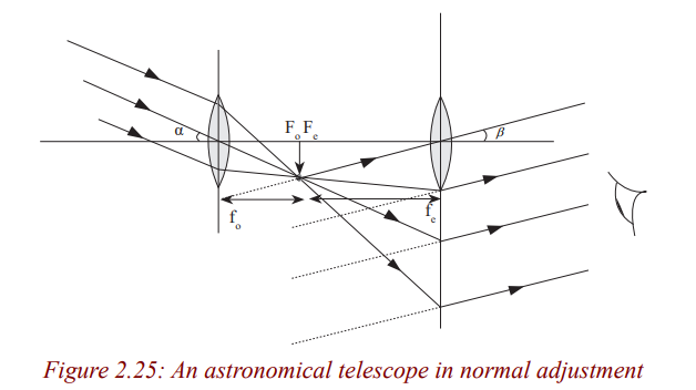
light. This parallel beam is focused by the objective lens and it forms a real,
diminished image at its principal focus Fo
. The eye piece is adjusted so that
this image lies in its focal plane. This image acts as the object for the eyepiece and the eye piece produces the image at infinity.
Note that in normal adjustment, the eye is relaxed or un accommodated whenviewing the image. In this case, the eye has minimum strain.
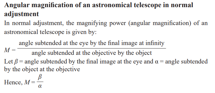
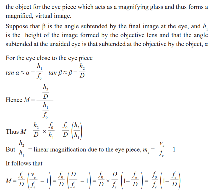
Magnifying power or angular magnification of an astronomicaltelescope
The magnifying power of a telescope is the ratio of the angle subtended by
the image to the eye when the telescope is used to the angle subtended at the
unaided eye by the object. Since the telescope length is very small compared
with the distance of the object from either lens, the angle subtended at the
unaided eye by the object is the same as that subtended at the objective by theobject.
Activity 33
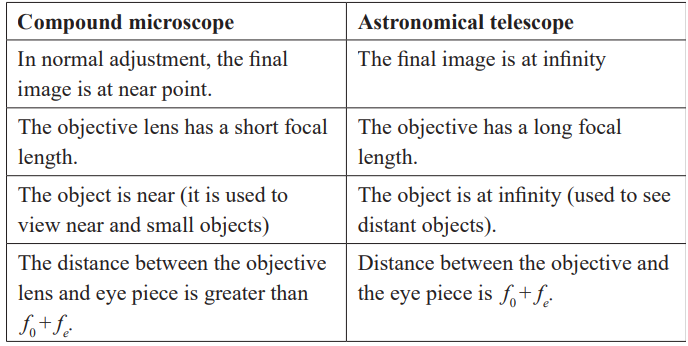
Discuss and give a summary of differences between a compoundmicroscope and an astronomical telescope.
The table below shows the differences between a compound microscope andan astronomical telescope.
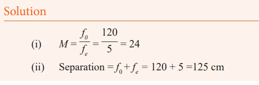
Example
An astronomical telescope has an objective lens of focal length 120
cm and an eye piece of focal length 5 cm. If the telescope is in normaladjustment, what is;
(i) The angular magnification (magnifying power)(ii) The separation of the two lenses?
Application activity 2.6
An astronomical telescope is used to view a scale that is 300 cm from the
objective lens. The objective lens has a focal length of 20cm and the eye
piece has a focal length of 2 cm. Calculate the angular magnification whenthe telescope is adjusted for minimum eye strain.
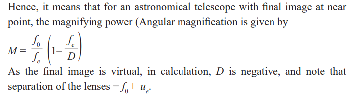
An astronomical telecope with the final image at the near point
In this case, the image is seen in detail but the telecope is not in normaladjustment (use) because the eyes are strained.
The eye ring
The eye ring is the best position to place the eye in order to be able to view
as much of the final image as possible. The best position for an observer to
place the eye when using a microscope is where it gathers most light from that
passing through the objective. In this case, the image is brightest and the field
of view is greatest. In case of the telescope, all the light from a distant object
must pass through the eye ring after leaving the telescope. So by placing
the eye at the eye ring, the viewer is able to see the final image as much aspossible.
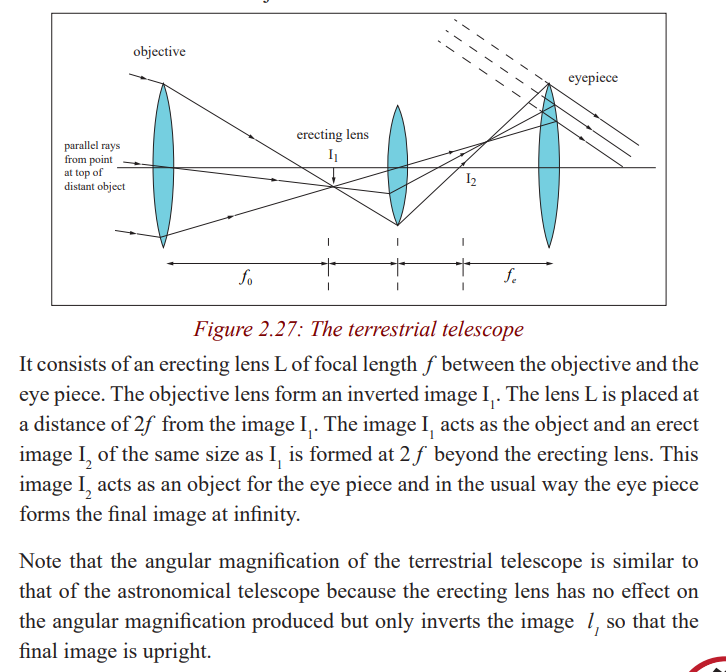
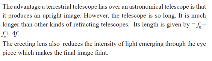
Terrestrial telescope
An astronomical telescope produces an inverted image, so it is not suitable
for viewing objects on the earth. It is suitable for viewing stars and other
heavenly bodies. A terrestrial telescope provides an erect image and thismakes it suitable to view objectives on the earth.
Activity 34
Discuss the advantages and disadvantages of a terrestrial telescope overan astronomical telescope.
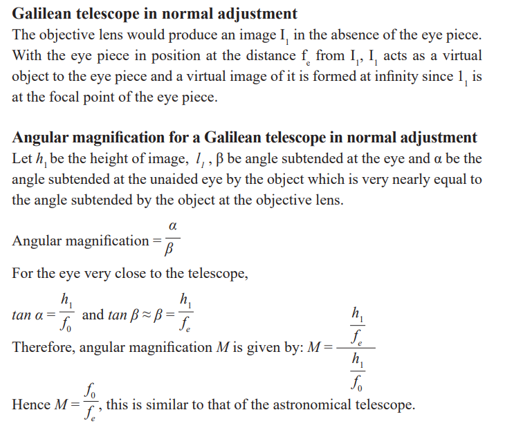
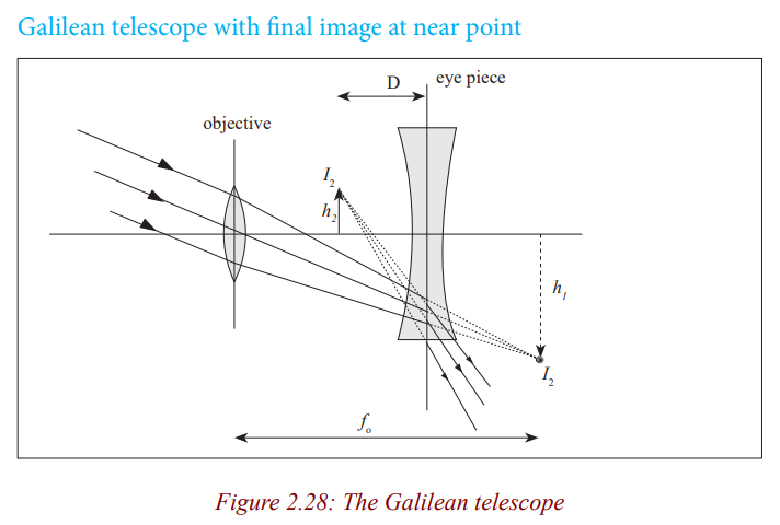
Galilean Telescope
Activity 35
(i) Hold a concave lens of focal length 5cm close to your eye.
(ii) Hold another convex lens of focal length 20cm at an arm’s length.
(iii) Use the lens combination to view distant objects.(iv) What is the nature of the image?
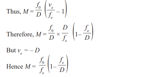
The above lens combination is a Galilean telescope. A Galilean telescope
consists of an objective lens which is a convex lens of long focal length and an
eye piece which is a concave lens of short focal length. It forms erect imagesboth in normal and not in normal adjustment.
Activity 36
Discuss the advantages and disadvantages of a Galilean telescope over anastronomical telescope and write them in your notebook.
Reflecting telescopes
Activity 37
Take the case of a TV satellite dish in the neighborhood. Discuss andexplain the functioning and principle of a satellite dish
Reflecting telescopes consist of a large concave mirror of long focal length as
their objective. There are three kinds of reflector telescopes, all named aftertheir inventors.
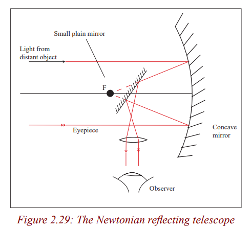
Cassegrain reflecting telescope
This is the type used in most observatories It consists of a concave mirror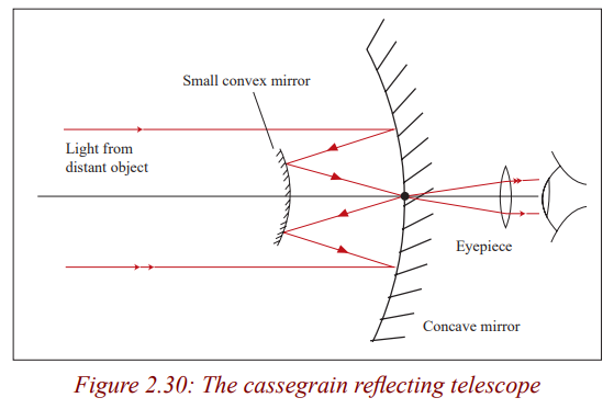
which acts as an objective, a small convex mirror and the eye piece lens. Light
from a distant object is reflected by the concave mirror to the convex mirror
which reflects it back to the centre of the concave mirror where there is a small
hole to allow the light through. So the convex mirror forms the final image(real) at the pole of the objective.
Coude Reflector Telescope
This is a combination of Newtonian and cassegrain reflector telescopes.
The plane and convex mirrors used in reflecting telescopes are used to bring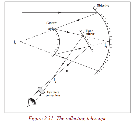
the light to a more convenient focus where the image can be photographed andmagnified several times by the eye piece for observation.
Activity 38
Discuss and explain the advantages of reflecting telescopes over refractingtelescopes.
The reflecting telescopes are free from chromatic aberration since no refraction
occurs. The image formed is brighter than in refracting telescopes where thereis some loss of light during refraction at the lens surfaces.
Spherical aberration can be eliminated by using a parabolic mirror instead of a
spherical mirror as an objective. They have a power because of higher ability
to distinguish two closely related objects because of the large diameter of
the parabolic mirror. We say that they have a high resolving power. They areeasier to construct since only one surface requires to be grounded.
Critical Thinking Exercise
What is meant by the resolving power of an optical instrument? Explain its
usefulness.
Explain why astronomers use reflecting telescopes rather than refractingtelescopes?
Prism binoculars
Activity 39
Have you ever asked yourself how tourists and scientists are able to see
distant animals and birds in a forest or any hidden places?Discuss with your neighbour and write in your notebook the observation.
Tourists and scientists use prism binoculars to view wild animals and birds inhidden places such as caves and forests.
These consist of a pair of refracting astronomical telescopes with two totally
reflecting prisms between each objective and eyepiece. The prisms use total
internal reflection to invert rays of light so that the final image is seen the
correct way. These prisms reflect up and down the light and by doing so, theyshorten the length of the instrument.
Prism A causes lateral inversion and prism B inverts vertically so that the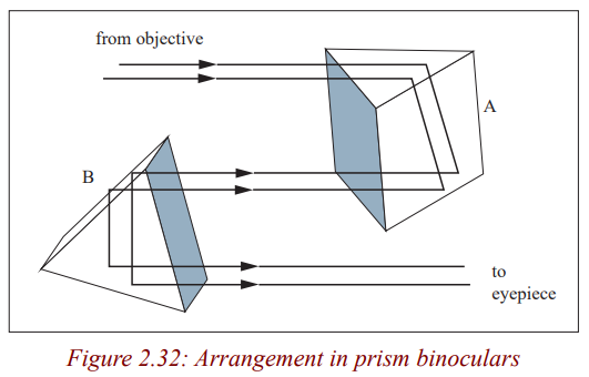
final image is the same way round and same way up as the object. Each prism
reflects light through 180o
. This makes the effective length of each telescope
three times shorter than the distance between the objective and the eye piece.So good magnifying power is obtained with compactness.
END UNIT ASSESSMENT
1. A certain nearsighted person cannot see distinctly objects beyond 80
cm from the eye. What is the power in diopters of the spectacle lensesthat that will enable him to see distant objects clearly?
2. Explain the difference between the terms magnifying power and
magnification, as used about optical systems. Illustrate this, by
calculating both, in the case of an object placed 5.0 cm from a simple
magnifying glass of focal length 6.0 cm, assuming that the minimumdistance of distinct vision for the observer is 25 cm.
3. An eyepiece is made of two positive thin lenses, each of focal length f
= 20 mm, separated by a distance of 16 mm.
(a) Where must a small object viewed by the eyepiece be placed so
that the eye receives parallel light from the eyepiece?
(b)Does the eye see an erect image relative to the object? Is it
magnified?(c) Use a ray-trace diagram to answer these questions by inspection.
4. A common telephoto lens for a 35 mm camera has a focal length of
200 mm; its range from to (a)What is the corresponding range of
aperture diameters? (b)What is the corresponding range of imageintensities on the film?
5. What is the maximum stop rating of a camera lens having a focal
length of and a diameter of ? If the correct exposure at , whatexposure is needed when the diaphragm setting is changed to ?