UNIT 5: DIVERSITY OF SPECIALIZED TISSUES
UNIT 5: DIVERSITY OF SPECIALIZED TISSUES
Key Unit Competence
Describe different specialized plant and animal cells and adaptation of tissues.
Learning objectives
By the end of this unit, I should be able to:– Define a tissue as a group of cells with similar structure working together forIntroductory activity
a function.
– Name the main types of animal and plant tissues.
– Define an organ as a structure made up of a group of tissues with related
functions working together to perform bodily functions.
– Explain how epithelial tissues are adapted to perform a diversity of functions
in the body.
– State the advantages and disadvantages of being unicellular.
– Observe and draw plant and animal tissues as seen under a light microscope.
– Interpret photomicrographs of plant and animal tissues
– Acknowledge the relationship between levels of organization
– Recognize the efficiency shown by multicellular organisms to explore more
modes of life that are not available to single celled organisms that show little
or no specialization
Read the following passage and use it to answer the following questions:
In an anthill, there are different groups of termites such as a queen, workers and
soldiers. Each group has a specific role to play in the colony. The structure termites
of each group is related to their role for example soldiers that protect the colony
have mouth parts shaped like a pair of scissors building and a slightly larger
abdomen for storing water. The queen is the largest of all and has a role of laying
eggs. Workers have mouth parts for cutting and chewing food or soil particles.
Some members of workers are in charge of caring for the young while others find
food and defend the colony or remove dead members. Their specialization anddivision of labor bring about efficiency in the colony.
1. Specify the message addressed by the above paragraph.
2. Explain how is the structure of termites related to their functions?
3. What is the significance of specialized tissues in multicellular organismslike plants and animals?
The study of tissues is known as Histology. A tissue is a group of associated,
similarly structured cells that perform specialized functions for the survival of the
organism. In histology, differentiation is the process by which structures become
modified and specialized to perform specific functions. Differentiation is also known
as ‘specialization’. In animals, the first type of cells in the developing embryo is stem
cells. These are unspecialized cells that go on to form all the different types of cellsin adult.
5.1. Specialized plant tissues
Activity 5.1.1
– Remove an epidermis layer from the ventral side of an onion leaf.
– Mount it on the slide containing a drop of iodine solution
– Observe your preparation under a light microscope
– Draw, label and describe your findings.
– From your discussion:1. What is a tissue?2. What is the role of epidermis in onion?
5.1.1. Plant tissues
Activity 5.1.2
The following figure represents the flow chart of subdivisions of plant tissues. Useit to answer the following questions.
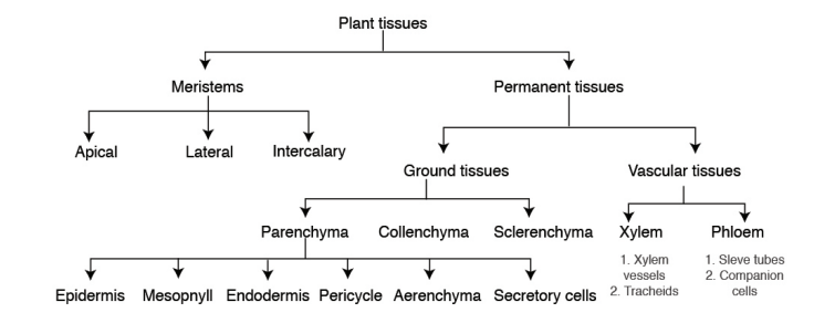 1. How do meristems differ from permanent tissues?
2. Plant tissues are classified into ground tissues and vascular tissues as
shown in the figure above. What is meant by the term vascular tissues?
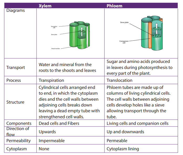
3. How is the structure of the xylem and phloem vessels related to their
function?4. From the flow diagram above, identify three types of ground tissues.Plant tissues can be divided into two main groups, Meristematic tissues (apical, lateral,
5. Write down short notes on each of the following types of meristems.
a. Apical meristems.
b. Lateral meristemsc. Intercalary meristems
and intercalary meristems) and Permanent tissues (ground tissues and vascular tissues).
5.1.2. Meristem tissues
Meristem tissue is a group of cells which retain the ability to divide by mitosis.
Meristematic tissues are specialized to carry out specific functions such as
reproduction, growth, photosynthesis and replacement of old or damage tissues.
The cells making a meristem tissue are small, have a central large nucleus and dense
cytoplasm, thin-walled, with no or small vacuole, and no specialized features. Thecells are rectangular and closely packed with no intercellular air spaces.
Types of meristematic tissues
Meristematic tissues are subdivided into apical meristems, lateral meristems
(cambium) and intercalary meristems
a. Apical meristems
They are located in the root and shoot apex (at the growing points of roots and
stems). They are responsible for primary growth, leading to the increase of primary
plant body.
b. Lateral Meristems (cambium)
Lateral meristems are in lateral parts of the plant, where they are responsible for
secondary growth. The cambium gives rise to secondary vascular tissues (secondary
xylem and secondary phloem) in dicotyledonous plants.
c. Intercalary meristems
These are found in the region of permanent tissues like at nodes of monocotyledonousplants (e.g. sugar cane). It allows growth in length to occur between internodes.
Functions of meristematic tissues– The main function of meristematic tissue is to produce new cells by mitosis.5.1.3. Permanent tissues
The cells elongate and differentiate to form new cells for primary growth of
shoot and root.
– Vascular cambium produces new cells to increase the diameter of stems and
roots during secondary growth.
– Cork cambium called (phellogen) produces the outer cork layer called phellem
which consists of suberized cells. The cork layer reduces water evaporation
from the plant and protects the plant against the entry of pathogens.
– The intercalary meristems allow growth and increase in length in regions otherthan the tips.
Permanent tissues consist of two groups of tissues such as: ground and vascular
tissues.
5.1.4. Ground tissues
The ground or fundamental tissues are plant tissues which function in storage,
metabolism and support. There are three types of ground tissues: parenchyma,collenchyma and sclerenchyma tissues.
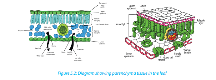
5.1.5. Parenchyma tissues
Parenchyma is a soft plant tissue made up of thin-walled cells that forms the
greater part of leaves, stem pith, roots, and fruit pulp. They are the main sites for
physiological and biochemical processes in the plants including photosynthesis,
protein synthesis and storage of starch and mineral ions. Parenchyma tissues can be
found in epidermis, mesophyll, endodermis, pericycle, aerenchyma and secretory
cells.
Characteristics– Parenchyma tissues consist of large living cells, with relatively thin wall
containing cellulose, pectin and hemicellulose.
– Parenchyma tissues consist of cells, usually having a large central vacuole.
They are often partially separated from each other.– Spongy cells present intercellular spaces that intervene in gaseous exchange
and transpiration through stoma. They are usually stuffed with plastids.– Parenchyma tissues consist of cells with polygonal and spherical shapes inFunctions of parenchyma tissues
the leaf. They form the mesophyll, and are located between upper and lowerepidermises. They are responsible for photosynthesis.
– In the leaves, parenchyma tissues form the mesophyll and are sites for
photosynthesis, gaseous exchange and transpiration.
– They store food substances such as starch, proteins and lipids
– They can be modified to form specialized cells to carry out other function inepidermis, endodermis, pericycle, parenchyma, and secretory cells.
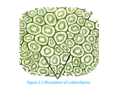
Adaptations of parenchyma for its function– Parenchyma tissues are made of unspecialized cells with variety of functions:5.1.6. Collenchyma tissues
– Parenchyma can become specialized to carry out specific functions e.g.
mesophyll has cells with many chloroplasts, and aerenchyma which has air
spaces. All of these adaptations help in photosynthesis and gas exchange.
– They have isodiametric cells and function as packing tissue and storage tissue.
– Cells are loosely packed with many large intercellular spaces. This permits
diffusion of gases.
– They have thin cellulose cell wall which is permeable so that it permits passage
of materials.
– The walls are transparent and permit entry of light in photosynthesis cells.
– Large cells with large vacuoles provides space for storage of substances, where
the entry of water causes vacuole to expand and cells become turgid
– Leucoplasts act as storage of starch while chromoplasts present in some cellse.g. in petals attract insects for pollination.
Their cells are elongated with irregularly thickened cell walls that provide structural
support, particularly in growing shoots and leaves. Their thick cell walls are composed
of cellulose and pectin. These cells are often found under the epidermis, or the outerlayer of cells in young stems and in leaf veins.
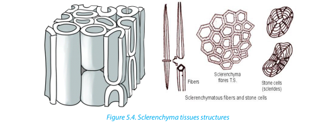
 Sclerenchyma is found in hard parts of the plant body. They are very common in
5.1.7. Sclerenchyma tissues
roots, stems, leaves and petioles. They may be present in patches, groups or layers.
The cells of the sclerenchyma are dead, they are elongated, narrow, and thick walled
and lignified. They are pointed at both ends where it gives strength, rigidity and
flexibility to the plant body. They consist of fibres and sclereids. Fibres are long,
narrow, thick and liquefied cells usually tapering at both ends. Sclereids cells arenormally short with very thick walls, irregular and not tapering at the ends.
5.1.8. Vascular tissues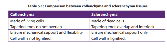
The vascular tissue system consists of two kinds of conducting tissues: the xylem
responsible for conduction of water and dissolved mineral nutrients, and the
phloem responsible for conduction of elaborated food.
a. Xylem
The xylem tissues are made of dead cells which have the cell walls removed at the
end of the cells, forming tubes through which the water and dissolved mineral ions
can flow. Xylem vessels are involved in the movement of water through a plant -
from its roots to its leaves via the stem. During this process water is absorbed from
the soil through root hair cells, moves by osmosis from root cell to root cell until it
reaches the xylem, and finally it is transported through the xylem vessels up thestem and then to the leaves.
Xylem vessels are hollow tubes or lumen with a thick strengthened cellulose cell
wall. The hollow tubes act like pipes allowing water and dissolved minerals to flow
through them. They develop from cylindrical cells arranged end to end, in which
the cytoplasm dies and the cell walls between adjoining cells breaks down leaving
a dead empty tube. The cell walls in xylem vessels contain a substance called ligninwhich strengthens the cells and gives structural support.
b. Phloem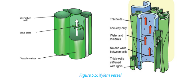
Phloem vessels are involved in translocation of elaborated substances. Dissolved
sugars, produced during photosynthesis, and other soluble food molecules are
moved from the leaves to growing tissues such as the tips of the roots and shoots
and storage tissues such as in the roots. In contrast to xylem, phloem consists of
columns of living cells. The cell walls of these cells do not completely break down,
but instead form small holes at the ends of the cell. The ends of the cell are referred
to as sieve plates. The connection of phloem cells effectively forms a tube which
allows dissolved sugars to be transported.
Phloem tubes carry food substances like sugar and amino acids produced in leaves
during photosynthesis to every part of the plant. The movement of food substancesthrough the plant is called translocation.
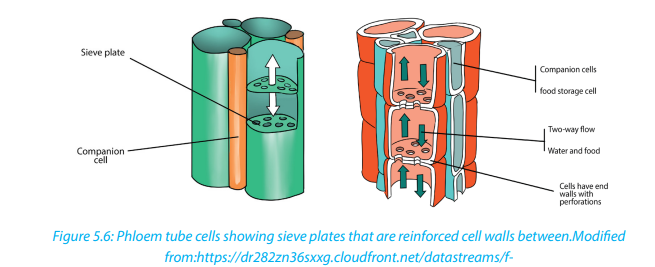
Table 5.2: Comparison between Xylem and Phloem tissues
Self-assessment 5.1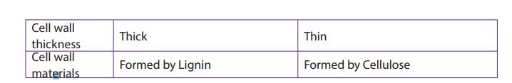
1. State where in a flowering plant you would find:a. Lateral meristem2. Give characteristics of meristematic cells.
b. Intercalary meristem
c. Apical meristem
3. What do you understand by the following terms?a. Differentiation4. Differentiate between Collenchyma and sclerenchyma
b. Cambium
c. Wood
d. Meristem
5. State the main structures (components) that make up a xylem and phloem
tissues.
6. Explain how the structure of Parenchyma and Xylem tissues are suitable to
their functions.
7. The diagram below shows a longitudinal section of two cells of phloem tissue
in a plant stem.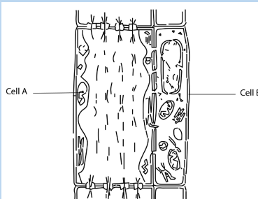
a. Name the cells labelled A and B on the diagram.b. State the function of phloem in a plant.
5.2. Animal tissues
Activity 5.2
Conduct a research by using different sources of information to find out the
structures and the main functions of the following four groups of animal tissues:epithelial, connective, muscular and nervous tissues.
There are four basic types of animal tissues such as epithelial tissue, muscle tissue,nervous tissue, and connective tissue.
5.2.1. Epithelial tissue
Epithelial tissue consists of closely packed cells arranged in single or multilayered
sheets. It is made up of layers of tightly packed cells that form the external surfaces
of the body and cover the outer and the inner surfaces of the organs. Some are
specialized to form glandular tissues (glands). The epithelium lining the inside of the
heart, blood vessels and lymph vessels is referred to as endothelium. Two criteria
for classifying epithelia are: the number of cell layers and the shape of cells on
the free surface. The following are the types of epithelium tissues:
a. Simple cuboidal epithelium
This is a tissue with cells that are cubical in shape. Cuboidal cells are specialized
for secretion and they make up the epithelia of kidney tubules and many glands
including salivary glands, and thyroid gland.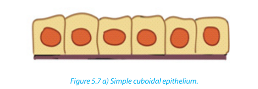
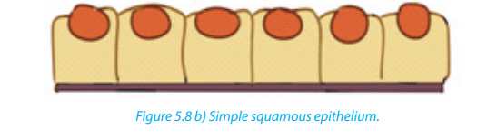
b. Simple squamous epithelium
It is thin, leaky and functions in the exchange of material by diffusion. This type of
epithelium lines blood vessels and the air sacs of lungs, where diffusion of nutrientsand gases is critical.
c. Simple columnar epithelium
These are columnar in shape with free surface containing extensions of micro villi.
It lines the intestines. This epithelium secretes digestive juices for the final stages of
digestion and absorbs nutrients to blood stream.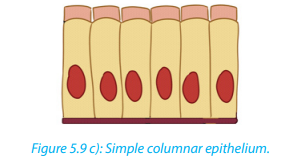
d. Pseudo-stratified ciliated columnar epithelium
It forms a mucous membrane that lines the nasal passages of many vertebrates. Thebeating cilia move the film of mucus along the surface.
e. Stratified squamous epithelium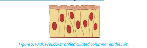
It regenerates rapidly by cell division near the basal lamina. The new cells are pushed
outward to replace cells that are sloughed off. This epithelium is commonly found
on surfaces subject to abrasion, such as the outer skin and lining of the esophagus,anus, and vagina.
f. Transitional epithelium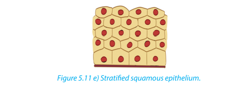
In this type of stratified epithelium, the surface cells change their shape from round
to squamous. Transitional epithelium lines urinary bladder. When the bladder is
empty, the surface cells are rounded. As the bladder fills urine, these cells become
flattened. Transitional epithelium enables the bladder to fill and stretch withouttearing the lining.
g. Stratified columnar epithelium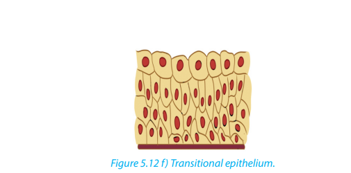
It is a rare type of the epithelial tissue composed of column shaped cells arranged in
multiple layers. They are found in the conjunctiva or the eye, in parts of the pharynx,anus, uterus, the male urethra and vas deferens.
h. Stratified cuboidal epithelium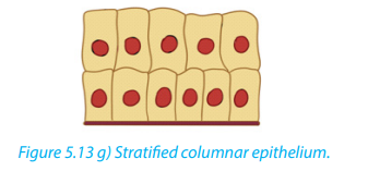
It is a type of epithelial tissue composed of multiple layers of cube-shaped cells. Only
the most superficial layer is made up of cuboidal cells and the other layers can be
cells of other type. It has several locations in the body including sweat gland ducts,egg-producing vesicles and ovaries.
5.2.2. Main characteristics of epithelial tissues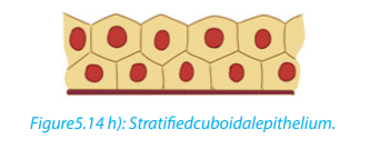
a. Polarity
All epithelia have a free surface and a lower attached basal surface that differ in
structure and function. For this reason, epithelium is described as showing polarity.
b. Supported by connective tissue
All epithelia are supported by connective tissue. For instance, deep to the basal
lamina is reticular lamina, an extracellular material containing collagen protein
fiber which forms the basement membrane. The basement membrane reinforces
the epithelium and helps it to resist stretching and tearing.
c. They are avascular; epithelia have no blood vessel in them. Nutrients and gasesare supplied by blood through the connective tissue by simple diffusion
d. Regeneration
Epithelium have a high regenerative capacity and can reproduce rapidly as long as
they receive adequate nutrition.
Functions of epithelium– Epithelium forms a protective layer: The epithelium of the skin protects the5.2.3. Muscular tissues
body from mechanical damage, entry of pathogens, ultraviolet rays and
dehydration. Epithelium lining the respiratory air passages secretes mucus
which traps inhaled dust particles and microbes.
– The ciliated epithelium cells have cilia that propel the mucus and trapped
particles to the throat.
– Glandular tissues secrete the digestive enzymes, hormones, mucus, sweat and
sebum.
– Acts as a barrier and regulates movement of substances across kidney
– Some epithelial cells can divide mitotically producing new cells to replace
damaged or dead cells.
– Some epithelial cells such as taste buds and retina cells are specialized to formsensory receptors.
Muscle tissues consist of elongated cells held together by connective tissue. Muscle
cells are highly specialized in that they are able to shorten to a half or even a third of
their resting length by the process of contraction. The contraction is caused by two
types of fibrous proteins: myosin and actin.
Muscles in the body provide the necessary force for the motion and they convert
chemical energy into kinetic or mechanical energy. There are three types of muscle
tissue:– Smooth muscle which is found in the inner linings of organs;Smooth and cardiac muscles are involuntary muscles whereas skeletal muscles are
– Skeletal or striated muscle, which is attached to bone and helps in movement
of the body;
– Cardiac muscle which is found only in the heart.called voluntary muscles because they are under voluntary (conscious) control.
a. Smooth Muscle
Smooth muscle is also called unstriated, unstriped, involuntary or visceral muscle.
It is found in the walls of the hollow internal organs such as blood vessels, intestinal
tract, urinary bladder, and uterus. Smooth muscles have the following features;– It is under control of the autonomic nervous system; they cannot be controlled
consciously, so they are also called involuntarily muscle. They do not have
striations.– Smooth muscle cells contract slowly and rhythmically
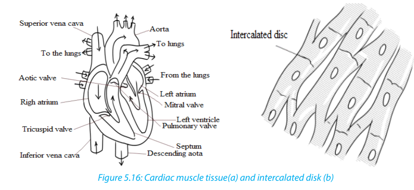
b. Cardiac tissue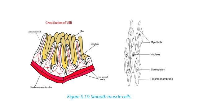
Cardiac tissue (figure 5.10 a) is found in the walls of the heart and it is under control
of the autonomic nervous system. Cardiac muscle has the flowing basic features.
– It contracts and relaxes continuously.
– It is branched and connected to other cardiac muscle fibers through
intercalateddiscs (Figure 5.16 b), which are reinforced membranes that hold the
cells together during contractions. These interconnections or intercalated discs
between the fibers ensure a rapid and uniform spread of excitation throughout
the wall of the heart which in turn ensures a synchronous contraction.– They are myogenic (their contraction originate from within the heart itself).
c. Skeletal Muscle
Skeletal muscle is also called striated, striped, or voluntary. They are attached
to bone, and are responsible for body movements and body posture. There areapproximately 639 skeletal muscles in the human body.
Characteristics of skeletal muscles:
– They are under control of voluntary nervous system
– They are attached to bone and this is the reason why they are called skeletal
muscles.
– They are made of elongated and cylindrical muscle fibres
– They appear under microscope to have alternate light and dark bonds and this
is why they are called striated muscles.
– Their muscle fibres are multinucleated (many nuclei per cell)
– These muscle cells also contain light and dark stripes called striations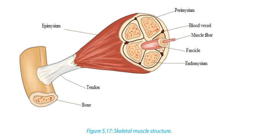
General functions of muscle
The main function of muscle is its contribution to motion, where body movements
such as walking, breathing, and speaking, as well as movements associated with
digestion and the flow of fluids take place. Muscles contribute to the heat production,
maintenance of posture and body support and communication through facial
expression, writing and speech.
5.2.4. Nervous tissue
Nervous tissue is composed principally of densely packed cells called the nerve cells
(neurons) that together form the nervous system including the brain and spinal
cord. Neurons are specialized for transmitting electrical nerve impulses.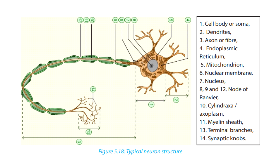
A typical neuron has three main parts: Cell body, dentrites and axon.
a. The cell body or soma– It is the main part from which, extensions derive (Axon and Dendron).all nerve cell activities.
– It is made of a great spherical nucleus, granular cytoplasm and controls
b. Dendrites (Dendron when single): small branches attached to the cell body
and receive nerve impulse from other neurons
c. Axon or cylindrax:– It is the thinner nerve fibre that carries messages away from the cell5.2.5. Connective tissues
body and can be as long as 1 m. In some neurones, the axons have a
fatty myelin sheath formed by Schwann cells which wrap themselvesaround the axon to increase the speed of impulse transmission.
Connective tissue is made up of many different types of cells that are all involved in
structure and support of the body. Bone, blood, fat, and cartilage are all connective
tissues. Connective tissues can be densely packed together, as bone cells are or
loosely packed, as adipose tissue (fat cells) are. A connective tissue is made up of a
variety of cells embedded in a large amount of intracellular substance called matrixand fibers which are non-living products of the cells.
a. Common functions of connecting tissues:
– Connective tissues protect and support the body and internal organs.
– They act as connecting systems, binding all other tissues together.– They also form surrounding sheaths to separate the various organs.
b. Cells of connective tissue
The specialized cells of the various connective tissues produce the extracellular
matrix. The names of the cells end with suffixes that identify the cell functions as
blasts, cytes, or clasts. Blasts create the matrix, cytes maintain it, and clasts break it
down for remodeling. For example: Fibroblasts are cells that form fibrous connective
tissue and fibrocytes maintain it, chondroblasts form cartilage and chondrocytes
maintain it, and osteoblasts form bone, osteocytes maintain it, and osteoclastsbreak it down
Adipose, or fat cells, also called Adipocytes, contain large amounts of lipid. The
lipid pushes the rest of the cell contents to the periphery, so that each cell appears
to contain a large and centrally located lipid droplet with a thin layer of cytoplasm
around it. Adipose cells are rare in some connective tissue types like cartilage but
they are abundant in others like loose connective tissue, and they are predominant
in adipose tissue.
Mast cells are commonly found beneath membranes in loose connective tissue
and along small blood vessels of organs. They contain chemicals such as heparin,
histamine and proteolytic enzymes. These substances are released in response toinjury such as trauma and infection and play important roles in inflammation.
White blood cells continuously move from blood vessels into connective tissues.
The rate of movement increases dramatically in response to injury or infection. In
addition, accumulations of lymphocytes, a type of white blood cell, are common
in some connective tissues, such as in the connective tissue beneath the epitheliallining of certain parts of the digestive system.
Macrophages are found in some connective tissue types. They are derived from
monocytes, a white blood cell type. Macrophages are either fixed and do not
move through the connective tissue in which they are found or are wandering
macrophages and move by amoeboid movement through the connective tissue.
Macrophages phagocyte foreign or injured cells, and they play a major role inproviding protection against infections.
Note that there are three structural major components of the extracellular matrix of
connective tissue such as fluid, ground substance consisting of non-fibrous protein
and other molecules and protein fibers. The structure of the matrix gives connective
tissue types most of their functional characteristics, such as the ability of bones and
cartilage to bear weight, tendons and ligaments to withstand tension, and dermis of
the skin to withstand punctures, abrasions, and other abuses.
c. Fiber connective tissues
Another type of connective tissues consists of fibers. Fibers are of different types
including:
– Connective tissue fibers: which are made of protein and are of three kinds:
collagenous, elastic and reticular fibers.
– Collagenous fibers: These provide strength combined with flexibility. They
are made up of collagen, probably the most abundant protein in the animal
kingdom.
– Elastic fibers: These are easily stretched but are also resilient, snapping back
to their original length when tension is released. Shaped as long threads,
elastic fibers are made of a protein called elastin.
– Reticular fibers: These are thin collagen fibers coated with glycoprotein. They
are very short, thin fibers that branch to form a network and appear different
microscopically from other collagen fibers. Reticular fibers are not as strong as
most collagen fibers, but networks of reticular fibers fill space between tissues
and organs.
d. Loose connective tissue
This is also called areola connective tissue and is the most widespread connective
tissue in all animal tissues. It binds epithelial tissues to underlying tissues and
functions as packing material, holding organs in place. Loose Connective tissue
has the following main components;– Fibers: collagen, elastic and reticular.e. Fibrous connective tissue
– Cells; fibroblasts and macrophages. Fibroblasts secrete the protein ingredients
of the extracellular fibers. Macrophages are cells that roam the maze of fibers,
engulfing both foreign particles and the debris of dead cells by phagocytosis.
Fibrous Connective tissue is dense with collagenous fibers. The fibers form parallel
bundles, which maximize non-elastic strength. Fibrous Connective tissue is found in
tendons, which attach muscles to bones, and ligaments, which connect bones atjoint.
f. Adipose tissue
Adipose tissue is a specialized form of loose connective tissue that stores fats in adipose
cells distributed throughout its matrix. Adipose tissue consists of adipocytes, or
fat cells, which contain large amounts of lipid. Unlike other connective tissue types,
adipose tissue is composed of large cells and a small amount of extracellular matrix
that consists of loosely arranged collagen and reticular fibers with some scattered
elastic fibers. Blood vessels form a network in the extracellular matrix. The fat cells
are usually arranged in clusters or lobules separated from one another by looseconnective tissue. Adipose tissue functions as:
– An insulator against heat loss
– A protective tissue to delicate internal organs– A site of energy storage in the form of fat.
g. Bone and Cartilage tissue
Cartilage has an abundance of collagenous fibers embedded in a rubbery matrix
made of a protein-carbohydrate complex called chondroitin sulfate. Cartilage is
composed of specialized cells, called chondrocytes, surrounded by a gelatinous
matrix of collagen, a tough protein. The cartilage surface is covered by a membrane
known as the perichondrium. There are three types of cartilage (hyaline cartilage,yellow elastic and white fibrous cartilage.)
– Hyaline cartilage is semi-transparent and is often stained light blue or pink inBone tissue
tissue sections. It is extremely very strong but very flexible and elastic. Hyaline
cartilage occurs in the trachea, larynx, tip of the nose, connection between the
ribs and the breastbone; and at the ends of bones where they form joints. It
also forms much of the fetal skeleton.
– Elastic cartilage is similar to hyaline cartilage, but in addition to the collagenous
fibers.The matrix of the elastic cartilage also contains an abundant network of
branched elastic fibers. This type of cartilage is found in the lobe of the ears,
the epiglottis and in the parts of the larynx. They provide flexibility and elastic
support.
– Fibro-cartilage(White fibrous cartilage) is an extremely tough tissue. It is found
as discs between the vertebrae, bones, anterior joint between the two halves
of pelvic girdle and at points where tendons inserted on bones near hyalinecartilage. It resists compression and absorbs shock in some joints.
This is a firmer and denser material that has the following features:– Hard and compactThe following are the main functions of bone tissue:
– Has many collagen fibres
– Its matrix has inorganic salts such is calcium carbonate and calcium phosphate
– Has few cells located in the lacunae in the matrix
– Has osteoblasts as mature and non-dividing cells
– Have a harversian canal– Consists of irregular cylinder with layer of matrix call lamellae
– Structural support of the bodyh. Blood tissue
– Protection of internal organs, heart and lungs.
– Attachment of the muscles to effect movement– Production of blood cells
Blood is a flowing made up of particles suspended in a fluid composed of fluid called
plasma, and several kinds of cells. Within the blood plasma, there are erythrocytes
(red blood cells), leukocytes (white blood cells), thrombocytes (platelets) and othersubstances. Blood performs the following important functions:
Transport– Blood transports absorbed substances such as glucose, amino acids, mineralHomeostasisions and vitamins from the small intestine.
– Blood transports the respiratory gases (Oxygen and Carbon dioxide).
– Blood transports the excretory wastes such as urea, uric acid to excretory
organs to be removed out of the body.
– Blood transports hormones e.g. insulin from pancreas to the liver where it isstored.
Na+ affects the water potential of the blood and regulates the diffusion of water
between blood and tissues. Hydrogen carbonates help to maintain the pH of the
blood.
Protection– Leucocytes such as neutrophils and macrophages engulf pathogens e.g.Self assessment 5.2
bacteria
– B-lymphocytes produce antibodies to destroy pathogens or to neutralize
toxins.
– T-lymphocytes destroy infected cells.
– Platelets, fibrinogen and prothrombin play an important role in blood clottingto reduce blood loss and the entry of pathogens.
You are provided with photomicrographs or slides of different plant and animal
tissue. Study them carefully and answer questions that follow.
Identify the different tissues provided and where they are located.One of the images is a blood smear. Draw a well labeled diagram of this tissue
5.3. Levels of organization: cell, tissue, organ and system
Activity 5.3
Visit a classroom block, administration block or any building in school which is
constructed with bricks and use it to answer the following questions.
1. What is the smallest unit or component of the classroom block?
2. How are bricks arranged?
3. Do you think the brick has other smaller particles in it?
4. How many bricks does a classroom block have?
5. How are walls, classrooms, washrooms and other apartments of the block
formed?
6. Arrange the following in their ascending order of size (from the smallest to
the largest); whole block, wall, a brick, a room, course (a line of bricks).
7. Relate the above arrangement of a building to levels of organization inmulticellular organisms, beginning with a cell and ending with an organism
The human body is organized into structural and functional levels of increasing
complexity. Each higher level incorporates the structures and functions of the
previous level. The simplest is the cells, organized into tissues, organs, and organ
systems. All of the levels of organization of the human body are represented in thefollowing figure.
5.3.1. Cells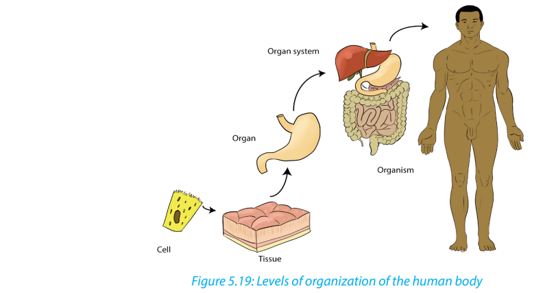
The smallest structural and functional living units of living things are cells. There are
many different types of human cells, though they all have certain similarities. Each
type of cell is made of chemicals and carries out specific chemical reactions.
5.3.2. Tissues
A tissue is a group of cells with similar structure and function. There are four groups
of tissues (Epithelial tissues, Connective tissues, Muscle tissues, Nerve tissue)
5.3.3. Organs
An organ is a group of tissues precisely arranged so as to accomplish specific
functions. Examples of organs are the kidneys, individual bones, the liver, lungs, and
stomach. The kidneys contain several kinds of epithelial or surface tissues, for their
work of absorption. The stomach is lined with epithelial tissue that secretes gastric
juice for digestion. Smooth muscle tissue in the wall of the stomach contracts to
mix food with gastric juice and propel it to the small intestine. Nerve tissue carriesimpulses that increase or decrease the contractions of the stomach.
5.3.4. Organ systems
An organ system is a group of organs that all contribute to a particular function.
Examples are the urinary system, digestive system, and respiratory system. For
example, the urinary system consists of the kidneys, ureters, urinary bladder, and
urethra. These organs all contribute to the formation and elimination of urine.
The Human body has 11 organ systems: circulatory, digestive, endocrine, and
excretory (urinary), the lymphatic, integumentary, muscular, nervous, reproductive,respiratory, and skeletal systems.
Table 5.3: Major organ systems of the human body
Self-assessment 5.3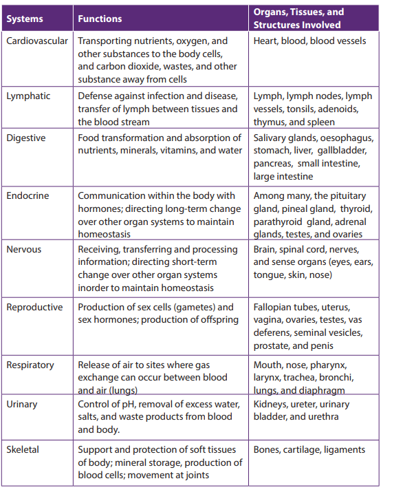
1. Answer by true or falsea. Organic chemicals are often very complex and always contain the element2. Explain why the cell as level of organization of human body is said to be:
carbon only.
b. A tissue is a group of cells with similar structure and function.
c. Integumentary organ system plays the role in protection of the human
body from injury and fluid loss.
d. An organ system is a group of organs that all contribute to a particular
function.a. Basic unit of human body5.4. Advantages and disadvantages of being Unicellular or
b. Structural unit of human bodyc. Functional unit of human body
Multicellular
Activity 5.4
Discuss the advantages and disadvantages of an organism being unicellular orMulticellular
5.4.1. Advantages of unicellular organisms– Unicellular organisms need fewer nutrients and can survive in unfavorable5.4.2. Disadvantages of unicellular organisms
conditions.
– Some of the organisms can generate energy through photosynthesis.
– Sometimes different bacteria work together to work to their advantages.
– Unicellular organisms can multiply quickly and have less energy/resourcedemands.
Unicellular organisms only have one cell that is used to function their entire being.
This is a disadvantage compared to multicellular organisms, which have many cells
and function more easily and properly.
5.4.3. Advantages of a multicellular state of an organism– Multicellular organism usually has a wider range of functions because of the
aggregation of different types of cells.
– Multicellular organisms have many more necessities and can only survive in
certain conditions.
– Multicellular organisms such as animals are unable to make their own food sothey survive by eating living things such as vegetables, fruits, and meat.Self-assessment 5.4They can also eat things that are produced by other living things such as eggs, milk, and honey.
1. Give the advantages and disadvantages of being Unicellular organisms.2. Describe how unicellular organisms perform their functions.
End of unit assessment 5
1. Which type of tissue forms glands?a. Epithelial2. What are the four types of animal tissues?
b. Connective
c. Nervous
d. Musclesa. Epithelial, squamous, muscular, connective3. Which type of the tissues form glands
b. Epithelial, connective, muscular, cardiac
c. Connective, muscular, epithelial, nervous
d. Cuboidal, ciliated, glandular, columnara. Epithelial4. Describe how epithelial tissues have adapted to their functions
b. Connective
c. Nervous
d. Muscle
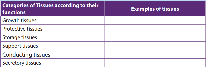
5. Describe the three main functions of the blood6. Complete the following table by filling in the examples of the respective tissues: