Topic outline
UNIT 1: INTRODUCTION TO BIODIVERSITY
Key Unit Competence
Explain how diversity is threatened by climate change and human activities
Learning objectives
By the end of this unit, I should be able to:– Define the terms: species, ecosystem and niche.
– Explain that biodiversity is considered at three different levels
– Evaluate the consequences of loss of biodiversity.
– Characterize the biotic and abiotic components that define Rwanda’s
ecosystems (example: freshwater, marine, and terrestrial).
– Apply Simpson’s Index of Diversity.
– Explain the importance of random sampling in determining the biodiversity
of an area.
– Use suitable survey methods such as frame quadrats, line and belt transects to
assess the distribution and abundance of organisms in a local area.
– Use Pearson’s linear correlation to analyze the relationships between the
distribution and abundance of species and abiotic or biotic factors.– Recognize that the biodiversity of the earth is threatened by human activitiesIntroductory activity: Biodiversity of Rwandaand climate change
Read the following text and answer the questions that follow
Rwanda is located at the heart of the Albertine Rift eco-region in the western
arm of the Africa’s Rift Valley. Habitats of Rwanda are equally varied, ranging from
Afro-Montana ecosystems in the northern and western regions to lowland forests,
savannah woodlands and savannah grasslands in the southern and eastern
regions. There are other habitats around volcanic hot springs and old lava flows,
especially in the northern and western part of the country.
Rwanda also has several lakes and wetlands which are rich in different species.
Though not yet well surveyed, all these ecosystems host a rich variety of fauna and
flora and micro-organisms. This rich biodiversity is mainly conserved in protected
areas including three national parks, natural forests and wetlands. These cover
almost 10 percent of the national territory while the rest of the country is densely
populated (507 people per square kilometer in 2018).
Many tourists visit Rwanda for its beautiful environment and biodiversity made
of different species of plants and animals such as Aloe vera (Igikakarubamba),
Muringa oleifera (Muringa), Phaseolus vulgaris (common bean), Nymphaea
thermarum (Endemic plant species that cannot be met elsewhere in the world,only found in Mashyuza minor locality harbors),
Colobus polykoma (White-black colobus monkey), Gorilla gorilla (mountain
gorilla) bird Laniarius mufumbiri (Bird species mainly found in Rweru- Mugera
wetland),etc.
The most attracting species in Rwanda is Gorilla gorilla whose habitat is the
mountains of Birunga where they make a large population. Another natural forest,
Nyugwe National Park is a terrestrial ecosystem that contains a large community
of different plants and animals.
Rwanda also has different lakes such as Muhazi and Rumira. They are aquatic
ecosystems made of few species of fish, such as tilapias. Tilapias from Lake Muhazi
are small, black and bony fish while those from Lake Rumira look red, big and soft.Tilapias from both lakes still belong in the same species but show variations.
Many species of animals and plants have been discovered in Rwanda but some
species also disappeared. Today the big garden snails known as Achatina achatina
have become rare in Bugesera. Other people poached Rhinoceros alba living in
Savanah of Akagera National Park.
Honey bees, butterflies and grasshoppers are small in size but still important for
different ecosystem services. Each organism is important for its niche in ecosystem.
We need to identify and protect the biodiversity of our ecosystem. Many tourists
enjoy visiting Rwanda for its biodiversity.1. Name the species not found elsewhere that attract the tourists and locate1.1. Meaning of key ecological terms and biodiversity
where it is found.
2. Mashyuza is a minor locality in western province in Rusizi district that
contributes to biodiversity of Rwanda. Give any other two locations.
3. Define each of the following biological terms and give an example from
the text
abovea) Species (b) Population (c) Community (d) Habitat (e) Ecosystem
(f)Variation (g) Niche
4. What causes some species to become extinct?
5. What can be the consequences of the loss of some species from our
biodiversity?6. Do you support tourism in Rwanda? Give a reason to justify your answer.
Activity 1.1
Using addition resources to your textbook available in your school such as the
books from the school library and search further information from the internet:
1. Describe the following terms: biodiversity, species, niche, population, and
community?2. Differentiate between ecological niche and habitat.
1.1.1. Key ecological terms
Species is a group of closely related organisms which are capable of interbreeding to
produce fertile offspring. Occasionally two organisms which are genetically closely
related but not of the same species can interbreed to produce infertile offspring. For
example:– A cross between a donkey and a horse produces a mule, which is infertile.
Thus, a donkey and a horse do not belong to the same species
– Lions and tigers belonging to different species. However, when a male tiger
mates with a female lion they can have fertile offspring called tiglons, althoughthe offspring of female tigers and male lions called ligers are not fertile
Note that normally, tigers are forest dwellers and lions are plains dwellers and they
are ecologically isolated. Breeding has only been observed in captivity.
An ecological population is a group of individuals of the same species which live in
a particular area at any given time.
An ecological community consists of populations of different species which live in
the same place at the same time, and interact with each other.
A habitat is a specific area or place in which an individual organism lives. When a
habitat is very small it is regarded as a microhabitat. Most ecosystems contain
several habitats, and one species can have more than one habitat constituting its
geographic range.
An ecological niche is the status or the role of an organism in its habitat or the mode
of life of an organism within its habitats. For example, insects are pollinating agents
and preys of insectivores.Biotic factors are the living organisms in the environment. They include organisms
Abiotic factor are non-living physical aspects of the environment such as thesunlight, soil, temperature, wind, water, and air.
and their interactions with each other.
An ecosystem is a natural unit consisting of biotic and abiotic factors through
which energy flows and nutrients recycle. In an ecosystem, nutrients pass between
different organisms in definite pathways. For example, nutrients in the soil are taken
up by plants, which are then eaten by herbivores, which in turn may be eaten bycarnivores and recycled by decomposers.
A biome is a group of ecosystems that have the same climate and similar dominant
communities. The highest level of organization is the entire biosphere.
The Biosphere is the whole of the earth’s surface, the sea and the air that is inhabited
by living organisms. The biosphere is made up of all ecosystems.
1.1.2. Biodiversity
Biodiversity is defined as the full range of variety and variability within and among
living organisms and the ecological complexes in which they occur.
Self-assessment 1.1
1. Describe the two main components of an ecosystem.
2. Hippopotamus has different habitats. It was found that the resting
habitat is different from the mating habitat, and these two habitats
are different from the area where this animal gets food. Explain theecological term given to this set of habitats.
1.2. Identification of biodiversity
Activity 1.2
Use books or other sources of information to answer the followings questions:
1. What kinds of initiatives and incentive mechanisms are put in place by
the Government of Rwanda to motivate local community in biodiversity
conservation?
2. Describe different ways used to identify biodiversity.
3. Discuss the values of biodiversity and ecosystem services in Rwanda.
4. Evaluate the contribution of biodiversity to human well-being.
1.2.1. Categories of biodiversity
Biodiversity can be categorized into three groups:– Genetic diversity: the combination of different genes found within a1.2.2. Importance of biodiversity
population of a single species, and the patterns of variation found within
different populations of the same species.
– Species diversity: the variety and abundance of different types of organisms
which inhabit an area.
– Ecosystem diversity: the variety of habitats that occur within a region, or withinthe mosaic of patches found within a landscape.
Biodiversity contributes to ecosystem goods and services. The ecosystem goods and
services include:– Provision of food, air, fire wood, medicines(Fig.1.2), energy, fresh water.
– Nutrient cycling such carbon, water and nitrogen cycles by microorganisms
and primary production by photosynthesis.
– Cultural or aesthetic service recreation, ecotourism, cultural and religious
inspiration.
1.2.3. The threats and consequences of biodiversity loss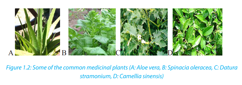
1.2.3.1. Causes of biodiversity loss
The main causes of biodiversity loss can be attributed to the influence of human
activities on ecosystems. Threats to biodiversity include:
a. Habitat loss and the degradation of the environment
The habitat loss and the degradation of the environment occur in different ways.
The most occurring, are tree cutting, agriculture and fires (Figure1.3). These human
activities lead to the alteration and loss of suitable habitats for biodiversity. As a
consequence, there is a loss of plant species as well as the decrease in the animalspecies associated to this plant diversity.
b. Introduction of invasive alien species and genetically modified organisms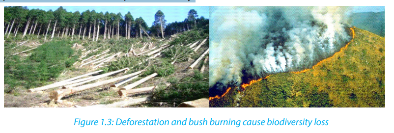
Species originating from a particular area are harmful to native species also called
endemic species when they are introduced into new natural environments. They
can lead to different forms of imbalance in the ecological equilibrium, so that
endemic species may fail to compete with introduced species, and they may affect
the abundance and distribution in natural habitat.
c. Pollution
Human activities such as excessive use of fertilizers, and increased pollutants from
industries and domestic sewage affect biodiversity. They contribute to the alteration
of the flow of energy, chemicals and physical constituents of the environment and
hence species may die as a result of toxic accumulation.
d. Overexploitation of natural resources
Increased hunting, fishing, and farming in particular areas lead to the decrease and
loss of biodiversity due to excessive and continuous harvesting without leaving
enough time for the organisms to reproduce and stabilize in their natural habitat.
e. Climate change
This is a change in the pattern of weather, related changes in oceans, land surfaces
and ice sheets due to global warming resulting from man’s activities. Increasing
global temperatures have resulted into melting of icebergs raising sea levels and soflooding coastal areas eventually affecting the niche.
1.2.3.2. Consequences of loss of biodiversity
They are various consequences of loss of biodiversity that include:– Desertification, is thought by scientists to be a consequence of climate change,Self-assessment 1.2
has been considered to be related to deforestation. Disrupting water cycles
and soil structure results into less rainfall in an area.
– Floods as a result of rising sea levels
– Habitat destruction for extensive farming, timber harvesting and infrastructure
and settlement
– Decrease in food production as result of change in pattern of weather that
affects productivity
– Large scale deforestation has a negative effect on nutrient recycling and can
accelerates soil erosion– Diseases that come as effects of floods and malnutrition due to famine
1. Define the term Extinction.1.3. Calculation of Simpson’s index
2. Suggest the causes of extinction of species in Rwanda.
3. Discuss the benefits of biodiversity to humans
4. Discuss the major factors leading to the degradation of ecosystems in
Rwanda
5. Discuss the contribution of ecosystems to cultural traditions in Rwanda.
6. In Rwanda different plants are used in traditional medicine to treat different
diseases. Conduct a research and list at least 20 medicinal plants and the
diseases they treat. From the list above describe at least one medicinal
plant and get ready to present your work. The project work should include:
written content of 2 pages in minimum and 4 pages in maximum, a
testimony of people that have used plant species.
7. Pollution is one of the causes of aquatic biodiversity loss.
a. What do you understand by water pollution?
b. Outline human activities that contribute to water pollutionc. Discuss how polluted water affects aquatic living organisms?
Activity 1.3
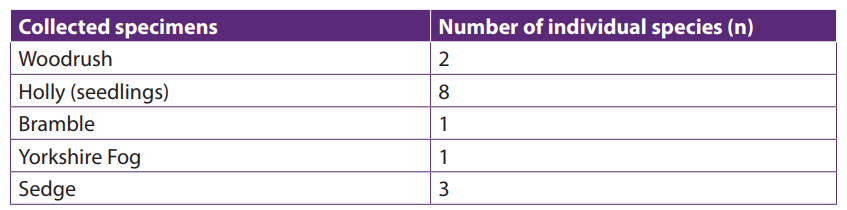
A survey on tree species was conducted in Gako forest by a group of students.
Five tree species (A to E) were identified and counted. The numbers found duringthis exercise are summarized in the following table:
 1. Describe the relative abundance of species A to E.There are many ways to measure diversity. The Simpson diversity index among
2. Based on the data in the above table, suggest how species diversity of treespecies can be calculated.
indices used to measure diversity. It is expressed in three related indices namelySimpson index, Simpson index of diversity and Simpson reciprocal index.
a. Simpson index D
Simpson index D can be expressed in two ways and takes into consideration the
total number of organisms of a particular species and the total number of organisms
of all species. It is calculated as follows: D =1-∑ (n/N) 2 or D = , with n: the total
number of organisms of a particular species and N: the total number of organisms
of all species. When the index equals or is nearby 0 there is an infinite diversity
of considered species. When it equals or is nearby 1, this means that there is no
diversity. The bigger the value of D, the lower the diversity and small is D, the biggeris the diversity.
b. Simpson index of diversity 1 – D The value of this index ranges between 0 and
1, but now, the greater the value, the greater the sample diversity. This makes
more sense. In this case, the index represents the probability that two individuals
randomly selected from a sample will belong to different species.
c. Simpson reciprocal index 1 / D
Another way of overcoming the problem of the counter-intuitive nature of Simpson’s
index is to take the Simpson’s reciprocal index 1 / D. The value of this index starts with
1 as the lowest possible figure. This figure would represent a community containing
only one species. The higher is the value of Simpson reciprocal index, the greater the
biological diversity.
Examples
1. In woodland, a quadrat was sampled for ground vegetation. Data collected were
recorded in the table 1.3.2. Find out the value of the Simpson index and draw the
conclusion about the biological diversity of the sampled area.Table 1.3.1: Recorded data on the vegetation from a woodland

Solution: Putting the figures into the formula for Simpson’s Index:
Based on the meaning of Simpson index, the quadrat presents a low diversity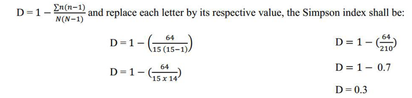
because the value of D is near zero and zero and below 0.5.
2. Calculate the value of Simpson’s Diversity Index (D) for a single quadrate sample
of ground vegetation in woodland from which the following sampling date wasobtained:
Solution:

Self-assessment 1.3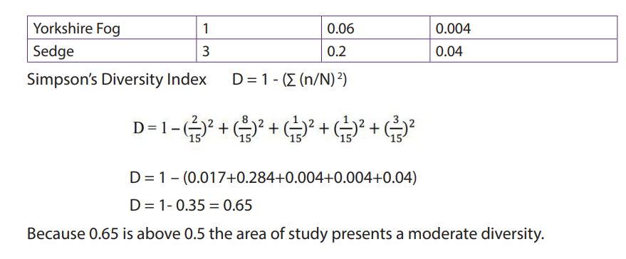 1. Differentiate between species richness and species evenness
2. Suggest precautions taken when measuring populations of aquatic animals
or plants.
3. Explain why a habitat with high diversity tends to be more stable than one
with lower diversity.
4. In a survey of trees in a tropical forest, students identified five tree species
(A to E).
They counted the numbers of trees in an area 100 m × 100 m and found
these results:
Calculate the Simpson’s Index diversity for identified species and explain the
advantage of using data on species diversity and abundance when calculating an
index of diversity.5. The Simpson’s Index of diversity for vegetation in an open area inhabited1.4. Sampling techniques to assess the distribution and
by grasslands was 0.8. For a similar sized area of vegetation beneath someconifer trees it was 0.2. What do you conclude from these results?
abundance of organismsActivity 1.4
From your school garden, sample different flowering plant species and answer
the following questions:
1. Specify the techniques used for collecting flowers of different species.To calculate Simpson’s index for a particular place:
2. What are the advantages of the technique you used for data collection?
3. Move around the school garden and collect different specimens of plant
species. Name the collected species by using their names. In case you don’t
know their names, use letters A, B, C ….
Calculate Simpson index D, Simpson index of diversity and Simpsonreciprocal index.
– Identify the habitat to be studied.To analyze the distribution and abundance of organisms in an area of study, there
– The number of individuals sampled for each species must be recorded.
are different sampling methods.
Note that, sampling only one quadrat would not give reliable estimate of thediversity of the ground flora in the wood.
a. Random sampling method
A random sampling method is a sampling method where samples are taken from
different positions within a habitat and those positions are chosen randomly.
b. Quadrat sampling method
A quadrat is a square area that is marked using a pre-made square of plastic, or
stakes and string and it can range in size. Different species and their numbers within
the quadrat are counted. Counting is repeated many times in different places in the
habitat to get an accurate representation of biodiversity.
c. Frame quadrats
Frame quadrats are small plot used to isolate a standard unit of area for the study
of the distribution of an item over a large area. While originally rectangular, modern
quadrats can be rectangular, circular, and /or irregular. The quadrat is suitable for
sampling plants, slow-moving animals such as millipedes and insect and some
aquatic organisms.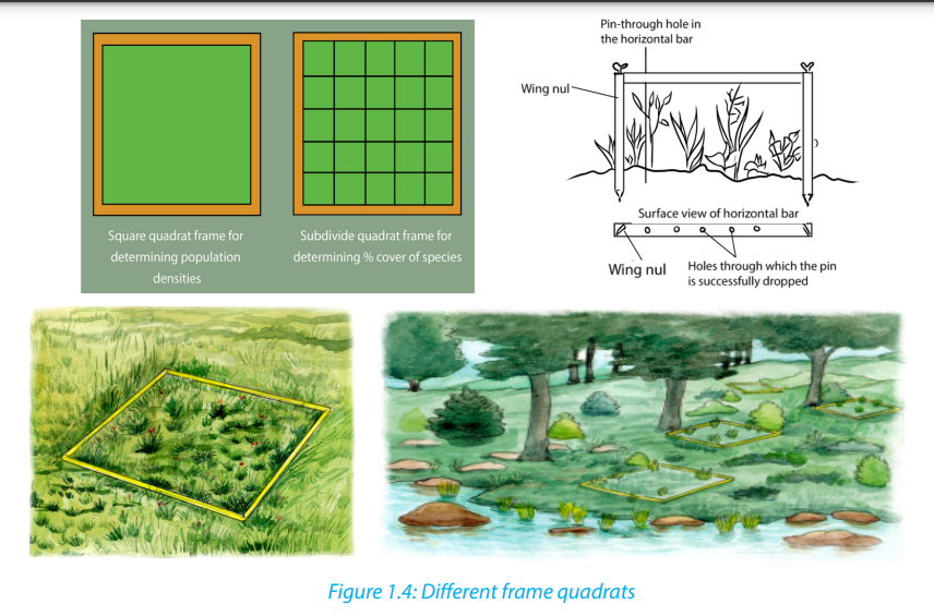
d. Transect sampling
Transect sampling is done using a transect line, which is usually a rope or measuring
tape that has been marked at set intervals, such as every meter. The line is unrolled
within the habitat. At every interval, the type and number of species along the
line are recorded. A measured line is laid across the area in the direction of the
environmental gradient. The species touching the line can be recorded along the
whole length of the line (continuous sampling) or at specific points along the line(systematic sampling).
e. Belt transects method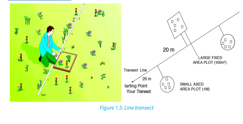
Belt transects method is the same as the line transects but widens the sampling
area. The samples are taken and the abundance, percentage cover in a defined areadetermined. Samples can be taken within the belt.
f. Netting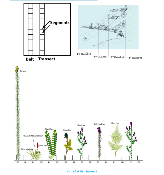
Netting is a sampling method where fine mesh nets are used to capture different
organisms that include insects, birds and bats. The technique is also used forsampling small aquatic organisms like daphnia, and water boatman.
g. Capture -recapture technique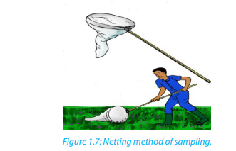
This method is useful for sampling non-fixed population and is suitable for animal
such as fishes, birds, lizards and insects. A sample of the population to be studied is
first captured and each individual is marked with a spot for identification. These are
then released and given enough time to mix up with the rest of the members in the
habitat. After a certain period of time, another sample is taken.
During the mark-release-recapture technique, the total population can be estimated
by the use of the formula: , where
n1 is a number caught and marked in first sample,
n2 is a number caught in second sample
n3 is a number in the second sample that had been marked.
To understand this application, let us use the following examples:1. A team of students used a sweep net to sample brown grasshoppersSolution
and each collect insect was marked with a very small spot of non-toxic
waterproof paint and then they were released in the field. The next day, a
second large sample was conducted and data were recorded as follows:
number of caught and marked in first sample (n1) = 247, number of caught
in second sample (n2) = 269, and the number in the second sample that hadbeen marked (n3) = 16. What is the number of estimated population?
The estimated number =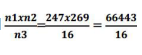 = 4152 grasshoppers
2. A student collected 16 butterflies which he marked and released. For a second
time he collected 18 butterflies among which 12 were already marked from thefirst sampling. Estimate the population size of butterflies in that area.
Solution
The estimated number
Self-assessment 1.4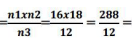 24 butterflies
1. Explain the advantages of the random sampling techniques.
2. Use suitable methods, such as frame quadrats, line transects, and belt
transects, to assess the distribution and abundance of insect species in a
school garden. Record your data and use the Simpson index of diversity (D)
to calculate the diversity of collected insects.
3. Suggest the benefits of using the following sampling techniques:a. Quadrats4. State the conditions in which quadrats, transect and mark recapture are
b. Transect
c. Mark-capture-recapturesuitable sampling methods.
1.5. Pearson’s linear correlation
Activity 1.5Some of the following figures indicate a positive, negative or non-correlation.
1. What do you understand by the term correlation?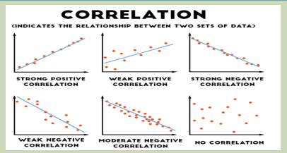
2. Categorize the graphs given as positive, negative or weak or no correlation
3. In which conditions results can indicate a positive correlation?
4. Conclude about your results when there is no correlation.
To decide if there is an association between collected data, a correlation coefficient
is calculated and plot scatter graph drawn in order to make a judgment. The
strongest correlation is present for studied items when all the points lie on a straightline. In this case, there is linear correlation, and the correlation coefficient equals
1. If a given variable X increases so does another variable Y, the relationship is a
positive correlation. If a variable X increases while the variable Y decreases, then
the relationship is a negative correlation. A correlation coefficient of 0 means there
is no correlation at all. These correlation coefficients are ways to test a relationship
observed and recorded to see if the variables are correlated and, if so, to find thestrength of that correlation.
a. Pearson’s correlation coefficient
Pearson’s correlation coefficient can only be used where there might be a linear
correlation and when there are collected quantitative data as measurements (for
example, length, height, depth, and light intensity, mass) or counts (for example
number of plant species in quadrats). The data must be normally distributed.
Where: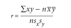 r is the correlation coefficientSelf-assessment 1.5
x is the number of species in a quadrat
y is the number of species in the same quadrat
n is the number of readings (From1 to n)
x is the mean number of species
y is the mean number of species
sx
is the standard deviation for x
s
yis the standard deviation for y
Use Pearson’s linear correlation to analyze the relationships between thedistribution and abundance of species and abiotic or biotic factors.
End of unit assessment 1
Section A: Answer as true or false1. Abiotic factors are the non-living physical aspects of the environment.
2. Capture –recapture is a method used to integrate the numbers of mobile
animals in a particular place.3. A correlation coefficient of 0 means that there is no correlation at all.4. A sample is a portion, piece, or segment that is representative of a whole
area of study.5. In the Simpson’s index, N represents the total number of organisms of aparticular species
Section B: Long and short answer based questions
1. What do you understand by the term biodiversity?
2. What do you think would happen to plants if there were no insects?
3. Suggest different ways to conserve our forests.
4. A student has randomly collected 5 types of species at the followingfrequencies.
Calculate the Simpson’s diversity index of this community.
5. A team of students conducted the capture- recapture sampling method
of tilapia from lake Muhazi at different times of the day as recorded in thedata below:
 a. Plot the graph for the date provided and describe the shape of the6. What do you understand by term endangered species?
graph.
b. From the graph, determine the appropriate time to have the most
catch.
7. Describe how diversity is threatened by climate change and humanactivities
UNIT 2:INTRODUCTION TO CLASSIFICATION
UNIT 2: INTRODUCTION TO CLASSIFICATIONKey Unit CompetenceApply the basic knowledge of classification to group living organisms into the threedomains.Learning objectives– Describe the classification of species into the taxonomic hierarchy of domain,kingdom, phylum, class, order, family, genus and species.– Outline the characteristic features of the three domains Archaea, Bacteria andEukarya.– Draw and label the structure of a typical bacterial cell.– Identify common bacterial diseases in plants and animals.– Outline the characteristic features of the kingdoms Protoctista, Fungi, Plantaeand Animalia.– Explain why viruses are not included in the three domain classification.– Outline how viruses are classified limited to type of nucleic acid and their host.– Describe the role of bacteria in the production of dairy products.– Describe methods of preventing common bacterial diseases.– Construct a dichotomous key for a group of organisms.– Recognize that microorganisms can survive in hot springs
Introductory activityCollect different fruits such as oranges, lemons, avocado, green paper, red paper,bananas, mangoes and tomatoes.1. Observe each of the above fruits and group them based on their externalfeatures.2. Based on groups made, which fruits are most closely related?
For more than 3.5 billion years, life on earth has been constantly changing. Naturalselection and other processes have led to a staggering diversity of organisms. Atropical rain forest, for example, may support thousands of species per meter square.Recall that a species is a population of organisms that share similar characteristicsand breed with another to produce fertile offspring. Biologists have identified andnamed about 1.5 million species so far, and they estimate that between 2 and 100million additional species have yet to be discovered.
2.1. Taxonomic hierarchyActivity 2.1You are provided with cards written on a list of words such as continent, district,country, cell, province, sector, village and family.1. Arrange the above words in increasing size2. What is your opinion about the people of the same family and those in thewhole country?3. Compare your arrangement above with 8 groups of the biologicaltaxonomic hierarchy.
Taxonomy is the study and practice of classification, which involves placing organismsin a series of taxonomic units, or taxa (singular: taxon). In biological classification,these taxa form a hierarchy. Each kind of organism is assigned to its own species, andsimilar species are grouped into a genus (plural: genera). Similar genera are groupedinto a family, families into an order, orders into a class, classes into a phylum (plural:phyla) and phyla into a kingdom. The domain is at the top of this hierarchical system.
The hierarchy classification starts from the largest group, the domain. The eightlevels of classification are known as taxa (taxon in singular), these include: Domain,Kingdom, phylum, class, order, family, genus and species. As one moves down thetaxonomic hierarchy, it follows that the number of individuals decreases but thenumber of common features increases. For example, there are numerous individualsin the domain Eukarya, with very few features in common.
Binomial nomenclature
When precision is not required one generally reverts to common names. The trouble
is that an organism may be known by different common names, and sometimes the
same name may be given to two quite different organisms because common names
are not internationally recognized and they change from one region to another one,
or from one country to another one. To solve this problem, the binomial system
also known as scientific name was introduced and it was pioneered by the Swedishnaturalist Carl Linnaeus (1707-1778).
In this system, each organism is given two Latin names: a generic name beginning
with a capital letter and a specific name beginning with a lower case letter based on
the physical characteristics of studied species. The scientific name is in italic whenprinted otherwise it is underlined, when hand written.
For example, many cats belong to the genus Felis but there are many species of cats:
A wild cat is Felis sylvestris while a domestic cat is Felis domesticus. These names are in
italic because this book was written by the use of computer. Hierarchy taxonomy ofhuman, earthworm and hibiscus plant are given in the table 2.1.
Table 2.1 Taxonomic classification of human being, earthworm and hibiscus
Scientific names present more advantages than common names.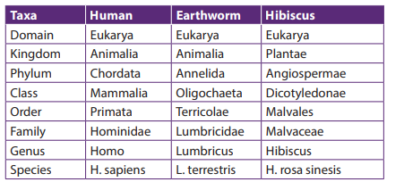 – They are necessary whenever precise identification is required, and theyenable scientists to communicate accurately with each other.– They are used worldwide and have the merit that every biologist knows exactlywhich organism is being referred to.Currently, with DNA technology, it is possible to investigate relationships based on
genes or DNA structure. As this new technology comes to greater use, it is possibleto find that some species had to be reclassified into different taxa.
Self-assessment 2.1
1. An African bush elephant belongs to order Proboscidae and family
Elephantae. Its scientific name is Loxodonta africana.a. Make a table indicating the hierarchy classification of African bushelephantb. Use the examples from table 2.1 to define the term “taxon”2. Classify each of the following organisms under the following kingdom,
phylum and class taxa: honey bee, cockroach, maize, and spider.3. Describe the system of naming species that Linnaeus developed.
2.2. Three domains: Archaea, Bacteria and Eukarya.
Activity 2.2.
Using text books and other sources identify the characteristics of each of the three
biological domains
Three domains are used by biologists to divide organisms into three large groups
based on their cell structure. The domain is the highest taxon in the hierarchy. The
prokaryotes are divided between the domains Bacteria and Archaea, while all theeukaryotes are placed into the domain Eukarya.
a. Domain Bacteria
Domain bacteria include prokaryotic organisms as their cells have no true nucleus.
They are all microscopic that vary in size between 0.2 to 10 micrometres. The
characteristic features of bacteria are:
– Cells with no true nucleus– DNA exists in circular chromosome and does not have histone proteinsassociated with it– No membrane-bound organelles (mitochondria, endoplasmic reticulum,Golgi body, chloroplasts)– Contain mesosomes as infolding of membrane and acts as sites for respiration.– Ribosomes (70 S) are smaller than in eukaryotic cells– Cell wall is always present and contains peptidoglycans in place of cellulose– Cells divide by binary fissionb. Domain Archaea– Usually exist as single cells or colonies
This contains bacteria that live in extreme environments where few other organisms
can survive. They are classified according to the environments they live in;– Methanogenic bacteria that live in habitats deprived of oxygen and give offmethane as a product of metabolism for example those that live in the guts ofruminant animals– Halophilic bacteria live only in salty conditions– Thermoacidophilic bacteria tolerate extreme acid and temperature thatc. Domain Eukaryaexceed boiling point of water and a pH below 2.
All the organisms classified into this domain have cells with nuclei and membranebound organelles. Their characteristic features are:
– Cells with a nucleus and membrane-bound organelles
– linear DNA associated with histones arranged within a chromosome in the
nucleus
– Ribosomes (80S) in the cytosol are larger than in prokaryotes, while chloroplasts
and mitochondria have 70S ribosomes, like those in prokaryotes.
– Chloroplast and mitochondrial DNA is circular as in prokaryotes suggesting an
evolutionary relationship between prokaryotes and eukaryotes
– A great diversity of forms: unicellular, colonial and multicellular organisms
– Cell division is by mitosis– Many different ways of reproduction including asexually and sexually.
Self-assessment 2.2
1. What are the three domains of living things?
2. Describe the ways in which a domain differs from a kingdom?
3. It is confirmed that: “Some bacteria can survive in extreme temperatures such
as hot springs”. Justify this statement.
4. How is the information about evolutionary or phylogenetic relationshipsuseful in classification of the living things?
2.3. Five kingdoms of organisms
Activity 2.3.
1. Collect organisms from a habitat near your school including a housefly, spider,
frog, gecko, bean/maize plant, moulds/mushroom, spirogyra (algae) and a hen. If
there are small rapidly moving land animals such as insects, anaesthetise them by
placing them in an ether/ethanol bottle for few seconds. Preserve the collected
specimens for future usea. Examine each organism, using a hand lens.b. Make a table of the features observed and identify the kingdom to whicheach organism belongs.2. Which feature do all animals (except sponges) have that distinguishes themfrom plants and fungi?
There are different ways of classifying the living world into kingdoms but the most
common and recommended is the five kingdom classification.
According to Kent (2000) the kingdoms are:– Kingdom Monera or prokaryote– Kingdom Protoctista– Kingdom Fungi or kingdom mycota– Kingdom Plantae2.3.1. Kingdom Protoctista– Kingdom Animalia
This kingdom is made up of a very diverse range of eukaryotic organisms, which
includes those that are often called protozoans and algae. Any eukaryote that is not
a fungus, plant or animal is classified as a protoctist. The characteristic features of
protoctists are listed according to the different phyla due to their diverse range:– Rhizopods that have pseudopodia for locomotion. Example, amoeba– Flagellates which are heterotrophic organisms with at least one flagellum forlocomotion. Example, trypanosoma.
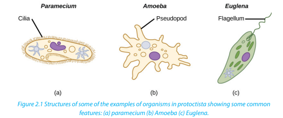
– Sporozoans which are mainly parasitic organisms that reproduces by multiplefission. Example plasmodium.– Ciliates which are organisms with cilia. Example paramecium– Euglenoid flagellates which are organisms with flagella but with a biochemistryquite distinct from that of flagellates. Example Euglena– Oomocytes which are similar to fungi except that they have cell wall withcellulose. Example Phytopthora infestans; potato blight– Green algae which are photosynthetic organisms with chlorophyll pigmentssimilar to the ones of plants. Example chlorella– Red aglae which are photosynthetic organisms with organelles with redpigment as well as chlorophyll. Example, chondrus– Brown algae which are photosynthetic organisms with organelles whichcontain brown pigments as well as chlorophy. Example Fucus, sea weedLiving things such as paramecium (a), amoeba (b), euglena (c) and plasmodia belongto the kingdom Protoctista.
2.3.2. Kingdom Fungi
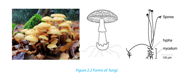
Fungi have some similarities with plants, but none of them is able to photosynthesise.
They are all heterotrophic, obtaining energy and carbon from dead and decaying
matter or by feeding as parasites on living organisms. There is a vast range in size
from the microscopic yeasts to what may be the world’s largest organisms. Other
characteristic features of fungi are:– Heterotrophic nutrition – they use organic compounds made by otherorganisms as their source of energy and source of molecules for metabolism– Reproduce asexually by means of spores and sexually by conjugation– Simple body form, which may be unicellular or made up of long threads calledhyphae (with or without cross walls).– Large fungi such as mushrooms produce large compacted masses of hyphaeknown as fruiting bodies to release spores– Cells have cell walls made of chitin or other substances
2.3.3. Kingdom Plantae
Plants are all multicellular photosynthetic organisms. They have complex bodies
that are often highly branched both above and below the ground. Characteristic
features of plants are:– Multicellular eukaryotes with cells that are differentiated to form tissues and2.3.4. Kingdom Animalia
organs.
– Few specialized cells
– Cells have large and often permanent vacuoles for support with cell walls
made of cellulose– Most plants store carbohydrates as starch or sucrose
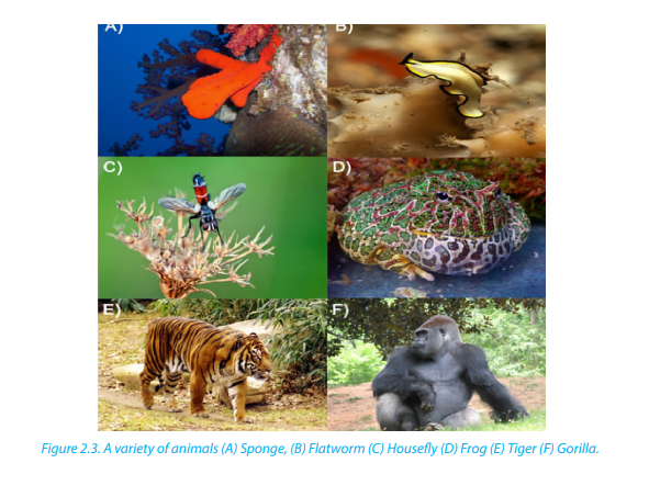
Animals (Fig 2.3) are multicellular organisms that are all heterotrophic with different
methods of obtaining their food. Organisms in this kingdom have other additional
features.– Different types of specialized cells
– Cells do not have chloroplasts and cannot photosynthesize (although some,
such as coral polyps have photosynthetic protoctists living within their tissues)
– Cell vacuoles are small and temporary (for example lysosomes and food
vacuoles)
– Cells do not have cell walls– Communication is by the nervous system
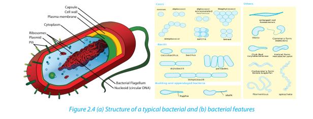
2.3.5. Kingdom Monera
Organisms in this kingdom have single cells that do not have a nucleus. They are
prokaryotic. They are the smallest and simplest organisms. Examples are bacteria
which form a diverse group with members that range widely in size and shape.
Some of them stick together to form chains or clusters while others are single cells.
The figure below (Figure 2.4) shows a typical structure of a bacterial cell whichcontains all the main features of prokaryotes
Self-assessment 2.3
1. The kingdom protoctista contains groups which do not appear to show an
evolutionary relationship. On this basis, is the five kingdom classification a
natural or artificial classification?
2. What are the three methods that protists use to obtain food?
3. Identify three characteristics of protists
4. The following is a list of organisms belonging to various kingdoms: housefly
(Musca domestica), maize (Zea mays), Frog (Rana spp), Bat and Eagle.a. Classify these organisms into their kingdoms
b. Name any two organisms that are not closely related and give a reason.5. How are fungi different from members of kingdom plantae?
2.4. Economic importance of bacteria
Activity 2.4
When an animal dies in a forest, it decays after a certain period of time. Once a
farmer grows beans in the soil with such dead animal decay, beans grow well.1. What cause the dead animal to decay?Bacteria are economically important because they are essential in many beneficial2. Why the beans have grown well?
biological and industrial processes. There exist some examples of bacteria that arepathogens as they cause disease and spoilage of food..
2.4.1. Useful bacteria
a. Biotechnology
Bacteria are used in biotechnology and industry. They are used to manufacture
products such as ethanol, acetone, organic acid, enzymes, and perfumes. In the
chemical industry, bacteria are most important in the production of pharmaceuticals.For example, E. coli is used for commercial preparation of riboflavin and vitamin K.
b. Genetic engineering
Bacteria are used in genetic engineering through the manipulation of genes, also
called recombinant DNA technology. In this case, bacterial cells are transformed and
used in production of commercially important products for example, production of
human insulin used in treatment of diabetes.
c. Decomposition
In addition, bacteria are important in decomposition of dead organisms and animal
wastes such as feces to form organic matter. This process improves soil fertility andplays an important role in mineral recycling in an ecosystem.
d. Fibre retting
Some bacteria including Clostridium butyricum are used to separate fibres in a
process called retting. In this process, fibres are formed to make ropes and sacks.
e. Nitrogen fixation
Some other bacteria are used to fix nitrogen in form of nitrates into the soil. For
example, Rhizobium bacteria which live in root nodules of leguminous plants. Such
bacteria help in improvement of soil fertility.
f. Digestion
Some bacteria living in the gut of ruminant animals such as cattle, horses and other
herbivores secrete cellulase, an enzyme that helps in the digestion of cellulose of
plant cell walls. Another example is Escherichia coli that live in the human largeintestine which synthesizes vitamin B and releases it for human use.
Self-assessment 2.4
Bacteria are both useful and harmful to humans”. Discuss the validity of thestatement.
2.5. Common bacterial diseases in plants and animals
Activity 2.5
Suppose there is cholera outbreak in your village and the executive secretary
invited you to sensitize people about preventive measures against cholera.Prepare a brief presentation for this purpose.
The bacteria that cause diseases are harmful to humans and other animals and are
referred to as pathogenic bacteria. The body is a home to many millions of bacteria
both useful and harmful to humans.
A bacterial disease is caused by entry of bacteria into a host where they can
grow, flourish then causing harm to the host. Bacterial diseases include cholera,
tuberculosis (TB), typhoid fever, pneumonia, tetanus, and diphtheria, and bacterialmeningitis, tooth decay in humans and anthrax in cattle.
Table 2.2. Common bacterial diseases in humans
2.5.1. Common Bacterial Diseases in Plants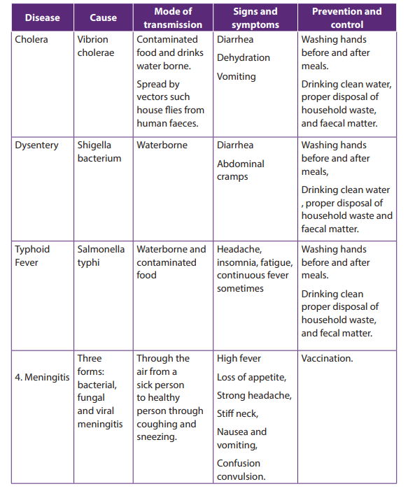
The table 2.3 common bacterial diseases in plants
Self-assessment 2.5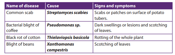
Mr. Green lives in one of the slums in a certain city. He prepares and sells chapattis
on street. He is usually very clean, but one morning, he is late for work so he does
not bother to wash his hands after visiting the toilet. That day he prepares 400
chapattis all of which are sold. Few hours later, his customer Sandra suffered from
a disease with the following signs and symptoms: severe diarrhea, excessive loss
of water leading to dehydration, and vomiting. Five dayslater, all his customers
were rushed and admitted in hospital due to the same problem.1. Suggest the disease that Mr. Green’s customers were suffering from and2.6. Structure and classification of Viruses
what caused the disease
2. Name three ways this disease might be spread around city.
3. After reading this scenario, what message do you have for people who are
like Mr. Green?
4. Suppose you were the health officer for the area in town with such a
problem. What steps would you take to prevent the disease from spreading
further?
5. House flies are described as vectors. Describe how houseflies transmit
diseases to humans.
Activity 2.6
Visit the internet and conduct a research to explain the reasons why viruses are
not classified in any of the five kingdoms of living organisms.
Viruses are microorganisms whose structure is only visible with electron microscopes.
Viruses are acellular and lack cellular structure. Viruses have none of the features that
we traditionally use for classification. They are particles made of proteins and nucleic
acids that are found in all cellular organisms, but show metabolism only once inside
the host cell.
When they infect cells, they use biochemical machinery and proteins of the host cell
to copy their nucleic acids and to make proteins coats often leading to destruction
of the host cells. The energy for these processes is provided by the ATP from the host
cell.
2.6.1. Structure of a virus
A typical virus consists of DNA or RNA within a protective protein coat called capsid.
The shape of the capsid may vary from one type of virus to another, as shown in
Figure 2.5 below.
Some viruses have an envelope of phospholipids and proteins. The envelope is made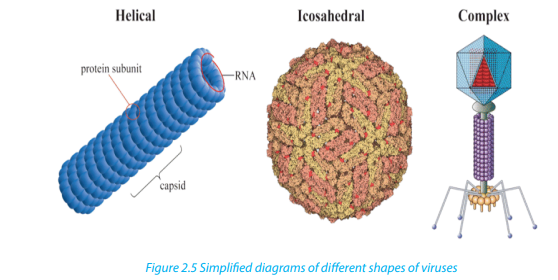
from portions of the host’s cell membrane. It surrounds the capsid and helps protect
the virus from the host’s immune system. The envelope may also have receptor
molecules that can bind with host cells and facilitate the virus to infect the cells.
2.6.2. Characteristics of viruses
An individual virus is called a virion. It is a tiny particle much smaller than
a prokaryotic cell. Because viruses do not consist of cells, they also lack cell
membranes, cytoplasm, ribosomes, and other cell organelles. Without these
structures, they are unable to make proteins or even reproduce on their own.
Instead, they must depend on a host cell to synthesize their proteins and to make
copies of themselves. Viruses infect and live inside the cells of living organisms.
2.6.3. Classification of viruses
Viruses can be classified according to:– Type of nucleic acid molecules of DNA or RNA, forming the core of the capsid:They are also regarded as parasites since they depend entirely on living cells for
Most animal viruses contain RNA while plant viruses contain DNA
– Type of host cell: plant or animal viruses as they are specific to their hosts– Presence or absence of the envelope: Plant viruses’ bacteriophage are nonenveloped while animal viruses like HIV and influenza virus are enveloped.
their survival. Although viruses are not classified as living things, they share twoimportant traits with living things: They have genetic material, and they can evolve.
2.6.4. Viruses and human disease
When viruses infect cells of their host, they cause disease. Examples of diseases
caused by viruses include HIV/AIDS, influenza (flu), chicken pox, and the common
cold. The human immunodeficiency viruses that causes AIDS is a retrovirus. Other
viral diseases include rabies, measles, diarrheal diseases, hepatitis A, B and C, polio,
and cold sores. One-way virus cause disease is by causing host cells to burst open
and die. Viruses may also cause disease without killing host cells. They may cause
illness by disrupting homeostasis in host cells.
Some viruses live in a dormant state inside the body. The virus that causes chicken
pox may infect a young child and causes the short-term disease chicken pox. Then
the virus may remain latent in nerve cells within the body for decades. The virus
may re-emerge later in life as the disease called shingles, where the virus causes
painful skin rashes with blisters. Some viruses can cause cancer. Examples include
the human papillomavirus (HPV) causing cancer of the cervix in females. Hepatitis B
virus causes cancer of the liver. A viral cancer is likely to develop only after a personhas been infected with a virus for many years.
Self-assessment 2.61. What is meant by the term virus?2.7. Dichotomous key of identification of organism
2. State the main components of a virus.
3. Describe the two ways how viruses cause an infection.
4. Differentiate between a bacteriophage and a retrovirus?
5. Do you think viruses should be considered as a form of life? Give reasonsfor your answer.
Activity 2.7.1
The figure below represents different types of plant leaves. Make a classificationof these plants based on the external structure of the leaves.
The dichotomous key is also referred to as biological identification key. It is made up
of a series of contrasting statements called leads indicated by the numbers 1, 2, 3…
where each lead deals with a particular observable characteristic. The characteristics
used in keys should be readily observable morphological features which may be
either qualitative, such as shape of abdomen, nature of legs, or quantitative, such
as number of antennae, number of pairs of legs and length of the antennae in case
of arthropods. It is essential to note that size and color are often less considered
as both can be influenced by the environment, the season, the age or state of theorganism at the time of identification.
2.7.1. Guidelines used in construction of a dichotomous key:
The following guidelines must be considered while constructing a dichotomous key.– Use morphological characteristics which are observable as much as possibleExample
such as leaf venation, nature of margin, apex, lamina and nature or length of
the petiole (leaf stalk).
– Start with a major characteristic that divide the organism or the specimen into
two large groups such as the type of a leaf.
– Select a single characteristic at a time and identify it using a number for
example: simple leaf………go to 2, compound leaf………go to 5. This means
that in 2 you will deal with only simple leaves and 5 only compound leaves.
– Use similar forms of words for two contrasting statements for example at 2:
leaf with parallel venation …………go to G and leaf with network venation
………go to 3.
– The first statement should always be positive.
– Avoid generalizations or overlapping variations, be specific and precise to thepoint.
– Collect leaves from the following plants: cassava, avocado, jacaranda, cassia,Solution: The dichotomous key of specimens A, B, C, D, E, F and G.
hibiscus bean, maize or paspalum grass,
– Label different leaves collected as, A, B, C, D, E, F and G
– Observe and familiarize with the specimens before starting the experiment
to minimize errors during the identification process
– Make a table summarising the specimens and steps followed to identify each
of them.
– Construct a dichotomous key based on the observable features(characteristics) and table of steps followed.
1. a. Simple leaves ---------------------------------------------------------------------go to 22.7.2. Common features used for identification of animals
b. Compound leaves ---------------------------------------------------------------go to 5
2. a. Parallel venation ------------------------------------------------------------------------G
b. Network venation -------------------------------------------- ------------------go to 3
3. a.Simple digitate ----------------------------------------------------- ----------------------A
b. Non simple digitate -------------------------------------------------------------go to 4
4. a. Leaf with serrated margin -------------------------------------------------------------E
b. Leaf with smooth margin -------------------------------------------------------------B
5. a.Leaf with three leaflets (compound trifoliate)-------------------------------------F
b. Leaves with more than three leaflets --------------------------------------go to 6
6. a. Pinnate leaf -------------------------------------------------------------- ---------------- Db. Bipinnate leaf ------------------------------------------------------------ --------------- C
Animals are classified based on the following features:– Locomotory structures such as legs, wings and fins2.7.3. Common features used for identification of plants
– Antennae (presence, nature and number)
– Presence or absence of eye and eye type
– Number of body parts for example insects have three body parts
– Body segments (nature and number)
– Body surface structures such as fur, hair, feathers and scales
– Feeding structures such as mouth parts in arthropods for example in insects– Type of skeleton present such as endoskeleton, exoskeleton and hydrostatic
Plants can be classified basing on the following features:– The leaf structure such as nature of apex, margin, venation, lamina and petiolePrecaution
– The flower structure including inflorescence type, flower shape and number
of floral parts
– The type of stem (woody, fleshy and herbaceous), shape (rectangular,
cylindrical) and texture of the stem (smooth, spiny and thorny) …– The type of root system, tap root, storage root, fibrous roots…
– Care must be taken while collecting and handling some organisms becauseActivity 2.7.2
some are poisonous, have thorns and others are able to sting
– Collection of specimen should be done a day or few days before the experiment
depending on nature of the experiment
– Avoid and try to minimize where possible, uprooting, cutting down or plucking
and pruning of plants as this may threaten the biodiversity as well as resultinto environmental degradation
Construct and interpret a dichotomous key of arthropods listed below.– Collect the following litter arthropods: honey bee, spider, millipede, butterfly,Self-assessment 2.7
sugar ant, centipede and mosquito and label each specimen as A, B, C, D, E, F
and G respectively
– Observe and familiarize yourself with the specimens before starting the
experiment.
– Use sharply contrasting external features of collected specimens /diagramsto construct a dichotomous key
Read and interpret the dichotomous tree below and use it to answer the followingquestions.
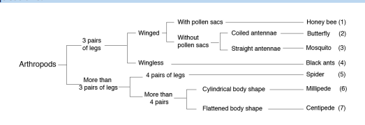 1. Specify the phylum of kingdom animalia represented by the above
dichotomous tree?
Give one observable reason to support your answer.
2. According to this dichotomous tree, which characteristic feature was used
to classify different insects?
3. Which observable characteristic feature distinguishes between a spider
and a mosquito?
4. How does a millipede differ from a centipede?
5. To which classes do a millipede and a centipede belong?
6. Which class of arthropods is not represented on the dichotomous tree?End of unit assessment 2
1. Which one of the following living organisms belongs to domain bacteria?a. Euglena3. Which one of the following is not a kingdom of living organisms?
b. Vibrio cholerae
c. Paramecium
d. moulds
2. The group of classification where organisms resemble one another and are
capable of interbreeding together to produce viable offspring is known as:
a. Species
b. kingdom
c. Genus
d. Phyluma. Monera4. Which one of the following is a characteristic feature common to fish, reptiles
b. Animalia
c. Annelida
d. Protoctista
and birds but absent in mammals?a. Possession of scales5. Which one of the following statements about fish is not correct?
b. Has no limbs
c. Possession of feathers
d. Undergo internal fertilizationa. Fish live both in water and on land and undergo external fertilization.6 Which one of the following is not a characteristic of all insects?
b. Most fish have bones while others are cartilaginous
c. Most fish have streamlined body, lateral line and swim bladder.
d. Gills are organs for gaseous exchange in fisha. They have three body parts namely head, thorax and abdomen.7. The following are characteristics of all mammals except;
b. They have three pairs of jointed legs attached on segment of the thorax.
c. They have four pairs of jointed legs
d. They have a pair of antennae attached on the head.a. They have mammary glands to secrete milk feed their young ones.8. The point where the leaf joins the stem is called;
b. Their skin is covered with hair.
c. Undergo internal fertilization and internal development of the embryo.
d. They have a pair of wings made up feathers.a. Apex9. Which of the following is less considered while identifying feature to construct
b. Margin
c. Leaf base
d. Lamina
e. Length of petiole.a dichotomous key of leaves?
a. Nature of margin10. The following are characteristics of arachnids except;
b. Nature of apexc. Size and color of leaf
a. Four pairs of jointed legs11. Match the structures with the organisms which possess them
b. Two body parts
c. Three body partsd. Do not have wings

12. A group of S4 students drew a Venn diagram below to summarize the five
kingdoms into which organisms are classified. Study the diagram and answerthe questions that follow:
a. Which kingdoms are represented by the letters x and y?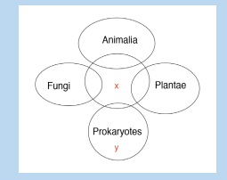
b. State one characteristic that organisms of x may share with:i. Prokaryotes13. Complete the table to summarize the characteristics of each class of phylum
ii. Fungi
iii. PlantaeArthropoda.
14. What is the significance of classification of living organisms?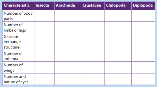
15. The binomial system of naming a blue monkey, Cercopithecus mitis, is
provided below;
Complete the table by filling the missing words.
UNIT 3: MICROSCOPY
UNIT 3: MICROSCOPY
Key Unit Competence
Distinguish between the types of microscopes and their principal uses.
Learning objectives
By the end of this unit, I should be able to:– Describe the main features and functions of the components of a compound
light microscope.
– Manipulate a compound light microscope to observe prepared slides.
– Show perseverance when using light microscopes.
– Pay attention when using a compound light microscope to avoid damage of
the lenses, mirrors and slides.
– State that magnification is the increase in the apparent size of the object.– State that resolution is the ability of the microscope to show two objects asIntroductory activity
separate.
– Appreciate the importance of magnifying instruments in Biology.
– Use of a microscope to determine the relationship between actual size of the
specimen and the image.
– Calculate the approximate size of different biological structures using an
appropriate unit of measurement
– State the advantages and disadvantages of using an electron microscope.
– State the principles and limitations of TEM (Transmission Electron Microscopy).
– State the advantages and disadvantages of using SEM (Scanning Electron
Microscopy).
– Compare light and electron microscopes
– Acknowledge the use of electron microscopes in modern science with
reference to electron micrographs.
– Observe and draw biological specimens under a light microscope.
– Prepare temporary slides for observation under light microscopes using
different objective lenses– Appreciate the importance of magnifying instruments in Biology\
Point out scientific activities that require the use of microscope in our daily lives.
A microscope is used to produce a magnified image of an object or specimen.
Anton Van Leeuwenhoek (1632-1723) was the first to invent a microscope powerful
enough to explore the world of microbes. His discoveries stimulated an explosion
of interest in scientific use of microscopes. Since the 18th century, many new types
have been invented of which the most commonly used today are the compoundlight microscope and the electron microscope.1 (Kent, 2000, p. 58)).
3.1. Compound Light Microscope
Activity 3.1.1
Some of the living things including Protoctista and fungi have small size to be
observed by naked eyes. Discuss the ways used by biologists to observe andidentify different parts of these living organisms.
The optical microscope, often referred to as light microscope is a type of microscopewhich uses visible light and a system of lenses to magnify images of small samples.
The different parts of light microscope are described below: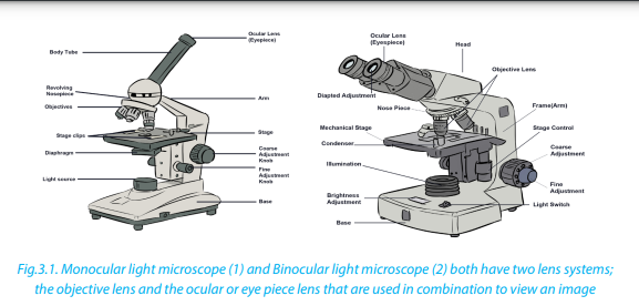 – Base: supports and stabilizes the microscope on the table or any other working
place
– Light source: It is made by lamp or mirror which provides light for viewing the
slide.
– Stage: is a platform used to hold the specimen in position during observation.
– Stage clips: are pliers used to fix and hold tightly the slide on stage.
– Arm: supports the body tube of microscope
– Body tube: maintains the proper distance between the objective and ocular
lenses
– Arm: used for holding when carrying the microscope and it holds the body
tube which bears the lenses.
– Coarse focus adjustment: moves stage up and down a large amount for
coarse focus– Fine focus adjustment: moves stage up and down a tiny amount for fine focus– Objective lenses: focuses and magnifies light coming through the slideRevolving nosepiece: rotates to allow use of different power objectives– Slide: is a transparent pane on which a specimen is placed.
– Eye piece/ocular lens: magnifies image produced by objective lens
– Condenser: It will gather the light from the illuminator and focus it on the
specimen lying on the stage. The function of the condenser is to focus the light
rays from the light source onto the specimen.
– Iris diaphragm lever: This allows the amount of light passing through thecondenser to be regulated to see the object.
Activity 3.1.2b. To observe under high power at a greater magnification, proceed as
Using the light microscope
a. To observe under low power and low magnification, proceed as follows:
– Objects (specimens) to be observed under the microscope are first placed on
a glass slide and covered with a cover slip.
– Place the specimen on the stage of your microscope; in other words, arrange
it so that the specimen is exactly at the center of the hole at the stage.
– Fix the slide in place with two clips.
– Rotate the nosepiece so that small objective lens is immediately above the
specimen.
– Set the angle of the reflector mirror so that light is directed up through the
microscope.
– Look down the microscope through the eye piece. Adjust the iris diaphragm
so that the field of vision is bright and not dazzling.
– Turn the course adjustment knob until the tip of the objective lens is close to
the slide.
– Now look down the microscope again. Slowly turn the course adjustment
knob in the other direction, so the tube gradually moves upwards. The
specimen on the slide should eventually come into view.
– Use the course and fine adjustment knobs to focus the object as sharply as
possible.
– If necessary readjust, the iris diaphragm so the specimen is correctly
illuminated. You will get a much better image if you don’t have too muchlight coming through the microscope.
follows:– Rotate the nosepiece so that the large objective lens (with higher magnifyingMicroscope uses transmitted light for observation. However, microscope uses
power) is immediately above the specimen. The nosepiece should click into
position, as before.
– If the specimen is not in focus, focus it with fine adjustment knob. Be careful
that the tip of the objective lens does not touch the slide.– Readjust the illumination if necessary.
specific light characteristics for specific samples, such as transparent specimens and
samples that do not pass light. All parts of a microscope work together, the light
from the illuminator passes through the aperture, through the slide, and through the
objective lens, where the image of the specimen is magnified. Then the magnified
image continues up through the body tube of the microscope to the eyepiece,which further magnifies the image the viewer can see.
Light from the source is focused on the specimen by the condenser lens. It then
enters the objective lens, where it is magnified to produce a real image. The real
image is magnified again by the ocular lens to produce a virtual image that is seenby the eye.
Care of the compound microscope
The microscope is an expensive instrument that must be given proper care. Always
general instructions have to be respected when using a microscope. These include:– Carry the microscope with both hands, one hand under the base, and theSelf-assessment 3.1
other on the arm.
– When getting ready to put the microscope away, always return it to the low
power or scanning power setting.
– When setting the microscope on a table, always keep it away from the edge.
– It is generally better to clear your lab table of items that are not being used.
– Never clean lenses with anything other than lens paper, don’t use towels and
other paper tissues because they scratch the lens.
– Inform the instructor or the biology lab technician if there is any microscope
damage or irregularity in its operation as soon as possible. Do not return a
faulty microscope without first informing the instructor or lab technician.– You are responsible for the microscope while using it treat it with care!
1. Complete the table below:
2. What is the importance of a light microscope?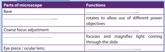
3. Suggest a reason why it is not advisable to clean the objective and eye piecelens with a wet cloth or towel?
3.2. Magnification and resolution of a compound light
microscope.
Activity 3.2.1
Work out the following equivalent measurements:
1. 1 millimetre (mm) =........... metre (m)
2. 1micrometre (µm) =............mmetre (m)
3. 1 nanometre (nm) =..............metre (m)
4. 1 metre (m) = .............mm =.......... µm =........nm,5. 1 kilometre (km) = .............m
a. Magnification
Magnification refers to increase in the apparent size of the object, while resolution
of a microscope is the ability to show two close objects as separate. The maximum
magnification of an ordinary light microscope is about x1500. Magnification must
be written on the right side and below the biological drawing and it does not have
units. The size of the image is measured in mm but converted into micrometers ornanometers to work out the actual size. It is calculated as follows:
Example
Calculate the magnification if the actual size is 5μm and the measured image of the
specimen has the size of 40mm.
Answer:
– Make the size of the image and the actual size in the same units by convertingmm in μm. This is done by multiplying 40mm by 1000 so that 40mm = 40000 μm
Note that the magnification of the specimen under a light microscope is calculated
by multiplying the magnification of the objective used to that of the eyepiece. For
example: 10x (objective) 10x (eyepiece) = x100.
b. Microscopic observation
Activity 3.2.2
Using prepared slides of microorganisms such as a bacterium, amoeba, and
paramecium.
Observe, draw and label the visible parts under a light microscope. Avail these
materials before you start: Petri-dishes, plate covers, pencil, transparent tape,
microscope, agar powder, and permanent slide of bacteria, amoeba, and
paramecium, Bunsen burner or any other source of heat.
Procedure– Prepare agar medium by boiling a mixture of 10g of agar powder with 50mlFor this experiment, light microscope allows to observe organisms of small size
of water
– Label a control and exposed petri dishes in which you pour prepared agar
medium.
– Cool both plates for 20 minutes until the medium hardens.
– Tape closed the cover of the control plate and removes the cover of the
exposed plate.
– Leave both plates for 5 minutes, and do not touch or breathe on the agar.
After five minutes, tape closed the lid of the exposed plate and store both
plates upside down in a warm place and draw your observations
– Repeat the observation by using mounted slides of amoeba and paramecium
and make a comparison between bacteria, amoeba and paramecium: what isyour conclusion?
including bacteria, amoeba and paramecium. Some other parts of macroscopic
organisms such as cells and tissues of plants and animals or some parts of these living
organisms such as stems and roots can also be observed under light microscope.
Some specimens can be observed directly after collection and preparation.
However, some of the details might not be clearly observed because specimens are
not colored. Also, some material distorts when you try to cut the specimen into thin
sections. To overcome this challenge, slides can be prepared in advance by the useof the following steps:
– Staining: colored stains are chemicals that bind to chemicals on or in the
specimens. This allow the specimen to be seen. Some stains bind to specific
cell structures. For example, acetic orcein stains DNA dark red, while gentian
violet stains bacterial cell walls.
– Sectioning: specimens are embedded in wax, where thin sections are then
cut without distorting the structure of the specimen. This is particularly useful
for making sections of soft tissue, such as brain. Safety measures might betaken. Make sure that hands are washed with soap and warm water after theActivity 3.2.3
experiment. Use a disinfectant to wipe down all surfaces where bacteria may
have been deposited for example. Be sure that some substances and animalsmight be harmful to the life.
Preparing of temporary slides and observation under light microscope
Make temporary preparation of slides of epidermis of onions young stems by
fixing, staining and mounting. Observe under low and high power of a light
microscope.
Preparation and procedures– Add a drop of water at the center of the microscopic slide to flatten the
membrane
– Pull of a thin membrane from the onion layer and lay it at the center of the
microscopic slide
– Add a drop of iodine solution or methylene blue on the onion membrane
– Gently lay a microscopic cover slip on the membrane and press it down
gently using a needle to remove air bubbles.
– Touch a blotting paper on one side of the slide to drain excess iodine/water
solution,
– Place the slide on the microscope stage under low power to observe.
– Adjust focus for clarity to observe.
– Make another slide without adding the stain to see the difference between a
stained slide and a non- stained slide.
– Draw and label the observed parts of each of the two slides and compare adrawing of a stained slide and that of a non-stained slid.
c. Measuring cells
Cells and organelles can be measured with a microscope by means of an eyepiece
called graticule. This is a transparent scale, usually having 100 divisions (Figure 3.4,
A). The eyepiece graticule is placed in the microscope eyepiece so that it can be seen
at the same time as the object to be measured (Figure 3.4, B). At this figure (Figure
3.4, B), the cell lies between 40 and 60 on the scale, so that it measures 20 eyepieceunits in diameter (60 – 40 = 20).
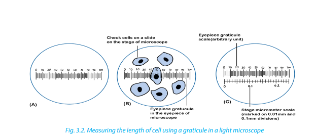
To calibrate the eyepiece graticule scale, a miniature transparent ruler called a stage
micrometer scale is placed on the microscope stage and is brought into focus. This
scale may be fixed onto a glass slide or printed on a transparent film. It commonly
has subdivisions of 0.1 and 0.01 mm. The images of the two scales can then be
superimposed (Figure 3.4, C). If in the eyepiece graticule, 100 units measure 0.25mm, the value of each eyepiece unit equals
By converting mm to μm, the value of eyepiece equals
 The diameter
of the cell shown superimposed (Figure 3.4, B) measures 20 eyepiece units. Its actual
diameter equals 20 × 2.5 μm = 50 μm. This diameter is greater than that of manyhuman cells because the cell is a flattened epithelial cell.
Use the following instructions to measure the length of one cell– Measure the distance in millimetre from the start of one cell to the end of 10Self-assessment 3.2.
cells
– Divide by 10 to find the length of one cell in the specimen.
– Convert this length in millimetre to micrometer by multiplying by 1000.
– Find the actual length of a cell by dividing this length by the magnification ofthespecimen got from the product of eye piece and objective lens used.
1. Calculate the magnification of an image with 50mm, and the object3.3 Electron microscopes
measuring 5µm. in length.
2. If a nucleus measures 100mm on a micrograph, with a magnification ofX10 000, what is the actual size of the nucleus?
Activity 3.3
Suggest the form and source of energy used by electron microscope. How doesthis differ from that used by a compound microscope?
An electron microscopes use a beam of accelerated electrons as a source of
illumination.
Electron beams have a much smaller wave length than light rays and therefore have
greater resolving powers and can produce higher effective magnifications than light
microscopes. There are two types of electron microscopes;– Transmission electron microscope (TEM)Electron microscopes are used to study the details of internal structures
– Scanning electron microscope (SEM)
(the ultrastructures) of cells. Most modern TEMs can distinguish objects as small as 0.2nm.
This means that they can produce clear images magnified up to 250,000 times.
Formation of an image by the TEM:– Extremely thin samples of the specimen are needed and are cut by using
diamond or glass knives as they are supported in resin block to prevent them
from collapsing
– The section is then impregnated with a heavy-metal stain
– As the beam passes through the specimen, electrons are absorbed by the
heavily stained parts but passes readily through the lightly stained parts.
– Electro magnets bend the electron beam to focus an image onto the florescentscreen or photographic film to form an electron micrograph

Scanning electron microscope (SEM)
The SEM is used to produce 3D images of surfaces of the specimens. Electrons are
reflected from the surface of the specimen stained with a heavy metal. This enablesthe SEM to produce images of all specimens, cells, tissues, or even organisms
a. Advantages of the electron microscope over light microscope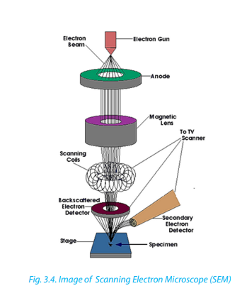
Electron microscope has a higher resolution and is therefore able of a higher effective
magnification estimated at up to 250,000 million times compared to the light
microscope which can show a useful magnification only up to 1000-2000 times. This
is because a light microscope uses a beam of light with a longer wave length while
Electron microscopes use a beam of electrons that have a short wave length.
b. Disadvantages of electron microscope
Despite the advantages, electron microscope presents a number of setbacks and
limitations.– They are extremely expensive and the maintenance costs are high.c. Comparison between light and electron microscopes
– Sample preparation is often much more technical requiring special training.
– Samples must be dead, exposed to high radiation and are placed in a vacuum
so that it is impossible to observe living specimens
– It is not possible to observe colors because electrons do not possess a color. The
image is only black-white, even if sometimes the image is colored artificially to
give a better visual impression.
– They require more training and experience in identifying artifacts that may
have been introduced during the sample preparation process.
Light and electron microscope presents the following similarities and differences.
The following are some of the similarities:
Both light and electron microscopes form larger (magnified) and more detailed
(highly resolved) images of small objects or small areas of larger objects– Both light and electron microscopes are used in biology study, research andDespite the similarities, light and electron microscope presents differences such as
medical sciences particularly histology, material sciences such as metallurgy
and other aspects of science.
– Specimens must be carefully prepared using techniques appropriate for both
the equipment and the sample including slicing, staining, and mountinthese summarized in the following table:
Table 3.1. Differences between light and electron microscopic
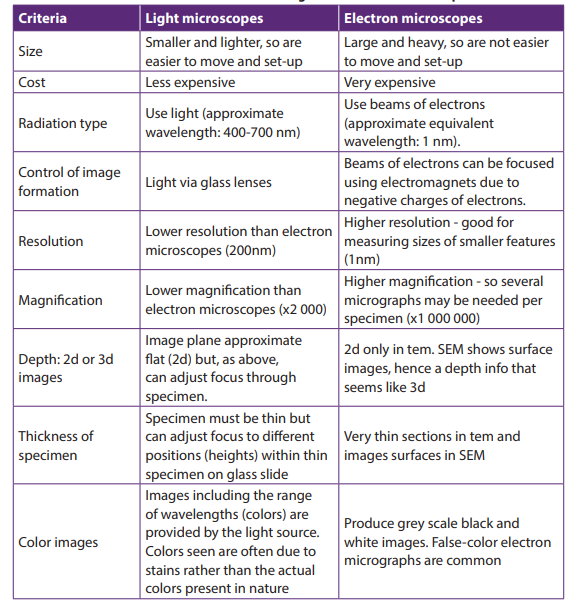
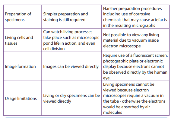
Self-assessment 3.3
1. How is magnification varied in;a. A light microscope2. Why is the resolving power of an electron microscope such better than
b. An electron microscope?
that of a light microscope?
3. Make a comparison between light and electron microscope, highlighting
the advantages and disadvantages for each type of microscope.Summarise the similarities and differences between light and electron microscopes
End of unit assessment 3
Section A. Multiple choice questions1. Which ranges can be viewed using a light microscope?
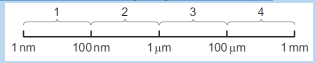 a. 4 only2. The figure below shows a mitochondrion drawn from an electron micrograph.
b. 1 and 2 only
c. 2 and 3 only
d. 3 and 4 onlyStudy it carefully and answer the following questions.
If the length of the mitochondrion line X Y is 3000 nm. What is the magnification
of the drawing of the mitochondrion?a. ×1003. A light microscope is used to observe two membranes that are 200 nm apart.
b. ×1000
c. ×10 000
d. ×100 000
How far apart are the membranes when the objective lens is changed from low
power (×40) to high power (×400)?a. 2 μm
b. 20 μm
c. 200 nm
d. 2000 nm4. The electron micrograph below is that of a chloroplast.
The length of the chloroplast along the line shown is 80 mm. The actual length of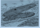
the chloroplast is 10 μm. What is the magnification of the chloroplast?a. ×8 × 1025. The following diagram below is drawn from an electron micrograph of an
b. ×8 × 10³
c. ×8 × 10⁴
d. ×8 × 10⁶animal cell.
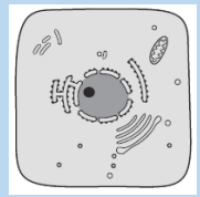
Which represents the same cell, seen under a light microscope at ×400magnification?
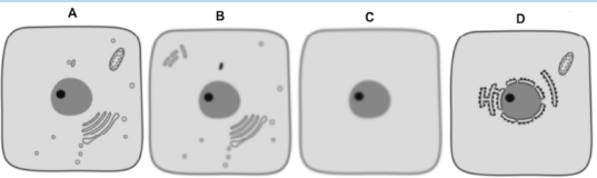
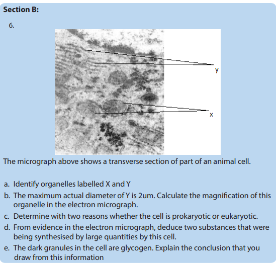
UNIT 4: CELL STRUCTURE AND SPECIALIZATION
UNIT 4: CELL STRUCTURE AND SPECIALIZATIONKey Unit CompetenceDescribe the structure and function of cells in an organism.Learning objectivesBy the end of this unit, I should be able to:– Identify plant and animal cell structures visible under a light microscope.– State functions of cell structures as seen under an electron microscope.– Describe the nature of artefacts.– State the importance of freeze fracturing for examining membrane structure.– Explain how cell organelles can be isolated by cell fractionation.– List the functions of cell membranes.– Describe the fluid mosaic structure of cell membranes.– Explain the role of the different components of a cell membrane.– Explain cell specialization as the differentiation of a cell or process to do aparticular function.– Interpret charts and micrographs to relate the structure of specialized cells totheir functions.– Prepare, observe and draw diagrams for specimens on temporary slides for:Wandering Jew, in plants and cheek cells under a light microscope.– Distinguish between ultra-structures of plant cells and animal cells.– Compare ultra-structures of prokaryotic and eukaryotic cells– Show resilience and be aware of artefacts when preparing temporary slides.– Appreciate the importance of cell specialization in multicellular organisms
Introductory ActivityUsing addition resources to your textbook available in your school such as thebooks from the school library and search further information from the internet:1. Differentiate between prokaryotic and eukaryotic cells.2. By using charts for the two cells, identify different organelles of eukaryoticcell that may perform functions similar to those of a prokaryotic cell.
4.1. Ultra-structure of a cellActivities 4.11. Observe the chart given for Ultra structure of a cell and identify parts thatare easily recognizable when compared with a photomicrograph form alight microscope.2. Identify the mitochondria and ribosomes and state their roles in the life ofthe cell.Cytology is the study of the structure and function of cells. A Cell is the basic unit oflife. All living organisms are made up of cells.Living organisms are classified into:– Unicellular organisms are made of only one cell, such as bacteria,– Multicellular organisms are animals and plants composed of many cells. Inmulticellular organisms, cells divide and then undergo differentiation orspecialisation for specific functions.
Cell theory.The cell theory states that all living organisms are made up of cells, and cells are thebasic unit of structure function in all living organisms.The main principles of cell theory are based on the following ideas.– All known living organisms are made up of one or more cells,– All cells come from pre-existing cells by division– Cells contain the hereditary information that is passed from cell to cell duringcell division.– Metabolism takes place in cells– Given suitable conditions, cells are capable of independent existence
When a cell is viewed under light microscope, the most obvious features observedare the very large nucleus and a clear cytoplasm surrounded by a cell membrane.However, under electron microscope, it is possible to identify a range of organellesin plant and animal cells. Ultrastructure is the detailed of cell as revealed by theelectron microscope.
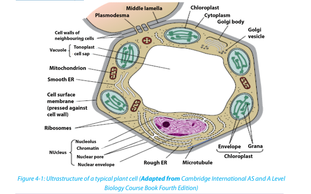
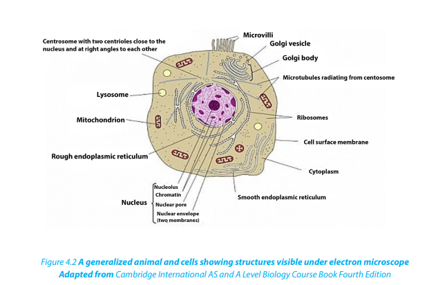 Similarities between animal cell and plant cell– Both have a cell membrane, a cytoplasm and a nucleus.– Both animal and plant cells have mitochondria, Golgi apparatus, Reticulumendoplasmic, lysosome, big ribosomes (80S), peroxisome, microtubules.
Table 4.1: The differences between animal and plant cell
Self-assessment 4.1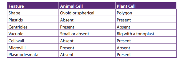
1. What structures do both animal and plant cells have in common?
2. State any five principles of the cell theory.
3. Give the major difference between a plant and animal cell. Which organellesdoes this difference relate to?
4.2. Prokaryotic cells
Activities 4.2
Under microscope, observe mounted slides of bacteria, and plant cells. Draw andlabel the parts that are common in both plant and bacterial specimens
A typical bacterial cell has a cell surface membrane enclosing the cytoplasm that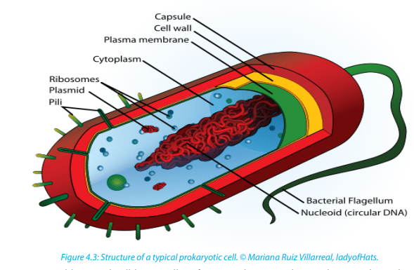
contains enzymes, ribosomes and food granules. The membrane is surrounded by
the cell wall and this may in turn be enclosed in a capsule. A bacterial cell lacks high
level of organization compared to animal or plant cell. It has no Golgi apparatus
or endoplasmic reticulum. The genetic material is a single strand of DNA usually
coiled up into the center of the cell to form a nucleoid. This nucleoid has no double
membraned nuclear envelope so is often described as an ‘ill-defined nucleus’.– Some bacterial cells contain plasmids with additional DNA.Comparison between prokaryotic and eukaryotic cells
– Respiration generally takes place in mesosomes which is an in-folding of the
cell surface membrane but lack mitochondria
– Photosynthesizing bacterial cells such as cyanobacteria (blue green
algae) have a special form of chlorophyll but it is not enclosed in a doublemembraned chloroplast
Table 4.2 Comparison between prokaryotic and eukaryotic cells
Self-assessment 4.2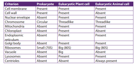
Organisms such as bacteria are known as prokaryotes.1. Which structure in a bacterial cell resembles a nucleus?4.3. Cell organelles2. How does it differ from the nucleus of eukaryotic cells?
Activities 4.3
By using iodine solution, methylene blue, a piece of onion leaf, a scalpel, forceps,
light microscope, slides and cover slips, clean cotton wool bud, and onion bulbs.
Observe cells from onion epidermis under light microscope.
Observation of a plant cell– Add a drop of diluted iodine solution on the slide.Why did you use iodine solution in this experiment?
– Remove a transparent layer of onion epidermis from the inner side that you
will mount on the slide and add iodine solution.
– Cover your preparation with a cover-slip and mount it on the stage.
– Observe the preparation under the low power and thereafter under high
magnification.What main parts of a plant cell are easily observed from a light microscope?
Observe animal cells from mouth cheek epithelium– By using a clean cotton wool bud, wipe over inside of your cheek.
– Smear cells over surface of a clean grass microscope slide containing a drop
of methylene blue stain
– Carefully put the cover-slip on the preparation and mount it on the stage to
observe.Draw both plant and animal cell and label the cell wall, nucleus and vacuole\
The cell nucleus contains nearly all the cell’s DNA with the coded instructions for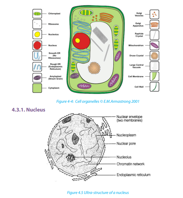
making proteins and other important molecules. The nucleus is surrounded by a
double nuclear envelope, which allow materials to move into and out of the nucleus
through nuclear pores. The granules found in the nucleus are called chromatin which
consist of DNA bound to protein. When a cell divides, the chromatin condenses into
chromosomes containing the genetic information. The nucleus contains a densespherical structure called nucleolus in which assembly of ribosomes occurs
The ER consists of a series of flattened membrane-bound sacs called cisternae. The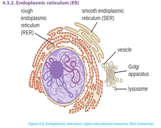
rough ER is surrounded with ribosomes. The rough ER transports proteins made on
attached ribosomes. The smooth ER is made of tubular cavities lacks ribosomes, and
it involves in synthesis of lipids that the cell needs. The number and distribution of
the ER relates to the functions of the cell; glandular cells are seen to have several RER
for synthesis of hormones and enzymes. Examples include liver cells, plasma cells,and pancreatic cells.
4.3.3. Golgi apparatus
The Golgi apparatus is a stack of membrane-bound, flattened sacs, which receives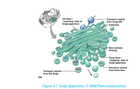
proteins from the ER and modifies them. It may add sugar molecules to them to form
glycoproteins or lipids to form glycolipids. The Golgi apparatus then packages the
modified substances into vesicles that can be transported to their final destinationsthroughout the cell or outside of the cell by exocytosis.
Mitochondrion have two membranes separated by a fluid-filled intermembrane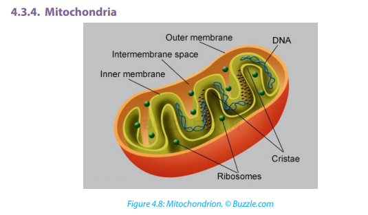
space. The inner membrane is highly folded to form cristae that plays a big role inaerobic respiration. The central part of the mitochondrion is called matrix.
The mitochondria are the site where Adenosine triphosphate (ATP) is producedduring aerobic respiration.
4.3.5. Chloroplasts
Chloroplasts are the site of photosynthesis in plant cells. These are found in plant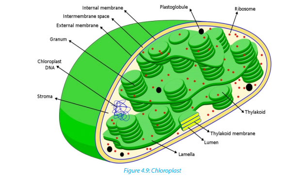
cells and in cells of some protoctists. They also have two membranes separated
by a fluid-filled space, circular DNA as in mitochondria. The inner membrane is
continuous, with thylakoids. A stalk of thylakoids is called a granum (plural:grana). Chlorophyll molecules are present on the thylakoid membranes.
These are spherical sacs surrounded by a single membrane. They contain powerful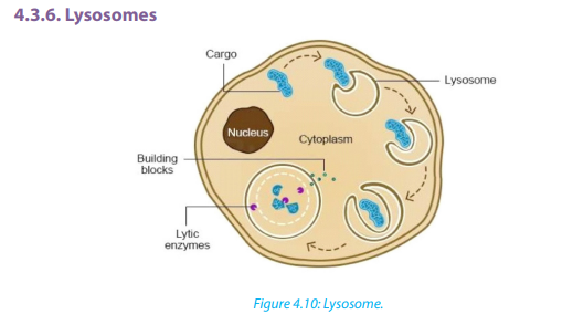
digestive enzymes. Their role is to break down materials such as worn out cell
organelles, and destroy foreign microorganisms that enter the body. In acrosome,
lysosomes help the sperm to penetrate the egg by breaking down the material
surrounding the egg. Lysosomes are also involved in autolysis, breakdown of dead
tissues or harmful objects inside the cell. Therefore, lysosomes are referred to as‘suicide bags’
Ribosomes appear as dark granules in the cytoplasm and are not surrounded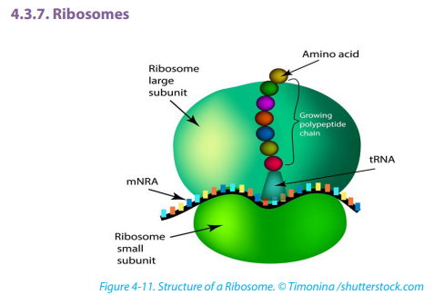
by a membrane. They have the same size as those found attached to the rough
endoplasmic reticulum- about 20nm in diameter and known 80S. Free ribosomes
make proteins that are as enzymes or in other forms in the cytoplasm. Ribosomesare made in a region of the nucleus called the nucleolus.
Centrioles are small tubes of protein fibers called microtubules which have many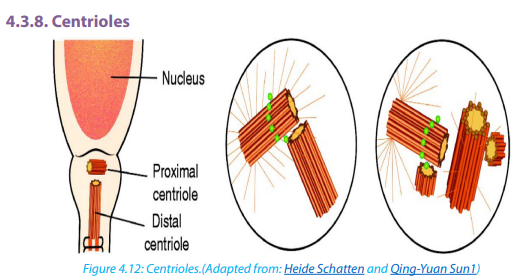 \
\
roles including moving chromosomes during nuclear division. Animal cells have
structures called centrioles which consist of two groups of nine triple microtubules.Centrioles form an anchor point for microtubules during cell division.
A vacuole is a saclike structure that stores materials such as water, salts, proteins, and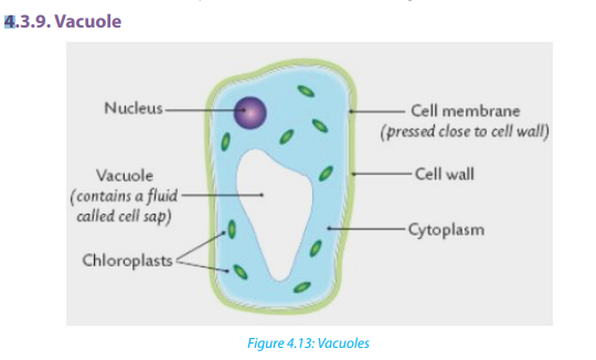
carbohydrates. In many plant cells there is a single and large central vacuole filled
with liquid. The pressure in the cells of central vacuole makes it possible for plants
to support heavy structures like leaves and flowers. Some animals and unicellular
organisms contain contractile vacuoles which contract to pump excess water outof the cell.
Self-assessment 4.3
1. Explain why muscle cells contain several mitochondria compared to fat
storage cells
2. What kind of information is contained in chromosomes?
3. Describe the functions of the endoplasmic reticulum, Golgi apparatus,
chloroplasts, mitochondria and nucleus in the cell.
4. Consider the 3D structures which would be visible in ultrastructure of a
plant cell.a. Identify their parts and label them4.4. Membrane structureb. State one function for each part
Activity 4.4
Learners mix a portion of cooking vegetable oil with water and shake the mixture
vigorously and leave it to settle. Note the way water and oil are distributed withinthe mixture and suggest a possible explanation for your observation.
Cell membranes cover surfaces of every cell. Some organelles in cytoplasm are
enveloped by membranes. The cell membranes ultrastructure is not easily visible
under a light microscope but is studied by electron microscopes, freeze structuring
and other modern techniques which reveal complex structures
A detailed study of a cell membrane reveals that it is 7-8nm wide and is made of a
phospholipid bilayer.
– Lipid component makes up 45% protein and 10% carbohydrate. Most of the
lipids are phospholipids
– Each molecule of phospholipid consists of a hydrophobic tail of two fatty acids
and a hydrophilic phosphate head. They arrange themselves in phospholipids
bilayer with their tails pointing inward away from the water both inside andoutside the cell
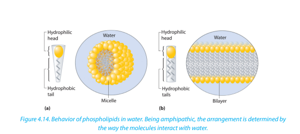 \
\
In 1972, Jonathan singer and Garth Nicolson proposed the fluid mosaic model of the
cell membrane structure. This was done after realizing that membranes must have a
complex structure to carry out a variety of activities. In their model;– Individual protein molecules shift and move on a fluid bilayer of phospholipids;
some spanning the width of the membrane (intrinsic proteins), others confined
to the outer or inner surface (extrinsic protein)
– Protein molecules are variable in structure and function but they all contributeto the mechanical strength of membranes
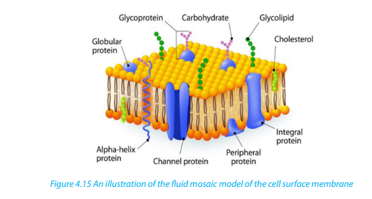
The membrane is referred to as;– A fluid because it appears to have the properties of a fluid rather than a solid as4.4.1. Properties of the cell membrane
the major constituent, lipids and proteins move about the structure
– Mosaic because protein and lipid components form a pattern of parches model– It is mainly made of lipids, proteins and carbohydrates.4.4.2. Roles of different components of cell membrane
– It is semi-permeable or partially permeable to allow some substances to pass
through but prevents others to cross depending on their size, charges and
polarity.
– It is positively charged outside and negatively charged inside and has a
hydrophilic pole and a hydrophobic pole
– It is a bilayered sensitive and flexible.It has inorganic ions and its proteins and
lipids may be mobile and contains different types of enzymes and coenzymes.
– It is perforated of pores and recognizes chemicals messengers includinghormones and neurotransmitters.
a. Cholesterol– Gives the membranes of some eukaryotic cells the mechanical stability.b. Channel proteins
– It fits between fatty acid tails and helps make the barrier more complete,
so substances like water molecules and ions cannot pass easily and directlythrough the membrane.
– Allow the movement of some substances across the membrane.channels.
– Large molecules like glucose enter and leave the cell using these protein
c. Carrier proteins– Actively move some substances across the cell membrane. For example,d. Receptor sites
magnesium and other mineral ions are actively pumped into the roots hair
cells from the surrounding soil.
– Nitrate ions are actively transported into xylem vessels of plants– Allow hormones to bind with the cell so that a cell response can be carried out.e. Enzymes and coenzymes
– Glycoproteins and glycolipids may be involved in cells signaling and they allow
the immune system to recognize foreign objects to the cells.
– Some hormone receptors are glycoprotein and some are glycolipid.– Some reactions including metabolic processes in photosynthesis take place in4.4.3. Functions of a cell surface membrane
membranes of chloroplasts.
– Some stages of respiration take place in membranes of mitochondria, where
Enzymes and coenzymes may be bound to these membranes.
– The more membrane there is, the more enzymes and coenzymes it can hold
and this helps to explain why mitochondrial inner membranes are folded to
form cristae, and why chloroplasts contain many stacks of membranes calledthylakoids.
– The cell membrane acts as a selective barrier at the surface of the cell, andSelf-assessment 4.4
controls the exchange between the cell and its environment.
Glycoproteins and glycolipids are involved in the cell protection, the process
by which cell adhesions are brought about and in the cell recognition.
– Receptor sites for hormones and neurotransmitters
– Transmission of nerve impulses
– Insulation of nerves to improve transmission speeds.
Internal membranes:
– Act as reaction surfaces
– Act as an intra cellular transport system
– Providing separate intra cellular compartment, isolating different chemicalreactions as in organelles.
1. What is meant by the fluid mosaic model of the cell membrane?
2. State at least three properties of the cell membrane.
3. Describe at least 4 types of the proteins in the cell membrane and their
roles.4. What is a partially permeable membrane?
5. What do the words hydrophilic and hydrophobic mean?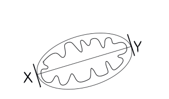
6. The diagram below shows the structure of a cell membrane. Study it carefully
and answer the following questions.
a. Name parts labelled A, B, C and D and give the function of the part B.
b. What types of molecule are likely to be involved in?i. Cell signaling and recognition7. What is the difference between rough and smooth endoplasmic reticulum?
ii. Allowing small charged molecules to pass through the cell membrane
iii. Site metabolic reactions
8. Describe the role of cytoskeleton
9. The photograph in the figure below shows an organelle of the living cell.
a. Name this organelle.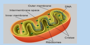
b. What is the function of this organelle?c. In which ways is this organelle similar to a chloroplast?
4.5. Specialized cells
Activity 4.5
By using the diagrams below, relate the structure of specialized cells to theirfunctions.
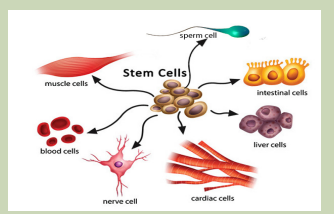
Differentiation refers to the changes occurring in cells of a multicellular organism so
that each different type of cell becomes specialized to perform a specific function.
In animals, the first type of cells in the developing embryo is stem cells. These are
unspecialized cells that go on to form all the different types of cells in adult. Cell can
differentiate in many ways, with changes to the shape of the cell, the number ofparticular organelles and the content of the cell.
4.5.1. Specialized animal cells and their functions4.5.1.1. Red blood cells
All blood cells are produced from undifferentiated stem cells in the bone marrow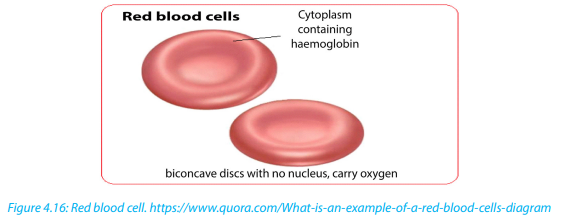
but the cells destined to become erythrocytes (red blood cells) lose their nucleus,
mitochondria, Golgi apparatus and rough endoplasmic reticulum. They are packed
full of the protein called haemoglobin. The shape of this cells change so that theybecome biconcave discs, and they are then able to transport Oxygen in the body.
Sperm cells are specialized to fertilize the egg. Its specialization involves many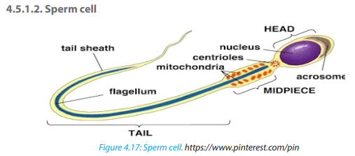
changes in shape and organelles content.
By shape: the sperm cells are very small, long and thin to help them to move easily,
and they have a flagellum which helps them to move up the uterine tract towardsthe egg.
By organelles content: sperm cells contain numerous mitochondria which
generate much energy for their movement. Their acrosome has specialized
lysosomes containing many enzymes that are released on the outside of the egg.
These enzymes lyse the wall of the egg, and facilitate the sperm nucleus to penetrate
easily. In content, the sperm cell nucleus contains the half number of chromosomes
of the germ cell in order to fulfil its role as a gamete of fertilizing the egg.
Did you know: As a sperm fuses with an ovum to form a zygote which grows into
an individual, in the same way: a man maries a woman to form a couple which willproduce children and form a family.
Nerve cells also known as neurons are specialized cells to carry nervous impulses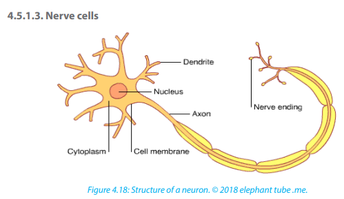
in the body. These signals between neurons occur via specialized connections
called synapses. Specialized animal cells have different functions. Some of them aresummarized in the following table.
Table 4.3: Specialized animal cells and their functions.
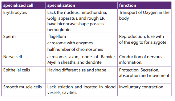
4.5.2. Specialized plant cells and their functions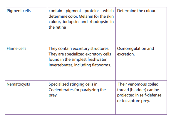
4.5.2.1. Root hair cells
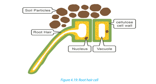
The root hair cells have hair-like projection from their surface out into the soil. This
increase the surface area of root available to absorb water and minerals from thesoil.
4.5.2.2. Palisade cells
Palisade cells are in leaves, right below the upper epidermis. They are vertically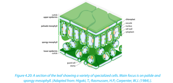
elongated, a different shape from the spongy mesophyll cells beneath them in
the leaf. Their large numbers of chloroplasts allow them have several chloroplasts
used in photosynthesis.
Parenchyma cells
Parenchyma is composed of relatively simple and undifferentiated parenchyma
cells. They function in storage, photosynthesis. In most plants, metabolic activity
such as cell division, respiration, and photosynthesis occurs in these cells because
they retain their active cytoplasm. .
4.5.2.3. Guard cells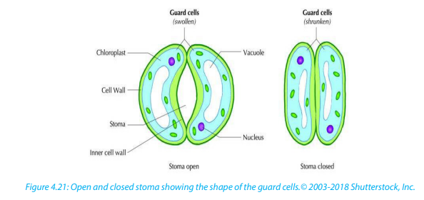
Guard cells are cells surrounding each stoma. Guard cells are specialized cells in the
epidermis of leaves, stems and other organs that are used to control gas exchange.
They are produced in pairs with a gap between them that forms a stomatapore.
Guard cells have the following feature:– Un even thick wallsSelf-assessment 4.5
– Possess chloroplasts; they are the epidermal cell that have chloroplasts anadaptive feature in controlling pore opening.
1. Explain why differentiation to produce erythrocytes involves a change in
shape.
2. Red blood cells cannot divide as they have no nucleus. State two other
biological processes that red blood cells cannot carry out.
3. Describe how the following are specialized for their roles:a. Neutrophil4. Explain why photosynthesis is carried out in palisade mesophyll more than
b. Sperm cell
c. Root hair cell
in spongy mesophyll.
5. In what kinds of organisms is cell specialization pronounced characteristic?6. Discuss the advantages of cell specialization in living things
End of unit assessment 4
Section A. Multiple choice questions
1. Which organelle converts the chemical energy in food into a form that cells can
use?a. Chromosome2. The cell membranes are constructed mainly of:
b. Chloroplast
c. Nucleus
d. Mitochondriona. Carbohydrate gates3. In many cells, the structure that controls the cell’s activities is the:
b. Protein pumps
c. Lipid bilayer
d. Free-moving proteinsa. Nucleus4. Despite differences in size and shape, all cells have cytoplasm and a
b. Nucleolus
c. Cell membrane
d. Organellea. Cell wall5. If a cell of an organism contains a nucleus, the organism is a (an)
b. Cell membrane
c. Mitochondria
d. Nucleusa. Plant6. Match each part of the cell (left column) to corresponding statement (right
b. Eukaryote
c. Animald. Prokaryote
column):
Nucleus controls movement of substances in and out of the cell
Mitochondrion where photosynthesis takes place
Chloroplast where aerobic respiration takes place
Smooth ER controls the activity of the cell
Ribosomes where lipids including steroids are made
Section B: Questions with short answers
1. How does a cell membrane differ from a cell wall?
2. Name the structures that animal and plant cells have in common, those found in
only plant cells, and those found only in animal cells.
3. List:a. Three organelles each lacking a boundary membrane4. Identify each cell structure or organelle from its description below.
b. Three organelles each bounded by a single membrane
c. Three organelles each bounded by two membranes (an envelope)a. Manufactures lysosomes and ribosomesSection C: Essay questions
b. Site of protein synthesis
c. Can bud off vesicles which form the Golgi body
d. Can transport newly synthesized protein round the cell
e. Manufactures ATP in animal and plant cells
f. Controls the activity of the cell, because it contains the DNA
g. Carries out photosynthesis
h. Can act as a starting point for the growth of spindle microtubules during
cell division
i. Contains chromatin
j. Partially permeable barrier only about 7 nm thick
k. Organelle about 25 nm in diameterl. Which two organelles other than the nucleus contain their own DNA
1. Describe the structure and function of the cell membrane and cell wall.4. The diagram below shows the structure of a liver cell as seen using an electron
2. Describe the basic structure of the cell membrane.
3. Explain two common characteristics of chloroplasts and mitochondria.Consider both function and membrane structure.
microscope.
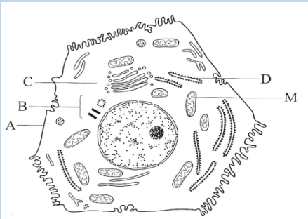 a. Name the parts labelled A, B, C and D.
b. The magnification of the diagram above is x12 000. Calculate the actual
length of the mitochondrion labelled M, giving your answer in µm. Show
your working.
c. Explain the advantage to have a division of labor between different cells inthe body.
UNIT 5: DIVERSITY OF SPECIALIZED TISSUES
UNIT 5: DIVERSITY OF SPECIALIZED TISSUES
Key Unit Competence
Describe different specialized plant and animal cells and adaptation of tissues.
Learning objectives
By the end of this unit, I should be able to:– Define a tissue as a group of cells with similar structure working together forIntroductory activity
a function.
– Name the main types of animal and plant tissues.
– Define an organ as a structure made up of a group of tissues with related
functions working together to perform bodily functions.
– Explain how epithelial tissues are adapted to perform a diversity of functions
in the body.
– State the advantages and disadvantages of being unicellular.
– Observe and draw plant and animal tissues as seen under a light microscope.
– Interpret photomicrographs of plant and animal tissues
– Acknowledge the relationship between levels of organization
– Recognize the efficiency shown by multicellular organisms to explore more
modes of life that are not available to single celled organisms that show little
or no specialization
Read the following passage and use it to answer the following questions:
In an anthill, there are different groups of termites such as a queen, workers and
soldiers. Each group has a specific role to play in the colony. The structure termites
of each group is related to their role for example soldiers that protect the colony
have mouth parts shaped like a pair of scissors building and a slightly larger
abdomen for storing water. The queen is the largest of all and has a role of laying
eggs. Workers have mouth parts for cutting and chewing food or soil particles.
Some members of workers are in charge of caring for the young while others find
food and defend the colony or remove dead members. Their specialization anddivision of labor bring about efficiency in the colony.
1. Specify the message addressed by the above paragraph.
2. Explain how is the structure of termites related to their functions?
3. What is the significance of specialized tissues in multicellular organismslike plants and animals?
The study of tissues is known as Histology. A tissue is a group of associated,
similarly structured cells that perform specialized functions for the survival of the
organism. In histology, differentiation is the process by which structures become
modified and specialized to perform specific functions. Differentiation is also known
as ‘specialization’. In animals, the first type of cells in the developing embryo is stem
cells. These are unspecialized cells that go on to form all the different types of cellsin adult.
5.1. Specialized plant tissues
Activity 5.1.1
– Remove an epidermis layer from the ventral side of an onion leaf.
– Mount it on the slide containing a drop of iodine solution
– Observe your preparation under a light microscope
– Draw, label and describe your findings.
– From your discussion:1. What is a tissue?2. What is the role of epidermis in onion?
5.1.1. Plant tissues
Activity 5.1.2
The following figure represents the flow chart of subdivisions of plant tissues. Useit to answer the following questions.
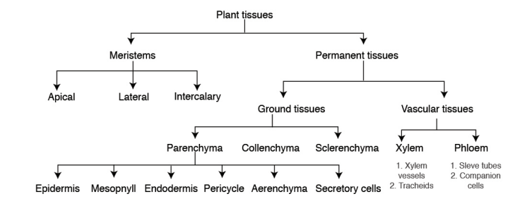 1. How do meristems differ from permanent tissues?
2. Plant tissues are classified into ground tissues and vascular tissues as
shown in the figure above. What is meant by the term vascular tissues?
3. How is the structure of the xylem and phloem vessels related to their
function?4. From the flow diagram above, identify three types of ground tissues.Plant tissues can be divided into two main groups, Meristematic tissues (apical, lateral,
5. Write down short notes on each of the following types of meristems.
a. Apical meristems.
b. Lateral meristemsc. Intercalary meristems
and intercalary meristems) and Permanent tissues (ground tissues and vascular tissues).
5.1.2. Meristem tissues
Meristem tissue is a group of cells which retain the ability to divide by mitosis.
Meristematic tissues are specialized to carry out specific functions such as
reproduction, growth, photosynthesis and replacement of old or damage tissues.
The cells making a meristem tissue are small, have a central large nucleus and dense
cytoplasm, thin-walled, with no or small vacuole, and no specialized features. Thecells are rectangular and closely packed with no intercellular air spaces.
Types of meristematic tissues
Meristematic tissues are subdivided into apical meristems, lateral meristems
(cambium) and intercalary meristems
a. Apical meristems
They are located in the root and shoot apex (at the growing points of roots and
stems). They are responsible for primary growth, leading to the increase of primary
plant body.
b. Lateral Meristems (cambium)
Lateral meristems are in lateral parts of the plant, where they are responsible for
secondary growth. The cambium gives rise to secondary vascular tissues (secondary
xylem and secondary phloem) in dicotyledonous plants.
c. Intercalary meristems
These are found in the region of permanent tissues like at nodes of monocotyledonousplants (e.g. sugar cane). It allows growth in length to occur between internodes.
Functions of meristematic tissues– The main function of meristematic tissue is to produce new cells by mitosis.5.1.3. Permanent tissues
The cells elongate and differentiate to form new cells for primary growth of
shoot and root.
– Vascular cambium produces new cells to increase the diameter of stems and
roots during secondary growth.
– Cork cambium called (phellogen) produces the outer cork layer called phellem
which consists of suberized cells. The cork layer reduces water evaporation
from the plant and protects the plant against the entry of pathogens.
– The intercalary meristems allow growth and increase in length in regions otherthan the tips.
Permanent tissues consist of two groups of tissues such as: ground and vascular
tissues.
5.1.4. Ground tissues
The ground or fundamental tissues are plant tissues which function in storage,
metabolism and support. There are three types of ground tissues: parenchyma,collenchyma and sclerenchyma tissues.
5.1.5. Parenchyma tissues
Parenchyma is a soft plant tissue made up of thin-walled cells that forms the
greater part of leaves, stem pith, roots, and fruit pulp. They are the main sites for
physiological and biochemical processes in the plants including photosynthesis,
protein synthesis and storage of starch and mineral ions. Parenchyma tissues can be
found in epidermis, mesophyll, endodermis, pericycle, aerenchyma and secretory
cells.
Characteristics– Parenchyma tissues consist of large living cells, with relatively thin wall
containing cellulose, pectin and hemicellulose.
– Parenchyma tissues consist of cells, usually having a large central vacuole.
They are often partially separated from each other.– Spongy cells present intercellular spaces that intervene in gaseous exchange
and transpiration through stoma. They are usually stuffed with plastids.– Parenchyma tissues consist of cells with polygonal and spherical shapes inFunctions of parenchyma tissues
the leaf. They form the mesophyll, and are located between upper and lowerepidermises. They are responsible for photosynthesis.
– In the leaves, parenchyma tissues form the mesophyll and are sites for
photosynthesis, gaseous exchange and transpiration.
– They store food substances such as starch, proteins and lipids
– They can be modified to form specialized cells to carry out other function inepidermis, endodermis, pericycle, parenchyma, and secretory cells.
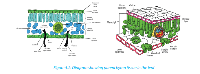
Adaptations of parenchyma for its function– Parenchyma tissues are made of unspecialized cells with variety of functions:5.1.6. Collenchyma tissues
– Parenchyma can become specialized to carry out specific functions e.g.
mesophyll has cells with many chloroplasts, and aerenchyma which has air
spaces. All of these adaptations help in photosynthesis and gas exchange.
– They have isodiametric cells and function as packing tissue and storage tissue.
– Cells are loosely packed with many large intercellular spaces. This permits
diffusion of gases.
– They have thin cellulose cell wall which is permeable so that it permits passage
of materials.
– The walls are transparent and permit entry of light in photosynthesis cells.
– Large cells with large vacuoles provides space for storage of substances, where
the entry of water causes vacuole to expand and cells become turgid
– Leucoplasts act as storage of starch while chromoplasts present in some cellse.g. in petals attract insects for pollination.
Their cells are elongated with irregularly thickened cell walls that provide structural
support, particularly in growing shoots and leaves. Their thick cell walls are composed
of cellulose and pectin. These cells are often found under the epidermis, or the outerlayer of cells in young stems and in leaf veins.
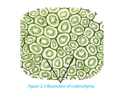 Sclerenchyma is found in hard parts of the plant body. They are very common in
5.1.7. Sclerenchyma tissues
roots, stems, leaves and petioles. They may be present in patches, groups or layers.
The cells of the sclerenchyma are dead, they are elongated, narrow, and thick walled
and lignified. They are pointed at both ends where it gives strength, rigidity and
flexibility to the plant body. They consist of fibres and sclereids. Fibres are long,
narrow, thick and liquefied cells usually tapering at both ends. Sclereids cells arenormally short with very thick walls, irregular and not tapering at the ends.
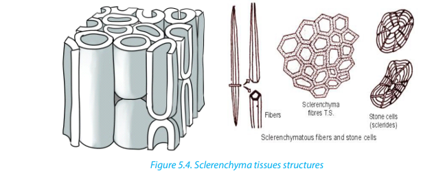
5.1.8. Vascular tissues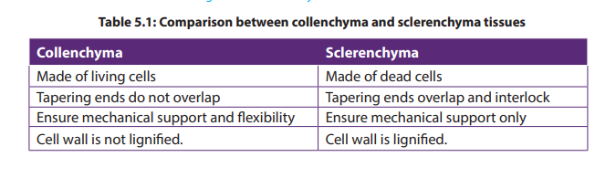
The vascular tissue system consists of two kinds of conducting tissues: the xylem
responsible for conduction of water and dissolved mineral nutrients, and the
phloem responsible for conduction of elaborated food.
a. Xylem
The xylem tissues are made of dead cells which have the cell walls removed at the
end of the cells, forming tubes through which the water and dissolved mineral ions
can flow. Xylem vessels are involved in the movement of water through a plant -
from its roots to its leaves via the stem. During this process water is absorbed from
the soil through root hair cells, moves by osmosis from root cell to root cell until it
reaches the xylem, and finally it is transported through the xylem vessels up thestem and then to the leaves.
Xylem vessels are hollow tubes or lumen with a thick strengthened cellulose cell
wall. The hollow tubes act like pipes allowing water and dissolved minerals to flow
through them. They develop from cylindrical cells arranged end to end, in which
the cytoplasm dies and the cell walls between adjoining cells breaks down leaving
a dead empty tube. The cell walls in xylem vessels contain a substance called ligninwhich strengthens the cells and gives structural support.
b. Phloem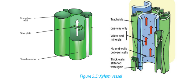
Phloem vessels are involved in translocation of elaborated substances. Dissolved
sugars, produced during photosynthesis, and other soluble food molecules are
moved from the leaves to growing tissues such as the tips of the roots and shoots
and storage tissues such as in the roots. In contrast to xylem, phloem consists of
columns of living cells. The cell walls of these cells do not completely break down,
but instead form small holes at the ends of the cell. The ends of the cell are referred
to as sieve plates. The connection of phloem cells effectively forms a tube which
allows dissolved sugars to be transported.
Phloem tubes carry food substances like sugar and amino acids produced in leaves
during photosynthesis to every part of the plant. The movement of food substancesthrough the plant is called translocation.
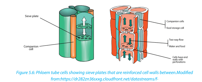
Table 5.2: Comparison between Xylem and Phloem tissues
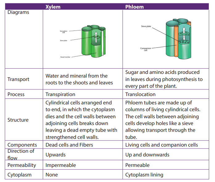
Self-assessment 5.1
1. State where in a flowering plant you would find:a. Lateral meristem2. Give characteristics of meristematic cells.
b. Intercalary meristem
c. Apical meristem
3. What do you understand by the following terms?a. Differentiation4. Differentiate between Collenchyma and sclerenchyma
b. Cambium
c. Wood
d. Meristem
5. State the main structures (components) that make up a xylem and phloem
tissues.
6. Explain how the structure of Parenchyma and Xylem tissues are suitable to
their functions.
7. The diagram below shows a longitudinal section of two cells of phloem tissue
in a plant stem.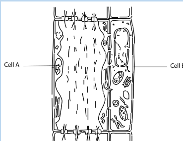
a. Name the cells labelled A and B on the diagram.b. State the function of phloem in a plant.
5.2. Animal tissues
Activity 5.2
Conduct a research by using different sources of information to find out the
structures and the main functions of the following four groups of animal tissues:epithelial, connective, muscular and nervous tissues.
There are four basic types of animal tissues such as epithelial tissue, muscle tissue,nervous tissue, and connective tissue.
5.2.1. Epithelial tissue
Epithelial tissue consists of closely packed cells arranged in single or multilayered
sheets. It is made up of layers of tightly packed cells that form the external surfaces
of the body and cover the outer and the inner surfaces of the organs. Some are
specialized to form glandular tissues (glands). The epithelium lining the inside of the
heart, blood vessels and lymph vessels is referred to as endothelium. Two criteria
for classifying epithelia are: the number of cell layers and the shape of cells on
the free surface. The following are the types of epithelium tissues:
a. Simple cuboidal epithelium
This is a tissue with cells that are cubical in shape. Cuboidal cells are specialized
for secretion and they make up the epithelia of kidney tubules and many glands
including salivary glands, and thyroid gland.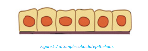
b. Simple squamous epithelium
It is thin, leaky and functions in the exchange of material by diffusion. This type of
epithelium lines blood vessels and the air sacs of lungs, where diffusion of nutrientsand gases is critical.

c. Simple columnar epithelium
These are columnar in shape with free surface containing extensions of micro villi.
It lines the intestines. This epithelium secretes digestive juices for the final stages of
digestion and absorbs nutrients to blood stream.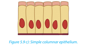
d. Pseudo-stratified ciliated columnar epithelium
It forms a mucous membrane that lines the nasal passages of many vertebrates. Thebeating cilia move the film of mucus along the surface.
e. Stratified squamous epithelium
It regenerates rapidly by cell division near the basal lamina. The new cells are pushed
outward to replace cells that are sloughed off. This epithelium is commonly found
on surfaces subject to abrasion, such as the outer skin and lining of the esophagus,anus, and vagina.
f. Transitional epithelium
In this type of stratified epithelium, the surface cells change their shape from round
to squamous. Transitional epithelium lines urinary bladder. When the bladder is
empty, the surface cells are rounded. As the bladder fills urine, these cells become
flattened. Transitional epithelium enables the bladder to fill and stretch withouttearing the lining.
g. Stratified columnar epithelium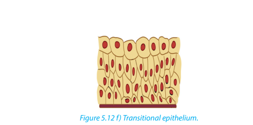
It is a rare type of the epithelial tissue composed of column shaped cells arranged in
multiple layers. They are found in the conjunctiva or the eye, in parts of the pharynx,anus, uterus, the male urethra and vas deferens.
h. Stratified cuboidal epithelium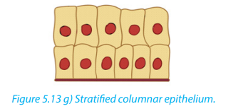
It is a type of epithelial tissue composed of multiple layers of cube-shaped cells. Only
the most superficial layer is made up of cuboidal cells and the other layers can be
cells of other type. It has several locations in the body including sweat gland ducts,egg-producing vesicles and ovaries.
5.2.2. Main characteristics of epithelial tissues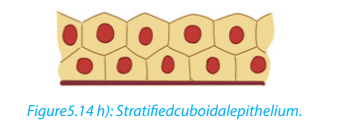
a. Polarity
All epithelia have a free surface and a lower attached basal surface that differ in
structure and function. For this reason, epithelium is described as showing polarity.
b. Supported by connective tissue
All epithelia are supported by connective tissue. For instance, deep to the basal
lamina is reticular lamina, an extracellular material containing collagen protein
fiber which forms the basement membrane. The basement membrane reinforces
the epithelium and helps it to resist stretching and tearing.
c. They are avascular; epithelia have no blood vessel in them. Nutrients and gasesare supplied by blood through the connective tissue by simple diffusion
d. Regeneration
Epithelium have a high regenerative capacity and can reproduce rapidly as long as
they receive adequate nutrition.
Functions of epithelium– Epithelium forms a protective layer: The epithelium of the skin protects the5.2.3. Muscular tissues
body from mechanical damage, entry of pathogens, ultraviolet rays and
dehydration. Epithelium lining the respiratory air passages secretes mucus
which traps inhaled dust particles and microbes.
– The ciliated epithelium cells have cilia that propel the mucus and trapped
particles to the throat.
– Glandular tissues secrete the digestive enzymes, hormones, mucus, sweat and
sebum.
– Acts as a barrier and regulates movement of substances across kidney
– Some epithelial cells can divide mitotically producing new cells to replace
damaged or dead cells.
– Some epithelial cells such as taste buds and retina cells are specialized to formsensory receptors.
Muscle tissues consist of elongated cells held together by connective tissue. Muscle
cells are highly specialized in that they are able to shorten to a half or even a third of
their resting length by the process of contraction. The contraction is caused by two
types of fibrous proteins: myosin and actin.
Muscles in the body provide the necessary force for the motion and they convert
chemical energy into kinetic or mechanical energy. There are three types of muscle
tissue:– Smooth muscle which is found in the inner linings of organs;Smooth and cardiac muscles are involuntary muscles whereas skeletal muscles are
– Skeletal or striated muscle, which is attached to bone and helps in movement
of the body;
– Cardiac muscle which is found only in the heart.called voluntary muscles because they are under voluntary (conscious) control.
a. Smooth Muscle
Smooth muscle is also called unstriated, unstriped, involuntary or visceral muscle.
It is found in the walls of the hollow internal organs such as blood vessels, intestinal
tract, urinary bladder, and uterus. Smooth muscles have the following features;– It is under control of the autonomic nervous system; they cannot be controlled
consciously, so they are also called involuntarily muscle. They do not have
striations.– Smooth muscle cells contract slowly and rhythmically
b. Cardiac tissue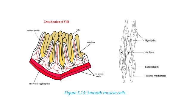
Cardiac tissue (figure 5.10 a) is found in the walls of the heart and it is under control
of the autonomic nervous system. Cardiac muscle has the flowing basic features.
– It contracts and relaxes continuously.
– It is branched and connected to other cardiac muscle fibers through
intercalateddiscs (Figure 5.16 b), which are reinforced membranes that hold the
cells together during contractions. These interconnections or intercalated discs
between the fibers ensure a rapid and uniform spread of excitation throughout
the wall of the heart which in turn ensures a synchronous contraction.– They are myogenic (their contraction originate from within the heart itself).
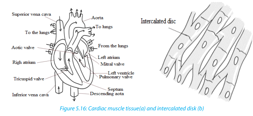
c. Skeletal Muscle
Skeletal muscle is also called striated, striped, or voluntary. They are attached
to bone, and are responsible for body movements and body posture. There areapproximately 639 skeletal muscles in the human body.
Characteristics of skeletal muscles:
– They are under control of voluntary nervous system
– They are attached to bone and this is the reason why they are called skeletal
muscles.
– They are made of elongated and cylindrical muscle fibres
– They appear under microscope to have alternate light and dark bonds and this
is why they are called striated muscles.
– Their muscle fibres are multinucleated (many nuclei per cell)
– These muscle cells also contain light and dark stripes called striations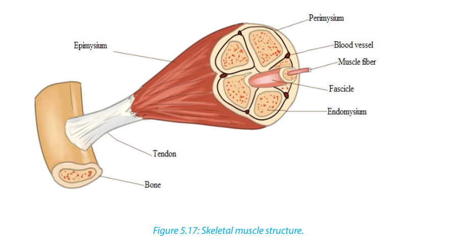
General functions of muscle
The main function of muscle is its contribution to motion, where body movements
such as walking, breathing, and speaking, as well as movements associated with
digestion and the flow of fluids take place. Muscles contribute to the heat production,
maintenance of posture and body support and communication through facial
expression, writing and speech.
5.2.4. Nervous tissue
Nervous tissue is composed principally of densely packed cells called the nerve cells
(neurons) that together form the nervous system including the brain and spinal
cord. Neurons are specialized for transmitting electrical nerve impulses.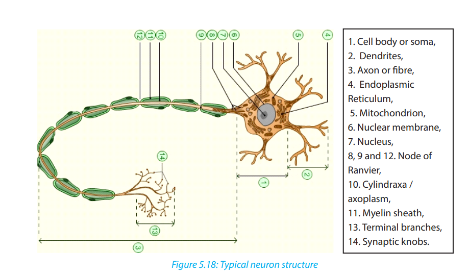
A typical neuron has three main parts: Cell body, dentrites and axon.
a. The cell body or soma– It is the main part from which, extensions derive (Axon and Dendron).all nerve cell activities.
– It is made of a great spherical nucleus, granular cytoplasm and controls
b. Dendrites (Dendron when single): small branches attached to the cell body
and receive nerve impulse from other neurons
c. Axon or cylindrax:– It is the thinner nerve fibre that carries messages away from the cell5.2.5. Connective tissues
body and can be as long as 1 m. In some neurones, the axons have a
fatty myelin sheath formed by Schwann cells which wrap themselvesaround the axon to increase the speed of impulse transmission.
Connective tissue is made up of many different types of cells that are all involved in
structure and support of the body. Bone, blood, fat, and cartilage are all connective
tissues. Connective tissues can be densely packed together, as bone cells are or
loosely packed, as adipose tissue (fat cells) are. A connective tissue is made up of a
variety of cells embedded in a large amount of intracellular substance called matrixand fibers which are non-living products of the cells.
a. Common functions of connecting tissues:
– Connective tissues protect and support the body and internal organs.
– They act as connecting systems, binding all other tissues together.– They also form surrounding sheaths to separate the various organs.
b. Cells of connective tissue
The specialized cells of the various connective tissues produce the extracellular
matrix. The names of the cells end with suffixes that identify the cell functions as
blasts, cytes, or clasts. Blasts create the matrix, cytes maintain it, and clasts break it
down for remodeling. For example: Fibroblasts are cells that form fibrous connective
tissue and fibrocytes maintain it, chondroblasts form cartilage and chondrocytes
maintain it, and osteoblasts form bone, osteocytes maintain it, and osteoclastsbreak it down
Adipose, or fat cells, also called Adipocytes, contain large amounts of lipid. The
lipid pushes the rest of the cell contents to the periphery, so that each cell appears
to contain a large and centrally located lipid droplet with a thin layer of cytoplasm
around it. Adipose cells are rare in some connective tissue types like cartilage but
they are abundant in others like loose connective tissue, and they are predominant
in adipose tissue.
Mast cells are commonly found beneath membranes in loose connective tissue
and along small blood vessels of organs. They contain chemicals such as heparin,
histamine and proteolytic enzymes. These substances are released in response toinjury such as trauma and infection and play important roles in inflammation.
White blood cells continuously move from blood vessels into connective tissues.
The rate of movement increases dramatically in response to injury or infection. In
addition, accumulations of lymphocytes, a type of white blood cell, are common
in some connective tissues, such as in the connective tissue beneath the epitheliallining of certain parts of the digestive system.
Macrophages are found in some connective tissue types. They are derived from
monocytes, a white blood cell type. Macrophages are either fixed and do not
move through the connective tissue in which they are found or are wandering
macrophages and move by amoeboid movement through the connective tissue.
Macrophages phagocyte foreign or injured cells, and they play a major role inproviding protection against infections.
Note that there are three structural major components of the extracellular matrix of
connective tissue such as fluid, ground substance consisting of non-fibrous protein
and other molecules and protein fibers. The structure of the matrix gives connective
tissue types most of their functional characteristics, such as the ability of bones and
cartilage to bear weight, tendons and ligaments to withstand tension, and dermis of
the skin to withstand punctures, abrasions, and other abuses.
c. Fiber connective tissues
Another type of connective tissues consists of fibers. Fibers are of different types
including:
– Connective tissue fibers: which are made of protein and are of three kinds:
collagenous, elastic and reticular fibers.
– Collagenous fibers: These provide strength combined with flexibility. They
are made up of collagen, probably the most abundant protein in the animal
kingdom.
– Elastic fibers: These are easily stretched but are also resilient, snapping back
to their original length when tension is released. Shaped as long threads,
elastic fibers are made of a protein called elastin.
– Reticular fibers: These are thin collagen fibers coated with glycoprotein. They
are very short, thin fibers that branch to form a network and appear different
microscopically from other collagen fibers. Reticular fibers are not as strong as
most collagen fibers, but networks of reticular fibers fill space between tissues
and organs.
d. Loose connective tissue
This is also called areola connective tissue and is the most widespread connective
tissue in all animal tissues. It binds epithelial tissues to underlying tissues and
functions as packing material, holding organs in place. Loose Connective tissue
has the following main components;– Fibers: collagen, elastic and reticular.e. Fibrous connective tissue
– Cells; fibroblasts and macrophages. Fibroblasts secrete the protein ingredients
of the extracellular fibers. Macrophages are cells that roam the maze of fibers,
engulfing both foreign particles and the debris of dead cells by phagocytosis.
Fibrous Connective tissue is dense with collagenous fibers. The fibers form parallel
bundles, which maximize non-elastic strength. Fibrous Connective tissue is found in
tendons, which attach muscles to bones, and ligaments, which connect bones atjoint.
f. Adipose tissue
Adipose tissue is a specialized form of loose connective tissue that stores fats in adipose
cells distributed throughout its matrix. Adipose tissue consists of adipocytes, or
fat cells, which contain large amounts of lipid. Unlike other connective tissue types,
adipose tissue is composed of large cells and a small amount of extracellular matrix
that consists of loosely arranged collagen and reticular fibers with some scattered
elastic fibers. Blood vessels form a network in the extracellular matrix. The fat cells
are usually arranged in clusters or lobules separated from one another by looseconnective tissue. Adipose tissue functions as:
– An insulator against heat loss
– A protective tissue to delicate internal organs– A site of energy storage in the form of fat.
g. Bone and Cartilage tissue
Cartilage has an abundance of collagenous fibers embedded in a rubbery matrix
made of a protein-carbohydrate complex called chondroitin sulfate. Cartilage is
composed of specialized cells, called chondrocytes, surrounded by a gelatinous
matrix of collagen, a tough protein. The cartilage surface is covered by a membrane
known as the perichondrium. There are three types of cartilage (hyaline cartilage,yellow elastic and white fibrous cartilage.)
– Hyaline cartilage is semi-transparent and is often stained light blue or pink inBone tissue
tissue sections. It is extremely very strong but very flexible and elastic. Hyaline
cartilage occurs in the trachea, larynx, tip of the nose, connection between the
ribs and the breastbone; and at the ends of bones where they form joints. It
also forms much of the fetal skeleton.
– Elastic cartilage is similar to hyaline cartilage, but in addition to the collagenous
fibers.The matrix of the elastic cartilage also contains an abundant network of
branched elastic fibers. This type of cartilage is found in the lobe of the ears,
the epiglottis and in the parts of the larynx. They provide flexibility and elastic
support.
– Fibro-cartilage(White fibrous cartilage) is an extremely tough tissue. It is found
as discs between the vertebrae, bones, anterior joint between the two halves
of pelvic girdle and at points where tendons inserted on bones near hyalinecartilage. It resists compression and absorbs shock in some joints.
This is a firmer and denser material that has the following features:– Hard and compactThe following are the main functions of bone tissue:
– Has many collagen fibres
– Its matrix has inorganic salts such is calcium carbonate and calcium phosphate
– Has few cells located in the lacunae in the matrix
– Has osteoblasts as mature and non-dividing cells
– Have a harversian canal– Consists of irregular cylinder with layer of matrix call lamellae
– Structural support of the bodyh. Blood tissue
– Protection of internal organs, heart and lungs.
– Attachment of the muscles to effect movement– Production of blood cells
Blood is a flowing made up of particles suspended in a fluid composed of fluid called
plasma, and several kinds of cells. Within the blood plasma, there are erythrocytes
(red blood cells), leukocytes (white blood cells), thrombocytes (platelets) and othersubstances. Blood performs the following important functions:
Transport– Blood transports absorbed substances such as glucose, amino acids, mineralHomeostasisions and vitamins from the small intestine.
– Blood transports the respiratory gases (Oxygen and Carbon dioxide).
– Blood transports the excretory wastes such as urea, uric acid to excretory
organs to be removed out of the body.
– Blood transports hormones e.g. insulin from pancreas to the liver where it isstored.
Na+ affects the water potential of the blood and regulates the diffusion of water
between blood and tissues. Hydrogen carbonates help to maintain the pH of the
blood.
Protection– Leucocytes such as neutrophils and macrophages engulf pathogens e.g.Self assessment 5.2
bacteria
– B-lymphocytes produce antibodies to destroy pathogens or to neutralize
toxins.
– T-lymphocytes destroy infected cells.
– Platelets, fibrinogen and prothrombin play an important role in blood clottingto reduce blood loss and the entry of pathogens.
You are provided with photomicrographs or slides of different plant and animal
tissue. Study them carefully and answer questions that follow.
Identify the different tissues provided and where they are located.One of the images is a blood smear. Draw a well labeled diagram of this tissue
5.3. Levels of organization: cell, tissue, organ and system
Activity 5.3
Visit a classroom block, administration block or any building in school which is
constructed with bricks and use it to answer the following questions.
1. What is the smallest unit or component of the classroom block?
2. How are bricks arranged?
3. Do you think the brick has other smaller particles in it?
4. How many bricks does a classroom block have?
5. How are walls, classrooms, washrooms and other apartments of the block
formed?
6. Arrange the following in their ascending order of size (from the smallest to
the largest); whole block, wall, a brick, a room, course (a line of bricks).
7. Relate the above arrangement of a building to levels of organization inmulticellular organisms, beginning with a cell and ending with an organism
The human body is organized into structural and functional levels of increasing
complexity. Each higher level incorporates the structures and functions of the
previous level. The simplest is the cells, organized into tissues, organs, and organ
systems. All of the levels of organization of the human body are represented in thefollowing figure.
5.3.1. Cells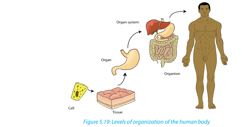
The smallest structural and functional living units of living things are cells. There are
many different types of human cells, though they all have certain similarities. Each
type of cell is made of chemicals and carries out specific chemical reactions.
5.3.2. Tissues
A tissue is a group of cells with similar structure and function. There are four groups
of tissues (Epithelial tissues, Connective tissues, Muscle tissues, Nerve tissue)
5.3.3. Organs
An organ is a group of tissues precisely arranged so as to accomplish specific
functions. Examples of organs are the kidneys, individual bones, the liver, lungs, and
stomach. The kidneys contain several kinds of epithelial or surface tissues, for their
work of absorption. The stomach is lined with epithelial tissue that secretes gastric
juice for digestion. Smooth muscle tissue in the wall of the stomach contracts to
mix food with gastric juice and propel it to the small intestine. Nerve tissue carriesimpulses that increase or decrease the contractions of the stomach.
5.3.4. Organ systems
An organ system is a group of organs that all contribute to a particular function.
Examples are the urinary system, digestive system, and respiratory system. For
example, the urinary system consists of the kidneys, ureters, urinary bladder, and
urethra. These organs all contribute to the formation and elimination of urine.
The Human body has 11 organ systems: circulatory, digestive, endocrine, and
excretory (urinary), the lymphatic, integumentary, muscular, nervous, reproductive,respiratory, and skeletal systems.
Table 5.3: Major organ systems of the human body
Self-assessment 5.3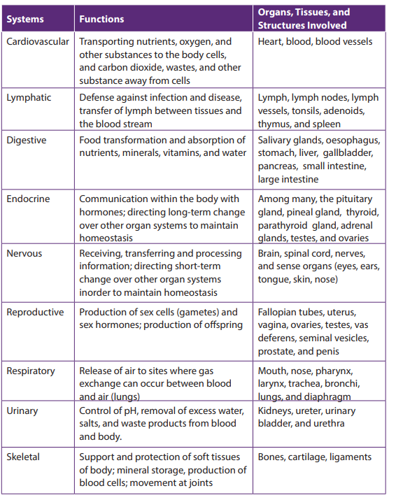
1. Answer by true or falsea. Organic chemicals are often very complex and always contain the element2. Explain why the cell as level of organization of human body is said to be:
carbon only.
b. A tissue is a group of cells with similar structure and function.
c. Integumentary organ system plays the role in protection of the human
body from injury and fluid loss.
d. An organ system is a group of organs that all contribute to a particular
function.a. Basic unit of human body5.4. Advantages and disadvantages of being Unicellular or
b. Structural unit of human bodyc. Functional unit of human body
Multicellular
Activity 5.4
Discuss the advantages and disadvantages of an organism being unicellular orMulticellular
5.4.1. Advantages of unicellular organisms– Unicellular organisms need fewer nutrients and can survive in unfavorable5.4.2. Disadvantages of unicellular organisms
conditions.
– Some of the organisms can generate energy through photosynthesis.
– Sometimes different bacteria work together to work to their advantages.
– Unicellular organisms can multiply quickly and have less energy/resourcedemands.
Unicellular organisms only have one cell that is used to function their entire being.
This is a disadvantage compared to multicellular organisms, which have many cells
and function more easily and properly.
5.4.3. Advantages of a multicellular state of an organism– Multicellular organism usually has a wider range of functions because of the
aggregation of different types of cells.
– Multicellular organisms have many more necessities and can only survive in
certain conditions.
– Multicellular organisms such as animals are unable to make their own food sothey survive by eating living things such as vegetables, fruits, and meat.Self-assessment 5.4They can also eat things that are produced by other living things such as eggs, milk, and honey.
1. Give the advantages and disadvantages of being Unicellular organisms.2. Describe how unicellular organisms perform their functions.
End of unit assessment 5
1. Which type of tissue forms glands?a. Epithelial2. What are the four types of animal tissues?
b. Connective
c. Nervous
d. Musclesa. Epithelial, squamous, muscular, connective3. Which type of the tissues form glands
b. Epithelial, connective, muscular, cardiac
c. Connective, muscular, epithelial, nervous
d. Cuboidal, ciliated, glandular, columnara. Epithelial4. Describe how epithelial tissues have adapted to their functions
b. Connective
c. Nervous
d. Muscle
5. Describe the three main functions of the blood6. Complete the following table by filling in the examples of the respective tissues:
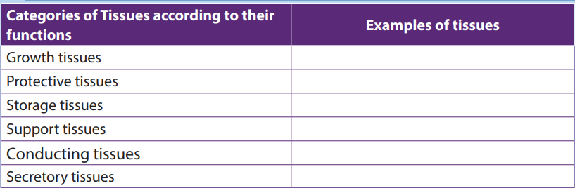
UNIT 6: TESTING FOR BIOLOGICAL MOLECULES
UNIT 6: TESTING FOR BIOLOGICAL MOLECULES
Key unit competence
Test for biological molecules in a variety of contexts, such as identifying the contentsof mixtures of molecules and to follow the activity of digestive enzymes
Learning objectives
By the end of this unit, I should be able to:– Write out procedures in the identification of biological moleculesIntroductory activity
– Explain the importance of the reagents used in the identification of biological
molecules.
– Carry out tests for the identification of biological molecules
– Compare reducing and non-reducing sugars
– Appreciate the importance of identification of food values in the food industry
and in processing and packaging.– Show resilience making observations on color changes during food tests
You are given solutions containing different food stuffs including maize flour,
vegetable cooking oil, and egg white sugar cane liquid and passion fruit. Using
prior knowledge of biological molecules to suggest the type of biological
molecule in each one of them. Suggest the chemical tests used to identify eachof the molecules.
6.1. Test for carbohydrates
Activity 6.1
Materials required:
Starch powder, Irish potatoes juice, prepared porridge, Iodine solution, beakers,
droppers, source of heat and test tubes
a. Test for starch
Procedure– Mix 1g of starch powder with 100ml of waterb. Test for reducing sugar
– Boil the mixture while stirring; then cool the solution
– Boil the mixture while stirring; then cool the solution
– Put 2ml of starch solution in a test tube labeled 1, 2ml of Irish potato juice in
a test tube labeled 2 and 2ml of prepared porridge in a test tube labeled 3
– In each test tube put 2 drops of Iodine solution and shake– Record your observation and draw a conclusion
Requirements
Glucose powder, beaker and test tube, Benedict solution, Bunsen burner, droppers
Procedures– In the beaker mix 1cm3 of water and 1g of glucose powder.Biological molecules are grouped into organic molecules including carbohydrates,
– Pour the prepared solution of glucose in a test tube and
– Add 2ml of benedict’s solution and heat– Record your observation.
proteins, lipids, nucleic acids and vitamins. They also contain inorganic molecules such
as minerals and water. The first four organic molecules are called macromolecules
because they are required in organism in large quantity. Carbohydrates including
starch, reducing and non-reducing sugars appear in this category and are the main
energy producers in the organisms. Others, including lipids and proteins are needed
for building organisms while vitamins protect the organisms against diseases. Weneed to ensure that what we take from diet have all required biological molecules.
a. Test for starch.
Carbohydrates such as starch are tested by mixing a sample with 2-4 drops of iodine
or Lugol’s solution. If the sample contains starch the solution will turn from a yellow-brown
color to a dark purple/dark blue (Figure 6.1). The color change is due to a
chemical reaction between the large carbohydrate molecule and the iodine ions. Ifthe sample does not contain starch the solution remains yellow-brown.
b. Testing for reducing and non-reducing sugar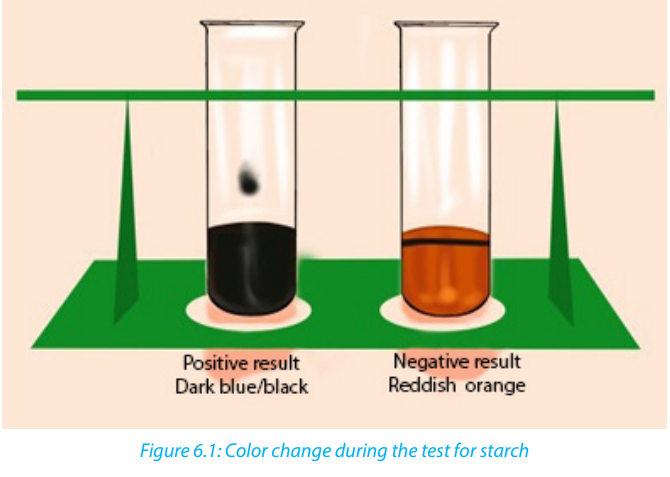
The presence of reducing sugar can be tested by using benedict reagent. Benedict
solution has copper ions that have a light blue color. When this solution is heated
in the presence of simple reducing sugars such as glucose, the blue color of copperions changes from a light green color to rusty orange-brown color (Figure 6.2).
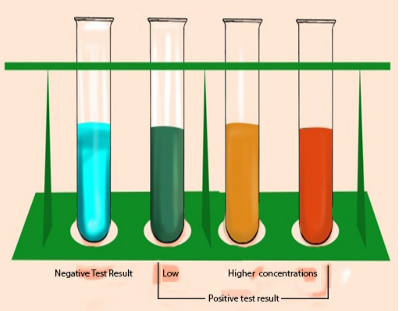
If the color of Benedict reagent persists, the sugar tested is not a reducing sugar.
Note that there is no special reagent to test for non-reducing sugar, but by the
addition of HCl, non-reducing sugars can be hydrolyzed to reducing sugars. To
test the presence of reducing sugars, a solution of sodium hydroxide is needed toneutralize the acidity because Benedict reagent works better in neutral solution
Self-assessment 6.1
A student prepared carbohydrate solution labeled C1. Perform the following
experiment to confirm whether C1 is starch, reducing sugar, or non-reducingsugar.
6.2. Test for proteins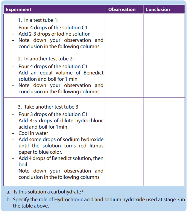
Activity 6.2
Materials required
Milk, eggs, soybeans, test tubes, beakers, mortar for crushing beans, 1% NaOH or
1% KOH solution, 0.1M of CuSO4 solution and Millon’s reagent.
Procedure– Extract the white fluid from an eggThe Biuret reagent is used to test for the presence of proteins. It contains copper ions
– Prepare an extra of soya bean and 10ml of fresh milk
– Put 2ml of albumen solution in a test tube labelled A1 and 2ml in A2
– Put 2ml of milk solution in a test tube labelled B1 and 2ml in B2
– Put 2ml of soya bean solution in a test tube labelled C1 and 2ml in C2
– Put 1ml of KOH or NaOH solution in each of the test tubes A1, B1, and C1.
Shake the mixture and add 1ml of CuSO4 solution in each (A1, B1, and C1)
test tube
– Put 1ml of Millon’s reagent in each of test tubes (A2, B2, and C2). Shake themixture and thereafter boil the three test tubes (A2, B2, and C2).
with blue characteristic color. During the copper ions react with protein molecules
and causes the biuret solution to turn from a light blue color to purple if proteins arepresent.
The test can also be done by using Millon’s reagent, which in the presence of proteins,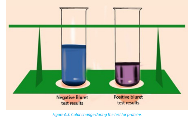
the Millon reagent changes from colorless to pink.
Self-assessment 6.2
1. You are provided with the sample of the substance M and A. Carry out thefollowing experiments and complete the table below.
2. Carry out the same experiment using the substance A and compare your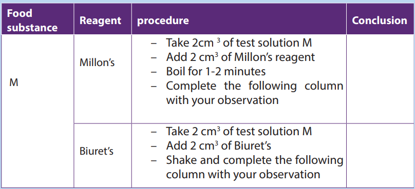
findings with M.3. Which of the substance A and M contain proteins?
6.3. Test for lipids
Activity 6.3
Materials required - Olive oil, test tubes, ethanol, water, Sudan III solution
Procedure:
Use olive oil to carry out the following experiments
Add 2 cm3
of olive oil in the test tube:– Add 5 cm3 of ethanol followed by 5 drops of water.The presence of lipids can be determined by using Sudan III indicators, which are fat-loving
– Shake the mixture and record your observation.
– To another test tube containing 2 cm3 of olive oil:
– add 5 drops of Sudan III solution
– Shake thoroughly and examine the mixture in the test tube after few minuteand record your observations
molecules that are colored. During the test for a solution containing lipids, two results are
likely to be found: there is either the separation of layers indicating the levels of water and
lipid, or the dye migrates toward one of the layers. If the mixtureis composed of water, the
conclusion is that the lipids are not present. In this case, the Sudan III indicator will form
small micelles/droplets and disperse throughout the solution. A positive result indicates
the lipid layers sitting on top of the water layer with a red-orange color. When using ethanolfor testing lipids the presence of the color changes from colorless to milky (emulsion test).
Self-assessment 6.3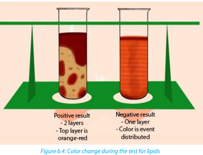
You are provided with a solution X. Use Sudan III indicator to test the presence oflipids in the solution X.
6.4. Test for vitamin C (Ascorbic Acid).
Activity 6.4Squeeze the orange fruits to extract the juice and carry out the following test.
Vitamin C is tested by using DCPIP (Dichlophenol Indophenol). Its positive (presence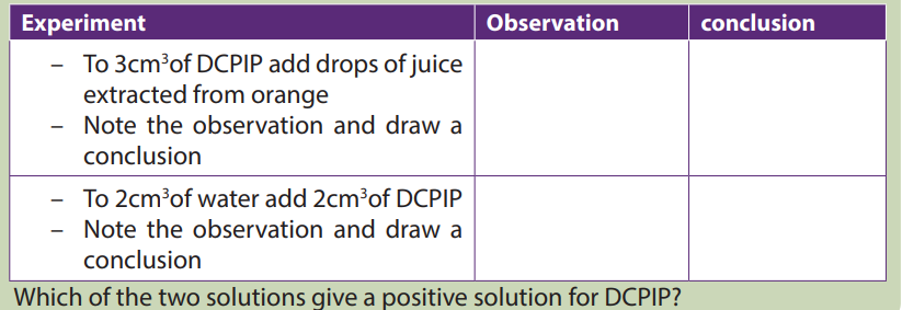
of vitamin C) test decolorizes DCPIP, while the negative (absence of vitamin C) test isindicated by the persistence blue color of DCPIP.
Self-assessment 6.4
In this experiment you are to press a tomato fruit (s) to get juice out of it. Use the
juice to carry out the test for vitamin C Draw a table of results that includes theprocedure, observation and conclusion.
End of unit assessment 6
1. Biological molecules are divided into:a. Organic molecules and inorganic molecules2. Name the reagents that are used to test for the following food substances
b. Carbohydrates and starch
c. Lipids, carbohydrates and water
d. Carbohydrates, food and potatoesa. Lipids3. You are provided with the following specimen:
b. Starch
c. Reducing sugarSpecimen A: Sorghum4. Some drops of fresh pineapple juice are added drop by drop to DCPIP solution.
Specimen B: Irish potatoes
Specimen C: Oranges
Specimen D: Sunflower seeds
a. Carry out chemical tests to determine the composition of the above seed to
tell whether they are composed of proteins, fats, starch or vitamin C.
b. Draw the table of used reagent, procedure and observation in (a)
The deep blue color of the DCPIP quickly fades.a. Explain why the blue colour disappeared?5. The result of food tests on unknown sample are shown below. Copy and
b. What is the importance of this food substance to the human body?
complete the table to show the conclusions which could be drawn from thesetests.
6. This is a practical question to be conducted using provided materials and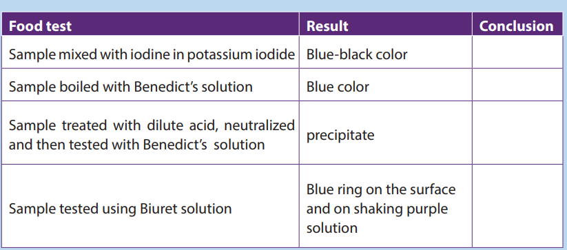
reagents to determine the food nutrients in each solution: You are provided with
the following solutions, A (sucrose 0.5%), B (1%starch), C (dilute hydrochloric
acid) and D (sodium hydroxide) and 6 test tubes labeled 1 to 6. Use the reagents
provided to determine the chemical nature of the substance present in thesolutions. Indicate your observations and conclusions in the table below:
a. Why was it necessary to boil solutions A and B with solution C in test (3)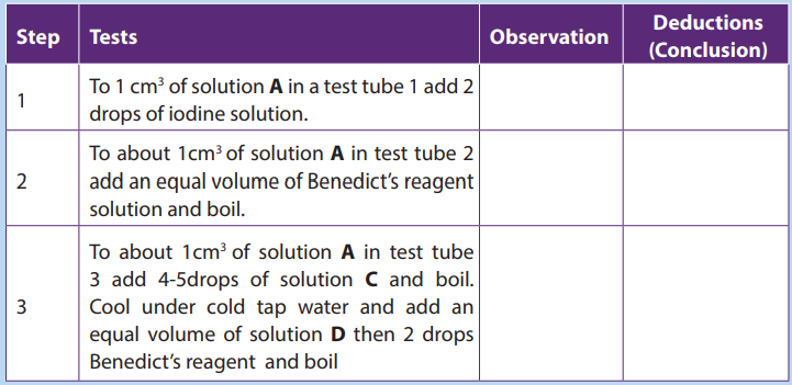
and (6)?b. Why was solution D added to test tubes 3 and 6?
UNIT 7: CARBOHYDRATES AND LIPIDS
UNIT 7: CARBOHYDRATES AND LIPIDS
Key Unit Competences
Explain the important roles of carbohydrates and lipids in the provision and storage
of energy and for a variety of other functions.
Learning objectives
By the end of this unit, I should be able to:– State the roles of carbohydrates and lipids.Introductory activity
– Recall the elements that make up carbohydrates and lipids.
– Explain the proportion of hydrogen in carbohydrates and lipids and relate this
to the amount of energy released when oxidized.
– Define the terms monomer, polymer, macromolecule, monosaccharide,
disaccharide and polysaccharide.
– Describe the ring forms of α-glucose and β-glucose structure.
– Explain the formation of glycosidic bonds.
– Describe the structure of phospholipids and relate to their functions in living
organisms.
– Describe the molecular structure and formation of triglycerides and
phospholipids, and give their functions in living organisms.
– Demonstrate that phospholipids have a hydrophilic head and hydrophobic
tails using a heterogeneous mixture made up of water and cooking oil.
– Interpret the charts and illustrations of molecular structure and the formation
of maltose and triglycerides.
– Demonstrate through a process of combustion that sugars and lipids are
biological fuel
– Differentiate between starch and cellulose.
– Appreciate the importance of carbohydrates and lipids in organisms.
– Be aware of the other roles of lipids in the formation of soap and withcarbohydrates and syrups in medicine
1. In the previous unit (test for biological molecules), we tested carbohydrates,
starch, reducing sugar, lipids, proteins, and vitamins. Where do you classify
monosaccharide, disaccharides and polysaccharides in the above tested
biochemical compounds?
2. Sometimes people say that fatty persons do not feel cold. What could be
the reasons?
7.1. Classes of monomers
Activity 7.1
Visit the internet and conduct a research the following:
1. The description of the term monomer
2. Where can we find monomers?
3. The biological importance of monomers?
A monomer is a molecule that can combine with others of the same kind to form
a polymer. A polymer is a large molecule or macromolecule composed of many
repeated sub-units (monomers). Because of their broad range of properties, both
synthetic and natural polymers play essential and ubiquitous roles in everyday life.
Polymers make up many of the materials in living organisms including proteins,
cellulose, and nucleic acids. Glucose molecules for example, are monomers that
combine to form the polymer cellulose. The examples of monomers are summarized
in the table 7.1.
Table 7.1: Biological molecules and their monomers
Carbohydrates comprise a large group of organic compounds which contain
carbon, hydrogen and oxygen. The word carbohydrate suggests that these organic
compounds are hydrates of carbon. Their general formula is In carbohydrates
the ration hydrogen-oxygen is usually 2:1. Carbohydrates are divided into three
groups including the monosaccharide (single sugars), disaccharides (double
sugars) and polysaccharides (many sugars). The most common monosaccharide of
carbohydrates is glucose with molecular formula
Self-assessment 7.1
1. What are some examples of polymers and monomers?
2. How are monomers, polymers and macromolecules related?
7.2. Ring form of α-glucose and β-glucose
Monosaccharides are group of sweet and soluble
Activity7.2
1. Based on the knowledge acquired during the lesson of monomers and further
information from books and internet:a. What are the examples of monosaccharide?crystalline molecules of relatively low molecular mass. They are named with the
b. Give the molecular formula of each of the monosaccharide stated above
c. Use the books to illustrate the structural formula of each of themonosaccharide stated above
suffix –ose. The general formula of a monosaccharide is , with n the number
of carbon atoms. The simplest monosaccharide has n=3 and it is a triose sugar.
When n = 5, this is a pentose sugar, and when n = 6, this is a hexose sugar. The two
common pentose sugars are ribose and deoxyribose, while the most known hexose
is glucose. Its molecular formula is . It is the most important simple sugar in
human metabolism called simple sugar or monosaccharide because it is one of the
smallest units which has the characteristics of this class of carbohydrates.
Monosaccharides can exist as isomers. The isomer is defined as each of two or more
compounds with the same formula but a different arrangement of atoms in the
molecule and different properties. For example, glucose, fructose and galactose
share the same molecular formula which is . However, they differ by their
structural formulae as follow: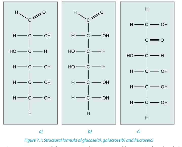
One important aspect of the structure of pentoses and hexoses is that the chain
of carbon atoms is long enough to close up on itself and form a more stable ring
structure. This can be illustrated using glucose as an example. When glucose forms
a ring, carbon atom number 1 joins to the oxygen on carbon atom number 5 (Figure
7.2)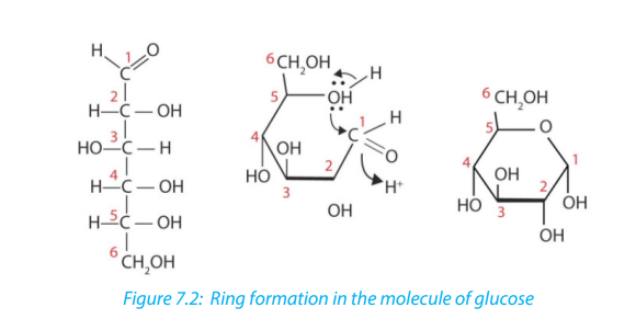
All hexoses sugars can exist as straight-chain structures but they tend to form ringstructures. Glucose, fructose, galactose can exist in ring structures (Figure 7.3).
Ring monosaccharides are said to be alpha (α) if the -OH group located on carbon 1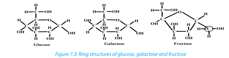
is below the ring and beta (β) when the -OH group is above the ring. The molecule
of glucose for example can exist as alpha and beta glucose denoted by α-glucoseand β-glucose (Figure 7.4)
Self-assessment 7.2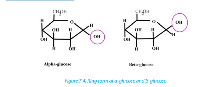
1. How do we call the monosaccharide with 3, 5 and 6 carbon atoms?
2. Differentiate between α and β glucose3. What are the properties of glucose?
7.3. Formation and breakdown of glycosidic bonds
Activity 7.3
1. Monomers are joined to form polymers, use a point as a monomer to
illustrate how a polymer can be formed
2. How do you call joining structures between atoms?
3. Use books or other sources to show how monosaccharide form adisaccharide.
7.3.1. Monosaccharides
Monosaccharides may combine together in pairs to give a disaccharide (double sugar).
The union involves the loss of a single molecule of water and is therefore a
condensation reaction. The bond which is formed is called a glycosidic bond. It is
usually formed between carbon atom1of one monosaccharide and carbon atom 4
of the other, hence it is called a -1, 4- glycosidic bond. Any two monosaccharides
may be linked together to form a disaccharide of which maltose, sucrose and lactoseare the most common.
The addition of water under suitable conditions is necessary if the disaccharide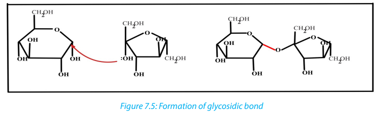
is to be split into its constituent monosaccharide. This is called hydrolysis \
water breakdown or more accurately, breakdown by water.
7.3.2. Disaccharides
These are carbohydrates made of two monosaccharides. They include maltose
(glucose + glucose), sucrose or saccharose (glucose +fructose), and lactose
(glucose+ galactose). The maltose is the sugar from the germinating seeds, sucrose
or saccharose is the common table sugar obtained from sugarcane, while lactose isthe sugar from the milk. In addition, sucrose is a non-reducing sugar.
Table 7.2: Types of disaccharides and their monomers
In maltose ring, the two ring of glucose are bonded by the -1, 4-glycosidic bond
while in sucrose the glucose and fructose are bonded by -1, 2-glycosidic bond.
All the disaccharides are non-reducing sugar, except maltose which behaves in
the same as a reducing sugar with benedict’s solution. All monosaccharides and
disaccharides have the following characteristics: sweet taste, soluble in water andlower molecular mass.
Self-assessment 7.3
1. Write the molecular structure of sucrose
2. How is the glycosidic link is formed
3. Sucrose is formed when two monosaccharide are assembled together:
a. Name those two monosaccharides.
b. Using the ring form of these monosaccharide named above to explain andshow sucrose formation?
7.4. Polysaccharides: starch, glycogen and cellulose
Activity 7.4
1. Based on the meaning of monosaccharide, what is a polysaccharide?
2. Classify the following compound into polysaccharide, monosaccharide and
disaccharidea. Glucose, fructose and galactose3. Use glucose to form any polysaccharide of your choice
b. Lactose, sucrose, and maltose
c. Starch, cellulose and glycogen
In the same way that two monosaccharides may combine in pairs to give a
disaccharide, many monosaccharides may combine by condensation reactions to
form a polysaccharide. The number of monosaccharides that combine is variable
and the chain produced may be branched or unbranched. Polysaccharide are manybut the most known are starch, glycogen and cellulose.
a. Starch
Starch is made up of two components: amylose and amylopectin. Amylose is a linear
unbranched polymer of 200 to 1500 α-glucose units in a repeated sequence of α-1,
4-glucosidic bonds. The amylose chain coils into helix held by hydrogen bonds
formed between hydroxyl groups. A more compact shape is formed. The amylose
helices are entangled in the branches of amylopectin to form a complex compact
three dimensional starch molecule.
Amylopectin is a branched polymer of 200 to 200,000 α-glucose units per starch
molecule. The linear chains of α-glucose units are held together by α-1, 4-glucosidic
bonds. Branches occur at intervals of approximately 25 to 30 where α-1, 6-glucosidic
bonds occur. Starch grains are found in chloroplast, potato tubers, cereals and
legumes. Starch is insoluble in cold water. It is digested by salivary amylase and
pancreatic amylase into maltose and the latter is hydrolyzed by maltase enzyme to
form glucose. Therefore, diabetic people should avoid tubers since they are rich instarch which in turn gives glucose (Figure 7.8).
b. Glycogen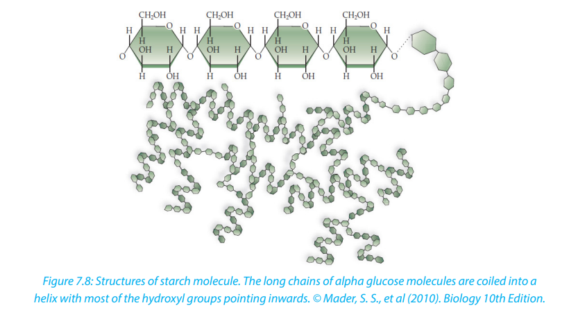
Glycogen is often called animal starch because it is a major polysaccharide storage
material in animals and fungi. The brain and other tissues require constant supply of
blood glucose for survival. Some tissues particularly the liver and skeletal muscles
store glycogen in the form that can be rapidly mobilized to form glucose. Like starch,
glycogen is made up of α-glucose and exists as granules. It is similar to amylopectin instructure but it has shorter chains (10-20 glucose unit) and is more highly branched.
c. Cellulose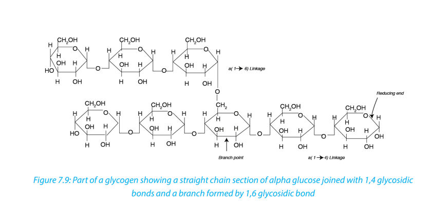
Cellulose is the structural polysaccharide in plant cell wall. It is found in vegetables
and fruits but it cannot be hydrolyzed by enzymes in the human digestive system.
Cellulose is composed of long unbranched chains of up to 10,000 β-glucose units
linked by β-1,4-glucosidic bonds. Each β-glucose unit is related to the next by a
rotation of 180 ͦ C with OH groups projecting outwards on either side of the chain.
Cellulose chains run parallel to one another. Unlike amylopectin and glycogen
molecules, there are no side chains (no branch) in the cellulose. This allows the linear
chains to lie close together. Many H-bonds are formed between the OH groups of
adjacent chains. The chains group together to form microfibrils arranged in larger
bundles of macrofibrils. The fibrils give the plant cell their high tensile strength andrigidity. The layers of fibrils are permeable to water and solutes.
Cellulose is formed from ß - glucose units linked by 1,4 glycosidic bonds. The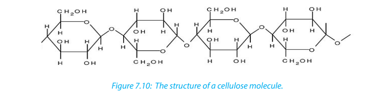
hydroxyl groups alternate on either side of the molecule forming straight chains
giving cellulose a fibrous structure. Cellulose are strengthened further by hydrogen
bonds that link adjacent chains.
d. Chitin
Chitin is one of naturally occurring Polymers. It forms a structural component of
many animals such as exoskeleton in arthropods. Chitin is a polymer of glucose
although in its structure a molecule of amino acid is added to each glucose. Thedigestion of chitin yields simple sugars and ammonia.
Self-Assessment 7.4
1. What type of reaction is involved in the formation of glucose from starch?
2. Use the type of reaction above to form glucose from sucrose molecule3. What are the 2 main components of starch? Give the difference between them
7.5. Lipids
Activity 7.5
Use textbooks and/or internet to
1. List the monomers that are present in lipids
2. Locate where can we find lipids?
3. Discuss the reasons why animals like pig do not like hot weather.
Lipids are a broad group of naturally occurring molecules which include fats,
waxes, sterols, fat soluble vitamins (such as vitamins A, D, E and K), monoglycerides,
diglycerides, Phospholipids and others. Lipids are grouped into fats which are solid
at room temperature and oils which are liquid at room temperature. Lipids are
made by carbon, hydrogen and oxygen, but the amount of oxygen in lipids is much
smaller than in carbohydrates. Lipids are made by two components namely glycerol
and fatty acids. The chemical formula for glycerol is with structural formula
as shown in the figure 7.11
In all lipids glycerol do not show any variation while fatty acids vary. Therefore, the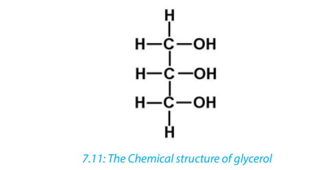
nature of lipid depends on the fatty acid it contains. There are two types of fatty
acids: unsaturated fatty acid characterized by the chain of hydrocarbon containing
one or more double and triple bonds; and saturated fatty acid characterized by the
chain of hydrocarbon without any double or triple bond.
Lipids are formed when glycerol combines with one, two, or three fatty acids to form
monoglyceride, diglyceride or Triglyceride. A bond is formed between the carboxyl
(-COOH) group of a fatty acid and one of the hydroxyl (-OH) groups of the glycerol.
This is a condensation process and water is lost. The resulting bond is known as ester
link, and the type of reaction is called esterification.
Lipids are of different types as it is summarized in the following table (Table 7.3)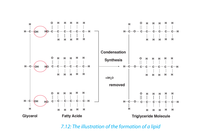
Table 7.3: Types of lipids, their structure, main role and features
a. Waxes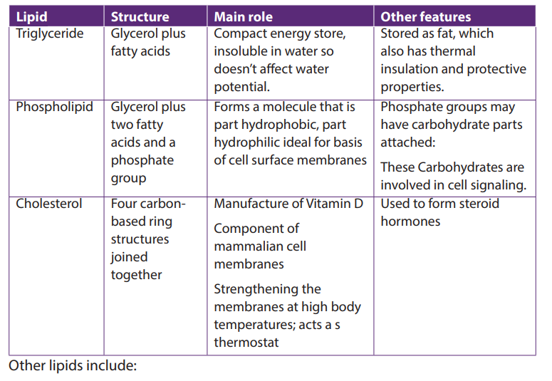
Waxes are similar to triglycerides but contain fatty acids bonded to long chain
alcohol rather than to glycerol. Waxes form the cuticle that protects the leaves andsurfaces of insects against the loss of water
b. Steroids
A steroid is an organic compound with four rings of carbon and hydrogen atoms
with various side chains. Steroids have several functions. It is a component of mostanimal hormones like estrogen, testosterone.
General functions of lipids
Lipids perform a number of functions within living organism:– Lipids are source of energy: due to the presence of C-H bond, lipids canSelf-assessment 7.5
generate more ATP compared to the carbohydrates of the same mass
– Lipids are storage of energy in adipose cells forming adipose tissue in fat of
organism
– Lipids act as insulators of the organism. For example, they reduce heat loss.
Lipids also are electrical insulators around the nerve cells, the Myelin sheath
– Lipids have a role of protection, in the cuticle of plant leaves against drying, in
exposed organ like hand and knees
– Synthesis of hormones such as steroid hormones (most of sex hormones) are
made by lipids– Lipids are used in production of soap by saponification reaction
1. Name the small units found in lipids2. Differentiate between fats and oils
End of unit assessment 7
1. Write the formula of a monosaccharide with 3 atoms of carbon
2. Compare the structure of fat(triglycerides)and the phospholipids
3. Give two examples of how carbohydrates are used in the body.
4. The formula for a hexose is What would be the formula of?a. Triose5. What type of chemical reaction would be involved in the formation of glucose
b. Pentose
from starch or glycogen?
6. Distinguish between:a. Alpha glucose and beta glucose
b. Glycogen and cellulosec. Amylopectin and amylose
UNIT 8: PROTEINS AND WATER
UNIT 8: PROTEINS AND WATER
Key Unit Competence
Describe how protein structure is related to function and the role of water as aspecial molecule with extraordinary properties that make life possible.
Learning objectives
By the end of this unit, I should be able to:– Describe the structure of an amino acid and the formation and breakage of athat make life possible on this planet.
peptide bond.
– Describe the primary, secondary, tertiary and quaternary structure of proteins.
– Describe the molecular structure of hemoglobin as an example of a globular
protein.
– Describe the functions with an emphasis on iron in the hemoglobin molecule.
– Explain the effect of heat, pH and chemicals on protein structure.
– Explain how hydrogen bonding occurs between water molecules and relate
the properties of water to its roles in living organisms.
– Devise an experiment to investigate the effect of temperature, pH and
chemicals on the structure of protein.
– Relate the structure of globular and fibrous proteins to their functions.
– Investigate the effect of lowering temperature on water.
– Distinguish between collagen molecules and collagen fibres
– Appreciate the importance of globular and fibrous proteins in biological
processes such as the transport of gases and providing support for tissues.
– Express that protein structure is central to many aspects of biology, such as
enzymes, antibodies and muscle contraction.
– Acknowledge that water is a special molecule with extraordinary properties
Introductory activity
Conduct a research by using different sources of information and answer the
following questions1. What is a protein?8.1. Proteins2. What do you understand by universal solvent in living organisms ?
Proteins are organic compounds of large molecular mass. For example, the
hemoglobin has a molecular mass of 64500. In addition to carbon, hydrogen
and oxygen, proteins always contain nitrogen, usually Sulphur and sometimes
phosphorus. Proteins are polymers of amino acids and they are not truly soluble inwater, but form colloidal suspensions.
8.1.1. Amino acids
Amino acids are group of over a hundred chemicals of which around 20 commonly
occur in proteins. They always contain a basic group, the amine group (-NH2
) and a
carboxylic acid group(-COOH) together with -R group or side chain (Figure 8.1). Allthe amino acid differs one to another by the structure of their side chain.
Amino acids are divided into two categories: essential amino acid and non-essential
amino acid. Essential amino acids are those amino acids which cannot be synthesized
by the body. Non –essential amino acids are synthesized by the organism. All 20amino acids can be found in diet from plant and animal tissues
Amphoteric nature of an amino acid
When an amino acid is exposed to basic solution, it is deprotonated (release
of a proton H+) to became negative carboxylate COO -while in acid solution it is
protonated (gains of a proton H+) to became ammonium positive ion -NH3+(Figure 8.1.3.a and Figure 8.1.3.b).
At a physiological pH, usually around 7, the amino acid exists as ZWITTERION (from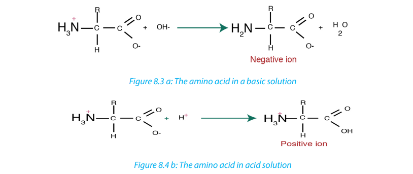
German means hermaphrodite) it is a molecule with two different charges (positiveand negative) at the same time (Figure 8.1.4).
8.1.2. Formation and breakage of peptide bond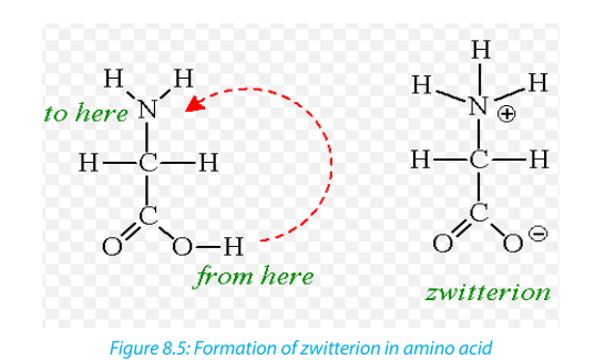
The formation of peptide bond follows the same pattern as the formation of
glycosidic bond in carbohydrates and ester bond in fats. A condensation reaction
occurs between the amino group of one amino acid and the carboxyl group ofanother, to form a dipeptide (fig 8.5).
A peptide bond is formed between two amino acids to form a dipeptide molecule.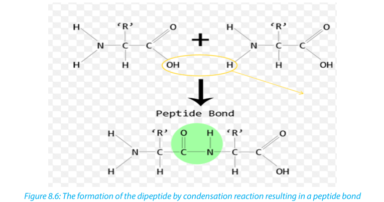
If three amino acids are assembled together they form a tripeptide while four
amino acids form a tetrapeptide and so on. A long chain of amino acid it is called a
polypeptide. The polypeptide chain or oligopeptide comprise more than 50 amino
acids joined together by peptide bonds.
During digestion, proteins are hydrolyzed to give amino acids that can be diffused
across the wall of intestine into blood stream. In hydrolysis the peptide bond breaksdown by the addition of a water molecule (Figure 8.5).
Self-assessment 8.1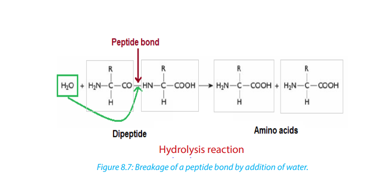 1. Explain what are essential amino acids?8.2. Structure and denaturation of proteins
2. Describe the formation of a peptide bond?
3. At physiological pH, the amino acid exists as zwitterions. What is a zwitterion?
4. Alanine is an amino acid with -CH3
as a side chain. Write its structural
formulae.
5. Most plants lack one or more of the essential amino acids needed by thebody. Explain how a vegetarian can obtain the essential amino acids.
Activity 8.2
1. From the books make a research on proteins and answer to the following
questions:a. What are different structures of proteins?2. Take a plastic rope cord, create the nodes bulk on it and suppose that those are
b. Differentiate globular proteins and fibrous proteins.
monomers of a long chain of polymer (the whole cord). Heat it using a Bunsenburner or another source of fire. Discuss the change that takes place.
8.2.1. Structure of proteins
The long chain of polypeptide can take different forms according to its molecular
weight and the types of bond that hold together atoms and molecules.
a. Primary structure of proteins
Primary structure of a protein is the sequence of amino acid that is made up of thepolypeptide chain or chains.
Ribonuclease is an enzyme found in pancreatic juice, which hydrolyses (digests)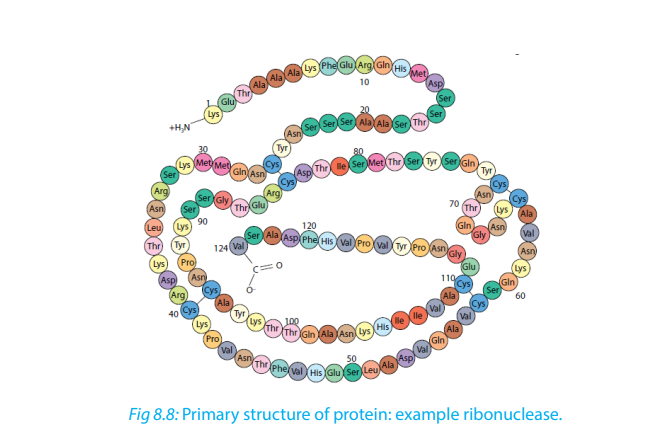
RNA. Notice that at one end of the amino acid chain there is an –NH3+ group, while
at the other end there is a –COO− group. These are known as the amino and carboxyl
ends, or the N and C terminals, respectively. (Adapted from Cambridge InternationalAS and A Level Biology Course Book Fourth Edition)
b. Secondary structure of proteins
The regular arrangement of amino acids in primary structure can induce the
interaction of the back bone of the polypeptide chain (side chain) by hydrogen
bonds. Those side chains are coiled and folded in the patterns that contribute to
the protein’s overall shape. One such secondary structure is α-helix and sometimeβ-pleated sheet (Figure 8.8).
c. Tertiary structure of proteins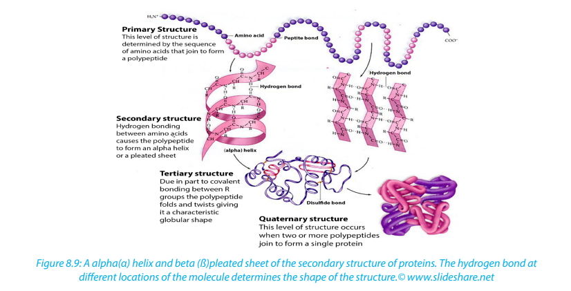
In addition to hydrogen and peptide bond in primary and secondary structure, the
tertiary structure of protein has other types that include:
– Hydrophobic interaction
– Ionic bond between positively and negatively charged r groups.– Disulfide bridges (-s-s)
d. Quaternary structure of proteins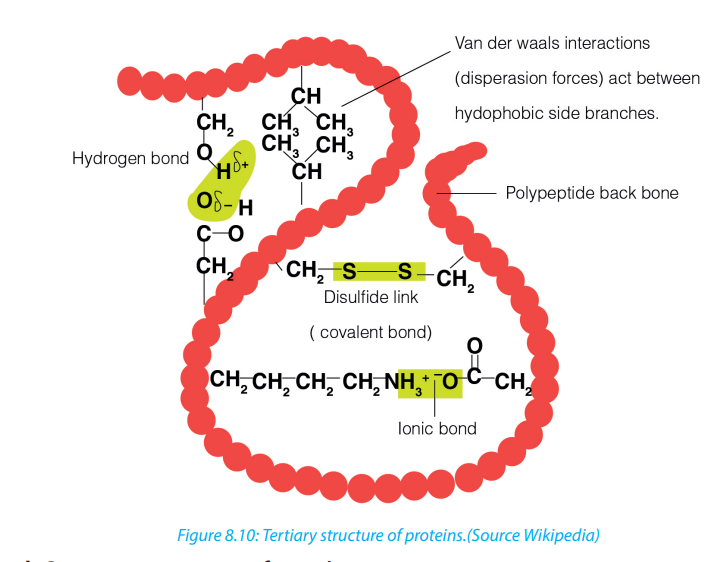
Quaternary structure involves more than one polypeptide chain chemically bonded
to each other. The quaternary structure refers to the way in which these polypeptide
chains are arranged in the protein. Examples, Hemoglobin that is composed of:– Four polypeptide subunits, two α- chains and two β- chains. Both α and β subunitsprimarily are α helical secondary structure polypeptide chain with 140Collagen: this is a fibrous protein consisting of three helical polypeptides that are
amino acids.
– Haeme composed of iron that binds with oxygen.supercoiled to form a rope like structure of great strength
– Soluble in water.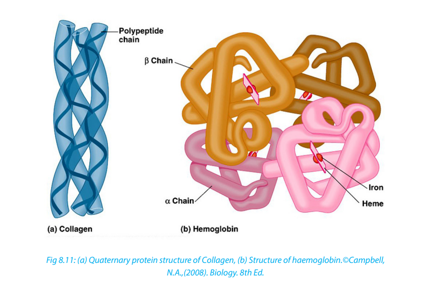 Globular protein
These are polypeptide chains that are tightly folded to form a spherical shape. Many
globular proteins are folded so that their hydrophobic groups are on the inside
of the molecule and the hydrophilic groups face outwards making these proteins
soluble in water.
Properties of globular proteins:
– They are spherical in shape
– Physiologically active
– May contain prosthetic group for example the iron (haeme)– Examples include hemoglobin and enzymes.
8.2.2. Protein denaturation
Protein denaturation is a process by which protein changes shape due to breakage
of bonds holding the polypeptide chains. Protein denaturation may be temporary
or permanent.
The agent of denaturation can be caused by;
– Extremely high temperatures beyond optimum,
– changes in pH,
– Ultra Violet (UV) rays,– High salt concentration and heavy metals.
8.2.3. Functions of proteins.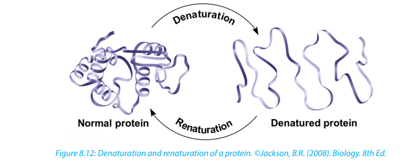 – Proteins such as lipase, pepsin and protease act as enzymes as they play aSelf-assessment 8.2
crucial role in biochemical reaction where they act as catalysts.
– Proteins play an important role in coordination and sensitivity (hormones and
pigments).
– Proteins have a transport functions. Example: Haemoglobin transport oxygen
– Proteins in the cell membrane facilitate the transport of substance across the
cell membrane.
– Proteins provide a mechanical support and strength.
– Proteins such as myosin and actin are involved in movement.
– Proteins play the role of defense of the organisms. Example: Antibodies areproteins
1. Describe the protein structures
8.3. Water
Activity 8.31. What is the medium of reaction in the organisms?Living organisms contain between 60% and 90% of water, the remaining being the
2. If two people are boiling the same quantity of cooking oil and water, which
one could evaporate first? Explain your choice.
dry mass. The function of water is defined by its physical and chemical propertiesthat differ from those of most liquids and make it effective in supporting life.
8.3.1. Biological significance of the physical properties of water
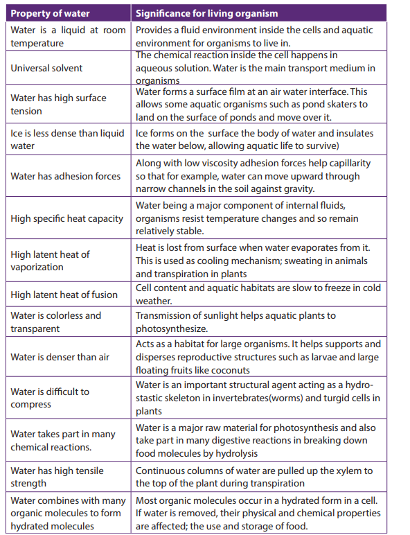
Functions of water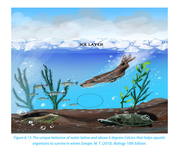 – Turgidity of plant cell which increases their size is due to the availability ofSelf-assessment 8.3
water.
– The transport of substances (minerals, nutrients in plant and animals) that are
dissolved in water.
– Excretion of waste product
– Support for hydrostatic skeleton.
– Temperature regulation in plant and animals
– Seed germination by breaking down the seed coat– Medium for biochemical reaction.
1. State the functions of water in animalsEnd of unit assessment 8
2. What do you understand by heat capacity?
3. Relate the high heat capacity of water to its biology functions.
4. Describe and explain how aquatic organisms live below frozen waterbodies
1. Certain drugs can break the covalent bond between two sulfur atoms
of nonadjacent amino acids. Which level of protein that can be affected most if the drug
is mixed with primary, secondary, tertiary and quaternary structure of proteins?
2. Complete the following statements by appropriate terms:
a. The formation of large molecules from small repeating units is called
………reaction.
b. A carbohydrate(polysaccharide)that is formed by the plant as a reserve
food supply and made up of only glucose molecules covalently bonded
together is……...
3. State the property of water that allows each of the following to take place. In
each case, explain its importance:
a. The cooling of skin during sweating
b. The transport of glucose and ions in a mammal
c. Much smaller temperature fluctuations in lakes and oceans than interrestrial (land-based) habitats.
4. Construct a three column table and relate the following terms with arrows to
indicate the correct match.
Phosphodiester linkages Monosaccharide Polypeptides
Peptide bonds Nucleotides Triacylglycerol
Glycosidic linkages Amino acids Polynucleotides
Ester linkages Fatty acids Polysaccharides5. Explain what happens during protein denaturation?
UNIT 9: VITAMINS AND MINERALS
UNIT 9: VITAMINS AND MINERALS
Key Unit Competence
Discuss the roles of minerals and vitamins in diet
Learning objectives
At the end of this unit you be able to:– State the mineral requirements for bodily functions.Introductory activity
– Identify the symptoms of mineral and vitamin deficiency.
– Outline the need for consumption of minerals and vitamins in small amounts.
– Organize a list of foods that are good sources of vitamins and mineral salts.
– Recognize the signs and symptoms of scurvy, night blindness, goiter, and
anaemia.
– Differentiate between water soluble and lipid soluble vitamins.
– Analyze one’s eating habits and suggest improvements
– Appreciate the importance of a balanced diet in relation to health and
economic prosperity.– Advocate for healthy feeding methods.
1. From the different food stuffs in our community, make a list of food stuffs that
are good sources of minerals and vitamins.
2. Using text books and other resources, make a list of e vitamins and mineral
deficiency diseases
9.1. Mineral nutrients in humans
Activity 9.1
Use textbooks and internet to list mineral nutrients found in human diet
Mineral nutrients are sometimes called mineral salts or just minerals. Mineral salts
are essential nutrients that our body needs. They are called essential not because
they are more important than other substances in our body but because our bodies
can not produce them. They include the inorganic substances found in daily diet.
They are dissolved in body fluids.
They are found in human body as ions (cations and anions). Organic food like proteins,
carbohydrates and fats provide the body with carbon, hydrogen, oxygen, nitrogen,
sulfur and phosphorus. But there are several more elements that the body needs
and occur as salts in the food we eat. They constitute about 1% of an organism by
weight. Even though they are required in a very small amount, they are nonethelessessential for body processes.
Some mineral nutrients are required by animals, plants, a few by both. Humans
require a number of minerals for the good functions of their bodies. Those are:
Self-assessment 9.1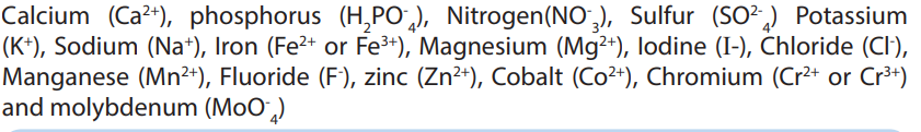
1. Outline ten mineral nutrients required in human diet.
2. Answer by true or false and justify your answer: “Minerals are called essentialnutrients because they are more important than others”.
9.2. Classification of mineral nutrients
Activity 9.2
Iodized table salt is advised to prevent goiter. In 100g of table salt there is 99% of
NaCl, and only 1% of iodine. Refer to the notes below to find the reason behindthis ratio.
The classification of minerals is based upon their requirement rather than on their relative
importance. Mineral nutrients are needed in a precise small amount. The five major mineralsneeded in human body include calcium

 Mineral nutrients are grouped into two groups:
 the macronutrients or major elements and the micronutrients or trace elements.
Macronutrients or major elements are minerals needed by humans in a relativelarge amount (greater than 200 mg/day). Their examples include nitrogen

Self-assessment 9.2 those which are needed in minute amount (a few parts per million). Examples include manganese

1. Categorize mineral nutrients according to their amount in human body.
2. Distinguish the two categories of mineral nutrients needed by the human
body.
3. From the minerals listed here, identify the five major minerals in the human
body: Sulfur (S), Potassium (K), Sodium (Na), Iron (Fe), Magnesium (Mg),
Iodine (I), Chloride (Cl), zinc (Zn), Cobalt (Co), chromium, Calcium (Ca),phosphorus (P), Nitrogen (N).
9.3. Sources, functions and deficiency symptoms of mineralnutrients in humans
Activity 9.3
Use your textbook to answer the following questions:
Hereunder is a variety of food stuffs: Banana, cassava, wholegrain, oranges,
pumpkin, potato, beans, water melon, green leafy vegetables, poultry, eggs,liver, and milk. Choose the food stuffs which are good sources of minerals.
Human body requires mineral nutrients to survive and to carry out daily functions
and processes. Minerals keep humans healthy and have key roles in several body
functions. Humans receive minerals by eating plants that absorb minerals from the
soil and by eating meat and other products from animals, which graze on plants. The
deficiency of mineral nutrients results into body functional disorders and diseases.
Most are found in the blood and cytoplasm of cells, where they assist basic functions.For example, calcium and potassium regulate nerve and muscle activity
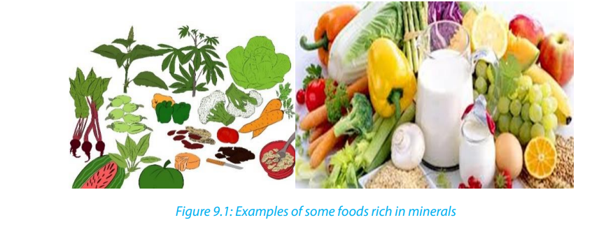
Table 9. 1 Minerals required in humans and their sources
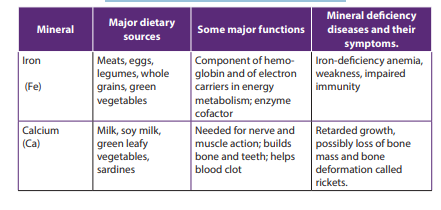
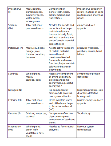
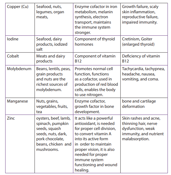
Self-assessment 9.3
1. Match the mineral nutrients with its function
a. Iodine 1. make bones hard
b. Fluorine 2. maintains the immune system stronger
c. Phosphorus 3. component of hemoglobin
d. Iron 4. prevents tooth decay
e. Copper 5. used in synthesis of thyroid hormone (thyroxin)
2. In a tabular form, identify the major dietary sources, the functions in human
bodies and the deficiency diseases of the following minerals: Ca, I, P, zinc, and3. Choose the best answer.
i. They are the minerals we need a lot in every day diet. How are they called?
a. Macronutrients
b. Micronutrients
c. Giant minerals
d. Monster minerals
ii. This mineral helps to build up strong teeth and bones. How is it called?
a. Calcium
b. Iron
c. Zinc
d. potassium
iii. What are foods that are natural good source of iron?
a. Roast Beef
b. Macaroni and cheese
c. Baked beans
d. Water melon
iv. Select 2 that are natural good sources of calcium?
a. Milk and cheese
b. Whole-wheat bread
c. Iceburg lettuced. Scanned salmon
v. The mineral that helps in oxygen transport to lungs is?
a. calcium
b. iron
c. zinc
d. potassium
vi. Which foods are good sources of zinc?
a. Milk and cheese
b. Lamb and pork
c. Macaroni and cheese
d. Peanuts and lentils
vii. Bananas are great source of this mineral, which helps our muscles and nervous
system to maintain your right water levels. What is it called?
a. Calcium
b. Iron
c. Zinc
d. Potassium
viii. Which mineral is important and needed by our body to fight off infection?
a. Calcium
b. Iron
c. Zinc
d. Potassium
ix. Which of the following mineral are needed in large amount every day?
a. Zinc
b. Iron
c. Calcium
d. selenium
x. Which is the type of mineral that keep your nervous system health?
a. Calcium
b. Iron
c. Zinc
d. Potassium
4. From the diseases listed below, what are those caused by the deficiency of
minerals?
Goiter, malaria, diabetes, rickets, beriberi, scaly skin, night blindness, anemia,impaired immunity, diarrhea
9.4. Vitamins and the classification of vitamins
Activity 9.4
Two students with different complains went to consult a medical doctor.
Student A says to the doctor that whenever he/she bleeds whenever she /he
brushes teeth.
Student B doesn’t see well objects around him/her,
The results from the doctor showed that they all have lack some vitamins.
1) What kind of vitamins that each student needs to take?2) Use your student textbook to explain your answer
Like minerals, vitamins are also essential for the human body. They are required
for metabolism, protection health and growth. Vitamins also assist in formation of
hormones, blood cells and genetic material. Vitamins are directly absorbed from
the small intestine into the blood stream. Water –soluble vitamins are absorbed in
the ileum while fat-soluble vitamins are absorbed in jejunum. Features shared by all
vitamins:
– They are not digested or broken down for energy
– They are not synthesized into the body structures
– Most are rapidly destroyed by heat.
– They are essential for good human health and needed in a very small amount
– They are required for chemical reactions in cells, working in association withenzymes.
There are thirteen vitamins required by human body. They are classified by their
solubility, whether they dissolve in water or in fats. Water-soluble vitamins including
vitamins C and B complex, and fat-soluble vitamins including vitamins A, D, E and
K (Table 9.2). Excess water- soluble vitamins are simply excreted in urine, while
fat soluble vitamins are stored in body fatty tissues to be used later if there is deficient
in diet. Excess intakes of these vitamins are stored in fatty tissues of the body,
where they can build up to toxic levels, especially if they are taken improperly insupplements.
Table 9.2 Water-soluble and fat-soluble vitamins Water-soluble vitamins Fat-soluble vitamins
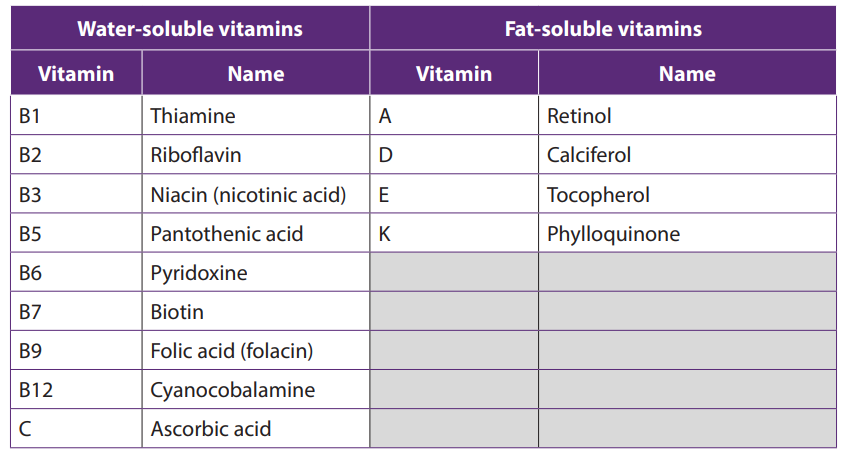
Self-assessment 9.4
1. How many vitamins does the human body needs to function properly?2. Describe the classification of vitamins.
9.5. Sources, functions and symptoms of vitamin deficiency
Activity 9.5Here is a number of foodstuffs rich in vitamins.

From the list of provided food stuffs (Banana, cassava, wholegrain, oranges,
pumpkin, potato, beans, water melon, green leafy vegetables, and milk). Can you
give some foods that are good sources of vitamins?
Some vitamins, including some vitamin B complex and Vitamin K are produced by
bacteria that normally live in the intestines, where they help to digest food. Vitamin
D is synthesized in the skin when it is exposed to UV radiation in sunlight.
Vitamins and their derivative are coenzymes; note that a coenzyme is an organic
molecule that combines temporaly with enzymes making them more efficient.
For example, Niacin or vitamin B3 is an essential component of coenzymes NAD
and NADP involved in lipid metabolism. It inhibits production of cholesterol and
catabolism of triglyceride. Thiamin or vitamin B1 is a coenzyme for many different
enzymes that break complex molecules such as carbohydrates to produce ATP.
Thiamin deficiency results into Beriberi anemia and stunted growth in children.
Vitamin K is an essential coenzyme for synthesis of several blood clotting factors.
Several vitamins, including vitamins C and E, act as antioxidants. An antioxidant is a
compound that neutralizes chemicals called free radicals. Free radicals are produced
naturally during cellular activities and may cause some types of cancer. Neutralizingfree radicals makes them harmless.
The table: 9.3. The major dietary sources, functions and possible symptoms of vitamindeficiency
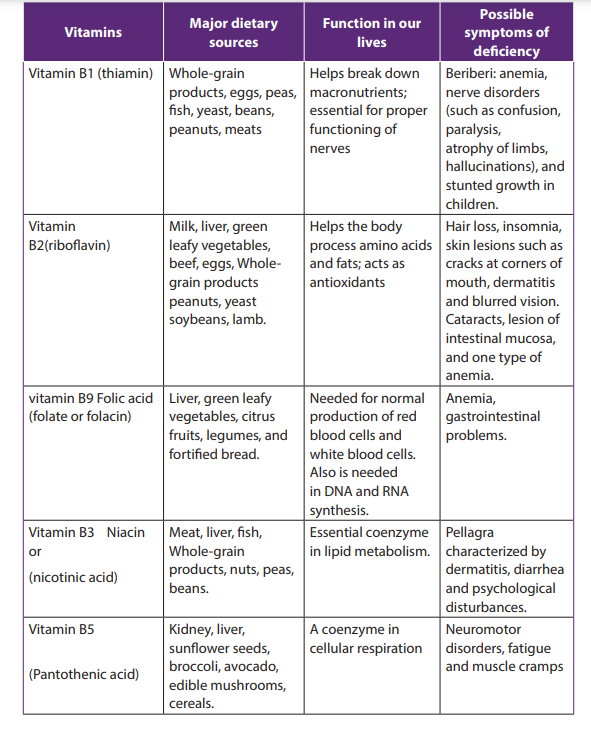
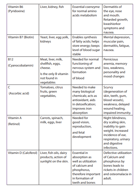
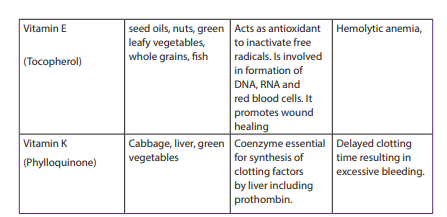
Many vitamin supplements are available in the market. However, it is always
advisable to obtain them from their natural sources by eating food rich in vitaminsdaily. Possible symptoms of vitamins deficiency are shown by the following pictures:
End of unit assessment 9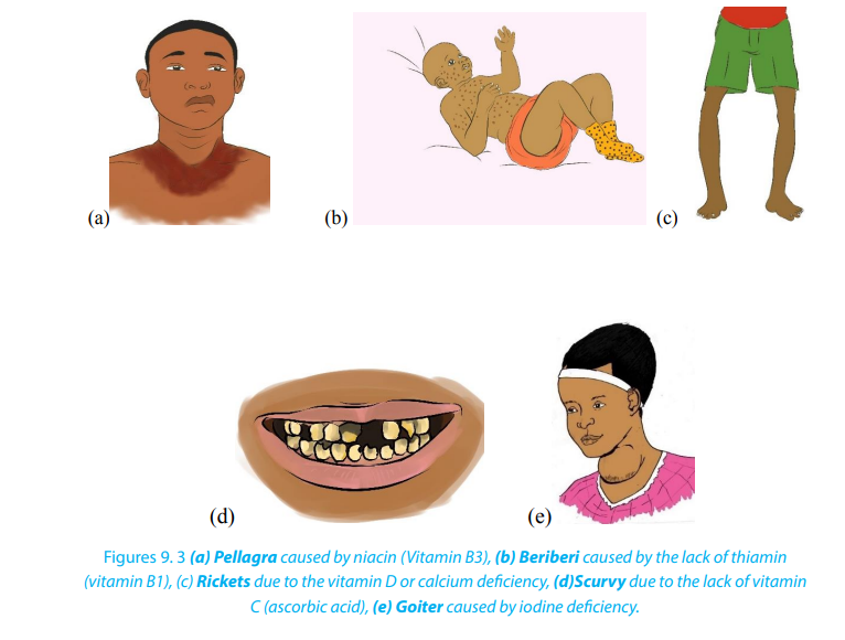
1. Choose a mineral which is an electrolyte and is found in almost every food. It
helps to lower blood pressure.a. Zinc2. choose a mineral which helps to make our blood vessels, tendons, and nerves
b. Potassium
c. Calcium
d. Iron
strong.a. Iron3. The following vitamins are part of Niacin and Thiamin minerals
b. Magnesium
c. Chromium
d. Coppera. Vitamins B1
b. Vitamins B2
c. Vitamins B3d. a and b
4. Vitamin C is required for the production and maintenance of:a. Collagen5. Vitamin C deficiency is called:
b. Hormone
c. Ascorbic Acid
d. Red Blood Cellsa. Scurvy6. Which of the following is a function of Vitamin A in the body?
b. Cold
c. Cancer
d. Ricketsa. Vision, bone and body growth7. Common food sources of Vitamin A are:
b. Immune defenses, maintenance of body linings and skin
c. Normal cell development and reproduction
d. All of the abovea. Milk, eggs, butter, cheese, cream, and liver8. Which of the following is a function of Vitamin B-12?
b. White sugar, honey, and sugar cane
c. Broccoli, apricots, Cantaloupe, Carrots, Sweet potato, Spinachd. Both a and c
a. Influences the cells that build bone tissue9. Vitamin B-12 deficiency caused by lack of intrinsic factor is called:
b. Is essential to the formation of bone
c. Helps to maintain acid-base balance
d. Maintains the sheaths that surround and protect nerve fibersa. Pernicious anemia10. What groups of people need additional Vitamin K?
b. Poor circulation of the red blood cells
c. Beriberi
d. None of the abovea. Premature newborns11. A common function of Thiamin, Riboflavin and Niacin is that:
b. People who do not have enough bile to absorb fat
c. Both A and B
d. None of the above answersa. They all are used in synthesis of blood clotting proteins12. The vitamin Folate works together with ______________ to produce new
b. They all work as a part of a coenzyme used in energy metabolism
c. They all help to strengthen blood vessel walls
d. They are used to stabilize cell membranes
red blood cells.a. Vitamin D13. Which of the following is a function of Vitamin B-12?
b. Vitamin A
c. Vitamin B-12
d. None of the abovea. Red blood cell formationd. Both A and B
b. Myelin sheath that protects nerve biers
c. Vision
14. Vitamin C helps in maintenance and repair of collagen which:a. Forms the base for all connective tissue in the body15. Which of the following is not a function of Vitamin D?
b. Aids in digestive processes
c. Promotes good eyesightd. Prevents PMS symptoms
a. Acts like a hormone16. Some food sources of Vitamin D are:
b. Stimulates maturation of cells
c. Maintains calcium cells
d. Builds tissuea. Fruits and vegetables17. Humans obtain vitamins from natural sources such as vegetables, fruits,
b. Salmon and egg yolks
c. Butter and fortified milk
d. Both B and C.
meat, fish and dairy products. What are the two vitamins that are not provided
by fruits and vegetables?
18. What would you advise someone starting to have symptoms of?a. Scurvy
b. Rickets
c. Teeth decay
d. Heart failuree. Pernicious anemia
UNIT 10: ENZYMES
UNIT 10: ENZYMES
Key Unit Competence
Describe the mode of action and factors affecting enzymes and their importance for
the existence of life
Learning objectives
At the end of this unit you be able to:– Define the term enzyme.Introductory activity
– Explain the criteria of naming enzymes.
– State that enzymes function inside cells and outside cells.
– Explain that enzymes are globular proteins that catalyze metabolic reactions.
– Describe the mode of action of enzymes in terms of the lock and key and the
induced fit hypotheses.
– Explain factors affecting enzyme activity.
– Define enzyme technology and its role in industry.
– Investigate the progress of an enzyme-catalyzed reaction by measuring rates
of formation of products.
– Investigate the effects of temperature, pH, enzyme and substrate concentration,
and inhibitors on enzyme activity.
– Interpret graphs of the effects of reversible and irreversible inhibitors on the
rate of enzyme activity.
– Investigate the effect of immobilizing an enzyme in alginate as compared with
its activity when free in solution.
– Use a computer to plot graphs of the rate of enzyme controlled reaction.
Calculate Q10 of an enzyme controlled reaction.
– Acknowledge that enzymes are essential in speeding up reactions that would
be too slow to sustain life.
– Appreciate the importance of planning and carrying out experiments under
controlled conditions.– Understand the roles of enzymes in industry and medicine
Discuss in pair the following questions and share with another pair your findings.1. What do you understand by the term enzyme?2. Two individuals want to reach the last floor of Kigali city tower. One climbs
up using the ladder but another one uses a lift. What advantage the lift
gives over the ladder?3. Why is it easy to digest hot foods than cold ones?
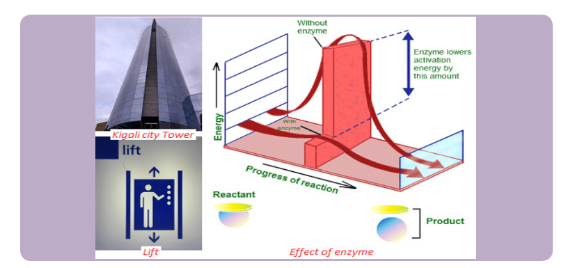
10.1. Criteria for naming enzymes
Activity 10.1You are provided with three groups of enzymes:
Make a research to find out:
a. specific role of each of the six enzymes mentioned aboveb. criterion followed to name enzymes of group A, B and C respectively
Enzymes are biological catalysts produced by a living organism to control the rate of
specific biochemical reactions by lowering the activation energy of reactants
First of all, individual enzymes are named by adding -ase to the name of the substrate
with which they react. The enzyme that controls urea decomposition is called urease;those that control protein hydrolyses are known as proteases.
A second way of naming enzymes refers to the enzyme commission number (EC
number) which is a numerical classification scheme for enzymes based on the
chemical reactions they catalyze. In a system of enzyme nomenclature, every EC
number is associated with a recommended name for the respective enzymecatalyzing a specific reaction. They include:
Oxidoreductases: catalyze redox reactions by the transfer of hydrogen,
oxygen or electrons from one molecule to another. Example: Oxidase catalyzes
the addition of oxygen to hydrogen to form water.
Glucose + oxygen gluconic acid +water
– Hydrolase: catalyzes the hydrolysis of a substrate by the addition of water.
Sucrose + water glucose+ fructose
– Ligases: catalyze reactions in which new chemical bonds are formed and use
ATP as energy source.Amino acid + tRNA
– Transferases: catalyze group transfer reactions. The transfer occurs from one amino acid-tRNA complex
molecule that will be the donor to another molecule that will be the acceptor.
Most of the time, the donor is a cofactor that is charged with the group about
to be transferred. Example: Hexokinase used in glycolysis.
– Lyases: catalyze reactions where functional groups are added to break double
bonds in molecules or the reverse where double bonds are formed by the
removal of functional groups. For example: Fructose bisphosphate aldolase
used in converting fructose 1, 6-bisphospate to G3P and DHAP by cutting C-C
bond.
– Isomerases: catalyze reactions that transfer functional groups within a
molecule so that isomeric forms are produced. These enzymes allow for
structural or geometric changes within a compound. Sometime the inter
conversation is carried out by an intramolecular oxidoreduction. In this case,
one molecule is both the hydrogen acceptor and donor, so there’s no oxidized
product. The lack of an oxidized product is the reason this enzyme falls under
this classification. The subclasses are created under this category by the type
of isomerism. For example: phosphoglucose isomerase for converting glucose
6-phosphate to fructose 6-phosphate by moving chemical group inside thesame substrate.
A third way of naming enzymes is by their specific names e.g. trypsin and pepsin
are proteases. Pepsin, trypsin, and some other enzymes possess, in addition, the
peculiar property known as autocatalysis, which permits them to cause their ownformation from an inert precursor called zymogen.
Self-assessment 10.1
1. How to name enzymes?
2. What is the role of peptidase?
10.2. Characteristics of enzymes
Activity 10.2
Requirement: Three test tubes, match box, about 1g of liver, 1g of sands, 1% H2
O2and MnO2
Procedure:
– Label three test tubes A, B and C respectively.
– Put about 0.1 g of MnO2
powder in test tube A and 1g of liver in tube B and 0.1g
of sand in tube C.
– Pour 5 ml of H2O2 (hydrogen peroxide) in each tube. What do you observe?
– Place a glowing splint in the mouth parts of each test tube. What do you observe?
Questions
1. Explain your observations.
2. Write down the chemical equation of the reaction taking place in tube A and B3. Carry out your further research to find out the characteristics of enzymes
Enzymes speed up the rate of metabolic reactions by allowing the reaction to go
through a more stable transition state than would normally be the case. As a result,
the rate of reaction is increased. In many chemical reactions, the substrate will not be
converted to a product unless it is temporarily given some extra energy referred toas activation energy (the minimum energy required the make a reaction take place).
Enzymes speed up the rate of biochemical reactions in the cell but remain unchanged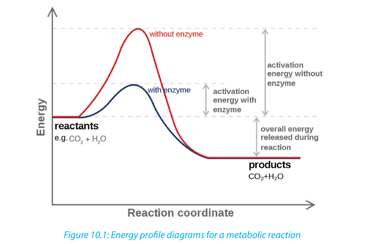
at the end of the reactions. An enzyme has no effect on the relative energy content
of products versus reactant. Chemical reactions catalyzed by enzymes are usually
reversible e.g. enzyme carbonic anhydrase catalyzes both synthesis and breakdownof carbonic acid.
An enzyme provides a reaction surface and a hydrophilic environment for a reaction
to take place. This is normally a hollow or cleft in the enzyme which is called the
active site, but it is normally hydrophobic in nature rather than hydrophilic.
A very small amount of enzymes is needed to react with a large amount of substrate.
The turnover number of an enzyme is the number or reactions an enzyme molecule
can catalyse in one second. Enzymes have a high turnover number e.g. the turnover
number of catalase is 200,000 i.e. one molecule of enzyme catalase can catalyse the
breakdown of about 200,000 molecules of hydrogen peroxide per second into waterand oxygen at body temperature.
A cofactor is the best general term to describe the non-protein substances required
by an enzyme to function properly. This term covers both organic molecules and
metal ions. A co-enzyme is an organic molecule that acts as a cofactor. A prostheticgroup is a cofactor that is covalently bound to the enzyme.
Self-Assessment 10.2
1. State any four properties of enzymes.
2. Enzymes have generally high turnover number. What is the significance ofthe high turnover of enzymes?
10.3. Mode of action of enzymes
Activity 10.3
There are two main hypotheses that explain the mode of action of an enzyme on
its substrate: the lock and key hypothesis and the induced-fit hypothesis. Carryout a research to find the relevance of each.
Enzymes do not change but substrates are converted into products. A substrate is a
molecule upon which an enzyme acts. In the case of a single substrate, the substrate
binds with the enzyme active site to form an enzyme-substrate complex. Thereafter
the substrate is transformed into one or more products, which are then released
from the active site. This process is summarized as follows:
Whereby: E = enzyme, S = substrate(s), ES = Complex Enzyme-Substrate and P=
product (s). There are two main hypotheses explaining the mechanism of enzyme
action:
a. The lock and key hypothesis by Emil Fischer
In this hypothesis the substrate is the key and enzyme is the lock. The active site isexactly complementary to the shape of the substrate as shown below.
b. The induced-fit hypothesis by Daniel Koshland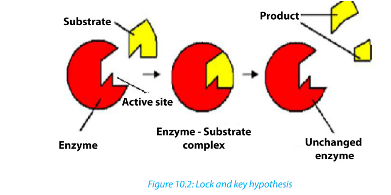
The induced-fit hypothesis is a modified version of the lock and key hypothesis and
is more widely accepted hypothesis. In this hypothesis, the active site is flexible andis not fully complementary with the shape of the substrate. An enzyme collides with
the substrate molecule and binds to the active site. This induces a slight change in
the shape of the enzyme making the substrate the fit more precisely. This reduces
the potential energy of the substrate and allows the reaction to occur. The products
formed move away from the active site and regains its original configuration readyfor the next reaction to take place.
Self-Assessment 10.3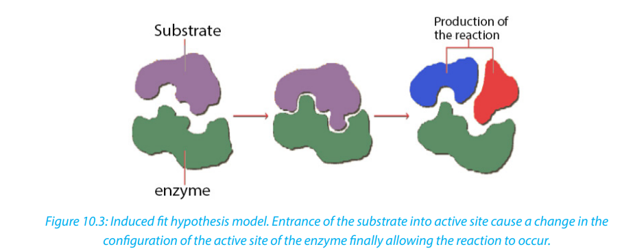
The key and lock hypothesis is a model that explain the mode of action of an
enzyme on the substrate. In the same context, analyse the diagram below andthen answer question that follow.
1. What does the lock represent?
2. What does the key represent?
3. Where is the active site?
4. Suggest another diagram that can better represent the induced fithypothesis. Write short notes to explain its functioning.
10.4 Factors affecting enzyme action
Activity 10.4
You will need
Eight test tubes containing 2 cm3
starch solution, amylase solution, cold water
(ice) water bath, iodine solution, HCl solution, and droppers
Procedure:
1. Label your test tubes A-D as follows:
2. Add 1 cm3 of starch solution to each test tube
3. Keep tube A and B in cold (ice) and tube C and D in the water bath at 35oC
for 5 minutes.
4. Add 1 cm3 of 1M HCl on test tubes B and D, then shake the mixture to stir.
5. Add 1 cm3 of amylase solution on each test tube. Shake and therefore
keep A and B in cold and C and D in water bath for 10 minutes.
6. Take a sample from each tube and mix it with one drop of iodine. Use a
different tile for each test tube. Record and interpret your observation andthen draw a conclusion.
Enzymes activities can be limited by a number of factors such as the temperature, the pH,
the concentration of the substrate or the enzyme itself and the presence of inhibitors.i. TemperatureAt zero temperature, the enzyme cannot work because it is inactivated. At low
temperatures, an enzyme-controlled reaction occurs very slowly. The molecules
in solution move slowly and take a longer time to bind to active sites. Increasing
temperature increases the kinetic energy of the reactants. As the reactant moleculesmove faster, they increase the number of collisions of molecules to form enzyme substrate complex.
At optimum temperature, the rate of reaction is at maximum. The enzyme is in
active state. The optimum temperature varies with different enzymes. The optimum
temperature for enzymes in the human body is about 37oc.
When the temperature exceeds the optimum level, the enzyme is denatured.
The effect is irreversible. However, some species are thermophilic that is they work
better at high temperatures; others are thermophobic, that is they work better at
low temperatures. For example, some thermophilic algae and bacteria can survive
in hot springs of 60oc.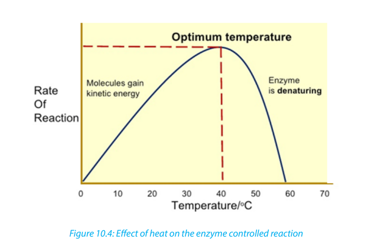
The rate doubles for each 10oC
rise in temperature between 0oC and 40oC (figure 10-
5). The temperature coefficient Q10 is the number which indicates the effect of rising
the temperature by 10oC on the enzyme-controlled reaction. The Q10 is defined as
the increase in the rate of a reaction or a physiological process for a 10°C rise in
temperature. It is calculated as the ratio between rate of reaction occurring at (X
+ l0) oC and the rate of reaction at XoC. The Q10 at a given temperature x can becalculated from:
Worked out example
The rate of an enzyme-controlled reaction has been recorded at different
temperatures as follows: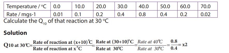
This means that the rate of the reaction doubles if the temperature is raised from
30°c to 40°c
Be aware that not all enzymes have an optimum temperature of 40°c. Some bacteria
and algae living in hot springs (e.g. Amashyuza in Rusizi) are able to tolerate very
high temperatures. Enzymes from such organisms are proving useful in variousindustrial applications because they do not denature up to 700c
ii. The pH
Most enzymes are effective only within a narrow pH range. The optimum pH is the
pH at which the maximum rate of reaction occurs. Below or above the optimum pH
the H+ or OH- ions react with functional groups of amino acids in the enzyme whichloses its tertiary structure and become natured.
Different enzymes have different pH optima (look in the table).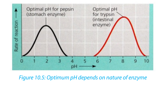 Table 10.1. Optimum pH of some digestive enzymes
iii. Enzyme concentration
The rate of an enzyme-catalyzed reaction is directly proportional to the concentration
of the enzyme if substrates are present in excess concentration and no other factorsare limiting.
iv. Substrate concentration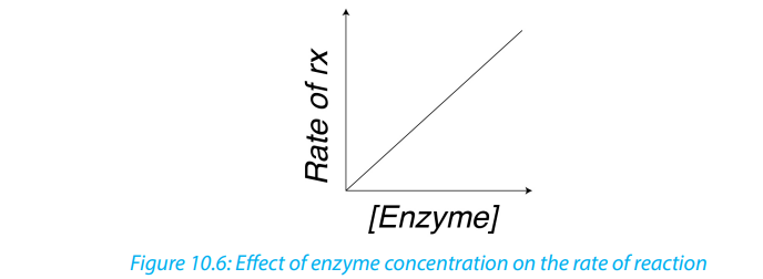
At low substrate concentration, the rate of an enzyme reaction increases with
increasing substrate concentration. The active site of an enzyme molecule can only
bind with a certain number of substrate molecules at a given time. At high substrate
concentration, there is saturation of active sites and the velocity of the reactionreaches the maximum rate.
b. Inhibitors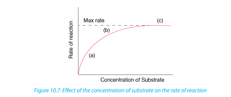
The inhibitors are chemicals or substances that prevent the action of an enzyme. An
inhibitor binds to an enzyme and then decreases or stops its activity. There are three
types of inhibitors:
i. Competitive inhibitors are molecules that have the similar shape as the
substrate. At high concentration, they compete with the substrate for the active
site of the enzyme e.g. O2
competes with CO2in RuBP-carboxylase.
ii. Non-competitive inhibitors are molecules that can be fixed to the other part
of enzyme (not to the active site) so that they change the shape of active site,due to this the substrate cannot bind to the active site of the enzyme.
iii. End product inhibitor, Allosteric inhibitor or Allostery.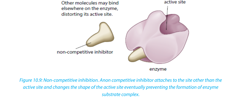
This is a chain enzymatic metabolic pathway where the final end product acts as
an allosteric reversible inhibitor for the first, the second or the third step in the
metabolic pathway. The shape of an allosteric enzyme is altered by the binding of
the end product to an allosteric site. This decreases enzymatic activity. By acting
as allosteric inhibitors of enzymes in an earlier metabolic pathway, the metabolites
can help to regulate metabolism according to the needs of organisms. This is anexample of negative feedback.
This often happens when few enzymes are working on a large number of substrate
e.g. ATP is an end-product inhibitor of the enzyme PFK (Phosphofructokinase) in
glycolysis during cell respiration. The end-product inhibitor leads to a negativefeedback.
The products of enzyme-catalyzed reactions are often involved in the feedback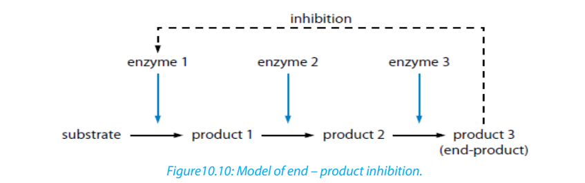
control of those enzymes. Glucose-1-phosphate is the product formed from this
enzyme-catalyzed reaction. As its concentration increases, it increasingly inhibitsthe enzyme.
Importance of reversible and irreversible inhibition
The nerve gas DIPF (DiIsopropyl Phosphor Fluoridate) is an irreversible inhibitor. It
binds permanently with enzyme acetyl cholisterase, altering its shape. The enzyme
cannot bind with and break down its substrate acetylcholine (neurotransmitter).
Acetylcholine molecules accumulate in the synaptic cleft. Nerve impulses cannot be
stopped causing continuous muscle contraction. This leads to convulsions, paralysisand eventually death.
Many pesticides such as organophosphate pesticides act as irreversible enzyme
inhibitors. Exposure to pesticides can produce harmful effects to the nervous and
muscular systems of humans. Heavy metal ions such as Pb2+, Hg2+, Ag+, As+ and
iodine-containing compounds which combine permanently with sulfhydryl
groups in the active site or other parts of the enzyme cause inactivation of enzyme.This usually disrupts disulphide bridges and cause denaturation of the enzyme.
Self-Assessment 10.4
1. What is Q10 of an enzyme controlled reaction?
2. You are provided with the table below of the rate of an enzyme controlledreaction.
Calculate the value of Q10 at:
a. 0° c
b. 10° c
c. 50° c
3. Explain why thermophile bacteria and algae are useful in some industrial
processes
4. The diagram below represents a metabolic pathway controlled by enzymes. – V is a substratea. Name the type of control mechanism which regulates production of
– W, X and Y are intermediate compounds
– Z is a product– e1, e2, e3, and e4 are enzymes
compound Zb. Explain how an excess of compound Z will inhibit its further production.
10.5. Importance of enzymes in living organisms
Activity10.5
Discuss and present your ideas about the need for different enzymes in livingorganisms.
Without enzymes, most of the biochemical reactions in living cells at body
temperature would occur very slowly or not at all. Enzyme can only catalyze reactions
in which the substrate shape fits that of its active site
There are thousands of metabolic reactions that place in the body that require
enzymes to speed up their rate of reaction, or will never happen. Enzymes are very
specific, so nearly each of these chemical reactions has its own enzyme to increase
its rate of reaction. In addition, the organism has several areas that differ from one
another by the PH. Therefore, the acid medium requires enzymes that work at low
pH while other media are alkaline and require enzymes that work at high pH. In
addition to digestion, enzymes are known to catalyze about 4,000 other chemical
reactions in your body. For example, enzymes are needed to copy genetic materialbefore your cells divide.
Enzymes are also needed to generate energy molecules called ATP, move fluid and
nutrients around the insides of cells and pump waste material out of cells. Most
enzymes work best at normal body temperature about at 370 c -- and in an alkaline
environment. As such, high fever and over-acidity reduce the effectiveness of mostenzymes. Some enzymes need co-factors or co-enzymes to work properly.
Self-Assessment 10.5
1. Fill the blank with appropriate terms:
Enzymes are biological ____________________ produced by
___________________________ cells. Enzymes reduce the amount of
____________________ energy required for reactions to occur. They consist of
globular ____________________ with _______________________ structure.
2. Answer the following questions:a. What is the main role of enzymes?10.6. Enzymes technologyb. What would happen if there are no enzymes in the cell?
Learning activity 10.6
Enzymes are needed in everyday life. At school you can use salivary amylase
to hydrolyse starch. There is industrial technique used to get large amounts of
enzyme amylase.
Read through the notes below and answer the following questions below:a. State the different processes in which enzyme technology is appliedThe market for enzymes is prosperous. The demand keeps on increasing as new
b. What is the role of thermophilic bacteria in this process?c. How is the effectiveness of an enzyme improved for used in industry?
applications of enzymes are discovered. Enzymes have been used in cheese-making,
in leather industries, and making washing powders.
Microbial cells are still the most sources of industrial enzymes because microorganisms
naturally produce enzymes inside their cells known as intracellular enzymes.
When microorganisms secrete their enzymes for an action outside their cells, the
enzymes are called extracellular enzymes. Microorganisms may have specific genes
introduced into their DNA by genetic engineering so that they produce enzymesnaturally made by other organisms.
Once enzymes are produced by the microorganisms they are isolated by
centrifugation in order to remove the large cell fragments. The enzyme is precipitated
from solution by a salt such as (NH4)2SO4 or an alcohol such as CH3-CHOH-CH3.
Thereafter the enzyme can be purified by the process known as electrophoresis or
column chromatography. The enzyme stability is a key factor in the industrial use of
enzymes. The stability of an enzyme is its ability to retain its tertiary structure undera wide range of conditions.
As many industrial processes require high temperatures and extreme pH, it is
recommended to use bacteria such as Bacillus subtilis which withstand harsh
conditions such as high temperature. Those thermophilic bacteria produce
thermostable enzymes that do not denature at high temperature because their
optimum temperature between 65 - 750c.
Some useful enzymes are not thermostable. Such enzymes should be improved by
the technique called immobilization i.e. the enzyme is attached to or located withinan unreactive support such as nylon that protects it from denaturation.
Self-Assessment 10.6
1. What is the role of alcohol or ammonium sulphate during the extraction of
enzymes?
2. Why is thermostability of enzymes so important for many industrial processes?End of unit assessment 10
1.
a. What is the meaning of the following terms related to enzyme activity?
i. Catalyst
ii. Activation energy
iii. Lock and key hypothesis
iv.Q10
b. Why are there hundreds of different enzymes in a cell?
c. How do enzymes reduce the activation energy of a reaction?
2. Enzyme activity is affected by a number of factors.
a. Explain why enzymes work faster at relatively high temperatures
b. Describe what happens to the enzyme structure if the temperature is raised
above the optimum temperature.
c. How are enzymes affected by pH?
d. Why do different enzymes have a different optimum pH?
e. What is the difference between a reversible and irreversible enzymeinhibitor?
3. Some bacteria and algae can survive in boiling water of hot springs. Enzymes
from those organisms are used in industrial processes. Why are those enzymes
useful?
4. The following set of data shows the effect of temperature on the completion
time of an enzyme reaction.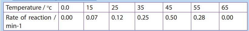
a. Plot the data on a graph
b. What is the optimum temperature of this reaction?
c. Describe the shape of the graph between 10 and 40oc
d. Calculate the rate of increase between 20 and 30oc.5. The table below shows the rate of an enzyme reaction at a range of temperature:
a. Fill that table with the values of the rate of reaction and plot a graph of rate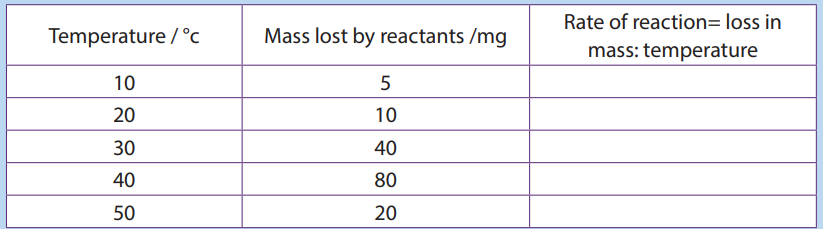
at different temperatures (use x-axis for temperature).
b. Calculate Q10 at 30°c.
c. Explain what happen between 20 and 30°c, and between 40 and 50°c.
6. The graph below shows the activity of a commercial enzyme alcalase atdifferent pH value. Alcalase is a protease enzyme.
a. What are the compounds digested by this enzyme?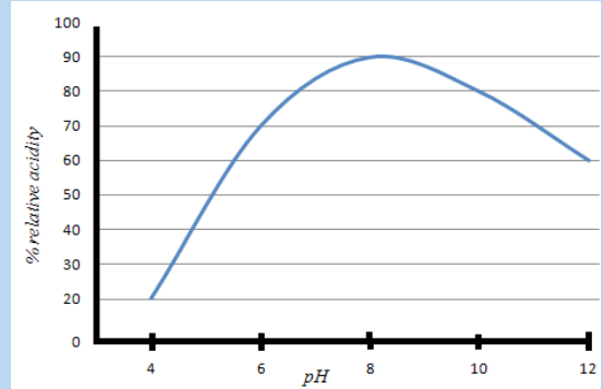
b. Describe the change in enzyme activity with PH.
c. How does this curve compare to the pH curve of a human digestive
enzyme such as pepsin?
7. Outline how a specific enzyme can be produced from bacteria.UNIT 1: PRINCIPLES OF GAS EXCHANGE SYSTEMS
UNIT 11: PRINCIPLES OF GAS EXCHANGE SYSTEMS
Key Unit Competence
Explain the principles of gaseous exchange systems
Learning objectives
At the end of this unit learners will be able to:– Explain the relationship between size and surface area to volume ratio.Introductory activity
– Describe how different respiratory surfaces are modified to speed up the
diffusion process.
– State the characteristics of gaseous exchange surfaces.
– Describe the effects of tar and carcinogens in tobacco smoke on gas exchange
system with reference to lung cancer and Chronic Obstructive Pulmonary
Disease (COPD).
– Describe the short-term effects of nicotine and carbon monoxide on the
cardiovascular system.
– Observe prepared slides of gaseous exchange surfaces and identify their
characteristics.
– Dissect fish gills and observe the surface area for gas exchange.
– Observe mammal’s lungs and state their adaptation for gaseous exchange.
– Use internet to make research and deduce the findings
– Appreciate the evolution of gaseous exchange surfaces from simple tocomplex.
Kalisa and Uwase wanted to rear tilapia at their home. They bought a nice
transparent plastic box. They filled it with 1.5L of clean mineral water, put in some
pieces of meat and plant leaves. They finally introduced a living tilapia in the box
and covered. After two days they were happy to see their fish swimming. But on
the third day, they become sad after finding it dead and yet the food was still in
water.What could have caused the death of the fish?
11.1. Relationship between size and surface area to volume
ratio
Activity 11.1
1. Use Manila paper, scissors, and graduate ruler to create three cubes: 3cm x 3cm,
2cm x 2cm, 1cm x 1cm
a. Calculate the surface area, the volume, and the surface area to volume ratio
of each cube. What do you conclude from these ratios?
b. Compare the surface area to the volume of a spherical alveolus having a
radius of 0.001m and that of another animal with a radius of 0.000001m.2. What do you understand by surface area to volume ratio?
The surface area to volume ratio is the relationship between the surface area and
the volume of an object. Small or thin objects have a large surface area compared tothe volume. For example, the surface area of a sphere is calculated by
As the length or radius of the sphere increases, the increase in the surface area is
squared (X2) and the increase in the volume is cubed (X3). The surface area to the
volume ratio gets smaller as the cell or animal gets larger. Thus, if the cell grows
beyond a certain limit, not enough material will be able to cross the membrane fastenough to accommodate the increased cellular volume.
As a cell grows, its surface area to volume ration decreases. At some point in its
growth its surface area to volume ratio becomes so small that its surface area is too
small to supply its raw materials to its volume. The cell will reach a size at which
substances cannot enter or leave the cell in sufficient time to sustain life. At this
point the cell cannot get larger. The volume of the cell will also be so large that
the diffusion rate will be too low to distribute necessary substances throughout the
cell within a reasonable time. This brings about the need of having a mechanism ofventilation that speeds up the rate of gaseous exchange.
The rate of oxygen consumption by an animal gives a relatively accurate indication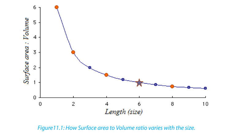
of the rate of its metabolic activity. The need of oxygen varies with the activity, the
size of the organism, and their health. In general, small mammals need more oxygenthan large mammals because:
– Small mammals have a big respiratory surface area to the volume ratio
– Small mammals are too motile than large mammals. Therefore, they need to
produce more energy through aerobic respiration– Small mammals reproduce more rapidly than large mammals.
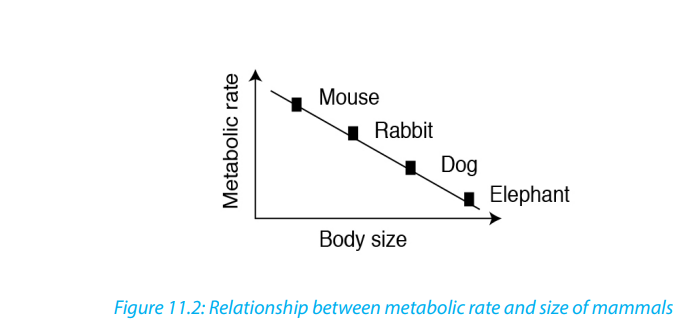
A running man needs a double volume of oxygen than a sleeping manand a pregnant woman needs more oxygen than a normal woman.
Self-Assessment 11.1Determine the surface area to volume ratio of a sphere having a diameter of 4 mm
11.2. Characteristics of gas exchange surfaces
The following are the characteristic features of gaseous exchange surfaces:
Large surface area: they should have a large surface area to allow adequate and
fast gaseous exchange in order to provide enough oxygen to cells and to get rid of
the carbon dioxide that is released.
Rich supply of blood: in animals with a transport system, the respiratory surface
areas found in the lungs and gills have rich supply in blood capillaries to quickly
transport gases to and from the cells. Gases diffuse into the blood and are carried to
and from the body cells.
Thin surface or thin wall: respiratory surfaces should have thin walls or thin
surface area to maximize the diffusion. The alveoli in the lungs have thin squamous
epithelium that enables gases to diffuse quickly between the alveoli and blood.
According to Fick’s law, the rate of diffusion is proportional to:
Moist surfaces area: to enable gases to dissolve and pass through the solution.
High diffusion deficit / concentration gradient: respiratory surface areas should
have a high diffusion deficit / concentration gradient to ensure faster diffusion of
respiratory gases.
Protection against injury and dry out: lungs and gills are protected by the bonesand cartilage and mucus protects them from drying out.
Self-Assessment 11.2
1. State the features common to all respiratory surfaces in living organisms
2. Explain how the following features of a respiratory surface helps gaseous
exchange:
3. Short diffusion distancea. Protection11.3. Modifications of gaseous exchange surfaces to speed up
b. A rich blood supplyc. Protection
the rate of gaseous exchange in different organism
Activity 11.3
Use appropriate laboratory equipment to extract gills in fish to show the gill
filaments. Draw and label to show the parts observed.
a. Insects
The spiracles are openings of small tubes running into the insect’s trachea system
that terminates into small fluid-filled tracheoles in which the gases are dissolved.
The fluid is drawn into the muscle tissue during physical exercise, and this increases
the surface area of air in contact with the cells.
Ventilation movements of the body during exercise may help this diffusion. The
spiracles can be closed by valves and may be surrounded by tiny hairs. The later help
to keep humidity around the opening to ensure that there is a lower concentrationgradient of water vapor, and so less is lost from the insect by evaporation.
b. Fish and tadpoles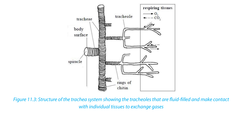
Fish and young amphibians (tadpoles) use gills for the gaseous exchange.
Gills have numerous folds that give them a very large surface area.– The rows of gill filaments have many protrusions called gill lamellae. These
filaments help in the exchange of respiratory gases
– They also have an efficient transport system within the lamellae which
maintains the concentration gradient across the lamellae. The arrangement
of water flowing passes the gills in the opposite direction to the blood (called
counter-current flow) means that they can extract oxygen at 3 times the ratea human can.
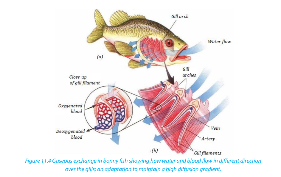
c. Amphibians, Reptiles, Birds and Mammals
These have alveoli in their lungs. Air reaches the alveoli via a system of tubes
(trachea, splitting into two bronchi - one for each lung - and numerous bronchioles):– Numerous alveoli - air sacs, providing a massive surface area over which gasesis regularly moving in and out of the lungs due to changes in volume and
can diffuse
– Have a short diffusion distance between the alveolus and the blood because
the lining of the lung and the capillary as they are only one cell thick.
– The blood supply is extensive, which means that oxygen is carried away to the
cells as soon as it has diffused into the blood.– Ventilation movements also maintain the concentration gradients because air
pressure
Activity11.3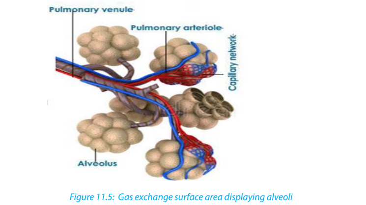
You will need: Lungs of a sheep or pig, newspaper, plastic sheets, dissecting
board, sharp scalpel, dissecting needles, scissors, dissecting tray, latex gloves, CPRmouth piece, soap to wash hands and surfaces.
Procedure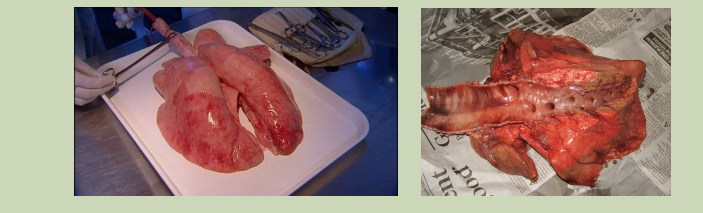
– Place the dissecting board on the newspaper and lay the lungs on the board.
– Use a scalpel to cut the lungs in half in longitudinal section.
– Identify the trachea, right lung, left lung, cartilage rings, bronchus, larynx, alveoli,
and bronchiole. You can use a magnifying hand lens to observe structures in
the lungs.
– Inflate the lungs by blowing through the CPR (cardio-Pulmonary Resuscitation)
mouth piece to see how the lungs expand.
– Feel the slippery inside of trachea, press the lung with your finger and look at
cartilaginous rings.
– Remember to wash your hand s with soap as you finish your experiment.
1. Explain what it feels like as you press the lungs with your fingers.
2. Look at cartilaginous rings. What function do they serve?
3. (a) List four features of respiratory surfaces you can identify from the
specimen.
(b) Examine the lung and explain how the lungs are suited for efficientgaseous exchange.
Table 11.1: Parts of the human gas exchange system and their respective functions
Self-Assessment 11.3
1. List the adaptations of the gills for gaseous exchange
2. List the structures through which air passes on its way from the nose to the
alveoli.
3. Give two reasons why mammals need lungs, rather than exchanging gasesthrough the skin.
11.4. Smoking and related risks
Activity 11.4
In groups, make research to find out main health risks related to smoking. Analysethe photographs below and answer questions that follow.
Between the lungs of individuals, A and B, which one is most likely that of the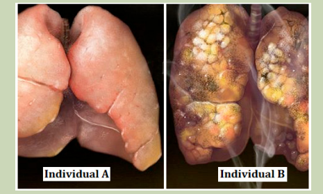
cigarette smoker?Read the notes below to identify at least three risks related to smoking cigarette.
Cigarette smoking harms nearly every organ of the body, causes many diseases, and
reduces the health of smokers in general (Figure 11.6). Quitting smoking lowers the
risk for smoking-related diseases and can increase the longevity. Inhaling cigarette
smoke is called passive smoking and presents a health hazard to people nearby whoinhale it. Of the thousands of chemicals in tobacco smoke three important ones are:
– Carbon monoxide (CO), a poisonous gas form incomplete combustion carbon.Tar in tobacco smoke is a mixture of chemicals that enter the respiratory tract. It is
CO in tobacco smoke combines easily, but irreversibly, with hemoglobin to
form carboxy hemoglobin and therefore reduces oxygen carrying capacity of
the blood. This can lead to hypotension and heart failure.
– Nicotine, a poisonous alkaloid drug that is addictive. Nicotine in tobacco
smoke stimulates the production of the hormone adrenaline by adrenal gland,
leading to an increase in the heart rate and raised blood pressure. Nicotine
also makes the red blood cell stickier and this leads to high risk of thrombosis
and hence of the strokes.
– Tar- is a sticky and brown substance. It appears in tobacco spoke minute
droplets.
an irritant and causes inflammation of the mucous membranes lining the trachea,
bronchi and bronchioles, resulting in producing more mucus. Tar also thickens the
epithelium and paralyses the cilia on its surface. As a result, cilia cannot remove the
mucus secreted by epithelium lining.
a. Short-term effects of smoking
Tar causes constriction of finer bronchioles by increasing resistance to the flow
of air.
– Tar paralyses the cilia which remove dirt and bacteria; the accumulation of
extra material in the air passage can restrict air flow.
– Smoke acts as an irritant; this causes secretion of excess mucus from goblet
cells and excess fluid into the airways, making it more difficult for the air to
pass through them.
– Mucus accumulating in the alveoli limits the air that they can contain and
lengthens the diffusion pathway.
– Coughing of many smokers, way of trying to remove the build-up of mucus
from the lungs, can cause damage to the airways and alveoli; scar tissue builds
up which again reduces air movement and rates of diffusion
– Infections arise because the cilia no longer remove mucus and pathogens
– Allergens such as pollen also accumulate, leading to further inflammation
of the airways, reduced air-flow in and out of the lungs, and possible asthmaattacks.
b. The long-term effects of smoking
– Bronchitis: Bronchitis is inflammation of the lining of the air passages and may
be acute or chronic.
– Emphysema: One in five smokers develop the crippling lung disease called
emphysema i.e. condition of gradual breakdown of the thin wall of the alveoli
leading to sensation of breathlessness as the gaseous exchange reduces.
– Lung cancer: Lung cancer usually starts in the epithelium of the bronchioles
and then spreads throughout the lungs as dividing cells cease to respond to
the normal signals around them and form unspecialized masses of cells called
tumours. The tar is carcinogen i.e. contains chemicals which cause cancer. The
irritation causes thickening of the epithelium by extra cell division and this maytrigger the cancer. Almost all people who die from lung cancer are smokers.
Self-Assessment 11.4Analyze the photograph and share ideas with your group members.
1. Between the baby and the parent who will suffer more the effects of
tobacco? Give reasons
2. Discuss any side effects of smoking.
3. Design a sign post to advocate against smoking.
End of unit assessment111. Match the terms in Column A with the correct definition in Column B.
Describe how the human lungs serve as good gaseous exchange organs.
3. Emphysema is a disease of the lungs. People who smoke cigarettes are more
likely to suffer from emphysema. The diagrams show lung tissue from a healthyperson and lung tissue from a person with emphysema.
a. Identify on the figure above by using E (for emphysema) and we (without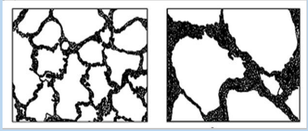
emphysema) and give a reason for your choice.
b. Explain how emphysema reduces the amount of oxygen which diffuses into
the blood.
c. What are the features that make the gill of fish an efficient respiratory organ?
d. Compare respiratory system of fish and with that of a mammal.
e. Why do people who smoke have high chances of developing lung cancer?
f. Design a simple model that shows the structure and functioning of gasexchange system in mammals.
UNIT 12: GAS EXCHANGE IN PLANTS
UNIT 12: GAS EXCHANGE IN PLANTS
Key Unit Competence
Describe structures of gaseous exchange organs in plants
Learning objectives
By the end of this unit I should be able to:
– Describe the structure of the stoma.
– Explain how stomata, lenticels and breathing roots are adapted to their
function.
– Explain the theories of opening and closure of stomata stating limitations of
each.
– Relate the differences between the structures of aquatic and terrestrial leaves
to a habitat.
– Draw and label a diagram of stoma as observed under alight microscope.
– Compare gaseous exchange structures of aquatic and terrestrial plants
– Relate the structure and function of aquatic and terrestrial plants
– Defend the relationship between structure and function in aquatic andterrestrial plants
Introductory activitySuggest the different parts of a plant that are used in gaseous exchange
12.1. Structure of stoma
Activity 12.1
Requirements
Light microscope, glass slide, cover slip, Camelina zebrine leaves, razor blade,forceps, Pasteur dropper and iodine solution.
Procedure
– Identify Commelina zebrina or commelina tradescantia plant nearby the
school. You can also use any other monocotyledonous plant with succulent
leaves.
– Remove a leaf from a plant. Then peel off gently the lower epidermis. It must
be thin enough to allow light to pass through
– Smear the epidermis on a slide containing one drop of dilute iodine solution.
– Put on a cover slip and then observe under the lower and medium
magnification.
– Repeat the observation in morning hours and in the afternoon hours.
Questions
1. Why should the sample used in the preparation must be transparent?
2. Draw and label structures observed under light microscope
Stomata (stoma in singular) are microscopic pores in epidermis of the leaves and
stems of terrestrial plants. They function in gas exchange between plant and the
atmosphere and in transpiration.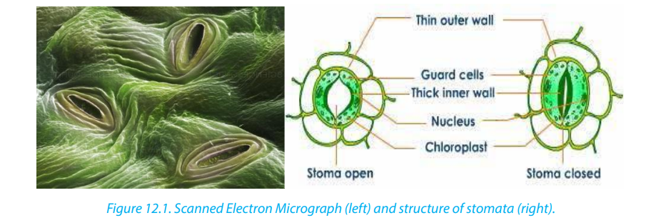
Each stoma is bordered by two saucer shaped cells called guard cells, which are
specialized epidermis cells whose movements control the size of the aperture
(pore). Unlike other epidermis cells, guard cells have kidney shape and have many
chloroplasts. Their inner cell wall is thick and less elastic while the outer cell wall is
thin and more elastic. Guard cells shrink when the plant has too little water. This
closes the stomata. When the plant has enough water, the guard cells swell up
again. This opens the stomata. In this way, the guard cells enable gaseous exchange.
Oxygen in the atmosphere diffuses through the stomata into the air spaces between
the cells of the spongy mesophyll tissue while carbon dioxide diffuses through thestomata out to the atmosphere.
Self – assessment 12.1Analyze the diagram below and answer to the following questions
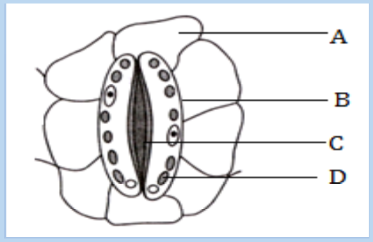
1. What title fits better to this diagram:a. an open stoma2. The part labelled C is:
b. a closed stoma
c. of a guard cell and neighbouring cells
d. of a stoma and neighbouring cellsa. Vacuole3. The part which better represents the neighbouring cell is:
b. Thick inner cell wall
c. Chloroplast
d. Thin outer cell walla. Part A4. If the guard cells become more turgid, what is more likely to happen?
b. Part B
c. Part C
d. Part Da. The cells A will swell5. Which of the following statements is false about that diagram?
b. The pore will increase its diameter
c. The number of structures C will decrease
d. The structure B will stretch in warda. The stoma is closed12.2. Theories used to explain the mechanism of opening and
b. The inner cell wall of guard is thicker than the outer cell wall
c. There are many chloroplasts in neighbouring cellsd. The guard cells have many chloroplasts.
closure of stomata
Activity12.2The diagram below shows a closed and an open stoma.
Carefully analyse the diagrams above and brainstorm your observation.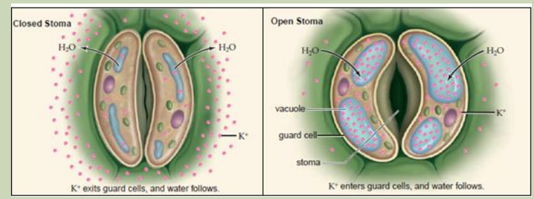
Illustrate how stomata open and close
Many theories have been proposed regarding opening and closing of stomata. The
four important theories of stomatal movement are the following:– Theory of photosynthesis in guard cellsThe combined outcome of the four theories shows that in general stomata open
– Theory of starch sugar inter-conversion
– Theory of glycolate metabolism and– Theory of active potassium pump.
during the day (light) and close during the night (dark). But how does this happen?
In light, guard cells are stimulated. They absorb K+ ions from the neighbouring cells.
K+ ions make the guard cells more permeable to CO2
. As the guard cells perform photosynthesis, the concentration of CO2 falls and the pH rises. Elaborated starch
therefore splits into malate. The high concentration of malate and the rise of pH in
guard cells develop a decrease in water potential. Hence, the guard cells withdraw
water from the neighbouring cells and extend backward leaving an open pore inbetween whereby water is lost by evaporation.
During the night, there is no light to stimulate neither the absorption of K+ ions nor the
photosynthesis. Guard cells undergo cell respiration using photosynthetic products
as source of energy (carbohydrates: malate, glucose). Therefore, the concentration
of malate decreases making the guard cells hypotonic than neighbouring cells. As
guard-cells lose their water content, they shrive and the pore in between closes.
Stomatal transpiration ceases.
192
During the night, there is no light to stimulate neither the absorption of K+ ions nor the
photosynthesis. Guard cells undergo cell respiration using photosynthetic products as source of
energy (carbohydrates: malate, glucose). Therefore, the concentration of malate decreases making
the guard cells hypotonic than neighbouring cells. As guard-cells lose their water content, they shriveand the pore in between closes. Stomatal transpiration ceases.
Plant physiologists are certain that stomatal aperture varies as a result of changes in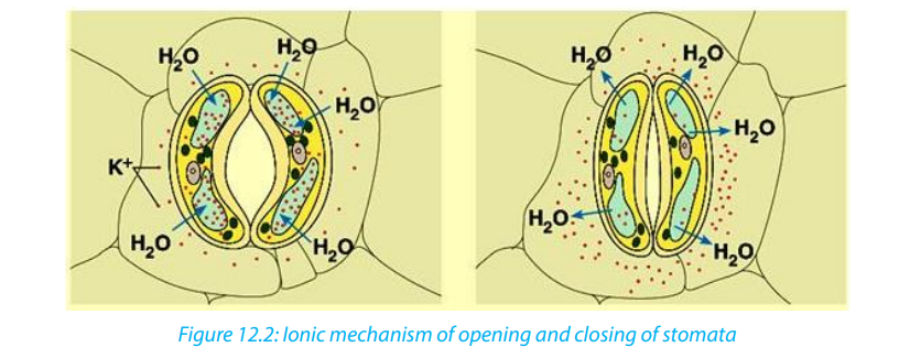 \
\
the turgidity of the guard cells. But they are less certain about how these changes
are brought about, though the following observations have been made:– Most stomata open during the day and close at nightPlants can therefore vary the stomatal aperture. This allows a compromise between
– Some stomata show a circadian (daily) rhythm of opening and closing even
when kept in constant conditions
– Stomata generally close when a plant suffers water stress, for example, when
transpiration exceeds water absorption
– The stomata of some desert plants close during the day and open at night toreduce transpiration
the need to conserve water and the need to exchange gases for photosynthesis.
The compensation point is the point when the rate of photosynthesis is equal to the
rate of respiration. This means that the CO2 released from respiration is equivalent to
that which is taken up during photosynthesis. The compensation point is reached as
light intensity increases. If the light intensity is increased beyond the compensation
point, the rate of photosynthesis increases proportionally until the point of light
saturation is reached, beyond which the rate of photosynthesis is no longer affectedby light intensity.
For a plant under water stress, its need to conserve water is greater than its need to
obtain carbon dioxide for photosynthesis. Under these conditions a plant secretes
abscisic acid (ABA). This is a chemical messenger which causes stomata to close. It is
thought that ABA triggers a metabolic pump which actively secretes potassium ionsout of guard cells, causing the cells to lose water and become flaccid.
Self-assessment 12.2
1. According to the ionic theory of opening and closing stoma, what is the role
of potassium ions in the guard cell?
2. What would happen to guard cells if the concentration of malate doubled?
3. What is meant by compensation point?
12.3. Structural adaptations and function of stomata, lenticels
and breathing roots.
Activity 12.3Observe the adaptations of these plants for gas exchange.
1. How is each of these plants adapted for gas exchange?
2. Read through the notes that follow and describe any two adaptations for
gas exchange
The exchange of atmospheric gases is essential to photosynthesis and cell respiration. In plants, the
gas exchange takes place through stomata, breathing roots, lenticels and cuticles. Most stomata are
on the lower epidermis of the leaves on plants. Unlike other plant epidermal cells, the guard cells
contain chlorophyll to carry out photosynthesis. This allows the cells to expand or contract to open
or close the stomata.
Guard cells swells, through the process of osmosis, to allow opening of the stomata for CO2 to enter
and excess O2 and H2O to leave, and they shrink in order to force the stomata shut either partially or
completely to prevent dehydration. The number of stomata on the epidermal surface depends on the
ecology of plants. Usually, plants on wet climate have fast growth and a high concentration of
stomata. Plants on dry weather have lower rates of photosynthesis, lower growth and lower
concentrations of stomata.
Self-assessment 12.2
1) According to the ionic theory of opening and closing stoma, what is the role of
potassium ions in the guard cell?
2) What would happen to guard cells if the concentration of malate doubled?
3) What is meant by compensation point?
Activity 12.3
Observe the adaptations of these plants for gas exchange.
Figure 12: (a) Mangrove (b) Breathing roots (c) Pneumatophores
1) How is each of these plants adapted for gas exchange?
2) Read through the notes that follow and describe any two adaptations for gas exchange
(a) Mangrove (b) Breathing roots (c) Pneumatophores
1. How is each of these plants adapted for gas exchange?
2. Read through the notes that follow and describe any two adaptations for
gas exchange
The exchange of atmospheric gases is essential to photosynthesis and cell
respiration. In plants, the gas exchange takes place through stomata, breathing
roots, lenticels and cuticles. Most stomata are on the lower epidermis of the leaves
on plants. Unlike other plant epidermal cells, the guard cells contain chlorophyll to
carry out photosynthesis. This allows the cells to expand or contract to open or close
the stomata.
Guard cells swells, through the process of osmosis, to allow opening of the stomata
for CO2 to enter and excess O2 and H2O to leave, and they shrink in order to force the
stomata shut either partially or completely to prevent dehydration. The number of
stomata on the epidermal surface depends on the ecology of plants. Usually, plants
on wet climate have fast growth and a high concentration of stomata. Plants on dry
weather have lower rates of photosynthesis, lower growth and lower concentrationsof stomata.
Xerophytic plants or xerophytes are plants that inhabit arid regions (desert). They
have the following adaptations:– Stomata sunken in grooves and reduced in numberEpidermis infolded to reduce the surface area
– Ability to fix CO2 at night, so the stomata are closed during the day.– Leaves reduced to scales or thorns to reduce the surface area for transpiration
Hydrophytes or water plants are plants that grow submerged or partially submerged
in water. To thrive in this environment, hydrophytes have the following features
– developed stomata on large upper surface of their leaves (rather than
underside) making gas exchange more efficient.
– large air space to facilitate evaporation from the mesophyll.
– little or no lignified supporting tissues on the submerged parts.
– poorly developed transport tissue,
– stems and leaves have little or no lower cuticle but large continuous air spaces,
forming reservoir of oxygen and CO2
which also provides buoyancy to theplant tissues when submerged.

A halophyte is a plant that grows in water of high salinity and they come into contact
with saline water through its roots or by salt spray, such as in saline semi-deserts,
mangrove swamps, and marshes. Halophytes are adapted in the flowing ways:– Store water in succulent tissues which have high concentration of salt. They
can thus take up water from the sea water by osmosis.
– Extensive air spaces throughout the stem and roots making air available to all
cells, and giving buoyancy to the stem and leaves at highest tides.
– They develop breathing roots called pneumatophores which grow upwardand protrude out of the ground. e.g. mangrove tress.
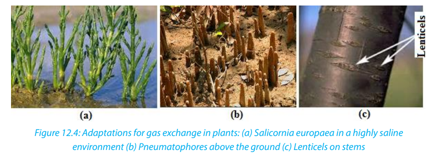
Self-assessment 12.3What features are common to plants living in desert and saline soils?
End of unit assessment 12
Section A: Objective questions
1. You are provided with the diagram below. Analyze it and then choose the
correct answer. Transpiration in the leaf depends on the transport of potassium
ions into:
a. Into O
b. Into P
c. From M to L
d. From M to Qe. From P to L.
2. The theory that says than during the light time, potassium pumps open and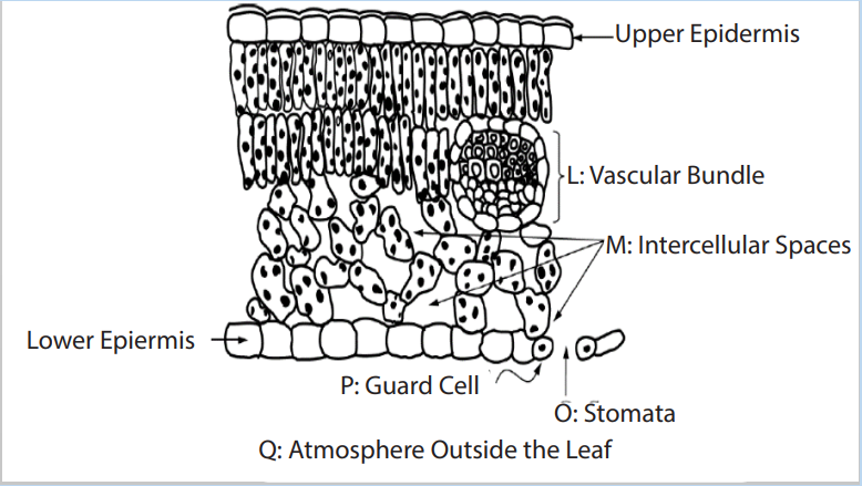
this brings about diffusion of CO2 from the atmosphere to the guard cells for
photosynthesis is called:a. Theory of photosynthesis in guard cells3. What is the main difference between the guard cells and the other epidermal
b. Theory of starch sugar inter-conversion
c. Theory of glycolate metabolismd. Theory of active Potassium Pump.
cells?a. Guard cells have chloroplast while the remaining epidermal cells have no4. Water lily is:
chloroplast
b. Guard cells have oval shape while other cells have cubic shape
c. Guard cells are beneath the spongy mesophyll
d. Guard cells are covered by a transparent cuticlea. Xerophytes5. Mangroves are plants adapted to estuaries or marine region with high salinity.
b. Halophyte
c. Hydrophyte
d. Helophyte
What statement does not describe the adaptations of mangroves?a. The presence of lenticels that help in gas exchange and evaporationSection B
b. Presence of large number of stomata on the upper side of the leaves
c. The presence of pneumatophores which are breathing rootsd. Presence of succulent tissues that have high concentration of salt
6. Explain how gaseous exchange occurs in the leaf.
7. How does gaseous exchange occur in woody stems?
8. Describe how roots get oxygen.
9. a. Draw a labelled diagram of a stoma
b. Draw arrows on the diagram to show how gaseous exchange occurs.
10.The drawing shows a 24-hour cycle for the opening and closing of stomata fromthe same plant.
a. Explain how this cycle of opening and closing of stomata is advantageous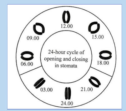
to the plant.
b. The diagram shows the potassium (K+) concentrations in the cells around
open and closed stomata in commelina. The concentrations are in arbitraryunits.
i. Explain how the movement of K+ ions accounts for the opening of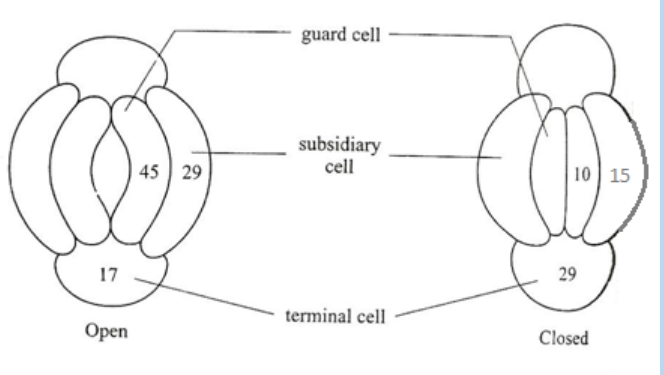
stomata.ii. Explain how K+ ions are moved against a concentration gradient.
UNIT 13: GROWTH AND DEVELOPMENT IN PLANTS AND ANIMALS
UNIT 13: GROWTH AND DEVELOPMENT IN PLANTS
AND ANIMALS.
Key Unit Competence
Account for the processes of growth and development in plants and animals
Learning Objectives
By the end of this unit, I should be able to:– Describe dormancy as a state of inactivity to absolute minimum due to theIntroductory activity
morphological and physiological state of a plant structure
– Explain how dormancy is maintained and broken
– State the conditions required for germination
– Outline the role of enzymes in the process of germination
– State types of plant growth hormones and their functions
– Identify the hypocotyl and coleoptile in a germinating seed
– Describe the stages and types of germination
– Recognize that a meristem is a growing point of the plant and the main
meristematic regions of a tree
– Describe current views about photoperiodic control of flowering
– Observe structures of endospermic and non-endospermic seeds
– Demonstrate how fruit and seed dispersal takes place
– Demonstrate hypogeal and epigeal germination
– Carry out an investigation to distinguish between primary and secondary
growth
– Appreciate the importance of fruit and seed dormancy and germination in the
life cycle of plants
– Carry out an experiment on the development of eggs at different temperatures
– Describe the process of metamorphosis in arthropods and amphibians
– Distinguish the various stages of development in frogs
– Analyse complete and incomplete metamorphosis
– Compare growth patterns in arthropods and vertebrates
– Appreciate the demands of the terrestrial environment to the adaptation of
amphibians
Suggest morphological differences by which different organisms develop and
grow to maturity.
13.1. Fruit, seed and bud dormancy
Activity 13.1– Put dry bean seeds and maize grains in boiling water for 10 minutes.A seed is a plant organ that develops from the fertilized ovule. As the seed embryo
– Use nails to make a longitudinal section of the bean and maize.
– Compare the two sections.
– From your point of view, do you think that all plants’ organs are always active?
Justify your point of view.
– Produce a picture showing how plant organ (seed and bud) behaves inrelation to oxygen, temperature and water.
develops from the zygote, the seed makes a food reserve of macronutrients including
carbohydrates, proteins and lipids. The amount of reserved food type depends on
the plant species. Legumes like peanuts and soybeans store proteins as well as
fats while beans store more starch and proteins. A seed consists of a plant embryo
surrounded by the food supply in cotyledon or endosperm and a protective coat
called seed-coat or testa. The plant embryo is composed of the radicle or embryonic
root, the hypocotyl which is the embryonic stem and the plumule, the embryonic
leaves.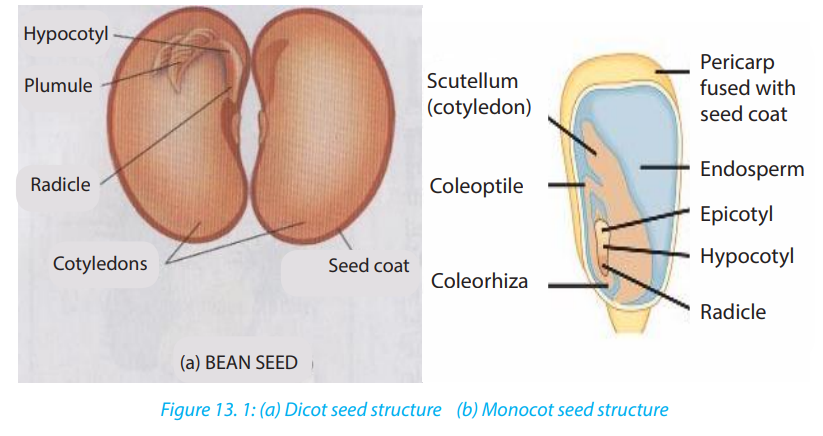
Dormancy is a period of cells’ inactivity due to a very low metabolism to prevent
growth when environmental conditions are unfavorable. The dormancy can be for
fruit, bud or seed.
a. Bud dormancy
Bud dormancy is a suspension of most physiological activities and growth that can
be reactivated. It may be a response to environmental conditions such as seasonality
or extreme heat, drought, or cold. The exit from bud dormancy is marked by theresumed growth of the bud. Bud dormancy may proceed to dormancy of the whole plant.
b. Seed dormancy
Seeds exhibit dormancy to avoid growth during unfavorable conditions. During the
last stages of its maturation, the seed dehydrates until it gets dry. The embryo which
is surrounded by a food supply (cotyledons, endosperm or both), enters dormancy.
Most seeds are enough durable that can last a year or two until conditions are
favorable for germinating. However, the length of time a dormant seed remain
viable and capable of germinating varies from a few days to years depending on the
plant species and environmental conditions. This may justify the reason why after
a bushfire or other environmental disruption, vegetation reappears as their seeds
have accumulated and stayed for longer in the soil. Many plants are easily grown
from seeds. Although its embryo is alive, a dormant seed cannot germinate until
it is exposed to certain environmental conditions to break the dormancy. Thus, itgerminates and starts to grow.
Conditions needed for seed germination
Germination is the process by which a seedling develops from a seed embryo. There
are requirements for germination to be successful. Environmental conditions such as
water, oxygen, and temperature trigger seed germination. For a seed to germinate,
it must be found in place where all these conditions are available. If one condition isnot available, germination becomes impossible. These conditions include:
a. Water
For a seed to germinate, it requires water. Mature seeds are very dry and must absorb
water by imbibition to germinate. Water softens the seed coat for embryo sprouting
and provides a medium for reactions during hydrolysis. Enzymes breakdown
macronutrients such as starch, proteins and fats stored in the cotyledons and the
endosperm to simple sugars such as glucose, amino acids which provides energy
for the embryo to grow.
b. Oxygen
This is needed for needed for cellular respiration by oxidizing glucose to liberate ATP
to provide energy for embryo development.
c. Temperature
Seeds germinate only if exposed to their optimum temperature varying between
5 to 40 0c depending on the seed species. Enzymes are sensitive to temperature
because enzymes need a favorable temperature to work efficiently. Burying seeds
too deeply in the soil prevents them from germinating as they are cut off fromtemperature and air.
Self-assessment 13.1
1. Define the term dormancy?
2. Explain how dormancy is maintained and broken?
3. Suggest the advantage of dormancy in plants?
4. What are conditions needed for seed germination?
5. Explain the role of enzymes during the process of seed germination.6. Draw and label endospermic and non-endospermic seeds
13.2. Types and stages of germination.
Learning activity 13.2
Conduct the experiment below to compare the two types of germination.
Requirements
Two bean seeds or soybean seeds, two sorghum or maize grains, water and two
plastic devices/containers
Procedure
– Fill each container with soil
– Put two grains of maize in the container labeled A and two seeds of bean in
the container labeled B
– Pour some water in each container simply to moisture the soil.
– Put both containers A and B in a secured place.
– Record your observations from day five to day twenty one with emphasis todevelopmental changes.
In conducive environment, a seed can germinate. Germination involves three mainstages: imbibition, radicle sprouting and plumule sprouting.
The dormant seed contains very little water, when placed in moist conditions, it
absorbs water by osmosis. The intake of water by a dry material is called imbibition.
As the seed absorbs water and oxygen, it swells, the embryo grows and the seed-coat
cracks, and the radicle also called primary root emerges downward. This is seed coat
rupture and radicle sprouting. The primary root is the first organ to appear during
embryo development. The plumule cells develop and the first leaf forms upwards inthe process known as plumule sprouting.
A germinating seed shows the primary root from which secondary roots start to
develop and the primary leaf which is surrounded by a protective sheath called
coleoptile. The Hypocotyl is the stem below the plumule while the epicotyl is thestem above the plumule.
13.2.1. Types of seed germination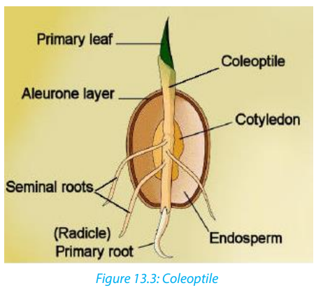
There are two types of seed germination: hypogeal and epigeal seed germination.
a. Epigeal germination
Epigeal germination is typical to non-endospermic seeds like beans, soybeans and
coffee seeds.In this type of germination, the cotyledons come above the soil surface
into air and light, due to rapid growth and elongation of hypocotyl. The cotyledons
are green and make food to be used by stem during growing season. They finally dry
off and seedlings become independent . The example of the epigeal germination is likethe one found in bean seeds.
b. Hypogeal germination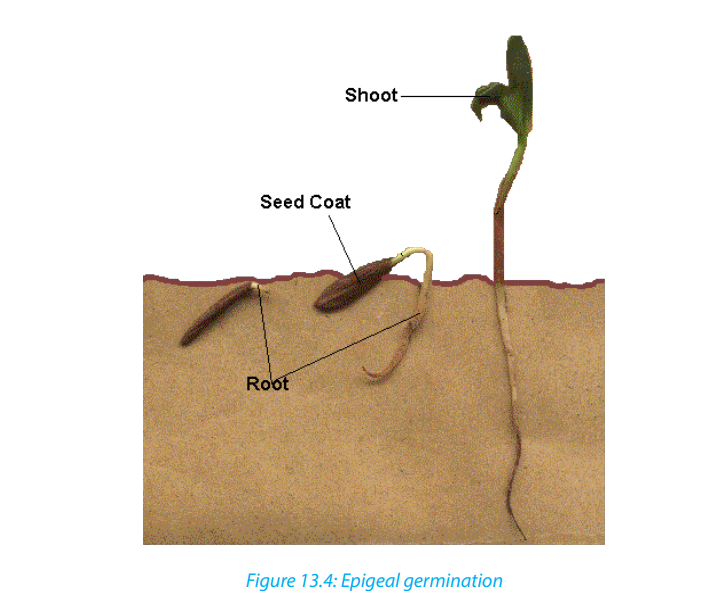
In this type of germination, the cotyledons remain in soil or just above the surface.
Here the epicotyl elongates, pushing plumule upwards. Cotyledons do not turn
green and gradually dry up and fall off. An example of this type of germination isfound in pea, mango, and groundnut seeds.
Self-assessment 13.2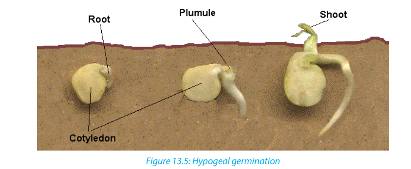
1. Use diagrams to demonstrate and distinguish the types of seed germination.2. Describe the three stages of seed germination.
13.3. Primary and secondary growth
Learning activity 13.3
Move around the school garden or anywhere in your surroundings, then collect
two plant species in which one is a monocotyledon and another is a dicotyledon.
1. By examining their physical characteristics, describe similarities and
differences between collected plants.
2. Predict the reason why all plants grow in length.
3. From your observation, suggest/ predict the reasons justifying whymonocotyledons do not grow in width while dicotyledons do.
Growth is a permanent increase in the size of an organism or of some parts of it.
It is brought by cell division and the assimilation of new material within the cells
which result from the division and the cell expansion which follows. Cell expansion
is particularly noticeable in plants, where rapid enlargement can occur as a result
of water taken up by osmosis. If the organism achieves its mature size, it starts
development, the formation of new structures or organs to perform specific functions.
That is the production of reproductive organs and locomotive or protective organs.It is controlled by cell differentiation. Growth is either primary or secondary.
a. Primary growth
Primary growth consists of the increase in length and formation of primary plant
organs including roots, stem, leaves, flowers, and fruits. It occurs in most herbaceous
plants. The roots elongate to penetrate the soil, and shoots elongate to reach the
sunlight. Primary growth is controlled by apical meristems. Meristematic cells
divide by mitosis. Some daughter cells absorb water and nutrients. As the cell takes
in water the cell walls stretches, the cell elongates and slightly enlarges. After this
growth by elongation, cells differentiate and specialize for specific functions. The
cells formed from apical meristems do not expand laterally and this limits their size.
The herbaceous plants exhibiting such growth tend to be short lived. They are called
annual plants because most of them do not live for more than one growing season(a year) after which they enter dormancy and survive as seeds.
b. Secondary growth
It consists of getting wider or thicker, and occurs in roots and stems of perennial
woody plants, all trees and shrubs. Woody plants grow taller than herbaceous plants
and they live longer, more than two years some plants while some others may live
for and over 30 years.
Secondary growth is controlled by lateral meristems: vascular cambium and cork
cambium. Vascular cambium is located between primary phloem and primary xylem.
Vascular cambium cells divide by mitosis. Some cells remain meristematic while
other cells expand sideways and differentiate. They form the secondary phloem
outwards and the secondary xylem inwards. As the secondary growth continues,
the outermost phloem cells die. Their death causes no matter to the whole plant as
dead cells are continually replaced by new ones.
The secondary xylem cells are strengthened by the accumulation of lignin and
cellulose. They form a wood also called secondary xylem providing the mechanical
support to very taller trees. The cork cambium situated between the epidermis and
secondary phloem produces the cork cells by mitosis. The cork cells are pushed
toward the epidermis and accumulate a waxy substance called suberin making
the cork waterproof. The cork gradually replaces the epidermis. Like the epidermis,
the cork protects the plant from dehydration and infection. The expansion of the
internal tissues results into the continual cork shedding. Because of meristematiccells that continuously divide, perennial plants have unlimited growth.
Self-assessment 13.3
1. In the table list the differences between primary growth and secondary
growth.
2. Describe briefly what the wood is and its major function.
3. What is the name given to a substance that makes the cork to resist plant
dehydration?
4. Identify the importance of apical and lateral meristems in plant growth
13.4. Determination of growth
Activity 13.4
In the learning activity 13.1 you have grown two types of seeds (bean and maize).
Based on that experiment do the following to investigate the primary growth in
a seedling.
– Use a centimeter ruler, measure the height of each plant once each five days.
– Use a thread and a centimeter ruler to measure the width.– Record your measurement in a tabular form as shown below.
1. On the basis of the period of time indicated in the above table, count the
number of leaves. What do you notice?
2. Among the two given plants, predict the one with an increased volume ofprotoplasm and dry mass.
The growth rate of an organism is measured by recording the variation in length,
in width and in mass through a period of time. The aspects that can allow the
measurement of the growth of a plant are the following:
– The increase in the dry mass
– The increase in the volume of protoplasm
– The increase in the length
– The increase in the thickness
– The ability to reproduce.
Experiments show that the growth rate is faster in young plants and starts todecrease as the plant gets older.
Self-assessment
1. A sign is hammered into a tree 2 m from the tree’s base. If the tree is 10 m tall
and elongates 1m each year, how high will the sign be after 10 years? A mark
is hammered into a tree 2 m from the tree’s base. If the tree is 10 m tall and
elongates 1m each year, how high will the mark be after 10 years
2. What features would enable you to conclude that this organism has grown?
3. Would you expect a tropical tree to have distinct growth rings? Why or why
not?
4. If a complete ring of bark is removed around a tree trunk (a process calledgirdling), the tree usually dies. Explain why?
13.5. Phytohormones
Activity 13.5
The immature avocado fruits often fall down without being mature as well as
sunflower plant which grows faster when young flourish in the opposite direction
to the sun. Have you ever think and wonder about this? From your point of
view, brainstorm the cause of the situations described above. Use internet and
textbooks, to outline roles played by cytokinines, Gibberellin and ethylene planthormones.
Plant growth is influenced by both external and internal factors. External factors
include;
– light,
– moisture
– Temperature, while internal factors include phytohormones or plant growth
factors.
Internal factors are chemical substances that are produced in the plants or
artificially synthetized for regulating plant growth. Those substances are capable
of accelerating, inhibiting or modifying growth in plants. If two hormones work
together to reinforce an effect, they are synergist but if the presence of one
hormone prevents the action of another they are antagonist.
The plant hormones include five major groups:
a. Auxins
It is produced in growing regions of plant such as shoots, tips, and young leaves,
and developing fruits. The most known auxin is Indol Acetic Acid (IAA). Artificially
produced auxins are widely used to:
– Promote the cell elongation in the region behind the apex of the stem
– Promote root formation on stem and leaf cuttings
– Increase number of fruit
– Prevent dropping of fruit– Prevent sprouting of stored potatoes and onions
b. Gibberellin or gibberellic acid (GA)
Produced in all parts of plants, especially in immature seeds. Gibberellin has different
functions:
– It promotes the parthenocarpy (formation of fruits without fertilization).
– It breaks down the bud dormancy
– It promotes the seed germination.
c. Cytokinins
They are produced in developing roots, fruits and seeds, cytokinin and have the
following roles:
– Promote cell division
– Promote the growth of lateral buds
– Promote the growth of fruits
– Are used to delay aging and death (senescence).
– Work with gibberellins to break down the bud dormancy and to promote theseed germination
d. Abscissic acid (ABA)
It is produced in leaves:
– Promotes the abscission i.e. falling of some organs of the plants
– Promotes the bud and seed dormancy
– Inhibits the stem growth during the stress
– Promotes stomatal closure
– Inhibits other hormones blocking thus the growth.
e. Ethene or ethylene (C2H4)
It is produced in fruits, flowers, leaves and roots and:
– Promotes ripening of fruit
– Promotes flowering in mangoes and pineapples– Promotes abscission (detachment of leaves).
Commercial application of synthetic phytohormones
Artificial auxins are widely used. For example, 2,4-D or 2,4-dichlorophenoxyacetic
acid and MCPA 4-chloro-2-methylphenoxyacetic acid are used as weeds killer
(selective herbicides). Synthetic auxins used in right concentration, cause excessive
growth and very rapid metabolism of broad-leaved dicotyledons herbaceous plants
that are weeds in cereals. As weeds grow faster, they soon die increasing thus thecereals crop yield.
Napthaleneacetic acid (NAA) or rooting powder is another kind of auxin sprayed
on stem cuttings for stimulating the development of adventitious root. This is
very important in vegetative propagation of plants of economic value. Synthetic
gibberellin helps to increase fruit crop yield because when sprayed on non-fertilized
flowers, they promote parthenocarpy (fruit formation without fertilization). It is
mostly used in production of seedless grapes such as seedless tomatoes or citrus.
Synthetic cytokinin is sprayed on cut flowers, fruits and vegetables to keep themfresh and extend shelf-life.
Self-assessment 13.5
1. What are the plants hormones?
2. Describe the role played by each plant hormone.3. Explain why some plants develop lateral shoots when the apex is cutoff.
13.6. Plant movements and photoperiodism
13.6.1. Plant movements
Activity 13.6.1
Take the container with the bean seedling in the classroom nearby the window
and observe the changes within one week.
Like animals, plants move as response to changes in their environment (internal or
external changes). Plant movements are grouped into two categories: tropisms and
nastic movements.
1. Tropism
A tropism is a movement of parts of a plant in response to external stimulus. The
movement is always a growth movement. External stimuli cause changes in the
direction of the plant’s growth, such as bending, turning or curving. Tropic responses
are described as positive or negative depending on whether growth is towards or
away from the source of the stimulus respectively. According to the type of stimulus,tropisms are classified as follow:
Table 13. 1: Classification of tropisms
a. Phototropism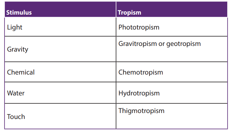
Phototropism is a directional growth depending on the direction of the light source.
Growth towards a light source is a positive phototropism, while growth away from
light is called negative phototropism. It is believed that light destroys auxin where it
strikes the stem, causing an imbalance in which the side of the stem that receives less
light has more auxin. This causes the plant to have elongated cells on the farthest
side from the light. Because more auxin is present, the cells on the darker side are
able to elongate more than the cells on the lighted side, causing the plant to bend
toward the light.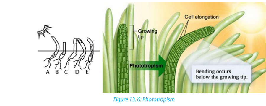
Phototropism is caused by unequal distribution of auxins in the plant stem. There is
more auxin on the side of the stem away from the light. This results in cell elongation,but only on that side. As the cells grow, the stem bends toward light.
b. Gravitropism
When a seed germinates, the young root turns downward regardless of the way in
which the seed is planted. This bending, known as positive geotropism, enables a
plant to anchor itself in the soil. The young stem, which turns upward away from the
earth, is said to be negatively geotropic. The gravity causes auxins to concentrate
on the down side than on the upper side. High concentration of auxins on the
down side of the stem promotes elongation of its cells. Therefore, the stem grows
and bends upward. But high concentration of auxins on the down side of the stem
inhibits cell elongation; therefore, the root grows and bends downward, that is whythe root grows shorter than the stem.
Use of clinostat in tropism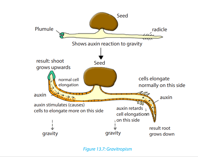
A clinostat is a piece of laboratory equipment with a turntable that allows a plant
placed on it to be exposed to a stimulus such as light or gravity equally on all sides.
As the clinostat is turning, all parts of the plant receive the stimulus equally and
therefore the plant will not bend but will grow straight away. In otherwise theclinostat is used to minimize the effect of the direction of a stimulus.
c. Chemotropism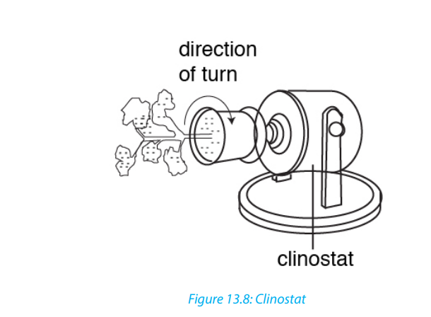
Chemotropism is a growth of a plant or plants parts, navigated by chemical stimulus
from outside of the organism. An example of chemotropic movement can be seen
during the growth of the pollen tube, where growth is always towards the ovules.
Fertilization of flowers by pollen is achieved because the ovary releases chemicals
that produce a positive chemotropic response from the developing pollen tube. It is
different from chemotaxis. The major difference being that chemotropism is related
to growth, while chemotaxis is related to locomotion. For example, the movement ofantherozoids (sperm) in ferns, swim toward the chemicals produced by archegonia.
d. Thigmotropism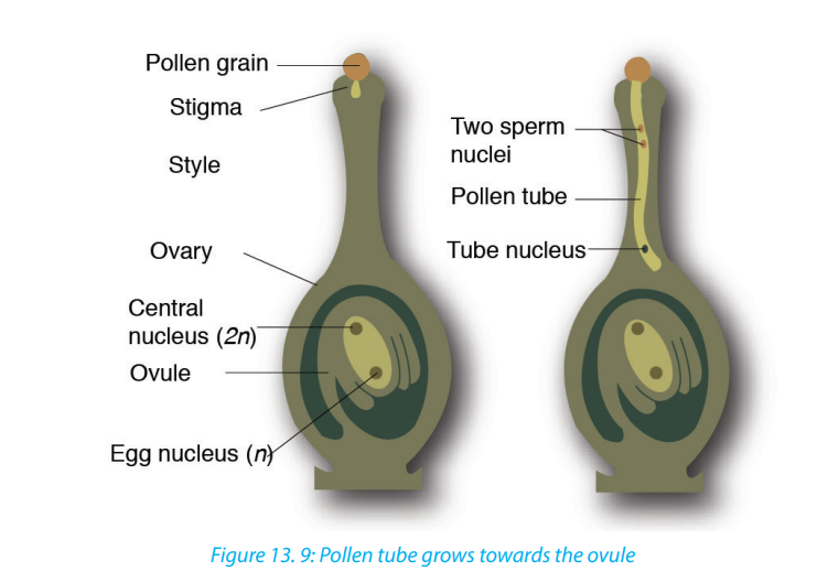
Thigmotropism is a movement in which an organism grows in response to touch or
contact stimuli. Usually thigmotropism occurs when plants grow around a surface,
such as a wall, pot, or trellis. Climbing plants, such as vines, develop tendrils that
coil around supporting objects. Touched cells produce auxin and transport it to
untouched cells. Some untouched cells will then elongate faster so cell growth
bends around the object. Some seedlings also inhibit triple response, caused by
pulses of ethylene which cause the stem to thicken (grow slower and stronger) andcurve to start growing horizontally.
2. Nastic movement
Nastic movements are non-directional responses to stimuli such as temperature,
humidity, light and irradiance. An example of such a response is the opening and
closing of flowers known as photonastic response and the opening and closing of
carnivorous plants known as thigmonastic response. The folding and unfolding of
some sensitive plants like Mimosa pudica when touched, is a nastic response which
protects them from insect damage or water loss during winds. They are named with
the suffix “-nasty” and have prefixes that depend on the stimuli:
– Photonasty (response to light),
– nyctinasty (movements at night or in the dark),
– chemonasty (response to chemicals or nutrients),
– hydronasty(response to water),
– thermonasty (response to temperature).
Self-assessment 13. 6
1. A bean seedling has been placed on horizontal position as shown by the diagrambelow.
a. Illustrate the expected shape of the shoot and that of the root after a week.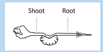
b. Suggest names (of what) for the expectations in the above experiment.
c. Based on the above experiment, draw a diagram illustrating the shape and
size of cells of both upward and downward side of the root.
d. Suggest a technique that can be used to minimize the effect of the stimulus
in this experiment.
2. a. What is the difference between ‘antagonistic’ and ‘synergistic’ when referring
to plant growth substances?
b. What are the two plant growth substances that act antagonistically and
which act synergistically?
3. Copy and complete the following table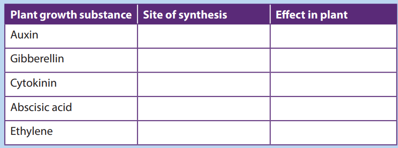
Activity 13.6.2
Most of plants grow toward the sunlight direction. The few which have been
observed respond to external stimuli like touching and temperature.
1. From your experience, brainstorm what will happen to the plant when:
– it is exposed to the direction of the sunlight
– its growing part is exposed to a physical material like a stone
– a living organism touches on it (Mimosa pudica)2. Use internet and textbooks to describe why some plants flourish during long
daylight while others do not?
3. What names can be given to the above processes.
13.6.2. Photoperiodism
The light provides energy that plants need to make its own food. The duration of
daylight affects the plant growth and plant development.
Photoperiodism is a plant physiological response to relative lengths of daylight and
darkness. Photoperiodism affects many plant processes, including the formation of
storage organs, flowering and bud dormancy.
Plants monitor changes in day length with a bluish, light-sensitive pigment called
phytochrome. The alternation of darkness and light triggers the phytochrome to
change from one chemical form to another. By detecting the type and the amount
of phytochrome present, plants determine the length of darkness and light eachday.
One of the effects of photoperiodism is that plants produce fruits at different times
and are classified into three categories:
– Short-day plants (SDP),
– Long-day plant (LDP)– Day–neutral plants (DNP).
– Short-day plants (SDP): they only flower when the days are short and the night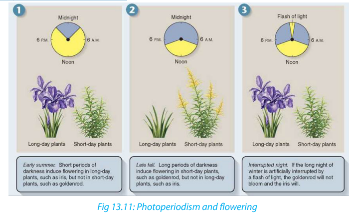
are longer than a certain length. Examples strawberry, blueberry, goldenrods,
cocklebur and soybeans, tobacco are short day plants for flowering.
– Long-day plant (LDP): they produce flower when the period of daylight
exceeds a critical minimum length. Radishes, asters, apple trees, squash trees,
and beets.
– Day–neutral plants (DNP) are not dependent on day length for flowering. They
produce flower regardless of the length of the daylight. Day neutral plants for
flowering include tomatoes, roses, corn, cucumber, carrot, cotton and beans.They can produce fruit throughout the entire growing season.
Self –assessment 13.6.2
1. What role does phytochrome play in photoperiodism?
2. What is the difference between a short-day plant and a long-day plant?3. What could happen if a short-day plant is grown in the long days of summer?
13.7. Metamorphosis and growth patterns in insects and
amphibians
Activity 13.7
From a pond or swamp, collect frog eggs together with water. Keep some eggs in
warm conditions at room temperature between 2 to 30 days.
1. Visit them regularly and note the observation.
2. Use the diagram below and relate your observation from question (1) tothe one given in the diagram.
1. Metamorphosis and growth patterns in animals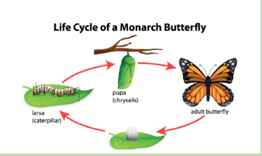
Metamorphosis is a process consisting of changes in body form of a young organism
before it reaches its adult size and becomes sexually mature, for example, the change
from tadpole to frog or from caterpillar to butterfly.
a. Metamorphosis and growth patterns in insects
All insects develop and grow by metamorphosis. Some insects show incomplete
metamorphosis. In incomplete metamorphosis, an immature nymph is hatched
from the egg that looks like the adult, but it is smaller, and its wings and reproductive
organs are undeveloped. It molts several times with each molt, the wings become
larger and more fully formed. The final molt transforms the nymph into an adult
that can reproduce and fly. Insects with incomplete metamorphosis are known
as heterometaboles e.g. weevil, cockroach (Periplaneta americana), Grasshopper;mayflies; dragonflies and termites.
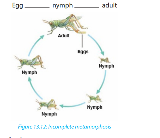
In complete metamorphosis eggs hatch into larvae which are morphologically,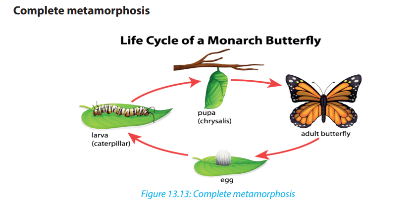
physiologically and behaviorally different from adult (wormlike larva) or caterpillar.
The caterpillar molts several times, when it reaches its full size, it prepares the pupa,
or chrysalis, a hard, sometimes thorny and oval structure. Inside the pupa, the larval
tissues break down and group of cells called imaginal disks develop into wings
and other tissues of the adult: imago stage, the pupa becomes sexually mature.Examples: Butterflies, moths, mosquitoes, beetles, bees, housefly.
Importance of metamorphosis
In life cycle based on complete metamorphosis, the larval and adult stages often
fulfill different functions, live in different habitat and eat different foods. Example:
mosquitoes.
Metamorphosis also enhances insect survival by helping insects survive harsh
period. Example: butterflies (caterpillars feed on leaves, but adult butterflies feed onnectar from flowers).
b. Metamorphosis and growth patterns in amphibians
Many amphibian species like the frog breed in water and their eggs are fertilized
externally. The fertilized eggs hatch into swimming, tailed larvae called tadpoles.
Tadpoles, which usually live in water, look somewhat like small fish. A tadpole has an
oval body, gills for breathing, and a long, muscular tail with fins along the upper and
lower edges for swimming.
Then the tadpole grows legs—the hind legs appear first—and resorbs its tail. It
loses its gills and grows lungs, and the structure of the heart, digestive system, and
skeleton changes. The horny beak and other mouthparts adapted for eating algae
disappear and are replaced by the long, sticky, projectile tongue that helps adult
frogs catch insects. Frogs reach reproductive age anywhere from several months toseveral years after metamorphosing.
Self-assessment 13.7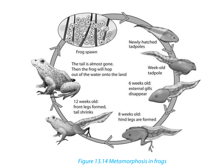
1. What is metamorphosis?
2. Describe the changes that occur during metamorphosis in frog.
3. Discus reasons why complete metamorphosis may have greater adaptive
value for an insect than incomplete metamorphosis.4. Compare metamorphosis of a butterfly and that of a grasshopper.
End of unit assessment 13
1. What do you understand by:a. Dormancy2. a. What factors can allow to measure the growth of a plant?
b. Gravitropism
c. Chemonasty
d. Phytochrome
e. Short-day plants
b. State any four external factors that can affect the growth of a plant.3. a. State any three characteristics of the phytohormones.c. What is meant by parthenocarpy? Give one example of a plant that shows
b. What is phytohormone (s) responsible for:
i. The falling of some plant organs during the stress
ii. The fruit ripening.
iii. Development of the lateral buds
iv. Stem growth and parthenocarpy
this phenomenon
4. A seedling has been grown in an opaque box receiving the light from a single
direction as shown by the diagram below.
a. What will happen on the coleoptile (seedling) as it grows?b. Suggest a name to the phenomenon investigated in this experiment.
UNIT 14: SUPPORT AND LOCOMOTION
UNIT 14: SUPPORT AND LOCOMOTION
Key Unit Competence
Explain and demonstrate modes of locomotion in protists, insects, fish, amphibians,
birds and mammals
Learning objectives
By the end of this unit, I should be able to:– Explain non-muscular movement in amoeba or paramecium.Introductory activity
– Describe support and movement on land.
– Describe skeletal modification in birds.
– Explain how movements and support of fish are brought about in water.
– Explain how support structures are related to the environment of the animal.
– Observe locomotion of animals and identify reasons for their movement.
– Demonstrate the arrangement of muscles in fish.
– Dissect a fish to observe its swim bladder.
– Observe and explain the relationship between muscles, joints and musculoskeletal attachments in fish, birds, amphibians and mammals.
– Compare the flight of birds and insects.
– Compare the jumping movement of grasshoppers and toads/frogs.
– Appreciate the need for locomotion in animals.– Recognize that the types of locomotion of animals depends on their habitat.
Animals have muscles and different types of skeleton.
What might happen if a large animal such as a cow does not have a skeleton.
How that animal would look like? What will happen to animal without skeleton
or muscles? Can you then think about the role of skeleton and muscles in livingorganisms?
14.1. Locomotion and its requirements
Activity 14.1
From your experience and knowledge from books and the internet:
1. Give details about the concept of locomotion
2. How do different animals move?
3. Explain why animals need to move from one place to another?
Living organisms particularly animals need to move from one place to another.
This is known as locomotion which should not be confused with movement which
occurs in plants. Movement is the displacement of part of an organism. Therefore,
movement is a characteristic of all living things.
Locomotion in animals is brought about by the action of muscles on a skeleton. A
skeleton is a rigid framework that maintains shape and supports the internal
organs and provides attachment for muscles, while a muscle is a soft tissue formed
by muscle cells which found in most animals. Each muscle cell contains actin and
myosin proteins that produce a contraction that changes both the length and the
shape of the cell. Thus, muscles function to produce force and motion. In animals
without muscles such as sea sponges, locomotion is brought by mesohyl or cellswhich act as actual muscles.
Depending on the type of animals, three types of skeletons are distinguished.
1. Hydrostatic skeleton
It is mostly seen in invertebrates and earthworm. These consist of fluid filled
body cavity surrounded by antagonistic sets of muscles. Movement results from
compressive contraction action of the contraction of these muscles on this fluid.
2. Exoskeleton
It characterizes the arthropod insects. It is a hard cuticle made of chitin which lies
outside the muscles. It sheds during molting when the organism outgrows it. It does
not grow because it is a dead material.
3. Endoskeleton
It is seen in vertebrates where the bones and cartilages are found within the interior
of the body on which muscles are attached. It is a living tissue and it grows with the
rest of the body.
For efficient locomotion, exoskeleton and endoskeleton provide a system of leversto which muscles are attached.
Types of locomotion
The locomotion can be either terrestrial, aerial or aquatic (swimming). In most
animals, the locomotion is by running, climbing, crawling, swimming, jumping,
gliding, hoping, and flying with aid of limbs or appendages. For animals without
limbs such as snake, its locomotion is by forming its body into zig - zag, gripping the
ground with its undersides and pushing itself forward. For ducks, their movement
in water is by floating. Some invertebrates like roundworms, flatworms, squids,
octopus, and jellyfish without special organs of locomotion are propelled by themuscular contractions.
Advantages of locomotion
Based on the types of locomotion mentioned above, an animal is capable to:
– Escape danger such as fire or predator– Look for food, water and shelter
Reproduce
– Avoid competition with other animals of the same or different species
– Avoid overcrowding which enables offspring to move to another place– Avoid unfavourable condition
Self-assessment 14.1
1. What is locomotion?
2. What are the requirements for locomotion?
3. Given the following animals: Frog, dragonfly, squid, spider, antelope,
kangaroo, fish, grasshopper, bee, duck, worm, zebra, snake, and cow.
Identify those which fly, crawl, hop, and or run/walk.4. Discuss why locomotion is very important in animals?
14.2. Support and locomotion in non-muscular organisms
Learning activity 14.2
From a culture of paramecium:
1. Use a microscope to observe the locomotion in Amoeba and Paramecium
2. From what you have observed identify Amoeba, Paramecium, Euglena andin Trypanosoma moves in relation to their structures / diagrams below
3. Discuss how is locomation performed in those organisms.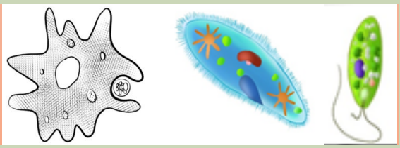
Non-muscular locomotion is identified in living organisms that belong into
Protoctista kingdom. Depending to individuals belonging to Protoctista, locomotionis either amoeboid, ciliary, flagellated or euglenoid type.
Amoeba moves by amoeboid locomotion i.e. by putting out pseudopodia.
Locomotion is not maintained in any particular direction for long. Amoeba is
constantly changing shapes as it changes direction. Amoeboid locomotion is
brought about by cytoplasmic streaming and between a gel and sol state.
These cytoplasmic streaming requires Ca2+ ions and ATP . Amoeboid locomotionis common to all rhizopodes including Amoeba and white blood cells of the vertebrates.
a. Amoeba
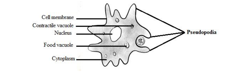
b. Paramecium
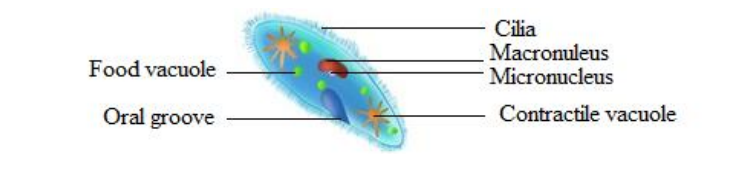
c. Euglena
Paramecium moves by means of cilia and Euglena move by the use of flagella. Cilia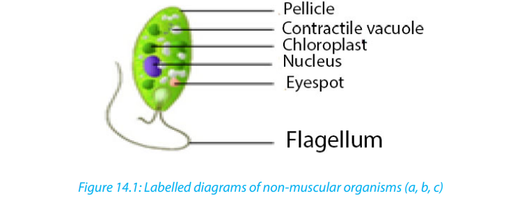
and flagella have similar structure except that cilia are short and many. Both cilia
and flagella consist of fine tubes composed of an extension of plasma membrane.
Euglenas have an intricate cell membrane called a pellicle. The latter is folded into
ribbon-like ridges and each ridge is supported by microtubules. The pellicle is tough
and flexible, letting euglenas crawl through mud when there is not enough water
for them to swim.
During cilia or flagellum locomotion, tubules slide past each other in a movement
similar to that of actin and myosin filaments in skeletal muscles. Hence Ca+
ions and ATP are also required in the ciliary locomotion.Self-assessment 14.2
1. Describe the type of locomotion found in:
a. Amoeba
b. Paramecium caudatum
c. Trypanosoma gambiense
d. Trypanoma vaginalis
e. Giardia intestinalis
2. How do cilia differ from flagellum?
3. Produce picture showing the locomotion of amoeba
14.3. Support and locomotion in fish
Activity 14.31. Observe the freshly collected fish or the figure, to label fins and lateral line.
 2. Dissect a fresh fish or observe the above given diagram. Redraw and showFish like other aquatic animals are adapted to such habitat in terms of locomotion
the swim bladder and the arrangement of muscles
3. If you have a live fish, put it in water and observe its locomotion.
4. From what you have observed, draw and label the external and internal
features that contribute to fish locomotion
due to its structural adaptive features particularly skeleton which gives shape as wellas muscles arrangement and swim-bladder.
Adaptive features of fish for locomotion in water
The streamlined body shape of the fish reduces friction between water and fish.
The body of fish is mostly covered by scales which overlap one another and point
backwards and lie close to the body. The scales are covered by mucus which reduces
the drag.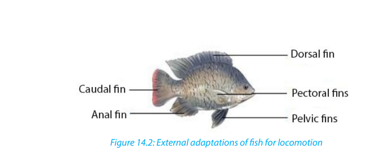
Tail or caudal fin has a large surface area, which increases the amount of water that
is displaced as it provides much of the push during swimming. Paired pectoral and
pelvic fins bring about downward and upward movement. With pectoral fins, the
control of direction of a fish in water is possible whereas the pelvic fins bring about
the balance, preventing diving and rolling. There are also unpaired dorsal and anal
fins for stabilizing the fish and thus preventing it from rolling or yawing.
Fish is also adapted to locomotion in water by its strong tail muscles and highly
flexible vertebrate column which enables the tail to move from side to side againstwater. In addition, inflexible head and neck maintain forward thrush.
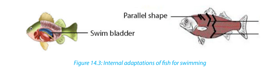
Internally, a fish is adapted to swimming by swim bladder and muscles. Air or gas filled sac called
swim-bladder, outgrowth of the pharynx, helps a fish to change its
buoyancy as it alters the gas pressure in the bladder. So that, it floats at any depth
in water without using its muscles. Swim-bladder also helps fishes to maintain a
density that is equal to that of the surrounding water. Muscles or myotomes /
myomeres (segments or sheets of muscles separated from its neighbor by a sheet of
connective tissue) enable fishes to move in water owing the shapes of muscles that
are located on either side of vertebral column.
Myotomes contribute to the mechanism of swimming by its arrangements. They may
be parallel, V-shape, or W-shape arranged in bundles or blocks that are separated by
myosepta.
Although there are such arrangements, the myoseptal organization and orientation
of fibres is complex. In bony fish, myomeres are V-shaped with new myomeres added
posteriorly. With those myotomes, a fish swim by passing a wave of contracting
muscle from anterior to posterior. Muscles near the head of the fish contract first and
contraction proceeds posteriorly down the length of the fish to the caudal fin. Thus,
a fish moves forward from the contraction and relaxation (antagonistic) of myotome
on either side of the body.
Undulatory swimming of the fish is also powered by the segmental body musculature
of the myotomes. Myotome and myosepta orient more perpendicularly to midline
to push aside. Therefore, the fish can bend laterally. With contraction muscle fibres
shorten by half their length while maintaining volume. Without myosepta, but simply
a series of interconnected muscle fibres, then the wave would be much dampened.
Self-assessment 14.31. How does swim bladder help the fish in locomotion?14.4. Support and locomotion in terrestrial animals
2. Illustrate how the arrangements of fish myotomes contribute to fish
locomotion.?
3. What does it make a fish to move in undulatory propulsion?
4. What are the anatomical structures that give rise to the direction of a fish
and preventing diving and rolling?
Activity 14.41. Through internet observe and think about how locomotion in dogs,All animals living on land move due to the musculoskeletal system. The rigid nature
chicken, frog and earthworm brought about.
2. Make a diagram showing how support and movement of different
animals such as dogs, chicken, frog and worm is brought about on land.
3. Show by using diagrams the relationship between muscles, joints andMusculo-skeletal attachment in mammals, birds, frog and earthworm.
of bone also gives a structure for muscles to pull, by their contraction, to create a
movement as they act as levers. The synovial joints also allow certain movements.
The support and movement differ from specimen to another. Thus, animals can
walk and run on land for moving from one place to another. This is possible by their
endoskeleton and its muscles. By its muscles, flexor (a muscle whose contraction
bends a limb or other part of the body) and extensor (a muscle whose contraction
extends or straightens a limb or other part of the body or any or a muscle that
increases the angle between members of a limb, as by straightening the elbow or
knee or bending the wrist or spine backward); contractions of those muscles cause
the limbs act as levers for them which result to the foot being pressed downwards
and backwards against the ground. For example, flexor and extensor work as
illustrated below: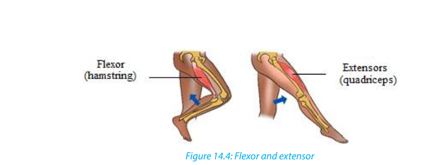
a. Locomotion in quadruped animals e.g. dog and frogs
When a dog walks, its vertebral column remains rigid, and the forward movement is
achieved by the activity of the hind limbs. When its extensor muscle contracts, each
hind limb, acting as a lever, extends and exerts a backward force against the ground,
thrusting the animal forward and slightly upwards. When the flexor contracts, the
limb is lifted clear of the ground and pulled forward. Only one limb is raised at any
one time, the other three providing a tripod of support which balances the rest ofthe body.
Beginning with the left forelimb in a stationary dog, the sequence of leg movement
is as follows when it walks forward: left forelimb-right hind limb-right forelimb-left
hind limb and so on. Such walking in quadrupedal animal is also identified in frogswhen they can walk on land.
b. Running of the dog
As a dog begins to run, it loses its quadrupedal movement which means, it developsa type of movement where the forelimbs move together, followed by the hind limbs.
C. Walking in bipedal animal e.g humans
Humans are bipedal, they walk on two legs. When standing upright, the weight is
balanced over the two legs. When a stride is taken by the right leg, the heel is raised
first by the contraction of the calf muscles. As this occurs, the weight of the body is
brought over the left foot which is still in contact with the ground and acting as theprop for the rest of the body.
When the right leg extends the heel is the first part of the foot to touch the ground.
The weight off the body is gradually transferred from the left side to a position over
the right heel and then the body continues to move forward, over the right toes,
backward pressure against the ground generally being exerted through the right
big toe. Like human does, a bird also can walk on ground through the movement ofcontractions of its leg muscles particularly flexor and extensor.
d. Crawling of earthworm
Earthworms are organisms having hydro skeleton with soft-bodied animals due to
fluid secreted within the body and surrounded by the muscles of the body wall.
They are capable to move by aid of their muscles. These muscles are not attached to
any structures and thus can pull against each other. The combined effect of musclecontraction and fluid pressure serves to maintain the shape and form of the animal.
Generally, there are two muscle layers, longitudinal in which muscle fibres are
arranged parallel to the long axis from one end of a segment to another and circular
with muscle fibres arranged in concentric circles to the circumference of the worm.
When those muscles act antagonistically against each other, locomotion is achieved.
The fluid which acts as pressurisable hydrostatic skeleton contained in body cavity
or coelom presses against the muscles which in turn are able to contract against the
fluid. Earthworm movement is also helped by bristles like setae or called chaetae(hair like structures on ventral surfaces) which anchor the worms to the substrate.
Contraction of the circular muscles makes the worm thinner, but because liquid is
essentially incompressible (and so maintains a constant volume) and the increase
in pressure forces the liquid outwards, stretching the worm, so the worm becomes
longer and thinner. Contraction of the longitudinal fibres shortens the worm, former
the coelomic liquid out to the sides and making the worm fatter. If the body is
segmented, then such pressure is localized and only certain segments will move orchange shape.
Self-assessment 14.4
1. What are the main muscles that contribute to locomotion in mammals,
amphibians and birds?
2. Draw an earthworm and illustrate the muscles that contribute to its
locomotion.
3. What type of skeleton system found in mammals, birds, amphibians and
annelids?
4. . Illustrate how flexor and extensor muscles contribute to lifting up a leg inhuman being.
14.5. Flight through air by birds and insects
Learning activity 14.5
Make a research on internet as well as books and do the following:
1. Observe pictures below (Figure 14.6) related to birds and make a description
of skeletal modification in birds. Illustrate the skeletal modification in birds
2. Draw a bird and show by using arrow the structures that enable a bird to fly
3. How will the external features of birds will behave when flying in high or
low atmospheric pressure
4. Make a table illustrating how does flight of birds and insects differ and
similar
5. Observe and compare the flight of birds and insects
Bird can fly either by flapping their wings or gliding by spreading its wings. Like
in animals moving on land, locomotion by flying in birds is brought about by the
action of flexor and extensor muscles as well some other structures given diagrambelow like pectoralis major, pectoralis minor and keel of sternum.
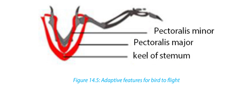
Based on the above diagram, wings move down by the contraction of pectoralis
major and then move up under the contraction of pectoralis minor.
Adaptive features of birds for movement in air
A number of features enable birds to aerial locomotion. Those features include
body shape, modified limbs, and modification in internal organs particularly bones.
The body of bird is highly streamlined and covered with light feathers that overlap
backwards thus reducing air resistance during flight. Those light features thatincrease the surface area of wings without increasing weight.
Differently from quadrupedal animals, birds are adapted to flying by modification
of fore limbs into wings. Such modification goes with a well-developed or large keel
sternum that provides large surface area for the attachment of the flight muscles
namely major and minor pectoral muscles which give the power to flap the wings in
flight. Also, birds have hollowed bones making the body light and vertebrae of trunk
are fused. As flight requires much energy, birds are adapted to that by an efficient
breathing system with air sacs attached to the lungs necessary to provide oxygenfor respiration and to remove the resulting carbon dioxide.
Other adaptations include a high metabolic rate for providing the high amount of
energy required, an efficient circulatory system necessary for transporting both the
nutrients and respiratory gases at speed related with the body needs, a high red
blood cell count for efficient oxygen transport, and a keen eye sight to enable themto judge distances correctly especially on landing.
Self-assessment 14.51. What are the muscles that enable the flight in birds?14.6. Hopping locomotion in grasshoppers and toads
2. Describe how bird skeleton contributes to its flight?3. Describe how birds are adapted to flying.
Activity 14.6
Use a collecting net to catch a grasshopper and toad from school compound. Put
them down on cemented ground for observing them very carefully when they
make a jump and then answer to the following:1. Identify and describe anatomic structures that enable grasshoppers to jumpSkeletal muscles such as extensor and flexor that occur in pairs are often antagonistic.2. Illustrate how legs’ muscles behave when they are resting and or jumping
With such antagonistic behaviour, when one contracts the other relaxes to producecontrolled movement in the opposite directions.
a. Locomotion of grasshopper
Insects have a skeleton which is on the outside of the body called an exoskeleton.
They can walk on the land but they are mostly adapted to hopping owing to their
muscles which are inside the hard shell as well as skeleton system. The muscles
which make them capable to move are flexors and extensors which are antagonists,
attached to internal surface of exoskeleton and the rear or back legs of a grasshopper
which are long and muscular, adapted for hopping. Additionally, there are two main
muscles inside are the extensor tibiae muscle which contracts to extends the leg,
and the flexor tibiae muscle which contracts to flex the leg as illustrated in figure
below. Those muscles pull on tendons which are attached to the tibia on either side
of the joint pivot.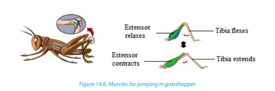
The back legs are much longer than the others for helping in hopping. With those
long legs, grasshopper is capable to make high jumping distance. As illustrated
above, flexor muscles bend a joint whereby extensor ones straighten it. The flexor
muscle contracts and the lower leg is pulled towards the body. Thus, the hind leg
is folded in a Z shape and ready for jumping. Being in resting or sitting position,
the extensor muscle contracts which enable then the legs jerk or move very quicklybackwards propelling the grasshopper.
b. Locomotion in toads and frogs
On land, frogs and toads move by hopping (going from place to place).
– When a frog is at rest, the hind legs are folded up in the shape of a letter Z.
– When it hops, the legs are quickly straightened out, lifting the animal of the
ground.
– The fore-limbs are used as shock absorbers on landing and they also prop up
(to give support) the front end of the body when the animal is at rest.They also hope but do not travel as high as far as a frog does at each hop.
Self-assessment 14.6
1. What are the muscles that contribute to high jumping in a grasshopper?
2. How do muscles (flexor and extensor) behave when toads and grasshopper
are resting?3. Draw a leg of grasshopper and the one of toad when are jumping
End of unit assessment 141. Describe ways of locomotion in Amoeba, Paramecium, Euglena and in
Trypanosom
2. Produce a cartoon showing different adaptive features of fish for aquatic
locomotion
3. Describe how different fish fins contribute to locomotion and balance
4. Describe how the movements and support of fish in water do they occur?
are brought in water
5. Show by diagrams the relationship between muscles, joints and
musculoskeletal attachment in mammals, birds, frog and earthworm.
6. Describe how flexor and extensor muscles work to enable the locomotion
on land, water and in air
7. What are the features that enable aerial locomotion in animals?8. Describe how a grasshopper and toad is adapted to jumping
UNIT 15: CLASSIFICATION AND PATTERNS OF DISEASE
UNIT 15: CLASSIFICATION AND PATTERNS OF DISEASE
Key Unit Competence
Describe the social factors that affect good health and apply knowledge gained in
familiar and unfamiliar contexts.
Learning objectives– By the end of this unit, I should be able to:Introductory activity
– Explain what is meant by health and disease.
– Identify different categories of disease and give an example of each.
– Explain the theory of the disease and the causes, sources, transmission,
symptoms and controls of the disease.
– Discuss how global patterns of disease are studied.
– Analyze and interpret records from a given hospital to identify diseases as
endemic, epidemic or pandemic.
– Apply knowledge gained to classify common diseases.
– Appreciate the importance of germ theory of disease by showing that thedeath rate related to infections is greater than those caused by accidents.
a. Suggest measures to be taken for addressing issues related to eating without15.1. Germ theory of diseases
washing hands.
b. Discuss on different communicable diseases got from eating without washinghands.
Activity 15.1
Discuss the following questionsa. What are the causes of death?The germ theory states that many diseases are caused by the presence and actions
b. Why it is difficult to eradicate malaria in Rwanda?
of specific microorganisms within the body. In 1677, Antoni van Leeuwenhoek was
the first to observe microorganisms in the droplets of water. But he did not make
the connection with disease. Later, Spallanzani and Louis Pasteur observed germs
in the blood of people suffering from disease. They suggested that the germs were
an effect of the disease rather than the cause. The observations and actions of
Ignaz Semmelweis, Joseph Lister and John Snow were a great contribution to the
acceptance of germ theory. However, the laboratory works of Louis Pasteur in the
1860s and Robert Koch in the following decades, provided the scientific proof for
germ theory. Their works opened the door to research related to the identification
of disease-causing germs and potential life-saving treatments.
15.1.1. The work of Louis Pasteur and Semmelweiss
The work of Eduard Jenner and Ignas Semmelweiss showed that infectious diseases
maybe caused by an infectious agent or germ. This was accepted as the germ theory
of disease for a very long time. The work of Robert Koch and Louis Pasteur led to a
wide acceptance of the germ theory
Pasteur made a number of important steps forward. He indicated that fermentation
is the result of the action of microorganisms (Yeast) on sugar. Huge number of people
at the time believed that living things could arise spontaneously from non-living
things and this theory is known as spontaneous generation. To reject this theory,
he showed that if broth is boiled in a sealed container, it would stay clear, but once
he added material which had been exposed to the air, microorganisms grew in the
broth. Finally, he designed a series of experiments using swan necked flasks which
showed once and for all that any microorganisms which appear in boiled broth
come from the air not arise spontaneously from nonliving organism such as broth.
In 1845-1846, Pasteur found a way of avoiding the disease of silkworms by observing
the infected eggs under microscope and thereby saving the silk industry. This was
the first clear evidence of microorganisms causing disease. Pasteur even developed
vaccines against a number of these diseases.
15.1.2. Summary of the contributions of Louis Pasteur in microbiology
and medicine
The contribution of Louis Pasteur in microbiology and medicine also include:– The fight against spontaneous generation theory15.1.3. The Germ Theory and Koch’s Postulates
– The technique of sterile culturing of microorganisms
– The technique of fermentation and conservation of drinks. This technique is
known as Pasteurization.
– The technique of antiseptic surgery to prevent contamination of wounds
during the surgical operations in hospitals.
Louis Pasteur developed the germ theory of disease which postulates that allcontagious and infectious diseases must be caused by pathogenic microorganisms.
Diseases can be spread by air, water, food, and human as well as animal vectors. In,
an english physician called John Snow (1854) and a German microbiologist called
Robert Koch (1884) found a relationship between polluted water and disease. Robert
Koch, has isolated the bacterium Vibrio cholera, the cause of cholera from Elbe River
water to provide the relationship.
Koch went on to formulate an established set of procedures to isolate and identify
the causative agent of a particular microbial disease. The following four steps, which
are still used today, are known as Koch’s Postulates:
Postulate 1: A specific organism must always be observed in association with the
disease.
Postulate 2: The organism must be isolated from an infected host and grown in
pure culture in the laboratory.
Postulate 3: When the organism from the pure culture is inoculated into a susceptible
host organism, it must cause the disease.
Postulate 4: The infectious organism must be re-isolated from the diseased organism
and grown in pure culture.
Self-assessment 15.1
1. What are the Koch’s Postulates?
2. Explain the theory of spontaneous generation.
15.2. Classification of diseases
Activity 15.2
Use the Knowledge gained to answer to the following questions:1. Propose two infectious diseases and for each disease, give:2. Does being healthy means just the absence of the disease? Explain.
a. their causal agents
b. causal agents’ type
c. their symptoms
d. their methods of prevention
e. Treatment
The following are the meaning of disease, signs and symptoms:– Disease is the disruption of normal body function.15.2.1. Infectious diseases
– Signs are indications of a disease that can be observed by examining the
patient.
– Symptoms are indications of disease perceive only by the patient.
The normal functioning of the body is disturbed, when the body is infected. Many
types of diseases are broadly divided into two categories: Infectious diseases andnon-infectious diseases
Infectious diseases are caused by microorganisms known as pathogens which
may include viruses, bacteria, fungi and protozoa. Those diseases are called
communicable diseases. as they can be transmitted from one person to another.
They include cholera, malaria, typhoid, HIV and AIDS…Malaria is one of the most
dangerous infectious diseases, endemic in Latin America, Africa and South-East Asia.
Some infectious diseases can also be from animals to humans.
The following are some technical terms used when discussing about infectious
disease.
– Aetiology: The study of the cause of disease.
– Epidemiology: The study of all the factors that contribute to the appearance
of a particular disease
– Causative agent: The organism which causes the disease
– Vector: An organism which carries the causative agent of the disease from
one person to another or from infected animal to human.
– Incubation period: The period of time between the original infection and the
appearance of signs and symptoms.
– Infective period: The time during which a person is capable of passing the
disease on to another person.
– Carrier: The person who has been infected but develop no signs and symptom,
the carrier can pass the disease on to another person
– Prevention: Measures taken to prevent diseases.
– Treatment: Measures taken to cure diseases Antibody: Is a protein produced
by the body’s immune system when it detects harmful substances called
antigen.
– Antigen: Is any substance that causes your immune system to produce
antibodies against it.
– Host: A host can be anything living organism ion which pathogens can survive
– Hygiene: Practices that help to maintain health and prevent the spread of
diseases
– Immunity: Is the ability of the body to resist to infections.
a. Important advances in the work against infectious disease
Table 15.1: The chronology of some of the most important advances in the work against
infectious disease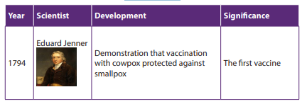
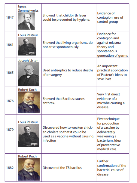
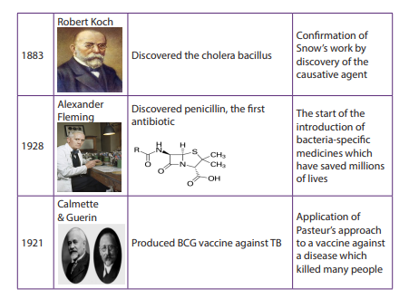
b. Some groups of communicable diseases
– Bacterial diseases: these are diseases caused by bacteria. They include
cholera, typhoid, tetanus, tuberculosis, etc.
– Viral diseases: these are diseases caused by viruses. They include AIDS, polio,
measles, Ebola, etc.
– Protozoan diseases: these are diseases caused by protozoa. They include
malaria, sleeping sickness, trichomoniasis, etc.
– Fungal diseases: these are diseases caused by fungi. They include candidiasis,
athlete’s foot, ring worms, etc.
– Worm diseases: these are diseases caused by worms. They include
elephantiasis, bilharzias, etc.
– Sexually transmitted diseases: these are diseases transmitted through
sexual contact. They include HIV-AIDS, syphilis, gonorrhea, etc.
c. Transmission of infectious diseases
Pathogens can spread when you have direct contact with an infected person. For
example, if you have contact with the person’s blood, body fluids or open wounds.
Pathogens can also be spread through contaminated food, water or air. Infectedanimals can spread pathogens to people.
The following conditions lead to the spread of an infectious disease:
– A pathogen which causes the disease.
– A source which is an infected organism.
Mode of transmission a pathogen must be able to enter the body of the new host
to cause an infection. Infectious diseases follow a pattern of development from the
time of infection. The pattern of development has five distinct periods, as described
in table below:Table 15.2: The pattern of development’s distinct periods for infectious diseases
d. Epidemiology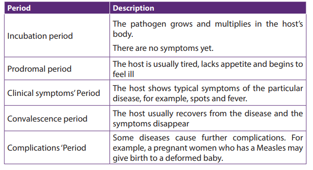
The study of patterns of disease and of the various factors that affect the spread of
disease is called epidemiology.
Epidemiologists try to discover the factors that cause a disease and develop methods
to prevent its spread. The main clue they use come from data about the number of
people in a particular area affected by specific diseases, and the number of death
The data are commonly expressed as incidence or morbidity and mortality rates.
The incidence rate is the number of new cases of disease in a given populationoccurring during a specific period (a week, month or a year). It is calculated as:
To find how many cases of a disease are new, this calculation requires information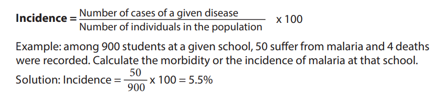
about the prevalence rate. This is the total number of individuals infected in a
population at any one time.
The mortality rate of a disease may be estimated for a whole population irrespectiveof whether they have the disease or not.

It may be calculated by using only those people who have disease
To make fair comparisons between different populations with the same disease,
epidemiological information is usually adjusted. For example, the mortality rate
among those with a particular disease is usually expressed as a percentage or ratio
per year. Hence, if in one year 7500 people in a given area die as a result of AIDS and
the total number of population infected was 30 000, the mortality rate would be25% for the rate.
Epidemiological studies are used to identify whether a disease is endemic, epidemic,
or pandemic:
– Endemic disease is a disease that is always present in a people e.g. malaria in
tropical Africa.
– Epidemic disease is a disease that spreads rapidly, suddenly, and unexpectedly
to affect many people. e.g. cholera in refugees’ camp. Pandemic disease is a
disease that affects people over very large area, such as a continent or eventhe whole world e.g. AIDS and TB are pandemic at present
15.2.2. Non-infectious diseases
These diseases are also called non-communicable diseases. They cannot be
transmitted from one person to other examples: albinism, kwashiorkor, cancer,diabetes, etc
Table 15.3: Six groups of non-communicable diseases
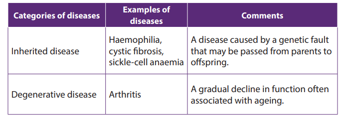
Lesson self-assessment 15.2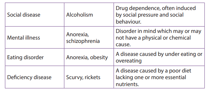
1.Answer by true or false
a. Epidemic disease is a disease that is always present in population.
b. The diseases that transmitted among people by pathogens are called
transmissible diseases
c. The study of patterns of disease and of the various factors that affect the
spread of disease is called epidemiology.
d. Cholera is infectious disease
e. Malaria is non-infectious disease whose vector is mosquito.2. Distinguish between morbidity and mortality
15.3. Common infectious diseases
Activity 15.3
Choose in the following list the infectious diseases and explain why:
Cholera, typhoid, Alcoholism, tetanus, tuberculosis, AIDS, Haemophilia, polio,
measles, Ebola, malaria, Anorexia, obesity, sleeping sickness, trichomoniasis,
sickle-cell anemia, candidiasis, athlete’s foot, ring worms, elephantiasis, bilharzias,syphilis, gonorrhea, cystic fibrosis, Arthritis, Anorexia, schizophrenia.
15.3.1. Measles
Measles is a contagious acute viral disease with symptoms that include a bright red
rash of small spots that spread to cover the whole body. Small white spots, known
as Koplik’s spots, appear in the mouth on the inside of the cheeks a few days beforethe rash appears and can be used in diagnosis.
Table15.4: The features of measles.
Failure to eradicate measles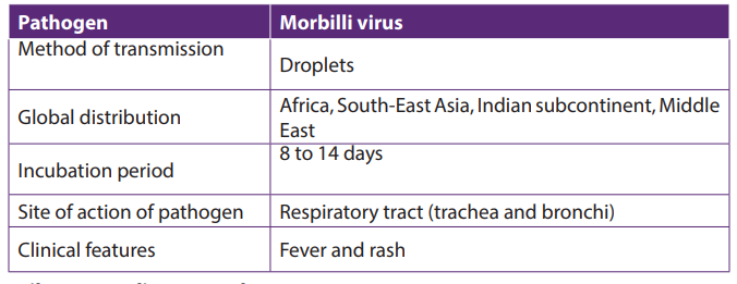
– Incubation period is short hence it is difficult to identify and isolate before
they become infectious
– It is transmitted through a carrier mother to healthy children hence it is hard
to eradicate.
– It targets young children who like playing together. This makes ease the spreadof the disease.
15.3.2. Typhoid
a. Causal agent of typhoid
Typhoid is waterborne disease caused by Salmonella typhus, a Gram negative
bacterium. The bacteria are derived from the feces of a patient. It has high infectivity
as low dosage of organisms is only needed for typhoid to spread. Common sourcesof typhoid infection are contaminated water, milk and food.
b. Development of typhoid illness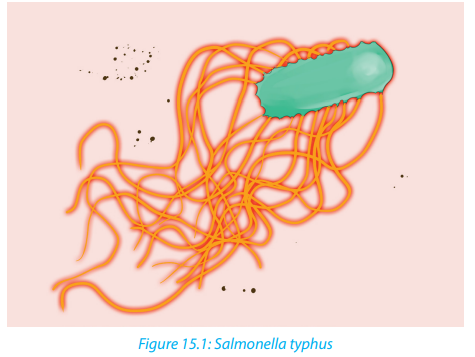
The incubation period is of ten days. After this time, the germs enter the bloodstream
and the patient develops the following symptoms: Headache, Muscular pains, Fever
reaching its peak after about 1 week, faint rash may appear, diarrhea in the second
week, mental confusion, etc. The third week shows the peak of the illness and thepatient may die if not treated.
c. Treatment and prevention of typhoid
The disease had a 20% fatality rate before the use of antibiotics. Chloramphenicol
and Ampicillin are effective and reduce fatality rate from 1 up 5%. Today, Ciprofloxine
is used as another antibiotic. The two most important preventive measures are;
proper treatment of sewage and disinfection of water supplies. Hygienic measures
in the food trade and at home, and control of flies, which can transfer fecal materialto food. Vaccine is formed by a polysaccharide from the capsule of the bacteria.
15.3.3. Cholera.
Cholera is a good example of a waterborne disease. It is endemic in parts of Asia,
particularly India. The organism which causes cholera is a comma shaped motile
bacterium called Vibrio cholerae.a. Transmission and symptoms of cholera
The main source of infection is water contaminated by feces with Vibrios. It is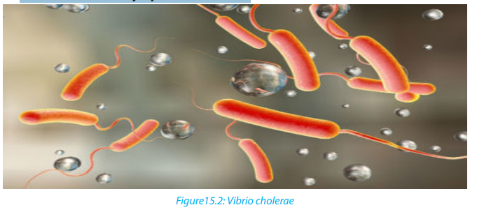
estimated that only about one infected person in 50 develops the disease, the rest
being carriers. Drinking contaminated water, or washing food or utensils in it, is the
most common means of transmission. Direct contamination of food with feces as a
result of poor hygiene is also possible, house flies being the main vector in this last
case.
b. Signs and symptoms of cholera
Vibrio cholerae multiply in the intestine, releasing a powerful toxin which results inviolent inflammation of the intestine and production of the watery diarrhea.
The main sign of the disease is severe diarrhea due to irritation of the bowel by toxins
from the Vibrios. The liquid of the feces is so profuse and cloudy like “rice water”.
Abdominal pain and vomiting are also common. Dehydration is rapid and quickly
results in death unless rehydration treatment is given. Fever is absent; in fact, theskin feels deathly cold and often damp.
Table15.5: The features of cholera.
c. Treatment of cholera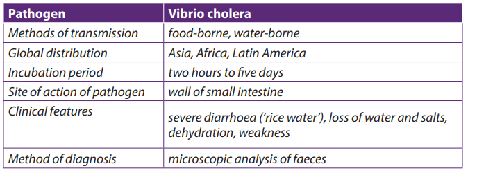
The primary cause of death from cholera is dehydration i.e. loss of water with its
minerals salts. For that, it is obligatory to rehydrate with oral serum which contain
mineral salts and sugar.
The loss fluid may be replaced by administration of a drip food into a vein.
Various antibiotics, such as tetracycline’s and chloramphenicol, are used to treatcholera. Chloramphenicol is effective against tetracycline-resistant Vibrios.
d. Prevention of cholera
– Use clean drinking water,
– Proper treatment of sewage and sanitation
– High standards of public and personal hygiene, particularly in relation to food
(such as washing hands after defecation)
– Health education
– Vaccination is recommended for people visiting areas where cholera is
endemic and for those living in such areas. But the vaccine lasts few months.– Isolation of patients and hygienic disposal of feces and vomit from patients.
e. Failure to eradicate cholera
– Vaccination is not very effective
– It is a waterborne disease i.e. transmitted through contaminated water– Poor sanitation condition in camps.
15.3.4. Tuberculosis (T.B)
TB spreads when infected people with the active form of the illness cough or sneeze
and the bacteria are carried in the air in tiny droplets of liquid.
a. Causal agent of tuberculosis
Tuberculosis is caused by bacterium called Mycobacterium tuberculosis, first
discovered by Robert Koch in 1882. It is sometimes referred to as the tubercle bacillus,
bacilli being rod-shaped bacteria. The common form is pulmonary T.B which infectsthe lungs, although other organs may be affected.
Two strains of the bacterium may cause the disease, the human and the bovine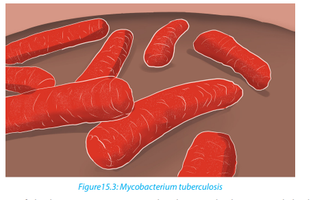
forms. The latter can be present in cattle and can enter the milk of cows. It is veryresistant and can remain alive for long time in milk products as well as in durst.

b. Transmission of tuberculosis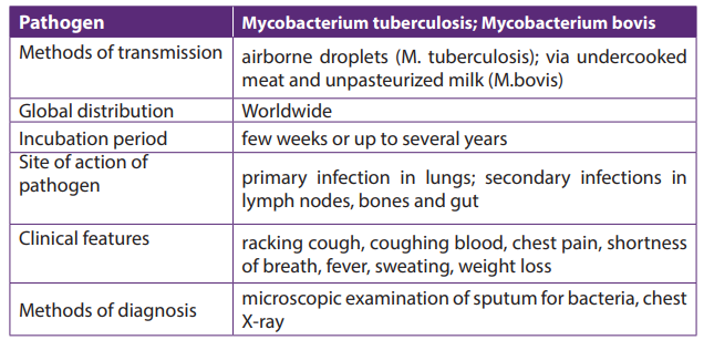
Tuberculosis is mainly airborne disease. The infection is done through the droplets
from the patient. It is much less infectious as it requires prolonged contact between
people, poor ventilation and overcrowded living conditions. In addition, TB is an
opportunistic infection, striking many people with a depressed immunity.
c. Signs and symptoms of tuberculosis
The disease is frequently characterized by vague symptoms such as: loss of appetite;
loss of weight; excessive sweating; coughing, appearance of blood in the sputum,
pains on the chest, shortness of breath (case of lung tuberculosis).
d. Treatment and prevention of tuberculosis
Vaccine against the disease has been developed by Albert Calmette and
Camileguerin). Antibiotics such as rifampicin, isoniazid and streptomycin are usedto treat tuberculosis.
e. Failure to eradicate tuberculosis
– Patients can carry pathogen and infection without showing symptoms.
Therefore, they are difficult to identify due to a long period of incubation
– Germs of tuberculosis can survive longer in the house dust
– The disease is related to poverty where many people share the same room and
have malnutrition.
– The disease is associated with AIDS that reduced the body immunity
– Long period of treatment (6-8 moths), hence patients give up when not yet
fully healed. The pathogens then form endospores that resists to medicines.
– The disease is also spread through milk from infected animals. Tuberculosis isan airborne disease i.e. spread in air
15.3.5. Malaria.
a. Causal agent
Malaria is caused by four species of plasmodium: Plasmodium falciparum, P. vivax, P.
ovale, and P. Malariae.The parasite is transmitted by the bite of female mosquitoes
(the vector) belonging to the genus Anopheles.
b. Symptoms
Malaria is characterized by severe chills, fever, sweating, fatigue and great thirst.
Victims die of anemia, kidney failure or brain damage.
c. Occurrence of malaria
The disease now occurs in tropical and subtropical regions of the world, and its
distribution is limited by conditions of the development of the mosquito vectorsuch as temperature and altitude.
Malaria is endemic in tropics because:
– Tropical climate provides the best breeding and living conditions for the
Anopheles mosquito which transmits malaria
– The Anopheles cycle requires areas of stagnant water, common within tropics
– In the tropical areas, there is presence of bushes or abundant vegetation which
makes suitable habitat for mosquitoes
– Plasmodium needs temperature in excess of 20ᵒC for it to complete its cyclewithin the mosquito.
Table15.7: The features of malaria.
d. Eradication and prevention of malaria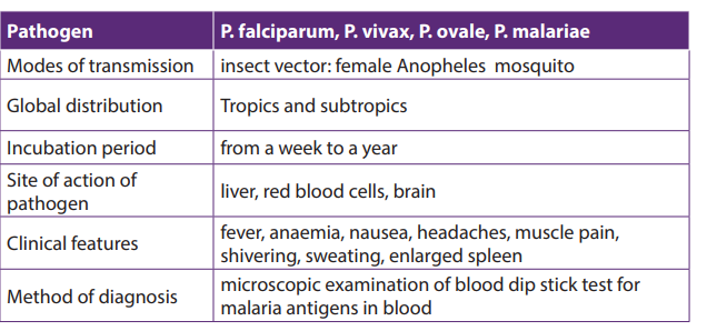
– Drainage of stagnant water: The larval stages of the mosquito live in stagnant
water, so drainage removes breeding sites.
– Destruction of the adult mosquitoes by spraying insecticide.
– Clean bushes nearest houses where mosquitoes lay eggs
– Sleeping under mosquito net during the night
e. Failure to eradicate malaria
– There is no effective vaccine against malaria
– The pathogens are transmitted by mosquitoes which are difficult to eradicate.
– The plasmodium has become resistant to different anti-malarial drugs
– Ignorance of some people toward the disease and how it is spread.
15.3.6. Smallpox
a. Cause of smallpox
Smallpox was a horrible viral disease caused by Variola virus (DNA virus), a pox virus.
It was a highly infectious disease transmitted by direct contact and it affects therespiratory passage.
b. Signs and Symptoms of smallpox
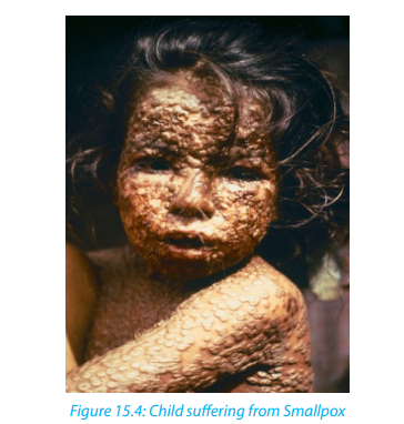
The following are Signs and Symptoms of smallpox:
– Obvious symptoms of the disease were red sports on the face, trunk, and
extremities that change to pea-sized blisters and became filled with pus. Highfever and generalized aching.
c. Modes of transmission
This disease spread through droplet infection (contagion possible via wounds in
skin, clothing, bedding and dressing)
d. Prevention and treatment
Large populations were vaccinated through Ring vaccination and people with the
disease were isolated.
The eradication of Smallpox was successful because:
– The smallpox virus is stable. so the same vaccine could be used
– The smallpox virus does not linger in the body after infection, nor does it infect
other animals, so it cannot remain hidden anywhere.
– The vaccine used was highly effective and easy to administer by scratching
technique.– It was easy to identify people with the disease.
Table15.8: The features of Smallpox
15.3.7. Tinea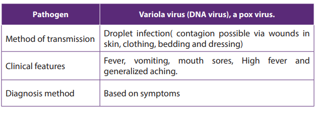
Tinea is a skin infection due to a fungus. Often, there are several patches of ringworm
on the skin at once. Tinea is also known as Ringworm.
a. Cause of Tinea
– Tinea is caused by a tiny fungus known as dermatophyte. These tiny organisms
normally live on the superficial skin surface, and when the opportunity is right,
they can induce an infection.
– The disease can also be acquired by person-to-person transfer usually via
direct skin contact with an infected individual. Animal-to-human transmission
is also common.
– Ringworm commonly occurs on pets (dogs, cats) and the fungus can be
acquired while petting or grooming an animal.
– Ringworm can also be acquired from other animals such as horses, pigs, ferrets
and cows.
– The fungus can also be spread by touching inanimate objects like personal
care products, bed linen, combs, athletic gear, or hair brushes contaminated
by an affected person.
Individuals at high risk of acquiring ringworm include those who:
– Sweat excessively, as sweat can produce a humid wet environment where
the pathogenic fungi can thrive.
– Wear tight, constrictive clothing with poor aeration.
– Have a weakened immune system
– Live in crowded, humid conditions.– Participate in close contact sports like soccer, rugby
b. Sign and symptoms of Tinea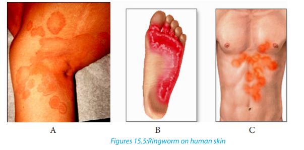
The following are Sign and symptoms of Tinea:
– Enlarging raised red rings with a central area of clearing (ringworm).
– The edge of the rash appears elevated and is scaly to touch.
– Sometimes the skin surrounding the rash may be dry and flaky.
– There will be hair loss in areas of the infection.c. Diagnosis
Superficial scrapes of skin examined under a microscope may indicate the presence
of a fungus. Diagnostic method called KOH Test is used, where the skin scrapings
are placed on a slide and immersed on a drop of potassium hydroxide solution
to dissolve the keratin on the skin scrapings thus leaving fungal elements such
as hyphae, septate or yeast cells viewable. If the skin scrapings are negative and
a fungus is still suspected, the scrapings are sent for culture. Because the fungus
grows slowly, the culture results do take several days to become positive.
d. Prevention
Basic prevention measures include:
– Serious washing of hands after handling animals, soil, and plants.
– Avoiding touching characteristic lesions on other people.
– Put on loose-fitting clothes.
– Promoting good hygiene when participating in sports that involve physicalcontact with other people.
e. Treatment
Application of topical antifungals creams to the skin. In extensive or difficult
cases, systemic treatment with oral medication may be required. Among theavailable prescription drugs are tolnaftate, terbinafine, naftifine, itraconazole.
15.3.8. Hookworm
Hookworm is a humans’ intestinal parasite. The adult worms and their larvae can
cause intestinal disease in which they live.
a. Cause of hookworm disease
The hookworm is caused by two main species of hookworm infecting humans which
are: Ancylostoma duodenale and Necatora mericanus
b. Method of transmission
If an infected person defecates near bushes, in a garden, or field, or if the feces from
an infected person are used as fertilizer, eggs are deposited on soil. They can then
mature and hatch, releasing larvae (immature worms). The larvae mature into a form
that can penetrate the skin of humans. Hookworm infection is transmitted primarily
by walking barefoot on contaminated soil.
c. Signs and symptoms of hookworm
– Itching and a localized rash are often the first signs of infection. These
symptoms occur when the larvae penetrate the skin.
– A person with a light infection may have no symptoms but a person with a
heavy infection may experience abdominal pain, diarrhea, loss of appetite,
weight loss, fatigue and anemia (pale skin etc.) and protein deficiency caused
by blood loss, constipation, congestive heart failure, excessive coughing
during larvae migration, stomach or chest pain, vomiting, weight loss.
– The physical and cognitive growth of children can be affected. There is a
decreased rate of growth and mental development in children (caused by
protein and iron deficiency)
d. Diagnostic method
Taking a stool sample and using a microscope to look for the presence of hookworm
eggs.
e. Prevention of hookworm
Avoid walking barefoot in areas where hookworm is common and where there may
be fecal contamination of the soil.
Avoid skin-to-soil contact and ingesting such soil.
Avoid defecating outdoors or using human feces as fertilizer, and by effective sewage
disposal systems.
f. Treatment for hookworm
Hookworm infections are generally treated for 1-3 days with medication prescribed
by your health care provider. The drugs are effective and appear to have few side
effects. Iron supplements may be prescribed if you have anemia.
g. Preventive treatment
– In developing countries, groups at higher risk for soil-transmitted infections
Biology Senior Four Student’s Book 257
are often treated without a prior stool examination. Treating in this way is
called preventive treatment.
– School-age children are often treated through school-health programs and
preschool children and pregnant women at visits to health clinics.h. Life cycle of Hookworm
Self-assessment 15.3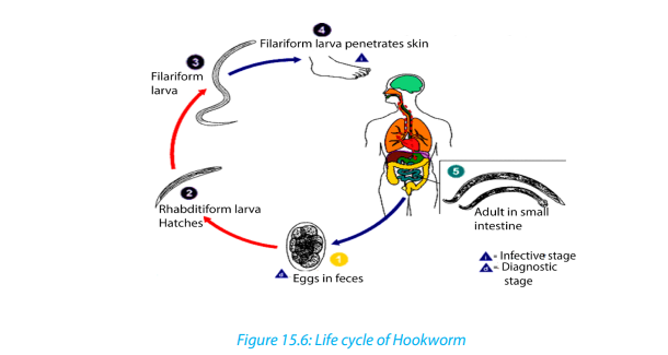
1. Which of the following diseases is transmitted by an insect vector?
a. Cholera
b. HIV/AIDS
c. Malaria
d. TB
2. What are the ways in which cholera is transmitted from person to person?
3. Explain why there is such a high risk of cholera following natural disasters such
as earthquakes, hurricanes, typhoons and floods.
4. Explain why there is a high death rate from TB in countries with a high proportion
of the population who are HIV-positive.
5. TB is an opportunistic infection. Why?
6. Describe how malaria is transmitted.
15.4. Health and community: criteria for good housing
Activity 15.4
Housing quality is associated with morbidity from different factors. State any
three factors
Housing refers to houses or buildings, accommodation of people. It is an importantdeterminant of health, and substandard housing is a major public health issue.
The public health community is aware of the importance of social determinants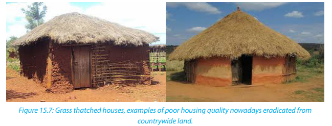
of health (including housing) in recent years. Yet defining the role of public health
practitioners in influencing housing conditions has been challenging. Responsibility
for social determinants of health is seen as lying primarily outside the scope of public
health. The quality and accessibility of housing is, however, a particularly appropriate
area for public health involvement.
An evolving body of scientific evidence demonstrates solid relations between
housing and health. The public health community is developing, testing, and
implementing effective interventions that yield health benefits through improved
housing quality.
Criteria for good housing
– Good housing must be well equipped.
– Good housing must be well localized
– Water and electricity
– Big size and ventilated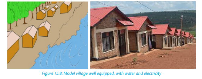
An increasing body of evidence has associated housing quality with morbidity from
infectious diseases, chronic illnesses, injuries, poor nutrition, and mental disorders.
Self-assessment 15.4
What measures are taken by Rwanda government to ensure high quality of
housing conditions?
15.5. Public health services
Activity 15.51. Suppose that you are one of Rwandans who have food industry in our15.5.1. Food inspection requirement
country, and you expect your production to be inspected. Outline the main
requirements for good production to be inspected?
2. Clean water is good for health. Discuss the ways you would use to obtainclean water at home.
a. Food inspection services
Food inspections services help to let you have a complete check of your running
production in factories or across the country. An inspector performs a random
selection checking on quantity, packing, labeling, dimension, weight and visual
aspects. Inspection allows to spot inconsistencies in your production lots before they
leave the factory: you can react timely and avoid costly rework, sorting or recalls.
15.5.2. Need for control of housing conditions
Living conditions affect people’s lives, be it at home or the workplace. Without good
living conditions, people’s health and work will be affected. Nowadays, the quality of
housing accompanied with good housing facilities is now improving.
15.5.3. Need for control of clean water.
For being healthy, only clean water must be used. Different materials are used to
clean water. They include “Sûr–eau”, heating or by using water purification etc. The
steps of water purification are: storage reservoir, aeration, filtration, disinfection,
reduction of chlorine concentration, covered service reservoir, distribution.
15.5.4. Need for control of hygiene
Many diseases can be prevented from having a damaging effect on the body by
the action of natural defenses, antibiotics or other medicines. However, there are
many steps that can be taken by individuals, and by the community as a whole,
to fight microbes even before they enter the body and cause disease. These steps
concern personal hygiene (cleanliness) and sanitation (public cleanliness involving
community efforts in disease prevention), both of which help to prevent disease.
Note that: The World of Health Organization defines health as a state of complete
physical, mental and social well-being and not merely the absence of disease and
infirmity. The responsibility for good health lies in the hands of each individual in
the community and the nation.
a. Personal hygiene– Hands frequently touch many things which may carry pathogens. They mustExercise, recreation and rest are other factors that are important in promoting health.
be always washed using a soap before preparing food, eating and after a visit
to a toilet.
– It is essential to bath frequently because sweat and oil secretions on the skin
enable bacteria and fungi to breed easily. This helps to prevent skin infections.
– Bath towels and sponges should not be shared Combs and hairbrushes should
not be shared.
– Hair should be washed frequently to avoid lice and mites. These can spread
typhus fever.
– Teeth should be cleaned at least twice a day, preferably after each meal
because the spaces between teeth where food particles are trapped provide
excellent breeding grounds for bacteria.
– Clothes would be clean and changed frequently.
– Shoes should be worn to prevent cuts and infection by hookworms.
– Finger nails and toe nails must be always kept short and clean so that they do
not provide breeding places for germs.
i. Exercise
Exercise makes the muscles strong so that they can support the body better.
It helps to get rid of excretory materials and to improve digestion.
It quickens blood circulation and improves the action of the glands and nerves.
ii. Recreation
– Gardening, playing games and reading can remove any dullness and mental
tiredness resulting from everyday work.
iii.Sleeping is the best form of rest.– Adults need about eight hours of sleep a day. A great deal of repair of wornout tissues in the body and the building up of new ones takes place whenLesson self-assessment 15.5
body rests.
– Other good personal habits include avoiding smoking, alcoholic drinks and
other drugs.
1. In which ways a personal cleanliness may be achieved.
2. How to promote a hygienic food preparation?3. Discuss about good personal Hygiene.
End of unit assessment 15
1. Answer to the following questions:a. What does the germ theory of disease mean?2. State any TWO diseases caused by:
b. State any four causes of diseases in our life.a. Bacteria
b. Protozoa
c. Microscopic fungi3. Match the following scientists with their scientific contributions:
4. List the reasons why smallpox is easier to eradicate than AIDS.
5. Suggest reason why Malaria is endemic disease in tropics.
6. Describe the biological factors that make malaria a difficult disease to
control.
7. Describe the precautions that people can take to avoid catching malaria.8. Explain what is meant by ring vaccination.
UNIT 16: A SEXUAL REPRODUCTION IN PLANTS
UNIT 16: ASEXUAL REPRODUCTION IN PLANTS
Key Unit Competence
Account for various methods of asexual reproduction as means of increasing crop
yield.
Learning Objectives
By the end of this unit, I should be able to:– Describe the various methods of asexual reproduction: fragmentation,Introductory activity
budding, and spore formation.
– Demonstrate asexual reproduction mechanisms in lower organisms.
– Discuss the advantages and disadvantages of asexual reproduction.
– Describe the characteristics of vegetative reproductive parts in a flowering
plant.
– Apply principles of artificial propagation in growing varieties of plants that
are economically important.
– Apply the knowledge acquired to produce economically important plants.– Appreciate the use of artificial propagation in increasing crop yields.
The kingdom Plantae comprises about 260,000 known species including flowering
and non-flowering plants. All plants have a general organization which includes
vegetative and reproductive organs. Plants reproduce through different ways:
Use the books and other source of information to1. Write on how lower organisms such unicellular plant and another like16.1. Asexual reproduction
cassava, sugar cane and apple reproduce.
2. Describe the techniques used by people to grow Irish potatoes, cassava
and bananas.
3. Describe each of the following methods of asexual reproduction:fragmentation, budding and spore formation.
Activity 16.1
Using textbooks or internet and search on asexual reproduction.
Asexual Reproduction
This is a type of reproduction without production of gametes. It usually results in the
production of identical offspring; the genetic variation arises as a result of random
mutation among the individuals.
Self-assessment 16.1
Write an essay on asexual reproduction.
16.2. Types of asexual reproduction
Activity 16.2.1
Discuss on asexual reproduction in lower organisms and write a report as an outof-class activity.
There are five common modes of asexual reproduction: fission, budding, vegetative
reproduction, spore formation and fragmentation.
16.2.1. Fission
An important form of fission is binary fission. In binary fission, the parent organism
is replaced by two daughter organisms, because it divides in two, i. e: Unicellular
Fungi. Another type of fission is multiple fission that occurs in many algae. The
nucleus of the parent cell divides several times by mitosis, producing several nuclei.
The cytoplasm then separates, creating multiple daughter cells.
16.2.2. Budding
Some cells split via budding resulting in a ‘mother’ and ‘daughter’ cell. The offspringorganism is smaller than the parent. Budding is also known on a multicellular level.
16.2.3. Vegetative reproduction
Vegetative reproduction is a type of asexual reproduction found in plants where
new individuals are formed without the production of seeds or spores. Examples
of vegetative reproduction include the formation of miniaturized plants called
plantlets on specialized leaves (for example in kalanchoe) and some produce new
plants out of rhizomes or stolon (for example in strawberry). Other plants reproduce
by forming bulbs or tubers (for example tulip bulbs and dahlia tubers). Some
plants produce adventitious shoots and suckers that form along their lateral roots.
Vegetative reproduction may form a clonal colony where all the individuals are
clones.
16.2.4. Spore formation
Many multicellular organisms form spores during their life cycle in a process called
sporogenesis. On the other hand, plants and many algae undergo sporic meiosis
where meiosis leads to the formation of haploid spores rather than gametes. These
spores grow into multicellular individuals called gametophytes. Fungi and some
algae can also utilize true asexual spore formation, which involves mitosis giving
rise to reproductive cells called mitospores that develop into a new organism after
dispersal.
Activity16.2.2
Demonstration of asexual reproduction by fragmentation in algae
Requirements:
Glass beakers of 500ml, Scalpel, Forceps, Pins, Spatula, Weighing balance, Labels,
Artificial, fertilizers, Clear river water and Spirogyra (algae).
Procedure1. Label five beakers of the same size as A, B, C, D and E. Pour water in eachDraw a table as this shown here and record your observation.
beaker. Weigh several measures of artificial fertilizers of 1 g each.
2. Transfer 1g of fertilizer to beaker A, then 2g to beaker B, 3g to beaker C,
4g to beaker D and 5g of fertilizers to beaker E. Note the concentration of
fertilizers is increasing from A- E.
3. Using forceps pick spirogyra and put it on a tile. Add several drops of
water to avoid drying. Tease off a piece of spirogyra using a pin. Cut that
piece into 5 fragments of the same length and transfer each piece into the
beaker.
4. Stand the beakers in a place where they can receive adequate sunlight for
the seven days. On the next day, start to examine the fragments in each
beaker every day and record any observable changes such the increase in
size of the spirogyra.Table16.1: Rate of growth
16.2.5. Fragmentation
Fragmentation is a form of asexual reproduction where a new organism grows
from a fragment of the parent. The fragment develops into a mature and fully
grown individual. Fragmentation is seen in many organisms such as fungiplants
and gemma in liverworts. Most lichens, which are a symbiotic union of a fungus
and photosynthetic algae, reproduce through fragmentation to ensure that new
individuals contain both symbionts. These fragments can take the form of soredia,
dust-like particles consisting of fungal hyphen wrapped around photobiont cells.
Self-assessment 16.2
1. Write a short essay on asexual reproduction.
2. What is fragmentation? give one example of plant which reproduces by
fragmentation
16.3. Advantages and disadvantages of asexual reproduction
16.3.1. Advantages of asexual reproduction.
Activity 16.3
Discuss on asexual reproduction in lower organisms and higher plants, and
outlining advantages and disadvantages.
Since asexual reproduction does not involve the process of gamete formation. It
can be completed much more quickly, allowing the individual to spread its genetic
material in a shorter period of time. It is also an advantage for small populations.
as there may not be an adequate number of plants to pollinate one another to
reproduce. If the parent plants are successful, it is a good idea to make clones of the
plant that can be successful as well.
16.3.2. Disadvantages of asexual reproduction.
Asexual reproduction does not have genetic diversity. With sexual reproduction,
mixing a gene pool can produce diversity. The organism becomes less adapted to
certain environmental changes when there is less variation produced in offspring.
The entire communities of the genetically identical organisms might not survive
such changes. Asexual reproduction usually leads to struggle for existence as well as
overcrowding. The main distinction of asexual reproduction from sexual reproduction
is that there is no need for two parents as well as special cells to reproduce. It means
that special the mechanisms of combining sex cells for fertilization is no longer
required. This form of reproduction process is simply mitosis that copy a parent
organism.
Self-assessment 16.3
Explain the advantages and disadvantages of asexual reproduction.
16.4. Vegetative and artificial propagation in flowering plants
Activity 16.4
Demonstration of asexual reproduction in plants by cuttings
Requirements
Growth medium or moist soil, sweet potatoes vines, elephant grass, sugarcane or
cassava stems, secateurs/sharp knife and rooting hormone.
Procedure1. Collect clean and healthy stems from cassava, sugarcane or potato plants.Draw and record what you will observe after 13 days on the development of roots
2. Using a secateurs/sharp knife, cut the stem of either cassava, sugarcane or
sweet potato stems into fragments of suitable sizes.
3. Place them in either suitable medium of growth with rooting hormone if
available or plant them in moist soil in the school garden.
4. Leave the set up for about 13 days, and then observe the development of
roots and leaves at nodes.
and leaves at nodes.
Artificial vegetative propagation is the deliberate production of new plants from
parts of old plants. This can be done by following three methods: Cutting, layering,
and grafting.
a. Cutting
This is a simple procedure in which part of the plant called a ‘cutting is removed
from the parent plant by cutting and placed in a suitable medium for grow. In this
method, one-year-old stem of root is cut from a distance of 20 to 30 cm and buried
in the moist soil in natural position. After sometime, roots develop from this cutting
and it grows into a new plant. This method is commonly used in rose and sugar cane.
Care is taken that nodes which were lower in parent plant (morphologically) are put
in the soil, while the morphologically higher nodes are kept up. Adventitious rootsare given off at the lower nodes.
b. Layering
This method of vegetative propagation is used in those plants with soft branches
such as jasmine plant. In this method, a branch of the plant which is near to the
ground is pulled towards the ground and covered with moist soil leaving the tip
of this branch above the ground. After sometime, roots develop from that part of
the branch which was buried in the soil. This branch is then cut of along with the
roots from the parent plant and develops into a new plant. This method of asexual
reproduction is also used in the production of plants such as Bougainvillea, guava,strawberries, lemon, China rose etc.
c. Grafting
In this method of vegetative propagation, the stems of two different plants are
joined together so as to produce a new plant containing the characters of both
plants. The plant of which the root system is taken is called ‘stock’, while the other
plant of which the shoot is selected is known as ‘scion’ or ‘graft’. These two stems i.e.
the stock and the scion are fitted together by making slanting cuts into them and
bound tightly with a piece of cloth and then covering it with a polythene sheet.
The diameter of the stock and scion chosen for grafting should be equal. Scion gets
the mineral and water from the soil through the stock and develops branches and
produce fruits. This method of propagation is used in mango, apple, banana, pear,grape, pineapple and peach.
Vegetative reproductive parts in flowering plants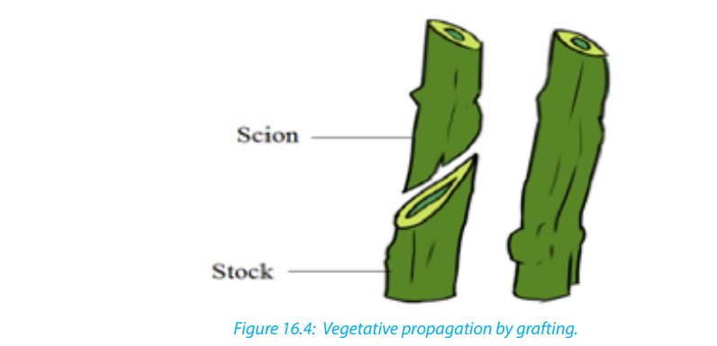
The reproductive part of the plant is a flower. The union of male and female
gametes to form a zygote is called fertilization. The transfer of pollen grains from the
anther to the stigma of the same flower or the different flower is called pollination.
Plants reproduce asexually in a variety of ways. The vegetative reproductive parts
in flowering plant are stem, branches, and leaves and they have the following
characteristics:
Characteristics of Stem:
Stem develops from the plumule of embryo. It is generally the ascending part of
the plant axis. It bears a terminal bud for growth in length and is differentiated into
nodes and internodes. The stem nodes possess dissimilar appendages called leaves.
The young stem is green and capable of performing photosynthesis. In the mature
state, stem bears flowers and fruits. Leaves and stem branches develop exogenously.
Stem exposes leaves, flowers and fruits to their most suitable position in the aerial
environment for optimum function. Stems usually show positively phototropic,negatively geotropic and negatively hydrotropic.
Characteristics of Leaf– It is dissimilar lateral flattened outgrowth of the stem,
– The leaf is exogenous in origin
– It is borne on the stem in the region of a node,
– An axillary bud is often present in the axil of the leaf.
– Leaf has limited growth. An apical bud or a regular growing point is absent,– The leaf base may possess two lateral outgrowths called stipules,Characteristics of branches
– A leaf is differentiated into three parts: leaf base, petiole and lamina.
– The lamina possesses prominent vascular strands called veins,
– It is green and specialized to perform photosynthesis,
– Leaf bears abundant stomata for exchange of gases and it is the major seat oftranspiration.
A branch is a woody structural member connected to but not part of the central
trunk of a tree. Large branches are known as boughs and small branches are known
as twigs. Due to a broad range of species of trees, branches and twigs can be found
in many different shapes and sizes.
Self-assessment 16.41. Discuss on the methods of artificial vegetative propagation.16. 5. Application of artificial propagation in production of
2. Cassava produces flowers, fruits and seeds. Why people prefer to grow
cassava by cutting rather than using seed?
3. Describe the characteristics of vegetative reproductive parts in a floweringplant
improved varieties of plants
Activity 16.5
Use textbooks and other sources of information to discuss on application ofartificial propagation in production of improved varieties of plants.
Artificial vegetative propagation is usually used in agriculture for the propagation
(or reproduction) of those plants which produce either very few seeds or do not
produce viable seeds. Such plants include: Banana, Pineapple, Orange, Grape, Rose,
etc.
Reasons for preferring vegetative rather than sexual means of reproduction vary,
but commonly include greater ease and speed of propagation of certain plants.
Another major attraction is that the resulting plants are clones of the parent plants.
The methods of vegetative reproduction range from rooting cuttings to grafting
and artificial propagation by laboratory tissue culture. In horticulture, a “cutting” is
a piece that has been cut off from a mother plant and then caused to grow into a
whole plant. A popular use of grafting is to produce fruit trees, sometimes with more
than one variety of the same fruit species growing from the same stem. Rootstocksfor fruit trees are either seedlings or propagated by layering.
Self-assessment 16.5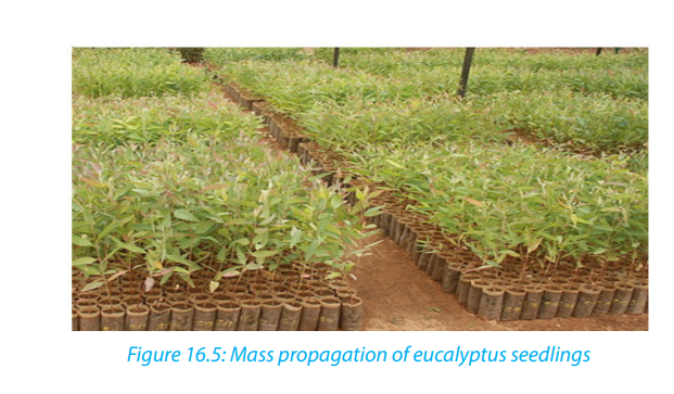
Explain the application of artificial propagation in production of improved
varieties of plants.
End of unit assessment16
A. Multiple choice questions: choose the best answers.
1. In vegetative propagation, cuttings are mainly taken froma. Leaves of parent plant2. Artificial methods of vegetative propagation includes
b. Roots or stems of parent plant
c. Shoots of parent plant
d. Buds of parent planta. Cloning3. Example of plant in which vegetative propagation is occurred by leaves is
b. Grafting
c. Cuttings
d. Both b and c
calleda. Cannabis4. Which of the following is NOT an advantage of asexual reproduction?
b. Chrysanthemum
c. Bryophyllum
d. Brassicaa. Rapid reproduction.B.Questions with short and long answers
b. High genetic diversity.
c. No need for a mate.
d. Low resource investment in offspring.1. Name the plants which are grown by grafting method.
2. What do you understand by grafting?
3. Show how vegetative propagation takes place in potatoes?
4. Explain the method by which the sugarcane and rose are propagated.
5. Give the names of the different methods of artificial vegetative reproduction.
6. Explain the term vegetative reproduction and give one example of plantwhich reproduces by using this type of asexual reproduction.
UNIT 17: SEXUAL REPRODUCTION IN PLANTS
UNIT 17: SEXUAL REPRODUCTION IN PLANTS
Key Unit Competence
Describe sexual reproduction in plants.
Learning objectives
By the end of this unit, I should be able to:– Explain the meaning of the term alternation of generations.Introductory activity
– Describe the types and structure of flowers.
– Describe pollination and fertilization in flowering plants.
– Explaining the events that takes place in a flower after fertilization.
– Describe the types and structure of seeds and fruits.
– Discuss the modes of dispersal of fruits and seeds.
– Observe and draw pollen grains.
– Draw and interpret floral formulae and diagrams.
– Relate the floral structures to the mode of pollination.
– Draw and label structures of fruits and seeds.– Appreciate the role of pollinating agents in flowering plants.
1. Observe the following pictures and suggest what is going on.2. How are the pictures below related to reproduction in flowering plants?

17.1. Alternation of generations in bryophytes and
pteridophytes
Activity 17.1
Using different resources to compare the life cycles of mosses and ferns.
The life cycle of an organism is the progressive sequence of changes which an
organism goes through from the moment of fertilization to death. During its life cycle,
the organism produces new generations of individuals which repeat continuously
the process. New generations are produced by reproduction, which may be sexual
or asexual. The life cycle involves the mitosis and meiosis. This unit concerns how
meiosis can affect the life cycle of living organisms.
The life cycle is seen in seaweeds, mosses, ferns and their relatives. Their life cycles
start with the sexually mature adult plants. Since they produce gametes, they are
called the gametophytes (gamete plants). These are haploid, as they produce
eggs and sperms. The egg and sperm fuse to produce a diploid zygote, but this does
not develop directly into a new gametophyte. Instead it grows (by mitotic divisions)
into another plant which is quite distinct from the gametophyte called sporophyte
(spore plant). The function of sporophyte is to produce spores. In bryophytes, the
sporophyte depends on gametophyte for nourishment. As spores are formed by
meiosis, they are haploid. When the later are dispersed by wind on suitable soil, they
germinate and grow by mitosis into gametophytes, which then repeat the sequence
of life cycle. Ferns and mosses consist of two distinct plants: haploid gametophyte
and diploid sporophyte, which alternate each other within the life cycle. Thisphenomenon is known as alternation of generation.
So, alternation of generation is a phenomenon in the plant life cycle in which a
diploid stage a sporophyte alternates with a haploid stage of gametophyte.
The importance of alternation of generation to organisms:– Spores produced can survive hash conditions and only germinate when
conditions are favorable.
– It ensures rapid multiplication of the plant species as spores are usually
produced in vast numbers.
– Interdependence between the gametophyte and sporophyte generations
ensures that both generations exist at any given time. This prevents extinctionof the plant species
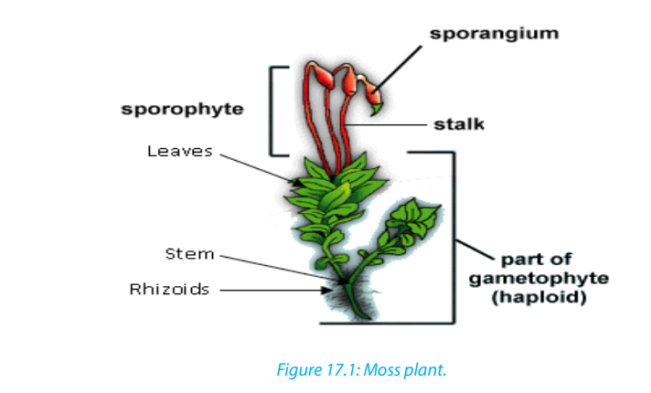
The life cycle in mosses involves alternation of generations between the diploid
(2n) sporophyte and haploidgametophyte. In bryophytes, the gametophyte
is a dominant stage of the life cycle, while the sporophyte is dependent on
gametophyte for supplying water and nutrients. Gametes are formed in special
reproductive organs at the tips of gametophytes. Sperms are produced in antheridia
(singular: antheridium), and eggs in archegonia (singular archegonium). Some
species produce both sperms and eggs on the same plant, whereas others produce
sperms and eggs on separate plants.
During fertilization which requires water, the sperms released from antheridia fuse
with the eggs and form a diploid (2n) zygote. The zygote grows into the sporophyte.
Sporophyte is a long stalk ending in a capsule in which haploid
formed by meiosis. When spores become mature enough, the capsule bursts and
spores are scattered by wind. Under suitable conditions, spores then germinate by
forming underground filaments called protonemata (singular: protonema). Small
buds produced by protonemata give rise to new gametophyte plants which can
start the cycle again.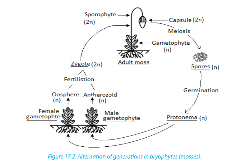
Checkpoint: why bryophytes grow in habitats where water is available constantly:
For fertilization to occur, the sperm of bryophyte must swim to an egg. Without water,
this movement is impossible. Because of this dependence on water for reproduction,
bryophytes must live in habitats where water is available at least part of the year.
17.1.1. Alternation of generations in pteridophytes (ferns)
Pteridophytes also exhibit alternation of generations. Ferns are formed of true
roots, stem and leaves (fronds) with vascular tissues. They have lignified tissues.
The horizontal underground stem is called rhizome which bears adventitious roots.
The leafy plant is a sporophyte. Mature leaves commonly called fronds bear yellow
or orange masses of sporangia which are grouped into the structures called sori(singular: sorus) on their lower side.
Ferns have a life cycle in which the diploid (2n) sporophyte is the dominant generation.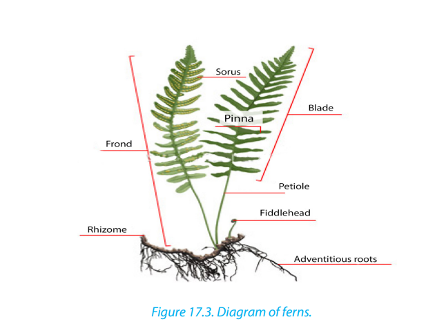
Sporophyte is large (some fern trees can have 7 m of height), and differentiated
into leaf, stem and roots with vascular tissues, while the haploid gametophyte
(prothallus) is very simple with few millimeters. In ferns, the sporophyte produces
haploid spores by meiosis. This is done on the underside of the leaf called frond insporangia (singular: sporangium). Sporangia are grouped into sori (singular: sorus).
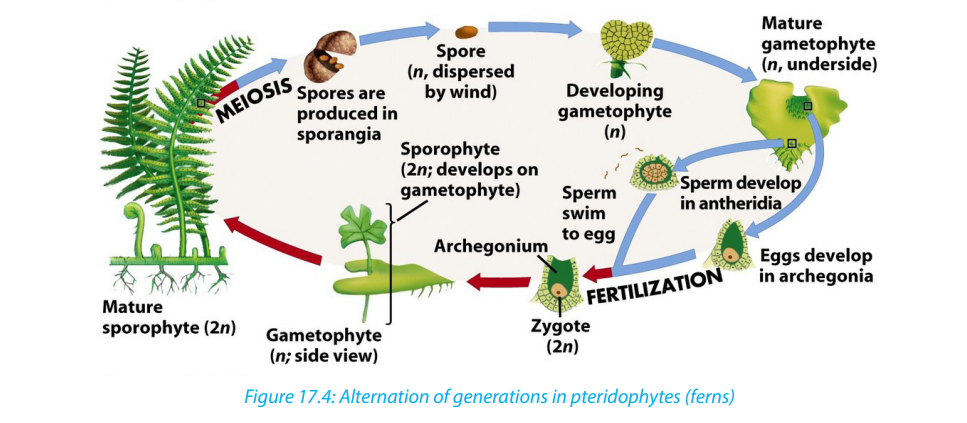
When spores are mature enough, the sporangia burst and spores are released on
ground. If conditions are favorable, spores germinate and grow into the haploid
heart-shaped gametophytes
Antheridia and archegonia found underside of gametophyte produce sperms
and eggs respectively. During fertilization, sperms swim towards eggs and fuse
together to form diploid zygotes, which grow and develop into new sporophytes.As sporophyte grows, the gametophyte dries and dies.
17.1.2. Differences between the mosses and fernsTable 17.1: Comparison between moss and fern.
Self-Assessment 17.1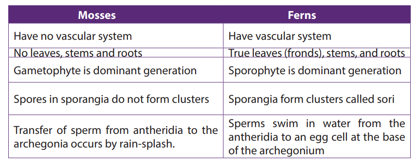 1. Explain the meaning of the term alternation of generation17.2. Types and structures of flowers
2. Why is water essential in the life cycle of a bryophyte?
3. What are the archegonium and antheridium?
4. Why are these structures important in the life cycle of a moss plant?
5. What is the dominant stage of the fern life cycle?
6. Explain the relationship between gametophyte and sporophyte phases of
the fern.
7. Compare gametophyte and sporophyte stages of the plant cycle. Which is
haploid? Which is diploid?8. How do bryophytes reproduce asexually?
Activities 17.2
Collect different forms of flowers from the school compound or around the school,
such as hibiscus, morning glory, sweet potato, or maize flower:
1. Observe and describe the structures of collected flowers.
2. How do collected flowers differ externally?3. Cut one of the flowers into two halves, draw and label one half of the flower.
17.2.1. Structure of a typical complete flowerA flower is a reproductive organ of a plant, which produces fruits and seeds.
A typical hermaphrodite or bisexual flower contains the following parts: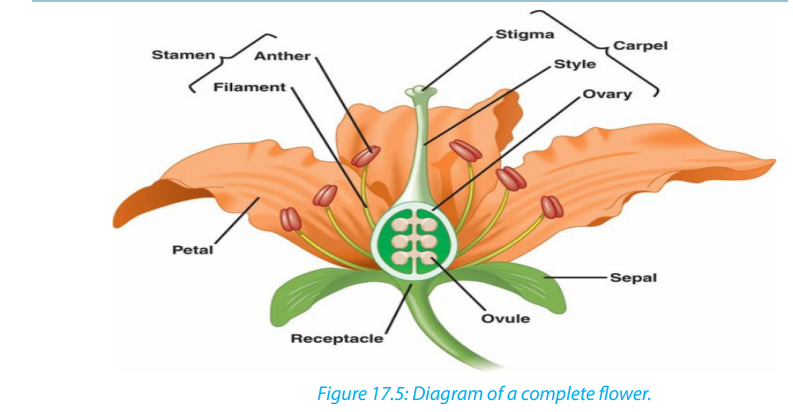
– Pedicel: it is the stalk which attaches the flower on the main floral axis.
– Receptacle: it is the swollen part at the end of the stalk where other parts of
the flower are attached.
– The calyx: it is the set of sepals, generally having green colour. They protect
the internal parts of the flower. In some plants, the sepals are coloured and are
called petaloids.
– The corolla: it is the set of petals, with different colours and nectar glands
that produce sugary substances which participate in attraction of pollinating
agents. In some plants, the petals are green and are called sepaloids. Both
calyx and corolla are collectively called perianth. They form a floral envelope
or accessory organs as they do not participate directly in reproduction, or in
formation of fruits and seeds, they all insure the protection of internal parts ofthe flower.
– Androecium: is the male reproductive organs of the flower. It consists of
many stamens. A stamen consists of: the filament which supports anther,
and anther which contains the pollen grains or male gametes.
– Gynoecium/pistil: is the female reproductive organ. It consists of many
carpels, and each carpel is made of: stigma (plural: stigmata), style and ovary
with ovules.
a. The stigma: receive pollen grains from anther during pollination.
b. Style: maintains the stigma in a good position to receive pollen grains.
c. Ovary: a sac where ovules are produced. Ovules become seeds afterfertilization.
17.2.2. Types of flowers
1. According to absence of some reproductive parts of the flower, we can
distinguish:
a. Unisexual flower: is a flower that consists of one type of reproductive
organ. This can be: staminate: unisexual male (with androecium only), or
carpellate: unisexual female (with gynoecium only). E.g. flower of papaya.
b. Bisexual or hermaphrodite flower: a flower with both male and female
reproductive organs (androecium and gynoecium). E.g. flowers of beans.
Dioecious plants are plants that have male flowers and female flowers on separate
plants (e.g. papaya/pawpaw) while monoecious plants are plants that have both
male and female flowers on the same plant (e.g. maize).
2. According to the position of ovary in the point of insertion of calyx, corolla
and stamen, we can distinguish:
a. A flower with inferior ovary: it is when the ovary is located below the point of
insertion of calyx, corolla and stamens.
b. A flower with superior ovary: it is when the ovary is located over the point of
insertion of calyx, corolla and stamens.
c. The semi-infer or semi-super flower: when ovary is neither inferior nor superior
but in the middle of receptacle which is hollowed.– When sepals are joined together, the flowers are called gamosepal, and where3. According to the shape and symmetry of the flower, we can distinguish:
are not joined together, the flower is called dialysepal.
– When petals are joined together, the flowers are called gamopetal, and when
are not joined together, the flower is called dial petal. When they are absent,the flower is called apetal.
i. Zygomorphic or irregular flower: a flower with a bilateral symmetry.Dichogany: it is when male and female organs of the flower mature at different
The flower cannot be divided into two similar halves. E.g. flowers of beans,
cassia.
ii. Actinomorphic or regular flower: a flower with a radial symmetry. The
flower can be divided into two or more planes to produce similar halves.
E.g. flowers of coffee, orange.
times. We can distinguish:1. Protandry: when stamens mature before pistil.Inflorescence is when two or more flowers are borne on a common stalk.
2. Protogyny: when pistil matures before stamen.
17.2.3. Representation of the number and characteristics of a flower
There are two ways by which we can present the number and characteristics ofdifferent parts of the flower. These ways include: floral diagram and floral formula.
Floral formula
The floral formula indicates the number and characteristics of different floral organs.
It varies from one flower to another. By convention, there are standard symbols that
are used to represent different parts of the flower and their characteristics:
K: for calyx, K5: calyx with five free sepals, K (5): calyx with five fused sepals.
C: for corolla, C5: corolla with five free petals, C (5): corolla with five fused petals.
P: for perianth, P4: four free tepals, P (4): four fused tepals, P2+2: four tepals in two
whorls of free each.
A: for Androecium or stamen, A5: androecium with five free stamens, A (5): five
fused stamens, A5+5: ten stamens in two whorls of five each, A0: stamens absent,
A ∞: stamens indefinite in number, A (9) +1: androecium of 10 stamens nine fused
together and one free.
G: for pistil or gynoecium, G2: two free carpels, G (2): two fused carpels, G0: carpels
absent, G (2): Bicarpellary, syncarpous semi-inferior ovary.
Representation of the symmetry of flower: zygomorphic or irregularflower:
Actinomorphic or regular flower.
Representation of sex of the flower: ♂: staminate flower, ♀: pistillate flower, ♀:
bisexual flower
The floral formula is specific to each species of plant. Examples:
Write a floral formula of coffee having
– The bilateral symmetry
– Hermaphrodite flower
– 1 calyx with 5 fused sepals
– 1 corolla with 5 petals
– 5 fused stamens
– 1 pistil with 2 carpels each one with infer ovary of 2 chambers– (5S) + (5P) +(5A) + 2C-2 or K (5) + C (5) + A (5) + G-2(2)
Write a floral formula of Irish potato having
– 5 free sepals
– 5 free petals
– 5 free stamens
– 2 fused carpels with 2 chambers having many ovules
– 1 infer ovary
– Bisexual flower
– Radial symmetry.– K (5) + C (5) + A5 + G-2(2) ∞
Self-assessment 17.21. What are the male and female structures of a flower?17.3. Pollination and double fertilization in flowering plants
2. What is the advantage for a plant to have many flowers together in a single
structure?
3. Where does the female gametophyte develop?4. Describe the flower and how it is involved in reproduction.
Activity 17.3
Use various resources to identify different pollinating agents and describe the
process of double fertilization in flowering plants.
Pollination is transfer of pollen grains from anther to the stigma.
Types of pollination: there are two types of pollination such as: self-pollination and
cross-pollination.
i. Self-pollination: it is the transfer of pollen grains from anther to the
stigma of the same flower, or of different flowers but of the same plants. It
involves one plant. E.g. flowers of maize and beans.
ii. Cross-pollination: it is the transfer of pollen grains from anther to the
stigma of the flower of another plant. It involves two plants. E.g. flowers ofpawpaw
17.3.1. Main Pollinating agents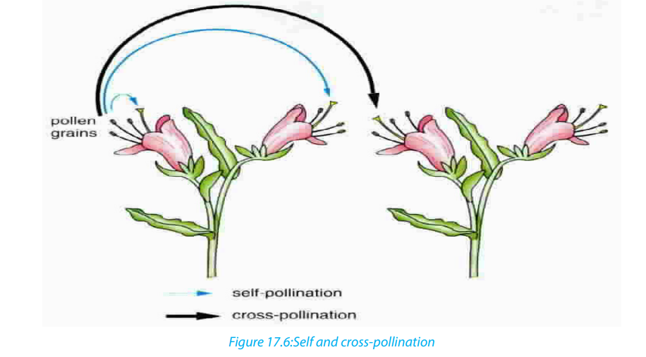
Flower structure is closely related with the way they are pollinated. This means that
flowers are adapted to specific agents or mode of pollination. The common agents
of pollination are: insects (entomophily), wind (anemophily), water (hydrophily),humans (anthropophily), and birds (ornithophily).
Characteristics of insect-pollinated flowers: (entomophilous flowers):– Flowers produce the nectar to attract pollinators.Characteristics of wind-pollinated flowers: (anemophilous flowers)
– Flowers have a large brightly coloured corolla to attract pollinators.
– Production of scents to attract pollinators.
– The surface of the stigma should be sticky to hold pollen grains.
– Pollen grains are sticky and rough enough to remain on the surface of stigma.– The flowers have large stigma to hold pollen grains.17.3.2. Double fertilization and events after fertilization in flowering
– The surface of the stigma should be sticky to hold pollen grains.
– Pollen grains are rough enough to remain on the surface of stigma.
– The flowers are or are not brightly-colored.
– They have or do not have scent.
– They do or do not secrete nectar.
– They produce large quantities of pollen grains, as much of them never reachthe stigmas.
plants
Double fertilization is a complex fertilization mechanism of flowering plants
(angiosperms). This process involves the joining of a female gametophyte(megagametophyte, also called the embryo sac) with two male gametes (sperms).
Development of pollen grains and plant ovules.
The pollen grains are produced in the anthers while the ovules are produced in the
ovary.
Pollen grains
Each anther has four pollen sacs which contain many diploid microspore mother cells
that undergo meiosis to form four microspores each. At first, the four microspores
remain together as tetrads. The nucleus of each microspore then divides by mitosis,
forming a generative nucleus and a tube or vegetative nucleus. At this point, the
content of the pollen grain may be considered as the male gametophyte.
A two layered wall forms around each pollen grain. The outer wall, the exine is thick and
sculptured. The inner wall, the intine is thin and smooth. There are many pores orapertures in the wall through which a pollen tube may emerge.
Plant ovule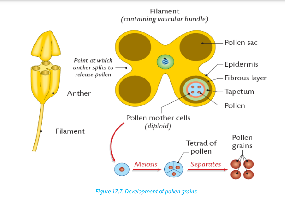
Each ovule is attached to the ovary wall by a short stalk called funicle.
The main tissue in the ovule is the nucellus which is enclosed and protected by
the integuments. At one end of the ovule, there is a small pore called micropyle. A
single diploid megaspore mother cell in the nucellus undergoes meiosis, producing
four megaspores. Three of the four megaspores degenerate, while the remaining
cell, called the embryo sac, grows to many times its original size. The nucleus of the
embryo sac divides mitotically three times, resulting in eight haploid nuclei which
are arranged in groups of four nuclei at the two poles. At this point, the contents of
the embryo sac may be regarded at the female gametophyte.
One nucleus from each pole migrates to the center of the embryo sac. These
two nuclei are called polar nuclei, and they fuse to form a single diploid nucleus.
Meanwhile, cell walls form around the remaining six nuclei and they form the
synergids, antipodals and the egg (ovum). Only the egg functions as the female
gamete.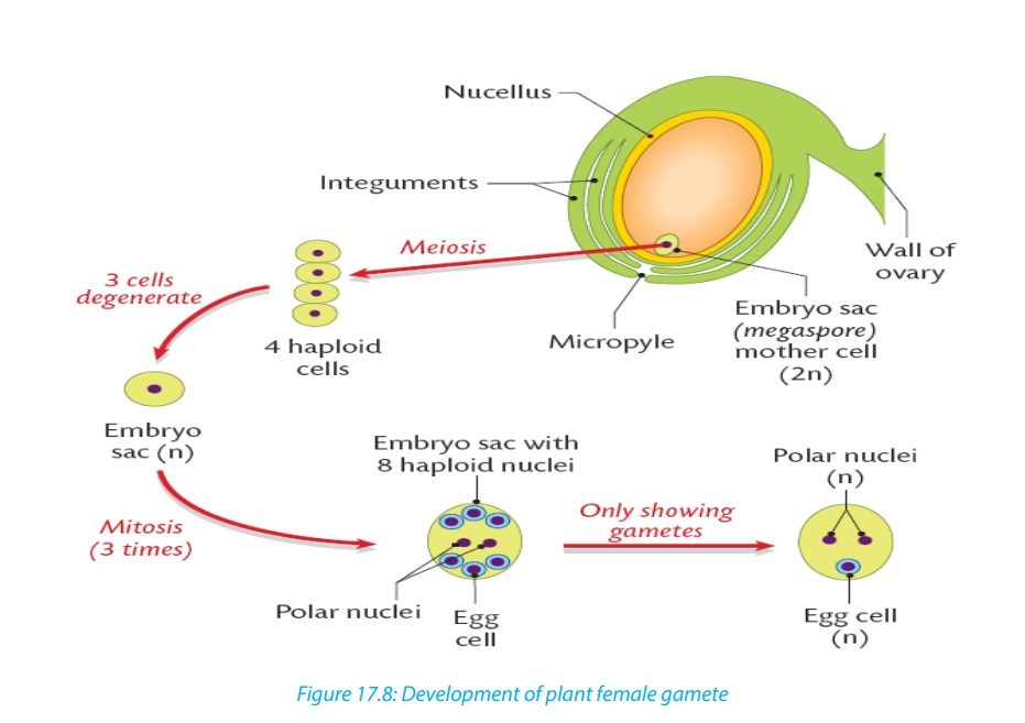
In summary, the pollen grain: contains two haploid nuclei: one called generativenucleus, and the other the tube nucleus.
On the other hand, the ovule or embryonic sac contains eight nuclei: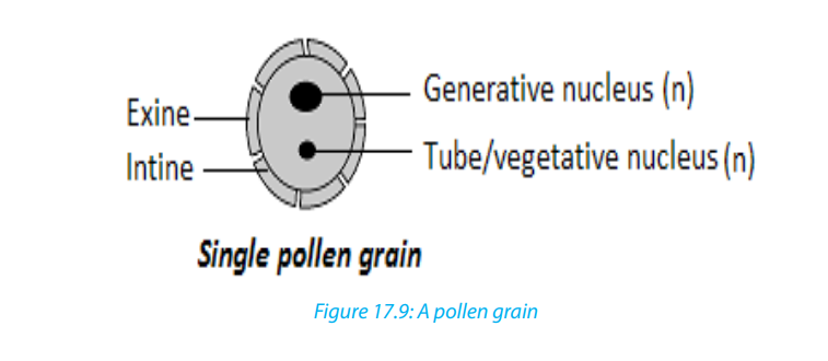 – Three antipodal nuclei/cells at one end
– Two polar nuclei/cells in the middle of ovule
– Two synergids (non-functional nuclei)
– One big egg cell.
The process of double fertilization: It begins when a pollen grain adheres to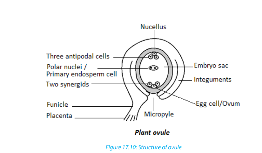
the stigma of the carpel, the female reproductive structure of a flower. The pollen
grain then takes in moisture and begins to germinate, forming a pollen tube that
extends down toward the ovary through the style.
The growth of the pollen tube is controlled by the pollen tube nucleus. In the pollen
tube, the generative nucleus divides mitotically into two haploid nuclei which are
the male gamete nuclei. These follow behind the tube nucleus as the pollen tube
grows down the style towards the ovule. The tip of the pollen tube then enters the
ovary and penetrates through the micropyle opening, releasing the two sperms in
the megagametophyte or ovule.
The tube nucleus degenerates, leaving a clear passage for the entry of male nuclei.
One nucleus fertilizes the egg cell to form a diploid zygote (2N), which will grow
into a new plant embryo; the other fuses with polar nuclei to form a triploid nucleus
(3N), which will grow into a food-rich tissue known as endosperm, which nourishesthe seedling as it grows.
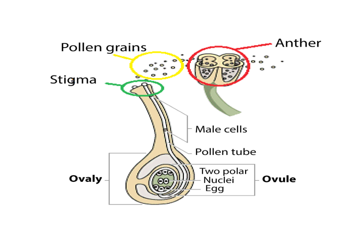
This process is described as double fertilisation and is typical of angiosperms. If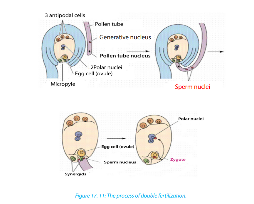
there is more than one ovule in the ovary, each must be fertilized by separate pollen
grain and hence the fruit will have many seeds genetically different from each other.
a. Events in a flower after fertilization
After fertilisation, the calyx, corolla, stamens and style may wither gradually and
fall off, but in some flowers the calyx may persist. The ovule forms the seed, the
two integuments of the ovule will form the seed coat, and the ovary will develop
into fruit, with the ovary wall forming the pericarp (fruit wall). The diploid zygote
undergoes cell division to form the embryo, the triploid primary endosperm nucleus
develops into endosperm, a store used by the developing embryo. This persists in
endospermic seeds of monocotyledons. The micropyle persists as a small hole in the
seed coat through which water is absorbed during germination.
Table 17.2: Floral parts and their fate after fertilization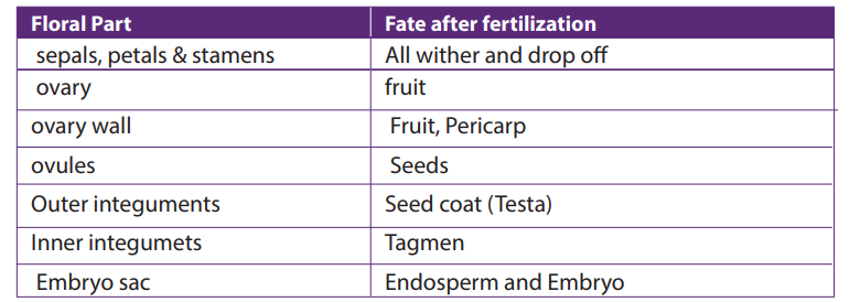
Self-assessment 17.31. Are angiosperms typically wind or animal pollinated? How does this process17.4. Structures and types of fruits and seeds
occur?
2. What is meant by the term endosperm?
3. What is the importance of brightly coloured petals to the plant?
4. What is double fertilization?
5. What happens to the antipodal cells and synergids cells after fertilization?
Activity 17.4
Observe slides containing micrographs of different fruits and seeds. According to
their characteristics:a. Differentiate fruits.
b. Draw and show a structure of seed as seen on microscopeBelow are some examples of fruits:

A fruit is a structure formed from the ovary of a flower, usually after the ovules have
been fertilized. It is normally produced only after fertilization of ovules has taken
place, but in many plants, largely cultivated varieties such as seedless citrus fruits,
grapes, bananas, and cucumbers, fruit matures without fertilization, a process
known as parthenocarpy. Ovules within fertilized ovaries develop to produce seeds.
In unfertilized varieties, seeds fail to develop, and the ovules remain with their
original size.
A fruit consists of two main parts; pericarp (fruit wall) and the seed. The pericarp has
three layers: epicarp or exocarp (outermost), mesocarpe (middle) and endocarp
(inner).
The fruit can have a dry pericarp or fleshy pericarp. The fruits with fleshy pericarp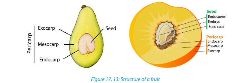
include: berry and drupe. Drupe is a fleshy fruit with only one seed,E. g. avocado.
Berry is a fleshy fruit having many seeds inside of it. E.g. tomatoes, orange, and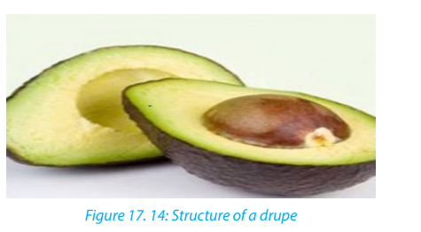
pawpaw.
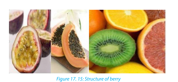
The fruits with dry pericarp include indehiscent fruit or dehiscent fruit. Indehiscent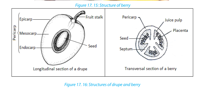
fruits do not open. Seeds remain inside of the fruits. E.g. fruits of coconuts. Dehiscent
fruits open and release seeds. They include: dehiscent fruits with one carpel, and
those with many carpels. Dehiscent fruits with one carpel include; those which open
along one side, e.g. follicle; and those which open along both sides, e.g. legume(beans). Fruits of eucalyptus are examples of dehiscent fruits with many carpels.
The major function of a fruit is the protection of developing seeds. In many plants,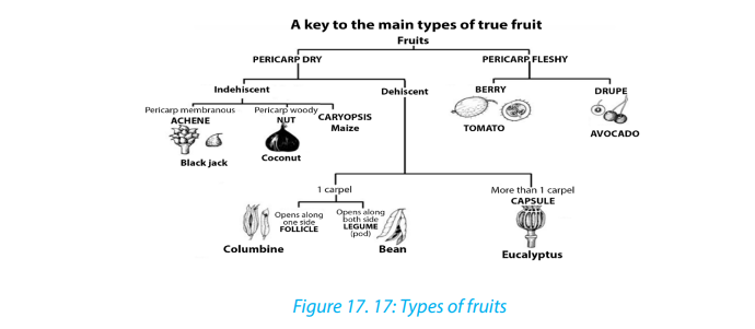
the fruit also aids in seed distribution (dispersal).
Food value
Fruits are eaten raw or cooked, dried, canned, or preserved. Carbohydrates,
including starches and sugars constitute the principal nutritional material of fruits.
Citrus fruits, tomatoes, and strawberries are primary sources of vitamin C, and most
fruits contain considerable quantities of vitamin A and vitamin B. In general, fruits
contain little protein or fat. Exceptions are avocados, nuts, and olives, which contain
large quantities of fat, and grains and legumes, which contain considerable protein.
A seed is an embryonic plant enclosed in a protective outer covering. The formation
of the seed is part of the process of reproduction in seed plants, the spermatophytes,
including the gymnosperm and angiosperm plants. Seeds are the product of the
ripened ovule, after fertilization by pollen and some growth within the mother plant.
The embryo is developed from the zygote and the seed coat from the integumentsof the ovule.
The main components of the embryo are: seed made up of a seed coat (testa), one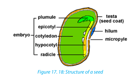
or two cotyledons and an embryonic axis. The embryonic axis is made up of a
plumule, an epicotyl, a hypocotyl and a radical. A seed which has one seed-leaf is
described as monocotyledonous and one which has two, as dicotyledonous. Maizeis monocotyledonous seed while bean is a dicotyledonous seed.
– The cotyledons: the seed leaves attached to the embryonic axis. There may
be one (Monocotyledons), or two (Dicotyledons). The cotyledons are also the
source of nutrients in the non-endospermic dicotyledons, in this case, they
replace the endosperm, and they are thick and leathery. In endospermic seeds,
the cotyledons are thin and papery.
– The epicotyl: the embryonic axis above the point of attachment of the
cotyledon(s).
– The plumule: It is located at the tip of the epicotyl and has a feathery appearance
due to the presence of young leaf primordial at the apex. It will become the
shoot upon germination.
– The hypocotyl: the embryonic axis below the point of attachment of the
cotyledon(s) connecting the epicotyl and the radicle, being the stem-root
transition zone.
– The radicle: the basal tip of the hypocotyl and it grows into the primary root.
Monocotyledonous plants have two additional structures in the form of sheaths.
The plumule is covered with a coleoptile that forms the first leaf while the radicle is
covered with a coleorhiza that connects to the primary root and adventitious roots
form from the sides. Here the hypocotyl is a rudimentary axis between radicle andplumule.
Self-Assessment 17.4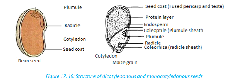 1. Describe the structure of a drupe17.5. Fruits and seeds dispersal with their adaptations
2. Differentiate between a drupe and a berry
3. What would happen to the fruit if ovules in the flower did not develop?
4. Compare the typical structure of seeds that are dispersed by animals to
those dispersed by wind and water.
Activity 17.5
Use books and internet and to answer to the following questions:1. Suggest the ways of fruits and seeds dispersal.
2. Explain adaptation of fruits dispersed by animals.
Dispersal of fruits and seeds is the scattering of fruits and seeds from their mother
plants. They are four methods of seeds and fruits dispersal such as: (1) Dispersal by
Wind (2) Dispersal by Water (3) Dispersal by Animals and (4) Mechanical Dispersal.
Seeds dispersed by wind or water are typically lightweight, allowing them to be
carried in air or to float on the surface of water. The wind carries also small seeds that
have wing-like structure. Seeds dispersed by animals are typically contained in sweet
and nutritious flesh fruits. They can be carried externally on their feet, fur, feathers, or
beaks. Those seeds with hooks or sticky substances rely on the chance that they will
attach themselves to a passing animal. Other seeds are eaten by animals and passed
out in the faeces. These seeds will germinate where the faeces will be deposited.
With mechanical dispersal: all dehiscent fruits scatter the seeds when they burst.
This dehiscence is accompanied by the expression of great force in many fruits so
that seeds are jerked at a considerable distance away from the mother plant. Such
fruits are called explosive fruits.
The dispersal of seeds is important for the survival of the plant species because:– It minimises overcrowding of plants growing around the parent plant thatSelf-assessment 17.5
could then result in too much competition for nutrients and light;
– It allows the plant species to colonise new habitats which can offer suitableconditions.
1. Why is it adaptive for some seeds to remain dormant before they germinate?
2. The seeds of a bishop pine germinate only after they have undergone a
forest fire. Evaluate the significance of this structural adaptation.3. Evaluate the importance of seed dispersal.
End of unit assessment 17
1. Answer by true or falsea. Mosses have life cycle that depends on water for reproduction.2. Choose the letter that best answers the question or complete the statement.
b. In ferns, the gametophyte depends on sporophyte.
c. In mosses, the sporophyte dominates over the gametophyte.
d. Seeds that are dispersed by animals are not contained in a flesh-sweet
tissue.
e. During pollination, pollen grains move from stigma to anthers.
a. Which of the following is not part of a flower?i. Stamensb. Which is the structure of a flower that includes all parts listed below?
ii. Petals
iii. Carpels
iv. Stem
v. Sepalsi. Stigmac. The thickened ovary wall of a plant may join with other parts of the flower to
ii. Carpel
iii. Ovary
iv. Stylev. Ovule
become thei. Cotyledond. In angiosperms, the structures that produce the male gametophyte are called
ii. Fruit
iii. Endosperm
iv. Seed
thei. Pollen tubese. The small and multicellular structures by which liverworts reproduce
ii. Stigma
iii. Anthers
iv. Sepals
asexually arei. Archegoniaf. In angiosperms, the mature seed is surrounded by a
ii. Gemmae
iii. Protonema
iv. Rhizoidsi. Flowerg. The leaves of ferns are called
ii. Fruit
iii. Cotyledon
iv. Conei. Sporesh. The most recognizable stage of a moss is the
ii. Fronds
iii. Sori
iv. Rhizomesi. Gametophyte3. Which are more likely to be dispersed by animals- the seeds of angiosperms or
ii. Archegonium
iii. Protonema
iv. Sporophyte
the spores of a fern? Explain your answer.
4. Pollination is a process that occurs only in seed plants. What is the process in
seedless plants is that is equivalent to pollination?
5. What is the dominant stage of the ferns life cycle? Explain the relationship
between gametophyte and sporophyte phases of the fern
6. Why is water essential in the life cycle of a bryophyte?
7. What is the characteristic responsible for the small size of bryophytes? Explain.
8. Briefly explain why a seed may remain dormant even when the environmental
conditions are favorable for germination.
9. Describe the relationship between the gametophyte and sporophyte in
mosses.
10. During the life cycle of a moss, what are the environmental conditions
necessary for fertilization to occur?
11. Describe the dominant stage in the life cycle of a fern.
12. Propose a hypothesis to explain why angiosperms have become the dominant
type of plant on the earth.
13. Moss plants are small. Ferns can grow as tall as small tree. Explain why. How
does your answer illustrate a major characteristic of the plant kingdom?14. Study the structure of the seed bellow
a. Name the parts A, B and C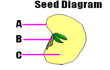
b. What is the importance of the part C for a growing seedling?
15. Many flowers have bright patterns of coloration that directly surround the
reproductive structures. Evaluate the importance of those bright-colored
patterns to plants.
16. What is the function of endosperm?
17. Some plants form flowers that produce stamens but no carpels. Could fruit
form on one of these flowers? Explain your answer.
18. Distinguish between pollination and fertilization.
19. Give names corresponding to the following to the letters: from A to J. Explainthe function of the parts represented by: B, G, and E.
20. Explain why the relationship between bees and flowers is described as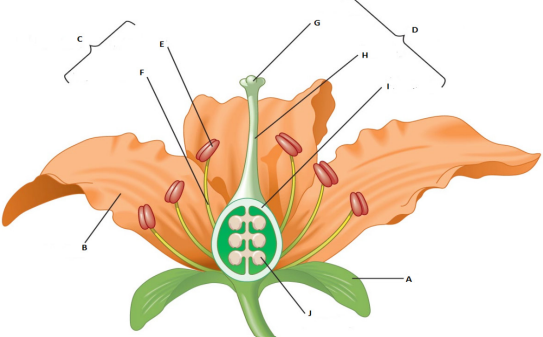
mutually beneficial.
21. What is the main advantage of cross-pollination?
22. Why are the stamens of wind-pollinated plants and insect-pollinated plants
different?
23. Differentiate wind-pollinated flowers from insect-pollinated flowers.
24. Give one example of a plant that uses each of the following dispersal
mechanism:a. An explosive device which works by being inflated with water.
b. A winged seed lifted by air currents
c. A buoyant seed carried by sea currentsd. A gluey substance which sticks the seed to an animal.
UNIT 18: MICROBIOLOGY
UNIT 18: MICROBIOLOGY
Key Unit Competence
Describe the structure and characteristics of viruses, bacteria, and fungal and nonfungal moulds.
Learning objectives
By the end of this unit, I will be able to:– Describe the basic structure of viruses.Introductory activity
– Explain how a retrovirus reproduces.
– Identify the effects of viruses (e.g. AIDS, influenza, measles, feline leukemia,
some human cancers) and prokaryotes (e.g. tuberculosis, bubonic plague,
cholera) on organisms.
– Describe how plant viruses can be transmitted.
– Explain how and why archaebacteria are thought to have been the first forms
of life.
– Describe the structure and life cycles of Escherichia coli
– Relate the structures and functions of Prokaryotes
– Describe the structure of fungal and non-fungal moulds and explain how they
reproduce– Appreciate the importance of microorganisms in life.
A student left fresh milk in a cup exposed to the air. After 6 hours, he/she found
that milk changed its state from fresh milk to stale milk. Why do you think this
happened?
Mukamukiza prepared food for dinner. Some of the food was immediately put in
tightly covered flask while the remaining food was left in the saucepan covered
with banana leaves. In the evening, food in the flask was warm and safe while
food in the saucepan has deteriorated. What is the cause of the food spoilage inthe saucepan?
18.1. Introduction to microbiology.
Activity 18.1.1
Discuss on the term microbiology and on the groups of microorganisms.
The term “microbiology” comes Greek words: ‘micros’ which means small, ‘bios’
which means life and ‘logos’ which means science. Microbiology is the study of
microorganisms which are too small organisms to be seen with the unaided eye and
require a microscope to be seen. They are also referred to as microbes. They include
bacteria, fungi, algae, protozoa and viruses, they are useful to humans and they play
a vital role in decay and recycling of nutrients in the environment. Some of them
cause diseases
Micro-organisms are everywhere: in the air, water soil, on plants, on rock surfaces
in very hot and cold places (ice). Before the invention of the microscope, microbes
were unknown and thousands of people died in devastating epidemics because,
vaccines and antibiotics were not available to fight against infectious diseases.Nowadays, microorganisms can be grown in the laboratory and studied.
a. The Prokaryotes
Prokaryotes can be categorized by their mode of nutrition and how they obtain
energy and the carbon used to build the organic molecules that make up cells.
Organisms that obtain energy from light are called phototrophs and those that
obtain energy from chemicals are called chemotrophs. Organisms that need
only inorganic compounds such as CO2 as a carbon source are called autotrophs.
Heterotrophs require at least one organic nutrient such as glucose to make other
organic compounds. Prokaryotes usually range in size from 1 to 5 micrometersmaking them much smaller than most eukaryotic cells.
b. Classification of prokaryotes
Traditionally, bacteria have been classified based on their structure, physiology,
molecular composition rather than on their evolutionary relationships. The bacteria
that we generally refer to as germs are classified in the domain Eubacteria. More
frequently, members of this kingdom are simply called bacteria. The other type of
bacteria is known as archaebacteria. These bacteria, which are more ancient than
the Eubacteria, are classified in the domain Archaebacteria. Taxonomists used toclassify all prokaryotes in kingdom Monera, yet they slightly differ in characteristics
18.1.2. Archaebacteria and Eubacteria
Activity 18.1.2
Discuss on the characteristics of given examples of both archaebacteria and
Eubacteria.
a. Archaebacteria
Taxonomists treat archaebacteria as a separate kingdom because they are so different
from other bacteria. Archaebacteria have unusual lipids in their cell membranes.
Their cell wall is characterized by the absence of peptidoglycans, a protein
carbohydrate compound found in the cell walls of Eubacteria. Archaebacteria were
first discovered in extreme environmental conditions such as swamps, salt lakes, hot
springs. Examples include:
1. Methanogens
– They have unique method of harvesting energy by converting H2 and CO2
in methane.
– Methanogens can live only in anaerobic condition, such as the bottom of a
swamp, and in sewage where they are the source of marsh gas, because
oxygen is a poison to them.2. Extreme halophiles– These are salt-loving archaebacteria living in environment with very high salt
concentration such as the Dead Sea. High salt concentration would kill most
bacteria.
– These organisms use salt to generate ATP.3. Thermoacidophiles– This third group of archaebacteria lives in extremely acidic environments thatHow and why Archaebacteria are thought to have been the first forms of life?
have extremely high temperature such as hot springs. Thermoacidophiles live
at 110ºC and at a pH of 2.
– Thermoacidophiles live near volcanic vents on land or near hydrothermalvents.
The Archaebacteria comprise a group of single-celled microorganisms that, like
bacteria, are prokaryotes that have no cell nucleus or any other organelles within
their cells. They are known to have an independent evolutionary history and have
numerous differences in their biochemistry compared to other forms of life.
Archaebacteria are now classified as in separate domain in the three-domain
system by Carl Woese who introduced three main branches of evolutionary descent
currently known as the Archaea, Eukarya and Bacteria. Classifying Archaea remains
difficult, since many of them have never been studied in the laboratory and haveonly been detected by analysis of their nucleic acids.
b. Eubacteria
They occur in many shapes and sizes and have distinct biochemical and genetic
characteristics. Eubacteria that are rod-shaped are called bacilli, sphere-shaped are
called cocci (sing. Coccus) and spiral-shaped are called spirilla (sing. Spirillum).1. The bacilli: bacteria with rod-shape. Ex: Clostridium tetani, Bacillus subtilis
2. Vibrios: comma-shaped with a single flagellum. eg: Vibrio cholera
3. The cocci: group of bacteria with spherical shape such as Streptococci.
Cocci that occur in chains are Staphylococci which are grapelike clusters of
cocci and Diplococci which is sphere shaped that are grouped two by two.4. The spirilla: bacteria with spiral shape. e.g.: Spirillum volutans.
18.1.3. Gram stain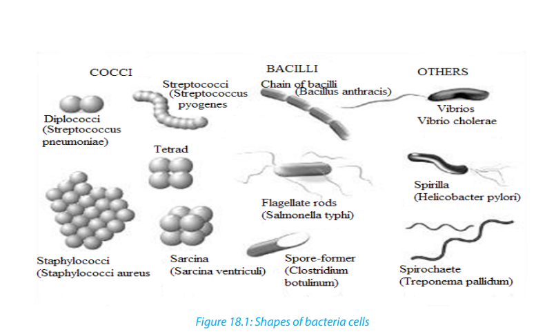
Bacteria have a peptidoglycan or murein cell wall that maintains cell shape, provides
protection and prevents the cell from lysis. Based on the composition of the cell wall,
bacteria can be classified as Gram-positive and Gram-negative. During the process
of Gram staining , some bacteria without a lipid layer along with their peptidoglycan
cell wall take the gram stain and appear violet (purple) and are therefore called
gram positive. Example streptococcus and staphylococcus. Bacteria having a lipid
layer along with their peptidoglycan cell wall do not take up the gram stain and aretherefore called gram negative.
Example: Escherichia coli, Azotobacter, Salmonella.
Self-assessment 18.11. Describe the characteristics of the two domains of prokaryotes.18.2. The structure and life cycle of Escherichia coli
2. What factors can be used to identify prokaryotes?
3. How do bacteria maintain equilibrium in the environment?
4. Identify the parts of a prokaryote.5. Describe briefly how some prokaryotes obtain their energy.
Activity 18.2.1
Using text books, videos or computer aided materials to describe the cycle life of
E. coli.
E. coli reproduce asexually by undergoing binary fission. This type of reproduction
begins with the replication of DNA molecule. Then, the copies of the genetic material \
attach themselves to the cell membrane. When the bacterium’s size has doubled
from its original size, the cell membrane starts pinching inward and a cell wall is
produced between the two DNA molecules. Finally, the cell wall divides the cell into
two daughter cells.
E. coli can also go through another process of reproduction known as
conjugation. Conjugation is a reproduction process which involves the transfer
of genetic material by the sex pili between two bacteria. This is not a sexual
reproduction because there is no combination of gametes. The process of
conjugation starts once the E. coli, called a donor, has finished to replicate its
genetic material in form of a plasmid. The enzyme of the donor can now send
signals to show that it is ready to mate. Once a mate is found, the donor attachesitself to the sex pilus of its mate. By doing so, the donor transfers the plasmid.
18.2. E. coli and food poisoning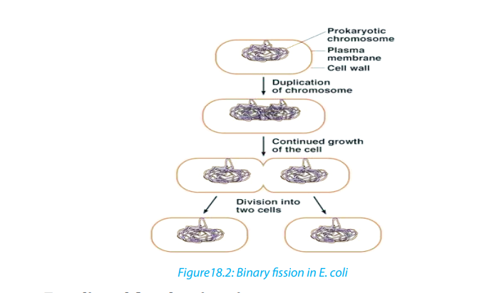
Activity 18.2.2
Using textbooks to brainstorm the process of food poisoning, evolution of harmful
strain of E. coli and food preservation
E. coli is a rod-shaped bacterium measuring about 2.5µm by 0.5µm. It is mainly found
in guts of vertebrates. It is chemoheterotrophic, capable of thriving on a variety of
the organic molecules. Its presence in water indicates contamination by faces.
E.coli reproduces asexually by binary fission. It can also take part in a primitive form of
sexual activity called conjugation where genetic material is passed in one direction
from bacterium to another through a pilus. Although conjugation does not in
itself produce new offspring, after the process has finished, the bacteria reproduceasexually, passing on their new genetic make-up to their offspring.
18.2.1. Evolution of harmful strain of bacteria
E. coli was thought to be a relatively harmless resident of the human gut which might
linked to the occasional upset stomach and mild diarrhoea. When massive colonies
of mutualistic bacteria are present in the gut, including most strains of E. coli, they
help to keep harmful bacteria away from starving them of food. They also help make
vitamin K. But in 1982, it became clear that a new strain of E. coli had evolved into
a much more troublesome organism. The strain had acquired a gene that enabled
it to produce a powerful toxin which damages the intestinal wall, causing severe
diarrhea and internal bleeding.
This may lead to internal serious dehydration in young children and elderly people,
and may result into death. In majority of the cases, infections of pathogenic strain ofE. coli are not fatal and the disease clears without treatment.
18.2.2. Sources of infection
Touching a source of contamination and not washing hands before handling food
may be sufficient to cause the infection.
In 1996, there was an outbreak which led to 20 deaths in Scotland due to
contaminated meat. In the same period, another one was traced due to apple juice
poisoning. Contaminated person can pass the bacteria on vegetables, and other
foods.We must practice good habits of dealing and handling food to minimise
cases of contamination. It is therefore, important to practice good hygiene. It is
also essential to store and package food. It might be vital to pasteurise all fresh fruit
juices just as milk is required to be pasteurised.
18.2.3. Food storage and packaging
The optimum storage conditions differ; raw meat and poultry are kept at around 00c,
meat products at 1oc - 40oc.
Canned foods and many vegetables in dry condition sat 10oc - 150oc,
and dried foods such as flour are stored, in air tight containers at10oc– 150oc.
For long term storage, meat and fish are vacuum-sealed or can be vacuum
packed in laminated plastic containers. For pasteurisation, food and drinks such as
milk are heated to a temperature that kills disease causing microorganisms. Example:Mycobacterium tuberculosis.
Self-assessment 18.21. Suggest the process by which E. coli reproduces.18.3. The structure and life cycle of viruses
2. What is the probable source of the gene that transforms harmless E. coli
into pathogenic E. coli?
3. At what temperature is E. coli in meat killed?
4. How is food poisoned?5. How can you minimise food and drink poisoning?
Activity18.3.1
Using textbooks, chart or videos to describe the structure, life cycle and effects of
viruses.
The term “virus” was first used in the 1890s to describe agents smaller than bacteria
that cause diseases. The existence of viruses was established in 1892, when, Russian
scientist, Dmitry Ivanovsky discovered later microscopic particles known as thetobacco mosaic virus
There are at least 3,600 types of virus. Hundreds of which are known to cause
diseases in animals, bacteria, and plants. Viruses consist of an inner core of either
ribonucleic acid (RNA) or deoxyribonucleic acid (DNA) plus a protein protective
coat called capsid made of protein or of protein combined with lipid or carbohydrate
components. An entire virus particle is called vibrios ?
The core confers infectivity, and the capsid provides specificity to the virus. In some
virions, the capsid is further enveloped by a fatty membrane. The later may causevirion inactivation by exposure to fat solvents such as ether and chloroform.
18.3.1. Characteristics of viruses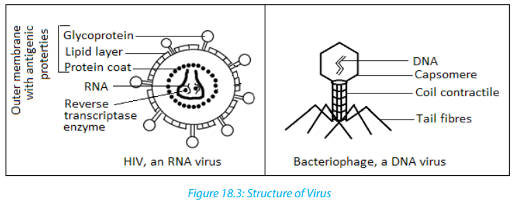 – Viruses are complex biochemical molecules having the following characteristics:18.3.2. Virus types
– Viruses are not visible under light microscope because they are very small than
bacteria.
– They possess a single type of nucleic acid either DNA or RNA enclosed in a
protein coat.
– They can reproduce and grow inside the host cell.
– They have no cell and no cell organelles.
– They are obligate parasite i.e. cannot survive outside a host cell.– They do not feed, respire and excrete.
DNA and RNA viruses differ in the way they use the host cell’s mechanisms to produce
new viruses.
For example, a DNA virus may act in one of the two ways:
The virus may directly produce RNA that is used to make more viral proteins or it
may join with the host cell’s DNA to direct the synthesis of new viruses.
RNA viruses replicate differently from DNA viruses. Upon entering the host cell, a viral
RNA is released into the host cell’s cytoplasm. There, it uses the host cell’s ribosomes.
Some RNA viruses known as retroviruses contain an enzyme called reverse
transcriptase in addition to RNA. Reverse transcriptase uses RNA as a template
to make DNA. The DNA then makes an RNA transcript of itself. This RNA is then
translated into proteins that become part of new viruses. Reverse transcriptase is so
named because it reverses the normal process of transcription, in which DNA servesas a template for producing RNA.
18.3.3. Viral replication
Because viruses are not cells, they can replicate only by invading a host cell and
using the enzymes and organelles of the host cell to make more viruses. Because
they depend on host cells for replication, viruses are called obligate intracellular
parasites. Outside the host cell, a virus is a lifeless particle with no control over its
movements. It is spread by wind, water, in food, or via blood or other body secretions.
18.3.4. Life cycle of Bacteriophage
Bacteriophage is a virus that infects bacteria. Bacteriophage is composed of an
icosahedral head that contains a nucleic acid. Beneath the head is a contractile tail
that includes a collar and a sheath.
The contractile tail helps to inject the nucleic acid into the host cell. The tail rests on
a base plate from which tail fibers emerge. These fibers assist the virus to attach to
a host cell.Viruses replicate by using either the lytic cycle or the lysogenic cycle:
a. The lytic cycle
Activity 18.3.2
Describe the sequence of events that occur during a lytic infection.
During the lytic cycle, a virus invades a host cell, produces new viruses, destroys the
host cell, and releases newly formed viruses. Viruses that undergo the lytic cycle are
called virulent because they cause disease. The lytic cycle consists of five phases:– The Bacteriophage first attaches to susceptible bacterium by attaching its tail
fibers to a receptor site. Receptor sites are specific sites that viruses recognize
and attach to on the host cell’s surface. If the Bacteriophage does not find a
receptor site, it cannot infect the cell.
– Next the Bacteriophage releases an enzyme that weakens a spot in the
cell wall of the host. Then the phage presses its sheath against the cell and
injects its DNA into the host cell through the weak spot in the cell wall. The
Bacteriophage leaves its capsid outside.
– The virus then takes control of the host’s protein synthesizing mechanisms,
transcribing mRNA from the viral DNA. The resulting Bacteriophage mRNA is
translated on ribosomes and proteins that are synthesized form B a capsid. So
the viral DNA is also replicated during this phase.
– Every replicated viral DNA is enclosed in the newly created viral capsid. The
assembly of new virus particles usually occurs in the cytoplasm.
– During the last phase of the lytic cycle, one of the enzymes that are produced by
the Bacteriophage genome causes the host cell to disintegrate, releasing new
Bacteriophage. The cell disintegration is called lysis. In case of the enveloped
viruses, the newly formed viruses move to the cell surface and force their waythrough the cell membrane.
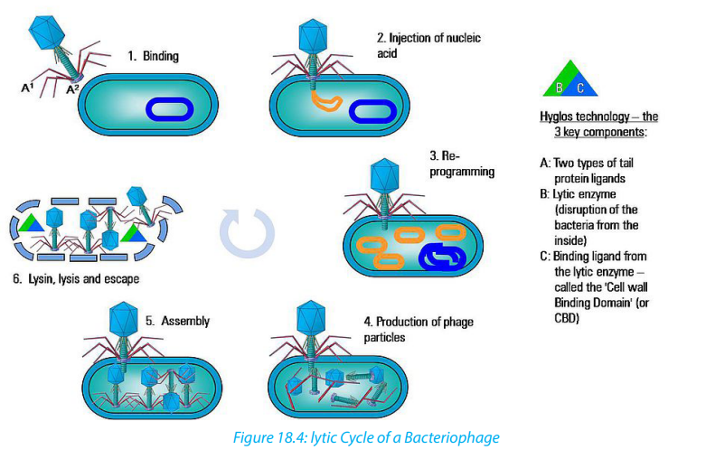
The first step in the replication of the phage in its host cell is called adsorption or
binding. The Bacteriophage adheres to the receptor site by means of its tail fibres.
Following adsorption, the phage injects its DNA into the bacterial cell.
The tail sheath contracts and the nucleic acid or the core is driven through the wall
to the membrane. This process is called penetration and it may be both mechanicaland enzymatic.
Immediately after injection of the viral DNA there is transcription and translation
of a section of the phage DNA to make a set of proteins that are needed to replicate
the phage DNA and proteins that make up the capsid and the various components
of the tail.
After making all viral parts, the assembly process follows. While the viruses are
assembling, produced lysozymes are used to break down the cell wall peptidoglycans
of the host bacteria. This is known as lysis and then mature viruses are released andspread to nearby cells for new infection.
b. The lysogenic cycle.
Activity 18.3.3
Using textbooks to describe what happens to the host cell infected by a temperate
virus.
Some viruses can infect a cell without causing its immediate destruction. Such
viruses stay in their host cell for an extended period of time: days, months or years
in a lysogenic cycle. A virus that replicates through lysogenic cycle and does not killthe host cell immediately is called a temperate virus.
Retroviruses, such as HIV, have RNA that is transcribed into DNA by the viral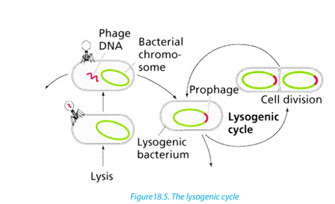
enzyme Reverse transcriptase upon entry into the cell. (The ability of retroviruses
to copy RNA into DNA earned them their name because this process is the reverse
of the usual transfer of genetic information, from DNA to RNA). The DNA form of the
retrovirus genome is then integrated into the cellular DNA and is referred to as the
provirus. The viral genome is replicated every time the host cell replicates its DNAand is thus passed on to daughter cells.
18.3.5. Some common viral diseasesTable 18.1: Some common viral diseases
18.3.6. Virus as living or non-living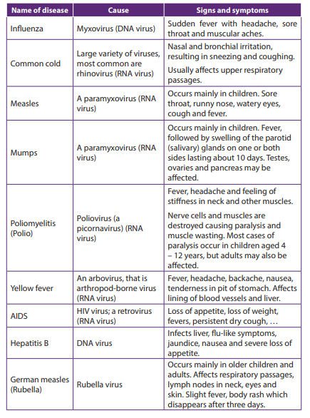
Activity 18.3.4
“Viruses are said to be on the border line of living organisms and non-living
things”. Discuss on this statement.
Viruses do not belong to any of the five kingdoms into which life is classified. It is
difficult to say whether they are living or non-living.
a. Features that make viruses to look like living things:– They have the genetic material composed of either DNA or RNA They causeb. Features that make viruses non-living things:
diseases to other living things: All viruses are infectious.
– They evolve as a result of mutation and natural selection.
– They reproduce /multiply only in other living things: they are obligate
intracellular parasites– They cannot metabolize
– They crystallize in isolation.
– They cannot reproduce outside of host.
– They are not made of cells. This means that they have a relatively simple noncellular organisation.
– They cannot respond to stimuli
– They have one type of nucleic acid, either DNA or RNA. But living cells containboth DNA and RNA.
Table 18.2: Comparison between viruses and cells
Self-assessment 18.3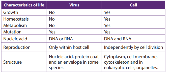 1. What are the parts of a virus?18.4. Moulds
2. Describe the two ways by which viruses cause infection.
3. Distinguish between Bacteriophage and a prophage.
4. What is meant by retrovirus?
5. What are the strengths and weaknesses of the tobacco mosaic virus
hypothesis?
6. Which characteristic feature is common to all viruses?
7. How is a capsid protein important to the functioning of a virus?8. What is the best way to protect humans against most viral diseases?
Activity 18.4
Using text books or computer aided materials to describe the life cycle of bread
mould.
Moulds pervade our world, living wherever moisture is present. Some are of great
benefit to humans, providing antibiotics, acting as decomposers so that nutrients
can be recycled, or taking part industrial processes. Other moulds cause diseaseswhich lead to serious damage.
Moulds have cells arranged in long thread-like filaments, the hyphae, that form a
mass called Mycelium. Moulds are usually considered as fungi, but mould may also
be formed by filamentous bacteria, slime moulds, and water moulds. Therefore,
there are two main types of moulds: fungal moulds and non-fungal moulds
18.4.1. Fungal moulds
All fungi that produce mycelia can be called moulds, but the term is usually used for
an organism in which the mycelium forms the main body of the fungus. In the black
bread mould Rhizopus and the pin mould Mucor, the mycelium consists of a tangled
mass of hyphae with many nuclei. These hyphae are called coenocytic because the
fungal tissue is not separated by cell walls.
Fungal hyphae have an outer cell wall made of chitin and inner lumen which contains
the cytoplasm and organelles. A cell surface membrane surrounds the cytoplasm
and sticks tightly to the cell wall.
Rhizopus and Mucor are Saprotrophic, obtaining their nutrients from dead organic
material. Rhizopus nigricans and Mucor mucedo can live on bread but some species
of Rhizopus feed on living plants, and Mucor commonly grows on rotting fruits and
vegetables, in the soil or on dung.
the food outside the organism and then absorb the soluble digestion products and
assimilate them.
a. Life cycle of Rhizopus and Mucor.
Rhizopus and Mucor belong to the fungal phylum Zygomycota. The phylum got its
name because its members produce two kinds of spores: Sexual zygospores as wellasexual sporangiospores.
The asexual sporangiospores formed by mitosis, develop in sporangium at the tip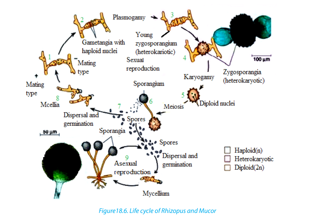
of hyphae. When sporangium busts, the spores are released.
In most species of Mucor, the sporangium dissolves then water enters the spore
mass, and the spores are dispersed by the raindrop or are transported by the insects.
In most Rhizopus species, the sporangium wall fractures and dry spores are released
by the wind.
The sexual reproduction involves conjugation. Usually the hyphae from mycelia of
different mating types meet and interconnect via outgrowths. The interconnecting
walls break down and their cytoplasm containing haploid nuclei mix, then the
diploid zygote formed by the fusion of two nuclei develops a thick, rough, black
coat and becomes a dormant zygospores. Meiosis probably occurs at the time of
germination; the zygospore cracks open to liberate several haploids spores whichcan give rise to asexual sporangia and mycelia of either mating strain.
b. Use of moulds
Even if species of Rhizopus and Mucor are responsible for the spoilage of food, they
are also useful as follow:– They are used to make the human foods. For example, Mucor is used with soya18.4.2. Non-fungal moulds
beans to make a cheese called sufu, in eastern Asia.In Indonesia, R. oligosporus
and R. oryzae are used to produce a food called tempeh from boiled skinless
soya beans.
– The fungal moulds belonging to the Zygomycota are used to make anaesthetics,
birth control pills, meat tenderisers, and the yellow colouring agents used inmargarines and butter substitutes
The following are different groups of non-fungal moulds:a. Bacterial moulds: including those of Streptomyces griseous, which secretes the1. Plasmodial slime moulds which have the following characteristics:
antibiotic streptomycin
b. Slime moulds: These are a peculiar group of organisms that resemble fungi in
appearance and lifestyle, but are more closely to protoctists such as Amoeba in
their cellular organization, reproduction, and life cycles. There are two types of
Slime moulds:– They have no connection with the parasitic protoctists belonging to the genus2. Cellular slime moulds (also called Acrasiomycotae) which have the following
Plasmodium which causes malaria.
– They exist as thin, streaming masses of colourful protoplasm that creep along
moist, rotting logs and leaves.
– They move in an amoeboid fashion, engulfing food particles by Phagocytosis.
– A single mould may extend for many centimetres, but it is not multicellular.
– They are made up of a continuous mass of cytoplasm with many nuclei called
coenocytic mass.
characteristics.– They have a unicellular feeding stage resembling an amoeba, with each cellc. Water moulds (Oomycota)
functioning individually.
– When food is scarce, the individual cells group into a mass resembling that of
Plasmodial slime moulds.
– The individual cells of Cellular slime moulds retain their identity and have
separate cell surface membranes– Although water moulds and fungi are closed related and have a similar
structure, water moulds are generally regarded as a separate and more ancientgroup belonging to the protoctists.
– Water moulds include rusts and mildews which consist of coenocytic masses(where “Oo” means egg)
of hyphae similar to fungi, for example Plasmodial slime moulds,
– Most water moulds have cell wall made of cellulose, while the cell wall of the
true fungi is made of chitin.
– Some of the most devastating plant diseases are caused by water moulds. For
example, the Phytophthora infestans causes potato blight, and Pythium which
is a relatively unexpected parasite attacks a great variety of plants causing soft
rot.
– Water moulds reproduce asexually by structure called conidia, and by moving
spores with flagella, called zoospores.
– They reproduce sexually by producing moving male gametes that fertilizes
large immobile egg cells. These egg cells give the group its name Oomycotae
Self-assessment 18.41. How are the cell walls of fungi similar to exoskeleton of insects?18.5. Penicillium and Saccharomyces
2. Distinguish between hyphae and mycelium.
3. What are conditions necessary for fungal spores to germinate?
4. Explain the basis of classification of fungi.
5. Why do many biologists think that Penicillium evolved from an ascomycete?
6. Briefly describe sexual and asexual reproduction in fungi.
7. The antibiotic penicillin is a natural secretion of a certain kind of fungus green
mould called Penicillium, penicillin kills bacteria. Why might a mouldspecies have evolved way of killing bacteria?
Activity 18.5
Make a research from the internet or textbooks to find out:
1. The structure of Penicillium, and yeast cell.
2. How saccharomyces reproduces.3. The explanation of budding.
18.5.1. Penicillium and antibiotics
Penicillium is highly known for producing penicillin, the first antibiotic discovered in
1928 by a scientist Alexander Fleming when he was culturing some Staphylococcus
bacteria during his medical research.
After leaving some Petri dishes for many days, he found a mouldy growth of
Penicillium notatum contaminating a corner of one of dishes. Then Fleming realised
that Staphylococcus next to the mould has been destroyed.
After studying Staphylococcus closely, Fleming concluded that the Penicillium
mould was producing a substance that killed the Staphylococcus. He carried on
with finding out if the broth of Penicillium mould contained penicillin which could
destroy pathogenic bacteria.
In 1931, Fleming dropped his research. Howard Florey and Ernst Chain went on to
produce purified penicillin. A successful work was reported 1940, and penicillin has
been used to treat wounded soldiers in Second World War. In 1945, Fleming, Floreyand Chain received the Nobel Prize for the discovery of penicillin.
a. The structure of Penicillium
Penicillium is septate; its hyphae have cross-walls called septa. However, the septa
are not formed by cell division, and at the Centre of septum there is a usually a
pore which allows cytoplasm to flow from one compartment to another. Each
compartment may contain one or more nuclei. Though Penicillium has septa, is a
coenocyte like the non-septate moulds Rhizopus and Mucor.
Penicillium is saprotroph, feeding on organic matter in damp soil, leather, bread, and
decaying fruit. The mycelia of Penicillium species form circular green, yellow, or blue
moulds (depending to the species).
Penicillium reproduces asexually by means of spores called conidia formed at the tip
of special hyphae called conidiophores.
Spores of Penicillium are exposed and free to be dispersed as they are mature.
18.5.1. Saccharomyces
a. Definition and characteristics– Saccharomyces is a genus of yeasts which include all unicellular fungi thatb. Structure of yeast
reproduce asexually by budding.
– They occur commonly on faeces, in the soil, and on the surfaces of plants and
animals.
– The most familiar and industrial important yeast is Saccharomyces cerevisiae.
– The tiny cells of this yeast are very active metabolically. They are usually aerobic
but in the absence of oxygen they use anaerobic metabolism, producing
carbon dioxide and ethanol (alcohol) as waste products which are industrially
useful
– Each cell of Saccharomyces cerevisiae has a single nucleus and is usually egg
shaped.– Cells contain most of organelles of a typical eukaryote.
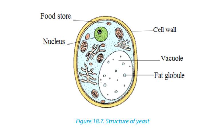
c. Mode of reproduction
Saccharomyces cerevisiae can reproduce either asexually or sexually.
In asexual reproduction, the single cell divides by budding and separate into two
cells. Some buds group together to form colonies; other separate to grow individuallyinto a new yeast.
In sexual reproduction, two cells fuse to form a diploid cell which then forms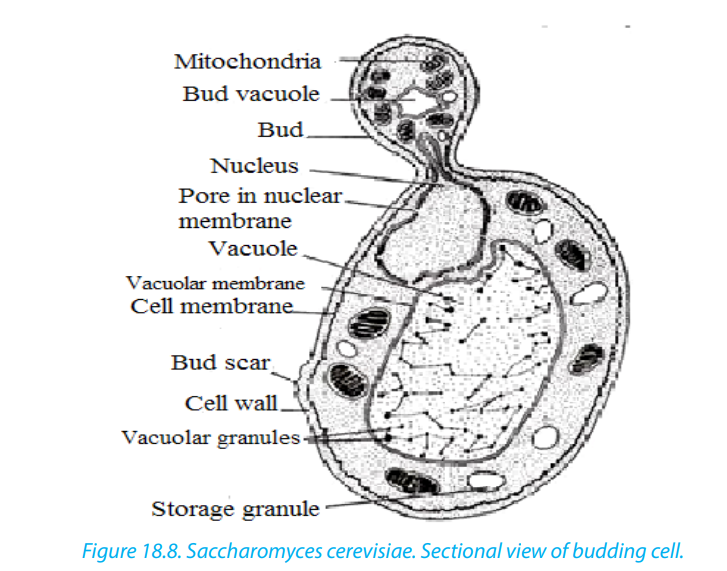
haploid spores by meiosis
Self-assessment 18.51. Which feature does all yeast have in common?18.6. Protozoa that cause disease
2. How do hyphae of Penicillium differ from those of Mucor.3. Describe the evidence for penicillin’s effectiveness.
Activity 18.6
Observe prepared slides of Entamoeba histolytica ,Plasmodium and Trypanosomato compare their structures.
18.6.1. Entamoeba histolytica
a. Characteristics of Entamoeba histolytica
Entamoeba histolytica is a protozoan parasite responsible for a disease called
amoebiasis. It occurs usually in the large intestine and causes internal inflammation
as its name suggests (histo which means tissue, lytic which means destroying). 50
million people are infected worldwide, mostly in tropical countries in areas of poor
sanitation. Inside humans Entamoeba histolytica lives and multiplies as Trophozoites.
Trophozoites are oblong and about 15–20 µm in length. In order to infect otherhumans, they encyst and exit the body.
b. Life cycle Entamoeba histolytica
Entamoeba histolytica life cycle does not require any intermediate host. Mature
cysts (spherical, 12–15 µm in diameter) are passed in the feces of an infected human.
Another human can get infected by ingesting them in fecally contaminated water and
food. If the cysts survive the acidic stomach, they transform back into Trophozoites
in the small intestine. Trophozoites migrate to the large intestine where they live
and multiply by binary fission. Both cysts and Trophozoites are sometimes present
in the feces. Cysts are usually found in firm stool, whereas Trophozoites are found in
loose stool. Only cysts can survive longer periods (up too many weeks outside the
host) and infect other humans. If trophozoites are ingested, they are killed by the
gastric acid of the stomach. Occasionally Trophozoites might be transmitted during
sexual intercourse.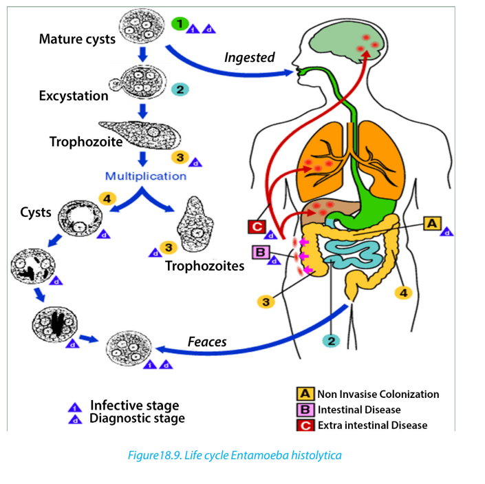
c. Symptoms
Many Entamoeba histolytica infections are asymptomatic and Trophozoites remain
in the intestinal lumen feeding on surrounding nutrients. About 10–20 % of the
infections develop into amoebiasis which causes 70 000 deaths each year. Minor
infections (luminal amoebiasis) can cause symptoms that include:– Gas (flatulence) intermittentSevere infections inflame the mucosa of the large intestine causing amoebic
– constipation loose stools
– stomach ache
– Stomach cramping.
dysentery. The parasites can also penetrate the intestinal wall and travel to organs
such as the liver via bloodstream causing extra-intestinal amoebiasis. Symptoms
of these more severe infections include: Anemia, Appendicitis (inflammation ofthe appendix), bloody diarrhea, fatigue, fever, gas (flatulence), genital and skin
lesions, intermittent constipation, liver abscesses (can lead to death, if not treated),
malnutrition, painful defecation (passage of the stool), peritonitis (inflammation of
the peritoneum which is the thin membrane that lines the abdominal wall), pleuropulmonary abscesses,
stomach ache, stomach cramping, toxic mega-colon (dilated
colon), Weight loss.
d. Prevention
To prevent spreading the infection to others, one should take care of personal
hygiene. Always wash your hands with soap and water after using the toilet and
before eating or preparing food. Amoebiasis is common in developing countries.
Some good practices, when visiting areas of poor sanitation:– Wash your hands often.Natural water can be made safe by filtering it through an “absolute 1 micron or less”
– Avoid eating raw food.
– Avoid eating raw vegetables or fruit that you did not wash and peel.
– Avoid consuming milk or other dairy products that have not been pasteurized.
– Drink only bottled or boiled water or carbonated (bubbly) drinks in cans orbottles.
filter and dissolving iodine tablets in the filtered water.
e. Methods of diagnosis
Amoebiasis is diagnosed by your health care provider under a microscope by
finding cysts and (rarely Trophozoites) from a stool sample. The results are usually
said to be negative, if Entamoeba histolytica is not found in three different stool
samples. But it still does not necessarily mean that you are not infected because
the microscopic parasite is hard to find and it might not be present the particular
samples. A blood test might also be available but is only recommended, if your
health care provider believes that the infection could have spread to other parts of
the body. Trophozoites can be identified under a microscope from biopsy samplestaken during colonoscopy or surgery.
18.6.2. Plasmodium spp.
a. Characteristics:– Plasmodium is the genus of the class of Sporozoa that includes the parasiteb. Life cycle of Plasmodium
that causes malaria. Plasmodium is a type of protozoa, a single-celled organism
that is able to divide only within a host cell.
– The main types of Plasmodium spp are P.falciparum, the species that causes
falciparum malaria, the most dangerous type of malaria; P. malariae, the species
that causes quartan malaria; P. ovale, a species found primarily in east and
central Africa that causes ovale malaria; and P. vivax, the species that causesvivax malaria, which tends to be milder than falciparum malaria.
Plasmodium species exhibit three life-cycle stages gametocytes, sporozoites, andmerozoites.
Gametocytes within a mosquito develop into sporozoites. The sporozoites are
transmitted via the saliva of a feeding mosquito to the human blood stream. From
there, they enter liver parenchyma cells, where they divide and form merozoites.
Inside the host’s liver cell, the Plasmodium cell undergoes asexual replication. The
products of this replication, called merozoites, are released into the circulatory
system. The merozoites invade erythrocytes and become enlarged ring-shaped
Trophozoites.
More erythrocytes are invaded, and the cycle is reinitiated. The merozoites are
released into the bloodstream and infect red blood cells. Rapid division of the
merozoites results in the destruction of the red blood cells, and the newly multiplied
merozoites then infect new red blood cells. Some merozoites may develop into
gametocytes, which can be ingested by a feeding mosquito, starting the life cycleover again.
The red blood cells destroyed by the merozoites liberate toxins that cause the
periodic chill-and-fever cycles that are the typical symptoms of malaria. P. vivax, P.
ovale, and P. falciparum repeat this chill-fever cycle every 48 hours (tertian malaria),
and P. malariae repeats it every 72 hours (quartan malaria). P. knowlesi has a 24-hourlife cycle and thus can cause daily spikes in fever.
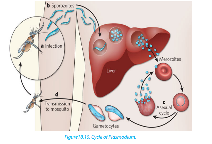
18.6.3. Trypanosoma spp.
a. Characteristics– Trypanosoma is the genus containing a large number of parasitic species whichb. Symptoms
infect wild and domesticated animals and humans in Africa.
– Commonly known as African sleeping sickness, human trypanosomiasis is
caused by the species Trypanosoma brucei and is transmitted to humans
through either a vector or the blood of ingested animals.
– The most common vector of Trypanosoma brucei is the tsetse fly, which may
spread the parasite to humans and animals through bites.
– Through a process called antigenic variation, some trypanosomes are able
to evade the host’s immune system by modifying their surface membrane,
essentially multiplying with every surface change. Trypanosoma bruceigradually infiltrates the host’s central nervous system.
Symptoms include: Headache, weakness, and joint pain in the initial stages; anaemia,
cardiovascular problems, and kidney disorders as the disease progresses; in its final
stages, the disease may lead to extreme exhaustion and fatigue during the day,
insomnia at night, coma, and ultimately death.
c. Occurrence
Human trypanosomiasis affects as many as 66 million people in sub-Saharan Africa.
Trypanosomes are also found in the Americas in the form of Trypanosoma cruzi,
which causes American human trypanosomiasis, or Chagas’ disease. This disease is
found in humans in two forms: as an amastigote in the cells, and as a trymastigote
in the blood.
d. Mode of transmission– The vectors for Trypanosoma cruzi include members of the order Hemiptera,e. Life cycle of Trypanosoma
such as assassin flies, which ingest the amastigote or trymastigote and carry
them to animals or humans.
– The parasites enter the human host through mucus membranes in the nose,
eye, or mouth upon release from the insect vectors. Left untreated, Chagas’
disease may cause dementia, megacolon, and megaesophagus, and damageto the heart muscle, and may result in death.
Trypanosoma’s cell structure plays a vital role in allowing the cell to morph into
three forms (trypomastigote, epimastigote, and amastigote) during its life cycle,
depending on where the cell is located in the host’s anatomy. The location of the
kinetoplast in relation to the nucleus and the flagellum emergence dictate in which
stage the trypanosome cell is found.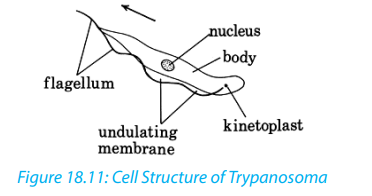
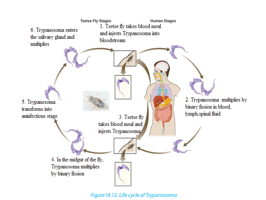
Role of Microbes
Microorganisms are usually associated with major diseases such as AIDS,
uncomfortable infections, or food spoilage.
However, the majority of microorganisms make crucial contributions to the welfare
of the world’s inhabitants by maintaining balance of living organisms and chemicals
in our environment. Therefore, microorganisms are essential for life on earth. They
have important beneficial biological functions such as:
1. Photosynthesis: Marine and freshwater microorganisms (Algae and some
bacteria) capture energy from sunlight and convert it to food, forming the
basis of the food chain in oceans, lakes, and rivers and generates oxygen which
is critical for life on Earth.
2. Decomposers: Soil microbes break down dead and decaying matter and
recycle chemical elements that can be used by other organisms.
3. Nitrogen Fixation: Some bacteria can take nitrogen from air and incorporate
it into organic compounds in soil, water, and air.
4. Digestion: Human and many other animals have microorganisms in their
digestive tract that are essential for digestion and vitamin synthesis. Examples
include:– Cellulose digestion by ruminants (cows, rabbits, etc.)5. Synthesis of chemical products: microorganisms have many commercial
– Synthesis of Vitamin K (for blood clotting) and Vitamin B (for metabolism) in
humans.
applications, such as the synthesis of acetone, organic acids, enzymes, alcohols.
6. Medicine: many antibiotics and other drugs are naturally synthesized by
microbes e.g. Penicillin is made by a mold.
7. Food industry: many important foods and beverages are made with microbes
e.g. vinegar, pickles, alcoholic beverages, green olives, soy sauce, buttermilk,
cheese, yogurt, and bread.
8. Genetic engineering: recombinant microbes produce importanta. Medical and therapeutic products: human growth hormone, insulin, blood9. Medical Research: Microbes are well suited for biological and medical
clotting factor, recombinant vaccines, monoclonal antibodies, etc.
b. Commercial products: cellulose, digestive aids, and drain cleaner.
research for several reasons:a. Relatively simple and small structures, easy to studyThough only minority of microorganisms is pathogenic (disease-causing), practical
b. Genetic material is easily manipulated.
c. Can grow a large number of cells very quickly and at low cost.
d. Short generation times make them very useful to study genetic changes.
knowledge on microbes is necessary for medicine and related health sciences. For
example, hospital workers must be able to protect patients from common microbes
that are normally harmless but pose a threat to the sick and injured.
Self-assessment 18.61. Name the causative agent of malaria.
2. The diagram below shows the life cycle of plasmodium. Analyse it andthen answer the questions that follow.
a. What is the vector of malaria?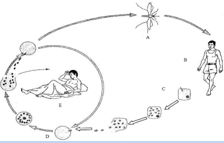
b. Between stages C and D, which one takes place in the red blood cells and
which one takes place in the hepatic cell (liver)?c. State any two symptoms of malaria displayed in individual in stage E.
End of unit assessment18
1. State any TWO diseases caused by:a. Bacteria2. What is the main feature of moulds?
b. Protozoa
c. Microscopic fungi
3. Why viruses are not generally considered to be living things?
4. The figure below shows the structure of a bacterial cell seen using an electronmicroscope.
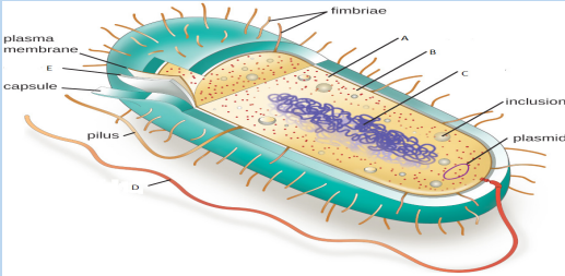 a. Name the parts labeled A, B, C and D5. The diagram below represents the structure of the human immunodeficiency
b. Describe the roles of parts B, C and Evirus (HIV/AIDS).
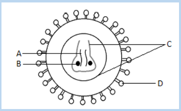 a. Name A, B, C, and D.6. Discuss the methods of reducing the risk of food poisoning by pathogenic
b. HIV/AIDS is under retroviruses. What is meant by retroviruses?
c. What type of leucocytes (white blood cells) are destroyed by HIV/AIDS?
bacteria
7. Why the hyphae of Mucor is called coenocytic?8. The figure below shows the life cycle of one of microorganisms.
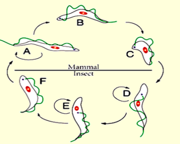 a. Which is the name of the microorganism having the life cycle represented
on this diagram of?
b. Name the parts labelled A, B, C, D, E and F9. Identify the following groups of bacteria
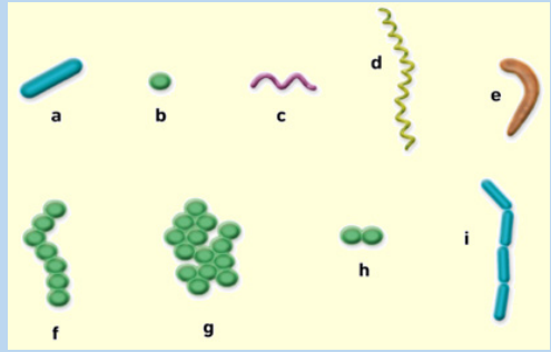
UNIT 19: CULTURING MICRO-ORGANISMS
UNIT 19: CULTURING MICRO-ORGANISMS
Key Unit Competence
Explain the process of culturing microorganisms and the factors affecting their
population growth.
Learning objectives– List and describe the roles of microorganisms and their requirements forIntroductory activity.
growth.
– Explain the role of environmental variables in culturing microorganisms.
– Describe the different types of culture media.
– Draw and interpret the graph of the population growth of bacteria.
– Carry out an experiment to stain bacteria for examination with a light
microscope.
– Observe and compare the numbers of bacteria present in fresh and stale milk.
– Distinguish between gram negative and gram positive bacteria.
– Describe the main features of aseptic techniques.
– Explain how pure cultures of pure bacteria can be obtained.
– Describe the methods of inoculation.
– Use sterile techniques to prepare agar plates to culture bacteria and fungi
– Carry out research on why microorganisms are particularly suitable for
industrial use.
– Appreciate the importance of culturing microorganisms.
– Show perseverance when inoculating a solid and liquid medium.
– Show concern for taking the basic precautions in the school laboratory whencarrying out routine microbiological work.
Use different books and visit internet make a short summary about the culture of
microorganisms and suggest why cultures are not incubated at 370 C in a school lab.
19.1. Requirements for culturing of microorganisms
Activity 19.1
Use textbooks and other sources of information to discuss the requirements of
growth of microorganisms.
Many microorganisms can be grown in the laboratory. This allows scientists to learn
a lot about them. We can find out which nutrients they need to survive and which
chemicals will kill them. We can also discover which microorganisms can be useful
to us and which cause deadly disease.
To find out more about microorganisms, you need to culture them. Culturing
microorganisms involves growing very large numbers of them so that you can see
the colony as a whole.
To culture microorganisms, you must provide them with everything they need. This
usually involves providing a culture medium containing carbohydrates to act as an
energy source. A long with this, various mineral ions some supplement of proteins
and vitamins are included.
The nutrients are often contained in an agar medium. Agar is a substance which
dissolves in hot water and sets to form a jelly. You pour hot agar containing all the
necessary nutrients into a Petri dish. Microorganisms are living organisms. Therefore,
they have requirements for their growth, maintenance and multiplication. These
include:
Optimum temperature (30-40ᵒC) for enzymes to work better.– Source of energy such as glucose, maltose, juice.The medium for culture of microbes can be the dead organic matters (food,
– Source of other nutrients (minerals such are as potassium, sodium, iron,
magnesium and calcium, vitamins, proteins
– Air for aerobic microbes or complete absence of air for anaerobic
microorganisms.
fruits, remaining of organism, juice, milk) or a prepared medium such as Agaragar
(universal medium for any germ), Lowenstein medium (selective medium for
tuberculosis bacillus). The medium can be wet or dry. Different types of media areused culture microorganisms.
19.1.1. Types of media
There are many different types of media described by their components or
ingredients.
Universal media: this allow the growth of every type of bacteria e.g. agar-agar
Differential/selective media: are specific to some types of bacteria for example
Lowenstein for tuberculosis bacteria. Their ingredients will favour growth of certain
types of bacteria.
A pure culture: this contains only one kind of microorganism. The pure cultures are
important for scientific method as they are free from other types of microorganisms.
19.1.2. Principles of sterile culturing– Wash hands before touching a sterile Petri-dish
– Open the Petri-dish as little as possible, and replace the lid quickly
– Never cough or sneeze near the dish
– Never touch the infected jiffy with fingers– When culturing is no longer required, they should be flooded with strongSafety precautions:disinfectant
After cleaning out the nutrient from Petri-dish, they should be washed and
disinfected, and then if they are glass, heat sterilize.
– Wash your hands thoroughly after all operation by using soap.
– Never push hands near the mouth during experimental work.
Bacteria grow and reproduce more quickly when they are warm than when they
are cold. It would be dangerous to incubate cultures at temperatures close to body
temperature (37°C) because doing so might allow the growth of pathogens harmful
to health. So the maximum temperature used in school and college labs is 25°C.
However, higher temperatures can be used industrially, and these produce faster
growth.
Self-assessment 19.11. What is meant by the term culturing bacteria?19.2. Culture media
2. What do bacteria need to grow?
3. Why do we culture microorganisms in the lab?
4. Explain why cultures are not incubated at 370 c in a school laboratory.
Activity: 19.2
Describe different types of media used in culturing microorganisms.
A medium is a solid or liquid preparation containing nutrients for the culture of
microorganisms. A pure microbial culture undergoes the following steps namely:– Choice of the culture medium.Microorganisms may be cultured in a solid medium or a liquid medium or broth.
– Sterilization of the culture medium.
– A culture with a collection of microbial cells growing on or, in a medium.
– Selection of a pure colony from a collection of microbial cells growing
– Introduction of a microorganism into a suitable growth medium
– Streaking to carrying out a pure culture.
When there is not a culture with a collection of microbial cells growing on or,
in a medium. A source of microorganisms is spread on the surface of an agar to
produce individual colonies. Once individual colonies are obtained, this collection
of microorganisms can then use to carry out a pure microbial culture.a. Solid medium.
Solid media are particularly suitable for bacteria and fungi and are prepared by mixing
the liquid nutrient solution with a gelling agent, usually agar, at a concentration ofabout 1-5%, thus, producing nutrient agar that allows the growth of colonies.
b. Liquid media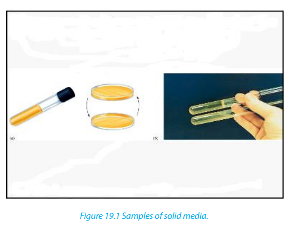
The liquid media are water – based solutions that are generally termed as broths,
milks and infusions.
Liquid media are often useful for measuring population growth. They may be placed
in a test tube, stopped by a plug of cotton wool or a metal cap, or in a glass, screw crapped
bottle such as a universal bottle which holds about 25cm2
enough for oneagar plate.
The medium must be sterilized and after, adding a small quantity of cells to the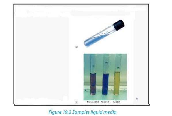
medium is called inoculation.
c. Enrichment media.
An enrichment medium is a medium in which substances are added to meet the
requirements of certain microorganisms in preference to others. As a result, certain
microorganisms grow better than others.
d. A selective medium
It is a medium in which one or more substances are added to favor the grown of
specific microorganisms and to inhibit the growth of others. Example, the addition
of penicillin to a culture to select for those organisms resisting to it, or the selectionof hybridizes cells during the production of monoclonal antibodies.
Self-assessment 19.21. How would you isolate from the soil an organism which could use19.3. Aseptic technique.
atmospheric nitrogen as its only source of nitrogen (a nitrogen-fixing
bacteria)?
2. What is meant by nutrient agar?
3. Distinguish between liquid media and solid media.4. Distinguish between enriched media and specific media.
Activity 19.3.1
Carry out a procedure of culturing fungi on a nutrient agar using sterile techniques.
Aseptic technique is using sterilized equipment and solutions and preventing their
contamination. Sterilization is the removal or destruction of all living microorganisms,
including spores (inactive structures that enable some microorganisms to
survive unfavorable periods). Bacterial and fungal spores are abundant in most
environments including laboratories. A range of special techniques and apparatus
are designed to prevent contamination of nutrients media. Autoclaves are used
to sterilize equipment and culture media before experiments and also to sterilize
equipment and specimens before disposal.
In addition, after sterilization, a great care is taken during experiments to ensure thatthere is no infection.
19.3.1. Spread plate technique
This is one of the most basic and useful of microbiological techniques. Petri dishes
are specially designed as shallow circular dish made of glass or plastic. The shape
of the lid allows avoiding contamination, but gas molecules can diffuse between
the inside of the dish and the environment through where the base meets the lid.Oxygen can therefore reach the culture and carbon dioxide can escape.
The spread plate technique involves using a sterilized spreader with a smooth
surface made of metal or glass to apply a small amount of bacteria suspended in
a solution over a plate. The plate needs to be dried at room temperature so that
the agar can absorb the bacteria more readily. A successful spread plate will havea countable number of isolated bacterial colonies evenly distributed on the plate.
19.3.2. Methods of inoculation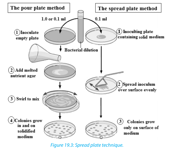
The introduction of a small number of microorganisms into a nutrient medium is
called inoculation. Aseptic technique must be used to avoid contamination. Theprocedure differs for solid and liquid media.
a. Inoculating a solid medium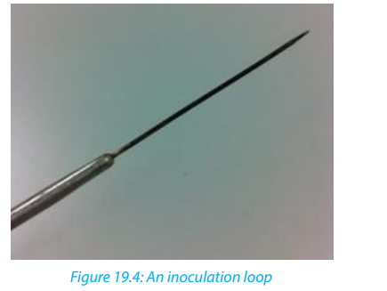
We use a wire loop. The loop is firstly flamed and after it is then used to lift a thin
film of a liquid suspension or a small amount of solid material containing the
microorganisms being investigated from the previous culture or any source of
microorganisms. The loop is gently stroked across the surface of the medium in a
series of sets of streaks.
b. Inoculating a liquid medium
If the cells to be inoculated are in a liquid, for example water or a broth, a sterile wire
loop is used to transfer a sample to the medium, which is often a test medium. The
wire loop is simply agitated gently inside the medium. If the cells to be inoculated
are in or a solid medium such as soil nutrient agar, a wire loop may be used for
transfer to the liquid medium. It can be rubbed on the inside surface of the vessel
containing the liquid medium to ensure successful transfer.
c. Carrying out a pure culture
Pure culture technique is a method of culturing microorganisms in which all of the
individuals in a culture have descended from a simple individual. The basis of pure
technique is the isolation in colonies of individual cells. This is done so as to allow thecharacterization of specific types of microorganisms.
d. Incubation on agar-agar.
During incubation, the nutrients are contained in agar medium. Agar is a substance
which dissolves in hot water and sets to form a jelly. You pour hot agar containing all
necessary nutrients such as carbohydrates, proteins and vitamins into a Petri- dish.
Then leave it to cool and set before you add any microorganism. The other way to
provide nutrients to grow microorganisms is as a broth in a culture flask. The stepsof culturing agar –agar are shown in the following activity.
Activity: 19.3.2– Boil a mixture of 50 ml of water and 20g of agar-agar powder for 15 minutes
as you are stirring
– Pour the jelly mixture into four pre-sterilized glass Petri-dishes. Then allow
the broth to coagulate at room temperature.
– Number the dishes; 1, 2, 3 and 4 respectively; on the bases.
– Place a nail scarping from between the teeth onto the jelly in dish 1 and 2,
wave the dish 3 on latrine for 1minute and do not put anything on dish 4.
– Warm the dishes 2 and 4 on the top of water vapour stream for 15 minutes
and then cool them (do not open them)
– Then fix the lids tightly to the bases of the four Petri-dishes with clear adhesive
tape and place them upside down in an oven/incubator at 37 ˚C for 3 days.– Record and interpret your results.

19.3.3 Alcoholic fermentation
Activity 19.3.3
Describe how yeast would be used in alcoholic fermentation.
Yeast releases digestive enzymes which allow the transformation of glucose into
ethanol as result of anaerobic fermentation. The presence of bubbles is the evidence
that carbon dioxide is released as waste product of the alcoholic fermentation.
Making Beer depends on a process called malting. You soak and keep barley grains
in water. As germination begins, enzymes break down the starch in the barley grains
into a sugary solution. You then extract a solution produced by malting and use it
as an energy source for the yeast. The mixture of yeast and sugar solution is then
fermented to produce alcohol. Hops are added at this stage to give flavour. The beer
is given time to clear and develops its flavour before putting it in bottles or to be
sold. Interestingly, alcohol in large quantities is toxic to yeast as well as to people.
This is why the alcohol content of wine is rarely more than 14%. Once it gets much
higher, it kills all the yeast and stops fermentation.
Self-assessment 19.3
From questions 1-5, circle the letter corresponding to the right answer
1. The method of culturing microorganisms in which all of the individuals in a
culture have descended from a single individual is called:a. Pure culture technique2. Inoculating liquid medium, various instruments are used. Which one of the
b. Spread plate technique
c. Aseptic technique
d. Liquid media method
following is used to transfer the sample to the medium?a. Sterile wire loop3. Large amounts of alcohol are dangerous to yeast during alcoholic fermentation.
b. Inoculating needle.
c. Petri-dishes
d. None of these.
Which of the following explains the reason?a. It kills all the yeast and stops fermentation.4. The technique of using sterilized equipment and solutions and preventing their
b. Motivate yeasts
c. It kills some few bacteria.
d. Temperature affects fermentation.
contamination is referred to as:a. Pure culture technique5. Petri dishes are specially designed as a shallow circular dish made of glass or
b. Spread plate technique
c. Aseptic technique
d. Petri-dish technique.
plastic with a lid. Which one of the following best explains the function of the
lid?a. Prevent contamination, but gas molecules can diffuse.19.4. Population growth of bacteria
b. Spread the bacteria on the plate.
c. Allows contamination.d. None of the above.
Activity: 19.4.1
Use text books and other sources of information to interpret the graphs of bacterial
growth.
When bacteria or any other microorganisms are incubated in a suitable culturing
medium, they reproduce by binary fissions and the number of individuals increases.
The ordinary growth of population is described as sigmoid curve or S-shaped curve
made of 4 main phases:
– The lag phase: period of adaptation of microorganisms to the new habitat
(new environment)
– The log or exponential phase: period of high rate of reproduction. Bacteria are
sensitive to the limiting factors of the growth or anti-microbial agents
– The stationary phase: Stationary phase of plateau-growth slows down. The
population remains constant because the rate of dividing/growth is equal to
the rate of death within the population. The maximum number that a habitat
can accommodate for a long period is known as the carrying capacity.
– The decline or death phase: period of high rate of death than the rate of
dividing/growth due to the scarcity of food, the abundance of metabolic waste
products, presence of antibiotics or any other drugs killing the germs. Figure19.5 shows the phases explained above.
19.4.1. Measuring population growth of bacteria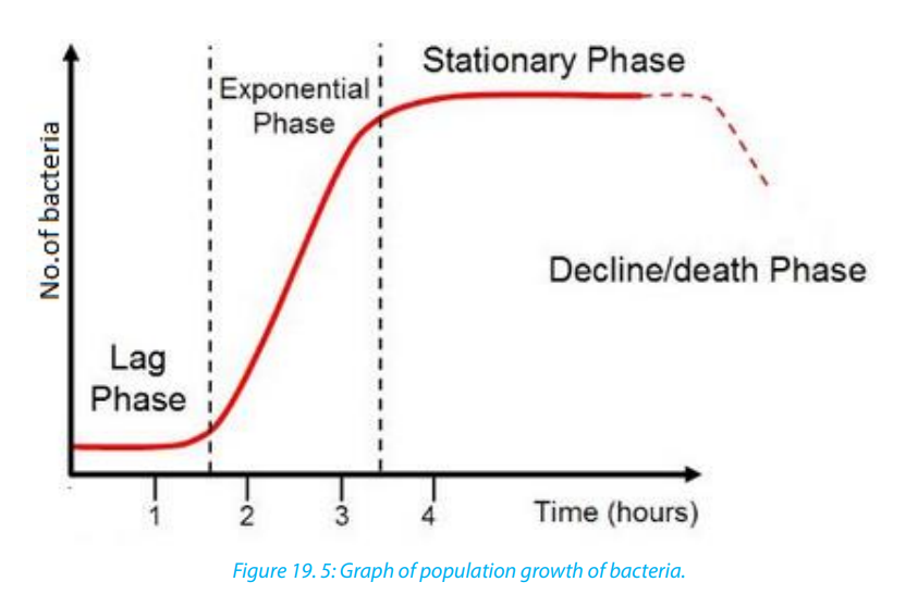
The typical growth curve of a population of bacteria is similar to the growth curve
expected for yeast, a unicellular fungus or the growth of any population. When
measuring the growth of a population of bacteria or yeast, we can carry out direct
counting of the numbers of cells or indirectly by measuring some indication of thenumber of cells such as the coldness of a solution, or production of a gas
It is usual to inoculate a small sample of the microorganisms in a sterilized nutrient
medium and to place the culture in an incubator at the optimum temperature for
growth. Other conditions are pH, oxygen concentration and ionic and osmotic
balance. Growth can be measured from the time of inoculation. Two types of cell
count are possible, namely viable count and total count. The viable count is the total
of living cells only and total count is the total number of both living and dead cellsand is easier to measure.
Activity 19.4.2
Investigating the bacterial content of fresh and stale milk.
Materials required: Four sterile nutrient agar plates, inoculating loop, Bunsen
burner, indelible marker or wax pencil, Fresh pasteurized milk, Stale milk (milk left
at room temperature for 24hours) and Incubator set at 350C
Procedure:– Place the inoculating loop in the Bunsen burner flame until the loop is red hot.
– Allow the loop to cool and then dip into a sample of fresh, well shaken milk.
– Lift the lid of the sterile agar plate slightly with the other hand and lightly
spread the contents of the inoculating loop over the surface the agar.
– Close the lid of the plate and return the loop to the Bunsen burner flame until
red hot.
– Label the base of the plate with an indelible marker or pencil.
– Repeat the process with the second plate and another sample of fresh milk.
– Flame the loop again and after cooling, place it in a sample of stale milk.
– Spread the contents of the loop over the surface of a third plate and then
close the lid.
– Label the base of the plate with an indelible marker or pencil.
– Repeat the process with the fourth plate and second sample of stale milk.
– Put the four plates in an incubator at 350
c for about 3 days. They should be
placed upside down to prevent condensation falling onto the cultures. After
incubation, the two halves of each plate should tape together for safety.
– Record the appearance of the coloniesGive the general comment based on your observations
Self-assessment 19.4
1. A culture of yeast, Saccharomyces cerevisiae, had been carried out in the banana
juice for 7 days at 30°C.The table below shows the change in number of yeastswithin that time:
 a. Draw a curve showing the growth of the yeast population2. Design an experiment to test the hypothesis that contact of an agar plate with a
b. What is the role of banana juice in that experiment?
c. State any two conditions that should be maintained constant during that
experiment.
d. Describe the trend of the graph you have drawn in afinger results in more bacterial growth than exposing the plate to classroom air.
19.5. Staining of bacteria
Activity 19. 5
“Staining bacteria for practical purpose is important”. Discuss the validity of the
statement.
The microorganisms or parts of microorganisms that pick up the stain are clearly
distinctively observed from the rest of the background.
In simple staining, all the cells and structures in general stain the same colour. In
positive staining, cells structures take in the stain e.g. methylene blue while in
negative staining the cells repel the stain and it is taken by the background e.g. Indian
ink. Negative staining is mostly useful in viewing capsules and such structures thatsurround the bacteria.
Differential staining on the other hand, multiple staining reactions are used to
take advantage of the fact that particular types of microorganisms and/or specified
structures of microorganism display varied staining reactions that are readily
distinguishable by different colours. The stain must be fixed immediately and the
dyed specimen is treated in some ways, e.g. by chemicals or heat to tightly bind thestain to the organism or its structures.
19.5.1. The purpose of staining bacteria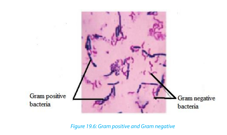
The purpose of staining bacteria is to see, for example, how thick of a layer of
peptidoglycan their cell wall has. In the Gram stain, gram-negative bacteria will stain
red or pink because the rinse took out the primary dye and the Safrinin (secondary
dye) took over the coloring as the counterstain. In gram-positive bacteria, since it
has a thick-layer of peptidoglycan, not all of the Crystal violet color will be rinsed out
of the cell wall, so it will be blue or purple. The following are reasons to explain why
stained:– It’s for helping classifying and determining what the bacteria are composed of.
– It’s very useful tool to help identify bacteria without necessarily killing the cell.– Gram staining is performed to distinguish between gram positive and negative
bacteria.– To enable the person to visualize its physical features- shape, size, arrangement,19.5.2. Procedure of staining and their corresponding stains.
etc the bacterial cells are stained with specific dyes or stains
Activity 19.5.1:
carry out an experiment to stain bacteria for examination under the light
microscope
In staining bacteria, we use various staining procedures each having specific set of
stains or dyes. Some of them are:1. Gram’s Staining - Crystal violet, Iodine and SafrininObserve and identify some of the staining methods on figure19.6 as shown below:
2. Capsule staining - Nigrosin, Safrinin or India Ink, Safrinin
3. Spore staining - Malachite Green and Safrinin
4. PHB staining - Sudan black.
5. Using decolorizer – Alcohol wash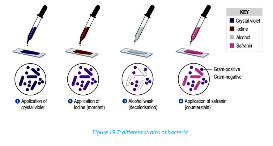
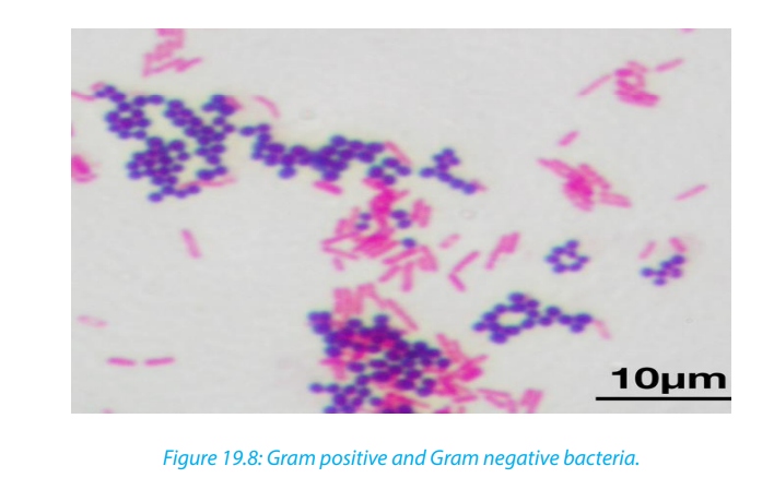
19.5.3 Growing viruses
The culture of viruses is made more difficult than the culture of bacteria or fungi because
viruses can only grow and multiply inside living cells. This can be done by infecting whole
Figure 19.8: Gram positive and Gram negative bacteria.
19.5.3. Growing viruses
The culture of viruses is made more difficult than the culture of bacteria or fungi
because viruses can only grow and multiply inside living cells. This can be done
by infecting whole organisms such as plants or animals but, where possible, cell,
tissue cultures are now used. An early technique was to grow certain viruses in chickembryos while the embryo was still growing inside the egg.
19.5.4. Tissue Culture of Animal Viruses
Viruses cannot be grown in standard microbiological broths or on agar plates;
instead they have to be cultured inside suitable host cells. Note the following facts:– Tissue culture is a useful method for cultivating clinical samples suspected ofCell culture is the complex process by which cells are grown under controlled
harboring a virus. This method helps with the detection, identification, and
characterization of viruses in the laboratory.
– Tissue culture of animal viruses involves growing animal cells in flasks using
various broth media and then infecting these cells with virus.
– Transfect ion can be carried out using calcium phosphate, by electroporation,
or by mixing a cationic lipid with the material to produce liposome’s, which
fuse with the cell membrane and deposit their content inside.
– Cytopathic effect is a non-lyrics damage that viruses cause to cells. These vary
in their manifestation and damaging effect.
– Cell culture is complex process by which cells are grown under controlled
conditions, generally outside of their natural environment.
conditions, generally outside of their natural environment. The term “cell culture” is
defined as the culturing of cells derived from multi-cellular eukaryotes, especially
animal cells. However, there are also cultures of plants, fungi, and microbes, includingviruses, bacteria, and protists.
2. Explain why it is more difficult to culture viruses than culturing bacteria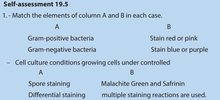
3. How/why are viruses specific to the cells they infect?
4. Distinguish between vaccines and antibiotics
End of unit assessment19
1. What are different types of media used in the laboratories for culturing
microorganisms?
2. Define a pure culture.
3. How do biologists differentiate between Gram –positive and Gram –negative
bacteria?
4. Describe the three methods of preventing bacterial growth in food.
5. How does temperature affect the growth of bacteria in culture media?
6. Assuming that you have a bacterial infection, would you ask for vaccination
against the bacteria? Why or why not?
7. How do bacteria maintain the balance in the environment?
8. Explain why an infection by Gram–negative bacteria are more difficult to treat
than Gram-positive bacteria.
9. How would you investigate that temperature affect the bacterial growth?
10.Write short notes on each of the following term related to the culture of
microorganisms.a. Aseptic techniques.
b. Staining bacteria
c. Growing viruses11. Explain why microorganisms are particularly suitable for industrial use.
UNIT 20: BIOTECHNOLOGY AND ITS APPLICATION
UNIT 20: BIOTECHNOLOGY AND ITS APPLICATION
Key Unit Competence
Explain the biotechnology involved in the production of ethanol, biogas and bread
making.
Learning Objectives
By the end of this unit, I should be able to:– State that bacteria are useful in biotechnology and genetic engineering dueIntroductory activity:
to their rapid reproduction rate and their ability to make complex molecules.
– Discuss why bacteria are useful in biotechnology and genetic engineering.
Focus on: lack of ethical concerns over their manipulation and growth, genetic
code shared with all other organisms, and presence of plasmids.
– Show concern for the role of bacteria in genetic engineering.
– Investigate and describe the use of pectinase in fruit juice production and
lactase to produce lactose-free milk.
– Describe the role of anaerobic respiration in yeast during bread-making.
– Compare leavened and unleavened bread.
– Appreciate the role of anaerobic respiration in the production of ethanol and
in yeast during bread-making.
– Explain how fermenters are used in the production of penicillin.
– Describe the role of the fungus Penicillium in the production of the antibiotic
penicillin.
– Interpret and explain graphs showing how the pH and the concentration of
penicillin in a culture changes over time when the pH is controlled and not
controlled.
– Defend the role played by antibiotics in treatment of bacterial diseases.
– Describe the three stages of biogas production and the role of bioreactors in
economically poor rural communities
– Apply the knowledge of bioreactors, using cow dung, agricultural waste and
domestic waste to prepare and produce biogas.
– Appreciate the role of biogas production in reducing the environmentaldegradation.
Biotechnology is a broad discipline in which biological processes, organisms, cells
or cellular components are exploited to develop new technologies, remember
that biotechnology is useful and applied in our daily life activities such as in
beverages and food industries, agricultures, medicines.Brainstorm on the role
of microorganisms in biotechnology and genetic engineering.Can you think on
your own understanding on howbread, juice and beer are made? Why do bacteriabecome resistant to antibiotics? Make discuss on the biogas production.
20.1. Role of bacteria in Biotechnology and genetic
engineering
Activity 20.1
Using addition resources to your textbook available in your school such as the
books from the school library and search further information from the internet.
Discuss the role of bacteria in biotechnology and genetic engineering.
Biotechnology is a broad discipline in which biological processes, organisms, cells
or cellular components are exploited to develop new technologies. New tools
and products developed by biotechnologists are useful in research, agriculture,
industry and the clinic. For example, the use of living cells, bacteria, etc., to make
useful products (such as crops that insects are less likely to destroy or new kinds of
medicine).
The wide concept of “biotech” or “biotechnology” encompasses a wide range of
procedures for modifying living organisms according to human purposes, going
back to domestication of animals, cultivation of the plants, and “improvements” to
these through breeding programs that employ artificial selection and hybridization.
Modern usage also includes genetic engineering as well as cell and tissue culture
technologies.
The bacteria have an economic importance which derives from the fact that bacteria
are exploited by humans in a number of beneficial ways. Despite the fact that some
bacteria play harmful roles, such as causing diseases and spoiling food, the economicimportance of bacteria includes both their useful and harmful aspects.
20.1.1. Useful Bacteria in Biotechnology
Biotechnology or Industrial microbiology is defined as the use of microorganism
such as bacteria, fungi and algae for the manufacturing and services industries.
These include:– Fermentation processes, such as brewing, baking, cheese and butter
manufacturing, Bacteria, often Lactobacillus bulgaricusin combination with
yeasts and fungi, is used to make yoghurt and cheese have been used for
thousands of years in the preparation of fermented foods such as cheese,
pickles, soy sauce, sauerkraut, vinegar, and wine.
– In the chemical industry, bacteria are most important in the production of
pure chemicals for use as pharmaceuticals or agrochemicals.
– Bacteria are also used in chemical manufacturing such as ethanol, acetone,
organic acid, enzymes, and perfumes.
– Bacteria can also be used in the place of pesticides in Biological Pest Control.
This commonly uses Bacillus thuringiensis (also called BT), a Gram-positive,
soil-dwelling bacterium.– Saprophytic bacteria help in breaking of complex organic substance to simpler
forms. Thus, in this process, they help to convert farm refuse, dung and other
wastes to manure.– Number of anti-bacterial and anti-fungal antibiotics such as Hamycin,20.1.2. Useful Bacteria in Genetic engineering
Polymyxin, and Trichomycin are obtained from fungal mycelia and bacteria
(like Streptomyces). Similarly, Bacillus is used for production of antibiotics such
as Bacitracin and Gramicidin.
– Different kinds of vitamins are produced from bacteria like Riboflavin from
Clostridium butylicum, Vitamin B12 from Bacillus megatherium and Vitamin Kand B-complex from Escherichia coli.
Genetic engineering is the manipulation of genes. It is also called recombinant DNA
technology. In genetic engineering, the genetic information for many biological
products and biological processes can be introduced into microbes in order to
genetically engineer them to produce a substance or conduct a process. The
genes can come from any biological source: human, animal, plant or microbes. This
opens the possibility for microbial production of foods, fuels, enzymes, hormones,
diagnostic agents, medicines, antibiotics, vaccines, antibodies, natural insecticides
and fertilizers, and all sorts of substances useful in our civilization and society.
The pieces of DNA (genes) are introduced into a host by means of a carrier (vector)
system. The foreign DNA becomes a permanent feature of the host, being replicated
and passed on to daughter cells along with the rest of its DNA. Microorganisms
especially bacteria play a central role in recombinant DNA technology and genetic
engineering. Important tools of biotechnology are microbial cells (bacteria, fungi),microbial genes and microbial enzymes.
Bacterial cells are transformed genetically and used in production of commercially
important products. For example, bio medical technology bacteria can be
bioengineered for the production of therapeutic proteins like: Human Insulin (used
against diabetes), Human Growth Hormone (somatotropins used to treat pituitary
dwarfism), and others which can be used to fight against viral diseases. Antibiotics are
produced in nature by molds such as Penicillium and bacteria such as Streptomycesand Bacillus.
Self-assessment 20.11. What is biotechnology?
2. What do you understand by genetic engineering?20.2. Immobilization of enzymes3. Discuss on the role of bacteria in Biotechnology and genetic engineering.
Activity 20.2
Carry out research on the action of enzymes with reference to pectinase in fruit
juice production and lactase to produce lactose-free milk.
Enzymes catalyze biological reactions in our body, but they can also be used to
catalyze industrial reactions outside the body. These enzymes are often bound toa support (‘immobilized’) and can be used for a wide range of purposes.
20.2.1. The advantages of immobilized enzymes
Enzymes have an enormous range of commercial applications for example: in
medicine, food technology and industrial processing. Enzymes are expensive. No
company wants to have to keep buying them over and over again if it can recycle
them in some way. One of the best ways of keeping costs down is to use immobilized
enzymes. Using immobilized enzymes means that you can keep and re-use the
enzymes, and that the product is enzyme-free. Another advantage of this process
is that the immobilized enzymes are more tolerant of temperature changes and
pH changes than enzymes in solution. This may be partly because their molecules
are held firmly in shape by the alginate in which they are embedded, and so do
not denature as easily. It may also be because the parts of the molecules that areembedded in the beads are not fully exposed to the temperature or pH changes.
Using enzymes instead of other molecules in reactions is useful because enzymes
catalyze specific reactions and work at much lower temperatures than chemical
catalysts.
The molecule that an enzyme acts on is called a substrate. Enzymes can either be
mixed freely with the substrate in solution or immobilized to a solid support so they
do not mix freely. There are many advantages of immobilization, one of which is that
the enzymes can be reused catalyzing the same reaction many times. Binding the
enzymes to a surface also makes those more stable and less likely to denature (lose
their shape). In addition, there will be no enzyme left in the product at the end, sopurification is not necessary
20.2.2. The disadvantages of immobilized enzymes
There are some disadvantages: immobilization requires extra time, equipment and
work; there may be a reduction in reaction rates if enzymes cannot mix freely with
the substrate; and immobilized enzymes cannot be used if one of the substrates is
insoluble.
20.2.3. Advantages of Using Immobilized Enzymes
The advantages of using immobilized enzymes are: (i) reuse (ii) continuous use (iii)
less labor intensive (iv) saving in capital cost (v) minimum reaction time (vi) less
chance of contamination in products, (vii) more stability (viii) improved process
control and (ix) high enzyme: substrate ratio. The first immobilized enzymes to be
scaled up to pilot plant level and industrial manufacture were immobilized aminoacid acylase, penicillin G-acylase and glucose isomerase.
20.2.4. Methods of Enzyme Immobilization
There are five different techniques of immobilizing enzymes: (i) adsorption, (ii)
covalent bonding, (iii) entrapment, (iv) copolymerization or cross-linking, and (v)
encapsulation. For the purpose of immobilization of enzymes carriers i.e. the supportmaterials such as matrix system, a membrane or a solid surface are used.
i. Adsorption
An enzyme may be immobilized by bonding to either external or internal surface of
a carrier or support such as mineral support (aluminum oxide, clay), organic support
(starch), and modified sapharose and ion exchange resins. Bonds of low energy are
involved e.g. ionic interactions, hydrogen bonds, van der Waals forces, etc. If the
enzyme is immobilized externally, the carrier particle size must be very small in
order to achieve appreciable surface of bonding. These particles may have diameter
ranging from 500 A to about 1 mm. Due to immobilization of enzymes on externalsurface, no pore diffusion limitations are encountered.
In addition, the enzyme immobilized on an internal surface is protected from
abrasion, inhibitory bulk solutions and microbial attack, and a more stable and active
enzyme system may be achieved. There are four procedures for immobilization by
adsorption : (i)static process (enzyme is immobilized on the carrier simply by allowing
the solution containing the enzyme to contact the carrier without stirring (ii)the
dynamic batch process (carrier is placed into the enzyme solution and mixed by
stirring or agitated continuously in a shaker), (iii) the reactor loading process (carrier
is placed into the reactor that will be subsequently employed for processing, then
the enzyme solution is transferred to the reactor and carrier is loaded in a dynamic
environment by agitating the carrier and enzyme solution), and (iv) the electrode
position process (carrier is placed proximal to one of the electrodes in an enzyme
bath, the current put on, the enzyme migrates to the carrier and deposited on thesurface).
ii. Covalent bonding
Covalent bond is formed between the chemical groups of enzyme and chemical
groups on surface of carrier. Covalent bonding is thus utilized under a broad range
of pH, ionic strength and other variable conditions. Immobilization steps are
attachment of coupling agent followed by an activation process, or attachment of a
functional group and finally attachment of the enzyme. Different types of carriers are
used in immobilization such as carbohydrates proteins and amine-bearing carriers,
inorganic carriers, etc. Covalent attachment may be directed to a specific group (e.g.
amine, hydroxyl, tyrosyl, etc.) on the surface of the enzyme. Hydroxyl and amino
groups are the main groups of the enzymes with which it forms bonds, whereassulfhydryl group least involved.
There are different methods of covalent bonding such as: (i)diazoation (bonding
between the amino group of the support e.g. aminobenzyle cellulose, aminosilanised
porous glass, aminoderivatives and a tyrosyl or histidyl group of the enzyme), (ii)
formation of peptide bond (bond formation between the amino or carboxyl group
of the support and amino or carboxy group of the enzyme), (iii) group activation
(use of cyanogen bromide to a support containing glycol group i.e. cellulose,
syphadex, sepharose, etc.), and (iv) poly functional reagents (use of a bifunctional
or multifunctional reagent e.g. glutaraldehyde which forms bonding between the
amino group of the support and amino group of the enzyme). The major problem
with covalent bonding is that the enzyme may be inactivated by bringing about
changes in conformation when undergoes reactions at active sites. However, this
problem can be overcome through immobilization in the presence of enzyme’s
substrate or a competitive inhibitors or protease. The most common activatedpolymers are celluloses or polyacrylamides
iii. Entrapment
Enzymes can be physically entrapped inside a matrix (support) of a water soluble
polymer such as polyacrylamide type gels and naturally derived gels e.g. cellulose
triacetate, agar, gelatin, carrageenan, alginate, etc. The form and nature of matrix
vary. Pore size of matrix should be adjusted to prevent the loss of enzyme from the
matrix due to excessive diffusion. There is possibility of leakage of low molecular
weight enzymes from the gel. There are several methods for enzyme entrapment:
(i)inclusion in gels (enzyme entrapped in gels), (ii) inclusion in fibers (enzyme
entrapped in fiber format), and (iii)inclusion in microcapsules (enzymes entrapped
in microcapsules formed monomer mixtures such as polyamine and polybasic
chloride, polyphenol and polyisocyanate). The entrapment of enzymes has been
widely used for sensing application, but not much success has been achieved with
industrial process.
iv. Cross - linking or Co-polymerization
Cross-linking is characterized by covalent bonding between the various molecules
of an enzyme via a polyfunctional reagent such as glutaraldehyde, diazonium salt,
hexamethylenedisocyanate, and ethylene bismaleimide. The demerit of using
polyfunctional reagents is that they can denature the enzyme. This technique is
cheap and simple but not often used with pure proteins because it produces very
little of immobilized enzyme that has very high intrinsic activity. It is widely used incommercial preparation.
v. Encapsulation
The encapsulation is the enclosing of a droplet of solution-of enzyme in a
semipermeable membrane capsule. The capsule is made up of cellulose nitrate and
nylon. The method of encapsulation is cheap and simple but its effectiveness largely
depends on the stability of enzyme although the catalyst is very effectively retained
within the capsule. This technique is restricted to medical sciences only. In this
method a large quantity of enzyme is immobilized but the biggest disadvantage isthat only small substrate molecule is utilized with the intact membrane.
20.2.5. How are immobilized enzymes used in food?
i. Immobilization of enzymes use of pectinase in fruit juice production
Pectinases find commercial application in fruit juice, wine, oil, tea, coffee, textile and
paper‐making industries using a wide variety of carriers and methods. One of the
vital applications is the clarification and depectinization of fruit juices. The raw fruit
juice obtained after pressing is very turbid viscous and contains a significant amount
of colloidal compounds, mainly pectin that causes cloudiness; therefore, clarification
of fruit juices involves the removal of juice haze by enzyme hydrolysis with pectolytic
enzymes. Although pectinases enhance the clarification of juices, immobilization of
these enzymes proves to be beneficial for industrial use. Immobilization of pectinase
on celite through adsorption is a simple, cheap and effective method. For the
clarification of pineapple juice, excellent results were observed using immobilizedpolygalacturonase in comparison with free enzyme.
Fruits contain pectin, carbohydrates found in the cell wall that holds the plant cells
together. Immobilized pectinase can be used to break down this pectin, loosening
the connections between cells. This increases the amount of juice you can get fromthe fruit, makes the juice runnier and gets rid of the cloudiness that pectin can cause.
ii. Making lactose-free milk
The enzyme lactase breaks down the sugar lactose, which is found in milk, into the
sugars glucose and galactose. Most people produce this enzyme in their bodies,
but some people (and most cats) don’t, meaning that they are lactose intolerant.
Because they can’t break down lactose, it builds up in their digestive system where
bacteria feed on it, causing digestive problems.
Immobilized lactase can be used to produce lactose-free milk: normal milk is poured
down a column containing the immobilized lactase enzymes, which break down
the lactose. After the milk has passed through this system, it will only contain the
products of the reaction (glucose and galactose), so lactose-intolerant people (and
cats) can drink it. The enzyme lactase can be immobilized using alginate beads.
The figures below show one way in which enzymes can be immobilized. The enzyme
is mixed with a solution of sodium alginate. Little droplets of this mixture are then
added to a solution of calcium chloride. The sodium alginate and calcium chloride
instantly react to form jelly, which turns each droplet into a little bead. The jelly bead
contains the enzyme.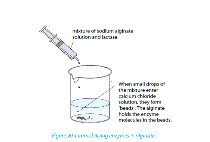
The enzyme is held in the bead, or immobilized. These beads can be packed gently
into a column. Milk is then allowed to run through the column of lactase-containing
beads. The lactase hydrolyses the lactose in the milk to glucose and galactose. The
milk is therefore lactose-free, and can be used to make lactose free dairy products
for people who cannot digest lactose. The product continues to trickle down the
column, emerging from the bottom, as illustrated in the diagram below, where it
can be collected and purified. Not only would you lose the lactase, but also you
would have milk contaminated with the enzyme.
iii. Biological washing powders containing enzymes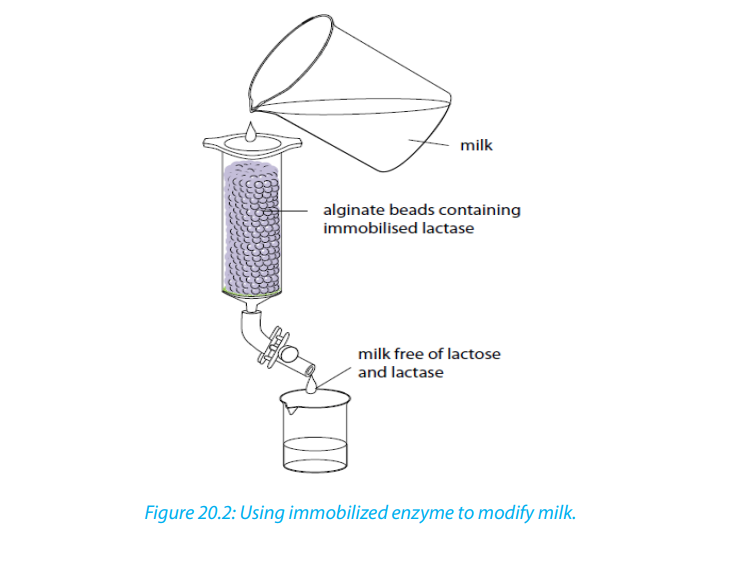
The biological washing powders contain enzymes like protease and lipase to remove
protein stains and fat/grease from clothes. The enzymes break down proteins or fats
on the fabric, forming water-soluble substances that can be washed away. Because
stains are made of different types of molecules, a range of enzymes are needed to
break them down. Proteases break down proteins, so are good for blood, egg, gravy,
and other protein stains. Amylases break down starches, and lipases break down fats
and grease.
For example: Blood contain the red protein Haemoglobin (Hb). The Proteases in
biological washing powder break Hb molecules into smaller molecules, which
are not colored and which dissolve in water and can be washed away. This makes
the washing powder more effective than detergent alone, especially at lower
temperatures. This save energy (no need to boil water), but if the temperature istoo high, the enzyme will be denatured.
iv. Fruit juices
Fruits contain pectin, carbohydrates found in the cell wall that holds the plant
together. Immobilized pectinase enzyme can be used to break down this pectin,
loosening the connections between cells. This increases the amount of juice you can
get from the fruit, makes the juice runnier and gets rid of the cloudiness that pectincan cause.
20.2.6. How are immobilized enzymes used in biosensors?
The specificity of enzymes means that they can be used to test for a unique substance,
which is exactly what a biosensor does.
Glucose test strips
People with type 1 diabetes lack the hormone insulin, so they have to test their
blood sugar levels regularly to ensure they stay within a healthy range. They do this
by measuring the amount of glucose in their blood with a glucose test strip. On
the test strip is the immobilized enzyme glucose oxidase; when glucose is present,
the enzyme catalyzes a reaction that changes glucose into hydrogen peroxide and
gluconic acid.
There is also another mediator molecule on the test strip, which catalyses a reaction
involving the products of the enzyme reaction. In the early test strips, this second
reaction caused a color change, with the color indicating the amount of glucose
present. In most modern tests, this second reaction produces electrical current,
which can be measured by a meter to give the exact concentration of glucose in theblood.
Self-assessment 20.2
1. Discuss the advantages and disadvantages of immobilized enzymes.
2. Write on the use of pectinase in fruit juice production.
3. Explain the role of lactase in making lactose-free milk.4. How are immobilized enzymes used in biosensors?
20.3. Application of enzyme in technology.
Activity 20.3
Visit a nearby bakery and verify how bread is prepared. Write a short report on theraw materials and procedures used in making bread up to the final product.
20.3.1. Enzymes in Brewing
Enzymes increase processing capacity and improve economy in the fruit juice
and wine industries. The most commonly used enzymes in these industries are
pectinase. Pectinase increases juice yields and accelerate juice clarification. They
produce clear and stable single-strength juices, juice concentrates and wines, from
not only core-fruits such as apples and pears, but also stone fruits, berries, grapes,
citrus-fruits, tropical fruits and vegetables like carrots, beets and green peppers.
Future aspects focus on a wider application of enzymes to brew with high amounts
of inexpensive raw materials like barley. Barley contains starch that has to be broken
down to fermentable sugars before the yeast can make alcohol. Therefore, traditional
brewing contains an extra step compared with wine-making, namely malting in
which enzymes needed for the degradation of starch into fermentable sugars areproduced.
20.3.2. Enzymes perform many functions in beverages
The most important field of application for enzymes in the beverage industry is the
extraction of fruit juice and vegetable juice. Pectinases, in particular, are employed
for apple and pear juice and for juices made from berries and tropical fruits. They
break down pectins found in the plant cell walls as supporting substances. This
increases the quality of juice extracted and reduces fruit waste. Enzymes can be
used in winemaking to increase the preliminary juice extraction and to obtain more
high-quality wine. Pectinase not only increase juice yields, but also increase the
colour and health-promoting antioxidants in fruit and vegetable juices. They also
increase colour extraction and juice volume by reducing fruit and vegetable mash
viscosity and improving solid/liquid separation, Pectinase and Amylase enzyme
solutions speed up filtration and prevent storage or post-packaging haze formationby depectinizing and reducing starch in raw juices.
20.3.3. Medical applications of enzymes
Development of medical applications for enzymes has been at least as extensive
as those for industrial applications, reflecting the magnitude of the potential
rewards: for example, pancreatic enzymes have been in use since the nineteenth
century for the treatment of digestive disorders. The variety of enzymes and their
potential therapeutic applications are considerable. At present, the most successful
applications are extracellular: purely topical uses, the removal of toxic substances
and the treatment of life-threatening disorders within the blood circulation.
20.3.4. Applications of enzymes in baking
For decades, enzymes such as malt and fungal alpha-amylases have been used in
bread-making. Rapid advances in biotechnology have made a number of exciting
new enzymes available for the baking industry. The importance of enzymes is likely
to increase as consumers’ demand more natural products free of chemical additives.
For example, enzymes can be used to replace potassium bromate, a chemical
additive that has been banned in a number of countries.
20.3.5. Application of enzymes in cheese
The most obvious use of enzyme action in the dairy industry is the coagulation of milk
by chymosin. Yet there are many other examples of the involvement of enzymes in
determining the quality of milk and milk products that, when the role of the enzyme
is properly understood, could be used by the industry to improve the profitability,
quality and safety of milk production, and product manufacture. Compared with
sectors such as starch hydrolysis, the volume of enzyme use in the dairy sector is low,
yet there are many opportunities for specialized applications in product ripening,
quality control, preservation and genetic improvements to fermentation cultures.
20.3.6. Application of enzymes in yoghurt
Like cheese, yoghurt is produced from milk by the action of lactate producing
bacteria, especially lactobacillus bulgaricus and streptococcus thermophiles. These
bacteria are commonly used in yoghurt starter cultures. Fermentation produces
lactate which brings the pH down to about 4.0. Fermentation-by products, including
ethanal and methanoic acid, give yoghurt its characteristics flavor. Sometimes fruit
pulp, coloring and flavors are added before packaging. Some yoghurt is heat-treated
before or after packaging to kill any bacteria, but most yoghurt contain live bacteria.
20.3.7. Application of enzymes in breads making.
Bread production involves harvesting the wheat, separating the grain from the husk,
crushing the grain to make flour, mixing the flour with water and then finally baking
it. The main difference between unleavened and leavened bread is that leavened or
risen bread uses leavened dough, and unleavened bread does not. If the leavened
bread is desired, then one adds yeast and allowing the bread to sit for a specificamount of time, depending on the type of bread being made.
Types of Unleavened Bread
1. Chapatti: Our staple chapatti is widely consumed across India and is a great
example of unleavened bread. It is made using atta flour although there are
variations that replace atta with wheat, gram, corn flour, or a combination of all
three.
2. Matzah: Jews only consume matzah during the Jewish Passover, which is
unleavened bread. This bread is consumed in remembrance of the Jewish exodus
of Egypt, during which the Jews fled in such haste that there was no time to allow
their breads to rise up. Matzah is made according to strict interpretations of the
Torah using kosher flour whole grain wheat flour.
3. Tortilla: Commonly eaten in Mexico and Spain, tortillas are made from corn flour
or wheat flour and are similar in appearance to the chapati. Tortillas are flattened
and browned over a skillet.
4. Pancakes: Pancakes without yeast are considered to be unleavened. Most
pancakes are cooked on a griddle and flipped over once the first side has beencooked.
Types of Leavened Breads
Yeast is commonly used to leaven bread and is typically added with sugar or honey
to catalyse and activate the yeast in order for the bread to rise. Breads made with
yeast is normally allowed to rest for an hour so that it can rise and double in size. It
is then punched down and allowed to rise once again before baking. Most types of
yeast breads include standard sandwich bread, pizza crust, donuts, and loaf breads
and so on.
While yeast is a commonly used leavening agent, it is not the only ingredient that
can be used for leavening. Quick breads are any type of breads that are made with
an ingredient other than eggs or yeast as a leavening agent. Baking soda and baking
powder are common leavening agents and both usually have salt added to the
recipe to activate the leavening agent. Quick breads, unlike yeasted breads, are not
let to rest before baking. Common types of quick breads include biscuits, muffins,
scones, banana bread and cornbread. There are also loaf breads like soda breads
which are a type of quick breads. Some donut and pizza recipes are made in thequick bread version.
Steps involved in bread making
The dough that we make in our bakeries follows all of these 10 steps from start to
finish. This ensures we produce the best quality bread without compromising taste,
texture, nutrition or our artisan craft. As a home baker, if you follow these 10 steps
when making breads at home, you will be on the right path to creating superb loaves.
1. Ingredients used to make breads
Using good quality ingredients is crucial to making good bread. The main ingredients
include: bread-flour, dry yeast (‘rapid rise’), levain (sourdough), salt, water, sugar, and
eggs.
2. Mixing
There are two stages to the mixing process: the first is to incorporate ingredients,
the second is to develop the structure of the dough, otherwise known as the gluten
network. Dough can be kneaded by hand, or mixed in a table top mixer. When using
a table top mixer, keep it to the lower speeds to avoid damaging the motor.
3. Primary Fermentation
Also referred to as rising, or proofing, this is where the yeast starts to do its work,
converting sugars into carbon dioxide, alcohol and organic acids. Every dough has
a different primary fermentation time, depending on its formulation. We work with
time as well as our senses to determine when the dough is properly fermented.
4. Divide and Pre-Shape
When the dough is properly fermented, it is time to divide it to the desired size and
give the divided pieces a preshape. A preshape is an intermediate shape a loose
suggestion to the dough of where it’s headed that will make final shaping easier.
5. Bench Rest
After the dough has been preshaped, it needs to rest for a short time before final
shaping. Bench rest is typically 15-20 minutes long and during that time, the gluten
network, which has been made more elastic through handling, will relax and become
more extensible.
6. Final Shaping
There are four basic shapes in bread making: the baguette (stick), the boule (round),
the bâtard (a football-like shape) and the pan loaf. After shaping, the dough must
be set somewhere to rest during its final fermentation. For baguettes and bâtards,
we use baker’s linen and wooden boards; for boules, we often use wooden proofing
baskets. The linen and the baskets help to hold the shape of the dough during the
final fermentation.
7. Final Fermentation
After shaping, the dough must rest and continue to ferment. The length of the final
fermentation varies from dough to dough; it could be anywhere from 15 minutes to
12 or more hours. Again, we work with time and with our “dough sense” to determinewhen the dough is properly fermented.
8. Scoring
Most loaves will be scored, or cut, just before they are baked. Scoring has a decorative
function, and it allows the dough to spring properly as the carbon dioxide gas that
has accumulated during fermentation expands in the heat of the oven. Scoring istypically done with a razor blade or a small serrated blade.
9. Baking
Lean dough (those like baguettes and levain breads made without fats, sugars, eggs,
etc.) are typically baked at a very high temperature, around 450-475°F. Enriched
breads (brioche, challah, sweet breads) are typically baked around 350-400°F. In
most cases, a smaller loaf should be baked at a higher temperature than a larger
one, so that it will take on the right amount of color in its baking time. There are
a few different ways to determine that a loaf is properly baked by color, by the
hollow sound you hear when you knock on the bottom of the loaf, and by internal
temperature (at least 190°F for lean breads, 165°F for enriched breads).
10. Cooling
Although it is tempting to eat hot bread right of the oven, that’s not the best way to
really taste its subtle flavors. When bread first comes out of the oven, it is still filled
with excess moisture and carbon dioxide. The bread needs time to cool so that the
moisture and gas will dissipate. After cooling, the texture, flavor and aroma of the
bread will have developed into what they should be and you will have a flavorful,palate-pleasing loaf.
Self-assessment 20.3
1. Explain the application of enzymes in brewing.
2. Explain the application of enzymes in cheese and yoghurt.
3. Discuss the steps involved in bread making.
20.4. Fermentation and fermenters and production of
penicillin
Activity 20.4
Use charts, internet, text books and illustrations to explain how fermentation isinvolved in production of penicillin.
20.4.1. Fermentation and fermenters
Fermentation is anaerobic breakdown of organic compounds by living cells
(microorganisms) that produces ethanol and carbon dioxide or lactate (lactic acid).
It occurs in yeast and bacteria, but also in oxygen-starved muscle cells, as in the case
of lactic acid. Fermentation is also used more broadly to refer to the bulk growth of
microorganisms on a growth medium, often with the goal of producing a specific
chemical product. French microbiologist Louis Pasteur is often remembered for his
insights into fermentation and its microbial causes. The science of fermentation is
known as zymology. To many people, fermentation simply means the production
of alcohol: grains and fruits are fermented to produce beer and wine. If a foodsoured, one might say it was ‘off’ or fermented. Fermentation react NADH with an
endogenous, organic electron acceptor. Usually this is pyruvate formed from the
sugar during the glycolysis step. During fermentation, pyruvate is metabolized to
various compounds through several processes:
a. Ethanol fermentation, alcoholic fermentation, is the production of ethanol and
carbon dioxide.
b. Lactic acid fermentation refers to two means of producing lactic acid:
Homolactic fermentation is the production of lactic acid exclusively.
Heterolactic fermentation is the production of lactic acid as well as other acids and
alcohols.
Sugars are the most common substrate of fermentation, and typical examples of
fermentation products are ethanol, lactic acid, Carbon dioxide, and hydrogen gas
(H2). However, more exotic compounds can be produced by fermentation, such
as butyric acid and acetone. Yeast carries out fermentation in the production of
ethanol in beers, wines, and other alcoholic drinks, along with the production of
large quantities of Carbon dioxide. Fermentation occurs in mammalian muscle
during periods of intense exercise where oxygen supply becomes limited, resultingin the creation of lactic acid.
A fermenter also known as bioreactors are an apparatus that maintains optimal
conditions for culture and growth of microorganisms (on liquid or solid media) to
be used in large-scale fermentation and in the commercial production of antibiotics
and hormones. The processes that take place in fermenters refers as fermentationwhich includes aerobic and anaerobic processes.
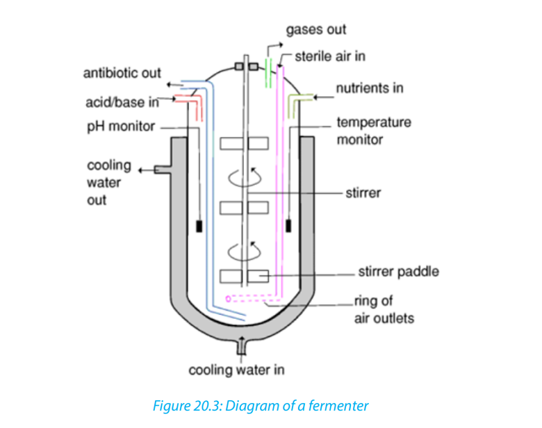
20.4.2. Production of penicillin: Antibiotic
Penicillin, an important part of our anti-microbial armament, had a significant impact
on the second half of the twentieth century. Deep-fermentation methods, which
were primarily developed for the production of penicillin during the war, gave rise
to the development of antibiotics and contributed to the nascent biotechnology
industry which appeared in the 1970s.
Penicillin production
In laboratory, it is relatively easy to grow microbes on a small scale in petri dishes,
test tubes and flasks, given a suitable nutrient medium, and good environmental
conditions. Producing chemicals like penicillin antibiotic from microbes on an
industrial scale becomes more complicated as a big number of organisms have to
be grown for the venture to be commercially viable. Laboratory procedure should
be modified so that it can be used on an industrial scale. This is called scaling up.
With scaling up, microorganisms are grown in very large vessels called fermenters
or bioreactors. Scaling up to be effective, it requires specialized biologists andengineers to deal with the following problems:
– Avoiding risks of contamination. Only desired organisms must be allowed to
grow in the vessel. Others are excluded.
– Big fermenters are built to very strict and specific design.
– Microorganisms should be kept in conditions that allow the optimum
production of required substances. This requires installing highly sensitive
equipment that maintains PH, temperature and fluid volume within very strict
limits.
– To keep nutrients at optimum levels as microbial population grows.
– Removing large amount of heat generated by high levels of microbial activity
via a heat exchanger, so that a constant temperature can be maintained.
– Minimizing the build-up of end-products (inhibitors) which may reduce
production.– Monitoring and controlling formation of the foam (unavoidable consequenceTypes of culture (of fermentation): there are two main types of culture used in
of carbon dioxide production in a nutrient-rich solution).
– Providing adequate amount of Oxygen to cultures of aerobic organisms by
aeration with small bubbles of sterile air which have a large surface area tovolume ratio.
industrial processes such as batch culture and continuous cultures.
In Batch cultures or batch fermentation (closed system), cells are grown in a
fixes volume of liquid medium in a closed vessel The conditions are set up and not
changed from outside once fermentation starts; for example: no microorganisms,
nutrients, or fluid are added or removed from the culture during the incubation
period. That is why the process is described as a closed system. The process is
stopped once sufficient products have been formed. The contents of fermenter are
then removed, isolated, microorganisms discarded and fermenter is cleaned, and
set up for a fresh batch.
Batch cultivation is used to produce secondary metabolites such as penicillin and
other antibiotics which are relatively unstable, and not essential for growth of the
culture. These secondary metabolites can be extracted economically only when
they reach a high concentration in the culture suspension.
In continuous cultures (open system), nutrients are added and cells harvested at
a constant rate, so that the volume of suspension is also kept constant. This means
that fermenters does not have to be emptied, cleaned and refilled very often. The
production is almost continuous. Continuous cultures are very expensive because
they need high equipment to maintain constant conditions, and highly skilled staffto operate the equipment.
Table 20.1: Advantages and disadvantages of batch and continuous culture
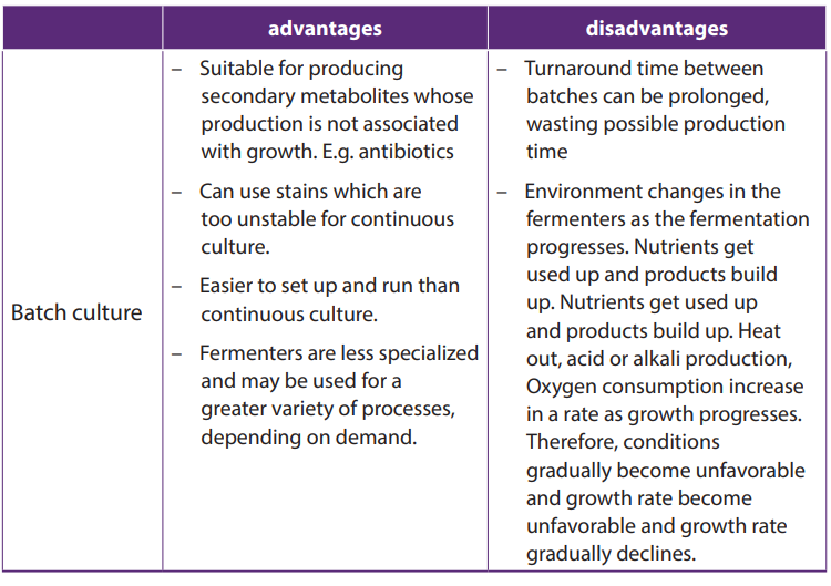
The industrial production of penicillin was generally classified into two processes: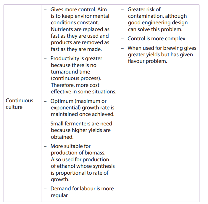
Upstream processing and downstream processing. Upstream processing
encompasses any technology that leads to the synthesis of a product and includes
the exploration, development and production. Downstream processing refers as the
extraction and purification of a biotechnological product from fermentation or at
the end of culture process. Usually the contents of fermenter are first separated into
liquid component and a solid component which contain the cells. This is usually
done by filtration or centrifugation. The liquid may contain the desired product in
solution or it may be the cells or some products inside the cells that it needs.
Penicillin is produced commercially by growing the fungus Penicillium chrysogenumin
large stirred fermenters. A solution of essential salts and a nitrogen source are put
into the fermenter together with an inoculum of the fungus. All procedures areperformed aseptically. The PH of the medium is regulated with ammonium salts
at 6.5 to 7.0. Lactose (a slowly hydrolysed disaccharide) is added to promote cell
growth and reproduction and minimize penicillin production. On completion of
fermentation (usually 6-7 days) the broth is separated from the fungal mycelium and
penicillin extracted. This penicillin can then be modified by chemical procedures toyield a variety of semisynthetic penicillins.
Modern Production Methods
Significant improvements in modern production methods have increased
production and decreased cost. Today, commercial producing strains of Penicillium
chrysogenum are grown using submerged culture in constantly agitating and
aerated 50,000- gallon stainless steel tanks. These industrial strains can now produce
40-50 grams of penicillin per liter of culture with a 90% recovery yield. This is an
overwhelming improvement from the earliest Peoria farmer’s market strain that
only produced 0.15 grams per liter with very low recovery rates. In order to achieve
these production rates, modern Penicillium strains display a host of genetic and
cellular modifications that result in increased production, including amplification of
the penicillin biosynthesis gene cluster, an increased number of peroxisomes, and
elevated levels of transporter proteins that secrete newly produced penicillin out of
the peroxisomes and the cell.
Temperature and pH are normally controlled in the fermenter. Temperature is kept
constant, while pH is held at a value of 5.5 for the first stage of the fermentation andthen raised to 6.8 and kept constant for the remainder of the fermentation period.
Self-assessment 20.4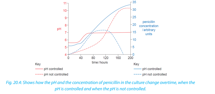 1. What is fermenter?20. 5. Antibiotics
2. Write on upstream processing and downstream processing
3. Write on your own word penicillin
4. Contrast commercial-scale production from laboratory-scale production
of penicillin.
5. Explain why the continuous culture is described as open system.
6. Explain why the batch culture is described as closed system.
7. Discuss advantages and disadvantages of batch culture?
8. Explain why continuous culture is very expensive.
9. What are Advantages and disadvantages of continuous culture?
Activity 20.5
Using addition resources to your textbook available in your school such as the
books from the school library and search further information from the internet:
Brainstorm on the antibiotic resistance and implications of antibiotic use.
Antibiotics are powerful medicines that fight certain infections by either stopping
bacteria from reproducing or by destroying them. Before bacteria can multiply
and cause symptoms, the body’s immune system can usually kill them. The word
antibiotic means “against life.” Any drug that kills germs in your body is technicallyan antibiotic.
How do antibiotics work?
Antibiotics are used to treat bacterial infections. Some are highly specialized and are
only effective against certain bacteria. Others, known as broad-spectrum antibiotics,
attack a wide range of bacteria, including ones that are beneficial to us.
There are two main ways in which antibiotics target bacteria. They either prevent
the reproduction of bacteria, or they kill the bacteria, for example by stopping
the mechanism responsible for building their cell walls. There are now hundreds
of different types of antibiotics, but most of them can be broadly classified into six
groups. These are outlined below.
Penicillin – widely used to treat a variety of infections, including skin infections,
chest infections and urinary tract infections.
Cephalosporins – can be used to treat a wide range of infections, but are also
effective for treating more serious infections, such as septicaemia and meningitis.
Aminoglycosides – tend to only be used to treat very serious illnesses such as
septicaemia, as they can cause serious side effects, including hearing loss and kidney
damage; they break down quickly inside the digestive system, so they have to be
given by injection, but are also used as drops for some ear or eye infections.
Tetracyclines – can be used to treat a wide range of infections; commonly used to
treat moderate to severe acne and rosacea, which causes flushing of the skin andspots.
Macrolides – can be particularly useful for treating lung and chest infections; can
also be a useful alternative for people with a penicillin allergy or to treat penicillinresistant strains of bacteria.
Fluoroquinolones – broad-spectrum antibiotics that can be used to treat a wide
range of infections. They include: Hypocholesterolemic agents, Lipopeptide,
Macrolides, Monobactams, Nitrofurans, Oxazolidinones, Polypeptides, Quinolones,
Sulfonamides, Tetracyclines, Lincosamides, Glycopeptides, Immunosuppressive
agents, Anti-migraine agents, Anti-bacterials, Antifungals, Penicillins,
Aminoglycosides, Ansamycins, Carbapenems, Cephalosporins (1,2, 3, 4, 5generations), and Fluoroquinolones.
20.5.1. Antibiotic resistance
Antibiotic resistance occurs when an antibiotic has lost its ability to effectively control
or kill bacterial growth; in other words, the bacteria are “resistant” and continue tomultiply in the presence of therapeutic levels of an antibiotic.
Why do bacteria become resistant to antibiotics?
Antibiotic resistance is a natural phenomenon. When an antibiotic is used, bacteria
that can resist that antibiotic have a greater chance of survival than those that are
“susceptible.” Susceptible bacteria are killed or inhibited by an antibiotic, resulting in
a selective pressure for the survival of resistant strains of bacteria.
Some resistance occurs without human action, as bacteria can produce and use
antibiotics against other bacteria, leading to a low-level of natural selection for
resistance to antibiotics. However, the current higher-levels of antibiotic-resistant
bacteria are attributed to the overuse and abuse of antibiotics. In some countries
and over the Internet, antibiotics can be purchased without a doctor’s prescription.
Patients sometimes take antibiotics unnecessarily, to treat viral illnesses like thecommon cold.
How do bacteria become resistant?
Some bacteria are naturally resistant to certain types of antibiotics. However, bacteria
may also become resistant in two ways: by a genetic mutation or by acquiring
resistance from another bacterium.
Mutations, rare spontaneous changes of the bacteria’s genetic material, are thought
to occur in about one in one million to one in ten million cells. Different genetic
mutations yield different types of resistance. Some mutations enable the bacteria
to produce potent chemicals (enzymes) that inactivate antibiotics, while other
mutations eliminate the cell target that the antibiotic attacks. Still others close up
the entry ports that allow antibiotics into the cell, and others manufacture pumping
mechanisms that export the antibiotic back outside so it never reaches its target.Bacteria can acquire antibiotic resistance genes from other bacteria in several ways.
By undergoing a simple mating process called “conjugation,” bacteria can transfer
genetic material, including genes encoding resistance to antibiotics (found on
plasmids and transposons) from one bacterium to another. Viruses are another
mechanism for passing resistance traits between bacteria. The resistance traits from
one bacterium are packaged into the head portion of the virus. The virus then injects
the resistance traits into any new bacteria it attacks. Bacteria also have the ability
to acquire naked, “free” DNA from their environment. Any bacteria that acquire
resistance genes, whether by spontaneous mutation or genetic exchange with
other bacteria, have the ability to resist one or more antibiotics. Because bacteria
can collect multiple resistance traits over time, they can become resistant to manydifferent families of antibiotics.
How does antibiotic resistance spread?
Genetically, antibiotic resistance spreads through bacteria populations both
“vertically,” when new generations inherit antibiotic resistance genes, and
“horizontally,” when bacteria share or exchange sections of genetic material with
other bacteria. Horizontal gene transfer can even occur between different bacterial
species. Environmentally, antibiotic resistance spreads as bacteria themselves move
from place to place; bacteria can travel via airplane, water and wind.
People can pass the resistant bacteria to others; for example, by coughing or
contact with unwashed hands.
Can bacteria lose their antibiotic resistance?
Yes, antibiotic resistance traits can be lost, but this reverse process occurs more
slowly. If the selective pressure that is applied by the presence of an antibiotic is
removed, the bacterial population can potentially revert to a population of bacteria
that responds to antibiotics.
20.5.2. Implications of antibiotic use
Antibiotics are considered the keystone of modern medicine, but their excessive
use continues to generate unwanted side effects. While specialists are making
strides to preserve the effectiveness of antibiotics and to slow potential infections
through better policy, the overuse of antibiotics continues to have severe healthconsequences around the world.
Self-assessment 20.51. What do you understand by antibiotic resistance?20.6. Biogas production
2. Explain how bacteria become resistant.
3. Discuss on how bacteria lose their antibiotic resistance.
4. Write on implications of antibiotic use.5. Talk on how antibiotic resistance spreads
Activity 20.6
Use diagrams or illustrations and visiting a biogas plants in your region, describe
the stages of biogas production and its significance in your area (a simple biogas
generator can also be made in schools).
Biogas typically refers to a mixture of different gases produced by the breakdown of
organic matter (methanogens or archaebacterial) in the absence of oxygen. Biogas
is produced by anaerobic fermentation of organic wastes such as agricultural waste,
manure, municipal waste, plant material, sewage, green waste, or food waste. It is
a renewable energy source and in many cases exerts a very small carbon footprint.
Biogas is primarily methane (CH4) and carbon dioxide (CO2) and may have small
amounts of hydrogen sulphide (H2S), moisture and siloxanes. The gases methane,
hydrogen, and carbon monoxide (CO) can be combusted or oxidized with oxygen.
This energy released allows biogas to be used as a fuel; it can be used for any heatingpurpose, such as cooking.
It can also be used in a gas engine to convert the energy in the gas into electricity
and heat. Biogas can be compressed, the same way the natural gas is compressed
to compressed natural gas (CNG), and used to power motor vehicles. In the UK, for
example, biogas is estimated to have the potential to replace around 17% of vehicle
fuel. It qualifies for renewable energy subsidies in some parts of the world. Biogas can
be cleaned and upgraded to natural gas standards, when it becomes bio methane.Production
Biogas is produced as landfill gas (LFG), which is produced by the breakdown of
biodegradable wastes inside a landfill due to chemical reactions and microbes,
or as digested gas, produced inside an anaerobic digester. A biogas plant is the
name often given to an anaerobic digester that treats farm wastes or energy
crops. It can be produced using anaerobic digesters (air-tight tanks with different
configurations). These plants can be fed with energy crops such as maize silage or
biodegradable wastes including sewage sludge and food waste. During the process,
the microorganisms transform biomass waste into biogas (mainly methane and
carbon dioxide) and digestate (remaining organic matter not transformed into
biogas).
The biogas is a renewable energy that can be used for heating, electricity, and
many other operations that use a reciprocating internal combustion engine, such
as a General Electrical (GE) Jenbacher or Caterpillar gas engines. Other internal
combustion engines such as gas turbines are suitable for the conversion of biogas
into both electricity and heat. The remaining organic matter that was not transformedinto biogas. It can be used as an agricultural fertilizer.
There are two key processes: mesophilic (A mesophyll is an organism that grows
best in moderate temperature, neither too hot nor too cold, typically between 20
and 45oC) and thermophilic (A thermophile is an organism, a type of extremophile,
that thrives at relatively high temperatures, between 41 and 122 °C) digestion which
is dependent on temperature. The production of biogas involves three stages and
three communities of microorganisms namely
1. Anaerobic fermentation by eubacteria including lactobacillus, which
converts the organic waste into a mixture of organic acids and alcohol, with
some Hydrogen, Carbon dioxide, and acetate.
2. Acetogenic (acetate-producing) reaction by bacteria such as
acetobacterium which, in addition to acetate, produce hydrogen and
Carbon dioxide from the organic acid and alcohol.
3. Methanogenic (methane-producing) reactions by archaebacteria,
including Methanobacterium, Metanococcus, and Methanospirillum. Thearchaebacteria generate methane either:
– By reducing the carbon dioxide:

– By converting acetate: CH3 COOH :
Composition
The composition of biogas varies depending upon the origin of the anaerobicdigestion process. Landfill gas typically has methane concentrations around 50%.
Table 20.2: Typical composition of biogas
In some cases, biogas contains siloxanes. They are formed from the anaerobic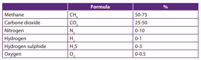
decomposition of materials commonly found in soaps and detergents. During
combustion of biogas containing siloxanes, silicon is released and can combine with
free oxygen or other elements in the combustion gas.
Applications
Biogas can be used for electricity production on sewage works, in a combined heat
and power (CHP) gas engine, where the waste heat from the engine is conveniently
used for heating the digester; cooking; space heating; water heating; and process
heating. If compressed, it can replace compressed natural gas for use in vehicles,
where it can fuel an internal combustion engine or fuel cells and is a much moreeffective displacer of carbon dioxide than the normal use in on-site CHP plants.
Self-assessment 20.6
1. What part do acetogenic reactions play in the production of biogas?
2. The archaebacteria generate methane either by reducing the carbon
dioxide, or by converting acetate: write chemical equations for the two
processes.
End of unit assessment 20
Multiple choice questions
1. During penicillin production, temperature is maintained ata. room temperature2. In penicillin production, pH of culture medium is maintained between
b. 26 °C
c. 36 °C
d. 46 °Ca. 5 and 63. To produce penicillin, main fermentable source in culture is
b. 4 and 6
c. 6 and 7
d. 4 and 5a. glucose4. Penicillin production is optimum in
b. lactose
c. sulphate
d. sugarsa. batch operation systems5. What is fermentation?
b. continuous operation systems
c. discontinuous operation system
d. unique operation system
6. The senior four Biology teacher said: “the biogas can contribute to the economic
development of Rwanda” defend his idea.
7. Explain how are immobilized enzymes made?
8. Explain the medical applications of enzymes.
9. Explain the importance of using yeast in bread making.
10. What Are the Main Ingredients of Bread?
11. Summarize the advantages of using immobilized enzymes rather than enzyme
solutions.
12. Describe the composition of biogas.
13. Describe three stages that are involved in production of biogas.
REFERENCES
Becket, B.S. (1986). Biology: A Modern introduction. GCSE Education, Oxford University
press.
Biggs, A., Kaicka, C., and Lundgren, L. (1995). Biology: The dynamics of life. McGrawHill, Westerville, USA.
Brainard, J. (2015). CK-12 Biology Foundation textbook next generation.
GoR [Government of Rwanda] (2016). National biodiversity and action plan, Kigali,
Rwada.
Government, W. (2005). Guidance for teaching. England: Charles Darwin House.
Hocking, S., Kennedy, P. and Sochacki, F. (2008). Biology. OCR and Heinemann, UK.
Ibrahim, S. (2006). Solutions to examination paper: UCE Biology paper 1and 2. PEAK
Publisher Ltd, Kampala, Uganda.
Jones, M., Fosbery, R., Gregory, J., and Taylor, D. (2014). Cambridge International AS
and A Level Biology coursebook fourth Edution. United Kingdom: Cambridge University
Press.
Jovanovich, H. B. (1986). Biology. Orlando, Harcourt Brace Jovanovich.
Juliet, M., and Magondu.J., M. V. (2017). Biology for Rwanda. Kigali: Easter African
Educational publishers
Karen, A., Camp, S., Pamela, Jenner V. J., and Zalisko J. E. (1994). Biology: A Journal into
life. 3rd Ed. Saunders College Publishing
Kay, I. (1998). Introduction to Animal Physiology, MMU, Manchester, UK.
Kennedy, P., and Sochck, F. (2008). OCR Biolgy. British: OCR and Heinemann.
Kent, M. (2000). Advanced Biology: A new Mainsream text for the new specifications.
Oxford University Press. New York, USA.
Kent, M. (2000). Advanced Biology: A new Mainsream text for the new specifications.
Oxford University Press. New York, USA.
Kinyua, S. and Oyugi, O. (1998). Secondary Biology, A practical approach; KLB, Nairobi,
Kenya.
Lee Ching and Arnasalam, J. (2008). Biology.Vol.1, Pre-U Text STPM, Pearson-Longman
Malaysia.
Level Biology coursebook fourth Edution. United Kingdom: Cambridge University
Press.
Mackean.D.G, and Hayward, D. (2014). Cambridge IGSCE biology 3rd edition. London:
Hadder education
Mader, S. S., Baldwin, A., Roush, R., & Stephanie Songer, M. T. (2010). Biology 10th
Edition. McGraw-Hill companies, Boston, UK.
Martin, E. and Hine, S.R. (2008). Oxford Dictionary of Biology, 6th edition, Oxford
University Press, Oxford, UK.
Mary, J., & Forcebery, R. (2004). Cambridge International AS and A Level. United
Kingdom: CAMBRIDGE University press.
Miller L. (2006). Biology: Florida students’ and teachers’edution. Prentice Hall Biology,
Boston. UK.
Morgan, S. (2000). Practice in Biology: Progressive questions for AS and A level.
Cambridge
Neil A. Campbell, Reece J.B., Urry L.A., Cain M.L., Wasserman S.A., Minorsky P.V., and
Jackson R.B. (2008). Biology. Pearson Benjamin Cummings. 8th Ed. San Francisco, US.
Ones, M., Fosbery, R., Gregory, J., & Taylor, D. (2014). Cambridge International AS and A
Owaka M., and Kavita P. (2006). Test it and Fix it. KCSE Revision Biology.Oxford
University Press, Nairobi, Kenya.
Peter K; Frank S. (2008). OCR Biology. British: British Library.
Roberts, M.B.B (1986). Biology for life. 2nd edition, Thomas Nilsson and sons Ltd,
London UK
Sequeira, L. (2010). Certificate Biology, form 4, Pupils’book. EAEP, Nairobi, Kenya.
Sochacki, F., and Kennedy, P. (2008). OCR Biology. Pearson Education Limited, China
The Perfect Guide for Students of Biology at School or University (2008). A Dictionary
of Biology, Oxford University Press.
Verrgilio, O.K. (2013). Senior secondary certificate biology for Rwanda, East African
Publishers Limited, Kigali, Rwanda.
Wilf Stout and Nigel Green (1990). A-level Biology. Macmillan work out series,
Macmillan.
Electronic links
https://study.com/academy/practice/quiz-worksheet-louis-pasteur-germ-thioryof-disease.html
http://printerfriendly.adam.com/content.aspx?productId=117&pid=1&gid=001439
&c-custid=758
https://www.cdc.gov/parasites/hookworm/gen_info/faqs.html
http://www.parasitesinhumans.org/hookworms.html
https://en.wikipedia.org/wiki/Tinea_corpori
