UNIT 4: CELL STRUCTURE AND SPECIALIZATION
UNIT 4: CELL STRUCTURE AND SPECIALIZATIONKey Unit CompetenceDescribe the structure and function of cells in an organism.Learning objectivesBy the end of this unit, I should be able to:– Identify plant and animal cell structures visible under a light microscope.– State functions of cell structures as seen under an electron microscope.– Describe the nature of artefacts.– State the importance of freeze fracturing for examining membrane structure.– Explain how cell organelles can be isolated by cell fractionation.– List the functions of cell membranes.– Describe the fluid mosaic structure of cell membranes.– Explain the role of the different components of a cell membrane.– Explain cell specialization as the differentiation of a cell or process to do aparticular function.– Interpret charts and micrographs to relate the structure of specialized cells totheir functions.– Prepare, observe and draw diagrams for specimens on temporary slides for:Wandering Jew, in plants and cheek cells under a light microscope.– Distinguish between ultra-structures of plant cells and animal cells.– Compare ultra-structures of prokaryotic and eukaryotic cells– Show resilience and be aware of artefacts when preparing temporary slides.– Appreciate the importance of cell specialization in multicellular organisms
Introductory ActivityUsing addition resources to your textbook available in your school such as thebooks from the school library and search further information from the internet:1. Differentiate between prokaryotic and eukaryotic cells.2. By using charts for the two cells, identify different organelles of eukaryoticcell that may perform functions similar to those of a prokaryotic cell.
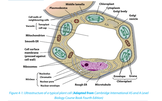
4.1. Ultra-structure of a cellActivities 4.11. Observe the chart given for Ultra structure of a cell and identify parts thatare easily recognizable when compared with a photomicrograph form alight microscope.2. Identify the mitochondria and ribosomes and state their roles in the life ofthe cell.Cytology is the study of the structure and function of cells. A Cell is the basic unit oflife. All living organisms are made up of cells.Living organisms are classified into:– Unicellular organisms are made of only one cell, such as bacteria,– Multicellular organisms are animals and plants composed of many cells. Inmulticellular organisms, cells divide and then undergo differentiation orspecialisation for specific functions.
Cell theory.The cell theory states that all living organisms are made up of cells, and cells are thebasic unit of structure function in all living organisms.The main principles of cell theory are based on the following ideas.– All known living organisms are made up of one or more cells,– All cells come from pre-existing cells by division– Cells contain the hereditary information that is passed from cell to cell duringcell division.– Metabolism takes place in cells– Given suitable conditions, cells are capable of independent existence
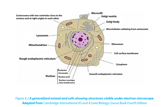
When a cell is viewed under light microscope, the most obvious features observedare the very large nucleus and a clear cytoplasm surrounded by a cell membrane.However, under electron microscope, it is possible to identify a range of organellesin plant and animal cells. Ultrastructure is the detailed of cell as revealed by theelectron microscope.
 Similarities between animal cell and plant cell– Both have a cell membrane, a cytoplasm and a nucleus.– Both animal and plant cells have mitochondria, Golgi apparatus, Reticulumendoplasmic, lysosome, big ribosomes (80S), peroxisome, microtubules.
Table 4.1: The differences between animal and plant cell
Self-assessment 4.1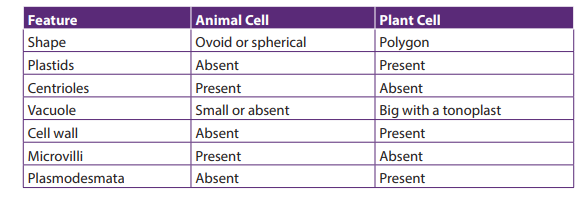
1. What structures do both animal and plant cells have in common?
2. State any five principles of the cell theory.
3. Give the major difference between a plant and animal cell. Which organellesdoes this difference relate to?
4.2. Prokaryotic cells
Activities 4.2
Under microscope, observe mounted slides of bacteria, and plant cells. Draw andlabel the parts that are common in both plant and bacterial specimens
A typical bacterial cell has a cell surface membrane enclosing the cytoplasm that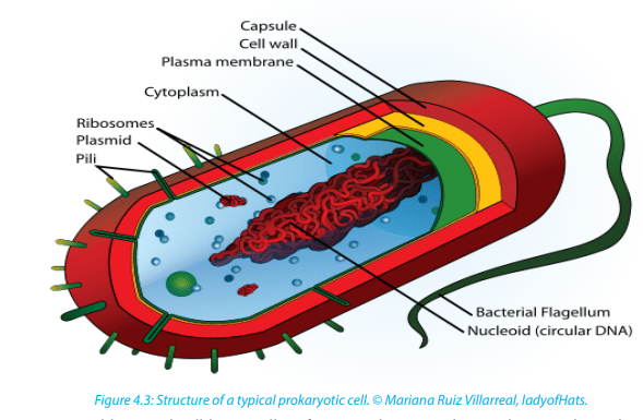
contains enzymes, ribosomes and food granules. The membrane is surrounded by
the cell wall and this may in turn be enclosed in a capsule. A bacterial cell lacks high
level of organization compared to animal or plant cell. It has no Golgi apparatus
or endoplasmic reticulum. The genetic material is a single strand of DNA usually
coiled up into the center of the cell to form a nucleoid. This nucleoid has no double
membraned nuclear envelope so is often described as an ‘ill-defined nucleus’.– Some bacterial cells contain plasmids with additional DNA.Comparison between prokaryotic and eukaryotic cells
– Respiration generally takes place in mesosomes which is an in-folding of the
cell surface membrane but lack mitochondria
– Photosynthesizing bacterial cells such as cyanobacteria (blue green
algae) have a special form of chlorophyll but it is not enclosed in a doublemembraned chloroplast
Table 4.2 Comparison between prokaryotic and eukaryotic cells
Self-assessment 4.2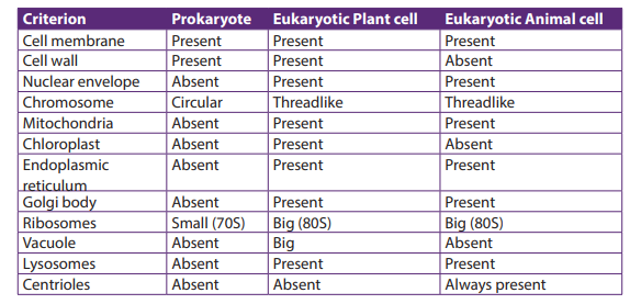
Organisms such as bacteria are known as prokaryotes.1. Which structure in a bacterial cell resembles a nucleus?4.3. Cell organelles2. How does it differ from the nucleus of eukaryotic cells?
Activities 4.3
By using iodine solution, methylene blue, a piece of onion leaf, a scalpel, forceps,
light microscope, slides and cover slips, clean cotton wool bud, and onion bulbs.
Observe cells from onion epidermis under light microscope.
Observation of a plant cell– Add a drop of diluted iodine solution on the slide.Why did you use iodine solution in this experiment?
– Remove a transparent layer of onion epidermis from the inner side that you
will mount on the slide and add iodine solution.
– Cover your preparation with a cover-slip and mount it on the stage.
– Observe the preparation under the low power and thereafter under high
magnification.What main parts of a plant cell are easily observed from a light microscope?
Observe animal cells from mouth cheek epithelium– By using a clean cotton wool bud, wipe over inside of your cheek.
– Smear cells over surface of a clean grass microscope slide containing a drop
of methylene blue stain
– Carefully put the cover-slip on the preparation and mount it on the stage to
observe.Draw both plant and animal cell and label the cell wall, nucleus and vacuole\
The cell nucleus contains nearly all the cell’s DNA with the coded instructions for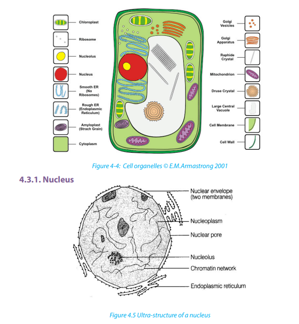
making proteins and other important molecules. The nucleus is surrounded by a
double nuclear envelope, which allow materials to move into and out of the nucleus
through nuclear pores. The granules found in the nucleus are called chromatin which
consist of DNA bound to protein. When a cell divides, the chromatin condenses into
chromosomes containing the genetic information. The nucleus contains a densespherical structure called nucleolus in which assembly of ribosomes occurs
The ER consists of a series of flattened membrane-bound sacs called cisternae. The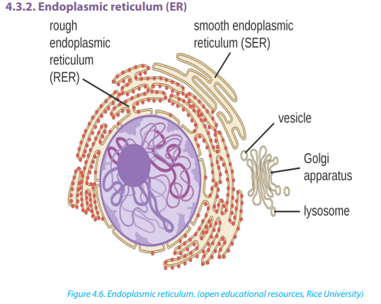
rough ER is surrounded with ribosomes. The rough ER transports proteins made on
attached ribosomes. The smooth ER is made of tubular cavities lacks ribosomes, and
it involves in synthesis of lipids that the cell needs. The number and distribution of
the ER relates to the functions of the cell; glandular cells are seen to have several RER
for synthesis of hormones and enzymes. Examples include liver cells, plasma cells,and pancreatic cells.
4.3.3. Golgi apparatus
The Golgi apparatus is a stack of membrane-bound, flattened sacs, which receives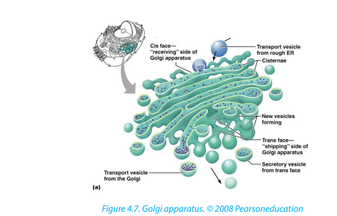
proteins from the ER and modifies them. It may add sugar molecules to them to form
glycoproteins or lipids to form glycolipids. The Golgi apparatus then packages the
modified substances into vesicles that can be transported to their final destinationsthroughout the cell or outside of the cell by exocytosis.
Mitochondrion have two membranes separated by a fluid-filled intermembrane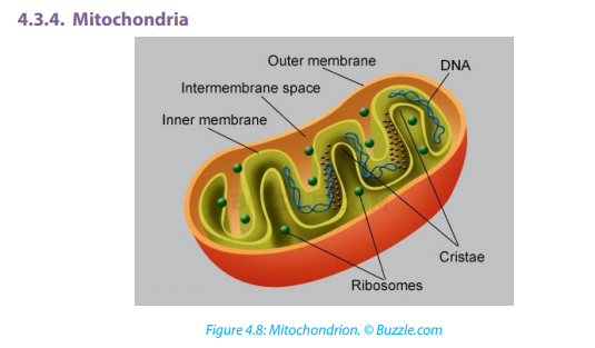
space. The inner membrane is highly folded to form cristae that plays a big role inaerobic respiration. The central part of the mitochondrion is called matrix.
The mitochondria are the site where Adenosine triphosphate (ATP) is producedduring aerobic respiration.
4.3.5. Chloroplasts
Chloroplasts are the site of photosynthesis in plant cells. These are found in plant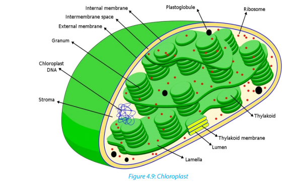
cells and in cells of some protoctists. They also have two membranes separated
by a fluid-filled space, circular DNA as in mitochondria. The inner membrane is
continuous, with thylakoids. A stalk of thylakoids is called a granum (plural:grana). Chlorophyll molecules are present on the thylakoid membranes.
These are spherical sacs surrounded by a single membrane. They contain powerful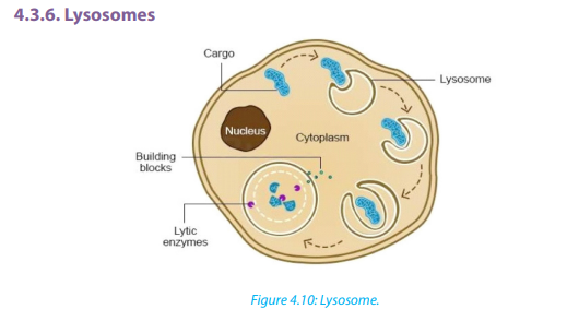
digestive enzymes. Their role is to break down materials such as worn out cell
organelles, and destroy foreign microorganisms that enter the body. In acrosome,
lysosomes help the sperm to penetrate the egg by breaking down the material
surrounding the egg. Lysosomes are also involved in autolysis, breakdown of dead
tissues or harmful objects inside the cell. Therefore, lysosomes are referred to as‘suicide bags’
Ribosomes appear as dark granules in the cytoplasm and are not surrounded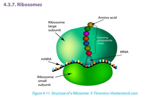
by a membrane. They have the same size as those found attached to the rough
endoplasmic reticulum- about 20nm in diameter and known 80S. Free ribosomes
make proteins that are as enzymes or in other forms in the cytoplasm. Ribosomesare made in a region of the nucleus called the nucleolus.
Centrioles are small tubes of protein fibers called microtubules which have many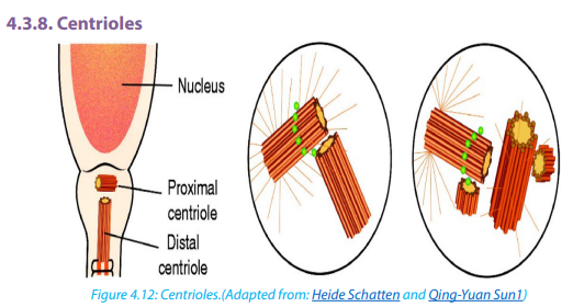 \
\
roles including moving chromosomes during nuclear division. Animal cells have
structures called centrioles which consist of two groups of nine triple microtubules.Centrioles form an anchor point for microtubules during cell division.
A vacuole is a saclike structure that stores materials such as water, salts, proteins, and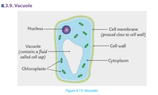
carbohydrates. In many plant cells there is a single and large central vacuole filled
with liquid. The pressure in the cells of central vacuole makes it possible for plants
to support heavy structures like leaves and flowers. Some animals and unicellular
organisms contain contractile vacuoles which contract to pump excess water outof the cell.
Self-assessment 4.3
1. Explain why muscle cells contain several mitochondria compared to fat
storage cells
2. What kind of information is contained in chromosomes?
3. Describe the functions of the endoplasmic reticulum, Golgi apparatus,
chloroplasts, mitochondria and nucleus in the cell.
4. Consider the 3D structures which would be visible in ultrastructure of a
plant cell.a. Identify their parts and label them4.4. Membrane structureb. State one function for each part
Activity 4.4
Learners mix a portion of cooking vegetable oil with water and shake the mixture
vigorously and leave it to settle. Note the way water and oil are distributed withinthe mixture and suggest a possible explanation for your observation.
Cell membranes cover surfaces of every cell. Some organelles in cytoplasm are
enveloped by membranes. The cell membranes ultrastructure is not easily visible
under a light microscope but is studied by electron microscopes, freeze structuring
and other modern techniques which reveal complex structures
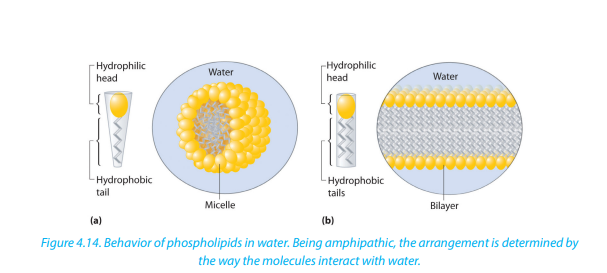
A detailed study of a cell membrane reveals that it is 7-8nm wide and is made of a
phospholipid bilayer.
– Lipid component makes up 45% protein and 10% carbohydrate. Most of the
lipids are phospholipids
– Each molecule of phospholipid consists of a hydrophobic tail of two fatty acids
and a hydrophilic phosphate head. They arrange themselves in phospholipids
bilayer with their tails pointing inward away from the water both inside andoutside the cell
 \
\
In 1972, Jonathan singer and Garth Nicolson proposed the fluid mosaic model of the
cell membrane structure. This was done after realizing that membranes must have a
complex structure to carry out a variety of activities. In their model;– Individual protein molecules shift and move on a fluid bilayer of phospholipids;
some spanning the width of the membrane (intrinsic proteins), others confined
to the outer or inner surface (extrinsic protein)
– Protein molecules are variable in structure and function but they all contributeto the mechanical strength of membranes
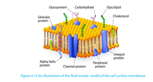
The membrane is referred to as;– A fluid because it appears to have the properties of a fluid rather than a solid as4.4.1. Properties of the cell membrane
the major constituent, lipids and proteins move about the structure
– Mosaic because protein and lipid components form a pattern of parches model– It is mainly made of lipids, proteins and carbohydrates.4.4.2. Roles of different components of cell membrane
– It is semi-permeable or partially permeable to allow some substances to pass
through but prevents others to cross depending on their size, charges and
polarity.
– It is positively charged outside and negatively charged inside and has a
hydrophilic pole and a hydrophobic pole
– It is a bilayered sensitive and flexible.It has inorganic ions and its proteins and
lipids may be mobile and contains different types of enzymes and coenzymes.
– It is perforated of pores and recognizes chemicals messengers includinghormones and neurotransmitters.
a. Cholesterol– Gives the membranes of some eukaryotic cells the mechanical stability.b. Channel proteins
– It fits between fatty acid tails and helps make the barrier more complete,
so substances like water molecules and ions cannot pass easily and directlythrough the membrane.
– Allow the movement of some substances across the membrane.channels.
– Large molecules like glucose enter and leave the cell using these protein
c. Carrier proteins– Actively move some substances across the cell membrane. For example,d. Receptor sites
magnesium and other mineral ions are actively pumped into the roots hair
cells from the surrounding soil.
– Nitrate ions are actively transported into xylem vessels of plants– Allow hormones to bind with the cell so that a cell response can be carried out.e. Enzymes and coenzymes
– Glycoproteins and glycolipids may be involved in cells signaling and they allow
the immune system to recognize foreign objects to the cells.
– Some hormone receptors are glycoprotein and some are glycolipid.– Some reactions including metabolic processes in photosynthesis take place in4.4.3. Functions of a cell surface membrane
membranes of chloroplasts.
– Some stages of respiration take place in membranes of mitochondria, where
Enzymes and coenzymes may be bound to these membranes.
– The more membrane there is, the more enzymes and coenzymes it can hold
and this helps to explain why mitochondrial inner membranes are folded to
form cristae, and why chloroplasts contain many stacks of membranes calledthylakoids.
– The cell membrane acts as a selective barrier at the surface of the cell, andSelf-assessment 4.4
controls the exchange between the cell and its environment.
Glycoproteins and glycolipids are involved in the cell protection, the process
by which cell adhesions are brought about and in the cell recognition.
– Receptor sites for hormones and neurotransmitters
– Transmission of nerve impulses
– Insulation of nerves to improve transmission speeds.
Internal membranes:
– Act as reaction surfaces
– Act as an intra cellular transport system
– Providing separate intra cellular compartment, isolating different chemicalreactions as in organelles.
1. What is meant by the fluid mosaic model of the cell membrane?
2. State at least three properties of the cell membrane.
3. Describe at least 4 types of the proteins in the cell membrane and their
roles.4. What is a partially permeable membrane?
5. What do the words hydrophilic and hydrophobic mean?
6. The diagram below shows the structure of a cell membrane. Study it carefully
and answer the following questions.
a. Name parts labelled A, B, C and D and give the function of the part B.
b. What types of molecule are likely to be involved in?i. Cell signaling and recognition7. What is the difference between rough and smooth endoplasmic reticulum?
ii. Allowing small charged molecules to pass through the cell membrane
iii. Site metabolic reactions
8. Describe the role of cytoskeleton
9. The photograph in the figure below shows an organelle of the living cell.
a. Name this organelle.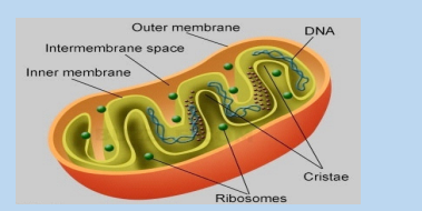
b. What is the function of this organelle?c. In which ways is this organelle similar to a chloroplast?
4.5. Specialized cells
Activity 4.5
By using the diagrams below, relate the structure of specialized cells to theirfunctions.
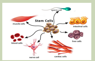
Differentiation refers to the changes occurring in cells of a multicellular organism so
that each different type of cell becomes specialized to perform a specific function.
In animals, the first type of cells in the developing embryo is stem cells. These are
unspecialized cells that go on to form all the different types of cells in adult. Cell can
differentiate in many ways, with changes to the shape of the cell, the number ofparticular organelles and the content of the cell.
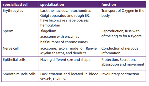
4.5.1. Specialized animal cells and their functions4.5.1.1. Red blood cells
All blood cells are produced from undifferentiated stem cells in the bone marrow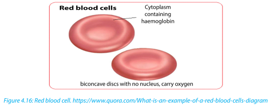
but the cells destined to become erythrocytes (red blood cells) lose their nucleus,
mitochondria, Golgi apparatus and rough endoplasmic reticulum. They are packed
full of the protein called haemoglobin. The shape of this cells change so that theybecome biconcave discs, and they are then able to transport Oxygen in the body.
Sperm cells are specialized to fertilize the egg. Its specialization involves many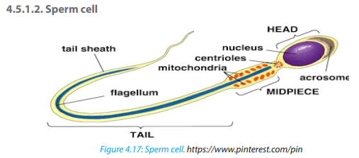
changes in shape and organelles content.
By shape: the sperm cells are very small, long and thin to help them to move easily,
and they have a flagellum which helps them to move up the uterine tract towardsthe egg.
By organelles content: sperm cells contain numerous mitochondria which
generate much energy for their movement. Their acrosome has specialized
lysosomes containing many enzymes that are released on the outside of the egg.
These enzymes lyse the wall of the egg, and facilitate the sperm nucleus to penetrate
easily. In content, the sperm cell nucleus contains the half number of chromosomes
of the germ cell in order to fulfil its role as a gamete of fertilizing the egg.
Did you know: As a sperm fuses with an ovum to form a zygote which grows into
an individual, in the same way: a man maries a woman to form a couple which willproduce children and form a family.
Nerve cells also known as neurons are specialized cells to carry nervous impulses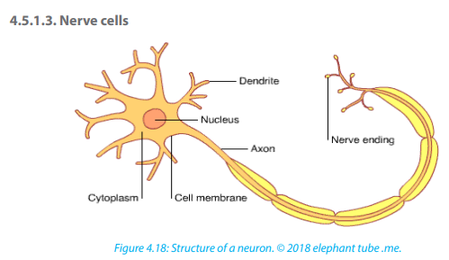
in the body. These signals between neurons occur via specialized connections
called synapses. Specialized animal cells have different functions. Some of them aresummarized in the following table.
Table 4.3: Specialized animal cells and their functions.
4.5.2. Specialized plant cells and their functions
4.5.2.1. Root hair cells
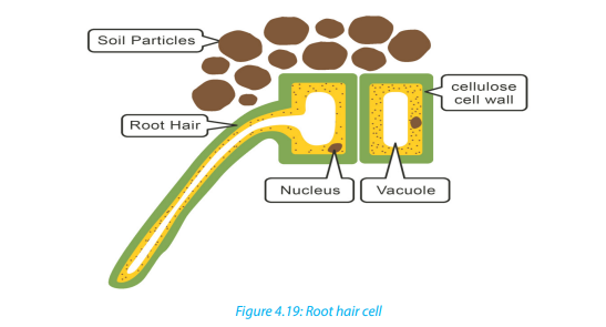
The root hair cells have hair-like projection from their surface out into the soil. This
increase the surface area of root available to absorb water and minerals from thesoil.
4.5.2.2. Palisade cells
Palisade cells are in leaves, right below the upper epidermis. They are vertically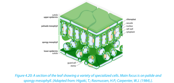
elongated, a different shape from the spongy mesophyll cells beneath them in
the leaf. Their large numbers of chloroplasts allow them have several chloroplasts
used in photosynthesis.
Parenchyma cells
Parenchyma is composed of relatively simple and undifferentiated parenchyma
cells. They function in storage, photosynthesis. In most plants, metabolic activity
such as cell division, respiration, and photosynthesis occurs in these cells because
they retain their active cytoplasm. .
4.5.2.3. Guard cells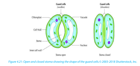
Guard cells are cells surrounding each stoma. Guard cells are specialized cells in the
epidermis of leaves, stems and other organs that are used to control gas exchange.
They are produced in pairs with a gap between them that forms a stomatapore.
Guard cells have the following feature:– Un even thick wallsSelf-assessment 4.5
– Possess chloroplasts; they are the epidermal cell that have chloroplasts anadaptive feature in controlling pore opening.
1. Explain why differentiation to produce erythrocytes involves a change in
shape.
2. Red blood cells cannot divide as they have no nucleus. State two other
biological processes that red blood cells cannot carry out.
3. Describe how the following are specialized for their roles:a. Neutrophil4. Explain why photosynthesis is carried out in palisade mesophyll more than
b. Sperm cell
c. Root hair cell
in spongy mesophyll.
5. In what kinds of organisms is cell specialization pronounced characteristic?6. Discuss the advantages of cell specialization in living things
End of unit assessment 4
Section A. Multiple choice questions
1. Which organelle converts the chemical energy in food into a form that cells can
use?a. Chromosome2. The cell membranes are constructed mainly of:
b. Chloroplast
c. Nucleus
d. Mitochondriona. Carbohydrate gates3. In many cells, the structure that controls the cell’s activities is the:
b. Protein pumps
c. Lipid bilayer
d. Free-moving proteinsa. Nucleus4. Despite differences in size and shape, all cells have cytoplasm and a
b. Nucleolus
c. Cell membrane
d. Organellea. Cell wall5. If a cell of an organism contains a nucleus, the organism is a (an)
b. Cell membrane
c. Mitochondria
d. Nucleusa. Plant6. Match each part of the cell (left column) to corresponding statement (right
b. Eukaryote
c. Animald. Prokaryote
column):
Nucleus controls movement of substances in and out of the cell
Mitochondrion where photosynthesis takes place
Chloroplast where aerobic respiration takes place
Smooth ER controls the activity of the cell
Ribosomes where lipids including steroids are made
Section B: Questions with short answers
1. How does a cell membrane differ from a cell wall?
2. Name the structures that animal and plant cells have in common, those found in
only plant cells, and those found only in animal cells.
3. List:a. Three organelles each lacking a boundary membrane4. Identify each cell structure or organelle from its description below.
b. Three organelles each bounded by a single membrane
c. Three organelles each bounded by two membranes (an envelope)a. Manufactures lysosomes and ribosomesSection C: Essay questions
b. Site of protein synthesis
c. Can bud off vesicles which form the Golgi body
d. Can transport newly synthesized protein round the cell
e. Manufactures ATP in animal and plant cells
f. Controls the activity of the cell, because it contains the DNA
g. Carries out photosynthesis
h. Can act as a starting point for the growth of spindle microtubules during
cell division
i. Contains chromatin
j. Partially permeable barrier only about 7 nm thick
k. Organelle about 25 nm in diameterl. Which two organelles other than the nucleus contain their own DNA
1. Describe the structure and function of the cell membrane and cell wall.4. The diagram below shows the structure of a liver cell as seen using an electron
2. Describe the basic structure of the cell membrane.
3. Explain two common characteristics of chloroplasts and mitochondria.Consider both function and membrane structure.
microscope.
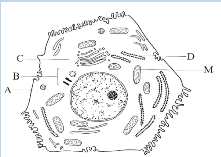 a. Name the parts labelled A, B, C and D.
b. The magnification of the diagram above is x12 000. Calculate the actual
length of the mitochondrion labelled M, giving your answer in µm. Show
your working.
c. Explain the advantage to have a division of labor between different cells inthe body.