UNIT 19: CULTURING MICRO-ORGANISMS
UNIT 19: CULTURING MICRO-ORGANISMS
Key Unit Competence
Explain the process of culturing microorganisms and the factors affecting their
population growth.
Learning objectives– List and describe the roles of microorganisms and their requirements forIntroductory activity.
growth.
– Explain the role of environmental variables in culturing microorganisms.
– Describe the different types of culture media.
– Draw and interpret the graph of the population growth of bacteria.
– Carry out an experiment to stain bacteria for examination with a light
microscope.
– Observe and compare the numbers of bacteria present in fresh and stale milk.
– Distinguish between gram negative and gram positive bacteria.
– Describe the main features of aseptic techniques.
– Explain how pure cultures of pure bacteria can be obtained.
– Describe the methods of inoculation.
– Use sterile techniques to prepare agar plates to culture bacteria and fungi
– Carry out research on why microorganisms are particularly suitable for
industrial use.
– Appreciate the importance of culturing microorganisms.
– Show perseverance when inoculating a solid and liquid medium.
– Show concern for taking the basic precautions in the school laboratory whencarrying out routine microbiological work.
Use different books and visit internet make a short summary about the culture of
microorganisms and suggest why cultures are not incubated at 370 C in a school lab.
19.1. Requirements for culturing of microorganisms
Activity 19.1
Use textbooks and other sources of information to discuss the requirements of
growth of microorganisms.
Many microorganisms can be grown in the laboratory. This allows scientists to learn
a lot about them. We can find out which nutrients they need to survive and which
chemicals will kill them. We can also discover which microorganisms can be useful
to us and which cause deadly disease.
To find out more about microorganisms, you need to culture them. Culturing
microorganisms involves growing very large numbers of them so that you can see
the colony as a whole.
To culture microorganisms, you must provide them with everything they need. This
usually involves providing a culture medium containing carbohydrates to act as an
energy source. A long with this, various mineral ions some supplement of proteins
and vitamins are included.
The nutrients are often contained in an agar medium. Agar is a substance which
dissolves in hot water and sets to form a jelly. You pour hot agar containing all the
necessary nutrients into a Petri dish. Microorganisms are living organisms. Therefore,
they have requirements for their growth, maintenance and multiplication. These
include:
Optimum temperature (30-40ᵒC) for enzymes to work better.– Source of energy such as glucose, maltose, juice.The medium for culture of microbes can be the dead organic matters (food,
– Source of other nutrients (minerals such are as potassium, sodium, iron,
magnesium and calcium, vitamins, proteins
– Air for aerobic microbes or complete absence of air for anaerobic
microorganisms.
fruits, remaining of organism, juice, milk) or a prepared medium such as Agaragar
(universal medium for any germ), Lowenstein medium (selective medium for
tuberculosis bacillus). The medium can be wet or dry. Different types of media areused culture microorganisms.
19.1.1. Types of media
There are many different types of media described by their components or
ingredients.
Universal media: this allow the growth of every type of bacteria e.g. agar-agar
Differential/selective media: are specific to some types of bacteria for example
Lowenstein for tuberculosis bacteria. Their ingredients will favour growth of certain
types of bacteria.
A pure culture: this contains only one kind of microorganism. The pure cultures are
important for scientific method as they are free from other types of microorganisms.
19.1.2. Principles of sterile culturing– Wash hands before touching a sterile Petri-dish
– Open the Petri-dish as little as possible, and replace the lid quickly
– Never cough or sneeze near the dish
– Never touch the infected jiffy with fingers– When culturing is no longer required, they should be flooded with strongSafety precautions:disinfectant
After cleaning out the nutrient from Petri-dish, they should be washed and
disinfected, and then if they are glass, heat sterilize.
– Wash your hands thoroughly after all operation by using soap.
– Never push hands near the mouth during experimental work.
Bacteria grow and reproduce more quickly when they are warm than when they
are cold. It would be dangerous to incubate cultures at temperatures close to body
temperature (37°C) because doing so might allow the growth of pathogens harmful
to health. So the maximum temperature used in school and college labs is 25°C.
However, higher temperatures can be used industrially, and these produce faster
growth.
Self-assessment 19.11. What is meant by the term culturing bacteria?19.2. Culture media
2. What do bacteria need to grow?
3. Why do we culture microorganisms in the lab?
4. Explain why cultures are not incubated at 370 c in a school laboratory.
Activity: 19.2
Describe different types of media used in culturing microorganisms.
A medium is a solid or liquid preparation containing nutrients for the culture of
microorganisms. A pure microbial culture undergoes the following steps namely:– Choice of the culture medium.Microorganisms may be cultured in a solid medium or a liquid medium or broth.
– Sterilization of the culture medium.
– A culture with a collection of microbial cells growing on or, in a medium.
– Selection of a pure colony from a collection of microbial cells growing
– Introduction of a microorganism into a suitable growth medium
– Streaking to carrying out a pure culture.
When there is not a culture with a collection of microbial cells growing on or,
in a medium. A source of microorganisms is spread on the surface of an agar to
produce individual colonies. Once individual colonies are obtained, this collection
of microorganisms can then use to carry out a pure microbial culture.a. Solid medium.
Solid media are particularly suitable for bacteria and fungi and are prepared by mixing
the liquid nutrient solution with a gelling agent, usually agar, at a concentration ofabout 1-5%, thus, producing nutrient agar that allows the growth of colonies.
b. Liquid media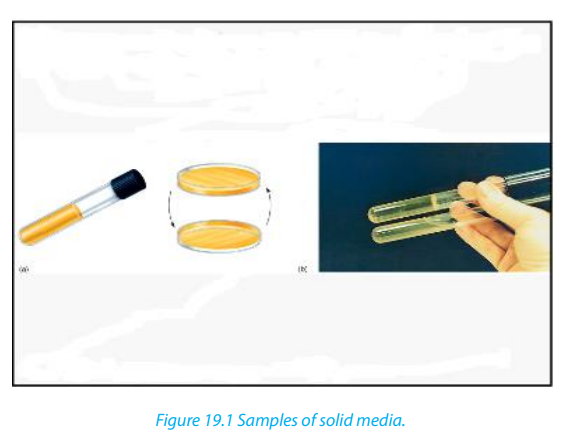
The liquid media are water – based solutions that are generally termed as broths,
milks and infusions.
Liquid media are often useful for measuring population growth. They may be placed
in a test tube, stopped by a plug of cotton wool or a metal cap, or in a glass, screw crapped
bottle such as a universal bottle which holds about 25cm2
enough for oneagar plate.
The medium must be sterilized and after, adding a small quantity of cells to the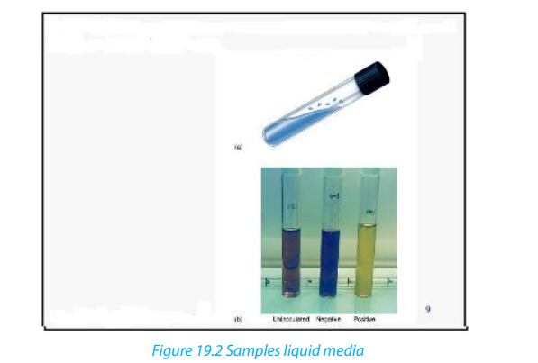
medium is called inoculation.
c. Enrichment media.
An enrichment medium is a medium in which substances are added to meet the
requirements of certain microorganisms in preference to others. As a result, certain
microorganisms grow better than others.
d. A selective medium
It is a medium in which one or more substances are added to favor the grown of
specific microorganisms and to inhibit the growth of others. Example, the addition
of penicillin to a culture to select for those organisms resisting to it, or the selectionof hybridizes cells during the production of monoclonal antibodies.
Self-assessment 19.21. How would you isolate from the soil an organism which could use19.3. Aseptic technique.
atmospheric nitrogen as its only source of nitrogen (a nitrogen-fixing
bacteria)?
2. What is meant by nutrient agar?
3. Distinguish between liquid media and solid media.4. Distinguish between enriched media and specific media.
Activity 19.3.1
Carry out a procedure of culturing fungi on a nutrient agar using sterile techniques.
Aseptic technique is using sterilized equipment and solutions and preventing their
contamination. Sterilization is the removal or destruction of all living microorganisms,
including spores (inactive structures that enable some microorganisms to
survive unfavorable periods). Bacterial and fungal spores are abundant in most
environments including laboratories. A range of special techniques and apparatus
are designed to prevent contamination of nutrients media. Autoclaves are used
to sterilize equipment and culture media before experiments and also to sterilize
equipment and specimens before disposal.
In addition, after sterilization, a great care is taken during experiments to ensure thatthere is no infection.
19.3.1. Spread plate technique
This is one of the most basic and useful of microbiological techniques. Petri dishes
are specially designed as shallow circular dish made of glass or plastic. The shape
of the lid allows avoiding contamination, but gas molecules can diffuse between
the inside of the dish and the environment through where the base meets the lid.Oxygen can therefore reach the culture and carbon dioxide can escape.
The spread plate technique involves using a sterilized spreader with a smooth
surface made of metal or glass to apply a small amount of bacteria suspended in
a solution over a plate. The plate needs to be dried at room temperature so that
the agar can absorb the bacteria more readily. A successful spread plate will havea countable number of isolated bacterial colonies evenly distributed on the plate.
19.3.2. Methods of inoculation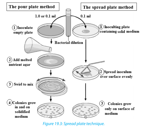
The introduction of a small number of microorganisms into a nutrient medium is
called inoculation. Aseptic technique must be used to avoid contamination. Theprocedure differs for solid and liquid media.
a. Inoculating a solid medium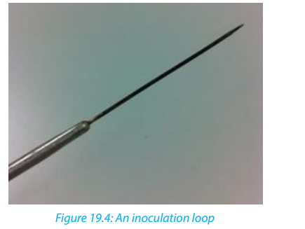
We use a wire loop. The loop is firstly flamed and after it is then used to lift a thin
film of a liquid suspension or a small amount of solid material containing the
microorganisms being investigated from the previous culture or any source of
microorganisms. The loop is gently stroked across the surface of the medium in a
series of sets of streaks.
b. Inoculating a liquid medium
If the cells to be inoculated are in a liquid, for example water or a broth, a sterile wire
loop is used to transfer a sample to the medium, which is often a test medium. The
wire loop is simply agitated gently inside the medium. If the cells to be inoculated
are in or a solid medium such as soil nutrient agar, a wire loop may be used for
transfer to the liquid medium. It can be rubbed on the inside surface of the vessel
containing the liquid medium to ensure successful transfer.
c. Carrying out a pure culture
Pure culture technique is a method of culturing microorganisms in which all of the
individuals in a culture have descended from a simple individual. The basis of pure
technique is the isolation in colonies of individual cells. This is done so as to allow thecharacterization of specific types of microorganisms.
d. Incubation on agar-agar.
During incubation, the nutrients are contained in agar medium. Agar is a substance
which dissolves in hot water and sets to form a jelly. You pour hot agar containing all
necessary nutrients such as carbohydrates, proteins and vitamins into a Petri- dish.
Then leave it to cool and set before you add any microorganism. The other way to
provide nutrients to grow microorganisms is as a broth in a culture flask. The stepsof culturing agar –agar are shown in the following activity.
Activity: 19.3.2– Boil a mixture of 50 ml of water and 20g of agar-agar powder for 15 minutes
as you are stirring
– Pour the jelly mixture into four pre-sterilized glass Petri-dishes. Then allow
the broth to coagulate at room temperature.
– Number the dishes; 1, 2, 3 and 4 respectively; on the bases.
– Place a nail scarping from between the teeth onto the jelly in dish 1 and 2,
wave the dish 3 on latrine for 1minute and do not put anything on dish 4.
– Warm the dishes 2 and 4 on the top of water vapour stream for 15 minutes
and then cool them (do not open them)
– Then fix the lids tightly to the bases of the four Petri-dishes with clear adhesive
tape and place them upside down in an oven/incubator at 37 ˚C for 3 days.– Record and interpret your results.

19.3.3 Alcoholic fermentation
Activity 19.3.3
Describe how yeast would be used in alcoholic fermentation.
Yeast releases digestive enzymes which allow the transformation of glucose into
ethanol as result of anaerobic fermentation. The presence of bubbles is the evidence
that carbon dioxide is released as waste product of the alcoholic fermentation.
Making Beer depends on a process called malting. You soak and keep barley grains
in water. As germination begins, enzymes break down the starch in the barley grains
into a sugary solution. You then extract a solution produced by malting and use it
as an energy source for the yeast. The mixture of yeast and sugar solution is then
fermented to produce alcohol. Hops are added at this stage to give flavour. The beer
is given time to clear and develops its flavour before putting it in bottles or to be
sold. Interestingly, alcohol in large quantities is toxic to yeast as well as to people.
This is why the alcohol content of wine is rarely more than 14%. Once it gets much
higher, it kills all the yeast and stops fermentation.
Self-assessment 19.3
From questions 1-5, circle the letter corresponding to the right answer
1. The method of culturing microorganisms in which all of the individuals in a
culture have descended from a single individual is called:a. Pure culture technique2. Inoculating liquid medium, various instruments are used. Which one of the
b. Spread plate technique
c. Aseptic technique
d. Liquid media method
following is used to transfer the sample to the medium?a. Sterile wire loop3. Large amounts of alcohol are dangerous to yeast during alcoholic fermentation.
b. Inoculating needle.
c. Petri-dishes
d. None of these.
Which of the following explains the reason?a. It kills all the yeast and stops fermentation.4. The technique of using sterilized equipment and solutions and preventing their
b. Motivate yeasts
c. It kills some few bacteria.
d. Temperature affects fermentation.
contamination is referred to as:a. Pure culture technique5. Petri dishes are specially designed as a shallow circular dish made of glass or
b. Spread plate technique
c. Aseptic technique
d. Petri-dish technique.
plastic with a lid. Which one of the following best explains the function of the
lid?a. Prevent contamination, but gas molecules can diffuse.19.4. Population growth of bacteria
b. Spread the bacteria on the plate.
c. Allows contamination.d. None of the above.
Activity: 19.4.1
Use text books and other sources of information to interpret the graphs of bacterial
growth.
When bacteria or any other microorganisms are incubated in a suitable culturing
medium, they reproduce by binary fissions and the number of individuals increases.
The ordinary growth of population is described as sigmoid curve or S-shaped curve
made of 4 main phases:
– The lag phase: period of adaptation of microorganisms to the new habitat
(new environment)
– The log or exponential phase: period of high rate of reproduction. Bacteria are
sensitive to the limiting factors of the growth or anti-microbial agents
– The stationary phase: Stationary phase of plateau-growth slows down. The
population remains constant because the rate of dividing/growth is equal to
the rate of death within the population. The maximum number that a habitat
can accommodate for a long period is known as the carrying capacity.
– The decline or death phase: period of high rate of death than the rate of
dividing/growth due to the scarcity of food, the abundance of metabolic waste
products, presence of antibiotics or any other drugs killing the germs. Figure19.5 shows the phases explained above.
19.4.1. Measuring population growth of bacteria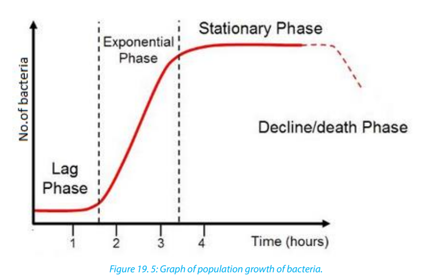
The typical growth curve of a population of bacteria is similar to the growth curve
expected for yeast, a unicellular fungus or the growth of any population. When
measuring the growth of a population of bacteria or yeast, we can carry out direct
counting of the numbers of cells or indirectly by measuring some indication of thenumber of cells such as the coldness of a solution, or production of a gas
It is usual to inoculate a small sample of the microorganisms in a sterilized nutrient
medium and to place the culture in an incubator at the optimum temperature for
growth. Other conditions are pH, oxygen concentration and ionic and osmotic
balance. Growth can be measured from the time of inoculation. Two types of cell
count are possible, namely viable count and total count. The viable count is the total
of living cells only and total count is the total number of both living and dead cellsand is easier to measure.
Activity 19.4.2
Investigating the bacterial content of fresh and stale milk.
Materials required: Four sterile nutrient agar plates, inoculating loop, Bunsen
burner, indelible marker or wax pencil, Fresh pasteurized milk, Stale milk (milk left
at room temperature for 24hours) and Incubator set at 350C
Procedure:– Place the inoculating loop in the Bunsen burner flame until the loop is red hot.
– Allow the loop to cool and then dip into a sample of fresh, well shaken milk.
– Lift the lid of the sterile agar plate slightly with the other hand and lightly
spread the contents of the inoculating loop over the surface the agar.
– Close the lid of the plate and return the loop to the Bunsen burner flame until
red hot.
– Label the base of the plate with an indelible marker or pencil.
– Repeat the process with the second plate and another sample of fresh milk.
– Flame the loop again and after cooling, place it in a sample of stale milk.
– Spread the contents of the loop over the surface of a third plate and then
close the lid.
– Label the base of the plate with an indelible marker or pencil.
– Repeat the process with the fourth plate and second sample of stale milk.
– Put the four plates in an incubator at 350
c for about 3 days. They should be
placed upside down to prevent condensation falling onto the cultures. After
incubation, the two halves of each plate should tape together for safety.
– Record the appearance of the coloniesGive the general comment based on your observations
Self-assessment 19.4
1. A culture of yeast, Saccharomyces cerevisiae, had been carried out in the banana
juice for 7 days at 30°C.The table below shows the change in number of yeastswithin that time:
 a. Draw a curve showing the growth of the yeast population2. Design an experiment to test the hypothesis that contact of an agar plate with a
b. What is the role of banana juice in that experiment?
c. State any two conditions that should be maintained constant during that
experiment.
d. Describe the trend of the graph you have drawn in afinger results in more bacterial growth than exposing the plate to classroom air.
19.5. Staining of bacteria
Activity 19. 5
“Staining bacteria for practical purpose is important”. Discuss the validity of the
statement.
The microorganisms or parts of microorganisms that pick up the stain are clearly
distinctively observed from the rest of the background.
In simple staining, all the cells and structures in general stain the same colour. In
positive staining, cells structures take in the stain e.g. methylene blue while in
negative staining the cells repel the stain and it is taken by the background e.g. Indian
ink. Negative staining is mostly useful in viewing capsules and such structures thatsurround the bacteria.
Differential staining on the other hand, multiple staining reactions are used to
take advantage of the fact that particular types of microorganisms and/or specified
structures of microorganism display varied staining reactions that are readily
distinguishable by different colours. The stain must be fixed immediately and the
dyed specimen is treated in some ways, e.g. by chemicals or heat to tightly bind thestain to the organism or its structures.
19.5.1. The purpose of staining bacteria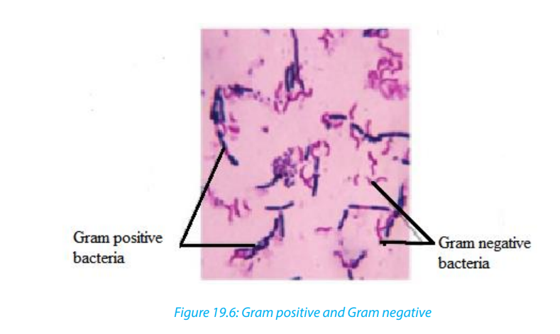
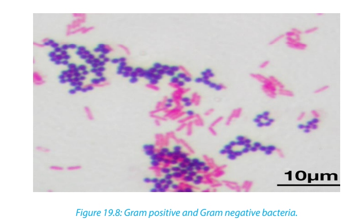
The purpose of staining bacteria is to see, for example, how thick of a layer of
peptidoglycan their cell wall has. In the Gram stain, gram-negative bacteria will stain
red or pink because the rinse took out the primary dye and the Safrinin (secondary
dye) took over the coloring as the counterstain. In gram-positive bacteria, since it
has a thick-layer of peptidoglycan, not all of the Crystal violet color will be rinsed out
of the cell wall, so it will be blue or purple. The following are reasons to explain why
stained:– It’s for helping classifying and determining what the bacteria are composed of.
– It’s very useful tool to help identify bacteria without necessarily killing the cell.– Gram staining is performed to distinguish between gram positive and negative
bacteria.– To enable the person to visualize its physical features- shape, size, arrangement,19.5.2. Procedure of staining and their corresponding stains.
etc the bacterial cells are stained with specific dyes or stains
Activity 19.5.1:
carry out an experiment to stain bacteria for examination under the light
microscope
In staining bacteria, we use various staining procedures each having specific set of
stains or dyes. Some of them are:1. Gram’s Staining - Crystal violet, Iodine and SafrininObserve and identify some of the staining methods on figure19.6 as shown below:
2. Capsule staining - Nigrosin, Safrinin or India Ink, Safrinin
3. Spore staining - Malachite Green and Safrinin
4. PHB staining - Sudan black.
5. Using decolorizer – Alcohol wash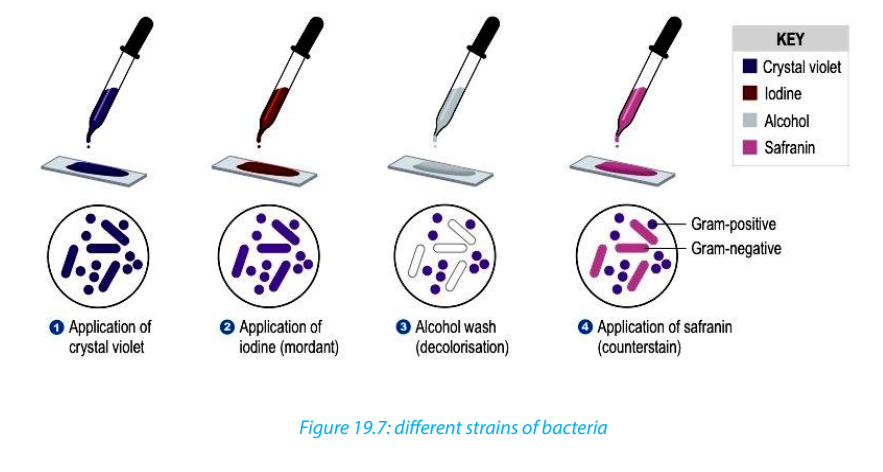
19.5.3 Growing viruses
The culture of viruses is made more difficult than the culture of bacteria or fungi because
viruses can only grow and multiply inside living cells. This can be done by infecting whole
Figure 19.8: Gram positive and Gram negative bacteria.
19.5.3. Growing viruses
The culture of viruses is made more difficult than the culture of bacteria or fungi
because viruses can only grow and multiply inside living cells. This can be done
by infecting whole organisms such as plants or animals but, where possible, cell,
tissue cultures are now used. An early technique was to grow certain viruses in chickembryos while the embryo was still growing inside the egg.
19.5.4. Tissue Culture of Animal Viruses
Viruses cannot be grown in standard microbiological broths or on agar plates;
instead they have to be cultured inside suitable host cells. Note the following facts:– Tissue culture is a useful method for cultivating clinical samples suspected ofCell culture is the complex process by which cells are grown under controlled
harboring a virus. This method helps with the detection, identification, and
characterization of viruses in the laboratory.
– Tissue culture of animal viruses involves growing animal cells in flasks using
various broth media and then infecting these cells with virus.
– Transfect ion can be carried out using calcium phosphate, by electroporation,
or by mixing a cationic lipid with the material to produce liposome’s, which
fuse with the cell membrane and deposit their content inside.
– Cytopathic effect is a non-lyrics damage that viruses cause to cells. These vary
in their manifestation and damaging effect.
– Cell culture is complex process by which cells are grown under controlled
conditions, generally outside of their natural environment.
conditions, generally outside of their natural environment. The term “cell culture” is
defined as the culturing of cells derived from multi-cellular eukaryotes, especially
animal cells. However, there are also cultures of plants, fungi, and microbes, includingviruses, bacteria, and protists.
2. Explain why it is more difficult to culture viruses than culturing bacteria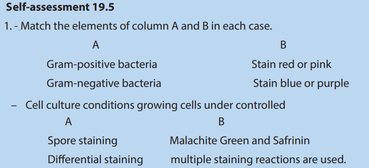
3. How/why are viruses specific to the cells they infect?
4. Distinguish between vaccines and antibiotics
End of unit assessment19
1. What are different types of media used in the laboratories for culturing
microorganisms?
2. Define a pure culture.
3. How do biologists differentiate between Gram –positive and Gram –negative
bacteria?
4. Describe the three methods of preventing bacterial growth in food.
5. How does temperature affect the growth of bacteria in culture media?
6. Assuming that you have a bacterial infection, would you ask for vaccination
against the bacteria? Why or why not?
7. How do bacteria maintain the balance in the environment?
8. Explain why an infection by Gram–negative bacteria are more difficult to treat
than Gram-positive bacteria.
9. How would you investigate that temperature affect the bacterial growth?
10.Write short notes on each of the following term related to the culture of
microorganisms.a. Aseptic techniques.
b. Staining bacteria
c. Growing viruses11. Explain why microorganisms are particularly suitable for industrial use.