UNIT 18: MICROBIOLOGY
UNIT 18: MICROBIOLOGY
Key Unit Competence
Describe the structure and characteristics of viruses, bacteria, and fungal and nonfungal moulds.
Learning objectives
By the end of this unit, I will be able to:– Describe the basic structure of viruses.Introductory activity
– Explain how a retrovirus reproduces.
– Identify the effects of viruses (e.g. AIDS, influenza, measles, feline leukemia,
some human cancers) and prokaryotes (e.g. tuberculosis, bubonic plague,
cholera) on organisms.
– Describe how plant viruses can be transmitted.
– Explain how and why archaebacteria are thought to have been the first forms
of life.
– Describe the structure and life cycles of Escherichia coli
– Relate the structures and functions of Prokaryotes
– Describe the structure of fungal and non-fungal moulds and explain how they
reproduce– Appreciate the importance of microorganisms in life.
A student left fresh milk in a cup exposed to the air. After 6 hours, he/she found
that milk changed its state from fresh milk to stale milk. Why do you think this
happened?
Mukamukiza prepared food for dinner. Some of the food was immediately put in
tightly covered flask while the remaining food was left in the saucepan covered
with banana leaves. In the evening, food in the flask was warm and safe while
food in the saucepan has deteriorated. What is the cause of the food spoilage inthe saucepan?
18.1. Introduction to microbiology.
Activity 18.1.1
Discuss on the term microbiology and on the groups of microorganisms.
The term “microbiology” comes Greek words: ‘micros’ which means small, ‘bios’
which means life and ‘logos’ which means science. Microbiology is the study of
microorganisms which are too small organisms to be seen with the unaided eye and
require a microscope to be seen. They are also referred to as microbes. They include
bacteria, fungi, algae, protozoa and viruses, they are useful to humans and they play
a vital role in decay and recycling of nutrients in the environment. Some of them
cause diseases
Micro-organisms are everywhere: in the air, water soil, on plants, on rock surfaces
in very hot and cold places (ice). Before the invention of the microscope, microbes
were unknown and thousands of people died in devastating epidemics because,
vaccines and antibiotics were not available to fight against infectious diseases.Nowadays, microorganisms can be grown in the laboratory and studied.
a. The Prokaryotes
Prokaryotes can be categorized by their mode of nutrition and how they obtain
energy and the carbon used to build the organic molecules that make up cells.
Organisms that obtain energy from light are called phototrophs and those that
obtain energy from chemicals are called chemotrophs. Organisms that need
only inorganic compounds such as CO2 as a carbon source are called autotrophs.
Heterotrophs require at least one organic nutrient such as glucose to make other
organic compounds. Prokaryotes usually range in size from 1 to 5 micrometersmaking them much smaller than most eukaryotic cells.
b. Classification of prokaryotes
Traditionally, bacteria have been classified based on their structure, physiology,
molecular composition rather than on their evolutionary relationships. The bacteria
that we generally refer to as germs are classified in the domain Eubacteria. More
frequently, members of this kingdom are simply called bacteria. The other type of
bacteria is known as archaebacteria. These bacteria, which are more ancient than
the Eubacteria, are classified in the domain Archaebacteria. Taxonomists used toclassify all prokaryotes in kingdom Monera, yet they slightly differ in characteristics
18.1.2. Archaebacteria and Eubacteria
Activity 18.1.2
Discuss on the characteristics of given examples of both archaebacteria and
Eubacteria.
a. Archaebacteria
Taxonomists treat archaebacteria as a separate kingdom because they are so different
from other bacteria. Archaebacteria have unusual lipids in their cell membranes.
Their cell wall is characterized by the absence of peptidoglycans, a protein
carbohydrate compound found in the cell walls of Eubacteria. Archaebacteria were
first discovered in extreme environmental conditions such as swamps, salt lakes, hot
springs. Examples include:
1. Methanogens
– They have unique method of harvesting energy by converting H2 and CO2
in methane.
– Methanogens can live only in anaerobic condition, such as the bottom of a
swamp, and in sewage where they are the source of marsh gas, because
oxygen is a poison to them.2. Extreme halophiles– These are salt-loving archaebacteria living in environment with very high salt
concentration such as the Dead Sea. High salt concentration would kill most
bacteria.
– These organisms use salt to generate ATP.3. Thermoacidophiles– This third group of archaebacteria lives in extremely acidic environments thatHow and why Archaebacteria are thought to have been the first forms of life?
have extremely high temperature such as hot springs. Thermoacidophiles live
at 110ºC and at a pH of 2.
– Thermoacidophiles live near volcanic vents on land or near hydrothermalvents.
The Archaebacteria comprise a group of single-celled microorganisms that, like
bacteria, are prokaryotes that have no cell nucleus or any other organelles within
their cells. They are known to have an independent evolutionary history and have
numerous differences in their biochemistry compared to other forms of life.
Archaebacteria are now classified as in separate domain in the three-domain
system by Carl Woese who introduced three main branches of evolutionary descent
currently known as the Archaea, Eukarya and Bacteria. Classifying Archaea remains
difficult, since many of them have never been studied in the laboratory and haveonly been detected by analysis of their nucleic acids.
b. Eubacteria
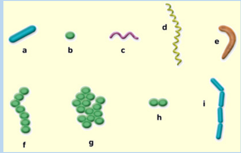
They occur in many shapes and sizes and have distinct biochemical and genetic
characteristics. Eubacteria that are rod-shaped are called bacilli, sphere-shaped are
called cocci (sing. Coccus) and spiral-shaped are called spirilla (sing. Spirillum).1. The bacilli: bacteria with rod-shape. Ex: Clostridium tetani, Bacillus subtilis
2. Vibrios: comma-shaped with a single flagellum. eg: Vibrio cholera
3. The cocci: group of bacteria with spherical shape such as Streptococci.
Cocci that occur in chains are Staphylococci which are grapelike clusters of
cocci and Diplococci which is sphere shaped that are grouped two by two.4. The spirilla: bacteria with spiral shape. e.g.: Spirillum volutans.
18.1.3. Gram stain
Bacteria have a peptidoglycan or murein cell wall that maintains cell shape, provides
protection and prevents the cell from lysis. Based on the composition of the cell wall,
bacteria can be classified as Gram-positive and Gram-negative. During the process
of Gram staining , some bacteria without a lipid layer along with their peptidoglycan
cell wall take the gram stain and appear violet (purple) and are therefore called
gram positive. Example streptococcus and staphylococcus. Bacteria having a lipid
layer along with their peptidoglycan cell wall do not take up the gram stain and aretherefore called gram negative.
Example: Escherichia coli, Azotobacter, Salmonella.
Self-assessment 18.11. Describe the characteristics of the two domains of prokaryotes.18.2. The structure and life cycle of Escherichia coli
2. What factors can be used to identify prokaryotes?
3. How do bacteria maintain equilibrium in the environment?
4. Identify the parts of a prokaryote.5. Describe briefly how some prokaryotes obtain their energy.
Activity 18.2.1
Using text books, videos or computer aided materials to describe the cycle life of
E. coli.
E. coli reproduce asexually by undergoing binary fission. This type of reproduction
begins with the replication of DNA molecule. Then, the copies of the genetic material \
attach themselves to the cell membrane. When the bacterium’s size has doubled
from its original size, the cell membrane starts pinching inward and a cell wall is
produced between the two DNA molecules. Finally, the cell wall divides the cell into
two daughter cells.
E. coli can also go through another process of reproduction known as
conjugation. Conjugation is a reproduction process which involves the transfer
of genetic material by the sex pili between two bacteria. This is not a sexual
reproduction because there is no combination of gametes. The process of
conjugation starts once the E. coli, called a donor, has finished to replicate its
genetic material in form of a plasmid. The enzyme of the donor can now send
signals to show that it is ready to mate. Once a mate is found, the donor attachesitself to the sex pilus of its mate. By doing so, the donor transfers the plasmid.
18.2. E. coli and food poisoning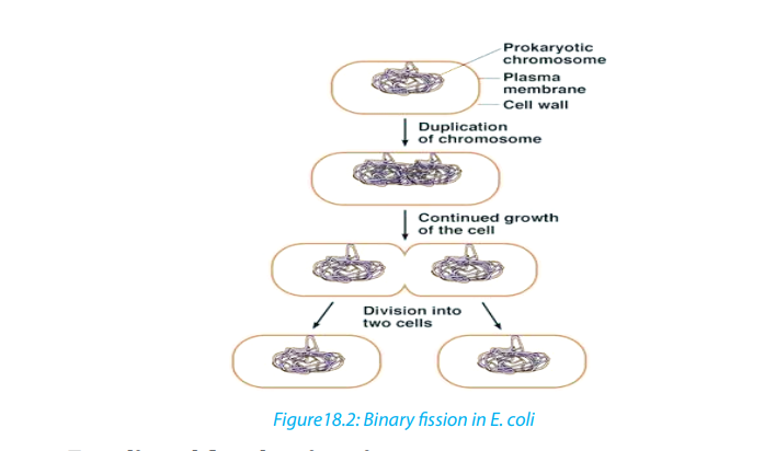
Activity 18.2.2
Using textbooks to brainstorm the process of food poisoning, evolution of harmful
strain of E. coli and food preservation
E. coli is a rod-shaped bacterium measuring about 2.5µm by 0.5µm. It is mainly found
in guts of vertebrates. It is chemoheterotrophic, capable of thriving on a variety of
the organic molecules. Its presence in water indicates contamination by faces.
E.coli reproduces asexually by binary fission. It can also take part in a primitive form of
sexual activity called conjugation where genetic material is passed in one direction
from bacterium to another through a pilus. Although conjugation does not in
itself produce new offspring, after the process has finished, the bacteria reproduceasexually, passing on their new genetic make-up to their offspring.
18.2.1. Evolution of harmful strain of bacteria
E. coli was thought to be a relatively harmless resident of the human gut which might
linked to the occasional upset stomach and mild diarrhoea. When massive colonies
of mutualistic bacteria are present in the gut, including most strains of E. coli, they
help to keep harmful bacteria away from starving them of food. They also help make
vitamin K. But in 1982, it became clear that a new strain of E. coli had evolved into
a much more troublesome organism. The strain had acquired a gene that enabled
it to produce a powerful toxin which damages the intestinal wall, causing severe
diarrhea and internal bleeding.
This may lead to internal serious dehydration in young children and elderly people,
and may result into death. In majority of the cases, infections of pathogenic strain ofE. coli are not fatal and the disease clears without treatment.
18.2.2. Sources of infection
Touching a source of contamination and not washing hands before handling food
may be sufficient to cause the infection.
In 1996, there was an outbreak which led to 20 deaths in Scotland due to
contaminated meat. In the same period, another one was traced due to apple juice
poisoning. Contaminated person can pass the bacteria on vegetables, and other
foods.We must practice good habits of dealing and handling food to minimise
cases of contamination. It is therefore, important to practice good hygiene. It is
also essential to store and package food. It might be vital to pasteurise all fresh fruit
juices just as milk is required to be pasteurised.
18.2.3. Food storage and packaging
The optimum storage conditions differ; raw meat and poultry are kept at around 00c,
meat products at 1oc - 40oc.
Canned foods and many vegetables in dry condition sat 10oc - 150oc,
and dried foods such as flour are stored, in air tight containers at10oc– 150oc.
For long term storage, meat and fish are vacuum-sealed or can be vacuum
packed in laminated plastic containers. For pasteurisation, food and drinks such as
milk are heated to a temperature that kills disease causing microorganisms. Example:Mycobacterium tuberculosis.
Self-assessment 18.21. Suggest the process by which E. coli reproduces.18.3. The structure and life cycle of viruses
2. What is the probable source of the gene that transforms harmless E. coli
into pathogenic E. coli?
3. At what temperature is E. coli in meat killed?
4. How is food poisoned?5. How can you minimise food and drink poisoning?
Activity18.3.1
Using textbooks, chart or videos to describe the structure, life cycle and effects of
viruses.
The term “virus” was first used in the 1890s to describe agents smaller than bacteria
that cause diseases. The existence of viruses was established in 1892, when, Russian
scientist, Dmitry Ivanovsky discovered later microscopic particles known as thetobacco mosaic virus
There are at least 3,600 types of virus. Hundreds of which are known to cause
diseases in animals, bacteria, and plants. Viruses consist of an inner core of either
ribonucleic acid (RNA) or deoxyribonucleic acid (DNA) plus a protein protective
coat called capsid made of protein or of protein combined with lipid or carbohydrate
components. An entire virus particle is called vibrios ?
The core confers infectivity, and the capsid provides specificity to the virus. In some
virions, the capsid is further enveloped by a fatty membrane. The later may causevirion inactivation by exposure to fat solvents such as ether and chloroform.
18.3.1. Characteristics of viruses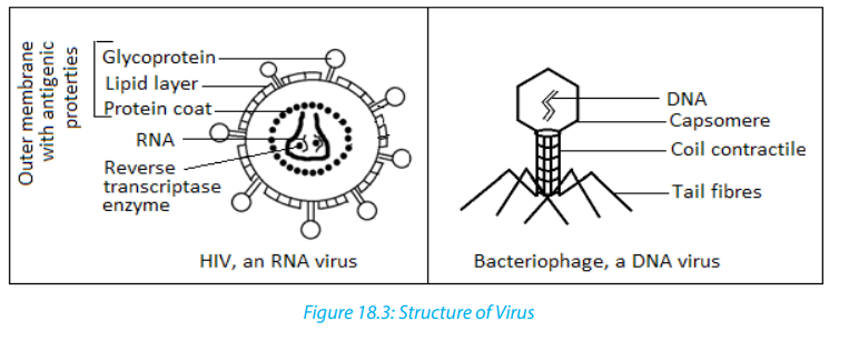 – Viruses are complex biochemical molecules having the following characteristics:18.3.2. Virus types
– Viruses are not visible under light microscope because they are very small than
bacteria.
– They possess a single type of nucleic acid either DNA or RNA enclosed in a
protein coat.
– They can reproduce and grow inside the host cell.
– They have no cell and no cell organelles.
– They are obligate parasite i.e. cannot survive outside a host cell.– They do not feed, respire and excrete.
DNA and RNA viruses differ in the way they use the host cell’s mechanisms to produce
new viruses.
For example, a DNA virus may act in one of the two ways:
The virus may directly produce RNA that is used to make more viral proteins or it
may join with the host cell’s DNA to direct the synthesis of new viruses.
RNA viruses replicate differently from DNA viruses. Upon entering the host cell, a viral
RNA is released into the host cell’s cytoplasm. There, it uses the host cell’s ribosomes.
Some RNA viruses known as retroviruses contain an enzyme called reverse
transcriptase in addition to RNA. Reverse transcriptase uses RNA as a template
to make DNA. The DNA then makes an RNA transcript of itself. This RNA is then
translated into proteins that become part of new viruses. Reverse transcriptase is so
named because it reverses the normal process of transcription, in which DNA servesas a template for producing RNA.
18.3.3. Viral replication
Because viruses are not cells, they can replicate only by invading a host cell and
using the enzymes and organelles of the host cell to make more viruses. Because
they depend on host cells for replication, viruses are called obligate intracellular
parasites. Outside the host cell, a virus is a lifeless particle with no control over its
movements. It is spread by wind, water, in food, or via blood or other body secretions.
18.3.4. Life cycle of Bacteriophage
Bacteriophage is a virus that infects bacteria. Bacteriophage is composed of an
icosahedral head that contains a nucleic acid. Beneath the head is a contractile tail
that includes a collar and a sheath.
The contractile tail helps to inject the nucleic acid into the host cell. The tail rests on
a base plate from which tail fibers emerge. These fibers assist the virus to attach to
a host cell.Viruses replicate by using either the lytic cycle or the lysogenic cycle:
a. The lytic cycle
Activity 18.3.2
Describe the sequence of events that occur during a lytic infection.
During the lytic cycle, a virus invades a host cell, produces new viruses, destroys the
host cell, and releases newly formed viruses. Viruses that undergo the lytic cycle are
called virulent because they cause disease. The lytic cycle consists of five phases:– The Bacteriophage first attaches to susceptible bacterium by attaching its tail
fibers to a receptor site. Receptor sites are specific sites that viruses recognize
and attach to on the host cell’s surface. If the Bacteriophage does not find a
receptor site, it cannot infect the cell.
– Next the Bacteriophage releases an enzyme that weakens a spot in the
cell wall of the host. Then the phage presses its sheath against the cell and
injects its DNA into the host cell through the weak spot in the cell wall. The
Bacteriophage leaves its capsid outside.
– The virus then takes control of the host’s protein synthesizing mechanisms,
transcribing mRNA from the viral DNA. The resulting Bacteriophage mRNA is
translated on ribosomes and proteins that are synthesized form B a capsid. So
the viral DNA is also replicated during this phase.
– Every replicated viral DNA is enclosed in the newly created viral capsid. The
assembly of new virus particles usually occurs in the cytoplasm.
– During the last phase of the lytic cycle, one of the enzymes that are produced by
the Bacteriophage genome causes the host cell to disintegrate, releasing new
Bacteriophage. The cell disintegration is called lysis. In case of the enveloped
viruses, the newly formed viruses move to the cell surface and force their waythrough the cell membrane.
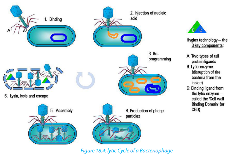
The first step in the replication of the phage in its host cell is called adsorption or
binding. The Bacteriophage adheres to the receptor site by means of its tail fibres.
Following adsorption, the phage injects its DNA into the bacterial cell.
The tail sheath contracts and the nucleic acid or the core is driven through the wall
to the membrane. This process is called penetration and it may be both mechanicaland enzymatic.
Immediately after injection of the viral DNA there is transcription and translation
of a section of the phage DNA to make a set of proteins that are needed to replicate
the phage DNA and proteins that make up the capsid and the various components
of the tail.
After making all viral parts, the assembly process follows. While the viruses are
assembling, produced lysozymes are used to break down the cell wall peptidoglycans
of the host bacteria. This is known as lysis and then mature viruses are released andspread to nearby cells for new infection.
b. The lysogenic cycle.
Activity 18.3.3
Using textbooks to describe what happens to the host cell infected by a temperate
virus.
Some viruses can infect a cell without causing its immediate destruction. Such
viruses stay in their host cell for an extended period of time: days, months or years
in a lysogenic cycle. A virus that replicates through lysogenic cycle and does not killthe host cell immediately is called a temperate virus.
Retroviruses, such as HIV, have RNA that is transcribed into DNA by the viral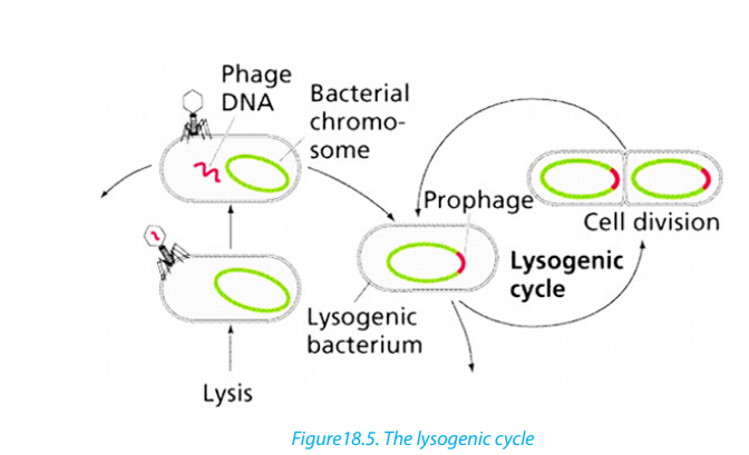
enzyme Reverse transcriptase upon entry into the cell. (The ability of retroviruses
to copy RNA into DNA earned them their name because this process is the reverse
of the usual transfer of genetic information, from DNA to RNA). The DNA form of the
retrovirus genome is then integrated into the cellular DNA and is referred to as the
provirus. The viral genome is replicated every time the host cell replicates its DNAand is thus passed on to daughter cells.
18.3.5. Some common viral diseasesTable 18.1: Some common viral diseases
18.3.6. Virus as living or non-living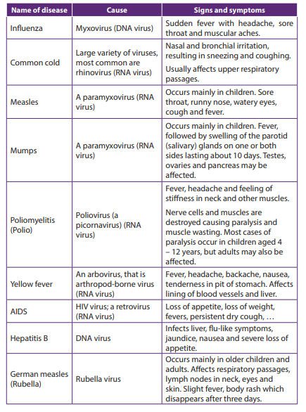
Activity 18.3.4
“Viruses are said to be on the border line of living organisms and non-living
things”. Discuss on this statement.
Viruses do not belong to any of the five kingdoms into which life is classified. It is
difficult to say whether they are living or non-living.
a. Features that make viruses to look like living things:– They have the genetic material composed of either DNA or RNA They causeb. Features that make viruses non-living things:
diseases to other living things: All viruses are infectious.
– They evolve as a result of mutation and natural selection.
– They reproduce /multiply only in other living things: they are obligate
intracellular parasites– They cannot metabolize
– They crystallize in isolation.
– They cannot reproduce outside of host.
– They are not made of cells. This means that they have a relatively simple noncellular organisation.
– They cannot respond to stimuli
– They have one type of nucleic acid, either DNA or RNA. But living cells containboth DNA and RNA.
Table 18.2: Comparison between viruses and cells
Self-assessment 18.3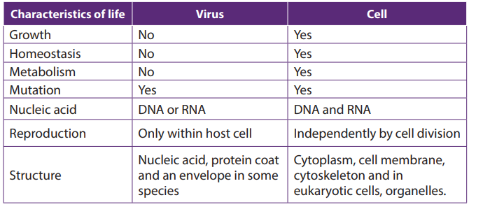 1. What are the parts of a virus?18.4. Moulds
2. Describe the two ways by which viruses cause infection.
3. Distinguish between Bacteriophage and a prophage.
4. What is meant by retrovirus?
5. What are the strengths and weaknesses of the tobacco mosaic virus
hypothesis?
6. Which characteristic feature is common to all viruses?
7. How is a capsid protein important to the functioning of a virus?8. What is the best way to protect humans against most viral diseases?
Activity 18.4
Using text books or computer aided materials to describe the life cycle of bread
mould.
Moulds pervade our world, living wherever moisture is present. Some are of great
benefit to humans, providing antibiotics, acting as decomposers so that nutrients
can be recycled, or taking part industrial processes. Other moulds cause diseaseswhich lead to serious damage.
Moulds have cells arranged in long thread-like filaments, the hyphae, that form a
mass called Mycelium. Moulds are usually considered as fungi, but mould may also
be formed by filamentous bacteria, slime moulds, and water moulds. Therefore,
there are two main types of moulds: fungal moulds and non-fungal moulds
18.4.1. Fungal moulds
All fungi that produce mycelia can be called moulds, but the term is usually used for
an organism in which the mycelium forms the main body of the fungus. In the black
bread mould Rhizopus and the pin mould Mucor, the mycelium consists of a tangled
mass of hyphae with many nuclei. These hyphae are called coenocytic because the
fungal tissue is not separated by cell walls.
Fungal hyphae have an outer cell wall made of chitin and inner lumen which contains
the cytoplasm and organelles. A cell surface membrane surrounds the cytoplasm
and sticks tightly to the cell wall.
Rhizopus and Mucor are Saprotrophic, obtaining their nutrients from dead organic
material. Rhizopus nigricans and Mucor mucedo can live on bread but some species
of Rhizopus feed on living plants, and Mucor commonly grows on rotting fruits and
vegetables, in the soil or on dung.
the food outside the organism and then absorb the soluble digestion products and
assimilate them.
a. Life cycle of Rhizopus and Mucor.
Rhizopus and Mucor belong to the fungal phylum Zygomycota. The phylum got its
name because its members produce two kinds of spores: Sexual zygospores as wellasexual sporangiospores.
The asexual sporangiospores formed by mitosis, develop in sporangium at the tip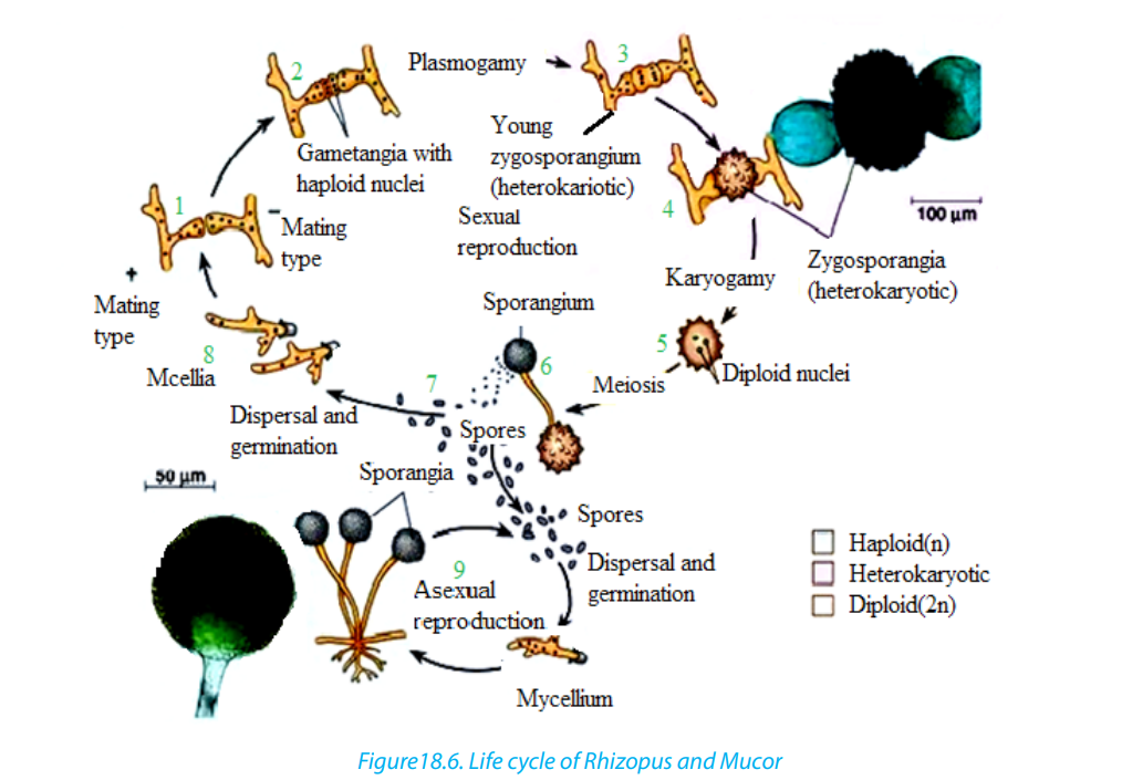
of hyphae. When sporangium busts, the spores are released.
In most species of Mucor, the sporangium dissolves then water enters the spore
mass, and the spores are dispersed by the raindrop or are transported by the insects.
In most Rhizopus species, the sporangium wall fractures and dry spores are released
by the wind.
The sexual reproduction involves conjugation. Usually the hyphae from mycelia of
different mating types meet and interconnect via outgrowths. The interconnecting
walls break down and their cytoplasm containing haploid nuclei mix, then the
diploid zygote formed by the fusion of two nuclei develops a thick, rough, black
coat and becomes a dormant zygospores. Meiosis probably occurs at the time of
germination; the zygospore cracks open to liberate several haploids spores whichcan give rise to asexual sporangia and mycelia of either mating strain.
b. Use of moulds
Even if species of Rhizopus and Mucor are responsible for the spoilage of food, they
are also useful as follow:– They are used to make the human foods. For example, Mucor is used with soya18.4.2. Non-fungal moulds
beans to make a cheese called sufu, in eastern Asia.In Indonesia, R. oligosporus
and R. oryzae are used to produce a food called tempeh from boiled skinless
soya beans.
– The fungal moulds belonging to the Zygomycota are used to make anaesthetics,
birth control pills, meat tenderisers, and the yellow colouring agents used inmargarines and butter substitutes
The following are different groups of non-fungal moulds:a. Bacterial moulds: including those of Streptomyces griseous, which secretes the1. Plasmodial slime moulds which have the following characteristics:
antibiotic streptomycin
b. Slime moulds: These are a peculiar group of organisms that resemble fungi in
appearance and lifestyle, but are more closely to protoctists such as Amoeba in
their cellular organization, reproduction, and life cycles. There are two types of
Slime moulds:– They have no connection with the parasitic protoctists belonging to the genus2. Cellular slime moulds (also called Acrasiomycotae) which have the following
Plasmodium which causes malaria.
– They exist as thin, streaming masses of colourful protoplasm that creep along
moist, rotting logs and leaves.
– They move in an amoeboid fashion, engulfing food particles by Phagocytosis.
– A single mould may extend for many centimetres, but it is not multicellular.
– They are made up of a continuous mass of cytoplasm with many nuclei called
coenocytic mass.
characteristics.– They have a unicellular feeding stage resembling an amoeba, with each cellc. Water moulds (Oomycota)
functioning individually.
– When food is scarce, the individual cells group into a mass resembling that of
Plasmodial slime moulds.
– The individual cells of Cellular slime moulds retain their identity and have
separate cell surface membranes– Although water moulds and fungi are closed related and have a similar
structure, water moulds are generally regarded as a separate and more ancientgroup belonging to the protoctists.
– Water moulds include rusts and mildews which consist of coenocytic masses(where “Oo” means egg)
of hyphae similar to fungi, for example Plasmodial slime moulds,
– Most water moulds have cell wall made of cellulose, while the cell wall of the
true fungi is made of chitin.
– Some of the most devastating plant diseases are caused by water moulds. For
example, the Phytophthora infestans causes potato blight, and Pythium which
is a relatively unexpected parasite attacks a great variety of plants causing soft
rot.
– Water moulds reproduce asexually by structure called conidia, and by moving
spores with flagella, called zoospores.
– They reproduce sexually by producing moving male gametes that fertilizes
large immobile egg cells. These egg cells give the group its name Oomycotae
Self-assessment 18.41. How are the cell walls of fungi similar to exoskeleton of insects?18.5. Penicillium and Saccharomyces
2. Distinguish between hyphae and mycelium.
3. What are conditions necessary for fungal spores to germinate?
4. Explain the basis of classification of fungi.
5. Why do many biologists think that Penicillium evolved from an ascomycete?
6. Briefly describe sexual and asexual reproduction in fungi.
7. The antibiotic penicillin is a natural secretion of a certain kind of fungus green
mould called Penicillium, penicillin kills bacteria. Why might a mouldspecies have evolved way of killing bacteria?
Activity 18.5
Make a research from the internet or textbooks to find out:
1. The structure of Penicillium, and yeast cell.
2. How saccharomyces reproduces.3. The explanation of budding.
18.5.1. Penicillium and antibiotics
Penicillium is highly known for producing penicillin, the first antibiotic discovered in
1928 by a scientist Alexander Fleming when he was culturing some Staphylococcus
bacteria during his medical research.
After leaving some Petri dishes for many days, he found a mouldy growth of
Penicillium notatum contaminating a corner of one of dishes. Then Fleming realised
that Staphylococcus next to the mould has been destroyed.
After studying Staphylococcus closely, Fleming concluded that the Penicillium
mould was producing a substance that killed the Staphylococcus. He carried on
with finding out if the broth of Penicillium mould contained penicillin which could
destroy pathogenic bacteria.
In 1931, Fleming dropped his research. Howard Florey and Ernst Chain went on to
produce purified penicillin. A successful work was reported 1940, and penicillin has
been used to treat wounded soldiers in Second World War. In 1945, Fleming, Floreyand Chain received the Nobel Prize for the discovery of penicillin.
a. The structure of Penicillium
Penicillium is septate; its hyphae have cross-walls called septa. However, the septa
are not formed by cell division, and at the Centre of septum there is a usually a
pore which allows cytoplasm to flow from one compartment to another. Each
compartment may contain one or more nuclei. Though Penicillium has septa, is a
coenocyte like the non-septate moulds Rhizopus and Mucor.
Penicillium is saprotroph, feeding on organic matter in damp soil, leather, bread, and
decaying fruit. The mycelia of Penicillium species form circular green, yellow, or blue
moulds (depending to the species).
Penicillium reproduces asexually by means of spores called conidia formed at the tip
of special hyphae called conidiophores.
Spores of Penicillium are exposed and free to be dispersed as they are mature.
18.5.1. Saccharomyces
a. Definition and characteristics– Saccharomyces is a genus of yeasts which include all unicellular fungi thatb. Structure of yeast
reproduce asexually by budding.
– They occur commonly on faeces, in the soil, and on the surfaces of plants and
animals.
– The most familiar and industrial important yeast is Saccharomyces cerevisiae.
– The tiny cells of this yeast are very active metabolically. They are usually aerobic
but in the absence of oxygen they use anaerobic metabolism, producing
carbon dioxide and ethanol (alcohol) as waste products which are industrially
useful
– Each cell of Saccharomyces cerevisiae has a single nucleus and is usually egg
shaped.– Cells contain most of organelles of a typical eukaryote.
c. Mode of reproduction
Saccharomyces cerevisiae can reproduce either asexually or sexually.
In asexual reproduction, the single cell divides by budding and separate into two
cells. Some buds group together to form colonies; other separate to grow individuallyinto a new yeast.
In sexual reproduction, two cells fuse to form a diploid cell which then forms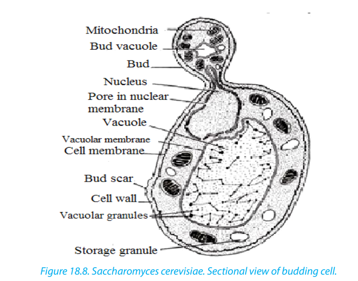
haploid spores by meiosis
Self-assessment 18.51. Which feature does all yeast have in common?18.6. Protozoa that cause disease
2. How do hyphae of Penicillium differ from those of Mucor.3. Describe the evidence for penicillin’s effectiveness.
Activity 18.6
Observe prepared slides of Entamoeba histolytica ,Plasmodium and Trypanosomato compare their structures.
18.6.1. Entamoeba histolytica
a. Characteristics of Entamoeba histolytica
Entamoeba histolytica is a protozoan parasite responsible for a disease called
amoebiasis. It occurs usually in the large intestine and causes internal inflammation
as its name suggests (histo which means tissue, lytic which means destroying). 50
million people are infected worldwide, mostly in tropical countries in areas of poor
sanitation. Inside humans Entamoeba histolytica lives and multiplies as Trophozoites.
Trophozoites are oblong and about 15–20 µm in length. In order to infect otherhumans, they encyst and exit the body.
b. Life cycle Entamoeba histolytica
Entamoeba histolytica life cycle does not require any intermediate host. Mature
cysts (spherical, 12–15 µm in diameter) are passed in the feces of an infected human.
Another human can get infected by ingesting them in fecally contaminated water and
food. If the cysts survive the acidic stomach, they transform back into Trophozoites
in the small intestine. Trophozoites migrate to the large intestine where they live
and multiply by binary fission. Both cysts and Trophozoites are sometimes present
in the feces. Cysts are usually found in firm stool, whereas Trophozoites are found in
loose stool. Only cysts can survive longer periods (up too many weeks outside the
host) and infect other humans. If trophozoites are ingested, they are killed by the
gastric acid of the stomach. Occasionally Trophozoites might be transmitted during
sexual intercourse.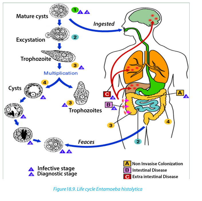
c. Symptoms
Many Entamoeba histolytica infections are asymptomatic and Trophozoites remain
in the intestinal lumen feeding on surrounding nutrients. About 10–20 % of the
infections develop into amoebiasis which causes 70 000 deaths each year. Minor
infections (luminal amoebiasis) can cause symptoms that include:– Gas (flatulence) intermittentSevere infections inflame the mucosa of the large intestine causing amoebic
– constipation loose stools
– stomach ache
– Stomach cramping.
dysentery. The parasites can also penetrate the intestinal wall and travel to organs
such as the liver via bloodstream causing extra-intestinal amoebiasis. Symptoms
of these more severe infections include: Anemia, Appendicitis (inflammation ofthe appendix), bloody diarrhea, fatigue, fever, gas (flatulence), genital and skin
lesions, intermittent constipation, liver abscesses (can lead to death, if not treated),
malnutrition, painful defecation (passage of the stool), peritonitis (inflammation of
the peritoneum which is the thin membrane that lines the abdominal wall), pleuropulmonary abscesses,
stomach ache, stomach cramping, toxic mega-colon (dilated
colon), Weight loss.
d. Prevention
To prevent spreading the infection to others, one should take care of personal
hygiene. Always wash your hands with soap and water after using the toilet and
before eating or preparing food. Amoebiasis is common in developing countries.
Some good practices, when visiting areas of poor sanitation:– Wash your hands often.Natural water can be made safe by filtering it through an “absolute 1 micron or less”
– Avoid eating raw food.
– Avoid eating raw vegetables or fruit that you did not wash and peel.
– Avoid consuming milk or other dairy products that have not been pasteurized.
– Drink only bottled or boiled water or carbonated (bubbly) drinks in cans orbottles.
filter and dissolving iodine tablets in the filtered water.
e. Methods of diagnosis
Amoebiasis is diagnosed by your health care provider under a microscope by
finding cysts and (rarely Trophozoites) from a stool sample. The results are usually
said to be negative, if Entamoeba histolytica is not found in three different stool
samples. But it still does not necessarily mean that you are not infected because
the microscopic parasite is hard to find and it might not be present the particular
samples. A blood test might also be available but is only recommended, if your
health care provider believes that the infection could have spread to other parts of
the body. Trophozoites can be identified under a microscope from biopsy samplestaken during colonoscopy or surgery.
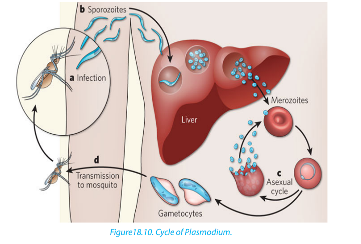
18.6.2. Plasmodium spp.
a. Characteristics:– Plasmodium is the genus of the class of Sporozoa that includes the parasiteb. Life cycle of Plasmodium
that causes malaria. Plasmodium is a type of protozoa, a single-celled organism
that is able to divide only within a host cell.
– The main types of Plasmodium spp are P.falciparum, the species that causes
falciparum malaria, the most dangerous type of malaria; P. malariae, the species
that causes quartan malaria; P. ovale, a species found primarily in east and
central Africa that causes ovale malaria; and P. vivax, the species that causesvivax malaria, which tends to be milder than falciparum malaria.
Plasmodium species exhibit three life-cycle stages gametocytes, sporozoites, andmerozoites.
Gametocytes within a mosquito develop into sporozoites. The sporozoites are
transmitted via the saliva of a feeding mosquito to the human blood stream. From
there, they enter liver parenchyma cells, where they divide and form merozoites.
Inside the host’s liver cell, the Plasmodium cell undergoes asexual replication. The
products of this replication, called merozoites, are released into the circulatory
system. The merozoites invade erythrocytes and become enlarged ring-shaped
Trophozoites.
More erythrocytes are invaded, and the cycle is reinitiated. The merozoites are
released into the bloodstream and infect red blood cells. Rapid division of the
merozoites results in the destruction of the red blood cells, and the newly multiplied
merozoites then infect new red blood cells. Some merozoites may develop into
gametocytes, which can be ingested by a feeding mosquito, starting the life cycleover again.
The red blood cells destroyed by the merozoites liberate toxins that cause the
periodic chill-and-fever cycles that are the typical symptoms of malaria. P. vivax, P.
ovale, and P. falciparum repeat this chill-fever cycle every 48 hours (tertian malaria),
and P. malariae repeats it every 72 hours (quartan malaria). P. knowlesi has a 24-hourlife cycle and thus can cause daily spikes in fever.
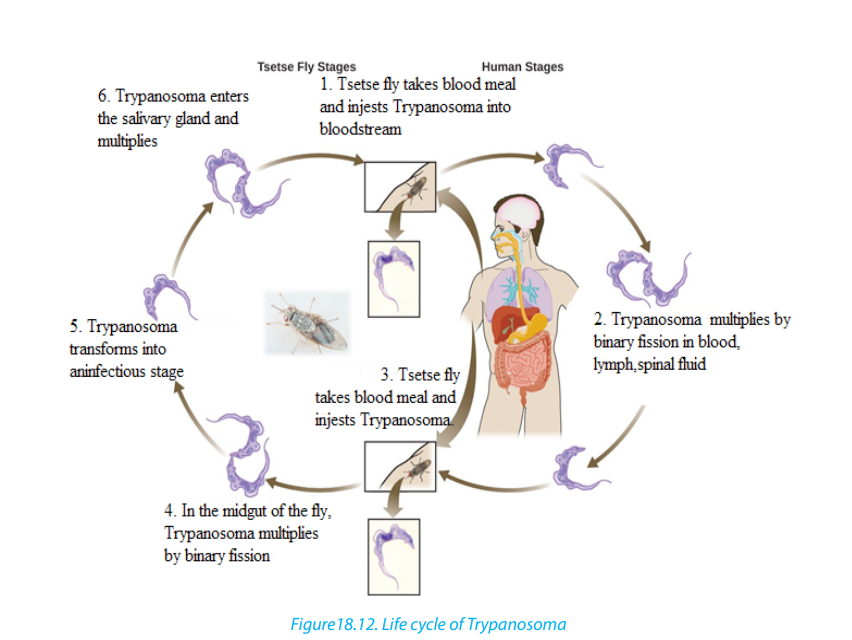
18.6.3. Trypanosoma spp.
a. Characteristics– Trypanosoma is the genus containing a large number of parasitic species whichb. Symptoms
infect wild and domesticated animals and humans in Africa.
– Commonly known as African sleeping sickness, human trypanosomiasis is
caused by the species Trypanosoma brucei and is transmitted to humans
through either a vector or the blood of ingested animals.
– The most common vector of Trypanosoma brucei is the tsetse fly, which may
spread the parasite to humans and animals through bites.
– Through a process called antigenic variation, some trypanosomes are able
to evade the host’s immune system by modifying their surface membrane,
essentially multiplying with every surface change. Trypanosoma bruceigradually infiltrates the host’s central nervous system.
Symptoms include: Headache, weakness, and joint pain in the initial stages; anaemia,
cardiovascular problems, and kidney disorders as the disease progresses; in its final
stages, the disease may lead to extreme exhaustion and fatigue during the day,
insomnia at night, coma, and ultimately death.
c. Occurrence
Human trypanosomiasis affects as many as 66 million people in sub-Saharan Africa.
Trypanosomes are also found in the Americas in the form of Trypanosoma cruzi,
which causes American human trypanosomiasis, or Chagas’ disease. This disease is
found in humans in two forms: as an amastigote in the cells, and as a trymastigote
in the blood.
d. Mode of transmission– The vectors for Trypanosoma cruzi include members of the order Hemiptera,e. Life cycle of Trypanosoma
such as assassin flies, which ingest the amastigote or trymastigote and carry
them to animals or humans.
– The parasites enter the human host through mucus membranes in the nose,
eye, or mouth upon release from the insect vectors. Left untreated, Chagas’
disease may cause dementia, megacolon, and megaesophagus, and damageto the heart muscle, and may result in death.
Trypanosoma’s cell structure plays a vital role in allowing the cell to morph into
three forms (trypomastigote, epimastigote, and amastigote) during its life cycle,
depending on where the cell is located in the host’s anatomy. The location of the
kinetoplast in relation to the nucleus and the flagellum emergence dictate in which
stage the trypanosome cell is found.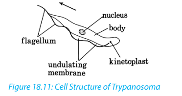
Role of Microbes
Microorganisms are usually associated with major diseases such as AIDS,
uncomfortable infections, or food spoilage.
However, the majority of microorganisms make crucial contributions to the welfare
of the world’s inhabitants by maintaining balance of living organisms and chemicals
in our environment. Therefore, microorganisms are essential for life on earth. They
have important beneficial biological functions such as:
1. Photosynthesis: Marine and freshwater microorganisms (Algae and some
bacteria) capture energy from sunlight and convert it to food, forming the
basis of the food chain in oceans, lakes, and rivers and generates oxygen which
is critical for life on Earth.
2. Decomposers: Soil microbes break down dead and decaying matter and
recycle chemical elements that can be used by other organisms.
3. Nitrogen Fixation: Some bacteria can take nitrogen from air and incorporate
it into organic compounds in soil, water, and air.
4. Digestion: Human and many other animals have microorganisms in their
digestive tract that are essential for digestion and vitamin synthesis. Examples
include:– Cellulose digestion by ruminants (cows, rabbits, etc.)5. Synthesis of chemical products: microorganisms have many commercial
– Synthesis of Vitamin K (for blood clotting) and Vitamin B (for metabolism) in
humans.
applications, such as the synthesis of acetone, organic acids, enzymes, alcohols.
6. Medicine: many antibiotics and other drugs are naturally synthesized by
microbes e.g. Penicillin is made by a mold.
7. Food industry: many important foods and beverages are made with microbes
e.g. vinegar, pickles, alcoholic beverages, green olives, soy sauce, buttermilk,
cheese, yogurt, and bread.
8. Genetic engineering: recombinant microbes produce importanta. Medical and therapeutic products: human growth hormone, insulin, blood9. Medical Research: Microbes are well suited for biological and medical
clotting factor, recombinant vaccines, monoclonal antibodies, etc.
b. Commercial products: cellulose, digestive aids, and drain cleaner.
research for several reasons:a. Relatively simple and small structures, easy to studyThough only minority of microorganisms is pathogenic (disease-causing), practical
b. Genetic material is easily manipulated.
c. Can grow a large number of cells very quickly and at low cost.
d. Short generation times make them very useful to study genetic changes.
knowledge on microbes is necessary for medicine and related health sciences. For
example, hospital workers must be able to protect patients from common microbes
that are normally harmless but pose a threat to the sick and injured.
Self-assessment 18.61. Name the causative agent of malaria.
2. The diagram below shows the life cycle of plasmodium. Analyse it andthen answer the questions that follow.
a. What is the vector of malaria?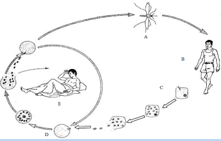
b. Between stages C and D, which one takes place in the red blood cells and
which one takes place in the hepatic cell (liver)?c. State any two symptoms of malaria displayed in individual in stage E.
End of unit assessment18
1. State any TWO diseases caused by:a. Bacteria2. What is the main feature of moulds?
b. Protozoa
c. Microscopic fungi
3. Why viruses are not generally considered to be living things?
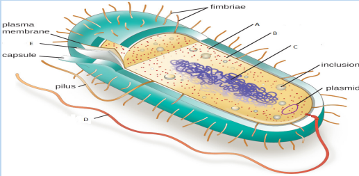
4. The figure below shows the structure of a bacterial cell seen using an electronmicroscope.
 a. Name the parts labeled A, B, C and D5. The diagram below represents the structure of the human immunodeficiency
b. Describe the roles of parts B, C and Evirus (HIV/AIDS).
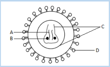 a. Name A, B, C, and D.6. Discuss the methods of reducing the risk of food poisoning by pathogenic
b. HIV/AIDS is under retroviruses. What is meant by retroviruses?
c. What type of leucocytes (white blood cells) are destroyed by HIV/AIDS?
bacteria
7. Why the hyphae of Mucor is called coenocytic?8. The figure below shows the life cycle of one of microorganisms.
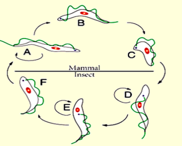 a. Which is the name of the microorganism having the life cycle represented
on this diagram of?
b. Name the parts labelled A, B, C, D, E and F9. Identify the following groups of bacteria