UNIT 14: SUPPORT AND LOCOMOTION
UNIT 14: SUPPORT AND LOCOMOTION
Key Unit Competence
Explain and demonstrate modes of locomotion in protists, insects, fish, amphibians,
birds and mammals
Learning objectives
By the end of this unit, I should be able to:– Explain non-muscular movement in amoeba or paramecium.Introductory activity
– Describe support and movement on land.
– Describe skeletal modification in birds.
– Explain how movements and support of fish are brought about in water.
– Explain how support structures are related to the environment of the animal.
– Observe locomotion of animals and identify reasons for their movement.
– Demonstrate the arrangement of muscles in fish.
– Dissect a fish to observe its swim bladder.
– Observe and explain the relationship between muscles, joints and musculoskeletal attachments in fish, birds, amphibians and mammals.
– Compare the flight of birds and insects.
– Compare the jumping movement of grasshoppers and toads/frogs.
– Appreciate the need for locomotion in animals.– Recognize that the types of locomotion of animals depends on their habitat.
Animals have muscles and different types of skeleton.
What might happen if a large animal such as a cow does not have a skeleton.
How that animal would look like? What will happen to animal without skeleton
or muscles? Can you then think about the role of skeleton and muscles in livingorganisms?
14.1. Locomotion and its requirements
Activity 14.1
From your experience and knowledge from books and the internet:
1. Give details about the concept of locomotion
2. How do different animals move?
3. Explain why animals need to move from one place to another?
Living organisms particularly animals need to move from one place to another.
This is known as locomotion which should not be confused with movement which
occurs in plants. Movement is the displacement of part of an organism. Therefore,
movement is a characteristic of all living things.
Locomotion in animals is brought about by the action of muscles on a skeleton. A
skeleton is a rigid framework that maintains shape and supports the internal
organs and provides attachment for muscles, while a muscle is a soft tissue formed
by muscle cells which found in most animals. Each muscle cell contains actin and
myosin proteins that produce a contraction that changes both the length and the
shape of the cell. Thus, muscles function to produce force and motion. In animals
without muscles such as sea sponges, locomotion is brought by mesohyl or cellswhich act as actual muscles.
Depending on the type of animals, three types of skeletons are distinguished.
1. Hydrostatic skeleton
It is mostly seen in invertebrates and earthworm. These consist of fluid filled
body cavity surrounded by antagonistic sets of muscles. Movement results from
compressive contraction action of the contraction of these muscles on this fluid.
2. Exoskeleton
It characterizes the arthropod insects. It is a hard cuticle made of chitin which lies
outside the muscles. It sheds during molting when the organism outgrows it. It does
not grow because it is a dead material.
3. Endoskeleton
It is seen in vertebrates where the bones and cartilages are found within the interior
of the body on which muscles are attached. It is a living tissue and it grows with the
rest of the body.
For efficient locomotion, exoskeleton and endoskeleton provide a system of leversto which muscles are attached.
Types of locomotion
The locomotion can be either terrestrial, aerial or aquatic (swimming). In most
animals, the locomotion is by running, climbing, crawling, swimming, jumping,
gliding, hoping, and flying with aid of limbs or appendages. For animals without
limbs such as snake, its locomotion is by forming its body into zig - zag, gripping the
ground with its undersides and pushing itself forward. For ducks, their movement
in water is by floating. Some invertebrates like roundworms, flatworms, squids,
octopus, and jellyfish without special organs of locomotion are propelled by themuscular contractions.
Advantages of locomotion
Based on the types of locomotion mentioned above, an animal is capable to:
– Escape danger such as fire or predator– Look for food, water and shelter
Reproduce
– Avoid competition with other animals of the same or different species
– Avoid overcrowding which enables offspring to move to another place– Avoid unfavourable condition
Self-assessment 14.1
1. What is locomotion?
2. What are the requirements for locomotion?
3. Given the following animals: Frog, dragonfly, squid, spider, antelope,
kangaroo, fish, grasshopper, bee, duck, worm, zebra, snake, and cow.
Identify those which fly, crawl, hop, and or run/walk.4. Discuss why locomotion is very important in animals?
14.2. Support and locomotion in non-muscular organisms
Learning activity 14.2
From a culture of paramecium:
1. Use a microscope to observe the locomotion in Amoeba and Paramecium
2. From what you have observed identify Amoeba, Paramecium, Euglena andin Trypanosoma moves in relation to their structures / diagrams below
3. Discuss how is locomation performed in those organisms.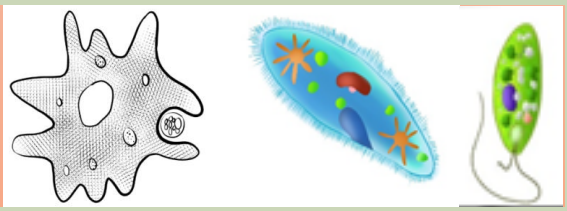
Non-muscular locomotion is identified in living organisms that belong into
Protoctista kingdom. Depending to individuals belonging to Protoctista, locomotionis either amoeboid, ciliary, flagellated or euglenoid type.
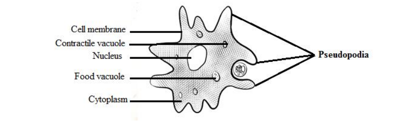
Amoeba moves by amoeboid locomotion i.e. by putting out pseudopodia.
Locomotion is not maintained in any particular direction for long. Amoeba is
constantly changing shapes as it changes direction. Amoeboid locomotion is
brought about by cytoplasmic streaming and between a gel and sol state.
These cytoplasmic streaming requires Ca2+ ions and ATP . Amoeboid locomotionis common to all rhizopodes including Amoeba and white blood cells of the vertebrates.
a. Amoeba
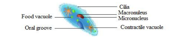
b. Paramecium
c. Euglena
Paramecium moves by means of cilia and Euglena move by the use of flagella. Cilia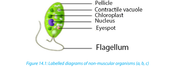
and flagella have similar structure except that cilia are short and many. Both cilia
and flagella consist of fine tubes composed of an extension of plasma membrane.
Euglenas have an intricate cell membrane called a pellicle. The latter is folded into
ribbon-like ridges and each ridge is supported by microtubules. The pellicle is tough
and flexible, letting euglenas crawl through mud when there is not enough water
for them to swim.
During cilia or flagellum locomotion, tubules slide past each other in a movement
similar to that of actin and myosin filaments in skeletal muscles. Hence Ca+
ions and ATP are also required in the ciliary locomotion.Self-assessment 14.2
1. Describe the type of locomotion found in:
a. Amoeba
b. Paramecium caudatum
c. Trypanosoma gambiense
d. Trypanoma vaginalis
e. Giardia intestinalis
2. How do cilia differ from flagellum?
3. Produce picture showing the locomotion of amoeba
14.3. Support and locomotion in fish
Activity 14.31. Observe the freshly collected fish or the figure, to label fins and lateral line.
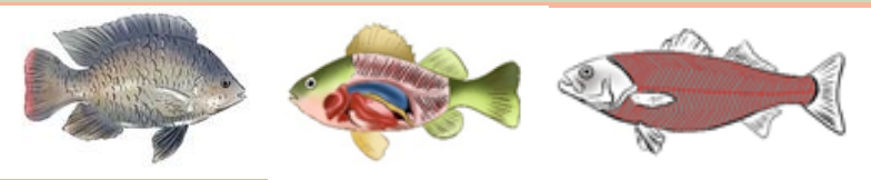 2. Dissect a fresh fish or observe the above given diagram. Redraw and showFish like other aquatic animals are adapted to such habitat in terms of locomotion
the swim bladder and the arrangement of muscles
3. If you have a live fish, put it in water and observe its locomotion.
4. From what you have observed, draw and label the external and internal
features that contribute to fish locomotion
due to its structural adaptive features particularly skeleton which gives shape as wellas muscles arrangement and swim-bladder.
Adaptive features of fish for locomotion in water
The streamlined body shape of the fish reduces friction between water and fish.
The body of fish is mostly covered by scales which overlap one another and point
backwards and lie close to the body. The scales are covered by mucus which reduces
the drag.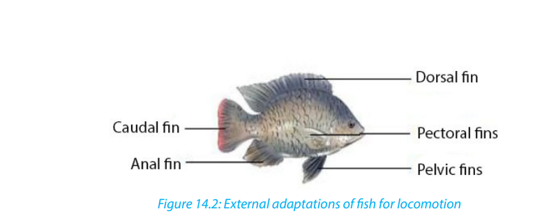
Tail or caudal fin has a large surface area, which increases the amount of water that
is displaced as it provides much of the push during swimming. Paired pectoral and
pelvic fins bring about downward and upward movement. With pectoral fins, the
control of direction of a fish in water is possible whereas the pelvic fins bring about
the balance, preventing diving and rolling. There are also unpaired dorsal and anal
fins for stabilizing the fish and thus preventing it from rolling or yawing.
Fish is also adapted to locomotion in water by its strong tail muscles and highly
flexible vertebrate column which enables the tail to move from side to side againstwater. In addition, inflexible head and neck maintain forward thrush.
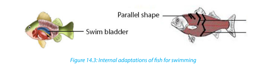
Internally, a fish is adapted to swimming by swim bladder and muscles. Air or gas filled sac called
swim-bladder, outgrowth of the pharynx, helps a fish to change its
buoyancy as it alters the gas pressure in the bladder. So that, it floats at any depth
in water without using its muscles. Swim-bladder also helps fishes to maintain a
density that is equal to that of the surrounding water. Muscles or myotomes /
myomeres (segments or sheets of muscles separated from its neighbor by a sheet of
connective tissue) enable fishes to move in water owing the shapes of muscles that
are located on either side of vertebral column.
Myotomes contribute to the mechanism of swimming by its arrangements. They may
be parallel, V-shape, or W-shape arranged in bundles or blocks that are separated by
myosepta.
Although there are such arrangements, the myoseptal organization and orientation
of fibres is complex. In bony fish, myomeres are V-shaped with new myomeres added
posteriorly. With those myotomes, a fish swim by passing a wave of contracting
muscle from anterior to posterior. Muscles near the head of the fish contract first and
contraction proceeds posteriorly down the length of the fish to the caudal fin. Thus,
a fish moves forward from the contraction and relaxation (antagonistic) of myotome
on either side of the body.
Undulatory swimming of the fish is also powered by the segmental body musculature
of the myotomes. Myotome and myosepta orient more perpendicularly to midline
to push aside. Therefore, the fish can bend laterally. With contraction muscle fibres
shorten by half their length while maintaining volume. Without myosepta, but simply
a series of interconnected muscle fibres, then the wave would be much dampened.
Self-assessment 14.31. How does swim bladder help the fish in locomotion?14.4. Support and locomotion in terrestrial animals
2. Illustrate how the arrangements of fish myotomes contribute to fish
locomotion.?
3. What does it make a fish to move in undulatory propulsion?
4. What are the anatomical structures that give rise to the direction of a fish
and preventing diving and rolling?
Activity 14.41. Through internet observe and think about how locomotion in dogs,All animals living on land move due to the musculoskeletal system. The rigid nature
chicken, frog and earthworm brought about.
2. Make a diagram showing how support and movement of different
animals such as dogs, chicken, frog and worm is brought about on land.
3. Show by using diagrams the relationship between muscles, joints andMusculo-skeletal attachment in mammals, birds, frog and earthworm.
of bone also gives a structure for muscles to pull, by their contraction, to create a
movement as they act as levers. The synovial joints also allow certain movements.
The support and movement differ from specimen to another. Thus, animals can
walk and run on land for moving from one place to another. This is possible by their
endoskeleton and its muscles. By its muscles, flexor (a muscle whose contraction
bends a limb or other part of the body) and extensor (a muscle whose contraction
extends or straightens a limb or other part of the body or any or a muscle that
increases the angle between members of a limb, as by straightening the elbow or
knee or bending the wrist or spine backward); contractions of those muscles cause
the limbs act as levers for them which result to the foot being pressed downwards
and backwards against the ground. For example, flexor and extensor work as
illustrated below: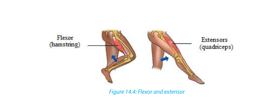
a. Locomotion in quadruped animals e.g. dog and frogs
When a dog walks, its vertebral column remains rigid, and the forward movement is
achieved by the activity of the hind limbs. When its extensor muscle contracts, each
hind limb, acting as a lever, extends and exerts a backward force against the ground,
thrusting the animal forward and slightly upwards. When the flexor contracts, the
limb is lifted clear of the ground and pulled forward. Only one limb is raised at any
one time, the other three providing a tripod of support which balances the rest ofthe body.
Beginning with the left forelimb in a stationary dog, the sequence of leg movement
is as follows when it walks forward: left forelimb-right hind limb-right forelimb-left
hind limb and so on. Such walking in quadrupedal animal is also identified in frogswhen they can walk on land.
b. Running of the dog
As a dog begins to run, it loses its quadrupedal movement which means, it developsa type of movement where the forelimbs move together, followed by the hind limbs.
C. Walking in bipedal animal e.g humans
Humans are bipedal, they walk on two legs. When standing upright, the weight is
balanced over the two legs. When a stride is taken by the right leg, the heel is raised
first by the contraction of the calf muscles. As this occurs, the weight of the body is
brought over the left foot which is still in contact with the ground and acting as theprop for the rest of the body.
When the right leg extends the heel is the first part of the foot to touch the ground.
The weight off the body is gradually transferred from the left side to a position over
the right heel and then the body continues to move forward, over the right toes,
backward pressure against the ground generally being exerted through the right
big toe. Like human does, a bird also can walk on ground through the movement ofcontractions of its leg muscles particularly flexor and extensor.
d. Crawling of earthworm
Earthworms are organisms having hydro skeleton with soft-bodied animals due to
fluid secreted within the body and surrounded by the muscles of the body wall.
They are capable to move by aid of their muscles. These muscles are not attached to
any structures and thus can pull against each other. The combined effect of musclecontraction and fluid pressure serves to maintain the shape and form of the animal.
Generally, there are two muscle layers, longitudinal in which muscle fibres are
arranged parallel to the long axis from one end of a segment to another and circular
with muscle fibres arranged in concentric circles to the circumference of the worm.
When those muscles act antagonistically against each other, locomotion is achieved.
The fluid which acts as pressurisable hydrostatic skeleton contained in body cavity
or coelom presses against the muscles which in turn are able to contract against the
fluid. Earthworm movement is also helped by bristles like setae or called chaetae(hair like structures on ventral surfaces) which anchor the worms to the substrate.
Contraction of the circular muscles makes the worm thinner, but because liquid is
essentially incompressible (and so maintains a constant volume) and the increase
in pressure forces the liquid outwards, stretching the worm, so the worm becomes
longer and thinner. Contraction of the longitudinal fibres shortens the worm, former
the coelomic liquid out to the sides and making the worm fatter. If the body is
segmented, then such pressure is localized and only certain segments will move orchange shape.
Self-assessment 14.4
1. What are the main muscles that contribute to locomotion in mammals,
amphibians and birds?
2. Draw an earthworm and illustrate the muscles that contribute to its
locomotion.
3. What type of skeleton system found in mammals, birds, amphibians and
annelids?
4. . Illustrate how flexor and extensor muscles contribute to lifting up a leg inhuman being.
14.5. Flight through air by birds and insects
Learning activity 14.5
Make a research on internet as well as books and do the following:
1. Observe pictures below (Figure 14.6) related to birds and make a description
of skeletal modification in birds. Illustrate the skeletal modification in birds
2. Draw a bird and show by using arrow the structures that enable a bird to fly
3. How will the external features of birds will behave when flying in high or
low atmospheric pressure
4. Make a table illustrating how does flight of birds and insects differ and
similar
5. Observe and compare the flight of birds and insects
Bird can fly either by flapping their wings or gliding by spreading its wings. Like
in animals moving on land, locomotion by flying in birds is brought about by the
action of flexor and extensor muscles as well some other structures given diagrambelow like pectoralis major, pectoralis minor and keel of sternum.
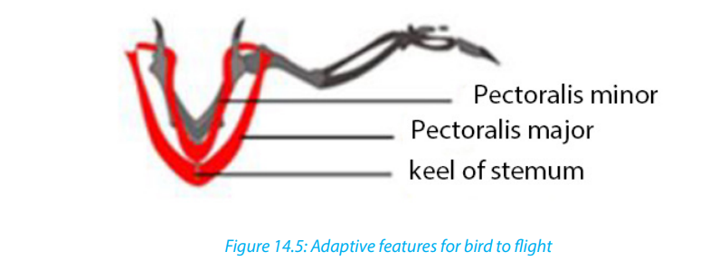
Based on the above diagram, wings move down by the contraction of pectoralis
major and then move up under the contraction of pectoralis minor.
Adaptive features of birds for movement in air
A number of features enable birds to aerial locomotion. Those features include
body shape, modified limbs, and modification in internal organs particularly bones.
The body of bird is highly streamlined and covered with light feathers that overlap
backwards thus reducing air resistance during flight. Those light features thatincrease the surface area of wings without increasing weight.
Differently from quadrupedal animals, birds are adapted to flying by modification
of fore limbs into wings. Such modification goes with a well-developed or large keel
sternum that provides large surface area for the attachment of the flight muscles
namely major and minor pectoral muscles which give the power to flap the wings in
flight. Also, birds have hollowed bones making the body light and vertebrae of trunk
are fused. As flight requires much energy, birds are adapted to that by an efficient
breathing system with air sacs attached to the lungs necessary to provide oxygenfor respiration and to remove the resulting carbon dioxide.
Other adaptations include a high metabolic rate for providing the high amount of
energy required, an efficient circulatory system necessary for transporting both the
nutrients and respiratory gases at speed related with the body needs, a high red
blood cell count for efficient oxygen transport, and a keen eye sight to enable themto judge distances correctly especially on landing.
Self-assessment 14.51. What are the muscles that enable the flight in birds?14.6. Hopping locomotion in grasshoppers and toads
2. Describe how bird skeleton contributes to its flight?3. Describe how birds are adapted to flying.
Activity 14.6
Use a collecting net to catch a grasshopper and toad from school compound. Put
them down on cemented ground for observing them very carefully when they
make a jump and then answer to the following:1. Identify and describe anatomic structures that enable grasshoppers to jumpSkeletal muscles such as extensor and flexor that occur in pairs are often antagonistic.2. Illustrate how legs’ muscles behave when they are resting and or jumping
With such antagonistic behaviour, when one contracts the other relaxes to producecontrolled movement in the opposite directions.
a. Locomotion of grasshopper
Insects have a skeleton which is on the outside of the body called an exoskeleton.
They can walk on the land but they are mostly adapted to hopping owing to their
muscles which are inside the hard shell as well as skeleton system. The muscles
which make them capable to move are flexors and extensors which are antagonists,
attached to internal surface of exoskeleton and the rear or back legs of a grasshopper
which are long and muscular, adapted for hopping. Additionally, there are two main
muscles inside are the extensor tibiae muscle which contracts to extends the leg,
and the flexor tibiae muscle which contracts to flex the leg as illustrated in figure
below. Those muscles pull on tendons which are attached to the tibia on either side
of the joint pivot.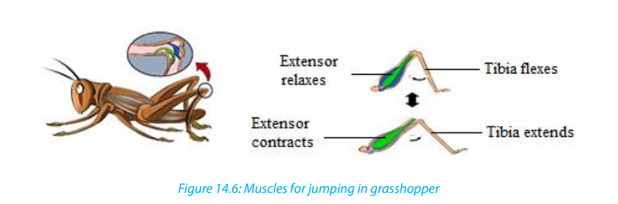
The back legs are much longer than the others for helping in hopping. With those
long legs, grasshopper is capable to make high jumping distance. As illustrated
above, flexor muscles bend a joint whereby extensor ones straighten it. The flexor
muscle contracts and the lower leg is pulled towards the body. Thus, the hind leg
is folded in a Z shape and ready for jumping. Being in resting or sitting position,
the extensor muscle contracts which enable then the legs jerk or move very quicklybackwards propelling the grasshopper.
b. Locomotion in toads and frogs
On land, frogs and toads move by hopping (going from place to place).
– When a frog is at rest, the hind legs are folded up in the shape of a letter Z.
– When it hops, the legs are quickly straightened out, lifting the animal of the
ground.
– The fore-limbs are used as shock absorbers on landing and they also prop up
(to give support) the front end of the body when the animal is at rest.They also hope but do not travel as high as far as a frog does at each hop.
Self-assessment 14.6
1. What are the muscles that contribute to high jumping in a grasshopper?
2. How do muscles (flexor and extensor) behave when toads and grasshopper
are resting?3. Draw a leg of grasshopper and the one of toad when are jumping
End of unit assessment 141. Describe ways of locomotion in Amoeba, Paramecium, Euglena and in
Trypanosom
2. Produce a cartoon showing different adaptive features of fish for aquatic
locomotion
3. Describe how different fish fins contribute to locomotion and balance
4. Describe how the movements and support of fish in water do they occur?
are brought in water
5. Show by diagrams the relationship between muscles, joints and
musculoskeletal attachment in mammals, birds, frog and earthworm.
6. Describe how flexor and extensor muscles work to enable the locomotion
on land, water and in air
7. What are the features that enable aerial locomotion in animals?8. Describe how a grasshopper and toad is adapted to jumping