UNIT 1: PRINCIPLES OF GAS EXCHANGE SYSTEMS
UNIT 11: PRINCIPLES OF GAS EXCHANGE SYSTEMS
Key Unit Competence
Explain the principles of gaseous exchange systems
Learning objectives
At the end of this unit learners will be able to:– Explain the relationship between size and surface area to volume ratio.Introductory activity
– Describe how different respiratory surfaces are modified to speed up the
diffusion process.
– State the characteristics of gaseous exchange surfaces.
– Describe the effects of tar and carcinogens in tobacco smoke on gas exchange
system with reference to lung cancer and Chronic Obstructive Pulmonary
Disease (COPD).
– Describe the short-term effects of nicotine and carbon monoxide on the
cardiovascular system.
– Observe prepared slides of gaseous exchange surfaces and identify their
characteristics.
– Dissect fish gills and observe the surface area for gas exchange.
– Observe mammal’s lungs and state their adaptation for gaseous exchange.
– Use internet to make research and deduce the findings
– Appreciate the evolution of gaseous exchange surfaces from simple tocomplex.
Kalisa and Uwase wanted to rear tilapia at their home. They bought a nice
transparent plastic box. They filled it with 1.5L of clean mineral water, put in some
pieces of meat and plant leaves. They finally introduced a living tilapia in the box
and covered. After two days they were happy to see their fish swimming. But on
the third day, they become sad after finding it dead and yet the food was still in
water.What could have caused the death of the fish?
11.1. Relationship between size and surface area to volume
ratio
Activity 11.1
1. Use Manila paper, scissors, and graduate ruler to create three cubes: 3cm x 3cm,
2cm x 2cm, 1cm x 1cm
a. Calculate the surface area, the volume, and the surface area to volume ratio
of each cube. What do you conclude from these ratios?
b. Compare the surface area to the volume of a spherical alveolus having a
radius of 0.001m and that of another animal with a radius of 0.000001m.2. What do you understand by surface area to volume ratio?
The surface area to volume ratio is the relationship between the surface area and
the volume of an object. Small or thin objects have a large surface area compared tothe volume. For example, the surface area of a sphere is calculated by
As the length or radius of the sphere increases, the increase in the surface area is
squared (X2) and the increase in the volume is cubed (X3). The surface area to the
volume ratio gets smaller as the cell or animal gets larger. Thus, if the cell grows
beyond a certain limit, not enough material will be able to cross the membrane fastenough to accommodate the increased cellular volume.
As a cell grows, its surface area to volume ration decreases. At some point in its
growth its surface area to volume ratio becomes so small that its surface area is too
small to supply its raw materials to its volume. The cell will reach a size at which
substances cannot enter or leave the cell in sufficient time to sustain life. At this
point the cell cannot get larger. The volume of the cell will also be so large that
the diffusion rate will be too low to distribute necessary substances throughout the
cell within a reasonable time. This brings about the need of having a mechanism ofventilation that speeds up the rate of gaseous exchange.
The rate of oxygen consumption by an animal gives a relatively accurate indication
of the rate of its metabolic activity. The need of oxygen varies with the activity, the
size of the organism, and their health. In general, small mammals need more oxygenthan large mammals because:
– Small mammals have a big respiratory surface area to the volume ratio
– Small mammals are too motile than large mammals. Therefore, they need to
produce more energy through aerobic respiration– Small mammals reproduce more rapidly than large mammals.

A running man needs a double volume of oxygen than a sleeping manand a pregnant woman needs more oxygen than a normal woman.
Self-Assessment 11.1Determine the surface area to volume ratio of a sphere having a diameter of 4 mm
11.2. Characteristics of gas exchange surfaces
The following are the characteristic features of gaseous exchange surfaces:
Large surface area: they should have a large surface area to allow adequate and
fast gaseous exchange in order to provide enough oxygen to cells and to get rid of
the carbon dioxide that is released.
Rich supply of blood: in animals with a transport system, the respiratory surface
areas found in the lungs and gills have rich supply in blood capillaries to quickly
transport gases to and from the cells. Gases diffuse into the blood and are carried to
and from the body cells.
Thin surface or thin wall: respiratory surfaces should have thin walls or thin
surface area to maximize the diffusion. The alveoli in the lungs have thin squamous
epithelium that enables gases to diffuse quickly between the alveoli and blood.
According to Fick’s law, the rate of diffusion is proportional to:
Moist surfaces area: to enable gases to dissolve and pass through the solution.
High diffusion deficit / concentration gradient: respiratory surface areas should
have a high diffusion deficit / concentration gradient to ensure faster diffusion of
respiratory gases.
Protection against injury and dry out: lungs and gills are protected by the bonesand cartilage and mucus protects them from drying out.
Self-Assessment 11.2
1. State the features common to all respiratory surfaces in living organisms
2. Explain how the following features of a respiratory surface helps gaseous
exchange:
3. Short diffusion distancea. Protection11.3. Modifications of gaseous exchange surfaces to speed up
b. A rich blood supplyc. Protection
the rate of gaseous exchange in different organism
Activity 11.3
Use appropriate laboratory equipment to extract gills in fish to show the gill
filaments. Draw and label to show the parts observed.
a. Insects
The spiracles are openings of small tubes running into the insect’s trachea system
that terminates into small fluid-filled tracheoles in which the gases are dissolved.
The fluid is drawn into the muscle tissue during physical exercise, and this increases
the surface area of air in contact with the cells.
Ventilation movements of the body during exercise may help this diffusion. The
spiracles can be closed by valves and may be surrounded by tiny hairs. The later help
to keep humidity around the opening to ensure that there is a lower concentrationgradient of water vapor, and so less is lost from the insect by evaporation.
b. Fish and tadpoles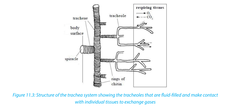
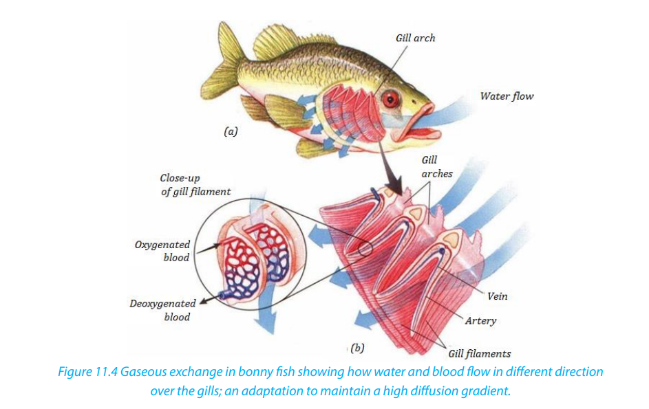
Fish and young amphibians (tadpoles) use gills for the gaseous exchange.
Gills have numerous folds that give them a very large surface area.– The rows of gill filaments have many protrusions called gill lamellae. These
filaments help in the exchange of respiratory gases
– They also have an efficient transport system within the lamellae which
maintains the concentration gradient across the lamellae. The arrangement
of water flowing passes the gills in the opposite direction to the blood (called
counter-current flow) means that they can extract oxygen at 3 times the ratea human can.
c. Amphibians, Reptiles, Birds and Mammals
These have alveoli in their lungs. Air reaches the alveoli via a system of tubes
(trachea, splitting into two bronchi - one for each lung - and numerous bronchioles):– Numerous alveoli - air sacs, providing a massive surface area over which gasesis regularly moving in and out of the lungs due to changes in volume and
can diffuse
– Have a short diffusion distance between the alveolus and the blood because
the lining of the lung and the capillary as they are only one cell thick.
– The blood supply is extensive, which means that oxygen is carried away to the
cells as soon as it has diffused into the blood.– Ventilation movements also maintain the concentration gradients because air
pressure
Activity11.3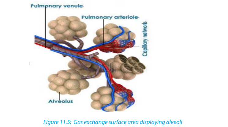
You will need: Lungs of a sheep or pig, newspaper, plastic sheets, dissecting
board, sharp scalpel, dissecting needles, scissors, dissecting tray, latex gloves, CPRmouth piece, soap to wash hands and surfaces.
Procedure
– Place the dissecting board on the newspaper and lay the lungs on the board.
– Use a scalpel to cut the lungs in half in longitudinal section.
– Identify the trachea, right lung, left lung, cartilage rings, bronchus, larynx, alveoli,
and bronchiole. You can use a magnifying hand lens to observe structures in
the lungs.
– Inflate the lungs by blowing through the CPR (cardio-Pulmonary Resuscitation)
mouth piece to see how the lungs expand.
– Feel the slippery inside of trachea, press the lung with your finger and look at
cartilaginous rings.
– Remember to wash your hand s with soap as you finish your experiment.
1. Explain what it feels like as you press the lungs with your fingers.
2. Look at cartilaginous rings. What function do they serve?
3. (a) List four features of respiratory surfaces you can identify from the
specimen.
(b) Examine the lung and explain how the lungs are suited for efficientgaseous exchange.
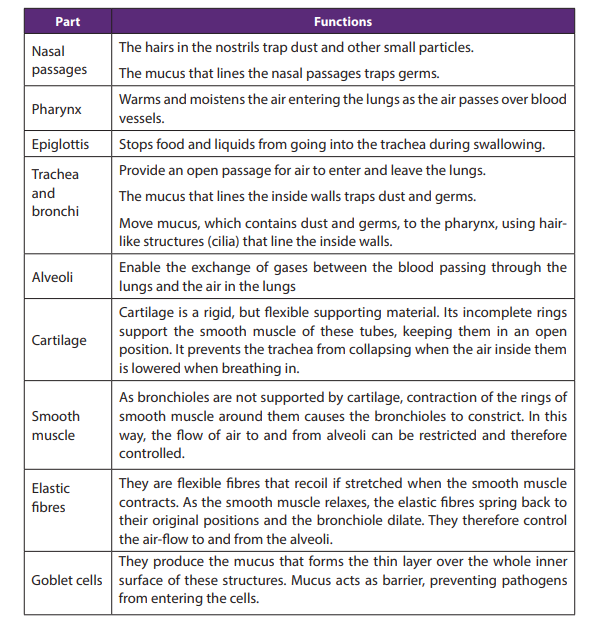
Table 11.1: Parts of the human gas exchange system and their respective functions
Self-Assessment 11.3
1. List the adaptations of the gills for gaseous exchange
2. List the structures through which air passes on its way from the nose to the
alveoli.
3. Give two reasons why mammals need lungs, rather than exchanging gasesthrough the skin.
11.4. Smoking and related risks
Activity 11.4
In groups, make research to find out main health risks related to smoking. Analysethe photographs below and answer questions that follow.
Between the lungs of individuals, A and B, which one is most likely that of the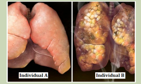
cigarette smoker?Read the notes below to identify at least three risks related to smoking cigarette.
Cigarette smoking harms nearly every organ of the body, causes many diseases, and
reduces the health of smokers in general (Figure 11.6). Quitting smoking lowers the
risk for smoking-related diseases and can increase the longevity. Inhaling cigarette
smoke is called passive smoking and presents a health hazard to people nearby whoinhale it. Of the thousands of chemicals in tobacco smoke three important ones are:
– Carbon monoxide (CO), a poisonous gas form incomplete combustion carbon.Tar in tobacco smoke is a mixture of chemicals that enter the respiratory tract. It is
CO in tobacco smoke combines easily, but irreversibly, with hemoglobin to
form carboxy hemoglobin and therefore reduces oxygen carrying capacity of
the blood. This can lead to hypotension and heart failure.
– Nicotine, a poisonous alkaloid drug that is addictive. Nicotine in tobacco
smoke stimulates the production of the hormone adrenaline by adrenal gland,
leading to an increase in the heart rate and raised blood pressure. Nicotine
also makes the red blood cell stickier and this leads to high risk of thrombosis
and hence of the strokes.
– Tar- is a sticky and brown substance. It appears in tobacco spoke minute
droplets.
an irritant and causes inflammation of the mucous membranes lining the trachea,
bronchi and bronchioles, resulting in producing more mucus. Tar also thickens the
epithelium and paralyses the cilia on its surface. As a result, cilia cannot remove the
mucus secreted by epithelium lining.
a. Short-term effects of smoking
Tar causes constriction of finer bronchioles by increasing resistance to the flow
of air.
– Tar paralyses the cilia which remove dirt and bacteria; the accumulation of
extra material in the air passage can restrict air flow.
– Smoke acts as an irritant; this causes secretion of excess mucus from goblet
cells and excess fluid into the airways, making it more difficult for the air to
pass through them.
– Mucus accumulating in the alveoli limits the air that they can contain and
lengthens the diffusion pathway.
– Coughing of many smokers, way of trying to remove the build-up of mucus
from the lungs, can cause damage to the airways and alveoli; scar tissue builds
up which again reduces air movement and rates of diffusion
– Infections arise because the cilia no longer remove mucus and pathogens
– Allergens such as pollen also accumulate, leading to further inflammation
of the airways, reduced air-flow in and out of the lungs, and possible asthmaattacks.
b. The long-term effects of smoking
– Bronchitis: Bronchitis is inflammation of the lining of the air passages and may
be acute or chronic.
– Emphysema: One in five smokers develop the crippling lung disease called
emphysema i.e. condition of gradual breakdown of the thin wall of the alveoli
leading to sensation of breathlessness as the gaseous exchange reduces.
– Lung cancer: Lung cancer usually starts in the epithelium of the bronchioles
and then spreads throughout the lungs as dividing cells cease to respond to
the normal signals around them and form unspecialized masses of cells called
tumours. The tar is carcinogen i.e. contains chemicals which cause cancer. The
irritation causes thickening of the epithelium by extra cell division and this maytrigger the cancer. Almost all people who die from lung cancer are smokers.
Self-Assessment 11.4Analyze the photograph and share ideas with your group members.
1. Between the baby and the parent who will suffer more the effects of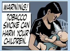
tobacco? Give reasons
2. Discuss any side effects of smoking.
3. Design a sign post to advocate against smoking.
End of unit assessment111. Match the terms in Column A with the correct definition in Column B.
Describe how the human lungs serve as good gaseous exchange organs.
3. Emphysema is a disease of the lungs. People who smoke cigarettes are more
likely to suffer from emphysema. The diagrams show lung tissue from a healthyperson and lung tissue from a person with emphysema.
a. Identify on the figure above by using E (for emphysema) and we (without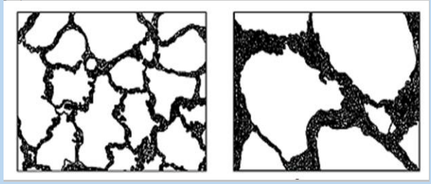
emphysema) and give a reason for your choice.
b. Explain how emphysema reduces the amount of oxygen which diffuses into
the blood.
c. What are the features that make the gill of fish an efficient respiratory organ?
d. Compare respiratory system of fish and with that of a mammal.
e. Why do people who smoke have high chances of developing lung cancer?
f. Design a simple model that shows the structure and functioning of gasexchange system in mammals.
