General
- Biology SME Y2 SB File Uploaded 1/11/21, 16:48
- Biology SME Y2 TG File Uploaded 1/11/21, 16:48
UNIT 2: NERVOUS AND HORMONAL
Key unit competence: Describe the structure of neurons, explain the mechanisms of impulse transmission and functions of endocrine glands in the body.
Introductory activity 2
2.1 Human nervous system
Activity 2.1
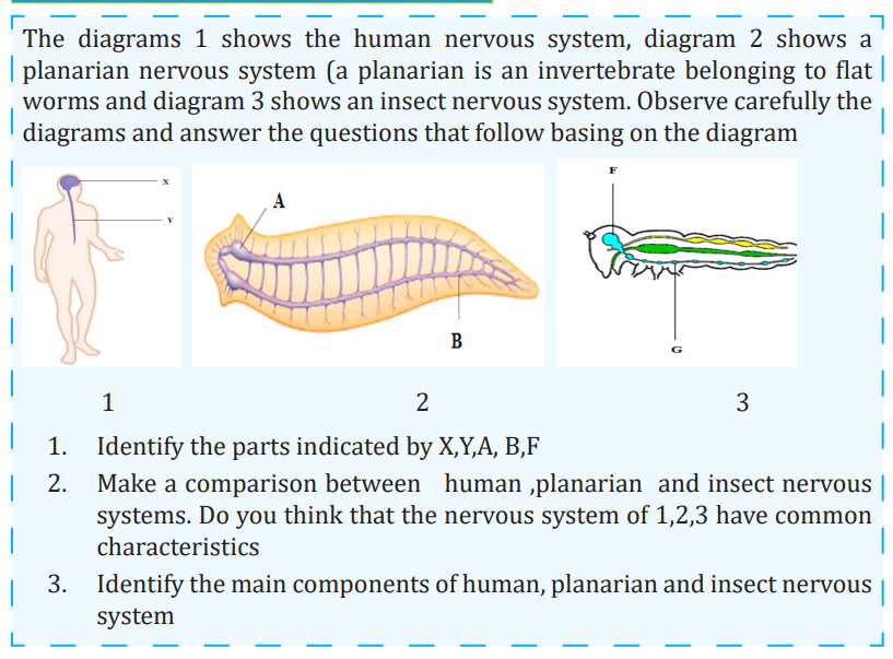
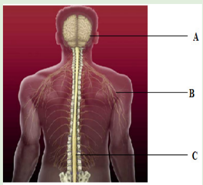
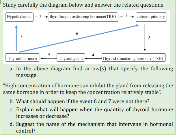
Human body is made up of many systems. The following diagram illustrates one of the systems of body. Observe carefully this diagram and use the school library in order to answer these questions
Coordination: It is the process in which body coordinate, ordinate and control different activities.
The nervous system is a system for gathering, transmitting and interpretation of information. It plays the main functions such as:
i. Sensory input: Sensory receptors present in skin and organs respond to external and internal stimuli by generating nerve impulseswhich are transmitted to the brain and spinal cord.
ii. Integration: The brain and spinal cord sum up the information from all over the body and send out nerve impulses
iii. Motor output: The nerve impulses from the brain and spinal cord are transmitted go to the effectors, which are muscles and glands.
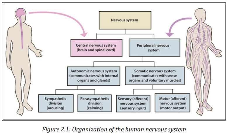
The Nervous system is divided into two main divisions: The central nervous system (CNS) and the peripheral nervous system (PNS) The central nervous system (CNS) consist of the brain and spinal cord, which are located in the midline of the body.The peripheral nervous system (PNS), which is further divided into the somatic division and the autonomic division, includes all the cranial and spinal nerves.
2.1.1 Some key word definitions
- Irritability or sensitivity. This is the ability of living organisms to detect and respond to a stimulus
- A stimulus: This is any change in the external or internal environment which provokes a response
- Receptors: These are specialized cells that detect a stimulus.
- Effectors: are organs that respond to the stimuli and bring about a response such as glands and muscles.
- A nervous system: This is a system for gathering transmitting and interpretation of information of information.
- The response may be to both the external and internal environments.
- Neurone or nerve cell: It is the basic functional unit of the nervous system. Neurones are cells specialized to generate and transmit nerve impulses (action potentials) are cells which transmit nerve impulses (action potentials).
2.1.2 Division of nervous system
The nervous system of a mammal comprises of the central nervous system (CNS) consisting of the brain and the spinal cord, and the peripheral nervous system (PNS) consisting of the cranial nerves from the brain, the spinal nerves from the spinal cord and the sensory organs (Figure 2.1).
1. Human brain
The brain is the enlarged end of the spinal cord. It is enclosed in the skull which protects it against mechanical damage. It is divided into three main parts namely: the fore brain, the mid brain, the hind brain.
a. Forebrain
This consist of: cerebrum, thalamus, hypothalamus and pituitary gland
• Cerebrum:
This is the largest part of the brain made up of two hemispheres called the right and the left cerebral hemispheres. The left cerebral hemisphere controls those activities of the right side of the body while the right cerebral hemisphere controls those of the left side of the body.
The functions of the cerebral hemisphere
- It is the centre of the judgment, memory, reasoning and imagination.
- It receives the impulses from the sensory organs: sight, taste, sound and touch.
- It controls all the body’s voluntary activities, e.g. walking, eating, singing,
• Thalamus:
This is a relay centre. It relays sensory information towards higher centre. It is the centre for the perception of pain and pleasure.
• Hypothalamus
It performs many functions such as; regulates and monitors the temperature of blood, monitors and regulates the water content of blood, a co-ordinating centre for activities of the internal organs, e.g. rate of heart beat, blood pressure. It is a centre of for feelings such as; hunger, thirst, sex drive, satisfaction, sleep, speech, etc. As an endocrine gland, it produces hormones i.e. anti-diuretic hormone (ADH) and oxytocin
• Pituitary gland:
It produces hormones such as: Follicle-stimulating hormone (FSH), Thyroid stimulating hormone (TSH), Adreno-cortico trophic hormone (ACTH), Prolactin hormone and Luteinizing hormone (LH). It also serves as a master gland i.e controls activities of other endocrine glands.
b. Mid brain
This acts as an association centre between the fore and the hind brain. It is a relay centre for audio and visual information. It is also responsible for movement of the head and the trunk.
The hind brain receives the impulse from the ear, the skin and the semi-circular canals. It consists of: The cerebellum and the medulla oblongata
The cerebellum: It lies behind the optic lobes. It receives impulses simultaneously from the eyes and the ears. It regulates and co-ordinate muscular movement, especially those concerned with maintaining body equilibrium and controls all the unconscious activities of the body.
The medulla oblongata: This controls all the involuntary movements of the body especially those concerned with respiration, digestion, heartbeat, breathing rate and sneezing.

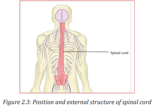
2. Spinal cord
The spinal cord is a dorso -ventrally flattened cylinder of nervous tissue running from the base of the brain down the lumbar region. It is protected by the vertebrate of the backbone and the meninges.
Functions of spinal cord include:
- It is a coordinating centre for simple reflex such as the knee-jerk response and the autonomic reflexes such as contraction of the bladder. - Providing a means of communication between peripheral nerves and the brain.
A transverse section of the spiral cord shows an H-shaped central core of grey matter. Grey matter is composed of nerve cell bodies, dendrites and synapses surrounding a central canal which contains cerebrospinal fluid. White matter: around the grey matter, is an outer layer containing nerve fibres whose fatty myelin sheaths give it its characteristics colour
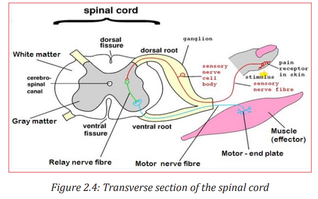
The spinal cord acts as a coordinating centre for simple reflex such as knee jerk response and autonomic reflexes. The spinal cord acts as means of communication between spinal nerves and the brain. It sends impulses to the brain through sensory neurons from the body and returns the motor impulses to the effectors which are muscles and glands
Application activity 2.1 1.
The diagrams A, B and C show a laptop, processor and RAM (Random Accessory Memory) respectively. How would you link what you learned to answer the following questions?

1. Which organ of the nervous system that correspond to A,B and C
2. Identify the parts of the nervous system that plays the same role as C.
2. Identifying of fish nervous system component
Materials needed
• (Dead fish.)
• Dissecting kit
• Dissecting board
• Cotton wool
• Pins
Procedure
• Place the fish on the dissection board. Dissect along, as shown in dotted lines below
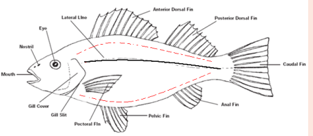
• Open the abdominal cavity
1. Locate the organs (brain and spinal cord) associated with Nervous systems
2. Draw and label the brain you are observing
2.2 Structure, types and functions of neurons
Activity 2.2
Research activity
a. Observe and label the following illustration
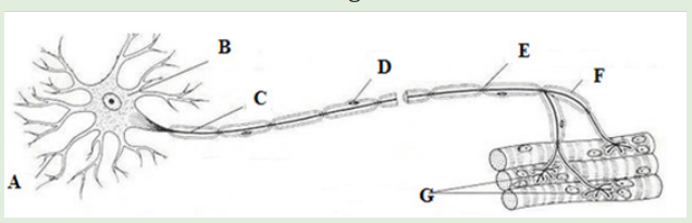
b. Using the school library or search engine, search on the structure of neuron, give the types and function of the above illustration
c. Make a table comparing different types of neurons
Neuron also called nerve cell is the basic functional unit of the nervous system. Neurons are cells specialized to generate and transmit nerve impulses (action potentials) are cells which transmit nerve impulses (action potentials).
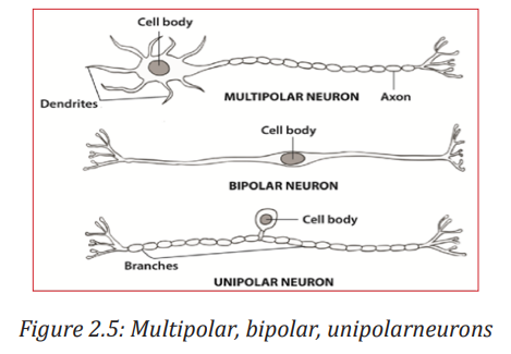
2.2.1 Types of neurons
Nerve cells may be grouped according to the number of processes they possess so that their types include:
- Unipolar neurons (sensory neuron): those with one process only, found mainly in invertebrates.
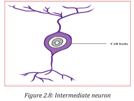
- Bipolar neurons (relay neuron): those with two separate processes such as neurons in the retina of the vertebrate eye.
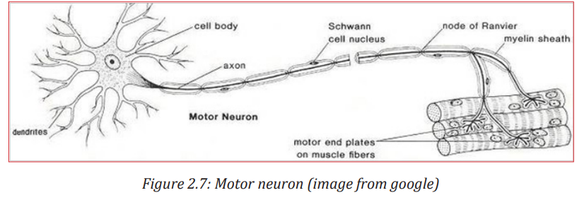
- Multipolar neurons (motor neuron): those with more than two processes such as most of the vertebrate neurons.
2.2.2 Classification of neurons by their functions
In vertebrates, it is also common to group neurons according to their functions. They include:
- Sensory or afferent neurons: transmit impulses from the receptors to the Central nervous system. In addition to sensory or afferent neurons.
- Motor or efferent neurons: that transmits impulses from the central nervous system to effectors motor organs such as muscles or glands that carry out the response. Most motor neurones are stimulated by impulses conducted by interneurons. However, there are some others that are stimulated directly by sensory neurons.
- Interneurons also known as intermediate or association, or relay or interneuron. They connect the pathways of sensory and motor impulses, and are found mainly in the central nervous system.
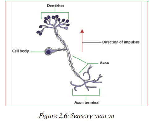
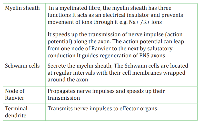
2.2.3 Parts of a neuron and their functions
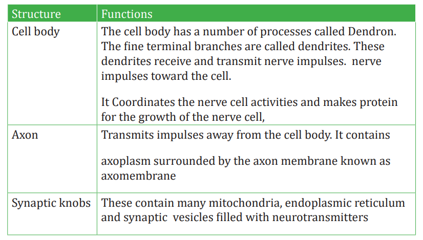
Each motor neuron possesses a cell body and cytoplasm with many mitochondria, endoplasmic reticulum, Golgi apparatus and ribosomes.
The Nissl granules which consist of endoplasmic reticulum and ribosomes function in protein synthesis. The table below (Table 2.1) shows all parts of neuron and their functions
Table 2.1: Parts of a neuron and their functions
2.2.4 Reflex Actions
A reflex action is a quick and involuntary response of the central nervous system to a stimulus. Examples are: The quick withdrawal of the hand from a hot object, knee jerk, eye blinking. When the spinal cord alone is involved, the reflex action is called spinal reflex and when the brain alone is involved, it is a cranial reflex e.g. blinking of eyes.
Reflex actions are described as involuntary actions and the same stimuli produce the same responses every time. Reflexes are useful because they make autonomic involuntary adjustments to changes in the external environment, such as the iris pupil reflex and the balance during locomotion. They also control the internal environment, such as breathing rate and blood pressure, and prevent damage to body as in cuts and burns. These help to maintain constant conditions, in other word they are involved in homeostasis.
Reflex arc is a pathway followed by a nerve impulse during a reflex action.
The components of reflex arc are:
- Stimulus
- Receptors
- The sensory receptor that detects the stimulus
- The sensory (or afferent) neurone along which the sensory impulse is transmitted;
- The relay neurone in the central nervous system to which the sensory impulse is passed on.
- The motor (or efferent) neurone along which the motor impulse is transmitted; and
- The effector (Muscle or gland) which the motor impulse triggers to bring about an appropriate response.
- CNS (Brain or spinal cord)

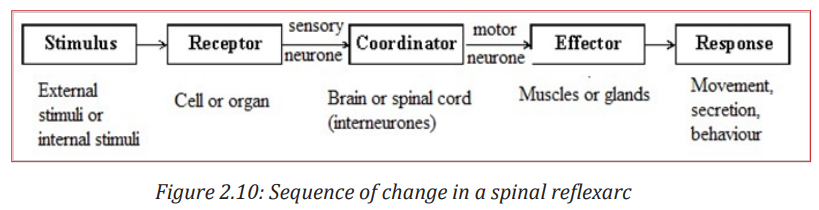
The sequences of changes that occur during a spinal reflex are:
- A sensory receptor receives a stimulus and impulse is generated in it
- The impulse is transmitted along a sensory neuron towards the spinal cord via the dorsal root
- Once the impulse reaches the grey matter inside the spinal cord, it is passed on to the relay neuron across a synapse
- The relay neuron then transfers the impulse to a motor neuron across another synapse.
- The motor neuron conveys the impulse to an effector such as a muscle where a response takes place.
The pathway that is followed by an impulse along the sensory neurons relay and motor neurone, during a reflex action is called reflex arc.
2.3 Nature, structure and function of synapse in the nervous system
Activity 2.3
Discuss the nature, structure and function of synapse in the nervous system
All cells in animal body tissues are electrically polarized in other words; they maintain a voltage difference across the cell’s plasma membrane, known as the membrane potential. This electrical polarization results from a complex interplay between protein structures embedded in the membrane called ion pumps and ion channels. Each excitable patch of membrane has two important levels of membrane potential: the resting potential, which is the value the membrane potential maintains as long as nothing passes along the cell, and a higher value called the threshold potential.
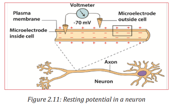
2.3.1 Resting potential in a neuron
A neuron is said to be in the resting state when it is not conducting an impulse. The membrane potential of an unstimulated excitable cell is called the resting potential. A resting potential is the difference in charge (electrical potential difference) which exists between the inside and the outside of the cell membrane. In excitable cells, the resting potential is about -70 millivolts (mV) and the threshold potential is around -55 mV. The negative sign indicates the interior of the cell is negative with respect to the exterior environment.
The resting potential difference across the neuron membrane is maintained by:
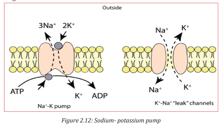
- The sodium–potassium pump (Na+ /K+ ). This is always working. Three sodium ions (Na+ ) are actively transported out of the cell for every two potassium ions (K+ ) pump into the cell. Energy supplied by ATP is used for the transport of ions against their electrochemical gradients.
- The axon membrane: It is more permeable to potassium ions than the sodium ions. This is due to the presence of more potassium ion non-gated, voltage independent channels and few sodium ion non-gated channels. More K+ ions can diffuse out back again faster than Na+ ions which can diffuse back in. The resting membrane potential is mainly determined by sodium-potassium pump, facilitated diffusion and electrochemical gradient of K+ ions across the membrane.
2.3.2 Action potential
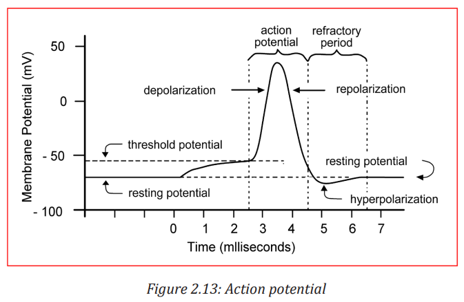
Action potential is the technical term for impulse. An action potential is rapid temporary reversal in the electrical potential difference of an excitable cell e.g. a neuron or a muscle cell. It is caused by changes in the permeability of the membrane following the application of a threshold stimulus. The action potential has a depolarization phase and a repolarization phase. There may be a short hyperpolarized phase after the repolarization phase. The time taken for an action potential is 2 to 3 milliseconds.
2.3.3 Depolarization
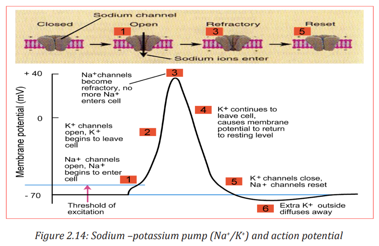
When a stimulus such as electric current reaches a resting neuron, some sodium voltage gated channels in the stimulated region of the axon membrane open. Sodium ions (Na+ ) move into the axon by facilitated diffusion down an electrochemical gradient. The initial influx of sodium ions is slow. The axon membrane becomes slightly depolarized and the sodium voltage gates are sensitive to voltage changes. More gates open allowing more Na+ ions to diffuse into the cell leading to further depolarization.
When the potential difference across the membrane reaches a threshold value (-50mV), many more sodium voltage gated channels open. This is an example of positive feedback. The rapid diffusion of Na+ ions leads to a sudden increase in the cell’s potential difference which becomes positive (+ 40mV). This reversal in the potential difference is known as depolarization and lasts for about 1 millisecond
2.3.4 Repolarization
The reversal in polarity to + 40 mV causes the voltage gated sodium channel to close. At the same time the voltage gated potassium channels open. The potassium ions K+ diffuse out of the cell down their electrochemical gradient to the tissue fluid outside. The axon membrane is repolarized. The action potential alters from + 40 mV to -70mV.
2.3.5 Hyperpolarization
The potassium voltage-gated channels are slow to close. An excess of K+ ions leave the axon. The inside of the membrane becomes more negative. The voltage falls slightly below -70mV and causes hyperpolarization. However, within a few milliseconds, the potassium voltage-gated channels close. The resting potential of -70mV is reestablished by the Na+ /K+ pump and different rates of facilitated diffusion of K+ and Na+ ions through the non-gated ion channels.
2.3.6 Frequency of action potentials
Information in axons is coded in the frequency of the action potentials. A weak stimulus above threshold produces fewer action potentials. A stronger stimulus produces a greater frequency of action potentials. As the intensity of stimulation increases, more action occurs.
Application activity 2.3
The graphs below show the changes that occur during an action potential in a membrane potential and the relative membrane permeability to sodium and potassium ions in a neurone. Observe well to answer the following questions:
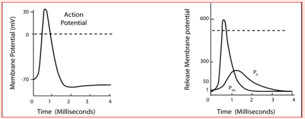
a. Describe the movement of ions during an action potential
b. Explain what is the effect of an action potential generation if there is a lowering of sodium ions in the extracellular fluid
2.4 Transmission of a nerve impulse
Activity 2.4: Research activity
The diagram below shows different nerves of a frog after dissection. Use school library and search additional information on internet and watch the movies on youtube related to frog dissection.
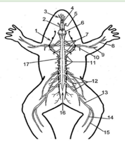
1. Redraw this diagram and label it from 1 to 17
2. Identify the main difference between the part numbered 13 from other parts
3. Identify the main function of 13
4. What would happen to a frog when 13 is damaged
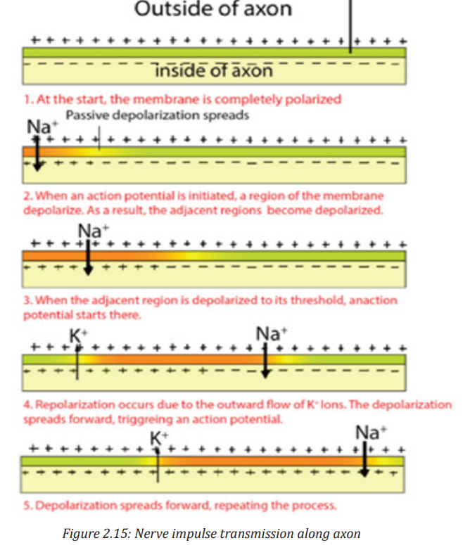
2.4.1 Mechanism of transmission of nerve impulses along an axon
- The neurons, like other cells, are positively charged outside and negatively charged inside. The membrane of the axon is said to be polarized. The potential difference (voltage) across their membranes is of – 70mV and is called resting membrane potential (RMP).
- A stimulus (heat, pain, bite, sound …) creates an action potential (AP) or an impulse that is transmitted along an axon by electro-chemical change.
- During an action potential, the membrane potential falls until the inside becomes positively charged with respect to the exterior. The membrane at this point is said to be depolarized. It takes few milliseconds to happen. In fact, the potential changes from – 75 mV to + 40 mV at the point of stimulation. That is an electrical change that runs along the axon.
- As the impulse is transmitted along the axon, the Na+ /K+ pumps of the axolemma are re-established. Sodium channels open first, allowing a large number of Na+ ions to flow in.
- The axoplasm becomes progressively more positive with respect to the outside of the axolemma. Then, almost instantly, the permeability of the membrane to Na+ ions ceases, and the net flow of Na+ ions stop. At the same time K+ ion channels start to open and K+ ions flow out from axoplasm where they are in high concentration. The counter-flow is of 3Na+ ions against 2K+ ions.
- The axoplasm now starts to become less positive again. This begins the process of re-establishing the resting potential difference of the membrane. That is an electro-chemical change.
a. Factors that affect transmission of nerve impulses along the axon membrane
Along the axon membrane, the transmissions of nerve impulses are affected as follows:
- The diameter of the axon: the greater the diameter the faster the speed of transmission of nerve impulses.
- The myelin sheath: myelinated neurones conduct impulses faster than non- myelinated neurones.
- The presence of nodes of Ranvier speeds up the movement of impulses in myelinated neurones.
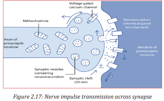
b. Structure of a synapse
Information from one neuron flows to another neuron across a synapse. The synapse is a small gap separating two adjacent neurons. The synapse consists of:
- A presynaptic ending that contains neurotransmitters, mitochondria and other cell organelles,
- A postsynaptic ending that contains receptor sites for neurotransmitters and,
- A synaptic cleft or space between the presynaptic and postsynaptic endings. It is about 20 nm wide.
The swollen tip of the axon of the presynaptic neuron, called synaptic knob or synaptic bulb contains many membranes bounded synaptic vesicles, mitochondria and microfilaments.
The synaptic vesicles contain neurotransmitter molecules such as acetylcholine or noradrenaline
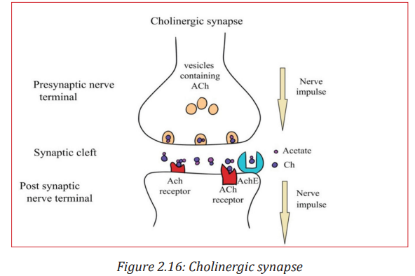
An example of synapse is the cholinergic synapse which is a synapse which uses acetylcholine (Ach) as neurotransmitter. Calcium and vesicles are involved in the release of neurotransmitter across the synaptic cleft in the mechanism of synaptic transmission to generate an excitatory post-synaptic potential.
c. Neurotransmitter
A neurotransmitter is a relatively small chemical found in the synaptic vesicle. It helps to transmit an impulse across a synapse or neuromuscular junction. 5050 There are about 50 different types of neurotransmitters in the human body. Examples are acetylcholine released by cholinergic neurons, noradrenaline (norepinephrine) released by adrenergic neurons, dopamine and serotonin including amino acids glutamate and glycine.
2.4.2 Mechanism of nerve impulse transmission across a synapse
- The arrival of an impulse on the synaptic knob causes the opening of Ca+2 ion channels on the presynaptic membrane, and Ca+2 ions flow in the presynaptic region from the synaptic cleft.
- The Ca+2 ions induce a few presynaptic vesicles to fuse with presynaptic membrane and to secrete their neurotransmitters (e.g. acetylcholine) by exocytosis into the synaptic cleft
- The neurotransmitter then binds with the receptor protein on the postsynaptic membrane. This causes the opening of Na+ channels on the postsynaptic neuron which in turn becomes depolarized.
- This causes a depolarization of the post-synaptic cell membrane, which may initiate an action potential, if the threshold is reached
- The action of the neurotransmitter does not persist because an enzyme cholinesterase catalyses the hydrolysis of acetylcholine into choline and acetate.
The breakdown products (choline) are absorbed by the pre-synaptic neuron by endocytosis and used to re-synthesize more neurotransmitter, using energy from the mitochondria.
2.4.3 Properties of a nerve impulse
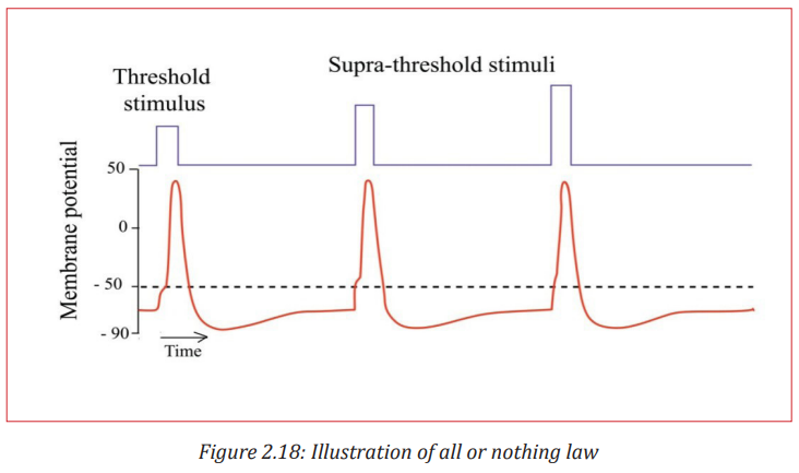
a. All or nothing law
An action potential can only be generated after the threshold value is exceeded. After the threshold is reached, the size of the action potential produced remains constant and is independent of the intensity of the stimulus. This is the all or nothing response. All action potentials are of the same amplitude.
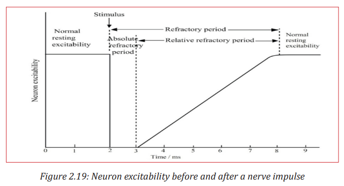
b. Refractory period
This is a brief period when an axon is unable to transmit an impulse following transmission of the same. It lasts about 5-10 milliseconds. It is divided into two; absolute and relative periods.
During the absolute refractory period which lasts about 1ms, the axon membrane is unable to respond to another stimulus, no matter how strong it is. An action potential cannot be produced. This is because there is conformational change in voltage-gated sodium channels which are still in a closed, inactive state. This also prevents the action potential from moving backwards.
Following the absolute refractory period, there is a relative refractory period which lasts around 5ms. During this period, the resting potential is gradually restored by Na+ /K+ pump and the relative permeability of membrane to facilitated diffusion of ions is also restored. A new action potential can then be produced if the stimulus is greater than the usual one. The refractory period therefore allows impulses to move only in one direction and limits the frequency at which successive impulses can pass along axon.
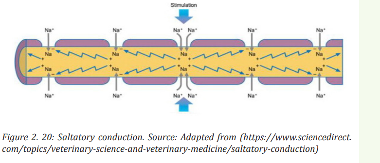
c. Saltatory conduction
It is movement or jump of nerve impulses from one node of Ranvier to another along the axon membrane of neurone.
Activity 2.4 1.
Suppose a cell’s membrane potential shifts from -70 mV to -50 mV. What changes in the cell’s permeability to K+ or Na+ could cause such a shift?
2. The diagram below shows the changes in potential difference across an axon membrane as a nerve impulse passes
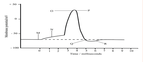
a. Explain what happens at M, N, O, P, Q and R as shown in the graph
b. Name two factors that can determine the speed of transmission of a nerve impulse and how each affects the speed
c. Explain why the initiation of an action potential is considered a positive feedback mechanism
2.5 Structure and function of the endocrine system in humans
Activity 2.5
Make a research using internet, watching movies, using different books from the school library and a charts showing different endocrine glands, make short notes on structure location and function of the endocrine system in humans
Endocrine glands secrete their products called hormones into the interstitial fluid surrounding the secretory cells rather than into ducts. From the interstitial fluid, hormones diffuse into blood capillaries and blood carries them to target cells throughout the body. Because most hormones are required in very small amounts, the circulating levels of hormones are typically low.
The word endocrine means internal secretion and endocrine glands are therefore glands of internal secretion. Since they shed their secretion into the bloodstream, they have no ducts and are hence known as ductless glands. Once in the bloodstream, the hormones are carried around the body, bringing about responses in various places. Structures that respond to them are called target organs. A hormone is a chemical messenger having the following properties:
- It travels in the blood
- It has its effect at a site different from the site where it is produced. The site where it has effect is called the target, while itself is called messenger
- It fits precisely into receptor molecules in the target like a key in a lock. It is therefore specific for a particular target;
- It is a small soluble molecule;
- It is effective in low concentrations
The ability of a target cell to respond to a hormone depends on the presence of receptors, within the cell or on its plasma membrane, to which the hormone can bind. Hormone receptors are dynamic structures. Changes in number and sensitivity of hormone receptors may occur in response to high or low levels of stimulating hormones.
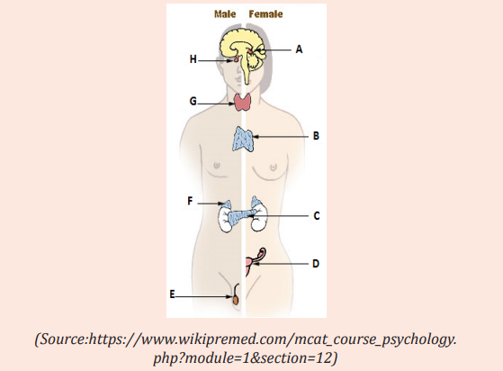
The endocrine glands include the pituitary, thyroid, parathyroid, adrenal, and pineal glands (Figure 10.1). Taken together, all endocrine glands and hormone-secreting cells constitute the endocrine system. The science of the structure and function of the endocrine glands and the diagnosis and treatment of disorders of the endocrine system is endocrinology (endo: within; crino: to secrete; -logy: study of).
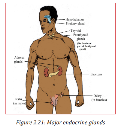
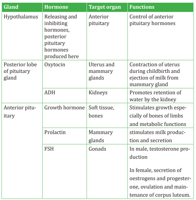
a. Pituitary gland
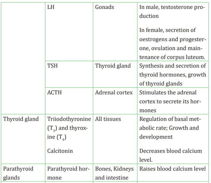
The pituitary gland also called hypophysis or master gland hangs from the base of the brain by a stalk and is enclosed by bone. It consists of a hormone-producing glandular portion called anterior pituitary and a neural portion called posterior pituitary, which is an extension of the hypothalamus. The hypothalamus regulates the hormonal output of the anterior pituitary and synthesizes two hormones that it exports to the posterior pituitary for storage and later release. Four of the six hormones produced by the pituitary gland are tropic hormones that regulate the function of other endocrine organs. Growth hormone (GH) or Somatotropic hormone is a hormone that stimulates growth of all body tissues but especially skeletal muscle and bone. GH mobilizes the use of fats, stimulates protein synthesis, and inhibits glucose uptake and metabolism.
- Thyroid-stimulating hormone (TSH) stimulates the normal development and activity of the thyroid gland. Thyrotropin-releasing hormone (TRH) stimulates its release; negative feedback of thyroid hormone inhibits it.
- Adrenocorticotropic hormone (ACTH) stimulates the adrenal cortex to release its hormones. ACTH release is triggered by corticotropin-releasing hormone (CRH) and inhibited by rising glucocorticoid levels.
- The gonadotropins: follicle-stimulating hormone (FSH) and luteinizing hormone (LH) regulate the functions of the gonads in both sexes.
- Prolactin (PRL) promotes the production of milk in human’s females. Its secretion is triggered by prolactin-releasing hormone (PRH) and inhibited by prolactin-inhibiting hormone (PIH).
The neurohypophysis stores and releases two hormones produced by the hypothalamus:
- Oxytocin stimulates powerful contractions of the uterus, which trigger labor and delivery of an infant, and milk ejection in nursing women. Its release is mediated reflexively by the hypothalamus and represents a positive feedback mechanism.
- Antidiuretic hormone (ADH) stimulates the kidney tubules to reabsorb and conserve water, resulting in small volumes of highly concentrated urine and decreased plasma osmolality.
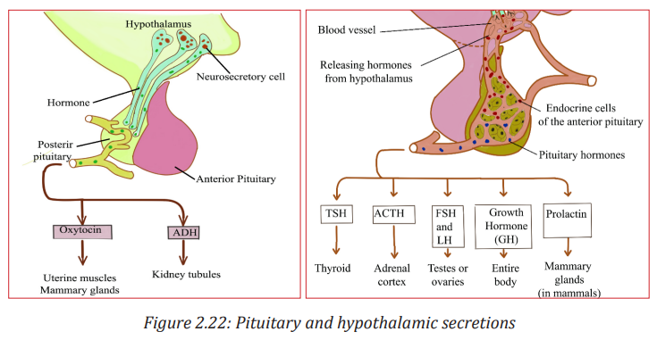
b. Hypothalamus
The hypothalamus plays an important role in integrating the vertebrate endocrine and nervous systems. The region of the lower brain receives information from nerves throughout the body and from other parts of the brain thus initiates endocrine signals appropriate to environmental conditions. A set of neurosecretory cells in the hypothalamus exerts control over the anterior pituitary by secreting two kinds of hormones into the blood: Releasing hormones which make the anterior pituitary to secrete its hormones and inhibiting hormones that make the anterior pituitary stop secreting hormones. Every anterior pituitary hormone is controlled at least by one releasing hormone and some have both a releasing and an inhibiting hormone.
Unlike the anterior pituitary, the posterior pituitary or neurohypophysis is an extension of the brain. It develops from a bulge of the hypothalamus that grows downward the mouth fold that forms the anterior pituitary. The posterior pituitary remains attached to the hypothalamus. It stores and releases two hormones that are made by a set of neurosecretory cells in the hypothalamus.
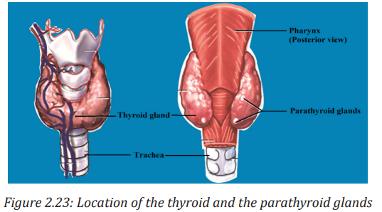
c. Thyroid gland
The thyroid gland is located in the anterior throat. Thyroid follicles store colloid containing thyroglobulin, a glycoprotein from which thyroid hormone is derived. Thyroid hormone (TH) includes thyroxine (T4) and triiodothyronine (T3), which increase the rate of cellular metabolism. Consequently, oxygen use and heat production rise. Calcitonin, produced by the parafollicular cells of the thyroid gland in response to rising blood calcium levels, decreases blood calcium levels by inhibiting bone matrix resorption and enhancing calcium deposit in bone.
d. Parathyroid glands
The parathyroid glands are located on the dorsal aspect of the thyroid gland and secrete parathyroid hormone (PTH), which causes an increase in blood calcium levels by targeting bone, the intestine, and the kidneys. PTH is the antagonist of calcitonin. PTH release is triggered by decreasing blood calcium levels and is inhibited by increasing blood calcium levels.
e. Pancreas
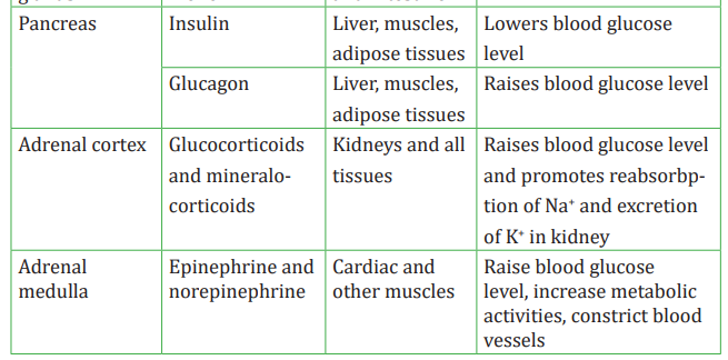
The pancreas is an organ located in the abdomen close to the stomach and is both an exocrine and an endocrine gland. The endocrine portion (islets of langerhans) releases insulin and glucagon and smaller amounts of other hormones such as somatostatine to the blood. Glucagon is released by alpha (α) cells when glucose levels in blood are low. Glucagon stimulates the liver to release glucose to the blood. Insulin is released by beta (β) cells when blood levels of glucose (and amino acids) are rising. It increases the rate of glucose uptake and metabolism by most body cells.
f. Gonads
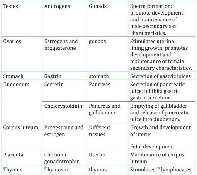
The ovaries of the female which are located in the pelvic cavity, release two main hormones. Secretion of estrogens by the ovarian follicles begins at puberty under the influence of FSH. Estrogens stimulate maturation of the female reproductive system and development of the secondary sex characteristics. Progesterone is released in response to high blood levels of LH. It works with estrogens in establishing the human menstrual cycle. The testes of the male begin to produce testosterone at puberty in response to LH. Testosterone stimulates the maturation of the male reproductive organs, development of secondary sex characteristics, and the production of sperm by the testes.
g. Adrenal Glands / Suprarenal Glands
Each adrenal gland weighs about 5 g and sits on the superior pole of the respective kidney, like a cap. The glands are included in the fatty capsule of the kidney and are noteworthy for their rich supply of nerves and vessels. A fresh adrenal gland section shows a bright yellow cortex, making up about 80% of the organ, and a more reddish-grey medulla. The endocrine activities of the adrenal cortex and the adrenal medulla differ both in development and function.
1. Adrenal cortex
Adrenal cortex makes mineralocorticoids (such as aldosterone and cortisol). Cortisol raises blood glucose level whereas aldosterone stimulates the reabsorption of Na+ and excretion of K+ in kidney.
2. Adrenal Medulla
The adrenal medulla makes two hormones epinephrine (adrenaline) (80 %) and norepinephrine (noradrenaline) (20 %). Epinephrine and norepinephrine are released into the bloodstream during stress and they act on the whole organism by preparing it for increased energy use. Both hormones, for instance, activate the liberation of fatty acids from fat depots and liberate glucose from glycogen storage in the liver (producing a rise in the blood sugar level).
They raise the blood pressure and stroke volume of the heart and may lead to vasoconstriction in certain defined areas.
h. Other hormone-producing structures
Many body organs not normally considered endocrine organs contain isolated cell clusters that secrete different hormones. Examples include the gastrointestinal tract organs (gastrin, secretin, and others), the placenta (hormones of pregnancy such as estrogen, progesterone, and others) and the kidneys (erythropoietin and renin).
Table 2.2: Major human endocrine glands, their functions and the control of their secretions
Application activity 2.5
The diagram below shows the main endocrine glands
1. Name the parts from A to H
2. Describe the roles of hormones produced by C
3. What would happen if the hormones produced by G are secreted in low quantity
2.6 Principles of the negative feedback mechanism of hormonal action
ACTIVITY 2.6
Feedback mechanisms are necessary in the maintenance of homeostatic mechanisms. All homeostatic control mechanisms have at least three interdependent components for the variable being regulated that work together. The receptor is the sensing component that monitors and responds to changes in the environment. When the receptor senses a stimulus, it sends information to a control center, the component that sets the range at which a variable is maintained. The control center determines an appropriate response to the stimulus. In most homeostatic mechanisms, the control center is the brain. The control center then sends signals to an effector, which can be muscles, organs or other structures that receive signals from the control center. After receiving the signal, a change occurs to correct the deviation by either enhancing it with positive feedback or depressing it with negative feedback.
The homeostatic mechanisms in mammals require information to be transferred between different parts of the body. 6464 There are two coordination systems in mammals that control this: the nervous system and the endocrine system
- In the nervous system, information in the form of electrical impulses is transmitted along nerve cells (neurons).
- The endocrine system uses chemical messengers called hormones that travel in the blood, in a form of long-distance cell signalling.
Positive feedback mechanisms are designed to accelerate or enhance the output created by a stimulus that has already been activated. Unlike negative feedback mechanisms that initiate to maintain or regulate physiological functions within a set and narrow range, the positive feedback mechanisms are designed to push levels out of normal levels. To achieve this purpose, a series of events initiates a cascading process that builds to increase the effect of the stimulus. This process can be beneficial but is rarely used by the body due to risks of the acceleration’s becoming uncontrollable.
One positive feedback example event in the body is the accumulation blood platelets, which, in turn, causes blood clotting in response to a break or tear in the lining of blood vessels. Another example is the release of oxytocin to intensify the contractions of the uterus that take place during childbirth.
Another example of a positive feedback mechanism is the production of milk by a mother for her baby. As the baby suckles, nerve messages from the mammary glands cause the mother’s pituitary gland to secrete a hormone called prolactin. The more the baby suckles, the more prolactin is released, which stimulates further milk production by the mother’s mammary glands. In this case, a negative feedback mechanism would not be helpful because the more the baby nursed, the less milk would be produced.
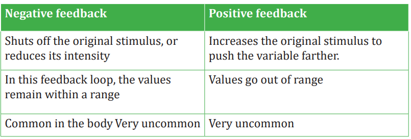
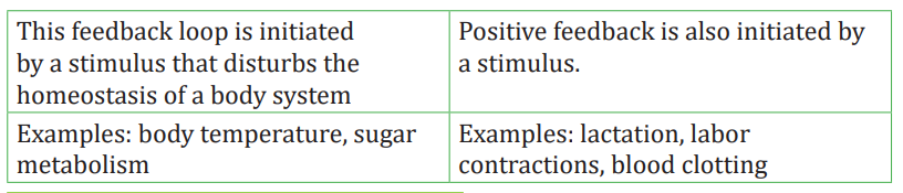
Table 2.3: Negative and positive feedback compared
Activity 2.7
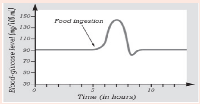
There are some events or steps that occur in human body when blood glucose increases above normal level or decreases below normal level. Describe 7 steps/events:
a. When glucose level in blood increases above normal level
b. When glucose level in blood decreases below normal level
2.7 Effects of hormonal imbalances
Activity 2.7
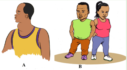
Observe carefully the photos below and suggest the type of disorders the following people may be suffering:
The disorders of the endocrine system often involve either the hyposecretion (hypo means too little or under), inadequate release of a hormone, or the hypersecretion (hyper means too much or above), excessive release of a hormone. In other cases, the problem is faulty hormone receptors, an inadequate number of receptors, or defects in second-messenger systems. Because hormones are distributed in the blood to target tissues throughout the body, problems associated with endocrine dysfunction may also be widespread.
2.7.1 Pituitary gland disorders
a. Pituitary dwarfism, gigantism, and acromegaly
Several disorders of the anterior pituitary involve human growth hormone. Hyposecretion of human growth hormone during the growth years slows bone growth, and the epiphyseal plates close before normal height is reached. This condition is called pituitary dwarfism. Other organs of the body also fail to grow, and the body proportions are childlike. Treatment requires administration of human growth hormone during childhood, before the epiphyseal plates close.
Hypersecretion of human growth hormone during childhood causes gigantism, an abnormal increase in the length of long bones. The person grows to be very tall, but body proportions are about normal. Hypersecretion of human growth hormone during adulthood is called acromegaly.
b. Diabetes insipidus
The most common abnormality associated with dysfunction of the posterior pituitary is diabetes insipidus.
This disorder is due to defects in antidiuretic hormone (ADH) receptors or an inability of the pituitary gland to secrete ADH. A common symptom of diabetes insipidus is excretion of large volumes of urine resulting in dehydration and thirst. Bed-wetting is common in afflicted children. Because so much water is lost in the urine, a person with diabetes insipidus may die of dehydration if deprived of water for only one day. Treatment of diabetes insipidus involves the injection of ADH.
2.7.2 Thyroid gland disorders
Thyroid gland disorders affect all major body systems and are among the most common endocrine disorders. Congenital hypothyroidism or the hyposecretion of thyroid hormones that is present at birth has devastating consequences if not treated quickly. Previously termed cretinism, it causes severe mental retardation and stunted bone growth. At birth, the baby typically is normal because lipid-soluble maternal thyroid hormones crossed the placenta during pregnancy and allowed normal development.
Hypothyroidism during the adult years produces a disorder called myxoedema. An indication of this disorder is oedema (accumulation of interstitial fluid) that causes the facial tissues to swell and look puffy. A person with myxoedema has a slow heart rate, low body temperature, sensitivity to cold, dry hair and skin, muscular weakness, general lethargy, and a tendency to gain weight easily. Because the brain has already reached maturity, mental retardation does not occur, but the person may be less alert.
The most common form of hyperthyroidism is Graves’ disease which is an autoimmune disorder in which the person produces antibodies that mimic the action of thyroid-stimulating hormone (TSH). The antibodies continually stimulate the thyroid gland to grow and produce thyroid hormones. A primary sign is an enlarged thyroid, which may be two to three times its normal size. Graves’ patients often have a peculiar oedema behind the eyes, called exophthalmos, which causes the eyes to protrude. Treatment may include surgical removal of part or all of the thyroid gland (thyroidectomy), the use of radioactive iodine to selectively destroy thyroid tissue, and the use of antithyroid drugs to block synthesis of thyroid hormones. A goitre is simply an enlarged thyroid gland. It may be associated with hyperthyroidism, hypothyroidism or by the lack of iodine.
2.7.3 Parathyroid gland disorders
Parathyroid gland disorders cause the hypoparathyroidism due to the too little parathyroid hormone leading to a deficiency of blood Ca2+, causing neurons and muscle fibres to depolarize and produce action potentials spontaneously. This leads to twitches, spasms, and tetany (maintained contraction) of skeletal muscle.
The main cause of hypoparathyroidism is accidental damage to the parathyroid glands or to their blood supply during thyroidectomy surgery.
Hyperparathyroidism or an elevated level of parathyroid hormone, most often is due to a tumour of one of the parathyroid glands. An elevated level of PTH causes excessive resorption of bone matrix, raising the blood levels of calcium and phosphate ions and causing bones to become soft and easily fractured. High blood calcium level promotes formation of kidney stones. Fatigue, personality changes, and lethargy are also seen in patients with high levels of parathyroid hormone.
2.7.4 Adrenal gland disorders
a. Cushing’s syndrome
Hypersecretion of cortisol by the adrenal cortex causes an endocrine disorder known as Cushing’s syndrome. The condition is characterized by breakdown of muscle proteins and redistribution of body fat, resulting in thin arms and legs accompanied by a rounded moon face and buffalo hump on the back. Facial skin is flushed, and the skin covering the abdomen develops stretch marks. The person also bruises easily, and wound healing is very slow. The elevated level of cortisol causes hyperglycaemia, osteoporosis, weakness, hypertension,increased susceptibility to infection, decreased resistance to stress, and mood swings.
b. Addison’s disease
Hyposecretion of glucocorticoids and aldosterone causes Addison’s disease (chronic adrenocortical insufficiency). The majority of cases are autoimmune disorders in which antibodies cause adrenal cortex destruction or block binding of ACTH to its receptors. Pathogens, such as the bacterium that causes tuberculosis, also may trigger adrenal cortex destruction. Symptoms, which typically do not appear until 90% of the adrenal cortex has been destroyed, include mental lethargy, anorexia, nausea and vomiting, weight loss, hypoglycemia, and muscular weakness. Loss of aldosterone leads to the elevated potassium and decreased sodium in the blood, low blood pressure, dehydration, decreased cardiac output and even cardiac arrest.
2.7.5 Pancreas disorders
The most common endocrine disorder is diabetes mellitus caused by an inability to produce or use insulin. According to the diabetes atlas of 2018, the prevalence of diabetes in Rwanda is about 3.16% of the population with 1,918 diabetes related deaths per year. On the world health day in 2016, the world health organization (WHO) addressed a call for action on diabetes, drawing attention to the need to step up prevention and treatment of this disease. The first WHO Global report on diabetes demonstrates that the number of adults living with diabetes has almost quadrupled since 1980 to 422 million adults. This dramatic rise is largely due to the rise in type 2 diabetes and factors driving it include overweight and obesity. In 2012 alone diabetes caused 1.5 million deaths. Its complications can lead to heart attack, stroke, blindness, kidney failure and lower limb amputation.
Because insulin is unavailable to aid transport of glucose into body cells, blood glucose level is high and glucose is found in the urine, the process known as glucosuria. The cardinal signs of diabetes mellitus are polyuria (excessive urine production due to an inability of the kidneys to reabsorb water), polydipsia (excessive thirst) and polyphagia (excessive eating).
Activity 2.8
The graph below blood–glucose level. Analyse it carefully and answer the following questions. What will happen after one eat food?
2.8 Comparison of hormonal and nervous systems
Activity 2.8
Make table comparing nervous system and endocrine system
The nervous and endocrine systems act together to coordinate functions of all our body systems. Remember that the nervous system acts through nerve impulses conducted along axons of neurons. At synapses, nerve impulses trigger the release of mediator molecules called neurotransmitters, while the endocrine system controls body activities by releasing mediators, called hormones. However, the means of control of the two systems are very different.
A basic similarity between the endocrine system and the nervous system is that both provide means of communication within the body of an organism. Both involve transmission of a message which is triggered by a stimulus and produces a response. Several chemicals function as both neurotransmitters and hormones including norepinephrine. Some hormones such as oxytocin are secreted by neuroendocrine cells; neurons that release their secretions into the blood. The target organs of a hormone are equivalent to nerve’s effectors.
The main differences between the two systems concern the nature of the message. In the endocrine system, the message takes the form of a chemical substance transmitted through the blood stream. In the nervous system it is a discrete-all or none action potential transmitted along a nerve fiber. All other differences arise from this fundamental one. They can be listed as follows:
- Because of the comparatively high speed at which impulses are transmitted along nerves, nerves responses are generally transmitted more rapidly than hormonal ones.
- Since it is conveyed by the bloodstream, there is nothing to stop a hormone being carried to every part of the body. Nervous impulses however are transmitted by particular neurons to specific destinations.
- As a result, hormones are often widespread, sometimes involving the participation of numerous target organs. In contrast, nervous responses may be much localized, involving perhaps the contractions of only one muscle.
- Hormonal responses frequently continue over a long period of time. Obvious examples of such long-term responses are growth and metabolism.
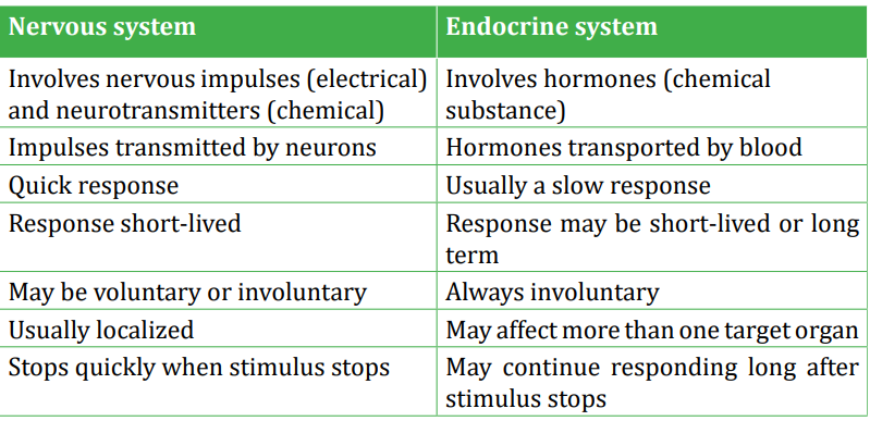
A comparison between nervous and endocrine system is summarized in the table 2.4
Table 2.4: Comparison between nervous and endocrine system
Skills lab 2
Objective : Determine the blood glucose level: Test for glucose level in blood by using a glucometer
Requirements: Blood, sterile needle, cotton wool, ethanol, glucometer Procedure:
1. Insert a disposable test strip into its place in the glucometer.
2. Wash your finger with alcohol using cotton wool.
3. Use a needle; get some drops of blood from one volunteer participant.
4. Place the drop of blood on a disposable test strip.
5. Read and calculate the blood glucose level.
Discuss what will happen when the amount of sugar in blood increases or decreases to the normal range of sugar level in blood.
End unit assessment 2
A. Multiple choice questions: Choose the best answer
1. What happens when a neuron’s membrane depolarizes?
a. There is a net diffusion of Na+ out of the cell.
b. The equilibrium potential of K+ becomes more positive.
c. The neuron’s membrane voltage becomes more positive.
d. The neuron becomes less likely to generate an actionpotential.
e. The inside of the cell becomes more negative relative to the outside.
2. Why action potentials are usually conducted in only one direction along an axon?
a. The nodes of Ranvier can conduct potentials in only one direction.
b. The brief refractory period prevents reopening of voltage gated Na+ channels.
c. The axon hillock has a higher membrane potential than the terminals of the axon.
d. Ions can flow along the axon in only one direction.
e. Voltage-gated channels for both Na+ and K+ open in only one direction.
3. During the repolarisation phase of an action potential, the permeability of the axon membrane to:
a. Na+ increases
b. K+ increases
c. Ca+ increases
d. Organic anions increases
4. What are the chemical messengers of the endocrine system called?
a. Neurons
b. Hormones
c. Blood cells
d. Carbohydrates
5. Endocrine glands
a. Function only after puberty
b. Function only before puberty
c. Release products through ducts
d. Release products into bloodstream
6. X and Y are hormones. X stimulates the secretion of Y, which exerts negative feedback on the cells that secrete X. Suppose the level of Y decreases. What should happen immediately afterwards?
a. Less X is secreted
b. More X is secreted
c. Secretion of Y stops
d. Secretion of X stops
7. Which one of the following hormones is secreted by the neurosecretory cells in mammals?
a. Adrenaline
b. Antidiuretic hormone
c. Insulin
d. Thyroxin
8. The graph shows the changes in the permeability of an axon to Na+ and K+ ions during an action potential
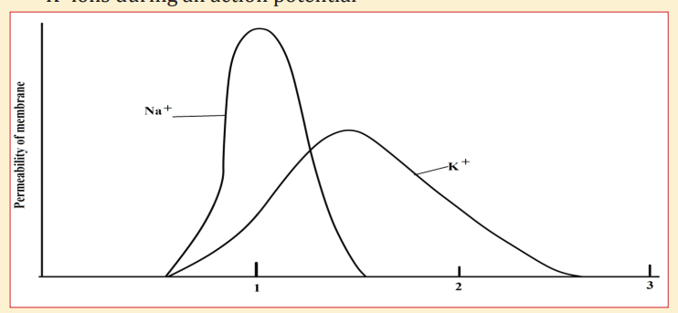
Which of the following shows the correct movement of these ions in the axon?
a. Na+ ions enter the axon, K+ ions leave the axon
b. Na+ ions leave the axon, K+ ions enter the axon
c. Both Na+ and K+ ions enter the axon
d. Both Na+ and K+ leave the axon
8. The graph shows the potential difference across an axon membrane. Which part of the graph shows the action potential?
9. The graph shows the potential difference across an axon membrane. Which part of the graph shows the action potential?
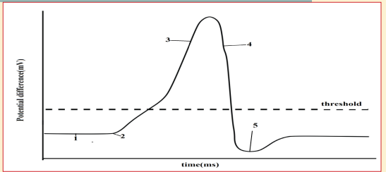
a. 3, 4 and 5
b. 2,3, 4 and 5
c. 1,2, 3 and 4
d. 1,2,3, 4 and 5
B. Short answer type questions
10. Name the hormone involved in the functions described below and the name of the gland which produces it:
a. Controls reabsorption of Na+ in the kidney.
b. Increases the permeability of convoluted distal tubule and collecting duct.
c. Increases heart rate.
d. Increases blood glucose level.
e. Decreases blood glucose level.
f. Repair and growth of the endometrium.
g. Stimulates the anterior pituitary gland to release FSH.
h. Stimulates contraction of the uterus.
i. Stimulates the mammary glands to secrete milk.
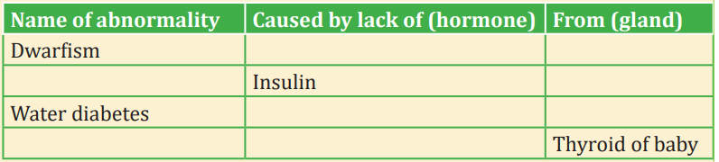
11. A number of metabolic diseases in mammals arise as a result of abnormal endocrine function. Complete the table below concerned with this:
12. The list describes the main stages in the process by which information is transmitted across cholinergic synapses.
- An action potential arrives at synaptic knob of presynaptic neurone. This causes…. the ions to enter the synaptic knob.
- Vesicles move to the………………. membrane.
- A neurotransmitter called……………….is released into the synaptic cleft
- This moves across the cleft by a process known as…………. the neurotransmitter combines with a………………. on the postsynaptic membrane.
- Influx of……………. ions cause local depolarisation and an action potential is set up in the postsynaptic neurone
a. Copy the list. Using the correct scientific terms, add the words that have been omitted.
b. Explain what happens to the neurotransmitter after it has passed information across a cholinergic synapse
c. Some nerves, especially those of the sympathetic nervous system, produce noradrenaline in their synaptic vesicles. Name this type of synapse
13. The graph shows the changes in permeability of the cell surface membrane of an axon to sodium and potassium ions during an action potential.
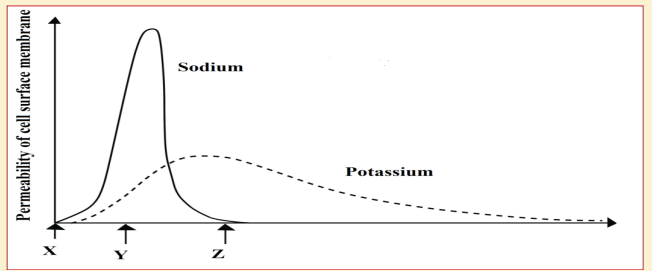
a. Explain how the events which take place between X and Y on the graph can lead to a change in the potential differences across the membrane
b. What happens to the potential difference across the membrane between times Y and Z?
c. Explain why a nerve impulse travels faster in myelinated neurone than in a non-myelinated one
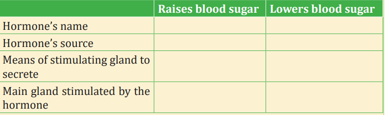
14. During the control of blood sugar in a mammal two antagonistic hormones are employed. Fill in the table about them
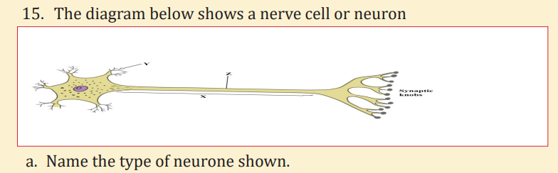
b. Name the structures labelled X and Y
c. A nerve impulse can be initiated by stimulation with a microelectrode.
What would be the effect of stimulation at point Z?
d. The synaptic knobs release a chemical transmitter, acetylcholine. Nerve gases prevent the breakdown of this chemical. From this information suggest
i. One early symptom of nerve gas poisoning
ii. One reason for this observed symptom
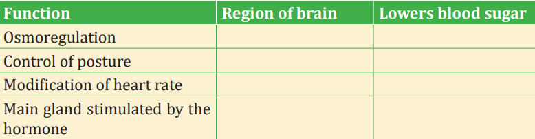
16. Complete the following table by stating which region of the brain controls each of the functions listed
17. The diagram below represents the structures visible at a synapse with the aid of electron microscopy.
a. Identify the structures labelled A and B
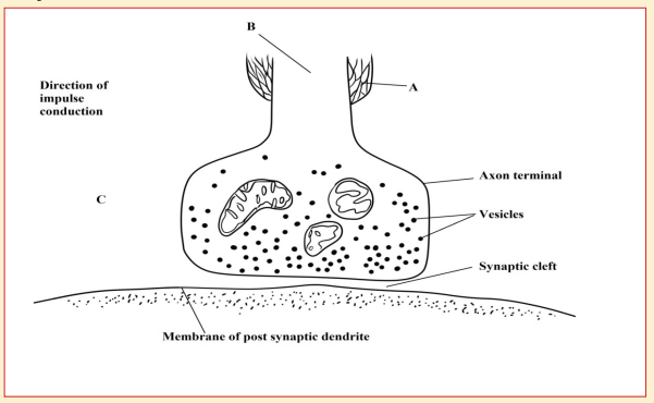
a. Name the chemical found in the numerous vesicles that occur in the synaptic knob
b. Identify the structure labelled C and suggests a reason for its presence in the synaptic knob
c. A powerful hydrolytic enzyme is found in the synaptic cleft. What is its function in normal synaptic transmission?
C. Long answer type questions
18. Describe what happens when an action potential arrives at a synaptic knob of an excitatory synapse
19. What is the difference between diabetes mellitus and diabetes insipidus? What are the characteristic signs of diabetes insipidus?
20. Use the following to describe a negative feedback mechanism: TSH, TRH, decreased metabolic rate, thyroxine and T3.
21. Compare the nervous system from hormonal system