General
- Biology S4 SB File Uploaded 28/01/22, 10:40
- S4: Biology TG File Uploaded 10/08/22, 16:31
UNIT 18: MICROBIOLOGY
Key Unit Competence
Describe the structure and characteristics of viruses, bacteria, and fungal and nonfungal moulds.Learning objectives
By the end of this unit, I will be able to:
–– Describe the basic structure of viruses.
–– Explain how a retrovirus reproduces.
–– Identify the effects of viruses (e.g. AIDS, influenza, measles, feline leukemia, some human cancers) and prokaryotes (e.g. tuberculosis, bubonic plague, cholera) on organisms.
–– Describe how plant viruses can be transmitted.
–– Explain how and why archaebacteria are thought to have been the first forms of life.
–– Describe the structure and life cycles of Escherichia coli
–– Relate the structures and functions of Prokaryotes
–– Describe the structure of fungal and non-fungal moulds and explain how they reproduce
–– Appreciate the importance of microorganisms in life.Introductory activity
A student left fresh milk in acupexposed to the air. After 6 hours, he/she found that milk changed its state from fresh milk to stale milk. Why do you think this happened?Mukamukiza prepared food for dinner. Some of the food was immediately put in tightly covered flask while the remaining food was left in the saucepan covered with banana leaves. In the evening, food in the flask was warm and safe while food in the saucepan has deteriorated. What is the cause of the food spoilage in the saucepan?
18.1 Introduction to microbiology.
Activity 18.1.1
Discuss on the term microbiology and on the groups of microorganisms.
The term “microbiology” comes Greek words: ‘micros’ which means small, ‘bios’ which means life and ‘logos’ which means science. Microbiology is the study of microorganisms which are too small organisms to be seen with the unaided eye and require a microscope to be seen. They are also referred to as microbes. They include bacteria, fungi, algae, protozoa and viruses,they are useful to humans and they play a vital role in decay and recycling of nutrients in the environment. Some of them cause diseasesMicro-organisms are everywhere: in the air, water soil, on plants, on rock surfaces in very hot and cold places (ice). Before the invention of the microscope, microbes were unknown and thousands of people died in devastating epidemics because, vaccines and antibiotics were not available to fight against infectious diseases.
Nowadays, microorganisms can be grown in the laboratory and studied.
a. The Prokaryotes
Prokaryotes can be categorized by their mode of nutrition and how they obtain energy and the carbon used to build the organic molecules that make up cells.
Organisms that obtain energy from light are called phototrophs and those that obtain energy from chemicals are called chemotrophs. Organisms that need only inorganic compounds such as CO2 as a carbon source are called autotrophs. Heterotrophs require at least one organic nutrient such as glucose to make other organic compounds. Prokaryotes usually range in size from 1 to 5 micrometers making them much smaller than most eukaryotic cells.
b. Classification of prokaryotes
Traditionally, bacteria have been classified based on their structure, physiology, molecular composition rather than on their evolutionary relationships. The bacteria that we generally refer to as germs are classified in the domain Eubacteria. More frequently, members of this kingdom are simply called bacteria. The other type of bacteria is known as archaebacteria. These bacteria, which are more ancient than the Eubacteria, are classified in the domain Archaebacteria. Taxonomists used to classify all prokaryotes in kingdom Monera, yet they slightly differ in characteristics.
18.1.2 Archaebacteria and Eubacteria
Activity 18.1.2Discuss on the characteristics of given examples of both archaebacteria and Eubacteria.a. ArchaebacteriaTaxonomists treat archaebacteria as a separate kingdom because they are so different from other bacteria. Archaebacteria have unusual lipids in their cell membranes. Their cell wall is characterized by the absence of peptidoglycans, a protein carbohydrate compound found in the cell walls of Eubacteria. Archaebacteria were first discovered in extreme environmental conditions such as swamps, salt lakes, hot springs. Examples include:1. Methanogens
–– They have unique method of harvesting energy by converting H2 and CO2 in
methane.
–– Methanogens can live only in anaerobic condition, such as the bottom of a
swamp, and in sewage where they are the source of marsh gas, because
oxygen is a poison to them.2. Extreme homophiles
–– These are salt-loving archaebacteria living in environment with very high salt
concentration such as the Dead Sea. High salt concentration would kill most
bacteria.
–– These organisms use salt to generate ATP.3. Thermoacidophiles
–– This third group of archaebacteria lives in extremely acidic environments that have extremely high temperature such as hot springs. Thermoacidophiles live at 110ºC and at a pH of 2.
–– Thermoacidophiles live near volcanic vents on land or near hydrothermal vents.How and why Archaebacteria are thought to have been the first forms of life?The Archaebacteria comprise a group of single-celled microorganisms that, like bacteria, are prokaryotes that have no cell nucleus or any other organelles within their cells. They are known to have an independent evolutionary history and have numerous differences in their biochemistry compared to other forms of life.Archaebacteria are now classified as in separate domain in the three-domain system by Carl Woese who introduced three main branches of evolutionary descent currently known as the Archaea, Eukarya and Bacteria. Classifying Archaea remains difficult, since many of them have never been studied in the laboratory and have only been detected by analysis of their nucleic acids.b. EubacteriaThey occur in many shapes and sizes and have distinct biochemical and genetic characteristics. Eubacteria that are rod-shaped are called bacilli, sphere-shaped are called cocci (sing. Coccus) and spiral-shaped are called spirilla (sing. Spirillum).1. The bacilli: bacteria with rod-shape. Ex: Clostridium tetani, Bacillus subtilis
2. Vibrios: comma-shaped with a single flagellum. eg: Vibrio cholera
3. The cocci: group of bacteria with spherical shape such as Streptococci.
Cocci that occur in chains are Staphylococci which are grapelike clusters of
cocci and Diplococci which is sphere shaped that are grouped two by two.
4. The spirilla: bacteria with spiral shape. e.g.: Spirillum volutans.
18.1.3 Gram stain
Bacteria have a peptidoglycan or murein cell wall that maintains cell shape, provides protection and prevents the cell from lysis. Based on the composition of the cell wall, bacteria can be classified as Gram-positive and Gram-negative. During the process ofGram staining t, some bacteria without a lipid layer along with their peptidoglycan cell wall take the gram stain and appear violet (purple) and are therefore called gram positive. Example streptococcus and staphylococcus. Bacteria having a lipid layer along with their peptidoglycan cell wall do not take up the gram stain and are therefore called gram negative.Example: Escherichia coli, Azotobacter, Salmonella.Self-assessment 18.1
1. Describe the characteristics of the two domains of prokaryotes.
2. What factors can be used to identify prokaryotes?
3. How do bacteria maintain equilibrium in the environment?
4. Identify the parts of a prokaryote.
5. Describe briefly how some prokaryotes obtain their energy.18.2 The structure and life cycle of Escherichia coli
Activity 18.2.1Using text books, videos or computer aided materials to describe the cycle life of E. coli.E. coli reproduce asexually by undergoing binary fission. This type of reproduction begins with the replication of DNA molecule. Then, the copies of the genetic material attach themselves to the cell membrane. When the bacterium’s size has doubled from its original size, the cell membrane starts pinching inward and a cell wall is produced between the two DNA molecules. Finally, the cell wall divides the cell into two daughter cells.E. coli can also go through another process of reproduction known as conjugation. Conjugation is a reproduction process which involves the transfer of genetic material by the sex pili between two bacteria. This is not a sexual reproduction because there is no combination of gametes. The process of conjugation starts once the E. coli, called a donor,has finished to replicate its genetic material in form of a plasmid.The enzyme of the donor can now send signals to show that it is ready to mate. Once a mate is found, the donor attaches itself to the sex pilus of its mate. By doing so, the donor transfers the plasmid.
18.2. E. coli and food poisoning
Activity 18.2.2Using textbooks to brainstorm the process of food poisoning, evolution of harmful strain of E. coli and food preservationE. coli is a rod-shaped bacterium measuring about 2.5µm by 0.5µm. I t is mainly found in guts of vertebrates. It is chemoheterotrophic, capable of thriving on a variety of the organic molecules. Its presence in water indicates contamination by faeces. E. coli reproduces asexually by binary fission. It can also take part in a primitive form of sexual activity called conjugation where genetic material is passed in one direction from bacterium to another through a pilus. Although conjugation does not in itself produce new offspring, after the process has finished, the bacteria reproduce asexually, passing on their new genetic make-up to their offspring.18.2.1 Evolution of harmful strain of bacteriaE. coli was thought to be a relatively harmless resident of the human gut which might linked to the occasional upset stomach and mild diarrhoea. When massive colonies of mutualistic bacteria are present in the gut, including most strains of E. coli, they help to keep harmful bacteria away from starving them of food. They also help make vitamin K. But in 1982, it became clear that a new strain of E. coli had evolved into a much more troublesome organism. The strain had acquired a gene that enabled it to produce a powerful toxin which damages the intestinal wall, causing severe diarrhoea and internal bleeding.This may lead to internal serious dehydration in young children and elderly people, and may result into death. In majority of the cases, infections of pathogenic strain of E. coli are not fatal and the disease clears without treatment.18.2.2 Sources of infectionTouching a source of contamination and not washing hands before handling food may be sufficient to cause the infection.In 1996, there was an outbreak which led to 20 deaths in Scotland due to contaminated meat. In the same period, another one was traced due to apple juice poisoning. Contaminated person can pass the bacteria on vegetables, and other foods.We must practice good habits of dealing and handling food to minimise cases of contamination. It is therefore, important to practice good hygiene. It is also essential to store and package food. It might be vital to pasteurise all fresh fruit juices just as milk is required to be pasteurised.18.2.3 Food storage and packagingThe optimum storage conditions differ; raw meat and poultry are kept at around 00c, meat products at 1oc - 40oc. Canned foods and many vegetables in dry conditions at 10oc - 150oc, and dried foods such as flour are stored, in air tight containers at10oc – 150oc. For long term storage, meat and fish are vacuum-sealed or can be vacuum packed in laminated plastic containers. For pasteurisation, food and drinks such as milk are heated to a temperature that kills disease causing microorganisms. Example: Mycobacterium tuberculosis.Self-assessment 18.2
1. Suggest the process by which E. coli reproduces.
2. What is the probable source of the gene that transforms harmless E. coli into pathogenic E. coli?
3. At what temperature is E. coli in meat killed?
4. How is food poisoned?
5. How can you minimise food and drink poisoning?18.3 The structure and life cycle of viruses
Activity18.3.1Using textbooks, chart or videos to describe the structure, life cycle and effects of virusesThe term “virus” was first used in the 1890s to describe agents smaller than bacteria that cause diseases. The existence of viruses was established in 1892, when, Russian scientist, Dmitry Ivanovsky discovered later microscopic particles known as the tobacco mosaic virusThere are at least 3,600 types of virus. Hundreds of which are known to cause diseases in animals, bacteria, and plants. Viruses consist of an inner core of either ribonucleic acid (RNA) or deoxyribonucleic acid (DNA) plus a protein protective coat called capsid made of protein or of protein combined with lipid or carbohydrate components. An entire virus particle is called vibrios?The core confers infectivity, and the capsid provides specificity to the virus. In some virions, the capsid is further enveloped by a fatty membrane. The later may cause virion inactivation by exposure to fat solvents such as ether and chloroform. 18.3.1 Characteristics of viruses
–– Viruses are complex biochemical molecules having the following characteristics:
–– Viruses are not visible under light microscope because they are very small than bacteria.
–– They possess a single type of nucleic acid either DNA or RNA enclosed in a protein coat.
–– They can reproduce and grow inside the host cell.
–– They have no cell and no cell organelles.
–– They are obligate parasite i.e. cannot survive outside a host cell.
–– They do not feed, respire and excrete.18.3.2 Virus typesDNA and RNA viruses differ in the way they use the host cell’s mechanisms to produce new viruses.For example, a DNA virus may act in one of the two ways:The virus may directly produce RNA that is used to make more viral proteins or it may join with the host cell’s DNA to direct the synthesis of new virusesRNA viruses replicate differently from DNA viruses. Upon entering the host cell, a viral RNA is released into the host cell’s cytoplasm. There, it uses the host cell’s ribosomes.Some RNA viruses known as retroviruses contain an enzyme called reverse transcriptase in addition to RNA. Reverse transcriptase uses RNA as a template to make DNA. The DNA then makes an RNA transcript of itself. This RNA is then translated into proteins that become part of new viruses. Reverse transcriptase is so named because it reverses the normal process of transcription, in which DNA serves as a template for producing RNA.18.3.3 Viral replicationBecause viruses are not cells, they can replicate only by invading a host cell and using the enzymes and organelles of the host cell to make more viruses. Because they depend on host cells for replication, viruses are called obligate intracellular parasites. Outside the host cell, a virus is a lifeless particle with no control over its movements. It is spread by wind, water, in food, or via blood or other body secretions.18.3.4 Life cycle of BacteriophageBacteriophage is a virus that infects bacteria. Bacteriophage is composed of an icosahedral head that contains a nucleic acid. Beneath the head is a contractile tail that includes a collar and a sheath.The contractile tail helps to inject the nucleic acid into the host cell. The tail rests on a base plate from which tail fibers emerge. These fibers assist the virus to attach to a host cell.Viruses replicate by using either the lytic cycle or the lysogenic cycle:a. The lytic cycleActivity 18.3.2Describe the sequence of events that occur during a lytic infection. During the lytic cycle, a virus invades a host cell, produces new viruses, destroys the host cell, and releases newly formed viruses. Viruses that undergo the lytic cycle are called virulent because they cause disease. The lytic cycle consists of five phases:– The Bacteriophage first attaches to susceptible bacterium by attaching its tail fibers to a receptor site. Receptor sites are specific sites that viruses recognize and attach to on the host cell’s surface. If the Bacteriophage does not find a receptor site, it cannot infect the cell.– Next the Bacteriophage releases an enzyme that weakens a spot in the cell wall of the host. Then the phage presses its sheath against the cell and injects its DNA into the host cell through the weak spot in the cell wall. The Bacteriophage leaves its capsid outside.– The virus then takes control of the host’s protein synthesizing mechanisms, transcribing mRNA from the viral DNA. The resulting Bacteriophage mRNA is translated on ribosomes and proteins that are synthesized form B a capsid. So the viral DNA is also replicated during this phase.– Every replicated viral DNA is enclosed in the newly created viral capsid. The assembly of new virus particles usually occurs in the cytoplasm.– During the last phase of the lytic cycle, one of the enzymes that are produced by the Bacteriophage genome causes the host cell to disintegrate, releasing new Bacteriophage. The cell disintegration is called lysis. In case of the enveloped viruses, the newly formed viruses move to the cell surface and force their way through the cell membrane. The first step in the replication of the phage in its host cell is called adsorption or binding. The Bacteriophage adheres to the receptor site by means of its tail fibres. Following adsorption, the phage injects its DNA into the bacterial cell.The tail sheath contracts and the nucleic acid or the core is driven through the wall to the membrane. This process is called penetration and it may be both mechanical and enzymatic.Immediately after injection of the viral DNA there is transcription and translation of a section of the phage DNA to make a set of proteins that are needed to replicate the phage DNA and proteins that make up the capsid and the various components of the tail.After making all viral parts, the assembly process follows. While the viruses are assembling, produced lysozymes are used to break down the cell wall peptidoglycans of the host bacteria. This is known as lysis and then mature viruses are released and spread to nearby cells for new infection.b. The lysogenic cycle.Activity 18.3.3Using textbooks to describe what happens to the host cell infected by a temperate virus.Some viruses can infect a cell without causing its immediate destruction. Such viruses stay in their host cell for an extended period of time: days, months or years in a lysogenic cycle. A virus that replicates through lysogenic cycle and does not kill the host cell immediately is called a temperate virus.
 Retroviruses, such as HIV, have RNA that is transcribed into DNA by the viral enzyme Reverse transcriptase upon entry into the cell. (The ability of retroviruses to copy RNA into DNA earned them their name because this process is the reverse of the usual transfer of genetic information, from DNA to RNA). The DNA form of the retrovirus genome is then integrated into the cellular DNA and is referred to as the provirus. The viral genome is replicated every time the host cell replicates its DNA and is thus passed on to daughter cells.18.3.5 Some common viral diseases
 18.3.6 Virus as living or non-livingActivity 18.3.4
“Viruses are said to be on the border line of living organisms and non-living things”. Discuss on this statement. Viruses do not belong to any of the five kingdoms into which life is classified. It is difficult to say whether they are living or non-living.a. Features that make viruses to look like living things:
–– They have the genetic material composed of either DNA or RNA They cause
diseases to other living things: All viruses are infectious.
–– They evolve as a result of mutation and natural selection.
–– They reproduce /multiply only in other living things: they are obligate
intracellular parasitesb. Features that make viruses non-living things:
–– They cannot metabolize
–– They crystallize in isolation.
–– They cannot reproduce outside of host.
–– They are not made of cells. This means that they have a relatively simple noncellular
organisation.
–– They cannot respond to stimuli
–– They have one type of nucleic acid, either DNA or RNA. But living cells contain both DNA and RNA.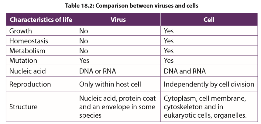 Self-assessment 18.3
1. What are the parts of a virus?
2. Describe the two ways by which viruses cause infection.
3. Distinguish between Bacteriophage and a prophage.
4. What is meant by retrovirus?
5. What are the strengths and weaknesses of the tobacco mosaic virus hypothesis?
6. Which characteristic feature is common to all viruses?
7. How is a capsid protein important to the functioning of a virus?
8. What is the best way to protect humans against most viral diseases?18.4 Moulds
Activity 18.4
Using text books or computer aided materials to describe the life cycle of bread mould.Moulds pervade our world, living wherever moisture is present. Some are of great benefit to humans, providing antibiotics, acting as decomposers so that nutrients can be recycled, or taking part industrial processes. Other moulds cause diseases which lead to serious damage.Moulds have cells arranged in long thread-like filaments, the hyphae, that form a mass called Mycelium. Moulds are usually considered as fungi, but mould may also be formed by filamentous bacteria, slime moulds, and water moulds. Therefore, there are two main types of moulds: fungal moulds and non-fungal moulds.18.4.1 Fungal mouldsAll fungi that produce mycelia can be called moulds, but the term is usually used for an organism in which the mycelium forms the main body of the fungus. In the black bread mould Rhizopus and the pin mould Mucor, the mycelium consists of a tangled mass of hyphae with many nuclei. These hyphae are called coenocytic because the fungal tissue is not separated by cell walls.Fungal hyphae have an outer cell wall made of chitin and inner lumen which contains the cytoplasm and organelles. A cell surface membrane surrounds the cytoplasm and sticks tightly to the cell wall.Rhizopus and Mucor are Saprotrophic, obtaining their nutrients from dead organic material. Rhizopus nigricans and Mucor mucedo can live on bread but some species of Rhizopus feed on living plants, and Mucor commonly grows on rotting fruits and vegetables, in the soil or on dung.Rhizopus and Mucor secrete hydrolytic enzymes onto their food source and digest the food outside the organism and then absorb the soluble digestion products and assimilate them.a. Life cycle of Rhizopus and Mucor.Rhizopus and Mucor belong to the fungal phylum Zygomycota. The phylum got its name because its members produce two kinds of spores: Sexual zygospores as well asexual sporangiospores.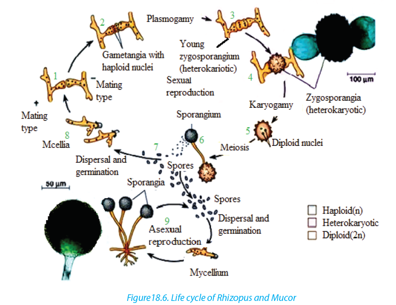 The asexual sporangiospores formed by mitosis, develop insporangium at the tip of hyphae. When sporangium busts, the spores are released.In most species of Mucor, the sporangium dissolves then water enters the spore mass, and the spores are dispersed by the raindrop or are transported by the insects. In most Rhizopus species, the sporangium wall fractures and dry spores are released by the wind.The sexual reproduction involves conjugation. Usually the hyphae from mycelia of different mating types meet and interconnect via outgrowths. The interconnecting walls break down and their cytoplasm containing haploid nuclei mix, then the diploid zygote formed by the fusion of two nuclei develops a thick, rough, black coat and becomes a dormant zygospores. Meiosis probably occurs at the time of germination; the zygospore cracks open to liberate several haploids spores which can give rise to asexual sporangia and mycelia of either mating strain.b. Use of moulds–– They are used to make the human foods. For example, Mucor is used with soya
beans to make a cheese called sufu, in eastern Asia.In Indonesia, R. oligosporus
and R. oryzae are used to produce a food called tempeh from boiled skinless
soya beans.
–– The fungal moulds belonging to the Zygomycota are used to make anaesthetics,
birth control pills, meat tenderisers, and the yellow colouring agents used in
margarines and butter substitutes.