General
- Y1: Integrated Science ECLPE SB File Uploaded 17/08/22, 09:41
- Y1: Integrated Science ECLPE TG File Uploaded 17/08/22, 09:44
UNIT 5: CELL STRUCTURE
Key unit competence
Distinguish between the types of microscopy and their principle uses and
relate the structure of cell to their functions.
Introductory ActivityCarefully, analyze the following diagram and the related questions
This is one of materials used in Biology.
a) Have you ever seen and manipulated it?
b) Observe the parts of the material, discuss and group them into thefollowing:
i. Suporting parts
ii. Magnifying parts
iii. Adjusting parts
iv. Lighting parts
c) Point out scientific activities that require the use of this material in
biology
5.1. Cell theory and microscopes
Activity 5.1
Using your knowledge acquired from ordinary level, textbooks and internet
search
Explain:
a) The meaning of cell theory
b) The functions of the components of the compound light microscope
5.1.1. Cell theory
Cytology is the study of the structure and function of cells. A Cell is the
basic unit of life surrounded by a cell membrane and containing organelles.
All living organisms are made of cells and nothing less than a cell can truly
be said to be alive. The word cell comes from the Latin cellula, meaning
a small room or cubicle, and was first used by Robert Hooke in his book
Micrographia, published in 1665. Hooke was observing slides of cork taken
from the bark of an Oak tree under the compound microscope. He decided
that the slides were made up of a lot of many small chambers that he called
cells that range in size are from 1μm – 1mm.
Living organisms are classified into unicellular organisms made by only one
cell, such as bacteria, whereas animals and plants are composed of many
millions of cells built into tissues and organs. They are called multicellular
organisms. In a multicellular organism, cells divide and thereafter they
undergo differentiation or specialisation for specific functions.
In biology, the historic scientific theory of cells is called cell theory. The cell
theory states that all living organisms are made up of cells, and cells are the
basic unit of structure function in all living organisms. The main principles
of cell theory is that all known living organisms are made up of one or more cells, all cells come from pre-existing cells by division and cells contain the hereditary information that is passed from cell to cell during cell division.
5.1.2. Microscopes: Compound-light microscope and Electron microscopes
Microscopy is the technical field of using microscopes to view objects and
areas of objects that cannot be seen with the naked eye. The microscope
was created by Zacharias Janssen in the late 16th century.
Prior to the invention of the microscope, the details of objects on slides were
limited. Single microscopes were similar to using a magnifying glass such as
hand lens. The invention of the light microscope in the 17th century by Antony
van Leeuwenhoek made cells visible for the first time and, for hundreds of
years afterwards. Electron microscope invented in the 1930s enables the
researchers to see very small organelles which cannot be seen by using
light microscopes. The purpose of the use of a microscope is to magnify
small objects such as cells or to magnify the fine details of a larger object in
order to examine minute specimens that cannot be seen by the naked eye.
a) Compound Light Microscope
The optical microscope, often referred to as light microscope is a type of
microscope which uses visible light and a system of lenses to magnifyimages of small samples.
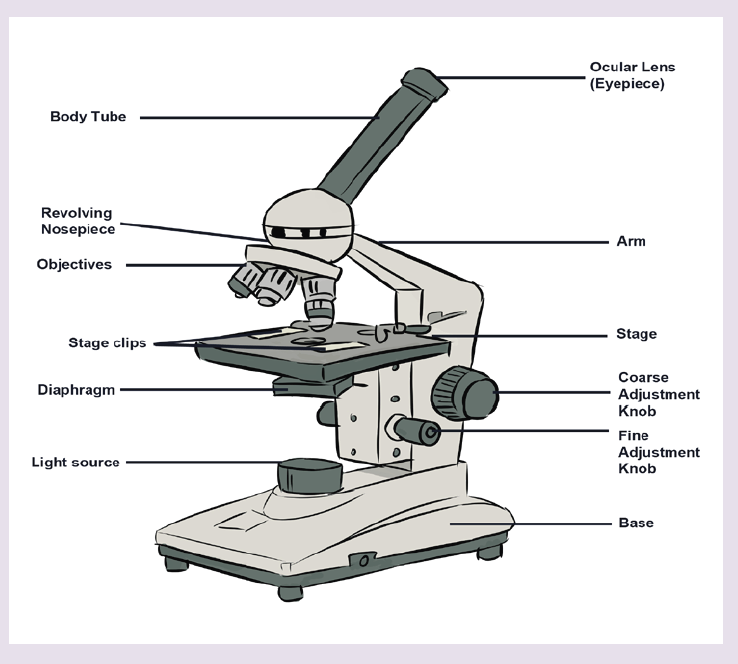
The parts of light microscope and their roles:
– Base: supports and stabilizes the microscope on the table or any other
working place
– Light source: It is made by lamp or mirror which provides light for
viewing the slide.
– Stage: is a platform used to hold the specimen in position during
observation.
– Stage clips: are pliers used to fix and hold tightly the slide on stage.
– Arm: supports the body tube of microscope
– Body tube: maintains the proper distance between the objective and
ocular lenses
– Arm: used for holding when carrying the microscope and it holds the
body tube which bears the lenses.
– Coarse focus adjustment: moves stage up and down a large amount
for coarse focus
– Fine focus adjustment: moves stage up and down a tiny amount for
fine focus
– Objective lenses: focuses and magnifies light coming through the
slide
– Revolving nosepiece: rotates to allow use of different power objectives- Slide: is a transparent pane on which a specimen is placed.
– Eye piece/ocular lens: magnifies image produced by objective lens
– Condenser: It will gather the light from the illuminator and focus it on
the specimen lying on the stage. The function of the condenser is to
focus the light rays from the light source onto the specimen.
– Iris diaphragm lever: This allows the amount of light passing through
the condenser to be regulated to see the object.
All parts of a microscope work together to magnify a specimen to be observed.
Light from the source is focused on the specimen by the condenser lens.
It then enters the objective lens, where it is magnified to produce a real
image. The real image is magnified again by the ocular lens to produce a
virtual image that is seen by the eye.
Advantages and limitations of the light microscope
Magnification: Most light microscopes can magnify a specimens up to a
maximum of X1500.
Resolution: It is the degree at which it is possible to distinguish clearly
between two objects that are very close together. It is the smallest distance
apart that two separate objects can be seen clearly as two objects. The
resolution for the:
– Human eye is 100μ.
– Light microscope is 200 nm
– Electron microscope is 0.20 nm.
The maximum resolution of the light microscope is 200 nm. This means
that if two objects are closer together than 200 nm, they will be seen as
one object. The light microscopes are used widely in education, laboratory
analysis and research. But because they do not have high resolution, they
cannot give detailed information about internal cell structure.
Specimens: a wide range of specimens are viewed using the light microscope.
These include: unicellular organisms, small sections from large plants and
animals, and smear preparation of blood or cheek cells.
Preparation of specimens for the light microscope
A lot of biological materials are not coloured, so you cannot see details. Also,
some material distorts when you try to cut it into thin sections. Preparation of
slides to overcome these problems involves the following steps:
a) Staining: Coloured stains are chemicals that bind on or in the
specimens. This allows the specimens to be seen. Some stains bind
to specific cell structures. For example, acetic orcein stains DNAdark red, while gentian violet stains bacterial cell walls.
b) Sectioning: Specimens are embedded in wax, where thin sections
are then cut without distorting the structure of the specimen. This is
particularly useful for making sections of soft tissue, such as brain.
Safety measures might be taken. Make sure that hands are washed
with soap and warm water after theexperiment. Use a disinfectant
to wipe down all surfaces where bacteria mayhave been deposited
for example. Be sure that some substances and animalsmight be
harmful to the life.
Measuring cells
Cells and organelles can be measured with a microscope by means of an
eyepiece called graticule. This is a transparent scale, usually having 100
divisions (Figure 5.2, A). The eyepiece graticule is placed in the microscope
eyepiece so that it can be seen at the same time as the object to be measured
(Figure 5.2, B). At this figure (Figure 5.2, B), the cell lies between 40 and 60on the scale, so that it measures 20 eyepiece units in diameter (60 – 40 =20).
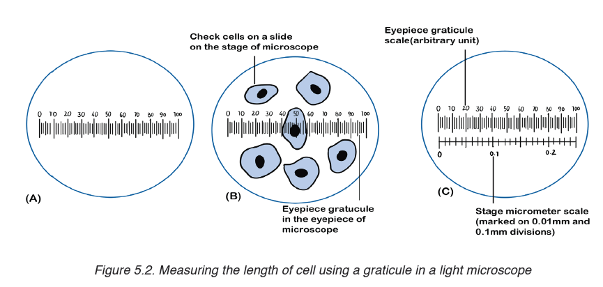
Note that it is not possible to know the actual size of the eyepiece units
until the eyepiece graticule scale is calibrated. To calibrate the eyepiece
graticule scale, a miniature transparent ruler called a stage micrometre scale
is placed on the microscope stage and is brought into focus. This scale may
be fixed onto a glass slide or printed on a transparent film. It commonly has
subdivisions of 0.1 and 0.01 mm. The images of the two scales can then be
superimposed (Figure 5.2, C). If in the eyepiece graticule, 100 units measure
0.25 mm, the value of each eyepiece unit equals
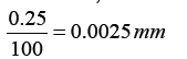
By converting mm to μm, the value of eyepiece equal
The diameter of the cell shown superimposed (Figure 5.2, B) measures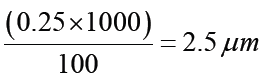
20 eyepiece units. Its actual diameter equals 20 × 2.5 μm = 50 μm. This diameter is greater than that of many human cells because the cell is a flattened epithelial cell.
There is a relationship between the actual size, magnification and image size, where:
Actual size = image size / magnification.
Magnification = image size / actual size.
Image size = Magnification x actual size.
c) Electron microscope
An electron microscopes use a beam of accelerated electrons as a source
of illumination. Electron beams have a much smaller wave length of 0.004
nm, 100 000 times shorter than light wavelength, and therefore have greater
resolving powers and can produce higher effective magnifications than light
microscopes. Electron microscopes are used to study the details of internal
structures (the ultrastructures) of cells. Most modern TEMs can distinguish
objects as small as 0.2nm. This means that they can produce clear images
magnified up to 500,000 times greater than that of the human eye.
There are two types of electron microscopes:
– Transmission electron microscope (TEM).– Scanning electron microscope (SEM)
a) Transmission electron microscope (TEM)
– The electron beam passes through a very thin prepared sample.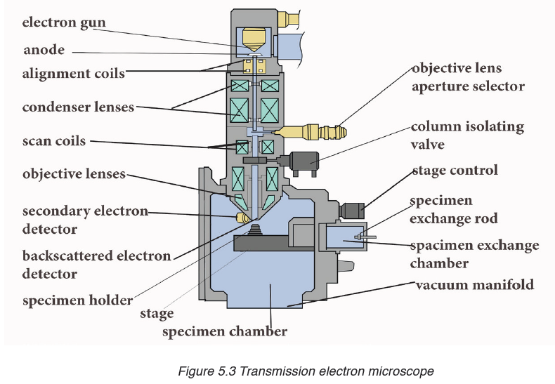
– Electrons pass through the denser parts of the sample less easily, so
giving some contrast.
– The final image produced is two-dimensional.– The magnification possible with a TEM is X500 000.
b) Scanning electron microscope (SEM)
– The electron beam is directed onto a sample. Electrons do not pass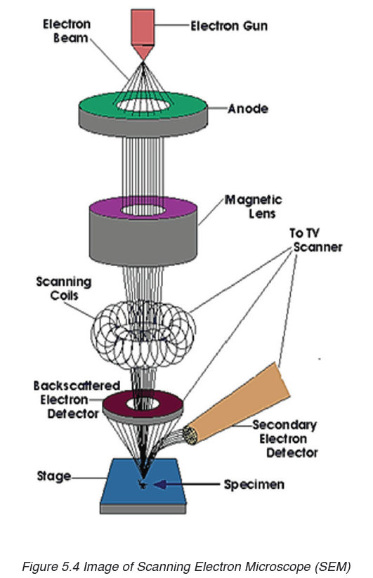
through the specimen.
– They are bounced off the sample.
– The final image produced is a three-dimensional.– The magnification possible with an SEM is about X100 000.
Advantages and disadvantages of the electron microscope
a) Advantages of electron microscope
Light microscope has a higher resolution and are therefore able of a higher
magnification estimated up to 2000 X more than in the light microscope.
Electron microscopes therefore allow for the visualization of too small
structures, such as cell organelles that would normally be not visible by
optical microscopy. The SEM produces a 3D images that can reveal the
detail of cellular and tissue arrangement. This is not possible by using light
microscopes.
b) Disadvantages of electron microscope
Despite the advantages, electron microscope presents a number of
disadvantages and limitations.
– These type of microscope are extremely expensive and the maintenance
costs are high.
– Samples must be completely dry so that it is impossible to observe
living specimens and moving specimens (they are dead).
– It is not possible to observe colors because electrons do not possess a
color. The image is only black-white images/ grey images.
– The energy of the electron beam is very high, the sample is therefore
exposed to high radiation, and therefore not able to live.
– The space requirements are high, so that they may need a whole room.
– Electron beams are deflected by molecules in air, so samples have to
be placed in a vacuum.
Application activity 5.1
1. What is the importance of a light microscope?
2. How can you apply microscope technique rules?
3. Discuss the advantages and disadvantages (limitations) of an
electron microscope.
4. Discuss the advantages and disadvantages of the types of electron
microscopes in medicine and biology research.
5. Make a comparative study between light and electron microscopefocussing on the advantages of each type of microscope.
5.2. Eukaryotic and prokaryotic cell
Activity 5.2
Under microscope, observe mounted slides of bacteria, and plant cells.
Draw and label the parts that are common in both plant and bacterial
specimens
Eukaryotic cells contain membrane-bound organelles, including a true
nucleus enclosed in a nuclear envelope. They include cells of: plants, animals,fungi and protoctista.
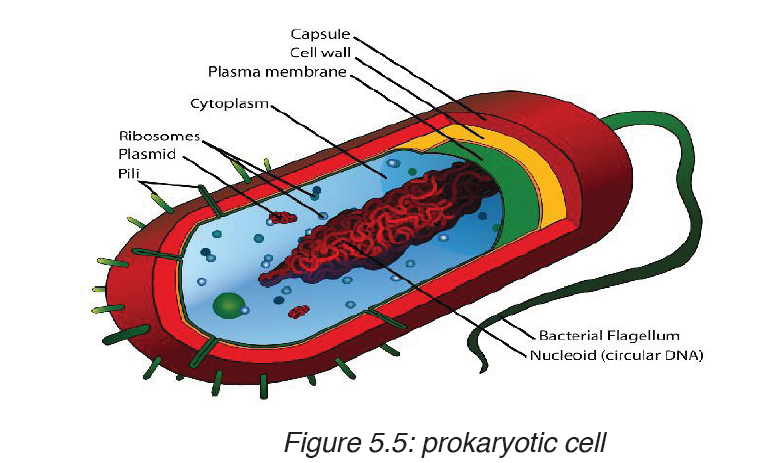
Prokaryotes are organisms having cells with no true nuclear envelope.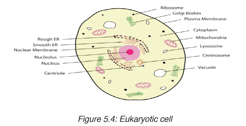
Prokaryotic cells do not contain a nucleus or any other membrane-bound
organelle. Prokaryotes include bacteria and blue-green algae. They makeup the monera kingdom.
Comparison between prokaryotic and eukaryotic cells
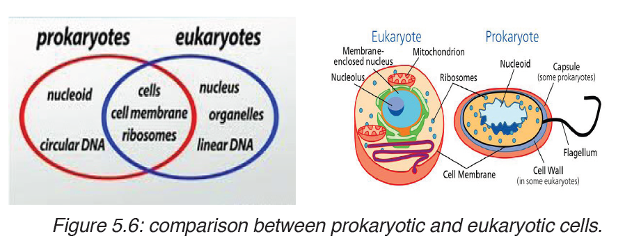
Table 5.3. Comparison between prokaryotic and eukaryotic cells
Application activity 5.2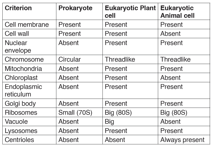
1. Define a prokaryote and eukaryote.
2. In a form of a table differentiate prokaryotic cells from eukaryoticcells.
5.3. Description of plant and animal cell
Activity 5.3
Observe two electron photographs, one containing a plant cell another
an animal cell.Record a description of their features, such as shape andinternal parts.
5.3.1. Structure of animal and plant cells
When viewed under light microscope, the most obvious features observed
are the very large nucleus and a clear cytoplasm surrounded by a cell
membrane. However, under electron microscope, it is possible to identify arange of organelles in plant and animal cells.
Ultrastructure of a plant cell contains different parts like: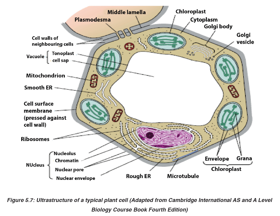
Cell wall, cell membrane, cytoplasm with organelles. Organelles found in the
cytoplasm of a plant cell include: chloroplast, mitochondria, Golgi apparatus,
endoplasmic reticulum, ribosomes, big central vacuole, and the nucleus
which contains chromosomes. The plant cell also has a regular shape, witha relatively bigger size than animal cell.
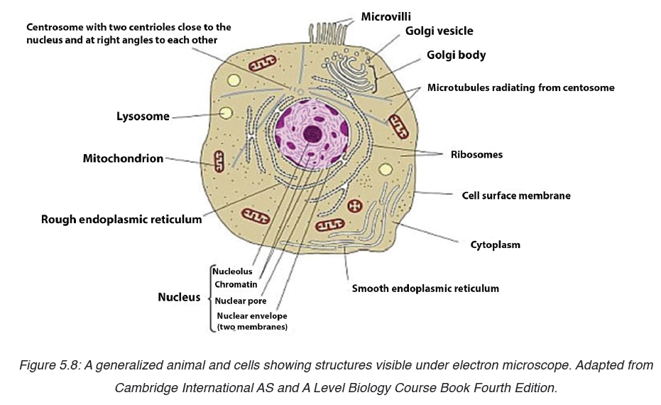 Ultrastructure of an animal cell contains different parts like:The cell nucleus contains nearly all the cell’s DNA with the coded instructions
Cell membrane, cytoplasm with organelles. Organelles found in the cytoplasm
of an animal cell include: mitochondria, Golgi apparatus, endoplasmic
reticulum, centrioles, ribosomes, small or absent vacuole, and the nucleus
which contains chromosomes. The animal cell also has irregular shape, witha relatively smaller size than a plant cell.
5.3.2. Functions of cell organelles
The previous sections described the structures of plant and animal cells.
This unit will explain the function of each part of both animal cell and plant cell.
1. Organelles surrounded by membranes
a) Nucleus
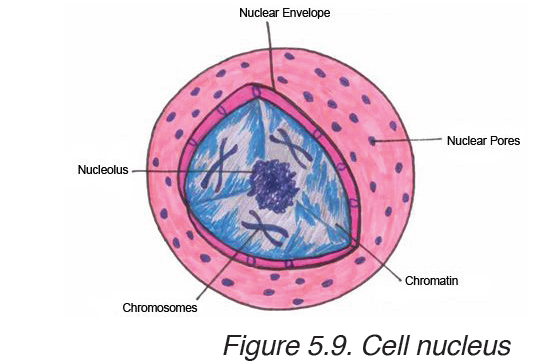
for making proteins and other important molecules. The nucleus is surrounded
by a double nuclear envelope, which allow materials to move into and out
of the nucleus through nuclear pores. The granules found in the nucleus
are called chromatin which consist of DNA bound to protein. When a cell
divides, the chromatin condenses into chromosomes containing the genetic
information that is passed from parents to their offspring. The nucleus
contains a dense spherical structure called nucleolus in which assembly ofribosomes begins. The nucleus controls all activities of the cell.
The ER consists of a series of flattened membrane-bound sacs calledb) Endoplasmic reticulum (ER)
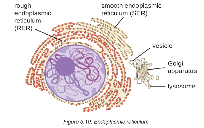
cisternae. The rough ER is surrounded with ribosomes. The rough ER
transports proteins made on attached ribosomes. The smooth ER does not
have ribosomes, and it involves in making lipids that the cell needs.c) Golgi apparatus
The Golgi apparatus is a stack of membrane-bound, flattened sacs, which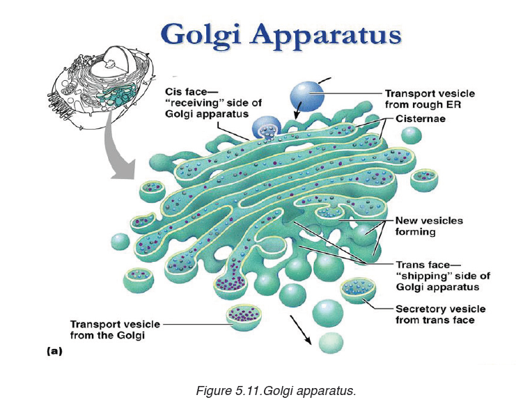
receives proteins from the ER and modify them. It may add sugar molecules
to them. The Golgi apparatus then packages the modified substances into
vesicles that can be transported to their final destinations throughout the cellor outside of the cell.
d) Mitochondria
Mitochondrion have two membranes separated by a fluid-filled space. The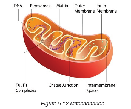
inner membrane is highly folded to form cristae. The central part of the
mitochondrion is called matrix. The mitochondria are the site where the
process of cell respiration takes place to produce Adenosine triphosphate
(ATP), a universal energy carrier to be used in cell metabolism.e) Chloroplasts
Chloroplasts are the site of photosynthesis in plant cells. These are found
in plant cells and in cells of some protoctists. They also have two membranes
separated by a fluid-filled space. The inner membrane is continuous, with
thylakoids. A stalk of thylakoids is called a granum (plural: grana).Chlorophyll molecules are present on the thylakoid membranes.
f) Lysosomes
These are spherical sacs surrounded by a single membrane. They contain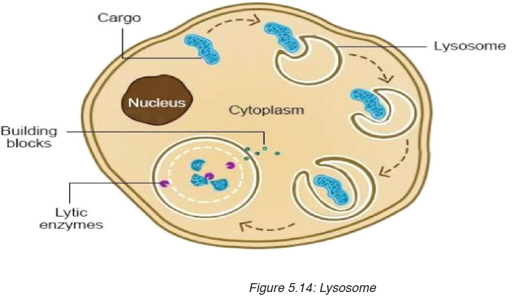
powerful digestive enzymes. Their role is to break down materials such
as white blood cells, and destroy invalid microorganisms. In acrosome,
lysosomes help the sperm to penetrate the egg by breaking down the
material surrounding the egg.Organelles without surrounding membranes
a) Ribosomes
Ribosomes are the site of protein synthesis in the cell. Some are in cytoplasm;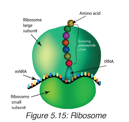
others are bound to ER. Ribosomes consist of two major components (two subunits): the small ribosomal subunit, which reads the RNA, and the large subunit, which joins amino acids to form a polypeptide chain. Each subunit
is composed of one or more ribosomal RNA (rRNA) molecules and a variety of ribosomal proteins (r-protein). Ribosomes act as an assembly line where coded information (mRNA) from the nucleus is used to assemble proteins
from amino acids. Cells that are more active in protein synthesis are often packed with ribosomes.b) Centrioles
Centrioles are small tubes of protein fibres (microtubules), which are involved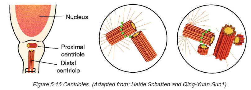
in animal cell division. They form fibres, known as spindle, which movechromosomes during nuclear division.
c) Vacuole
A vacuole is a saclike structure that is used to store materials such as water,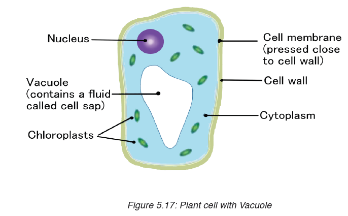
salts, proteins, and carbohydrates. In many plant cells there is a single, and
large central vacuole filled with liquid. The pressure of central vacuole in this
cells makes it possible for plants to support heavy structures such as leaves
and flowers. Some animals and some unicellular organisms contain
contractile vacuoles which contract rhythmically to pump excess water outof the cell.
d) Cytoskeleton
Cytoskeleton is a network of protein filaments that helps the cell to maintain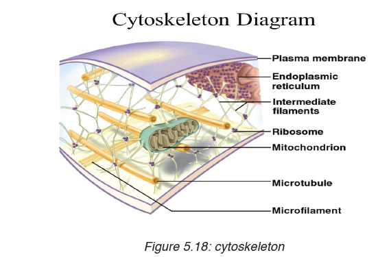
its shape. It is also involved in movement. The main components of
cytoskeleton are microfilaments made of a protein called actin, microtubules
made of a protein called tubulins, and intermediate filaments.
Did you realize that a cell has many organelles with different functions? But
all are important and work together for the survival of the cell. Likewise, in
human society, we can work together in peace and harmony despite of the
difference of our abilities, disabilities or physical appearance.
The structure of the cell membrane is based on fluid mosaic model. The
term fluid mosaic is used to describe the molecular arrangements in
membranes. The main features of the fluid mosaic model are:
– A bilayer of phospholipid molecules forming the basic structure.
– Many protein molecules floating in the phospholipid bilayer. Some are
free, others are bound to other components or to structures within the
cell.
– Some extrinsic proteins are partially embedded in the bilayer on the
inside or the outside face while other intrinsic proteins are completelyspanning the bilayer.
The basic structure of phospholipids has two parts: hydrophilic part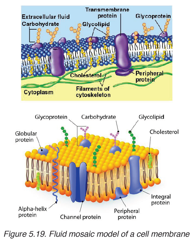
which means water loving and which consists of the phosphate head,and
hydrophobic part which means water hating and which consist of fatty acids.
If phospholipid molecules are completely surrounded by water, a bilayer can
form phosphate heads on each side of the bilayer stick into water, while the
hydrophobic fatty acid tails point towards each other.
Types of protein found in the cell membrane
Various types of proteins are found in the cell membrane. They include:
– Carrier proteins: They fix or attach molecules and facilitate them to
cross through the cell membrane by active transport.
– Channel proteins: they act as pores by pumping substances and
allow facilitated diffusion.
– Receptors: They act as receptors of enzymes and neurotransmitters
– Glycoproteins: They act as receptor proteins which recognize the– Integrated proteins: They define the shape of the cellsubstance to pass through the membrane
– Immune proteins (antigens): found in the membrane on the red blood
cell, they recognize the antibodies.
5.3.3. Roles of different components of cell membrane
a) Cholesterol
– Gives the membranes of some eukaryotic cells the mechanical stability.
– It fits between fatty acid tails and helps make the barrier more complete,
so substances like water molecules and ions cannot pass easily and
directly through the membrane.
b) Channel proteins
– Allow the movement of some substances across the membrane.
– Large molecules like glucose enter and leave the cell using these
protein channels.
c) Carrier proteins
– Actively move some substances across the cell membrane. For
example, magnesium and other mineral ions are actively pumped into
the roots hair cells from the surrounding soil.
– Nitrate ions are actively transported into xylem vessels of plants
d) Receptor sites
– Allow hormones to bind with the cell so that a cell response can be
carried out.
– Glycoproteins and glycolipids may be involved in cells signaling that
they are self to allow recognition by the immune system.
– Some hormone receptors are glycoprotein and some are glycolipid.
e) Enzymes and coenzymes
– Some reactions including metabolic processes in photosynthesis take
place in membranes of chloroplasts.
– Some stages of respiration take place in membranes of mitochondria,
where Enzymes and coenzymes may be bound to these membranes.
– The more membrane there is, the more enzymes and coenzymes it
can hold and this helps to explain why mitochondrial inner membranes
are folded to form cristae, and why chloroplasts contain many stacksof membranes called thylakoids.
Properties of the cell membrane
– It is mainly made of lipids, proteins and carbohydrates.
– It is semi-permeable or partially permeable and allow some substances
to pass through but prevents others to cross depending on their size,
charges and polarity.
– It is positively charged outside and negatively charged inside and has
a hydrophilic pole and a hydrophobic pole
– It is a bilayer, sensitive, flexible, has inorganic ions and its proteins
and lipids may be mobile and contains different types of enzymes and
coenzymes.
– It is perforated of pores and recognizes chemicals messengers
including hormones and neurotransmitters.
Functions of a cell membrane
– The cell membrane acts as a selective barrier at the surface of the cell,
and controls the exchange between the cell and its environment.
– The membrane proteins act as pores where chemicals pass through.
– Glycocalyx including glycoprotein and glycolipid are involved in the
cell protection, the process by which cell adhesions are brought about
and also in the uptake and entry of selected substances.
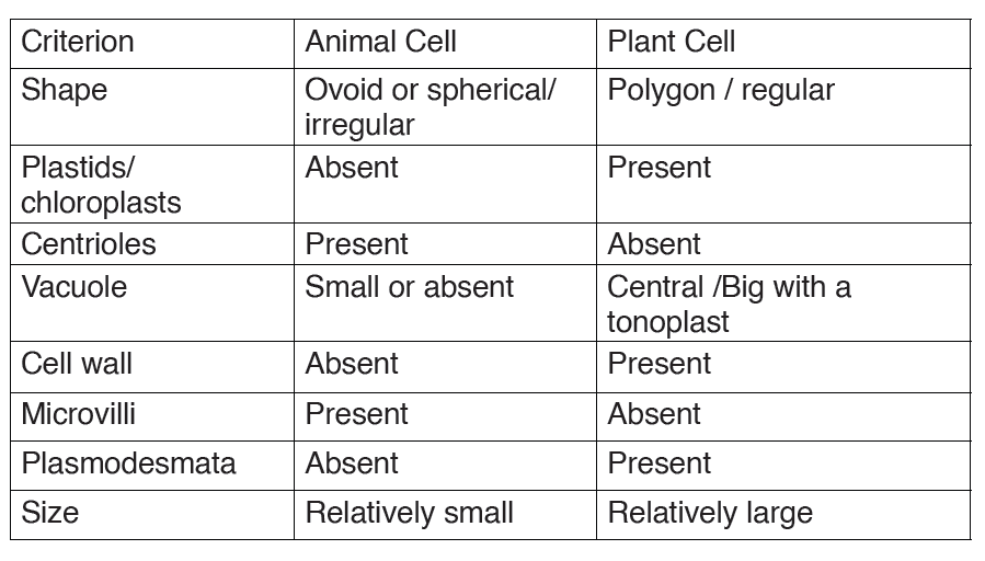
5.3.4. Comparison between animal and plant cell
By referring to the diagrams which show the structure of animal cell and
plant cell, similarities and differences between animal cell and plant cell can
be identified.
a) Similarities between animal cell and plant cell
– Both animal and plant cells have a cell membrane, a cytoplasm and a
nucleus.
– Both animal and plant cells have a true nucleus bounded by an
envelope.
– Both animal and plant cells have mitochondria, Golgi apparatus,
Reticulum endoplasmic, lysosome, big ribosomes (80S), peroxisome,
microtubules.
– The protoplasm is enveloped by a bounding cell membrane called
plasmalemma.
– The protoplasm is composed of a dense round structure called nucleus
which is surrounded by a less dense jelly-like cytoplasm.
– The cytoplasm contains numerous organelles such as mitochondria,
Golgi bodies, secretory vacuoles, endoplasmic reticulum.
– Mitochondria appear as very small darkly staining, rod-like structures.
– Golgi bodies are semi-transparent irregular, and membrane bound
structures.
– Vacuoles contain secretions, food- particles, or decomposing organic
substances.
– Chemically, both plant and animal cells are made up of water (80-90%),
proteins (7-13%), lipids (1-2%), carbohydrates (1-1.5%) and inorganic
salts.
– The cytoplasmic organelles are suspended in a semi-fluid jelly matrix
called cytosol.
b) Difference between animal and plant cellsTable 5.4: comparison between animal and plant cell
Application activity 5.3
1. Describe the structure of:
a) Animal cell
b) Plant cell
2. The diagram below shows the structures which would be visible ina plant cell examined under an electron microscope.
a) Identify the parts labelled in this plant cell and: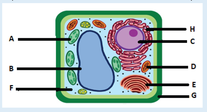
b) State one function for A, B, C, E,D,G, F, and H
c) Explain two functions of the cytoskeleton?
3. What structures do both animal and plant cells have in common?
4. Answer by true or false:
a) All organelles of a cell are well seen through a compound light
microscope.
b) Chloroplasts are found in both animal and plant cells.
c) Mitochondria are found only in animal cells.
5. With your pencil, draw and show the full ultrastructure of bothanimal and plant cells.
5.4 Specialized cells
Activity 5.4
By analyzing the figure, identify the cells on the figure and explain the importanceof those cells
5.4.1: Specialized animal cells and their functions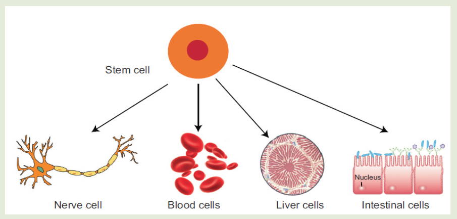
Differentiation refers to the changes occurring in cells of a multicellular
organism so that each different type of cell becomes specialized to
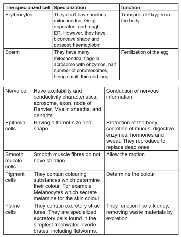
perform a specific function.a) Red blood cells
All blood cells are produced from undifferentiated stem cells in the bone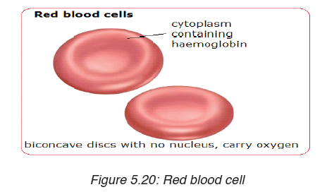
marrow but the cells destined to become erythrocytes (red blood cells)
lose their nucleus, mitochondria, Golgi apparatus and rough endoplasmic
reticulum. They are packed full of the protein called haemoglobin. The shape
of this cells change so that they become biconcave discs, and they are thenable to transport Oxygen in the body.
b) Sperm cell
Sperm cells are specialized to fertilize the egg. Its specialization involves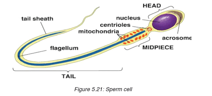
many changes in shape and organelles content.
By shape: the sperm cells are very small, long and thin to help them to move
easily, and they have a flagellum which helps them to move up the uterine
tract towards the egg.
By organelles content: sperm cells contain numerous mitochondria
which generate much energy for their movement. Their acrosome releases
specialized lysosomes contains many enzymes on the outside of the egg.
These enzymes lyse the wall of the egg, and facilitate the sperm nucleus to
penetrate easily. In content, the sperm cell nucleus contains the half number
of chromosomes of the germ cell in order to fulfil its role as a gamete offertilizing the egg.
c) Nerve cells
Nerve cells also known as a neuron are specialized cells to carry nervous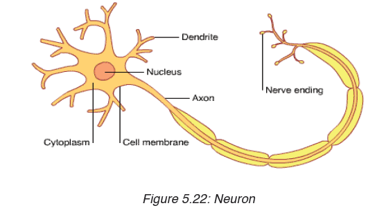
information in the body. It is an electrically excitable cell that receives,
processes, and transmits information through electrical and chemical
signals. These signals between neurons occur via specialized connections
called synapses. Specialized animal cells have different functions. Some ofthem are summarized in the following table.
Table 5.5: Specialized animal cells and their functions.
5.5.2: Specialized plant cells and their functions
a) Root hair cells
The root hair cells have hair-like projection from their surface out into the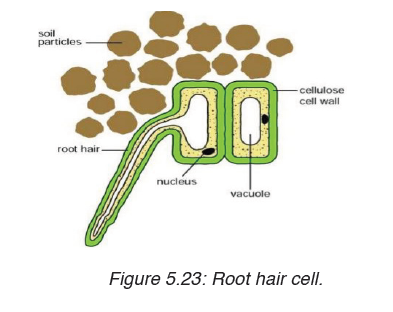
soil. This increase the surface area of root available to absorb water and
minerals from the soil.Root hairs are tip-growing cells that originate from
epidermal cells called trichoblasts. Their role is to extend the surface area ofthe root to facilitate absorption of mineral nutrients and water.
b) Palisade cells
Palisade cells are plant cells located in leaves, right below the epidermis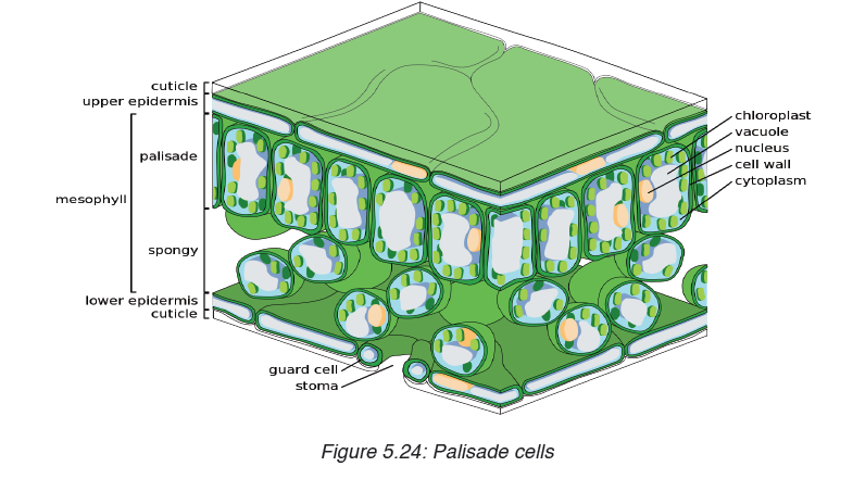
and cuticle. They are vertically elongated, a different shape from the spongy
mesophyll cells beneath them in the leaf. Their big number of chloroplasts
allow them to absorb a major portion of the light energy used by the leaf inthe process of photosynthesis.
c) Parenchyma cells:
Parenchyma cells are alive at maturity. They function in storage,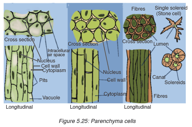
photosynthesis, and as the bulk of ground and vascular tissues. Palisade
parenchyma cells are elongated cells located in many leaves just below
the epidermis. Parenchyma is composed of relatively simple, undifferentiated
parenchyma cells. In most plants, metabolic activity such as respiration,
digestion, and photosynthesis occurs in these cells because they retain
their protoplasts(the cytoplasm, nucleus, and cell organelles) that carry out
these functions. Parenchyma cells are capable of cell division, even afterthey have differentiated into the mature form.
d) Guard cells
Guard cells are cells surrounding each stoma. They help to regulate the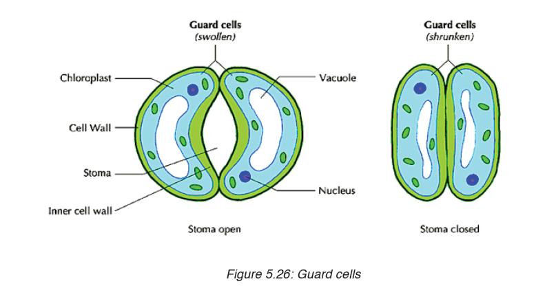
rate of transpiration by opening and closing the stomata. Guard cells are
specialized cells in the epidermis of leaves, stems and other organs that
are used to control gas exchange. They are produced in pairs with a gap
between them that forms a stomatal pore.
Application activity 5.4
1. Explain why differentiation to produce erythrocytes involves a
change in shape.
2. Red blood cells cannot divide as they have no nucleus. State two
other biological processes that red blood cells cannot carry out.
3. Discuss the advantages of cell specialization for living things
SKILLS LAB
After finishing the studies, assuming that the understood the unit 5, student teachers
will be able to use microscope to investigate the presence of
microorganisms that can cause diseases in food and beverages. This will helpin prevention of diseases
End unit assessment 5
Section A. Multiple choice questions
1. Which organelle converts the chemical energy in food into a form that
cells can use?
a) Chromosome
b) Chloroplast
c) Nucleus
d) Mitochondrion
2. The cell membranes are constructed mainly of:
a) Carbohydrate gates
b) Protein pumps
c) Lipid bilayer
d) Free-moving proteins
3. In many cells, the structure that controls the cell’s activities is the:
a) Nucleus
b) Nucleolus
c) Cell membrane
d) Organelle
4. Despite differences in size and shape, all cells have cytoplasm and a
a) Cell wall
b) Cell membrane
c) Mitochondria
d) Nucleus
5. If a cell of an organism contains a nucleus, the organism is a (an)
a) Plant
b) Eukaryote
c) Animald) Prokaryote
Section B: Questions with short answers
1. How does a cell membrane differ from a cell wall?
2. What is meant by the fluid mosaic model of the cell membrane?
3. State the properties of the cell membrane.
4. Discuss at least 4 types of the proteins in the cell membrane and their roles.
5. Explain why muscle cells contain a lot of mitochondria, whereas most fat storage cells do not.
6. What kind of information is contained in chromosomes?
7. You examine an unknown cell under the microscope and discover
that the cell contains chloroplasts. What type of organism could you decide that the cell came from?
Section C: Essay questions
1. Describe the structure and function of the cell membrane and cell wall.
2. Describe the basic structure of the cell membrane.
3. Explain two common characteristics of chloroplasts and mitochondria. Consider both function and membrane structure.4. The diagram below shows the structure of a liver cell as seen using an electron microscope.
a) Name the parts labelled A, B, C and D.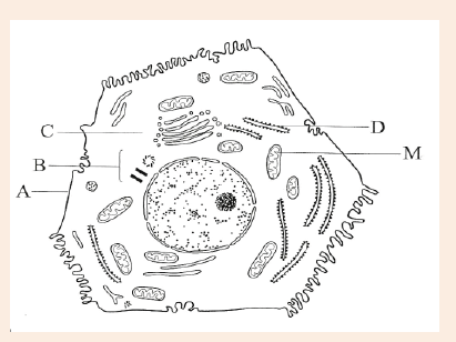
b) The magnification of the diagram above is 12 000. Calculate the
actual length of the mitochondrion labelled M, giving your answer
in μm. Show your working.
c) Explain the advantage to have a division of labour between differentcells in the body.