General
- S6: Biology SB File Uploaded 3/08/22, 12:46
- S6: Biology TG File Uploaded 3/08/22, 12:49
UNIT 7: EXCRETION AND OSMOREGULATION
Key Unit Competence
Explain the principles of excretion and osmoregulationLearning Objectives
By the end of this unit, the student should be able to:
–– Describe the structure and role of excretory organs in mammals.
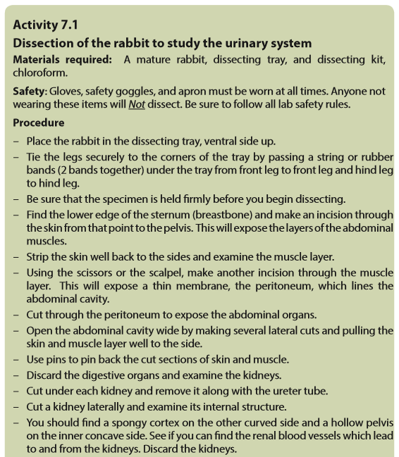
–– Dissect, display, draw and label the urinary system of a toad, rat/rabbit etc.
–– Describe the detailed structure of the nephron with its associated blood vessels.
–– Describe and outline the ornithine cycle and its role in the conversion ofammonia to urea.
–– Describe how the process of ultrafiltration and selective reabsorption are involved in the formation of urine in the nephron.
–– Describe the use of dialysis in kidney machines.
–– Describe how kidney transplants are performed.
–– Describe the role of hypothalamus, posterior pituitary, ADH and collecting ducts in osmoregulation.
–– Explain the principles of osmoregulation in organisms living in marine,
freshwater and terrestrial habitats.
–– Explain dialysis in terms of salt balance, the maintenance of glucose concentration and the removal of urea.
–– Explain why plants do not have specialised excretory organs.
–– State the excretory products of plants and how they are eliminated.
–– Dissect, display, draw and label the urinary system of a toad, rat/rabbit etc.
–– Interpret the ornithine cycle diagram with reference to urine formation.
–– Relate adaptations of different organisms to their habitat in terms of osmoregulation.
–– Compare the advantages and disadvantages of kidney transplants with dialysis machines.
–– Support the use of dialysis machine or kidney transplants in solving problems associated with kidney failure.
–– Appreciate the adaptation of organisms to different habitats in relation to osmoregulation.
7.1 Structure and functions of excretory organs in mammals

Excretion the removal of toxic waste products of metabolism from the body. The term is generally taken to mean nitrogenous wastes such as; urea, ammonia and uric acid but other materials like carbon dioxide and the bile pigments are also waste products of metabolism, and their removal is as much a part of excretion as the elimination of urea.
Excretion is an essential process in all forms of life. When cells metabolize or break down nutrients, waste products are produced. For example, when cells metabolize amino acids, nitrogen wastes such as ammonia are produced. Ammonia is a toxic substance and must be removed from the blood and excreted from the body.
Although the kidneys are the main organs of excretion of wastes from the blood, several other organs are also involved in the excretion, including the; liver, skin, and lungs.
–– The large intestine eliminates waste products from the bile synthesis.
–– The liver breaks down excess amino acids in the blood to form ammonia, and then converts the ammonia to urea, a less toxic substance. The liver also breaks down other toxic substances in the blood, including alcohol and drugs.
–– The skin eliminates water and salts in sweat.
–– The lungs exhale water vapour and carbon dioxide.The importance of excreting wastes
i. To maintain life processes, the body must eliminate waste products, many of these which can be harmful. The lungs eliminate carbon dioxide, one of the products of cellular respiration. The large intestine removes toxic wastes from the digestive system.
ii. The liver transforms ingested toxins, such as alcohol and heavy metals, into soluble compounds that can be eliminated by the kidneys.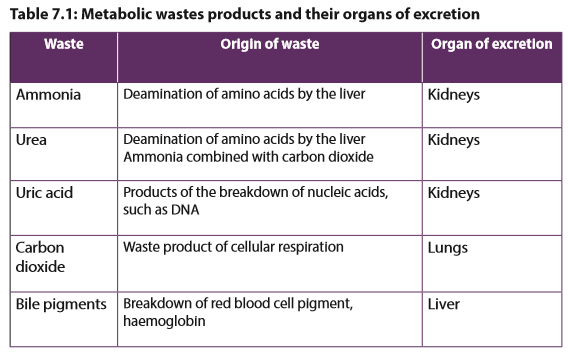

Kidneys and Excretion
The kidneys are part of the urinary system (Figure 7.1). The kidneys work together with other urinary system organs in the function of excretion

a. The Urinary System
In addition to the kidneys, the urinary system includes the; ureters, bladder, and urethra. The main functions of the urinary system are to; filter waste products and excess water from the blood and remove them from the body.
From the kidneys, urine enters the ureters. Each ureter is a muscular tube about 25 centimetres long. Peristaltic movements of the muscles of the ureter send urine to the bladder in small amount. Ureters carry urine to the bladder. The bladder is a hollow organ that stores urine. It can stretch to hold up to 500 millilitres. When the bladder is about half full, the stretching of the bladder sends a nerve impulse to the sphincter that controls the opening to the urethra. In response to the impulse, the sphincter relaxes and lets urine flow into the urethra.
The urethra is a muscular tube that carries urine out of the body. Urine leaves the body through another sphincter in the process of urination. This sphincter and the process of urination are normally under conscious control/voluntary system.
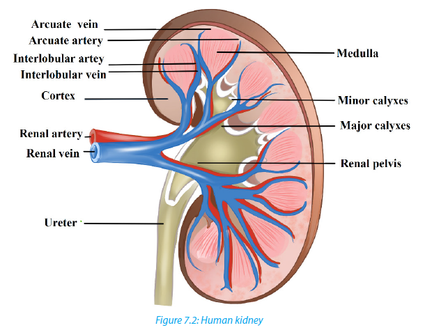
b. Kidneys
The kidneys are a pair of bean-shaped, reddish brown organs about the size of a fist. They are located just above the waist at the back of the abdominal cavity, on either side of the spine. The kidneys are protected by the ribcage. They are also protected by a covering of tough connective tissues and two layers of fat, which help cushion them. Located on top of each kidney is an adrenal gland. The two adrenal glands secrete several hormones. Hormones are chemical messengers in the body that regulate many body functions. The adrenal hormone aldosterone helps regulate kidney functions. The functional unit of a kidney is a nephron.
7.2 Structure and the functions of the nephron.
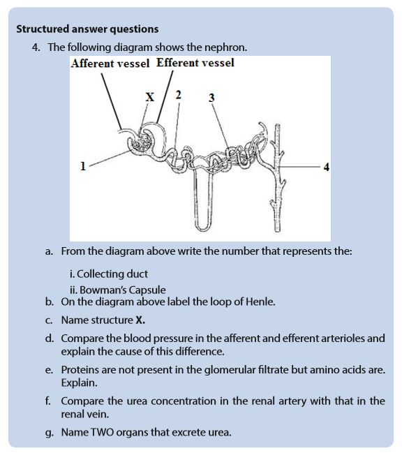
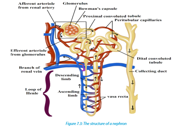
Nephrons are the structural and functional units of the kidneys. A single kidney may have more than a million nephrons. An individual nephron (Figure 7.3) includes a glomerulus, Bowman’s capsule, and renal tubule.
a. Parts of the nephron and their functions
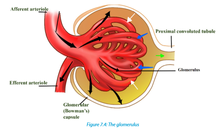
–– The glomerulus is a cluster of arteries that filters substances out of the blood.
–– Bowman’s capsule is a cup-shaped structure around the glomerulus that collects the filtered substances.
–– The renal tubule is a long, narrow tube surrounded by capillaries that reabsorbs many of the filtered substances and secretes other substances.b. Ultra-filtration, selective reabsorption and tubular secretion
The renal arteries, which carry blood into the kidneys, branch into the capillaries of the glomerulus of each nephron. The pressure of blood moving through these capillaries forces some of the water and dissolved substances in the blood through the capillary walls and into Bowman’s capsule. Bowman’s capsule is composed of layers. The space between the layers, called Bowman’s space, fills with the filtered substances.
The process of filtering substances from blood under pressure in the glomerulus is called ultra-filtration, while the fluid that collects in Bowman’s space is called glomerular filtrate. The filtrate is mainly composed of; water, salts, glucose, amino acids, hormones and urea. Larger structures in the blood including; the protein molecules, blood cells, and platelets do not pass into Bowman’s space. Instead, they remain in the main circulation.
From Bowman’s space, the filtrate passes into the renal tubule whose main function is reabsorption. Reabsorption is the return of needed substances in the glomerular filtrate back to the bloodstream. It is necessary because some of the substances removed from the blood by filtration including; water, salts, glucose, and amino acids which are useful and needed by the body. About 75 % of these substances are reabsorbed in the renal tubule.
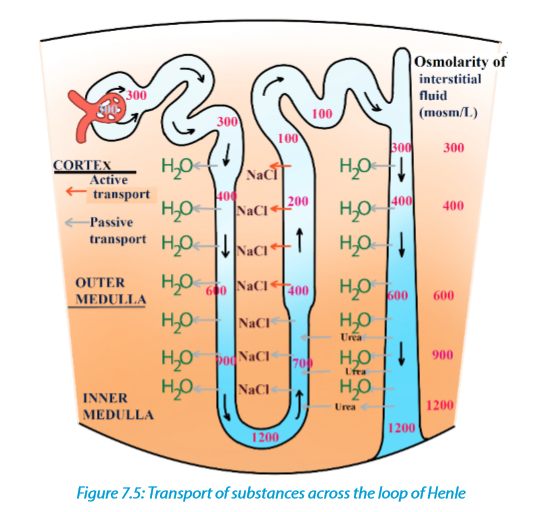
Under conditions in which the kidney conserves as much water as possible, urine can reach an osmolality of about 1200 milliosmoles (mOsm/L), considerably hypertonic to blood (about 300 mosm/L). Osmolarity is the solute concentration expressed as molarity. This ability to excrete nitrogenous wastes with a minimal loss of water is a key terrestrial adaptation of mammals. The loop of Henle is known as a countercurrent multiplier. The term counter-current refers to the fact that the fluid flows in opposite directions in the two sides of the loop, down one side and up in the other. The multiplier effect is seen by comparing the osmolality of the fluid in the cortex and that in the renal medulla at the hairpin end of the loop.
The remaining fluid enters the distal tubule. The distal tubule carries the fluid, now called tubular fluid, from the loop of Henle to a collecting duct. As it transports the fluid, the distal tubule also reabsorbs or secretes substances such as calcium and sodium following the influence of hormones (e.g. aldosterone). The process of secreting substances into the tubular fluid is called secretion.
7.3 Formation of urine and purification of blood
7.3.1 Urine formation

Urine formation depends on three processes including ultrafiltration, selective reabsorption and secretion/tubular secretion.
a. Ultra-filtration
Each nephron of the kidney has an independent blood supply, which moves through the afferent arteriole into the glomerulus, a high-pressure filter. Normally, pressure in a capillary bed is about 25 mm Hg. The pressure in the glomerulus is about 65 mm Hg. Dissolved solutes pass through the walls of the glomerulus into the Bowman’s capsule. Although materials move from areas of high pressure to areas of low pressure, not all materials enter the capsule.
b. Selective reabsorption
The importance of reabsorption is emphasized by examining changes in the concentrations of fluids as they move through the kidneys. On average, about 600 mL of fluid flows through the kidneys every minute. Approximately 20% of the fluid, or about 120 mL, is filtered into the nephrons. This means that if none of the filtrate were reabsorbed the quantity of around 120 mL of urine each minute would be formed and the amount of at least 1 L of fluids would be consumed every 10 minutes to maintain homeostasis.
Fortunately, only 1 mL of urine is formed for every 120 mL of fluids filtered into the nephron. The remaining 119 mL of fluids and solutes is reabsorbed. Selective reabsorption occurs by both active and passive transport. Carrier molecules move Na+ ions across the cell membranes of the cells that line the nephron. Negative ions, such as Cl-and HCO3- follow the positive Na+ ions by charge attraction. Numerous mitochondria supply the energy necessary for active transport. Reabsorption occurs until the threshold level of a substance is reached. Excess NaCl remains in the nephron and is excreted with the urine.
Other molecules are actively transported from the proximal tubule. Glucose and amino acids attach to specific carrier molecules, which shuttle them out of the nephron and into the blood. However, the amount of solute that can be reabsorbed is limited. For example; excess glucose will not be shuttled out of the nephron by the carrier molecules. The solutes that are actively transported out of the nephron create an osmotic gradient that draws water from the nephron. A second osmotic force, created by the proteins not filtered into the nephron, also helps reabsorption. The proteins remain in the bloodstream and draw water from the interstitial fluid into the blood. As water is reabsorbed from the nephron, the remaining solutes become more concentrated. Molecules such as urea and uric acid will diffuse from the nephron back into the blood, although less is reabsorbed than was originally filtered.
c. Secretion
Secretion is the movement of wastes from the blood back into the nephron. Nitrogencontaining wastes, excess H+ ions, and minerals such as K+ ions are examples of substances secreted.
Even drugs such as penicillin can be secreted. Cells loaded with mitochondria line the distal tubule. Like reabsorption, tubular secretion occurs by active transport, but, unlike reabsorption, molecules are shuttled from the blood into the nephron.
7.3.2 Formation of urea
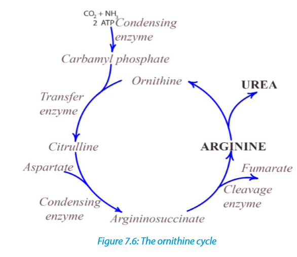
The body is unable to store proteins or amino acids, and any surplus is destroyed in the liver. Excess amino acids which are brought to the liver by the hepatic portal vein, are deaminated by the liver cells. In this process the amino (NH2) group is removed from the amino acid, with the formation of ammonia. The amino acid residue is then fed into carbohydrate metabolism and oxidized with the release of energy. Meanwhile the ammonia must not be allowed to accumulate because it is highly toxic even in small quantities. Under the influence of specific enzymes in the liver cells, the ammonia enters a cyclical series of reactions called the ornithine cycle, in which it reacts with carbon dioxide to form the less toxic nitrogenous compound urea. The urea is then shed from the liver into the bloodstream, and taken to the kidney which eliminates it from the body.

7.4 Role of hypothalamus, pituitary gland, adrenal gland and nephron in varying the blood osmotic pressure

The body adjusts for increased water intake by increasing urine output. Conversely, it adjusts for increased exercise or decreased water intake by reducing urine output. These adjustments involve nervous system and the endocrine system.
7.4.1 Regulation by antidiuretic hormone (ADH)
A hormone called antidiuretic hormone (ADH) helps to regulate the osmotic pressure of body fluids by causing the kidneys to increase water reabsorption. When ADH is released, more concentrated urine is produced, thereby conserving body water. ADH is produced by specialized nerve cells in the hypothalamus, and it moves along specialized fibres from the hypothalamus to the pituitary gland, which stores and releases ADH into the blood. Specialized nerve receptors, called osmoreceptors, located in the hypothalamus detect changes in osmotic pressure when there is a decrease in water intake or increase in water loss by sweating, causing blood solutes to become more concentrated. This increases the blood’s osmotic pressure.
Consequently, water moves into the bloodstream, causing the cells of the hypothalamus to shrink. When this happens, a nerve message is sent to the pituitary, signalling the release of ADH, which is carried by the bloodstream to the kidneys. By reabsorbing more water, the kidneys produce more concentrated urine, preventing the osmotic pressure of the body fluids from increasing any further.

7.4.2 Kidneys and Blood Pressure
The kidneys play a role in the regulation of blood pressure by adjusting for blood volumes. A hormone called aldosterone acts on the nephrons to increase Na+ reabsorption. The hormone is produced in the cortex of the adrenal glands which lies above the kidneys. Not surprisingly, as NaCl reabsorption increases, the osmotic gradient increases and more water move out of the nephron by osmosis.
Aldosterone is secreted by the adrenal cortex in response to a high blood potassium levels, to a low blood sodium levels, or to a decreased blood pressure. When aldosterone stimulates the reabsorption of Na+ ions, water follows from the filtrate back to the blood. This helps maintain normal blood volume and blood pressure. In the kidneys, aldosterone increases reabsorption of Na+ and water so that less is lost in the urine. Aldosterone also stimulates the kidneys to increase secretion of K+ and H+ into the urine. With increased water reabsorption by the kidneys, blood volume increases.

7.5 Kidney transplants and dialysis machines
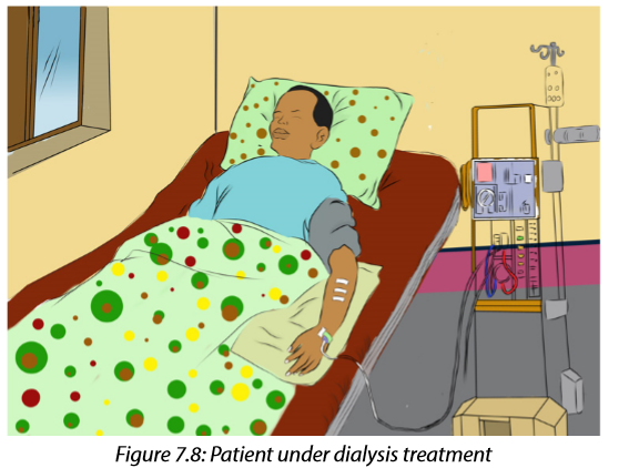
Dialysis is a medical procedure in which blood is filtered with the help of a machine. Blood from the patient’s vein enters the dialysis machine through a tube. Inside the machine, excess water, wastes, and other unneeded substances are filtered from the blood. The filtered blood is then returned to the patient’s vein through another tube. A dialysis treatment usually lasts three to four hours and must be repeated three times a week. Dialysis is generally performed on patients who have kidney failure. Dialysis helps them stay alive, but does not cure their failing kidneys.
Kidney transplants are sometimes performed on people who suffer from severe renal failure. Usually, the donor has suffered an accidental death and had granted permission to have his or her kidneys used for transplantation. An attempt is made to match the immune characteristics of the donor and recipient to reduce the tendency for the recipient’s immune system to reject the transplanted kidney. Even with careful matching, however, recipients have to take medication for the rest of their lives to suppress their immune systems so that rejection is less likely. The major cause of kidney transplant failure is rejection by the recipient’s immune system.
In most cases, the transplanted kidney functions well, and the tendency for the recipient’s immune system to reject the transplanted kidney can be controlled. The advantages and disadvantages of kidney transplants, compared with dialysis.
Advantages
–– The patient can return to a normal lifestyle – dialysis may require a lengthy session in hospital, three times a week, leaving the patient very tired after each session.
–– The dialysis machine will be available for other patients to use.Disadvantages
–– Transplants require a suitable donor – with a good tissue match. The donor may be from a dead person, or from a close living relative who is prepared to donate a healthy kidney (we can survive with one kidney).
–– The operation is very expensive.
–– There is a risk of rejection of the donated kidney; immunosuppressive drugs have to be used.
–– Transplants are not accepted by some religions.
7.6 Principles of osmoregulation in marine, freshwater and terrestrial organisms.
Organisms in aquatic and terrestrial environments must maintain the right concentration of solutes and amount of water in their body fluids. This involves excretion through the skin and the kidneys.
a. Marine animals
Marine bony fishes, such as the salmon, constantly lose water by osmosis. Such fishes balance the water loss by drinking large amounts of seawater. They then make use of both their gills and kidneys to rid themselves of salts. In the gills, specialized chloride cells actively transport chloride ions (Cl-) out, and sodium ions (Na+) follow passively. In the kidneys, excess calcium, magnesium, and sulphate ions are excreted with the loss of only small amounts of water.
b. Freshwater animals
The body fluids of fresh water animals must be hypertonic because animal cells cannot tolerate salt concentrations as low as those of lake or river water. Having internal fluids with an osmolality higher than that of their surroundings, freshwater animals face the problem of gaining water by osmosis and losing salts by diffusion through their gills. Many freshwater animals, including fishes, solve the problem of water balance by drinking almost no water and excreting large amounts of very dilute urine. At the same time, salts lost by diffusion and in the urine are replaced by those found in the food they eat.
c. Land animals
The threat of dehydration is a major regulatory problem for terrestrial plants and animals. Humans, for example, die if they lose as little as 12% of their body water. Adaptations that reduce water loss are key to survival on land. Much as a waxy cuticle contributes to the success of land plants, the body coverings of most terrestrial animals help prevent dehydration.
Examples are the waxy layers of insect exoskeletons, the shells of land snails, and the layers of dead, keratinized skin cells covering most terrestrial vertebrates, including humans. Despite these and other adaptations, most terrestrial animals lose water through many routes: in urine and faeces, across their skin, and from moist surfaces in gas exchange organs. Land animals maintain water balance by drinking and eating moist foods and by producing water metabolically through cellular respiration. A number of desert animals, including many insect-eating birds and other reptiles, are well enough adapted for minimizing water loss that they can survive without drinking water. A noteworthy example is the kangaroo rat loses so little water that 90% replaced by water generated metabolically; the remaining 10% comes from the small amount of water in its diet of seeds.

7.7 Excretion and osmoregulation in protoctista, insects, fish, amphibians and birds.

a. Osmoregulation in protists such as Amoeba
Amoeba makes use of contractile vacuoles to collect excretory wastes, such as ammonia, from the intracellular fluid by diffusion and active transport. As osmotic action pushes water from the environment into the cytoplasm, the vacuole moves to the surface and disposes the contents into the environment.
b. Excretion in insects
Insects and other terrestrial arthropods have organs called Malpighian tubules that remove nitrogenous wastes and also function in water balance. The Malpighian tubules extend from dead-end tips immersed in haemolymph (circulatory fluid) to openings into the digestive tract. The filtration steps which are common to other excretory systems are absent. Instead, the transport epithelium that lines the tubules secretes certain solutes, including nitrogenous wastes, from the haemolymph into the lumen of the tubule.
Water follows the solutes into the tubule by osmosis, and the fluid then passes into the rectum. There, most solutes are pumped back into the haemolymph and water reabsorption by osmosis follows. The nitrogenous wastes mainly insoluble uric acid, are eliminated as nearly dry matter along with the faeces. Capable of conserving water very effectively, the insect excretory system is a key adaptation contributing to their success on land.

c. Excretion in Birds and Reptiles
Most birds live in environments that are dehydrated. Like mammals, birds have kidneys with juxtamedullary nephrons that specialize in conserving water. However, the nephrons of birds have loops of Henle that extend less far into the medulla than those of mammals. Thus, bird kidneys cannot concentrate urine to the high osmolarities achieved by mammalian kidneys. Although birds can produce hyperosmotic urine, their main water conservation adaptation is having uric acid as the nitrogen waste molecule. Since uric acid can be excreted as a paste, it reduces urine volume.
The kidneys of reptiles having only cortical nephrons, produce urine that is osmotic or hypo-osmotic to body fluids. However, the epithelium of the chamber called the cloaca helps conserve fluid by reabsorbing some of the water present in urine and faeces. Also like birds, most reptiles excrete their nitrogenous wastes as uric acid.
Freshwater fishes and amphibians
Freshwater fishes are hyperosmotic to their surroundings, so they must excrete excess water continuously. In contrast to mammals and birds, freshwater fishes produce large volumes of very dilute urine. Their kidneys, which contain many nephrons, produce filtrate at a high rate. Freshwater fishes conserve salts by reabsorbing ions from the filtrate in their distal tubules, leaving water behind.
Amphibian kidneys function much like those of freshwater fishes. When in fresh water, the kidneys of frogs excrete dilute urine while the skin accumulates certain salts from the water by active transport. On land, where dehydration is the most pressing problem of osmoregulation, frogs conserve body fluid by reabsorbing water across the epithelium of the urinary bladder.
Marine bony fishes
The tissues of marine bony fishes gain excess salts from their surroundings and lose water. These environmental challenges are opposite to those faced by their freshwater relatives. Compared with freshwater fishes, marine fishes have fewer and smaller nephrons, and their nephrons lack a distal tubule. In addition, their kidneys have small glomeruli, and some lack glomeruli entirely. In keeping with these features, filtration rates are low and very little urine is excreted.
7.8 Excretion in plants
Compared to animals, plants do not have a well-developed excretory system to throw out nitrogenous waste materials. This is because of the differences in their physiology. Therefore, plants use different strategies for excretion.
The gaseous waste materials produced during respiration (carbon dioxide) and photosynthesis (oxygen) diffuse out through stomata in the leaves and through lenticels in other parts of the plant. Excess water evaporates mostly from stomata and also from the outer surface of the stem, fruits, etc., throughout the day. This process of getting rid of excess water is called transpiration. The waste products, like oxygen, carbon dioxide and water, are the raw materials for other cellular reactions such as photosynthesis and cellular respiration. The excess of carbon dioxide and water are used up in this way. The only major gaseous excretory product of plants is oxygen.
Many plants store organic waste products in their permanent tissues that have dead cells, for example in heartwood. Plants also store wastes within their leaves or barks, and these wastes are periodically removed as the leaves and barks fall off. Some of the waste products are stored in special cells or cellular vacuoles. Organic acids, which might prove harmful to plants, often combine with excess cations and precipitate out as insoluble crystals that can be safely stored in plant cells. Calcium oxalate crystals accumulate in some tubers like yam.
Aquatic plants lose most of their metabolic wastes by direct diffusion into the water surrounding them. Terrestrial plants excrete some wastes into the soil around them. Plants do not have complex excretory systems. This is because of the following reasons:
–– There is very little accumulation of toxic wastes. Often the plant wastes are utilized by the plant. For example, carbon dioxide is used for photosynthesis and oxygen for respiration.
–– The extra gaseous waste is removed from the plant by simple diffusion through the stomata and the lenticels.
–– Most of the waste substances formed in plants are not harmful and can be stored in the plant tissues.
–– Some plants store other waste such as resins in their tissues in a non-toxic form. These tissues or organs later fall off the plant.
–– Excess water and dissolved gases are removed by the process of transpiration through the stomata.
–– Some plants remove waste products by exudation, for example gums, resins, latex and rubber.
–– In some plants water with dissolved salts oozes out through hydathodes. This is called guttation.Note that hydathodes are specialized structures and they are mainly responsible for secreting water in liquid form. They are generally restricted to the apex or the serrated edges of the margins of leaves.