General
- S1: Biology TG File Uploaded 16/08/22, 11:10
- S1: Biology SB File Uploaded 16/08/22, 11:23
UNIT 8 Structure and functions of the human gas exchange system
Key unit
competenceTo be able to describe the structure and functions of the human gas exchange system.
Cross-cutting issue Environment and sustainability:
Breathing safe air ensures good health. Rwandans strive to maintain a clean environment by planting trees and conserving them. The areas of Umutara and Bugesera have been transformed by planting trees.Oral activity
In groups, think back to your earlier grades. Th en, discuss these questions.
1. What is
respiration?
2. Why does
respiration take
place in the cells of
the body?
3. Which gas is
needed for
respiration?
4. Which gas is
produced by
respiration?
5. Name the
respiratory organs
in humans.
6. Do all organisms
have the same
respiratory
organs?
7. Try to remember
the pathway that
air follows when
we breathe in
until it reaches the
respiratory organs.Figure 8.1 We need oxygen to make energy for exercise.
 Topic 2: Organisation and maintenance of life
Structure of the human gas exchange system
Most living organisms need oxygen gas (O2) for respiration. You learnt
in Unit 1 that respiration is a characteristic of living things. During
respiration, glucose is broken down using oxygen, to release energy in cells.
During this process, carbon dioxide (CO2) is produced as a waste product.
So, the cells must take in O2 and get rid of CO2 at the same time. The
movement of gases across a surface or a membrane in opposite directions is
called gas exchange. Gas exchange enables the movement of gases. It takes
place in our lungs and in our cells.How does gas exchange work?
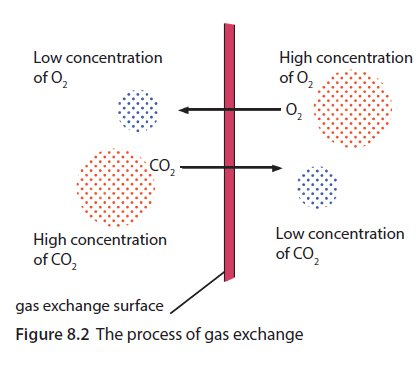
In Figure 8.2, you can see that O2
molecules move from one side of
the gas exchange surface to the other
side. The O2 molecules move from the
side where there are lots of them to
the side where there are fewer of them.
The CO2 molecules move in the opposite
direction through the gas exchange
surface. However, they too move from
where there is a high concentration of
CO2 molecules to where there is a lower
concentration of CO2 molecules.
The movement of molecules
from a place where they are in a high
concentration to a place where they are
in a lower concentration is called diffusion.
So, gas exchange takes place by diffusion.
Do not confuse gas exchange with breathing in mammals. Breathing is
the movement of air into and out of the lungs.The structure of the lungs
Air moves in and out of the lungs. Humans get the oxygen they need from
the air. They get rid of carbon dioxide in the air that leaves the lungs. The
lungs are part of the gas exchange system in humans.
Air is taken in from the outside, or inhaled, through the nose and
mouth. The nose has two nostrils, which lead to the nasal cavity. The nasal
cavity is lined with hairs and mucus, which filter and moisten the incoming
air. They also trap dirt and bacteria. Blood vessels warm the air as it passes through the nasal cavity.Unit 8: Structure and functions of the human gas exchange system

Air passes from the nasal cavity down a tube called the trachea, or windpipe. There is a flap of
cartilage at the top of the trachea that flaps over it during swallowing. This stops food from entering
the trachea. The trachea is held open by C-shaped rings of cartilage.
The trachea branches into two smaller tubes called bronchi (singular = bronchus). One
bronchus leads to each lung and branches into many smaller tubes called bronchioles. The
bronchioles end in many bunches of small, thinwalled air sacs called alveoli (singular = alveolus).
The alveoli are the gas exchange surfaces. The alveoli provide a very large surface area for gas
exchange. The inside surfaces of the alveoli are kept moist by water that diffuses out of the blood.
The walls of the alveoli are only one cell thick, and many small blood capillaries surround them.Experiment 8.1
Dissect a lung.
You will need: a dissecting set; plastic sheeting; a hand lens; a lung of
a goat, sheep or cow (with the trachea and bronchi intact); water; a
container; a towel; soap; disinfectant; rubber tubing
Procedure
1. Wash the lungs carefully with enough water to remove the blood. Do
not allow water to enter the trachea.
2. Place the lungs on a piece of plastic sheeting on a bench. Position the
lungs so that you can see the tubes leading into them. You may have to
cut away some pieces of flesh to fully expose the lungs.
3. Use Figure 8.3, above, to identify the various parts of the lungs.
4. Insert the rubber tubing into the trachea. Take a deep breath, and blow
into the lungs very hard. Do you notice any movement of the lungs?
Describe this movement. Do you think your lungs behave in a similar way when you breathe in? Explain your answer. Topic 2: Organisation and maintenance of life
5. Examine the sides of the trachea and bronchi very closely. Do you see
the rings on the trachea and bronchi? What colour are these rings?
Next, feel the sides of the trachea and bronchi, and then the surface of
the lungs. Do you notice any differences between them? Describe these
differences.
6. The thin layer of skin covering the lungs is called the pleural
membrane. What do you think is the function of this membrane?
7. Bend the trachea until the open end faces downwards, and then let
it go. Was it easy to bend? What happened to the trachea when you
let go? Next, try to close the trachea by squeezing it with your fingers,
and describe what you feel. What happens to the trachea after you stop
squeezing it? What function do the rings have?
8. Cut off one bronchus from the trachea to separate a lung. Next, cut
along the bronchus until you expose the inside of the lung. Use the
hand lens to see whether the bronchus divides further into smaller
tubes. What are these sub-divisions of the bronchus called? What
other details inside the lung have you observed? Describe the inside of
the lung.
9. Figure 8.3, on page 86, shows the human lung in detail. Could you see
this much detail when you examined the lung? Why can you not see
the alveoli?
10. Put the lungs and other parts into the container and bury them in the
ground. Carefully wash the bench and plastic sheeting with soap, and
then disinfect them.
11. Wash your hands thoroughly with soap.Table 8.1 describes the functions of the different parts of the human gas
exchange system.Table 8.1 Functions of parts of the human gas exchange system

Unit 8: Structure and functions of the human gas exchange system

Exercise 8.1
1. Draw a labelled diagram of the human lungs.
2. Describe the function of each of the following:
a) rings of cartilage
b) mucus inside the trachea and bronchi.
3. What are alveoli?
4. The inside of the lung is spongy. Explain the word ‘spongy’.
5. Copy the table below, and then complete it.
Gas exchange in humans
In humans, gas exchange takes place in two places: in the alveoli in the lungs and in the cells of the body.
Gas exchange in alveoli Air, which contains oxygen, is breathed into the lungs. Oxygen moves from the alveoli into the blood in the capillaries that surround the alveoli. Carbon dioxide moves out of the blood into the alveoli. Carbon dioxide is breathed out.
 Topic 2: Organisation and maintenance of life
Gas exchange in cells
Blood containing oxygen flows from the heart to the body cells.
Oxygen molecules move by diffusion from a high concentration in the blood towards a region where there is less oxygen inside thecells. Inside the cell, oxygen is used in the process of respiration.
Carbon dioxide is made during respiration in the cells. This means that there is a high concentration of carbon dioxide molecules inside the cells. Carbon dioxide moves out of the cells into the
blood where there is a lower carbon dioxide concentration.
Experiment 8.2
Make a model to demonstrate breathing.
You will need: rubber bands or string; a glass rod; a rubber sheet of a size that will cover the
bottom of a bell jar; a bell jar; two balloons; a Y-shaped tube; a rubber bung or cork
Procedure
1. Using a rubber band, tie the glass rod to the rubber sheet. Secure the rubber sheet
around the open end of the bell jar using rubber bands. The rubber sheet represents the
diaphragm.
2. Tie a balloon around each arm of the Y-shaped tube. Push the other end of the tube
through a rubber bung or a cork. The balloons represent the lungs and the Y-shaped tube
represents the trachea and bronchi. Assemble the apparatus as shown in Figure 8.6, below.
3. Pull the rubber sheet downwards using the glass rod. This represents an inhalation. Note
what happens to the balloons when the rubber sheet is pulled downwards. Explain what
happens.
4. Push the rubber sheet upwards using the glass rod. This represents an exhalation. Note
what happens to the balloons when the rubber sheet is pushed upwards. Explain what happens.
Unit 8: Structure and functions of the human gas exchange system

Activity 8.1
Work in pairs.
1. Use a microscope to examine the microscope slides that your teacher
will give you. You can also use the micrographs in Figure 8.7 if you do
not have slides.
2. Make a labelled drawing of what you see. What magnification did you
use?

Figure 8.7 Micrographs showing alveolar cells (A) and ciliated epithelium (B)
Exercise 8.2
1. Explain the difference between:
a) gaseous exchange and breathing
b) diffusion and breathing.
2. List the pathway for air from the nose into the lungs.
3. Name two places where gas exchange takes place in humans.
4. a) What type of cells line the trachea?
b) How are these cells specialised for their function?
5. Research gaseous exchange in the following animals:
a) insects
b) fish
c) spiders.Topic 2: Organisation and maintenance of life
Checklist of learning
In this unit, I have learned that:
Gas exchange is the movement of gas molecules across a surface or membrane which is called the
gas exchange surface.
The movement of gas molecules takes place by diffusion.
All organisms need to make energy through respiration, so they need oxygen and they produce
carbon dioxide.
In humans, gas exchange takes place inside the lungs and in the cells of the body.
The human gas exchange system consists of the nostrils, nasal passages, trachea, bronchi,
bronchioles and alveoli.
The walls of the alveoli are only one cell thick; the alveoli are surrounded by small blood capillaries.
Air, which contains oxygen, is breathed in; oxygen molecules diffuse through the walls of the alveoli
into the blood.
Blood that contains oxygen flows to the cells of the body; oxygen molecules diffuse across the cell
membrane into the cell, where they are used for respiration.
Carbon dioxide diffuses out of the cells into the blood.
Blood that contains carbon dioxide flows into the lungs; carbon dioxide molecules diffuse through
the walls of the alveoli into air in the lungs, and are breathed out.Self-assessment
Choose the correct answer.
1. What are the tiny sacs in the lungs called?
A bronchi B alveoli C capillaries
2. What happens during gas exchange in the lungs?
A Oxygen passes into the blood and carbon dioxide passes out of the blood.
B Oxygen passes out of the blood and carbon dioxide passes into the blood.
C Oxygen and carbon dioxide pass into the blood.
3. Which structures does the trachea lead to in the lungs?
A pleural membranes B bronchioles C bronchi
4. The alveoli are suited for gas exchange because they have:
A a small surface area B a large surface area C walls that are many cells thick
5. The cilia in the air passages:
A trap dust B trap bacteria C trap dust and bacteriaUnit 8: Structure and functions of the human gas exchange system
