General
- S6: Biology SB File Uploaded 3/08/22, 12:46
- S6: Biology TG File Uploaded 3/08/22, 12:49
UNIT 4: THE CIRCULATORY SYSTEM
Key Unit Competence
Relate the structures of the circulatory and lymphatic systems to their functions.Learning objectives
–– Explain the need for a transport system in animals.
–– Explain the advantages and disadvantages of different types of circulatory
systems.
–– Describe the external and internal structure of a mammalian heart.
–– Explain how a heartbeat is initiated.
–– Describe the main events of the cardiac cycle.
–– Explain the relationship between the structure and function of blood vessels.
–– Explain how blood circulation is controlled.
–– Describe the effects of exercise on respiration and on circulation.
–– Describe the process of blood clotting.
–– Recall the structure of haemoglobin and explain how haemoglobin transports
oxygen.
–– Explain how tissue fluid and lymph are formed.
–– Describe the risk factors associated with cardiovascular diseases.
–– Carry out an investigation on the effects of exercise on the pulse rate and blood
pressure.
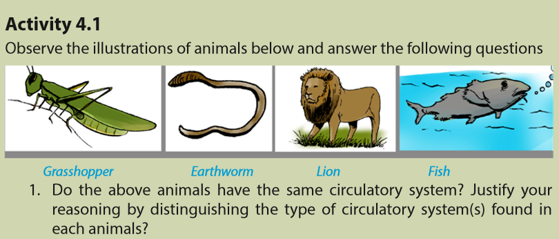
–– Distinguish between open and closed, single and double circulation with
reference to insects, earthworm, fish and mammals.
–– Recognize blood vessels from their structures using a light microscope.
–– Relate the structure of blood vessels to their functions.
–– Differentiate between blood, tissue fluid, and lymph.
–– Relate blood as a tissue to its functions.
–– Interpret oxygen dissociation curves for haemoglobin and other respiratory
pigments.
–– Appreciate the importance of the need for transport systems when animals
become larger, more complex and more active, to supply nutrients to, and
remove waste from, individual cells.
–– Recognize possible risk factors as diet, stress, smoking, genetic predisposition,
age and gender in relation to cardio vascular diseases.
Physical activities can make people including students to be stronger and healthier. They contribute also to lowering obesity rate. All individuals who practice physical activities tend to; have lower body mass indexes, benefit from developing muscles and burning calories. Physical activities help in lowering the rates of diabetes and high blood pressure. Doing physical exercises regularly contribute to better heart
and lung function.4.1 Blood circulatory system in animals
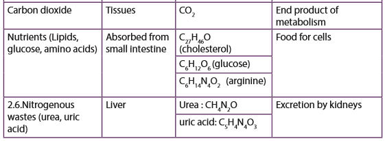
All, except the smallest and tiniest animals need a system to transport substances from cell to cell within themselves. The primary tasks of the system are to import, distribute/deliver nutrients and oxygen to every cell and then to remove waste products including carbon dioxide. The design of the transport system depends upon the size of the organism and on how active it is
In animals, there are two types of circulatory systems i.e.
i. Open circulatory system
ii. Closed circulatory systems:4.1.1 Open circulatory system and closed circulatory system
In animals, circulatory system is either open or closed. Table 4.1 below, shows differences between open and closed circulatory systems:
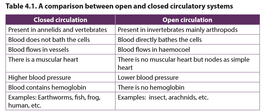
a. Open circulatory system
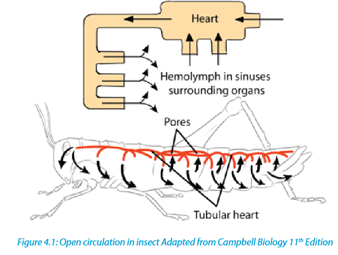
The open circulatory system is common to molluscs and arthropods. In this system, blood is pumped into a hemocoel where it comes into direct contact with body cells and there after goes back to the tubular ‘heart’ via openings called ostia/pores.
Insects and other arthropods have a heart which is an elongated tube located dorsally. The internal organs are suspended in a network of blood-filled sinuses which collectively form the haemocoel. Blood from the heart mixes with the interstitial fluid in the haemocoel to form haemolymph. The advantage this has, is the direct exchange of materials between body cells and haemolymph.
b. Closed circulatory system
Vertebrates, and a few invertebrates like earthworms, have a closed circulatory system. Closed circulatory systems have the blood closed at all times within vessels of different sizes and wall thickness. In this type of system, blood is pumped by the heart through vessels, and does not fill body cavities.
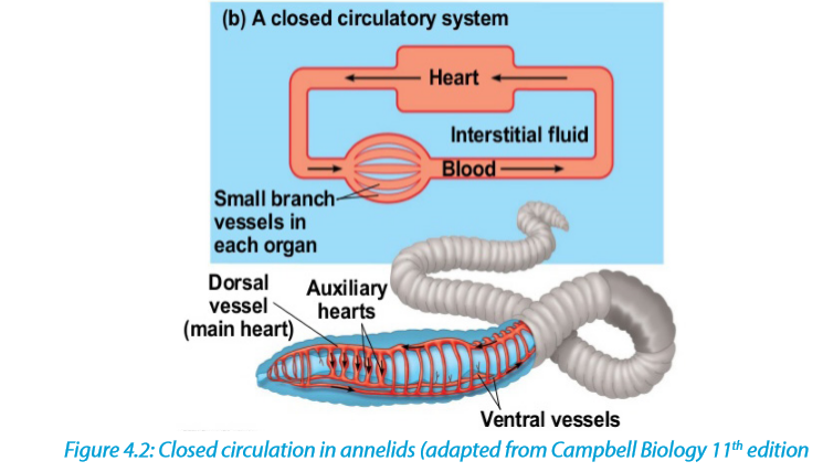
The earthworm possesses a closed circulation system whereby the blood is confined to a series of blood vessels and not permitted to mix with the body tissues. Blood is pumped around the system by muscular longitudinal and ventral vessels and five pairs of lateral pseudo-hearts in segments 7 to 11. Backflow of blood is prevented by valves. The blood itself contains haemoglobin dissolved in the plasma and some phagocyte cells. It is advantageous for an organism to have closed circulatory system because:
–– It helps in control of distribution of blood to different parts of the body.
–– Muscular walls of vessels can constrict and dilate to vary the amount of flow
through specific vessels
–– Blood pressures are fairly high and the circulation can be vigorous
–– It is more efficient hence the blood can reach further distances
–– Allows for more control over oxygen deliveryAll vertebrates including; fish, amphibians, reptiles, birds and mammals possess a prominent muscular heart which pumps blood around the body. The closed circulatory system can be single, partial and double.
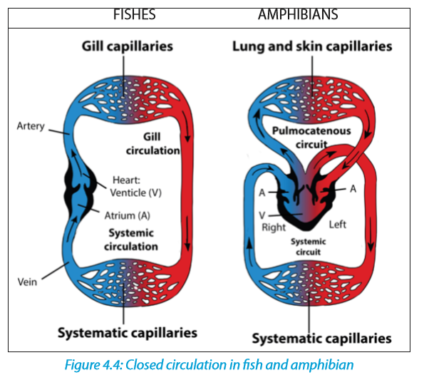
1. Single circulation in fish
Fish have a two-chambered heart made of one atrium and one ventricle. Deoxygenated blood from the body is pumped by the heart to the gills. Here blood is oxygenated before passing around the body and ultimately returning to the heart. Blood has to pass through two capillary systems, the capillaries of the gills and then those of the body before returning to the heart. The two system capillaries offer considerable resistance to the flow of blood. This means that in fish there is a marked drop in blood pressure before the blood completes a circuit. In this type of circulation, it is an advantage that the blood circulating in the body cells has already been oxygenated in the gills
2. Partial double circulation in amphibians
All amphibians and most of the reptiles possess a heart with two atria and one ventricle. Blood from the body enters the right atrium and is pumped to the lungs by the common ventricle. It returns to the heart and enters the left atrium before being pumped around the body. It is called partial because the only one ventricle received oxygenated and non-oxygenated blood which can be mixed as illustrated below:

A spiral valve called conus arteriosus helps to keep deoxygenated and oxygenated blood separate to some extent. The figures below distinguish how closed circulation occurs in fishes and in amphibians
3. Complete double circulation in mammals
This circulation is called double circulation because blood must pass twice in the heart for one complete circuit. The right side of the heart delivers oxygen poor blood to the capillary beds of the gas exchange tissue in lungs, where there is a net movement of O2 into the blood and of CO2 out of the blood. This part of the circulation is called a pulmonary circuit or pulmonary circulation
After the oxygen enriched blood leaves the gas exchange tissues (the lungs), it enters the left side of the heart. Contraction of the left part of the heart propels this blood to the capillary beds in organs and tissues throughout of the body. Following the exchange of O2 and CO2 as well as nutrient and waste products, then the oxygen poor blood returns to the right part of the heart, completing the systemic circuit or the systemic circulation.
Mammals and birds have a four-chambered heart and a complete double circulation. The following are some of the advantages of double circulation:
–– The heart can increase the pressure of the blood after it has passed through the lungs, so the blood flows more quickly to the body tissues.
–– There is no mixing of oxygenated blood with deoxygenated blood.
–– Blood is pumped exactly where it is needed
–– The blood pressure must not be too high in the pulmonary circulation, otherwise it may damage the delicate capillaries in the lungs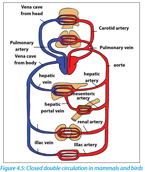
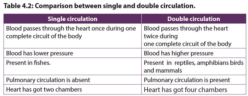
The following table 4.2 indicates the comparison between single and double circulation

4.2 Structure of the human heart
The human heart is made up of a cardiac muscle which contracts in order to propel blood throughout the body. It is located between the two lungs, behind the sternum in the thorax. The heart is surrounded by a tough sac called pericardium. A pericardial fluid is secreted between the membranes allowing them to move easily over each other. The pericardium protects the heart from overexpansion caused by elastic recoil when it is beating very fast. The heart (Figure 4.6) is divided into a left and a right side separated by the septum.
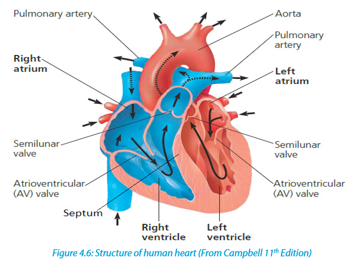
The heart of mammals and birds is composed of 4 chambers including 2 upper atria and 2 lower ventricles. The right side deals with deoxygenated blood and the left side with oxygenated blood. The muscular wall of the left ventricle is thicker than that of the right ventricle because the left ventricle has to pump blood to the whole body with much higher pressure.
The left atrium is separated from the left ventricle by a bicuspid or mitral valve, whilst a tricuspid valve separates the right atrium from the right ventricle. Jointly, these two valves are known as atrioventricular valves. Atrioventricular valves are pushed open when atria contract but, when ventricles contract they close and produce the first sound of the cardiac cycle, the second being that of the closing semilunar valves (aortic and pulmonary valves).
4.3 Heart beat and mammalian cardiac cycle
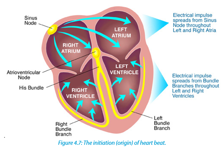
4.3.1. Initiation of a heartbeat
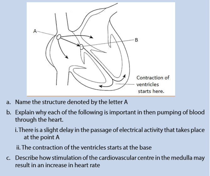
Heart beat is a rhythmic sequence of contractions of the heart. It is coordinated by two small groups of cardiac muscle cells called the sinoatrial (SA) and atrioventricular (AV) nodes. The sinoatrial node (SAN), often known as the cardiac pacemaker, is found in the upper wall of the right atrium and is responsible for the wave of electrical stimulation that starts atrial contraction by creating an action potential. The action potential causes the cardiac cells to contract. This wave of contraction spreads across the cells of the atria, reaching the atrioventricular node (AV node/ AVN) which is found in the lower right atrium.
The atrioventricular node/AVN conducts the electrical impulses that come from the SA node/SAN through the atria to the ventricles. The impulse is delayed there before being conducted through special bundles of heart muscle cells called the bundle of His. This delay allows for the ventricles to be filled with blood before they contract There is a collection of heart muscle cells (fibres) specialized for electrical conduction that transmits the electrical impulses from the AVN, through the Purkinje fibres, which leads to a contraction of the ventricles.
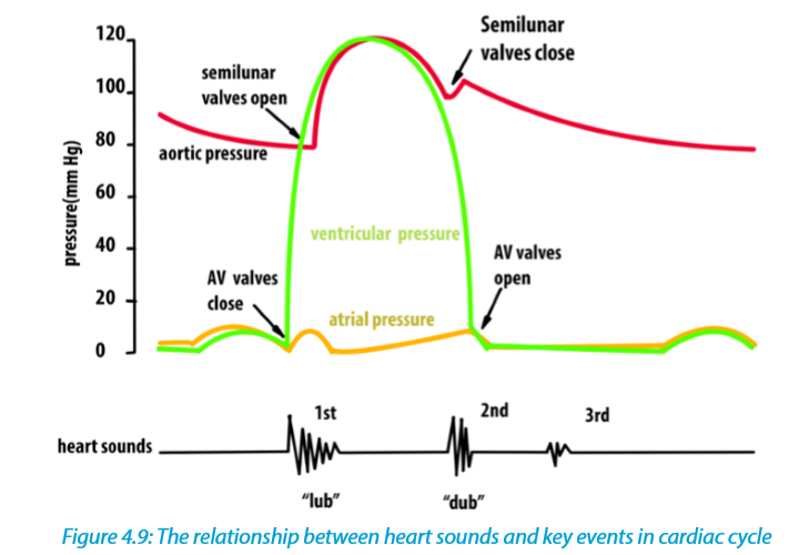
4.3.2. Mammalian cardiac cycle and cardiac sounds
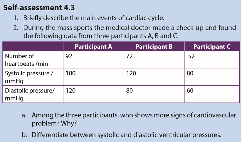
The cardiac cycle refers to the sequence of events which take place during the completion of one heartbeat. It involves repeated contraction (systole) and relaxation (diastole) of the heart muscle. The three steps in cardiac cycle are the followings:
1. Atrial systole and ventricular diastole
In this brief period of 0.1 seconds that follows atrioventricular diastole, blood from the vena cava and pulmonary vein enter the both atria and they get filled with blood. The walls of the atria undergo contraction (systole) forcing blood into the ventricles via bicuspid and tricuspid valves. During this time, the ventricles are relaxed and semilunar valves remain closed.
2. Ventricular systole and atrial disatole
During this stage, the ventriles undergo contraction (systole) hence forcing blood out of the heart via the semilunar valves into the aorta and pulmonary artery. At this time, the atria relax and expand waiting to be filled with blood. The contraction of ventricles causes the atrioventricular valves to close simultaneously in order to prevent back flow of blood. The closure of the valves produces the first heart sound termed as ‘lub’.
3. Atrioventricular diastole
Upon expelling of blood, ventricles relax and their pressure lowers compared to aorta and pulmonary artery pressures. This would cause back flow of blood to the heart but it is prevented by sudden closure of the semilunar valves. The closure of the semilunar valves causes a second heart sound called ‘dub’.
Note: The two sounds ‘lub’ and ‘dub’ are so close and often describes as ‘lub –dub’ and they form a single heartbeat
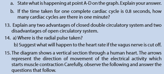
The atrioventricular diastole ends the cardiac cycles and is followed by the atrial systole. Hence the cycle restarts. When the heart rate is 75/min, which means 75 heartbeats per minute, the period of one cardiac cycle is 0.8 sec.

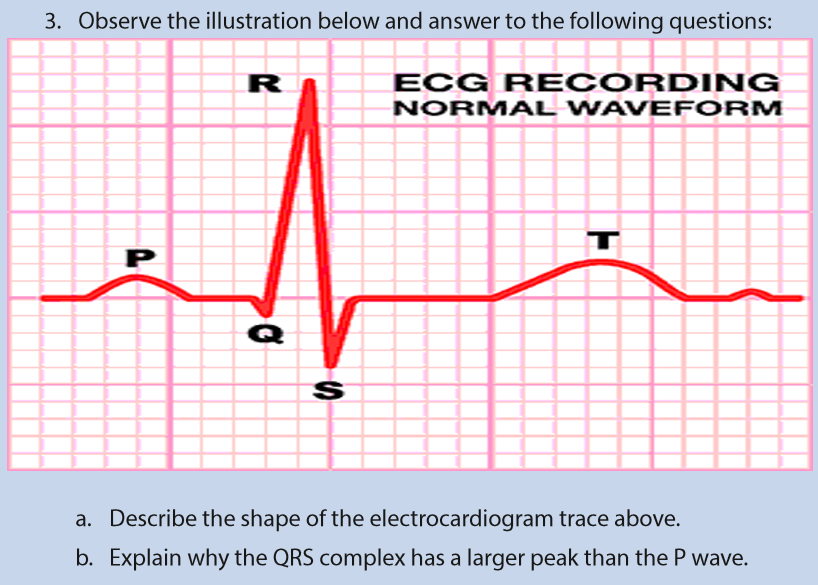
The electrical activity of the heart can be monitored using an Electrocardiogram (ECG) as shown in figure 4.10. This involves attaching of sensors to the skin. Some of the electrical activity generated by the heart spreads through the tissue next to the heart and onwards to the skin. The sensors on the skin pick up the electrical excitation created by the heart and convert this into a trace. The trace of a health person has particular shape. it consists of a series of waves that are labelled P, Q, R, S and T. Wave P shows the excitation of the atria, while QRS indicates the excitation of the ventricles and T shows diastole.
The shape of the ECG trace can sometimes indicates the parts of the heart muscles which are not healthy. It can show if the heart is being beating irregularly, fibrillation (the heart beat is not coordinated), or if it is suffering the heart attack (myocardial infarction). It can also show if the heart has enlarged or if the Purkinje fibre is not conducting electrical activity properly.

4.4 Control of the heart rate.
4.4.1. Nervous and hormonal control of heart rate
In the nervous control of the heartbeat, there is a cardiovascular center located in the medulla oblongata of the hindbrain which controls the activities of the SAN. The center has two nerves from the autonomic nervous system i.e. sympathetic nerve whose stimuli accelerates activity of the SAN (increases heartbeat) and vagus nerve whose stimuli slows down the activity of SAN (decreases heartbeat).
With regard to the hormonal control, the adrenal glands under influence of hypothalamus secrete the hormone adrenaline into blood. Upon reaching the heart, adrenaline will speed up the activity of the SAN thus increasing heartbeat. The reduction comes about when the levels of adrenaline reduce through a negative feedback mechanism.
4.4.2. Other factors controlling heart rate
Other factors affecting heart rate include; the levels of carbon dioxide, temperature, pH and mineral ions.
a. Carbon dioxide
Chemically, high CO2 levels stimulate the vasomotor Centre (VMC) to vasoconstrict arterioles. The resulting high blood pressure transports CO2 more rapidly to the lungs for expulsion and exchange with O2. Where tissues suddenly become active, they produce more CO2. This causes vasodilation of local blood vessels, thus increasing their blood supply and allowing more oxygen and glucose to reach them for respiratory purposes.
b. Body temperature
When the body temperature changes, so does the heart rate. This is one of the thermoregulatory changes that occur to prevent the body’s core temperature of 370C from increasing or decreasing. Heart rate increases when heat is gained by the body such as in hot climates and during physical exercise in order to transfer more heat away from the body. When the body loses heat such as in cold weather or a cold shower, heart rate decreases to preserve core temperature.
c. pH and mineral ions
The importance of plasma electrolytes and pH levels in determining heart rate is not yet well grounded. A significant heart rate increase was obtained after a decrease of potassium and calcium and an increase in pH levels and with no significant variations in indices of autonomic activity. The analysis revealed that changes in physiological range of; potassium, calcium, and pH could cause large heart rate variations from 60 to 90 bpm. It was concluded that electrolyte and pH changes in physiological range have an important complex impact on the pace making rhythm independently of autonomic outflow.
Effect of drugs, and physical activity on cardiac frequency
a. Physical exercise
The heart rate and blood pressure both rise during physical exercise. Over time, regular physical exercise can help lower the resting blood pressure and heart rate. This is because physical exercise training improves the health of the heart and blood vessels, allowing the cardiovascular system to function more efficiently. This enables increased blood flow to muscles without putting excess pressure on blood vessel walls. While blood pressure rises during exercise, it is too much smaller degree than the increase in heart rate. Like the heart rate, blood pressure returns to resting level a few minutes after the end of physical exercise.
b. Caffeine and Other Drugs
Caffeine found in coffee, tea and soda is a stimulant drug that influences the nervous system to increase heart rate. It mimics the effect of adrenaline, a natural hormone in the body responsible for elevating heart rate. Other stimulants such as cocaine and ephedrine work in a similar manner.
On the other hand, there are specific drugs used in lowering heart rate such as beta- and calcium channel blockers. Beta-blockers work by interfering with the receptors that adrenaline binds to, subsequently decreasing hormonal influence on heart rate. Calcium channel blockers reduce the amount of calcium that enters the heart muscle. Because calcium is needed for muscle to contract, the heart beats at a slower rate when this drug is taken.
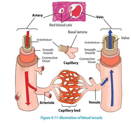
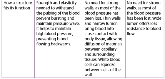
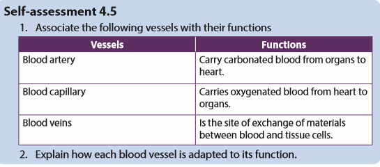
4.5 Blood vessels
Blood vessels include; arteries, capillaries and veins. Illustrations, structure of walls, lumen, valves, branching, and functions of arteries, capillaries and veins are summarized in the figure 4.1.1


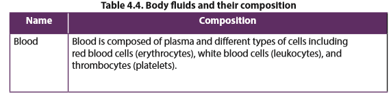
4.6 Body fluids, composition and functions
4.6.1. Main types of body fluids and their compositions
Body fluids are liquids originating from inside the body of living humans. The main body fluids are; blood, plasma, serum, tissue fluid and lymph which are described below in the table 4.4.

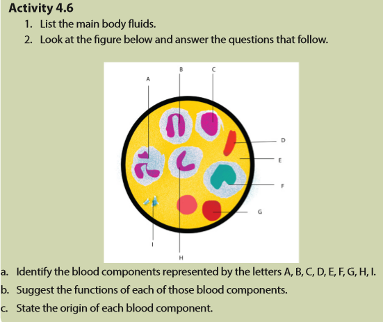
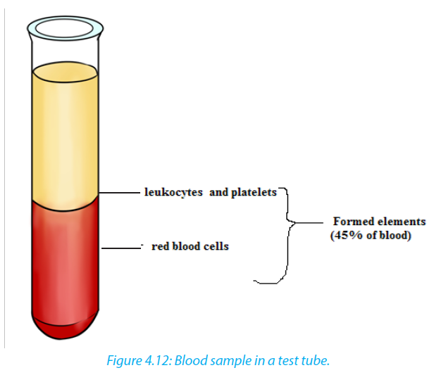
4.6.2. Composition and functions of blood
The main blood components are formed elements and plasma. Formed elements are erythrocytes (red blood cells), leukocytes (white blood cells) and thrombocytes (platelets).
a. Erythrocytes
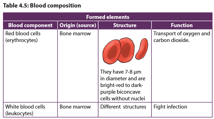
Erythrocytes also called red blood cells, their core function is to carry oxygen from the respiratory organs to tissues and their structure are well modified accordingly to perform the purpose. There are five million per cubic millimetre each having about 8 μm in diameter and 3 μm thick in widest part. The cell has red pigment called Haemoglobin a complex protein containing four iron haem groups.
b. Leukocytes
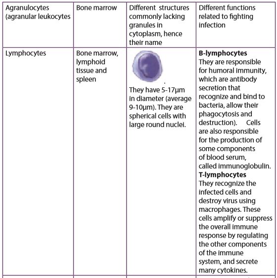
Leukocytes (white blood cells) are involved in immune system that fights against infections. . white blood cells are responsible for destroying infectious agents and infected cells, and secrete protective substances such as antibodies, which fight infections. Leukocytes are divided into:
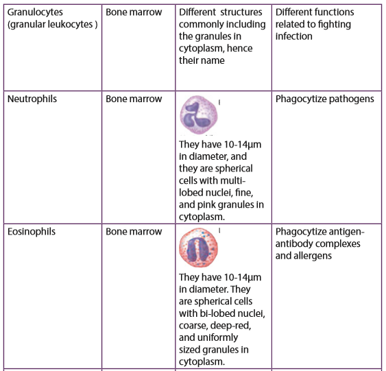
–– Granulocytes or polymorph nuclear cells. They are neutrophils, basophils eosinophils. They take the name from the possession of numerous granules in their cytoplasm.
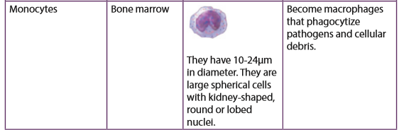
–– Agranulocytes or monomorphonuclear cells: They are lymphocytes and monocytes. They lack granules in the cytoplasm.Thrombocytes
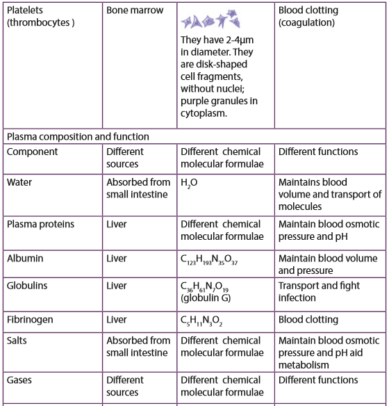
Thrombocytes are also called platelets, are small cell fragments with 2-3 mm in diameter. They are formed from cytoplasm of large cells (mega karyotypes. Normal quantitative value is between 250,000 and 450,000 platelets per mm³. They help in blood clotting. A comparison between formed elements is summarized in the table 4.5 below.


4.7 Transport of respiratory gases

a. Structure of haemoglobin of red blood cells.
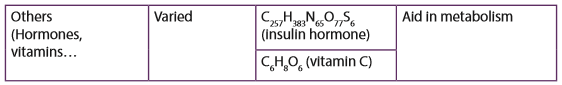
Haemoglobin is a red protein responsible for transporting oxygen in the blood of vertebrates. It is also involved in the transport of carbon dioxide. Haemoglobin is composed of haem and globin (polypeptide chains). Haem is an iron porphyrin compound. Iron occupies the centre of the porphyrin ring and establishes linkages with all the four nitrogen of all the pyrrole rings.
Globin part is made of four polypeptide chains, two identical α-chains and two identical β-chains in normal adult haemoglobin. Each chain contains a “haem” in the so called ‘haem pocket’ and one haemoglobin molecule possess four haem units. Haem pockets of α-subunits are of just adequate size to give entry to an O2 molecule. Entry of O2 into haem pockets of β-subunits is blocked by a valine residue.
b. Transport of carbon dioxide (CO2)
At systemic capillaries in the body cells, CO2 enters red blood cells. Some CO2
combines with Hb to form HbCO2 (Carbaminohaemoglobin):
I.e. Hb + CO2 →HbCO2 (Carbaminohaemoglobin)
Most CO2 is converted to HCO3- (bicarbonate ion), which is carried in the plasma.Haemoglobin is in relation with chloride shift. It is a process which occurs in
a cardiovascular system and refers to the exchange of bicarbonate (HCO3 −)
and chloride (Cl−) across the membrane of red blood cells (RBCs). The chloride shift
occurs in this way:
: H + Hb is reduced haemoglobin which is haemoglobin combined with hydrogen ion (H+).
c. Transport of oxygen
Haemoglobin gets oxygen in lungs from external environment to form a compound called oxyhaemoglobin (HbO8). , In this form, oxygen is transported to the body cells to sites where it is needed for aerobic respiration.

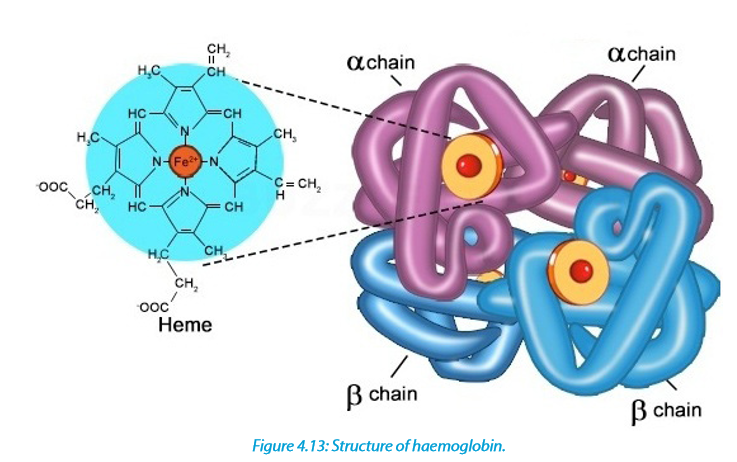
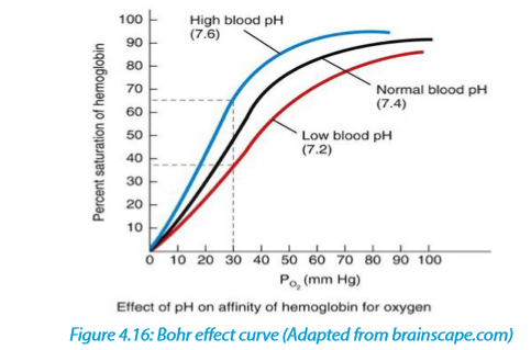
The curve above in figure 4.15 shows the oxygen dissociation curve by haemoglobin. Oxygen dissociation curves determined by plotting the partial pressure of oxygen in blood against the percentage of haemoglobin combined with oxygen in the form of ox haemoglobin. The S-shape of the oxygen dissociation curve can be explained by the behaviour of a haemoglobin molecule as it combines with or loses oxygen molecules. When an oxygen molecule combines with one haem group, the whole haemoglobin molecule is slightly distorted. The distortion makes it easier for a second and third oxygen molecules to combine the haem groups. It is then still easier for the fourth and final oxygen molecule to combine.
If all the oxygen binding sites contain oxygen, then the oxygen saturation is 100%. Oxygen saturation is defined as the ratio of oxyhaemoglobin to the total concentration of haemoglobin present in the blood The Bohr Effect is a physiological phenomenon in which a raise of carbon dioxide in the blood and a decrease in pH results in a reduction of the affinity of haemoglobin for oxygen. This causes the oxygen dissociation curve for haemoglobin to shift to the right. The Bohr Effect occurs in this way:
4.8 Blood clotting and common cardiovascular diseases
a. Blood clotting
Blood clotting also known as blood coagulation is the process by which blood becomes thick and stops flowing, forming a solid cover over any place where the skin has been cut or broken. Blood that has been converted from a liquid to a solid state is called blood clot. A blood clot called thrombus is stationary within a vessel or the heart. If a blood clot moves from that location through the bloodstream, it is referred to as an embolus.
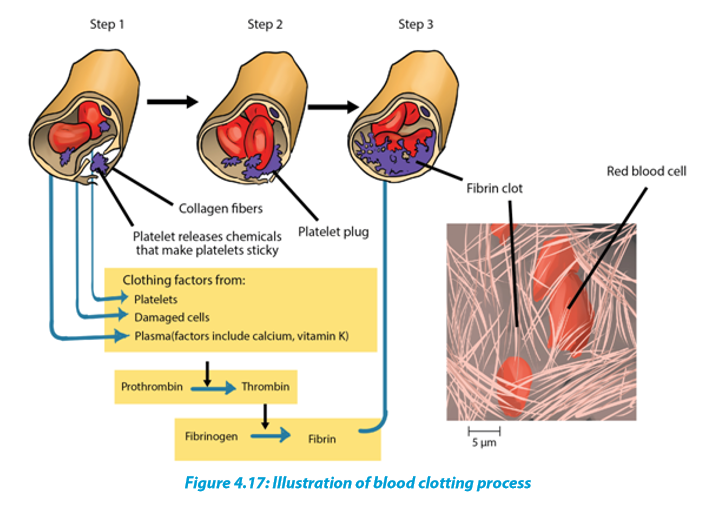
Blood clotting is a series of different processes:
Step 1: The blood coagulation process begins when the endothelium of a vessel is damaged, exposing the connective in the vessel wall to blood. Platelets adhere to collagen fibres in the connective tissue and release a substance that makes nearby platelets sticky.
Step 2: The thrombocytes form a plug that provides emergency protection against blood loss.
Step 3: This seal is reinforced by a clot of fibrin when vessel damage is severe. Fibrin is formed via a multistep process where clotting factors released from the clumped platelets or damaged cells mix with clotting factors in the plasma, forming an activation that converts a plasma protein called prothrombin to its active form, called thrombin. This is facilitated by calcium ions and vitamin K. Thrombin itself is an enzyme that catalyses the final step of the clotting process. This final step is the conversion of fibrinogen to fibrin. The threads of fibrin become interwoven into a patch. And the blood clot is formed. These threads trap red blood cells and other blood components, preventing the continuous bleeding
b. Common cardiovascular diseases
1. Stroke
Stroke is a cardiovascular disease due to the lack of oxygen to the brain which may lead to reversible or irreversible paralysis. The damage to a group of nerve cells in the brain is often due to interrupted blood flow, caused by a blood clot or blood vessel bursting. Since atherosclerosis is a body wide process, similar events can also occur in the arteries to other parts of the body, including the brain. A stroke is a loss of brain function due to a stoppage of the blood supply to the brain. It can be caused by a stationary blood clot known as thrombus, a free-floating clot moving blood clot or embolus that gets caught in a blood vessel, or by bleeding (haemorrhage). Hypertension or high blood pressure promotes atherosclerosis and increases the risk of heart attack and stroke.
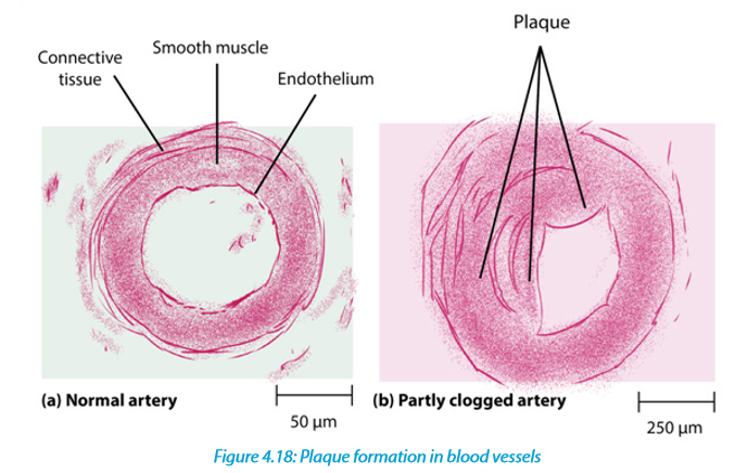
2. Atherosclerosis
Atherosclerosis is a cardiovascular disease characterized by the progressive narrowing and hardening of the arteries over time. Atherosclerosis normally begins in later childhood, and is usually found in most major arteries. It does not usually have any early symptoms. Causes of atherosclerosis include a high-fat diet, high cholesterol, smoking, obesity, and diabetes. Atherosclerosis becomes a threat to health when the plaque build-up interferes with the blood circulation in the heart known as coronary circulation or the brain known as cerebral circulation. A blockage in the coronary circulation, can lead to a heart attack, and blockage of the cerebral circulation can lead to a stroke.
3. Coronary heart disease
Coronary heart disease (CHD) is a disease in which a waxy substance called plaque builds up inside the coronary arteries. Cardiac muscle cells are fed by the coronary arteries. Blocked flow in a coronary artery can result in oxygen starvation and death of heart muscle. Most individuals with coronary heart disease have no symptoms for many years until the first sign, often a heart attack, happens.
c. Risk factors associated with cardiovascular diseases
There are several risk factors for heart disease. Some of those factors are controllable and others are uncontrolled. Uncontrollable factors include the gender (males are at greater risk), age (old people have higher risk), and family history in relation to heart diseases as well post-menopausal stages for females. Making some changes in lifestyle can reduce chance of having heart disease. Controllable risk factors include smoking, high blood pressure, physical inactivity, obesity, diabetes, stress and anger

4.9 Lymphatic system
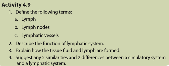
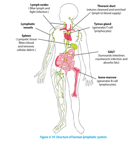
4.9.1 Structure of a lymphatic system
A lymphatic system is a system composed of tissues and organs, including; bone marrow, spleen, thymus, and lymph nodes that produce and store cells that fight infection and disease. The channels that carry lymph are also part of this system. So, the lymphatic system is part of the circulatory system and an important part of the immune system.
4.9.2 Functions of a lymphatic system
–– Drainage of fluid from blood stream into the tissues: The circulating blood through narrow vessels leads to leakage of fluid or plasma into the tissues carrying oxygen and nutrients to the tissues and taking waste materials from the tissues into the lymph channels. The leaked fluid drains into the lymph vessels.
–– Filtration of the lymph at the lymph nodes: The nodes contain white blood cells that can attack any bacteria or viruses they find in the lymph as it flows
through the lymph nodes.
–– Filtering blood: This is done by the spleen which filters out bacteria, viruses and other foreign particles.
–– Raise an immune reaction and fight infections: The lymphatic system especially the lymph nodes are over active in case of an infection the lymph nodes or glands often swell up in case of a local infection in so doing, the lymphocytes fight the foreign bodies trapped in the lymph nodes.4.9.3 Formation of tissue (interstitial) fluid
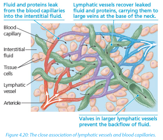
Fluids and some soluble proteins leak from the blood capillaries into the interstitial fluid that bathes the cells of tissues. This occurs due to the arterial end of capillary, where the blood pressure is greater than osmotic pressure so that fluid flows out of capillary into the interstitial fluid. This process is called pressure filtration or ultrafiltration
4.9.4 Formation of lymph
The lymph is the tissue fluid that moves within the lymphatic vessels. The lymphatic vessels recover some leaked fluid and proteins, and carry them to large veins at the base of the neck (figure 4.20).
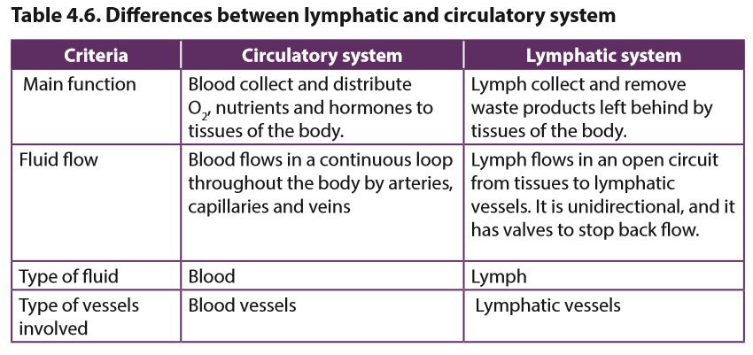
4.9.5 Comparison between lymphatic and circulatory systems
Both the cardiovascular and lymphatic systems are vascular networks carrying body fluids. Differences and similarities are summarized in the table 4.6

a. Identify the organs W, X, Y, Z shown on this figure
b. Describe the functions of the organs W, X, Y, Z.