General
- S6: Biology SB File Uploaded 3/08/22, 12:46
- S6: Biology TG File Uploaded 3/08/22, 12:49
UNIT 8: GENERAL PRINCIPLES OF RECEPTION AND RESPONSE IN ANIMALS.
Key Unit Competence
xplain the general principles of reception and response in animals.Learning Objectives
By the end of this unit, I should be able to:
–– Explain the necessity of responding to internal and external changes in the environment.
–– Describe the main types of sensory receptors.
–– Discuss the main functions of a sensory system.
–– Explain the significance of sensory adaptation.
–– Describe the structure of the human eye.
–– Describe the structure of the retina.
–– Explain how rods transduce light energy into nerve impulses.
–– Explain how retinal convergence improves sensitivity.
–– Explain how the cones achieve visual acuity.
–– Explain how cone cells produce colour vision.
–– Discuss the significance of binocular vision.
–– Describe the structure of the human ear and the functions of its main parts.
–– Describe the process of hearing and balance.
–– Locate the taste buds on the tongue and sensory cells in the skin.
–– Observe the structure of the skin, retina, cochlea and vestibular apparatus from prepared slides or micrographs and relate them to their functions.
–– Interpret graphs on sensory adaptation in response to a constant stimulus.
–– Relate the number of retinal cells to sensitivity and visual acuity
–– Recognise the role of sense organs in the perception of different stimuli.
–– Appreciate the role of sensory adaptation in protecting the sense organs from
overload with unnecessary or irrelevant information.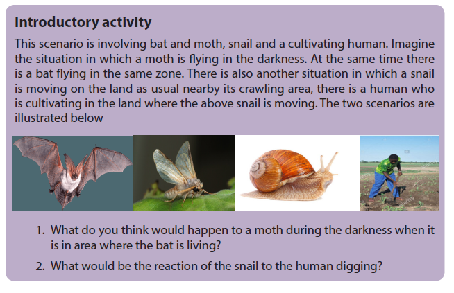
Animals realize different activities including searching for food, select a mate, and escape from predators. They also have the ability to feel changes in environmental factors and keep their internal environment within tolerable limits. These and other activities depend on the animal’s ability to gather information about what is happening inside and outside the body. The survival of animals depends upon the ability to respond in an appropriate way to environmental changes through the ability of detecting stimuli. Some other animals have become highly specialized to detect a particular form of energy by the use of specialized receptor cells which are able to perceive whichever form of energy and elaborate adequate response respond to nervous impulse
8.1 Types of sensory receptors and stimuli

The physical and chemical conditions in an animal’s internal and external environments are continually changing. A change that can be detected is called a stimulus. To some extent, all animal cells are sensitive to stimuli, and some cells called receptors have become especially sensitive to particular stimuli. There are a huge number of environmental variables that an animal could sense. However, each species has evolved receptors only to environmental variables that have an appreciable effect on its chances of survival. For example, humans can sense all the colors of the rainbow but can sense neither infrared nor ultraviolet light.
Classification of receptors
Receptors are commonly classified according to the type of stimulus energy they detect. The main types are:
–– Mechanoreceptors which detect changes in mechanical energy, such as movements, pressures, tensions, gravity, and sound waves.
–– Chemoreceptors which detect chemical stimuli, for example, through taste and smell.
–– Thermoreceptors which detect temperature changes.
–– Electroreceptors which detect electrical fields.
–– Photoreceptors which detect light and other forms of electromagnetic radiation.Receptors can also be classified according to their structure. Simple receptors, known as primary receptors, consist of a single neurone, one end of which is sensitive to a particular type of stimulus. A primary receptor gathers sensory information and transmits it to another neurone or an effector. For example, Pacinian corpuscles are mechanoreceptors located in the skin, tendons, joints and muscles. Their ends consist of concentric rings of connective tissue. Application of pressure against the connective tissue deforms stretch-mediated sodium ion channels in the cell surface membrane, causing an influx of sodium ions which leads to a generator potential.
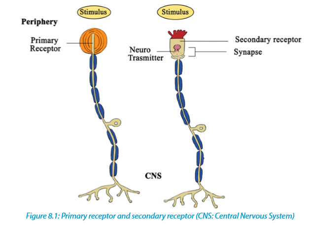
A secondary receptor is more complex. It consists of a modified epithelial cell which is sensitive to a particular type of stimulus. The cell senses changes and passes this information on to a neurone which transmits it as nervous impulse. Sense organs are complex stimulus – gathering structures consisting of grouped sensory receptors. In many sense organs, several receptors make synaptic connections with a single receptor neuron.
A third classification of receptors is based on the source of stimulation and includes exteroceptors responding to stimuli outside the body, interceptors responding to stimuli inside the body, and proprioceptors respond to changes of joint angle and amount of tension in muscles.

8.2 Components of the sensory system: transduction, transmission and processing
8.2.1 Sensory systems
Receptors are the first component of a sensory system, which has three main functions:
–– Transduction: Receptor cells gather sensory information and then convert it into a form of information that can be used by the animal (nerve impulses)
–– Transmission: Sensory neurones transmit nerve impulses from the receptors to the central nervous system
–– Processing: the central nervous system processes the information so that appropriate responses can be made to environmental changes.
A receptor converts the energy from the stimulus into an electrical potential that is proportional to the stimulus intensity. This graded electrical potential is known as the receptor potential or generator potential. If the stimulus is sufficiently high (above a critical threshold level) the graded potential is high enough to fire an action potential. If the stimulus is beneath the threshold, no action potential takes place.8.2.2 Sensory adaptation
Receptors are adapted to detect potentially harmful or beneficial changes in the environment. When given an unchanging stimulus, most receptors stop responding so that the sensory system does not become overloaded with unnecessary or irrelevant information. Loss of responsive is brought about by a process called sensory adaptation. An unchanging stimulus results in a decline in the generator potentials produced by sensory receptors. Consequently, the nerve impulses transmitted in sensory neurones become less frequent and may eventually stop. The mechanism of sensory adaptation involves changes in the membranes of receptor cells and explains why, for example, a person becomes insensitive to the touch of clothing on skin. Even a hair shirt becomes tolerable after wearing it for a long period of time.
8.2.2. Transferring information
After gathering and transducing the stimuli, the sensory system transmits information about the stimulus to the central nervous system and effectors. The frequency of nerve impulses propagated along a sensory neurone usually gives information about stimulus strength. The transfer of information is rarely direct. In mammals, much of the sensory information goes to sensory projection areas in the brain where information processing takes place.
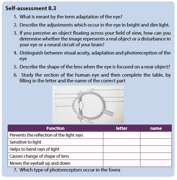
8.3 Structure and functioning of the eye

The eye is a complex light – sensitive organ that enables us to distinguish minute variations of shape, color, brightness, and distance. The function of eye is to transduce light (visible frequencies of electromagnetic radiation) into patterns of nerve impulses. These are transmitted to the brain, where the actual process of seeing is performed.
8.3.1. Functions of parts of eye
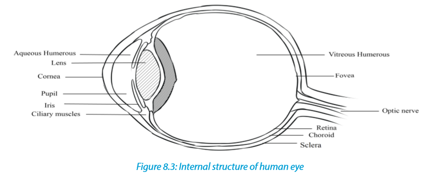
–– The lens: Refracts light and focuses it on retina. Made up of elastic material that adjusts when the eye focuses on far or near object.–– The ciliary body: Made up of muscle fibres which contract or relax to change the shape or curvature of the lens. It produces aqueous humour.
–– The suspensory ligament: The suspensory ligament is a tissue that attaches the edge of the lens to the ciliary body.
–– The iris: It is coloured part of the eye, it has radial and circular muscles which control the size of the pupil; it has melanin pigment that absorbs strong light to prevent blurred vision.
–– Pupil: It is a hole at the centre of the iris through which light pass into the eye.
–– Aqueous humour: Has fluids to maintain the shape of eye ball and to refract light rays. It contains oxygen and nutrient for cornea and lens. It is a transparent and allow light to pass through
–– Vitreous humour: It is the space behind the lens and it is filled with fluids, a transparent, jelly-like substance. Vitreous humour keeps the eyeball firm and helps to refract light onto the retina.
–– Cornea: Is transparent part of the eye and allows the passage of light. It refracts light ray. It is made up of tough tissues to strength the eye.
–– Choroid: The choroid is the middle layer of the eyeball that lies between the sclera and retina. It has two functions, one being able to prevent internal reflection of light as it is pigmented black. Secondly, it contains blood vessels that bring oxygen and nutrients to the eyeball and remove metabolic waste
product.
–– Retina: The retina is the innermost layer of the eyeball. It is the light sensitive layer on which images are formed. It contains light sensitive cells called photoreceptors. Photoreceptors consist of rods and cones. Cones enable us to see colours in bright light while rods enable us to see in black and dim light. The photoreceptors are connected to the nerve endings from the optic nerve.–– Blind spot: The blind spot is the region where the optic nerve leaves the eye. It does not contain any rods or cones. Therefore, it is not sensitive to light.
–– Optic nerve: It is a nerve that transmits nerve impulses to the brain for interpretation when the photoreceptors in the retina are stimulated.
–– Fovea or yellow spot: It is a small yellow depression in the retina. It is situated directly behind the lens. This is where images are normally focused. The fovea contains the greatest concentration of cones, but has no rods. The fovea enables a person to have detailed colour vision in bright light.
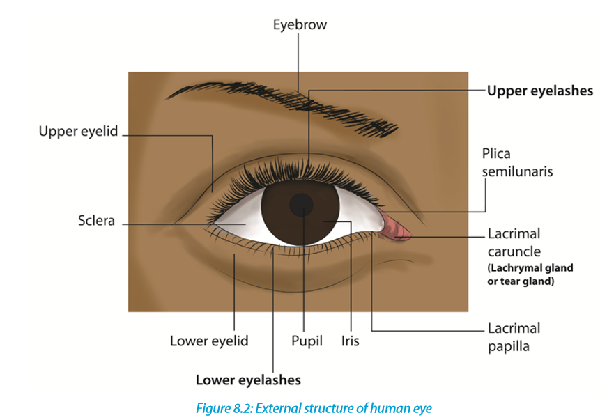
–– Conjunctiva: Thin and transparent to allow light to pass through.
–– Sclera: It is a tough, white outer covering of the eyeball, which is continuous with the cornea. It protects the eyeball from mechanical damage.
–– The eye brows: Prevent sweat and dust from entering the eye.
–– The eye lashes: Prevent dust particles from entering the eye.
–– The tears glands: Secrete tears that wash away dust particles in the eye and keep the eye moist.
8.3.2. Accommodation of the eye
The ability of the eye to see far and near objects on the retina is possible because the eye is able to adjust the size of the lens and its power to bend light. Adjustment of the size of the lens is done by the ciliary muscles inside the ciliary body which exert a force on the suspensory ligament and then onto the lens. Changes that occur in the edge of the lens to the ciliary body.
–– The iris: It is coloured part of the eye, it has radial and circular muscles which control the size of the pupil; it has melanin pigment that absorbs strong light to prevent blurred vision.
–– Pupil: It is a hole at the centre of the iris through which light pass into the eye.–– Aqueous humour: Has fluids to maintain the shape of eye ball and to refract light rays. It contains oxygen and nutrient for cornea and lens. It is a transparent and allow light to pass through
–– Vitreous humour: It is the space behind the lens and it is filled with fluids, a transparent, jelly-like substance. Vitreous humour keeps the eyeball firm and helps to refract light onto the retina.
–– Cornea: Is transparent part of the eye and allows the passage of light. It refracts light ray. It is made up of tough tissues to strength the eye.
–– Choroid: The choroid is the middle layer of the eyeball that lies between the sclera and retina. It has two functions, one being able to prevent internal reflection of light as it is pigmented black. Secondly, it contains blood vessels that bring oxygen and nutrients to the eyeball and remove metabolic waste
product.
–– Retina: The retina is the innermost layer of the eyeball. It is the light sensitive layer on which images are formed. It contains light sensitive cells called photoreceptors. Photoreceptors consist of rods and cones. Cones enable us to see colours in bright light while rods enable us to see in black and dim light. The photoreceptors are connected to the nerve endings from the optic nerve.–– Blind spot: The blind spot is the region where the optic nerve leaves the eye. It does not contain any rods or cones. Therefore, it is not sensitive to light.
–– Optic nerve: It is a nerve that transmits nerve impulses to the brain for interpretation when the photoreceptors in the retina are stimulated.
–– Fovea or yellow spot: It is a small yellow depression in the retina. It is situated directly behind the lens. This is where images are normally focused. The fovea contains the greatest concentration of cones, but has no rods. The fovea enables a person to have detailed colour vision in bright light.
–– Conjunctiva: Thin and transparent to allow light to pass through.
–– Sclera: It is a tough, white outer covering of the eyeball, which is continuous with the cornea. It protects the eyeball from mechanical damage.
–– The eye brows: Prevent sweat and dust from entering the eye.
–– The eye lashes: Prevent dust particles from entering the eye.
–– The tears glands: Secrete tears that wash away dust particles in the eye and keep the eye moist.
8.3.2. Accommodation of the eye
The ability of the eye to see far and near objects on the retina is possible because the eye is able to adjust the size of the lens and its power to bend light. Adjustment of the size of the lens is done by the ciliary muscles inside the ciliary body which exert a force on the suspensory ligament and then onto the lens. Changes that occur in the eye during accommodation include:a. Focusing on a near object: When a person is looking at a near object such as reading a book, diverging light rays reflecting off the near object are refracted through the cornea and the aqueous humour into the pupil.
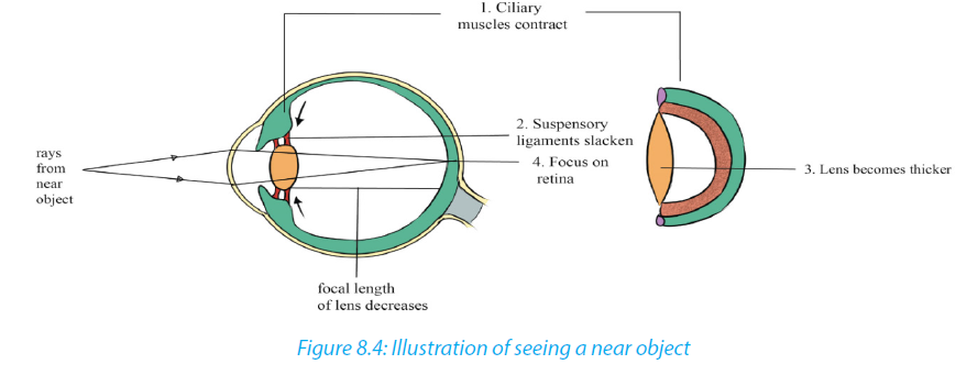
When the eye focuses on a near object, several changes occur:
–– The ciliary muscles contract, relaxing their pull on the suspensory ligaments.
–– The suspensory ligaments slacken, also relaxing their pull on the lens.
–– The lens, being elastic, becomes thicker and more convex, decreasing its focal length.
–– Light rays from the near object are sharply focused on the retina.
–– Photoreceptors are stimulated.
–– The nerve impulses produced are transmitted by the optic nerve to the brain.The brain interprets the impulses and the person sees the near object.b. Focusing on a distant object: When a person is looking at a distant object, the light rays reflecting off the object are almost parallel to each other when they reach the eye. These ‘parallel’ light rays are then refracted through the cornea and the aqueous humour into the pupil
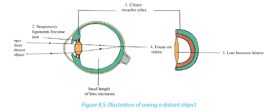
When the eye focuses on a distant object, several changes occur.
–– The ciliary muscles relax, pulling on the suspensory ligaments.
–– The suspensory ligaments then become taut, pulling the edge of the lens.
–– The lens become thinner and less convex, the focal length is increased. The
focal length is the distance between the middle of the lens and the point of
focus on the retina.
–– Light rays from the distant objects are sharply focused on the retina and
photoreceptors are stimulated.
–– The nerve impulses produced are transmitted by the optic nerve to the brain.
The brain interprets the impulses and the person sees the distant object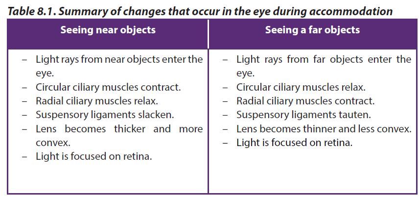
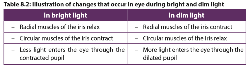
8.3.4. Some changes that occur in eye when you see in bright and dim light
In bright light
–– Circular iris muscle contracts.
–– The radial iris muscles relax.
–– The iris elongates in wards each other.
–– The pupil is reduced (narrowed).
–– Small amount of light rays enters the eye.
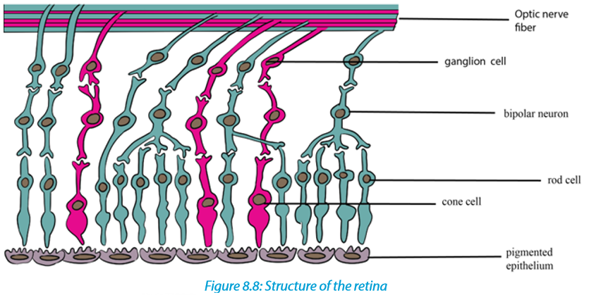
8.3.5. The retina of the eye
The retina possesses the photoreceptor cells. These are of two types, cones and rods. Both converts light energy into the electrical energy or nerve impulses. Both rods and cones are embedded in the pigment epithelial cells of the choroid layer. In cats and some other nocturnal mammals. They have reflecting layer called the tapetum which reflects light back into the eye and so allow the rod cells to absorb it.
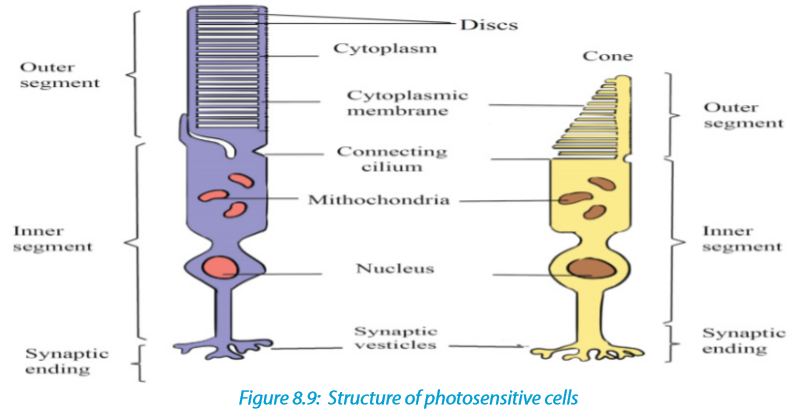
8.3.6. Adaptations of photosensitive cells.
–– They have numerous mitochondria to provide energy in form of ATP.
–– They have photosensitive pigment i.e. rhodopsin in rods and iodopsin in cones to absorb light rays.
–– They have lamellae (vesicles) to increase the surface area for holding the pigment molecules.
–– Many rods cells share a single bipolar neurone such that a single stimulation builds up a big generator potential.8.3.7. Changes which occur on rod cells when light strikes the retina
Each rod cell has in its outer segment up to 1000 vesicles, each containing a photosensitive pigment called rhodopsin. Rhodopsin is made up of the protein opsin and retinal, a derivative of vitamin A. Light causes retinal to change shape from its normal cis-isomeric form to trans-isomeric form. As result, retinal and opsin break apart. This process is called bleaching. This triggers a series of events which alters the permeability of rod’s cell surface membrane.
If light stimulation exceeds the threshold level, an action poetical is set up in a bipolar neurone, and then passes along a neurone in the optic nerve. The pattern of nerve impulses transmitted along different neurones is interpreted in the brain as patterns of light and dark. Before the rod cell can be activated again, the opsin and retinal must first be resynthesized into rhodopsin.
This re-synthesis is carried out by the mitochondria found in the inner segment of rod cell, which provide ATP for the process. Re-synthesis takes longer time than splitting of rhodopsin but is more rapid in lower light intensity. Rhodopsin of rods spits into opsin protein and retinal (derivative of vitamin A). About 3 minutes are required to reform again. That is why our eyes need some minutes to adapt to dark when we come from bright light.

The splitting of iodopsins of cone cells also produces an action potential (impulse) but they quickly re-form. There are three types of iodopsins and each responds to the wavelength of a particular colour: red – green – blue.
The impulses are then transmitted along the optic nerve to the visual area of the brain. There, the image is interpreted. Note that the image that is cast on the retina is virtual I to mean not real, small, inverted upside down and laterally, and reversed for example from right to left.
8.3.8. Changes which occur on cones when light strikes the retina
When light of high intensity strikes the cones, the iodopsin pigment decomposes into iodide ions and opsin, this process is called bleaching. On the contrary, when enough iodopsin is decomposed, the membrane develops an action potential when it reaches threshold level. An impulse is fired via bipolar neurone to the optic nerve to the brain for interpretation. A comparison between cone and rod cells is summarized in the table 8.3.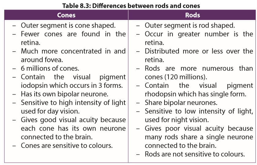
8.3.9. The process of vision
When light enters the eye, it is refracted by the curved surface of the cornea, the lens, the aqueous and vitreous humour. The refraction of light causes the image to be formed upside down on fovea centralis. When cones and rods are stimulated by light, they send impulses through the optic nerves to the brain where the correct impression of the object is formed
Colour vision in organism is explained by the trichromatic theory which states that, there are three forms of iodospin each responding to light of different wave length that is each responds on one of the three primary colours which are, blue, green and red. When these colours are mixed in appropriate intensities they can give rise to any other colour for example equal stimulation of red and green cones gives yellow perception. Alternative theory of colour vision known as the retinex theory, suggests that the brain cortex as well as retina is involved in colour perception. This would explain why we usually perceive a particular object as being the same colour under different types of illumination.
a. Stereoscopic vision: combining two images
Having two eyes (binocular vision) is better than having one because it gives a larger field of vision, a defect in one eye does not result in blindness. In animals with two forward facing eyes, it provides the potential for stereoscopic vision which depends on each eye being able to look at the same object from slightly different perspective. The visual centre in the brain combines the two views to make a three dimensional image. Stereoscopic vision provides information about the sizes and shapes of object
and enables distance to be judged accurately. However, because the eyes have to be relatively close together for stereoscopic vision, the field of vision is relatively small. Mammalian predators tend to have well developed stereoscopic vision, while herbivores tend to have eyes wide apart, sacrificing stereoscopic vision for a wide field of view
b. Nocturnal animals
Nocturnal animals have a lot of rods in their retinas, but no cones. The levels of light at night are very low, so even if the animals have lot of cones, they would not be able to see in colour because the level of light is too low to stimulate the cone cells. At night, animals need to be able to detect shape and movement and the very sensitive rod cells are ideal of this because they are stimulated by very low levels of light.
8.4 Structure and functioning of the ear
The human ear is a complex sensory organ that enables us to hear sounds, detect body movements, and maintain balance. The ear has three main parts: an air-filled (outer ear), an air-filled middle ear, and a fluid- filled inner ear

Each part of the ear has specifc feature and function as it is indicated in the table 8.4.
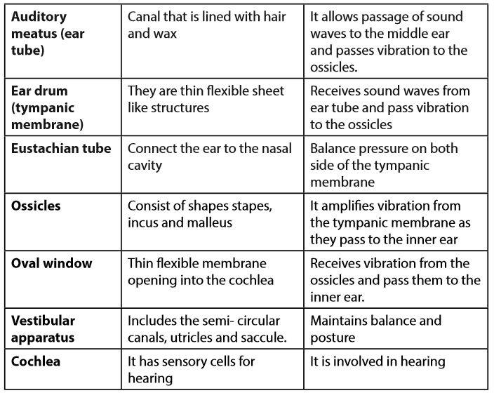
8.4.1. Sound perception in the ear (Hearing)
The most function of the ear is hearing. The hearing process include the following
processes:
–– Sound waves are collected by the pinna and directed to the auditory canal, which then strike the ear drum (tympanic membrane)
–– The sound waves cause the tympanic membrane to vibrate and the vibrations are sent to the ossicles.
–– The ossicles amplify the vibration and amplified vibration are received by the oval window that setting up vibration in the perilymph of tympanic and vestibular canal.
–– Vibration in perilymph cause movement of Reissner’s membrane which in turn displaced relative to the tectorial membrane, the sensory hair cell located between the basilar membrane and tectorial become distorted.
–– This distortion set up an action potential, which is transmitted along the auditory nerve to the brain which interprets the impulses as sound.
8.4.2. The cochlea and the organ of corti
The cochlea is coiled around above and their internal region is crossed by two membranes, i.e. upper Reissner’s membrane and lower basilar membrane. In between there is a membrane which is short called tectorial membrane. From the basilar membrane are sensitive sensory hair cells whose hair tips are close to the tectorial membrane. These cells have fibres which take impulses to the brain along the auditory nerve for interpretation. The upper and lower chambers of the cochlea are filled with perilymph while the middle chamber is filled with endolymph. The basilar membrane, tectorial membrane, Reissner’s membrane and sensitive hair cells are collectively known as the organ of corti and are directly concerned with hearing.

8.4.3. The vestibular apparatus and sense of balance
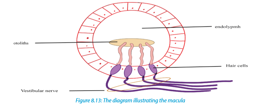
Our sense of balance and information about position and movement come from the vestibular apparatus in the inner ear. The vestibular apparatus consists of the semicircular canals, containing organs called cristae sacs including the saccule and utricle. The utricle and saccule are receptors containing sense organs called maculae that give information on the position of head in space in relation to gravity (static equilibrium).
These receptors consist of sensory hair cells which are embedded in fine granules of calcium carbonate called otoliths. According to the position of the head, the pull of gravity on the otolith will vary and otolith will be titled accordingly. The different distortions of the sensory cells that result from impulses discharge in the vestibular nerve fibres and this is interpreted by the brain, which sends impulses to the relevant organs which then restore the balance of the body
8.4.4. The role of semicircular canals in the maintenance of balance
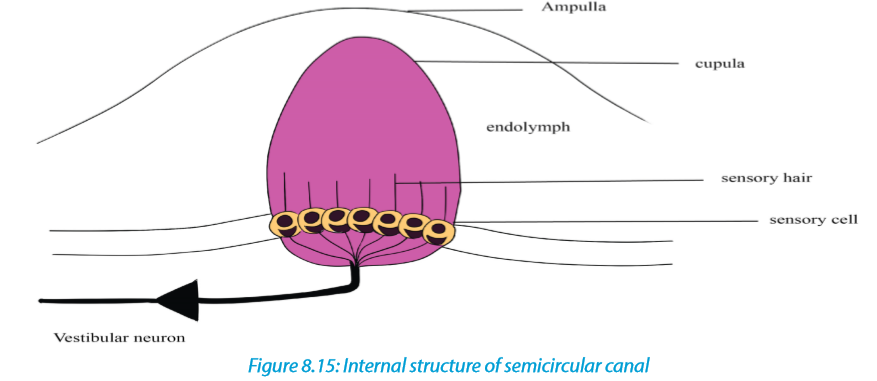
Semicircular canals are responsible for maintaining the balance of the body during motion (dynamic equilibrium). These are fluid – filled canals, three in number and arranged in three mutually perpendicular planes: vertical canals detect movement in the upward direction, horizontal canals detect back ward and forward motion while lateral canals detect sideways movement of the head.
A swelling, the ampulla in the canal contains the receptor. This consists of sensory hair cells supported by hairs embedded in a dome – shaped of a gelatinous structure called cupula. Movements of head in any of the planes causes the fluid in the relevant canal to move and therefore displacing the cupula. Due to inertia, the cupula is deflected in direction opposite to that of head. This put strain on the sensory cells and causes them to fire impulses in the different nerve fibres to the brain. The pattern of impulses sent to the brain varies depending on the canal stimulated. The brain interprets impulses and detects the speed and direction of movement of head. Then impulses from brain are sent to the relevant organs which then maintained the balance of the body.

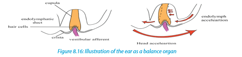
8.4.5 Ear as a balance organ
The vestibular apparatus is concerned mainly with detecting changes in the head position and body posture. When the head moves quickly, the cupula, knob in the ampulla, moves in the opposite direction. Sensory hairs below the cupula detect the impulse that is brought by a vestibular nerve to the brain.
Likewise, as the head moves by changing its posture, some crystals of CaCO3 known as otoliths also move. The membrane of the otoliths also moves pulling on the sensitive hairs and making them bend. The sense cells are stimulated to varying degrees, causing an action potential to be sent to the cerebellum (hindbrain) that actually controls the muscles in maintenance of body balance. The cerebellum sends out impulses to the muscles of the body which contract or relax or maintain body balance.
8.5 Structure and functioning of the tongue
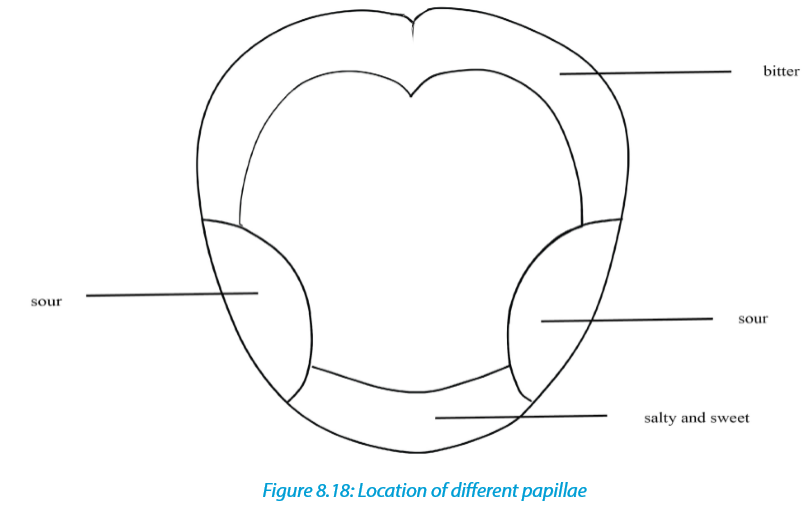
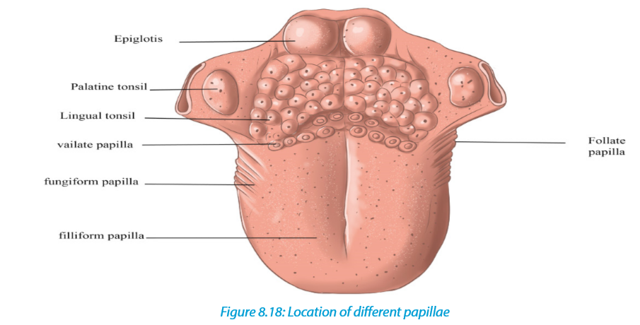
The tongue is the receptor organ for taste. Taste is due to chemicals taken into the mouth and for this reason the tongue is called chemoreceptor. The tongue is able to distinguish between four different kinds of taste including sweet, sour, salt and bitter which are also called primary taste. This is possible with the help of group of sensory cells found in taste buds located on the surface of the tongue in specific taste areas through four types of taste buds in which they are located in overlap as shown on the Figure 8.18, the detection of sour and bitter substances is important for they can be easily rejected if harmful. For a chemical to be tasted it must be dissolved in the moisture of the buccal cavity where it can stimulate the sensory cells grouped in taste buds.
Different types of taste and their sites on the tongue
In human, there are four kinds of taste including sweet, salty, sour and bitter. Different taste buds are sensitive to different chemicals: Those which are sensitive to sugary and salty fluids are usually found at the tip of the tongue while those at the sides of the tongue are sensitive to acidic substances and thus give the sensation of sourness while those at the back are responsible for the sensation of the bitterness.
8.6 Structure and functioning of the skin
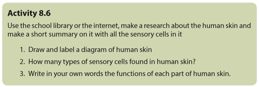
The human skin is the largest organ of the body. Being a vast organ, it has many functions including protection from microbes, regulation of the body temperature, and permits the sensations of touch, heat, and cold. This is possible thanks to the presence of different glands. The skin consists of three main layers: The epidermis, the outermost layer of skin that provides a waterproof barrier and creates our skin tone, the dermis, beneath the epidermis that contains tough connective tissue, hair follicles, and sweat glands and the deeper subcutaneous tissue called hypodermis that is made of fat and connective tissue. The epidermis consists of three regions:
–– The Cornfield layer also known as keratinized layer. This is the thin outermost layer made up of dead cells. It is resistant to bacterial infections and damage, and reduces water loss from the body. It is very thick on the soles of the feet and the palm and is also modified as nails.
–– The Granular layer that contains living cells which give way to the cornfield
layer.
–– Malpighian layer that is the continuous layer of living cells and they continuously divide to produce new cells. This layer has melanin pigment granules that determine the skin colour and act as screen against ultraviolet light.The dermis consists of the thick connective tissue. It consists of blood capillaries, receptors (sensory organs), lymphatic, sweat glands, sebaceous glands and hair follicles with different functions:
–– Capillaries supply food and oxygen, remove excretory waste products and help in temperature regulation.
–– Sweat glands are coiled tubes consisting of secretory cells with duct that passes sweat to the skin surface.
–– Hair follicles are deep pit (hole) of cells which divide and build the hair inside the follicle. They are richly supplied with sensory nerve endings which are stimulated by the hair movements.
–– Sebaceous gland opens into the hair and secretes oil which makes the hair waterproof.
–– Sensory nerve endings include sensory receptors for temperature, touch, pressure and pain.
Subcutaneous layer attaches dermis to underlying structures, composed of adipose
and connective tissue. It serves as shock absorbers for vital organs, it stores energy. It
varies in thickness according to age, sex, general health of individual.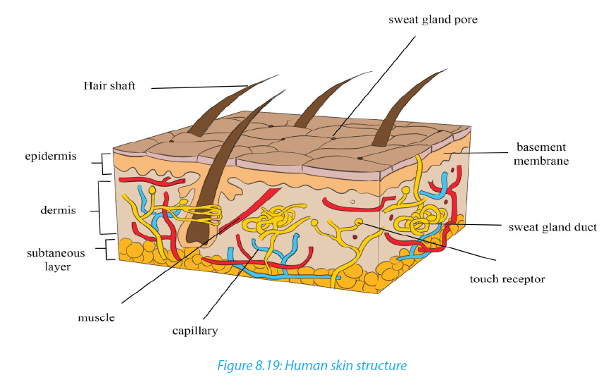
A comparative study of sense organs
Sense organs have different biological functions beneficial to the living organisms. A brief summary is given in the table 8.5.