General
- S6: Biology SB File Uploaded 3/08/22, 12:46
- S6: Biology TG File Uploaded 3/08/22, 12:49
UNIT 12: HUMAN REPRODUCTION
Key Unit Competence
Explain the role of hormones in human reproduction, stages of pregnancy and foetal development.Learning objectives
By the end of the lesson, I should be able to:
–– Define menstrual cycle
–– Describe main events of menstrual cycle
–– Describe the hormonal changes involved in menstrual cycle.
–– Distinguish oestrous and menstrual cycle
–– Describe how mammals mate
–– Explain how a sperm enters and fertilizes an ovum and how only one sperm fertilizes an ovum.
–– Outline the technique of in vitro fertilization (IVF).
–– Explain the physiological changes in females during pregnancy.
–– Explain how placenta forms and discuss its functions.
–– Explain the gestation period birth.
–– Describe the main stages of birth.
–– Discuss the significance of parental care in mammals
–– Explain how twins and multiple birth arise.
–– Describe the main types of birth control techniques.
–– Discuss advantages and disadvantages of different birth control methods.
–– State the causes and the ways of prevention of STIS and HIV.
12.1 Menstrual cycle
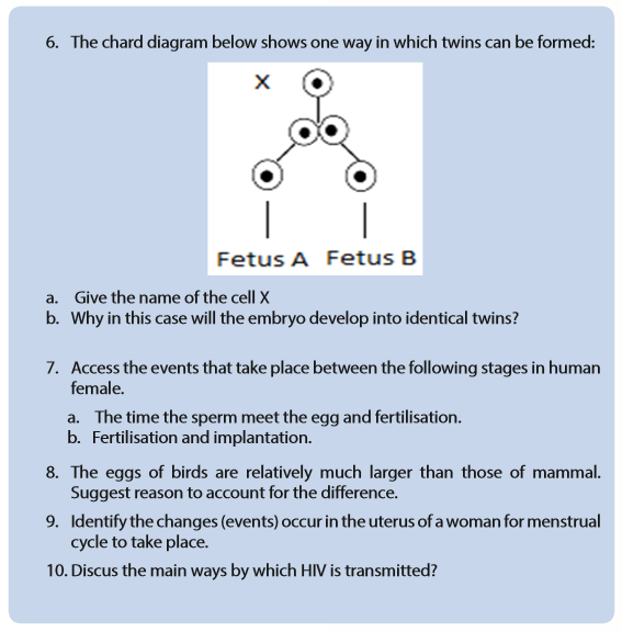
This refers to the periodical changes in the reproductive behaviour of a female which tend to occur in a sequence of events one after the other in the periodical circle. At the onset of puberty, the cycle begins and repeats after 28 days unless interrupted by pregnancy. The changes are stimulated by the gonadotrophic hormone such as; follicle stimulating hormone (FSH) and luteinizing hormone (LH). These hormones stimulate ovaries to secrete; oestrogen (steroid) and progesterone hormones. These four hormones are involved in menstrual cycle. Two of them including; FSH and LH are produced by pituitary gland and the other two are released by ovaries respectively. The most obvious sign of the cycle is the monthly discharge of blood a process called menstruation. The first day of menstruation is regarded as the first day of the cycle. Figure 12.2 and 12.3 show the stages of menstrual cycle. Menstrual cycle is divided into three phases or events:
a. Follicular phase
Menstrual cycle usually begins when blood is first discharged from the uterus during the first to fifth day (1-5 days). Following the reduction of progesterone, the hypothalamus releases gonadotropin releasing hormone (GnRH) which stimulates anterior pituitary gland to secrete follicle stimulating hormone (FSH). FSH brings about the following effects;
–– Stimulates the development of a primary follicle
–– Contributes to the shedding of uterine wall
–– Causes production of oestrogen by uterine cells. The oestrogen produced
promotes healing, repair and growth of uterine lining, inhibits further secretion of FSH. Oestrogen levels keep on raising until day 13 where they stimulate secretion of luteinizing hormone (LH) by anterior pituitary gland.b. Ovulatory phase
Around the 14th day, the high levels of oestrogen cause release of luteinizing hormone (LH) the release of LH brings about ovulation (release of mature egg from the ovary). Immediately after and slightly before ovulation, a woman is fertile and can conceive a baby if she has sexual intercourse or if sperm is present in her oviduct.
c. Luteal phase
After ovulation, the remains of ovarian follicle form corpus luteum also known as Yellow body, which secrete large amounts of progesterone hormone and smaller oestrogen. These two hormones; stimulate further development of mammary glands, inhibit release of FSH and thickening wall of uterus in anticipation of pregnancy. If oocyte (ovum) is not fertilized with in about 36 hours of being shed into oviduct, it dies and corpus luteum gets smaller. Thus levels of progesterone and oestrogen keep on reducing until day 28 days i.e. 14 days after ovulation. Low levels of progesterone remove the inhibitory effect on FSH, causing its release thus menstruation and the cycle starts again.
–– At menopause there are no more fertile follicle so follicular development and ovulation is ceased.
–– The menstrual cycle is controlled by hormones from both brain and the ovary.
–– The natural cycle repeats until there is either a pregnancy or the woman reaches menopause, the end of the reproductive phase of a woman’s life.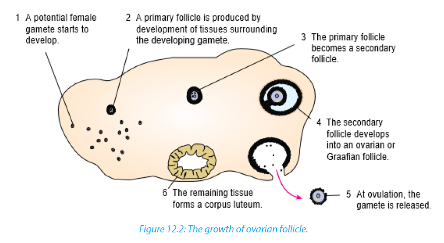

The uterine cycle also has three phases (events):
Proliferative phase: It stimulates the thickening of endometrium of the uterus. This thickness of endometrium is stimulated by oestrogen from follicles before ovulation. This results the development of ovary. It acts like follicular phase.Secretory phase: it occurs after ovulation for describes further thickening of endometrium (endometrium tissue become more complex) in preparation for implantation. This is stimulated by progesterone which is secreted by corpus luteum and this occurs when corpus luteum is functioning. It acts like lacteal phase.
Menstrual phase: when endometrium tissue is discharged and vaginal bleeding occurs at the end of ovulatory cycle if pregnancy has not occurred. It is called menstruation.it describes the shedding of endometrium when implantation does not occur. When pregnancy does not occur the level of progesterone falls and this results shedding of endometrium. Menstrual bleeding lasts between 3 and 5 days. The first day of the period is the first day of the cycle.
12.2 Oestrous cycle

The word oestrus is derived from the Latin language oestrus meaning sexual desire. It describes the phase when the female animal is sexually receptive to a male. Females of most species of mammals except human come into ‘heat’ known as oestrus in regular cycles at particular times of year. Oestrus is the time when females are both fertile and sexually receptive. Oestrus cycle is controlled by the same hormones as the human menstrual cycle. FSH and oestrogen control the process until ripe ova are released when LH and progesterone take over.
12.3 Copulation, fertilization and embryo development.

12.3.1 Copulation
It is act of mating where sperms from male are transferred into the female tract. Male mammals have an intromittent organ called penis which becomes erect at a moment of mating for insertion into female’s vagina. The erection of penis is brought by hydraulic action (penis becomes gorged with blood). This occurs as a result of sexual arousal which brings about by ejaculation (release of sperm). The semen’s are secreted from accessory glands into vas deferens and bladder sphincter closes preventing urine from entering urethra. Sperms are expelled from epididymis into vas deferens and out of the body by a series of muscle contraction of penis.
In a female, sexual arousal results in the swelling of clitoris and stimulates the secretion of mucus which lubricates vagina during sexual intercourse.
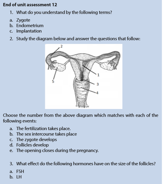
12.3.2 Fertilisation
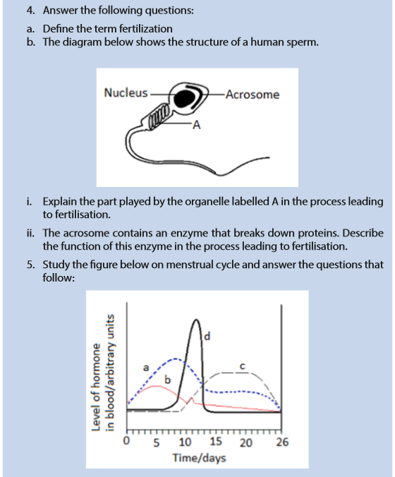
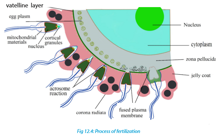
Fertilisation is the fusion of male and female nuclei to form zygote. Copulation results in the ejection of spermatozoa into vagina. The spermatozoa swim in the watery mucus of vagina and uterus up into the oviduct where the fertilisation takes place in the upper part of the oviduct. From the vagina or uterus spermatozoa propel using energy from mitochondria. If ovulation has already taken place, the egg and sperm meet in the upper part of oviduct and once they come into contact, acrosome raptures and release lytic enzyme which dissolve corona radiata of the egg and soften zona pellucida and vetelline membrane. The following processes take place:
a. Capacitation
This is a stage where by sperm undergoes essential changes while passing through female genital trackand this takes about 7 hours. These changes include the removal of a layer of glycoprotein from outer surface of sperm, by enzyme in uterus. Cholesterol also is removed to weaken the membrane.
b. Acrosome reaction
This involves the releasing of enzyme found in acrosome such as hyaluronidases and protease. These enzymes digest corona radiata (narrow path in the follicle cells) and the zona pellucida (a protective glycoprotein surrounding the plasma membrane of the egg).
c. Fusion
In this stage the head of sperm will fuse with the microvilli surrounding the secondary oocyte and penetrate its cytoplasm.
d. Cortical reaction
This stage involves the releasing of enzymes by lysosomes in cortical granules (outer region of the secondary oocytes); the enzymes cause the zona pellucida to thicken and harden forming a fertilization membrane. This cortical reaction prevents the entry of other sperm inside ovum (polyspermy).
e. Zygote formation
The secondary oocyte is stimulated to complete meiosis II, during this time of stimulation the nucleus of sperm and secondary oocyte are called pro-nuclei and then the two nuclei fuse to form the zygote (2n).
The movement of sperm in the female reproductive system;
Once sperm arrives the female reproductive tract, they moved largely by female reproductive system:
–– Around the time of ovulation, the vaginal mucus changes in PH in response to changing levels of sex hormones. It is normally so acidic which can tend to kill sperm. At the fertile time it becomes more alkaline to prevent sperm from damage.
–– The mucus which blocks the cervix, preventing the entry of pathogens and become less viscous, allowing sperm to move through it more easily.
–– Prostaglandin (local hormone) in semen and oxytocin hormone released by posterior pituitary gland during sexual intercourse. Initiate the contraction in uterus, helps semen to move towards fallopian tube.12.3.3 Embryonic development
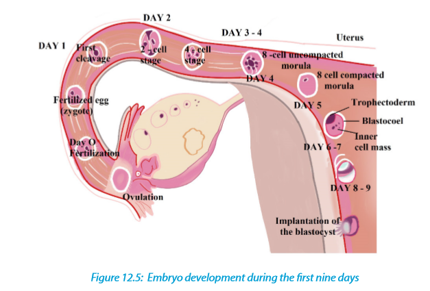
The zygote spends the next few days travelling down the oviduct (Fallopian tube) by peristaltic contraction and by beatings of the cilia in wall of the oviduct toward the uterus. As it travels, it divides by mitosis several times to form a ball of cells called a morula. The cell divisions, which are called cleavage, increase the number of cells but not their overall size. More cell divisions occur, and soon a fluid-filled cavity forms inside the ball of cells. At this stage, the ball of cells is called a blastocyst.
The blastocyst reaches the uterus and becomes embedded in the endometrium at roughly the 5th – 10th day. Once in the uterus the blastocyst burrows into the uterine wall a process called implantation. After implantation, the blastocyst becomes embryo. It grows through multiplication and differentiation of its cells forming tissues and organs. The heart and blood vessels are the first organs formed and embryo now called foetus.
a. Stages of embryo development:
There are three major stages of embryo development;i) Cleavage
The cleavage consists of the division of zygote without increase in mass into a ball of consisting of many daughter cells.ii) Gastrulation
It is the development of different layers of cells in the embryo. It generally occurs during the second week after fertilization. During gastrulation, cells of the embryo migrate to form three distinct cell layers: the ectoderm, mesoderm, and endoderm.
Each layer will eventually develop into certain types of tissues and cells in the body
of vertebrates.
–– Ectoderm—it forms tissues that cover the outer body; develops into cells such as nerves skin, hair, and nails.
–– Mesoderm—it forms tissues that provide movement and support; develops into cells such as muscles, bones, teeth, and blood.
–– Endoderm—it forms tissues involved in digestion and breathing; develop into organs such as lungs, liver, pancreas, and gall bladder.
-
iii) Organogenesis and Differentiation
Differentiation of cells leads to the development of specific organs and tissues within the three cell layers. This is called organogenesis. All the major organs begin to form during the remaining weeks of embryonic development.b. Extra-embryonic membranes
These membranes are part of placenta. The outer cells of the blastocyst, the trophoblast grow and develop into an outer layer or membrane called the chorion. This plays a major role in nourishing and removing waste products from the developing embryo.The amnion is a thin membrane covering the embryo like an umbrella and has a protective function. Between the embryo and the amnion is the amniotic fluid. The amniotic fluid supports the embryo and protects it from mechanical shocks. The yolk sac has no significant function in humans but is important in reptiles and birds, where it absorbs food from the separate yolk and transfers food to the gut of the developing embryo.
Note:
The first trimester of the development or the embryo is critical. There is high risk of spontaneous abortion or miscarriage due to alcohol, infection, radiations (X-rays), nutritional deficiencies, genetic mistakes or abnormalities in the developing embryo. From the 8th week until birth (around 38 weeks), the developing organism is called a foetus. The foetus is not as sensitive to damage from environmental exposures as the embryo, and toxic exposures often cause physiological abnormalities or minor congenital malformation. All major structures are already formed in the foetus, but they continue to grow and develop.
12.4 Role of Placenta in the development of embryo
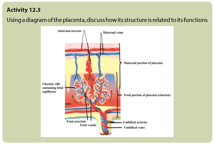
The placenta is a temporary organ in which nutrients and wastes are exchanged between the mother and the embryo or foetus.
The foetal part of the placenta consists of the allantoides and chorion. The chorion forms many large projections called chorionic villi which contain a dense network of foetal capillaries which in turn are connected to two umbilical arteries and umbilical vein in the umbilical cord. The umbilical arteries carry blood from the foetus to the placenta, while the umbilical vein carries blood in the opposite direction. Although maternal blood in the endometrium is in close proximity with the foetal blood in the umbilical capillaries, they do not mix because they separated by membranes of the villi and capillary.
12.4.1 Functions of the placenta:
–– Allows diffusion of nutrients such as water, glucose, amino acids, simple proteins and mineral salts from maternal blood.
–– It is a site of gaseous exchange: haemoglobin of the foetus has high affinity to oxygen compared to adult haemoglobin.
–– It offers passive natural immunity on the foetus. Certain maternal antibodies can cross the placental barrier.
–– It protects foetal circulation from the high pressure in the maternal circulation
–– Prevents mixing of maternal and foetal blood which would cause agglutination (clotting) if the two blood types are incompatible.
–– It produces and secretes hormones such as the HCG (human chorionic gonadotrophin), progesterone, oestrogen, and relaxin.Note that:
–– The action of HCG is similar to that of LH. HCG stimulates the corpus luteum to secrete progesterone and oestrogen throughout the first trimester. HCG is produced in such large quantities that some of it is excreted in the urine of a pregnant woman (positive test of pregnancy). Secretion of HCG declines
around tenth week and the corpus luteum reduces.
–– The placenta does not give complete protection to the foetus. Certain pathogens, toxins, and drugs can enter the foetal circulation and cause damage. Examples are; HIV, rubella toxins, alcohol, nicotine and heroin.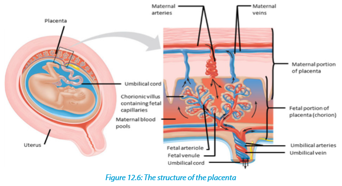
12.4.2 How the placenta works?
Blood from the mother enters the maternal blood vessels of the placenta under pressure, forcing the blood into the empty spaces. When the mother’s blood contacts the foetal blood vessels, gases are exchanged. Oxygen from the mother’s blood is exchanged with carbon dioxide from the foetus’s blood. A release of pressure brings the mother’s blood back from the placenta and into her veins.
– The substances that are moved from the mother to the foetus include:
–– Water
–– Glucose by passive diffusion
–– Hormones
–– Amino acids by active transport
–– Lipids by membrane lipid diffusion
–– Oxygen is released by the maternal haemoglobin. The haemoglobin of the foetus has a higher affinity for the oxygen.
–– Alcohol, many drugs, nicotine (if taken by mother during pregnancy)
–– Vitamins, minerals.The substances that are moved from the foetus to the mother include:
Carbon dioxide is taken up by the maternal plasma and transported to the lungs of the mother for excretion
–– Urea passes into the maternal blood and passes to her kidneys for excretion. The exchange between the mother and the foetus is possible because of specific structures in the placenta:–– The plasma surface membranes of the cells in the walls of the chorionic villi have
microvilli, which increase their surface area for the exchange of substances by diffusion, facilitated transport and pinocytosis.–– Numerous mitochondria are found in these cells. They provide the energy for the active transport and pinocytosis.
–– The cell surface membranes contain carrier molecules (protein) used in the uptake of materials into the villi by active transport.
–– Numerous small vesicles are found inside the cells of the villi as a result of materials being taken up from the blood by pinocytosis.
12.5 Physiological changes during pregnancy and parental care

Pregnancy refers to the development that take place between fertilisation of the ovum to birth of the foetus. When fertilised egg becomes implanted in uterine wall, pregnancy results. And a number of important events take place during this period. The period from fertilisation to birth is called gestation period. In human it is about nine months.
12.5.1. Changes during pregnancy
A pregnant woman’s body undergoes various; physiological, physical and behavioural changes.a. Some physiological changes during pregnancy:
–– Respiration rate rises for increased maternal oxygen consumption which is needed for demand of placenta, uterus and foetus.
–– More blood vessels grow and pressure of expanding uterus on large veins causes blood to slow in its return to the heart.
–– Rise up and out of pelvic cavity this action displaces the stomach and intestine.
–– Blood volume increase greatly.
–– Placenta produces large amount of progesterone and oestrogen by 10 to 12 week of pregnancy to control uterine activity.
–– Increased requirement of calcium due to increase of parathyroid gland.
–– Experiences warm (hot flashes) caused by basal metabolic rate and increased hormonal level.
–– Stretching of abdomen wall and ligaments that support uterus.
–– Kidney work extra hard to excrete waste products of both mother and foetus.
b. Some physical changes during pregnancy
–– Breast may become large and more tender because of increased level of oestrogen hormone progesterone thus breast gets even bigger to prepare for breast feeding.
–– Nipples may stick out more.
–– By the end of third trimester, a yellow, watery, pre-milk may leak from nipples.
–– Changes in hair and nail growth and texture due to hormone changes.
–– Leg cramp caused by fatigue from carrying pregnant weight.
–– Feet and ankles may swell because of extra fluid in the body during pregnancy.c. Some behavioural changes during pregnancy:
–– Physical discomfort such as urinary frequency can be frustrating.
–– Fear and anxiety lessen especially foetal movement are felt.
–– Self-introspection
–– Nesting behaviour begins. Some woman exhibit mood swings and emotional liability.12.5.2. Delivery process
By the end of pregnancy, near the time of birth, the amniotic sac raptures (breaks) and amniotic fluid drains through birth canal and labour usually begins which involves the contractions of muscular walls of the uterus.Initiation of birth: Uterine contractions starts when the foetal pituitary gland secretes adrenocorticotrophic hormone (ACTH) which stimulates foetal adrenal gland to secrete corticosteroids. These hormones pass into blood sinuses in placenta to cause maternal cells to secrete prostaglandins (local hormone) and cause uterine wall to contract. This contraction pushes the foetal head against the cervix to stimulating stretcher receptor to send information to mother’s brain and causes release of oxytocin hormone. The prostaglandin and oxytocin hormone together result intense contraction of uterine walls called labour which stimulates more release of oxytocin hormone and as positive feedback mechanism.
The delivery process can be summarized into three main stages:
–– Dilation stage: During this stage, water sac filled with amniotic fluid forms and precedes the head, widening soft tissue of birth canal, cervix, and vagina for canal of constant diameter. The amnion raptures and amniotic fluid drains through vagina.–– The expulsion stage: During this stage, cervix is fully dilated while abdominal muscle bear down in supporting rhythmic contraction of uterus shorten the uterine wall and baby is pushed into and through the birth canal. The head and shoulder align themselves first.
–– Placenta stage: This stage begins with complete expulsion of baby and ends with expulsion of foetal membrane. The cord is clamped and cut when delivery of baby is complete. This leads carbon dioxide enrichment into baby’s blood which activates respiratory centre and baby begins to breath with the first cry at the same time foetal circulation changes to baby’s own systemic and
12.5.3 Parental care
The degree of maturity in mammalian new-borns varies from one species to another. New-born in pigs can move around and eat solid food while new-born in humans, dogs and rat are quite helpless and require a lot of parental care to survive. All mammals feed their young ones by milk which contain all the nutrients required by new born for the first few days. Parents also protect new born from predators
and from unfavourable weather. Some species make nest just before delivering the new born. Some parents also become aggressive when they have young one. As the young one grow older the parent start gathering food for them. Once the new born get old enough to gather food for themselves can leave on their own. In humans’ parental care extends for very long time up over 18 years.In humans breastfeeding is associated with many benefits:
–– It makes earlier a closer contact between the mother and her infant
–– Breastfed babies do not get too fat
–– The infant has a better control over its own milk intake, this prevents over eating in late life
–– Fats and irons from breast milk are better absorbed than those in cow’s milk and milk is easily digested.
–– Breast feeding provides important antibodies that help to prevent respiratory
infections and meningitis,
–– Breastfeeding helps the mother’s reproduction organ return to a normal state more rapidly
–– Breast feeding promotes the secretion of LH (and prolactin) and this makes a delay in follicle development and ovulation,
–– The act of sucking on the breasts, promotes the development of the jaw, facial muscles and teeth (sucking from a bottle requires less effort).
–– Pulmonary circulation. After delivery, uterus contract so that placenta separates from
–– Uterine wall expelled out as the sign of birth end.
12.6 Twins and multiple births

Twins are individuals born to the same mother at the same time. Twins include;
–– Fraternal twins or non-identical twins or dizygotic twins: These are twins which develop from two separate egg cells fertilised by two different sperms.
Fraternal twins are genetically different since they develop from different gametes.–– Identical twins or monozygotic twins: these are twins which develop from the same fertilised egg. Identical twins are genetically similar since they develop from the same sperm and the same egg.
–– Siamese twins: are conjoint identical twins i.e. they have not completely separated during the embryo development. As consequence, they share same organs. Conjoint identical twins develop without separating completely and are born attached to one another. Such twins may be separated surgically.
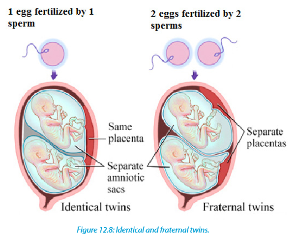
Multiple births arise when several eggs are released at the ovulation and are fertilised or when a zygote splits into several zygotes. It is commonly occurring in mammals such as; pigs, dogs and cats.

12.7 Infertility or barrenness
Infertility
Infertility is the failure to achieve pregnancy when no contraceptive method is used.
In females, infertility may be due to:
–– Failure to ovulate due to the lack of some hormones
–– Damage of the Fallopian tubes / oviducts, for example the tubes may be
completely blocked by nature or after an infection,
–– Damage on the uterus; for example, the endometrium can be destroyed
–– Damage on the cervix, for example the cervix may be narrow or too wide or may stop producing cervical mucus needed for the sperm to reach uterus
–– Antibodies against sperms, for example, the cervix, the uterus or the oviduct of a woman can produce antibodies against her husband’s sperms.Some causes of infertility/barrenness in males include:
–– Absence of sperms in the semen (Azoospermia).
–– Low sperm count e.g. when ones ejaculate less than 1cm3 of semen.
–– Abnormal sperm e.g. sperms with 2 tails, or without tail, or without acrosomes,
–– Auto-immunity e.g. antibodies attack one’s sperms
–– Premature ejaculation: the man has orgasm before copulation
–– Impotence i.e. inability to achieve or maintain an erection of the penis.
a. Some social consequences include:
–– Isolation including exclusion from ceremonies and social gathering.
–– Rejection being an outcast and physical abuse perpetrated by community
members.
–– Stigmatization or recognizable marginalization.
–– Status loss that is no respect and social fail.
–– Ridicule including insults and verbal abuse.Some economic consequences include:
–– Cost of infertility by either modern biomedical or traditional treatments.
–– A feeling of rejection.
–– Having few relations, receiving few gifts and less land.
–– Marital instability including fear of husband taking second wife.
–– Divorcing childless woman
–– Violence perpetrated by partner.Note:
While infertility may result into conflicts between couples and families, producing many children also brings about some economic challenges. Many children affect families’ financial wellbeing and some parents admit that children are expensive. Consequences of many children per one family include:
–– High rate of maternal depression.
–– Low rate of immunization and parental care.
–– Baby taxing both physical and emotional especially off work after birth.
–– I come tend to go up when new members of the family arrive. Men see the boost in their earnings after birth of child.
–– There is economic wellbeing decline in time around birth.b. Increasing fertility
Increasing fertility can be done in various techniques such as:
–– Fertility drugs: a synthetic chemical which stimulates ovulation by either proving gonadotrophins such as FSH which stimulates growth of follicles. Or proving chemical which inhibits natural production of oestrogen.
–– Artificial insemination: sperm from donor is inserted artificially through cervix of mother to be.
–– Using in-vitro fertilisation12.7.2 In-vitro-fertilisation
In-vitro fertilisation is the process of fertilisation where an egg is fertilised by sperm outside the body. It involves the fertilisation of egg cell outside the body which are then artificially implanted in the uterus to produce test tube baby. The process involves monitoring and stimulating of woman’s ovulatory process removing ovum (egg) from woman’s ovaries and letting sperm to fertilise them in liquid laboratory.
The fertilised egg (zygote) undergoes embryo cultured for 2 to 6 days and then transferred to the same or another uterus for successful pregnancy. The embryo is implanted in woman’s uterus.Advantages of in vitro-fertilization techniques include:
–– Simplicity: living organisms are extremely complex functional system with protein molecules, RNA molecules and genes. Therefore, the work of Vitro simplifies system under study to focus on small number of components.
–– Species specificity.in human cells in-vitro method can be studied without extrapolation from experimental animal’s cellular response.
–– Automation and convenience: In-vitro method can be automated, high yielding throughout screening methods for testing molecule in pharmacology.
–– In vitro- fertilisation can be used to achieve successful pregnancy but the process usually produces more embryos which some scientists wish for research design to improve our knowledge about disease.
12.8 Family planning: birth control and contraception
–– Birth control includes contraception, but is broader in meaning because it also includes any measures taken after fertilization which are designed to prevent birth. Contraceptionis preventing the fusion of the male gamete and female gamete. Both natural and artificial methods exist.
Artificial methods:
–– Oral Contraceptive pills: a chemical method of contraception. One version uses a combination of progesterone and oestrogen that inhibits ovulation. Others are single hormones that require very careful management when taken.
–– Intrauterine device (IUD) the coil is placed inside the uterus an exact understanding how this works is unclear. A possible explanation is that it ‘irritates’ the endometrium such that rejects implantation of embryos. The device is made from plastic or copper and inserted by a doctor. Nevertheless, this device is very effective.–– Condom is another mechanical method of contraception that prevents the sperm from reaching the egg. Composed of a thin barrier of latex this is placed over the erect penis and captures semen on ejaculation. This is also a good barrier to prevent the transmission of sexual diseases.
–– Cap (diaphragm) is another barrier method again made from latex. The cap is placed over the cervix to prevent the entry of sperm in semen. This technique requires that the cap is put in position in advance of sexual intercourse and that it is used in combination with a spermicidal cream. When used correctly
this is an effective contraceptive however this is not a barrier against the transmission of sexual diseases.–– Sterilisation is a surgical and near permanent solution for contraception such as: Vasectomy. In men this involves cutting the vas deferens and prevents sperm entering the semen. In this state, man still ejaculates normally and releases semen however this does not contain sperm.
–– Tubal ligation. Involves the cutting of fallopian tube so that eggs cannot reach the uterus. In women the surgery cuts or ties the oviducts thus preventing sperm from reaching the egg in fertilisation.
–– Natural method:
–– Natural birth control methods include specific actions that people can do
naturally to help prevent an unintended pregnancy.–– Abstinence: the individual makes the choice to delay sexual intercourse until the decision to conceive a child is made.
–– Withdrawal is a behavioural action where a man pulls his penis out of the
vagina before he ejaculates. The withdrawal method also relies on complete
self-control. You must have an exact sense of timing to withdraw your penis in
time.
–– Fertility awareness methods: This require a woman to monitor her body to determine when she is most fertile. You then avoid having unprotected sex around the time of ovulation.–– This natural birth control method involves paying attention to different body changes (such as basal body temperature or cervical mucus) and recording them to predict when you will ovulate. To be successful, you need to be willing to record and chart your fertility signs.
–– Then, you (and your partner) must agree to not have sex (or to use backup
birth control) for 7 days before and 2 days after you ovulate.–– Fertility awareness methods include the Billings Method, the Symptothermal Method, and the Standard Days method.
–– Continuous (Lactational Amenorrhea Method) can postpone ovulation for up to 6 months after giving birth. This natural birth control method works because the hormone required to stimulate milk production prevents the release of the hormone that triggers ovulation.Advantages and disadvantages of birth control
Some advantages of birth control/contraceptives
–– Gives great protection against unplanned pregnancy if one follows instructions.
–– Condoms to some extent protect against pregnancy and STDS.
–– Combinations of pills reduce/prevent cysts in breasts and ovaries.
–– Improved family wellbeing.
–– Improved maternal and infant health.
Some disadvantages of birth control/contraceptives
–– Necessity of taking medication continually.
–– High cost of medication.
–– Hormonal contraceptive does not protect against STDS.
–– Eggs may fail to mature in the ovary for a woman who uses hormonal
contraceptives.
–– Woman must remember to take them regularly.
–– Woman must begin using hormonal contraceptive in advance before they
become effective.
–– Some women experience several; headaches, breast tenderness, chest pain,
discharge from vagina, leg cramps and swelling or pain.
12.9 Causes and prevention of STIs and HIV
Sexual transmitted infections include:
1. Acquired Immune Deficiency Syndrome (AIDS)
It is a serious disease which suppresses body defence. It is characterised by suppression of immune system leading to development of a number of rare infectious diseases. It is caused by virus known as Human Immunodeficiency Virus (HIV). This virus can be transmitted from sick/infected person to healthy one in a number of ways:
–– None protected sexual intercourse either homosexually or heterosexually. It passes from infected semen or vagina fluid to blood of health person throughdamaged tissue in the vagina, penis or rectum.
–– From sick mother to her baby during birth or through breast milk during
suckling.
–– Through transfusion blood by contaminated needles.
–– Through sharing contaminated sharp instruments.HIV attach white blood cells (helper T cells) which is essential component of the body’s immune system. HIV is retrovirus invades its genetic materials into the host’s body and therefore its DNA remains dormant in host cells and being replicated leading host cells to divide. When HIV uses host cells to manufacture new viruses. New viruses burst out of host cells and eventually kill it and new host cells to infect to supress immune system thus HIV develop into AIDS and show number of diseases such as: tuberculosis, skin cancer, pneumonia and thrush and a person may show some symptoms such as: swelling of lymph glands, fever, sweating and fatigue, coughing, diarrhoea and unexplained loss of weight. The death may result as there is no known cure for AIDS but drugs reduce its progress but cannot stop it. Other
symptoms include:
–– Headache
–– Vomiting, and upset stomach
–– Mouth, genital, or anal sores
–– Rash or flaky skin
–– Short-term memory lossTreatment:
No specific treatment for AIDS but some drugs may be used to treat various infections that come about as result of AIDS.
HIV infection is not easy to treat. Some reasons why HIV is difficult to treat are as follow:–– HIV remains inactive in host cells for years and it cannot be targeted and destroyed.
–– Since its symptoms are not easily evident, the infected person may continue spreading the virus knowingly or unknowingly.
–– HIV is extraordinary variable therefore cells of immune system identify infective agents by shapes of antigen on their protein coats means that HIV cannot be detected easily by changing shape of its antigens.
–– HIV destroys helper T cells which help in body defence thus difficult to control it.
2. Syphilis:
–– It is serious sexually transmitted disease caused by bacteria Treponema pallidum. The symptoms of syphilis occurred in three stages if not cured.–– Stage I: it appears between 10 days to 3 months after the time between contact and appearance of first symptom (incubation period). The disease begins with painless sore which appear on sex organs and it heals itself.
–– Stage II: it appears between 2 to 6 months after contact with disease such as: headache, fever, pain in bones and joints and sore throat.
–– Stage III: it appears about 10 years after contact with disease such as: nervous system, heart and aorta therefore the result is serious damage to affected organs.Ways of transmission: Syphilis can be transmitted through sexual intercourse.
Treatment: Syphilis can be cured completely by antibiotics such as penicillin.3. Gonorrhoea
It is a common sexually transmitted disease caused by bacteria Neisseria gonorrhoea. It can also have transmitted from mother to baby during birth. The first symptoms appear from 3 to 5 days after sexual contact with infected individual and discharges from genital thus burning sensation during urination but in female there is no symptoms:
–– Pain or burning when urinating
–– Yellowish and sometimes bloody vaginal discharge
–– Bleeding between periods
–– Pain during sex
Ways of transmission: Gonorrhoea is transmitted through sexual intercourse. It can
also have transmitted through from mother to baby during birth thus affect newborn’s eyes.
Ways of treatment: It can be cured by antibiotics but if untreated it may lead sterility, heart disease and blindness.4. Genital herpes (simplex).
It is a sexually transmitted disease caused by herpes simplex virus. Symptoms include: small red bumps, blisters, or open sores where the virus entered the body, such as on the penis, vagina, or mouth. Its symptoms include:
–– Vaginal discharge
–– Fever
–– Headache
–– Muscle aches
–– Pain when urinating
–– Itching, burning, or swollen glands in genital area
–– Pain in legs, buttocks, or genital area
–– Symptoms may go away and then come back. Sores heal after 2 to 4 weeks
Ways of treatment: No specific cure for the disease but number of drugs may be
used to reduce pain and even further attach.5. Trichomoniasis
It is caused by protozoan Trichomonas vaginalis, transmitted through sexual contact, underwear and toilet seats. Its symptoms are; itching of urethra or vaginal in females, yellow discharge and smelly.
Ways of Prevention/control include: Avoiding indiscriminate sex, avoiding sharing linen and personal hygiene.6. Hepatitis
It is caused by virus hepatitis B through sexual contact, contaminated needles, blood transfusion and syringes. Its symptoms include; Fever, jaundice, nausea (sickness, vomiting), loss of appetite and yellow urine. Ways of prevention include; avoiding indiscriminate sex, use disposable needles and
syringes and strict personal hygiene.7. Candidiasis
It is caused by fungus Candida albicans through sexual contact, sharing linen and towels. Its symptoms include; Itching and burning sensation and white discharge from genitals.
Ways of prevention/ control include; Avoid indiscriminate sex and treat both partners
Ways of controlling STIs / STDs:
–– Abstaining from sexual intercourse in order to avoid STDS.
–– Using of condoms during sexual intercourse.
–– Going for blood check-up before engaging in sexual activities.
–– Not engaging in homosexuality/lesbianism reduces the risk of STDS.
–– avoiding multiple sexual patners
–– Getting medical attention as soon as possible in case of getting infections.