Topic outline
General
- Integrated Science LE_SSE Y3 TG File Uploaded 1/11/21, 16:27
- Integrated Science LE_SSE Y3 SB File Uploaded 1/11/21, 16:27
UNIT 1: BASIC BIOCHEMISTRY OF LIFE
Key Unit Competence:Explain the cellular respiration and photosynthesisIntroductory activity 1Observe the person in the picture below who is making physical exerciseand attempt the following questions:
 i) Where is the energy used by the person in the picture come from?ii) Why do all living organisms need a continuous supply of energy?iii) Identify the process exhibited by the person on the picture thatconsumes too much energy if compared with another one who is atrest.iv) How is the energy produced in our body? Does energy being produced
in our body serve for our various activities? If yes, how?
All living organisms require a continuous supply of energy to stay alive, eitherfrom the absorption of light energy or from chemical potential energy (energystored in nutrient molecules). The process of photosynthesis transfers lightenergy to chemical potential energy, and so almost all life on Earth dependson photosynthesis, either directly or indirectly. Photosynthesis supplies livingorganisms with two essential requirements: an energy supply and usable carboncompounds.
All biological macromolecules such as carbohydrates, lipids, proteins andnucleic acids contain carbon. All living organisms therefore need a source ofcarbon. Organisms that can use an inorganic carbon source in the form ofcarbon dioxide are called autotrophs. Those needing a ready-made organicsupply of carbon are heterotrophs.
Organic molecules can be used by living organisms in two ways. They canserve as ‘building bricks’ for making other organic molecules that are essentialto the organism, and they can represent chemical potential energy that canbe released by breaking down the molecules in respiration. This energy canthen be used for all forms of work. Heterotrophs depend on autotrophs forboth materials and energy.
1.1. Cellular respiration
Activity 1.1
1. When an ocelot breathes, it acquires oxygen, and when it feeds on a
lizard, it acquires glucose. Both molecules enter its bloodstream and are
carried to the body’s cells, where there is a specific biological processwhich uses both oxygen and glucose.
2. Which biological process is represented in the figure above? Where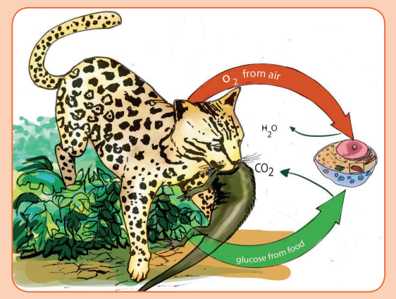
does that process take place in the organism? In a eukaryotic cell?i) Where does the biological process mentioned above take place in
the cell?
ii) Write the chemical equation of that biological process.
iii) The equation written in (iii) above is described in four steps. Conduct
research from the library textbooks or search engine to explain:– How is pyruvate formed from sugar/glucose?2. Yeast, sugar, water and flour are the main components in bread making
– What is the role of Coenzyme A in the link reaction?
– What is the role of reduced NAD+ and FAD in the Krebs cycle?
– What is the final acceptor of protons and electrons in the respiratory
chain?a) Why do bakers add yeast to flour and water when making bread?Cellular respiration is a set of metabolic reactions and processes that take
b) When yeast is added to grape juice at room temperature, vigorous
bubbling occurs. What gas produces the bubbles?
c) What type of beverage is produced by this process?d) What is the name of this process?
place in the cells of organisms to convert biochemical energy from nutrients
into adenosine triphosphate (ATP), and then release waste products.
Cellular respiration is the complex process in which cells make adenosine
triphosphate (ATP) by breaking down organic molecules. The energy stored
in ATP can then be used to drive processes requiring energy, including
biosynthesis, locomotion or transportation of molecules across cell membranes.
The main fuel for most cells is carbohydrate, usually glucose which is used by
most of the cells as respiratory substrate. Some other cells are able to breakdown fatty acids, glycerol and amino acids.
There are two types of cellular respiration, aerobic and anaerobic. Aerobic
respiration is more efficient and can be utilized in the presence of oxygen, while
anaerobic respiration does not require oxygen. Many organisms (or cells) will
use aerobic respiration primarily, however, if there is a limited oxygen supply,they can utilize anaerobic respiration for survival.
The breakdown of glucose can be divided into four stages: glycolysis, the link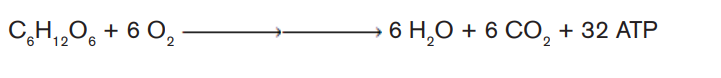
reaction, the Krebs cycle and oxidative phosphorylation.
1.1.1. Glycolysis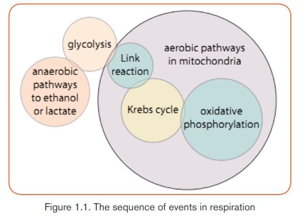
Glycolysis is the splitting, or lysis, of glucose. It is a multi-step process in which
a glucose molecule with six carbon atoms is eventually split into two molecules
of pyruvate, each with three carbon atoms. Energy from ATP is needed in the
first steps, but energy is released in later steps, when it can be used to make
ATP. There is a net gain of two ATP molecules per molecule of glucose brokendown. Glycolysis takes place in the cytoplasm of a cell.
In the first stage, phosphorylation, glucose is phosphorylated using ATP. Glucose
is energy-rich but does not react easily. To tap the bond energy of glucose,
energy must first be used to make the reaction easier. Two ATP molecules
are used for each molecule of glucose to make first glucose phosphate, then
fructose phosphate, then fructose bisphosphate, which breaks down to producetwo molecules of triose phosphate.
Hydrogen is then removed from triose phosphate and transferred to the carrier
molecule NAD (nicotinamide adenine dinucleotide). Two molecules of reduced
NAD are produced for each molecule of glucose entering glycolysis.
The hydrogens carried by reduced NAD can easily be transferred to other
molecules and are used in oxidative phosphorylation to generate ATP.
The end-product of glycolysis, pyruvate, still contains a great deal of chemical
potential energy. When free oxygen is available, some of this energy can be
released via the Krebs cycle and oxidative phosphorylation. However, the
pyruvate first enters the link reaction, which takes place in the mitochondria.
1.1.2. Link reaction
Pyruvate passes by active transport from the cytoplasm, through the outer and
inner membranes of a mitochondrion and into the mitochondrial matrix. Here it
is decarboxylated (this means that carbon dioxide is removed), dehydrogenated
(hydrogen is removed) and combined with coenzyme A (CoA) to give acetylcoenzyme A. This is known as the link reaction
Coenzyme A is a complex molecule composed of a nucleoside (adenine plus
ribose) with a vitamin (pantothenic acid), and acts as a carrier of acetyl groups
to the Krebs cycle. The hydrogen removed from pyruvate is transferred to NAD.
Fatty acids from fat metabolism may also be used to produce acetyl coenzyme
A. Fatty acids are broken down in the mitochondrion in a cycle of reactions in
which each turn of the cycle shortens the fatty acid chain by a two-carbon acetyl
unit. Each of these can react with coenzyme A to produce acetyl coenzyme A,which, like that produced from pyruvate, now enters the Krebs cycle.
1.1.3. The Krebs cycle
The Krebs cycle (also known as the citric acid cycle or tricarboxylic acid cycle)
was discovered in 1937 by Hans Krebs.
The Krebs cycle is a closed pathway of enzyme-controlled reactions.- Acetyl coenzyme A combines with a four-carbon compound (oxaloacetate)For each turn of the cycle, two carbon dioxide molecules are produced,
to form a six-carbon compound (citrate).
- The citrate is decarboxylated and dehydrogenated in a series of steps,
to yield carbon dioxide, which is given off as a waste gas and hydrogens
which are accepted by the carriers NAD and FAD.
- Oxaloacetate is regenerated to combine with another acetyl coenzyme A.
one FAD and three NAD molecules are reduced, and one ATP molecule isgenerated via an intermediate compound.
Although part of aerobic respiration, the reactions of the Krebs cycle make no
use of molecular oxygen.
However, oxygen is necessary for the final stage of aerobic respiration, which is
called oxidative phosphorylation. The most important contribution of the Krebs
cycle to the cell’s energetics is the release of hydrogens, which can be used inoxidative phosphorylation to provide energy to make ATP.
1.1.4. Oxidative phosphorylation and the electron transport
chain
In the final stage of aerobic respiration, oxidative phosphorylation, the
energy for the phosphorylation of ADP to ATP comes from the activity of the
electron transport chain. Oxidative phosphorylation takes place in the innermitochondrial membrane.
Reduced NAD and reduced FAD are passed to the electron transport chain.
Here, the hydrogens are removed from the two hydrogen carriers and each is
split into its constituent proton (H+) and electron (e−).
The energetic electron is transferred to the first of a series of electron carriers.
Most of the carriers are associated with membrane proteins, of which there are
four types. A functional unit, called a respiratory complex, consists of one of
each of these proteins, arranged in such a way that electrons can be passedfrom one to another down an energy gradient.
As an electron moves from one carrier at a higher energy level to another
one at a lower level, energy is released. Some of this energy is used to move
protons from the matrix of the mitochondrion into the space between the inner
and outer membranes of the mitochondrial envelope. This produces a higher
concentration of protons in the intermembrane space than in the matrix, setting
up a concentration gradient.
Now, protons pass back into the mitochondrial matrix through protein channels
in the inner membrane, moving down their concentration gradient. Associated
with each channel is the enzyme ATP synthase. As the protons pass through
the channel, their electrical potential energy is used to synthesise ATP in theprocess called chemiosmosis.
Finally, oxygen has a role to play as the final electron acceptor. In the mitochondrial
matrix, an electron and a proton are transferred to oxygen, reducing it to water.The process of aerobic respiration is complete.
The sequence of events in respiration and their sites are shown in Figure 1.1.
The balance sheet of ATP used and synthesised for each molecule of glucoseentering the respiration pathway is shown in Table 1.1.
Theoretically, three molecules of ATP can be produced from each molecule of
reduced NAD, and two molecules of ATP from each molecule of reduced FAD.
However, this yield cannot be achieved unless ADP and Pi are available inside
the mitochondrion. About 25% of the total energy yield of electron transfer isused to transport ADP into the mitochondrion and ATP into the cytoplasm.
Hence, each reduced NAD molecule entering the chain produces on average
two and a half molecules of ATP, and each reduced FAD produces one and a
half molecules of ATP. The number of ATP molecules actually produced varies in
different tissues and different circumstances, largely dependent on how muchenergy is used to move substances into and out of the mitochondria.
Table1.1: Balance sheet of ATP used and synthesized for each moleculeof glucose entering in respiration
Efficiency of aerobic and anaerobic respiration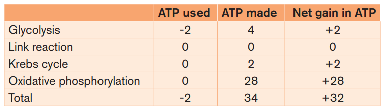
Without oxygen, pyruvate (pyruvic acid) is not metabolized by cellular respiration
but undergoes a process of fermentation. The pyruvate is not transported into
the mitochondrion, but remains in the cytoplasm, where it is converted to waste
products that may be removed from the cell. This serves the purpose of oxidizing
the electron carriers so that they can perform glycolysis again and removing the
excess pyruvate. Fermentation oxidizes NADH to NAD+ so it can be re-used in
glycolysis.
In the absence of oxygen, fermentation prevents the build-up of NADH in
the cytoplasm and provides NAD+ for glycolysis. This waste product varies
depending on the organism. In skeletal muscles, the waste product is lactic acid.
This type of fermentation is called lactic acid fermentation. In yeast and plants,
the waste products are ethanol and carbon dioxide. This type of fermentation is
known as alcoholic or ethanol fermentation. The ATP generated in this process
is made by substrate-level phosphorylation, which does not require oxygen.
Fermentation is less efficient at using the energy from glucose since only 2 ATP
are produced per glucose, compared to the 38 ATP per glucose produced by
aerobic respiration. This is because the waste products of fermentation stillcontain plenty of energy. Glycolytic ATP, however, is created more quickly.
Applications of anaerobic respiration
Some food products and drinks are produced by using anaerobic microorganisms:
- Production of beer
- Production of wine
- Production of yoghurt
- Production of cheese- Production of bread
In the bread-making process, it is the yeast that undergoes cellular respiration.
Anaerobic respiration also known as fermentation helps to produce beer and
wine and happens without the presence of oxygen. During bread production,
yeast starts off respiring aerobically, creating carbon dioxide and water and
helping the dough rise. After the oxygen runs out, anaerobic respiration begins,
although the alcohol produced during this process, ethanol, is lost through
evaporation when the bread is exposed to high temperatures during baking.
Yeast is crucial to making those soft, puffy loaves of bread and creating the
deep, craggy holes popular to traditional European breads, such as baguettes.
Yeast works as a leavening agent in bread, changing the sugars in dough into
gas, which creates the bubbles in the loaves. The longer the yeast is allowed
to work in the bread, this is the rising period of bread making and the more
flavorful the bread. However, because yeast will eventually switch from aerobic
to anaerobic respiration, the yeast will run out of nutrition of oxygen. When thebread is left to rise too long, the dough will slowly start to deflate.
To speed up the rising process, increase the sugar content, as well as add in
small amounts of vinegar, which encourages cellular respiration or fermentation
in the yeast. When you bake the bread after it has risen sufficiently, the cellular
respiration process stops, and the bubbles produced during the process arepreserved, making the holes in the bread.
The complete oxidation of glucose produces the energy estimated at 686 Kcal.
Under the condition that exists inside most of the cells, the production of a
standard amount of ATP from ADP absorbs about 7.3 Kcal. Glucose molecule
can theoretically generate up to 38 ATP molecules in aerobic respiration. Theefficiency of aerobic respiration (EAER) is calculated as follows:
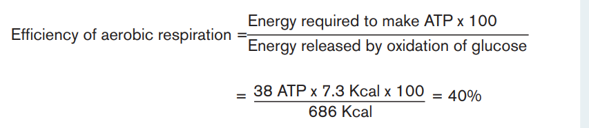
This result indicates that the efficiency of aerobic respiration equals 40%. The
remainder of the energy (around 60%) is lost from the cell as heat.
Due to the fact that anaerobic respiration produces only 2 ATP, the efficiency ofanaerobic respiration is less than that of aerobic respiration.
It is calculated as follows:
Oxygen debt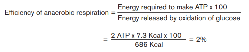
Standing still, the person absorbs oxygen at the resting rate of 0.2 dm3min−1.
(This is a measure of the person’s metabolic rate). When exercise begins,
more oxygen is needed to support aerobic respiration in the person’s muscles,
increasing the overall demand to 2.5 dm3min−1. However, it takes four minutes for
the heart and lungs to meet this demand, and during this time lactic fermentation
occurs in the muscles. Thus the person builds up an oxygen deficit. For the next
three minutes, enough oxygen is supplied. When exercise stops, the person
continues to breathe deeply and absorb oxygen at a higher rate than when
at rest. This post-exercise uptake of extra oxygen, which is ‘paying back’ the
oxygen deficit, is called the oxygen debt. The oxygen is needed for:- Conversion of lactate to glycogen in the liverThe presence of the lactic acid is sometimes described as an ‘ oxygen debt’.
- Re oxygenation of haemoglobin in the blood
- A high metabolic rate, as many organs are operating at above restinglevels.
This is because significant quantities of lactic acid can only be removed
reasonably quickly by combining with oxygen. However, the lactic acid was
only formed due to lack of sufficient oxygen to release the required energy to
the muscle tissue via aerobic respiration. Lactic acid can accumulate in muscle
tissue that continues to be over-worked. Eventually, so much lactic acid can
build-up that the muscle ceases working until the oxygen supply that it needs
has been replenished. To repay such an oxygen debt, the body must take in
more oxygen in order to get rid of the additional unwanted waste product lacticacid.
Muscle cramps
A muscle cramp is an involuntarily and forcibly contracted muscle that does not
relax. Muscle cramps can occur in any muscle; cramps of the leg muscles and
feet are particularly common.
Almost everyone experiences a muscle cramp at some time in their life. There
are a variety of types and causes of muscle cramps. Muscle cramps may occurduring exercise, at rest, or at night, depending upon the exact cause.
Overuse of a muscle, dehydration, muscle strain or simply holding a position
for a prolonged period can cause a muscle cramp. In many cases, however, the
cause isn’t known.
Although most muscle cramps are harmless, some may be related to an
underlying medical condition, such as:- Inadequate blood supply. Narrowing of the arteries that deliver blood to
your legs (arteriosclerosis of the extremities) can produce cramp-like pain
in your legs and feet while you’re exercising. These cramps usually go
away soon after you stop exercising.
- Nerve compression. Compression of nerves in your spine (lumbar stenosis)
also can produce cramp-like pain in your legs. The pain usually worsens
the longer you walk. Walking in a slightly flexed position such as you would
use when pushing a shopping cart ahead of you may improve or delay the
onset of your symptoms.
- Mineral depletion. Too little potassium, calcium or magnesium in your diet
can contribute to leg cramps. Diuretics or medications often prescribed
for high blood pressure also can deplete these minerals.The figure below accounts the energy yield per glucose molecule breakdownApplication activity 1.1
during cellular respiration in different cell organelles. Study it carefully and
answer questions that follow: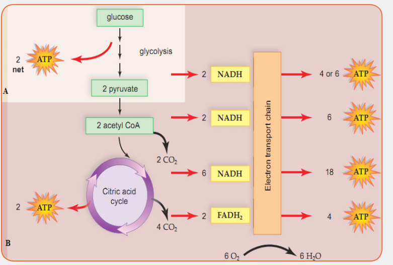 i) Where do the process labeled A and B take place in the cell?1.2. Photosynthesis
ii) Account for the total energy yield (ATP) per glucose molecule
breakdown during cellular respiration.
iii) What are the uses of energy produced during cellular respiration?
iv) What would happen to the total energy yield if glucose molecule
increases or decrease?
v) A student-teacher regularly runs 3 km each afternoon at a slow,
leisurely pace. One day, student–teacher runs 1 km as fast as she/he
can. Afterward, student-teacher is winded and feels pain in her chestand leg muscles. What is responsible for her symptoms?
Activity 1.2
Leaves and photosynthesis
Materials: test tubes, 500-mL beaker, potted houseplant with runners,
such as a spider plant, or a water plant (e.g., Elodea); wood splint; securedBunsen burner.
Procedure / protocol:
– Fill a 500-mL beaker with 400 mL of water.
– Fill a test tube with water and, without spilling, turn it upsidedown into
the water in the beaker. If an air bubble remains in the test tube, repeat
the procedure until there is no bubble or until the bubble is as small as
possible.
– Place the other test tube in the beaker repeating the steps above.
– Carefully place a spider-plant runner or sprig of a water plant into one
of the test tubes, as shown in the setup below and leave the other testtube filled with water only.
Leave the apparatus in bright sunlight or under a spotlight until there is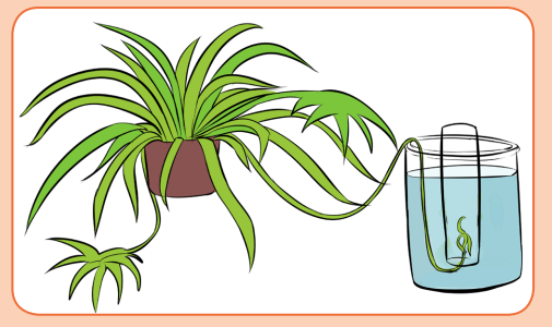
almost no water left in the tube containing the plant. Observe the test tubes
every 15 min over several hours.i) What happened to the glowing splint when it was lowered into the1.2.1. Autotrophic nutrition
test tube? Write the observation.
ii) What gas collected in the test tube?
iii) How do you know that the gas came from the plant?
iv) In which step does the gas mentioned above is produced?
Autotrophic nutrition is a process by which living organisms make their own
food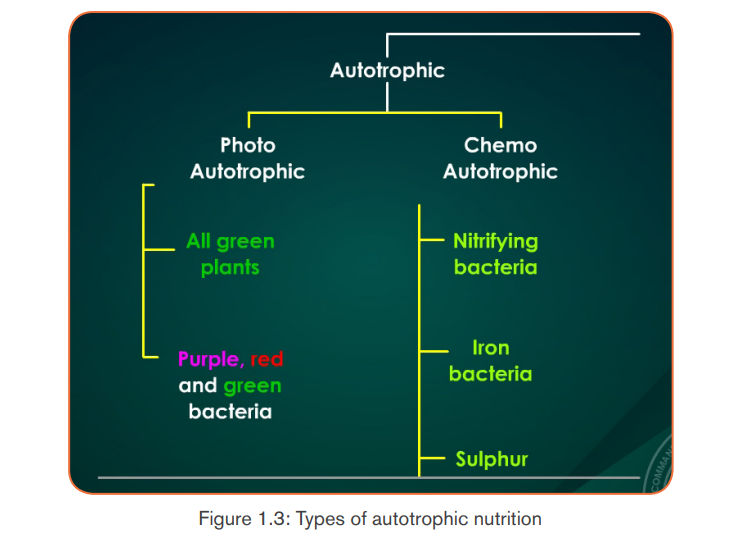 . This process is carried out by photoautotrophs like green plants, green
algae and green bacteria; and chemoautotrophs. Living organisms which make
their own food are called autotrophs, while others, including humans, whichcannot make their own food but depend on autotrophs, are called heterotrophs.
a. Types of autotrophic nutrition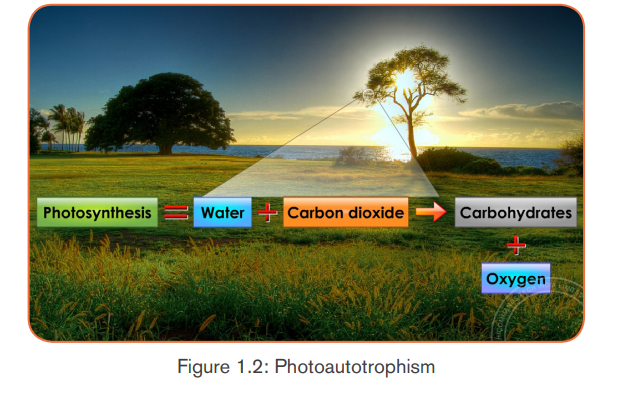

There are two types of autotrophic nutrition such as chemoautotrophic andphotoautotrophic nutrition.
a. 1 Chemoautotrophic nutrition
It is an autotrophic nutrition where organisms (mainly bacteria) get energy from
oxidation of chemicals, mainly inorganic substances like hydrogen sulphide andammonia.
a.2 Photoautotrophic nutrition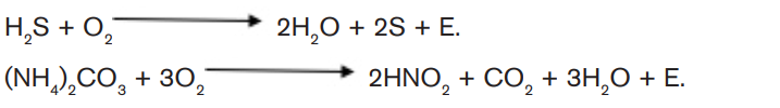
It is an autotrophic nutrition where organisms get energy from sunlight and
convert it into sugars. Green plants and some bacteria like green Sulphur
bacteria can make their own food from simple inorganic substances by a process
called photosynthesis. Photosynthesis is a process by which, autotrophs make
their own food by using inorganic substances in presence of light energy and
chlorophyll.
The function of thylakoids is to hold the chlorophyll molecules in a suitable
position for trapping the maximum amount of light. A typical chloroplast contains
approximatively 60 grana, each consisting of about 50 thylakoids. The space
outside the thylakoid membranes are made by watery matrix called stroma. Thestroma contains enzymes responsible for photosynthesis.
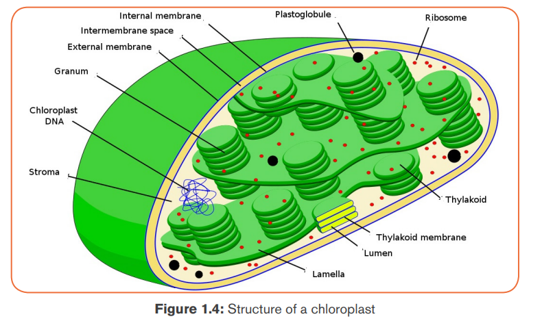
Note: Photosynthetic prokaryotes have no chloroplasts, but thylakoids often
occur as extensions of the plasma membrane and are arranged around theperiphery of the prokaryotic cell.
b. Chlorophyll
It is a sunlight- absorbing pigment, and it actually gets its green color because
it absorbs bleue and red wavelengths of light.
Structure of chlorophyll
The chlorophyll molecule is made of atoms of Carbon and Nitrogen joined in
a complex porphyrin ring containing an atom of Magnesium in the center of
the ring. The chlorophyll also has long hydrophobic carbon tail of 20 carbon
atoms (phytol) which hold it in the thylakoid membrane. In short, the chlorophyllconsists of a porphyrin ring and a phytol tail.
The chlorophyll a differs from the chlorophyll b in that: the porphyrin of the
chlorophyll a has the methyl group (-CH3) as a functional group, which is
replaced by an aldehyde group (-CHO) for chlorophyll b.
The difference between the chlorophyll a and the chlorophyll b shifts the
wavelength of light absorbed and reflected by chlorophyll b, so that thechlorophyll b is yellow-green, whereas the chlorophyll a is bright-green.
Adaptations for photosynthesis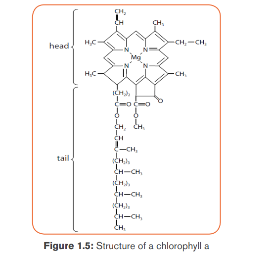
By considering both external and internal structures of the leaf, we can recognizeseveral adaptations for photosynthesis.
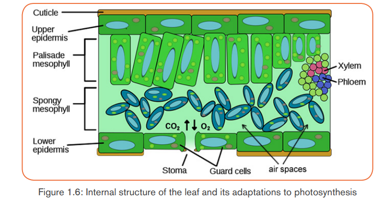
Note: when stomata are opened, the rate of photosynthesis may be 10 to 20
times as fast as the maximum rate of respiration. If the stomata are closed,
photosynthesis still can continue, using CO2 produced during cell respiration.The equilibrium can be reached between photosynthesis and cell respiration.
Photosynthesis uses CO2 from respiration, and respiration uses Oxygen from
photosynthesis. However, the rate of photosynthesis under these circumstances
will be much slower than when an external source of CO2 is available. The
stomata cannot remain closed indefinitely, they have to be open in order tomaintain transpiration of the plant.
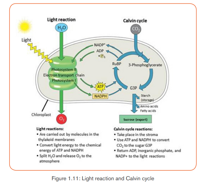
1.2.2. Mechanism of photosynthesis
The process of photosynthesis occurs through two main stages such as:- The light-dependent reactions: which take place in thylakoids, andA. The light-dependent reactions- The light-independent reactions (Calvin cycle): which take place in stroma.
They require light energy and occur in thylakoids. They produce Oxygen gasand convert ADP and NADP+ into ATP and NADPH.
The light-dependent reactions involve the following steps:
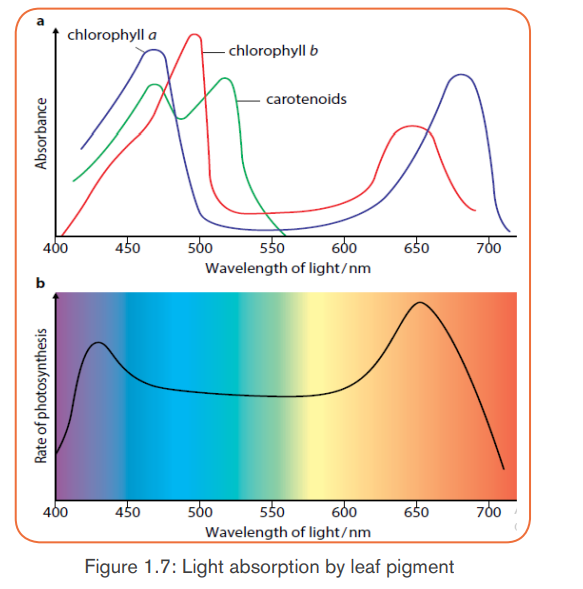
a. Absorption and action spectra
In addition to water and CO2, photosynthesis requires light and chlorophyll.
The chlorophyll pigment is found in the chloroplasts. The light that our eyes
perceive as white light is a mixture of different wavelengths. Most of them are
visible to our eyes and make up the visible spectrum. Our eyes see different
wavelengths of visible spectrum as different colors (violet, blue, green, yellow,
orange and red) except indigo which is not visible to our eyes. Plants absorb
the light energy by using molecules called pigments such as: chlorophyll a,
chlorophyll b, carotene (orange) and xanthophyll (yellow) but chlorophyll a
is the principle pigment in photosynthesis.
The chlorophyll absorbs light very well in blue-violet and red regions of visible
spectrum. However, chlorophyll does not absorb well the green light; instead it
allows the green light to be reflected. That is why young leaves and other partsof the plants containing large amount of chlorophyll appear green.
The chlorophyll a as a principle and abundant pigment, it is directly involved
in light reactions of photosynthesis. Other pigments (chlorophyll b, carotene,
xanthophyll and phaeophytin) are accessory pigments. They absorb light
colours that chlorophyll a cannot absorb, and this enables plants to capturemore energy from light.
The amount of energy that the pigment can absorb from the light depends on
its intensity and its wavelengths. So, the greater the intensity of light, the greater
amount of energy will be absorbed by the pigment in a given time.
Photosynthesis begins when the chlorophyll a in photosystem II absorbs light at
different wavelengths of light.- When the light energy hits the chlorophyll a, the light energy is absorbed
by its electrons, by raising their energy level.
- These electrons with high potential energy (electrons with sufficient
quantum energy) are passed to the electron-transport chain.
- Excited electrons are taken up by an electron acceptor (NADP+: oxidized
Nicotinamide Adenine Dinucleotide Phosphate), and pass along electron
transfer chain from photosystem II to the photosystem I. (Note: The
photosystems are the light-collecting units of the chloroplast).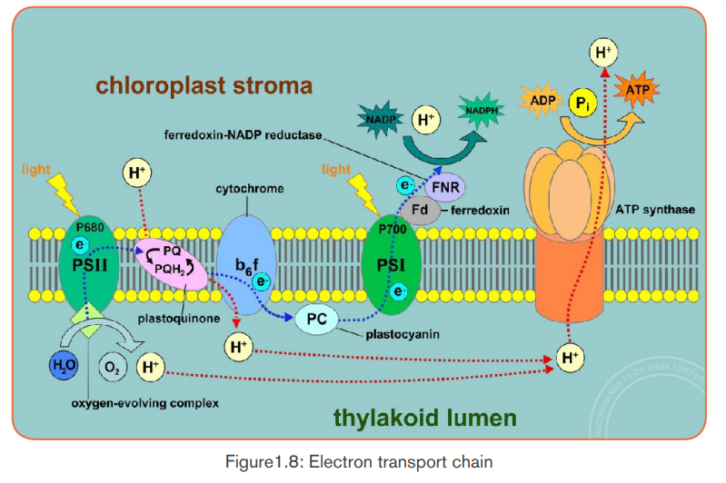
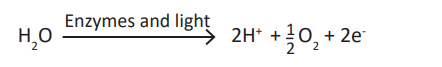
b. Enzymes in thylakoids and light absorbed by photosystem II
Enzymes in thylakoids and light absorbed by photosystem II are used to breakdown a water molecule into energized electrons, hydrogen ions H+, and Oxygen.
 - Oxygen produced is released to be used by living things in respiration.- The light-dependent reactions also allow generation of ATP (Adenosine
- Electrons and H+ from photolysis of water are used to reduce NADP+ toNADPH (Reduced Nicotinamide Adenine Dinucleotide Phosphate).
Triphosphate) by adding inorganic phosphate to ADP+ (AdenosineDiphosphate):

Generally, the light-dependent reactions use light energy, ADP, Pi, NADP+ andwater to produce ATP, NADPH and Oxygen. Or simply:
Both ATP and NADPH are energy carriers which provide energy to sugars
(energy containing sugars) in Light-independent reactions.
c. Photophosphorylation
The fixation of Pi to ADP+ to form ATP is called photophosphorylation.
Photophosphorylation can be done into two processes: cyclic
photophosphorylation, and non-cyclic photophosphorylation.
Cyclic photophosphorylation
It involves only photosystem I and not photosystem II. There is no production of
NADPH and no release of Oxygen. When the light hits the chlorophyll in PSI,the light-excited electron leaves the molecule.
This light-excited electron is taken up by an electron acceptor which passes
it along an electron transfer chain (a series of electron carriers) until it returns
to the chlorophyll molecule that it left (cyclic process). As an excited electron
moves along an electron transfer chain, it loses energy which will be used
for the synthesis of ATP from ADP+ and inorganic phosphate in the process
called chemiosmosis. Electron carriers can vary, but the principle includes thecytochromes.
Non-cyclic photophosphorylation
It is the main route of ATP synthesis. It is done in the following steps:- When the photosystem II (in chlorophyll) absorbs light, an electron is
excited to a higher energy level and captured by the primary electron
acceptor.
- Enzymes extract electrons from a water molecule replacing each electron
that the chlorophyll molecule lost when absorbed light energy. This reaction
dissociates a water molecule into hydrogen ions (2H+) and Oxygen which
is released for animals’ respiration.
- Excited electron moves from the primary electron acceptor of photosystemII to photosystem I, via an electron transport chain.
- When excited electron moves from the primary electron acceptor of
photosystem II to photosystem I, via an electron transport chain its energy
level lowers. The energy removed is used to synthesize ATP from ADP and
Pi in a process called: Non-cyclic phosphorylation.
- The hydrogen ions (2H+) produced from dissociation of water molecule
combines with NADP+ to form NADPH2.
- Both ATP and NADPH2 will be used in the light-independent reactions
(Calvin cycle) for synthesis of sugars.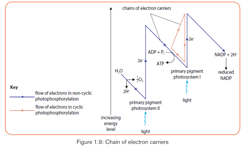
The significance of the cyclic phosphorylation
Non-cyclic photophosphorylation produces ATP and NADPH in equal quantities,
but the Calvin cycle consumes more ATP than NADPH. The concentration of
NADPH in a chloroplast may determine which pathway (cyclic versus noncyclic)
electrons pass through.
If a chloroplast runs low on ATP for the Calvin cycle, NADPH will accumulate as
the cycle slows down. The rise of NADPH may stimulate a shift from non-cyclic
(which produces ATP only) to cyclic electron pathway until ATP supply catcheswith the demand.
Table 1.2: Comparison between Non-cyclic and cyclicphotophosphorylation
B. The light-independent reactions (Calvin cycle)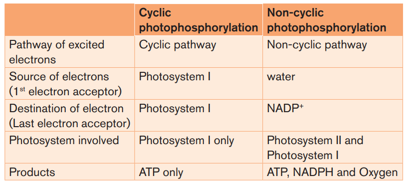
The light-independent reactions occur in stroma, and consist of reducing CO2
into sugars by using ATP and NADPH both coming from light-dependent
reactions in thylakoids. The Calvin cycle involves three main stages such as:
- Carbon fixation in form of CO2.
- Carbon reduction from CO2 to glucose.- Regeneration of RuBP.
a) Carbon fixation (Carboxylation) in form of CO2
The Calvin cycle begins with a 5-Carbon sugar phosphate called Riburose-1,
5 biphosphate (RuBP) which fixes the CO2 from air. This reaction is catalyzed
by an enzyme called RuBP carboxylase-oxygenase (RUBISCO), which makes
up about 30% of the total protein of the leaf, so it is probably one of the mostcommon proteins on the Earth.
The combination of RuBP and CO2 results in a theoretic 6-carbon compound
which is highly unstable. It immediately splits into two molecules of 3-carbon
known as phosphoglyceric acid (PGA) or glycerate 3-phosphate, or3-phosphoglycelate.
b) Carbon reduction from CO2 to glucose
With energy from ATP and reducing power from NADPH, the phosphoglyceric
acid is reduced into 3carbon molecules known as glyceraldehyde-3-phosphateor phosphoglyceraldehyde (PGAL).
Each molecule of PGA receives an additional phosphate group from ATP,
becoming 1, 3-biphosphoglycerate, and a pair of electrons and H+ from NADPH
reduces the carboxyl group of 3-phosphoglycerate to the aldehyde group ofPGAL which stores more potential energy.
ATP gives one phosphate group becoming ADP+, and NADPH gives H+ and
electrons to become NADP+. Both ADP+ and NADP+ will be used again in light
dependent reactions.
With 6 turns of Calvin cycle, the plant cell fixes 6CO2 molecules which are used
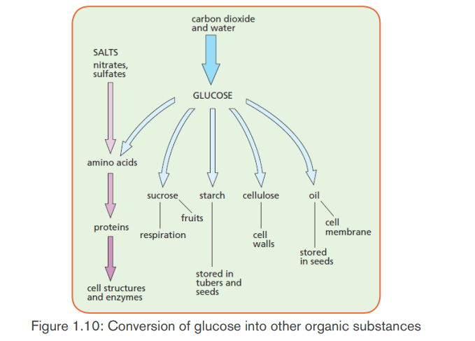
to synthesize 2 molecules of PGAL which leave the cycle and combine to makeone molecule of glucose or fructose. This glucose can be converted into:
Sucrose: when Oxygen combined with fructose. It is a form by which
carbohydrates are transported in plants.- Polysaccharides like starch for energy storage, and cellulose for structural
support.
- Amino acids when combined with nitrates,
- Nucleic acids when Oxygen combined with phosphates, and- Lipids.
c) Regeneration of RuBP
The remaining ten 3-carbon molecules (PGAL) are converted back into six
5-carbon molecules, ready to fix other CO2 molecules for the next cycle. Thelight-independent reactions can be summarized as:

Other carbon dioxide fixation pathways (C4 CAM)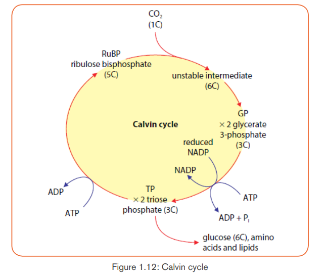 The most common pathway combines one molecule of CO2 with a 5-carbonsugar called ribulose biphosphate (RuBP). The enzyme which catalyzes this
reaction (nicknamed “Rubisco”) is the most abundant enzyme on earth! The
resulting 6-carbon molecule is unstable, so it immediately splits into two
3-carbon molecules. The 3 carbon compound which is the first stable moleculeof this pathway gives this largest group of plants the name “C-3 plants”
Dry air, hot temperatures, and bright sunlight slow the C-3 pathway for carbon
fixation. This is because stomata, which normally allow CO2 to enter and O2
to leave, must close to prevent loss of water vapor. Closed stomata lead to
a shortage of CO2. Two alternative pathways for carbon fixation demonstrate
biochemical adaptations to differing environments. Plants such as corn solvethe problem by using a separate compartment to fix CO2.
Here CO2 combines with a 3-carbon molecule, resulting in a 4-carbon molecule.
Because the first stable organic molecule has four carbons, this adaptation has
the name C-4. Shuttled away from the initial fixation site, the 4-carbon molecule
is actually broken back down into CO2, and when enough accumulates, Rubiscofixes it a second time!
In some temperate plants such as wheat, rice, potato and bean only Calvin cycle
occurs. Such plants are called C-3 plants. While in some other plants dual
carboxylation takes place: (1) carboxylation of phosphoenol pyruvate (PEP) and
(2) carboxylation of RuBP. Such plants are called C-4 plants e.g. maize, sugar
cane and sorghum. In these, the first product formed during carbon dioxide
fixation is a four carbon compound oxalo acetic acid (OAA). C-4 plants have
special type of leaf anatomy called Kranz Anatomy. They have special large cells
around vascular bundles called bundle sheath cells. These are characterized
by having large number of chloroplasts, thick walls and no intercellular spaces.
The shape, size and arrangement of thylakoids in chloroplasts are also different
in bundle sheath cell as compared to mesophyll cell chloroplasts.
The pathway followed by C-4 plants is called C-4 cycle or Hatch and Slack
pathway. This was discovered by Hatch and Slack in sugar cane. The primary
CO2 acceptor is a 3-carbon molecule phosphoenol pyruvate (PEP). The reaction
is catalyzed by PEP carboxylase or PEP case in mesophyll cell chloroplast.
It forms 4-carbon compounds like OAA, malic acid or aspartate, which are
transported to the bundle sheath cells. In bundle sheath cells, these acids are
broken down to release CO2 and 3-carbon molecule. The 3-carbon molecule
is transported back to mesophyll cells and converted to PEP again, while CO2
enters into C-3 cycle to form sugars. C-4 plants are more efficient than C-3
plants as in C-4 plants, photosynthesis can occur at low concentration CO2
and photorespiration is negligible or absent.
Cacti and succulent (water-storing) plants such as the jade plant avoid water
loss by fixing CO2 only at night. These plants close their stomata during the
day and open them only in the cooler and more humid nighttime hours. Leaf
structure differs slightly from that of C-4 plants, but the fixation pathways are
similar. The family of plants in which this pathway was discovered gives the
pathway its name, Crassulacean Acid Metabolism, or CAM. All carbon
fixation pathways lead to the Calvin cycle to build sugar.
The CAM pathway is similar to the C4 pathway in that carbon dioxide is first
incorporated into organic intermediates before it enters the Calvin cycle. The
difference is that in C4 plants, the initial steps of carbon fixation are
separated structurally from the Calvin cycle whereas in CAM plants, the
two steps occur at separate times.
The CAM pathway and the C4 pathway compared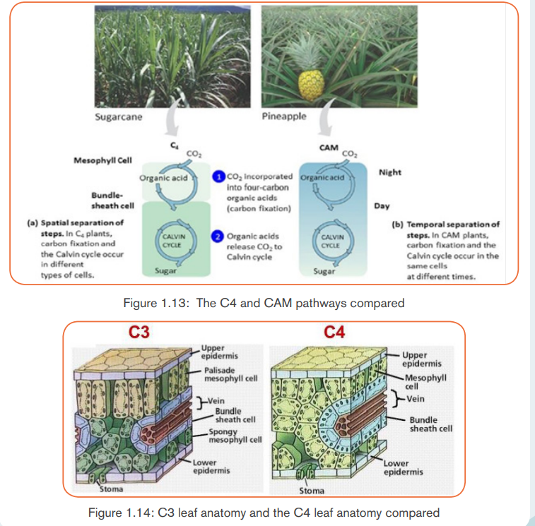
The table 1.3: Comparison between C3 and C4 plants
Photorespiration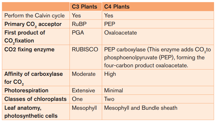
In most plants, initial fixation of carbon occurs via Rubisco, the Calvin cycle
enzyme that adds CO2 to ribulose biphosphate. Such plants are called C3
plants because the first organic product is a three carbon organic compound,
PGA. These plants produce less food when their stomata close on hot and dry
days.
The declining level of CO2 in the leaf starves the Calvin cycle. Making matter
worse, Rubisco can accept O2 in place of CO2. As O2 concentration overtakes
CO2 concentration within the air space, Rubisco adds O2 instead of CO2. The
product splits and one piece, a two-carbon compound is exported from thechloroplast. Mitochondria then break the two-carbon molecule into CO2.
The process is called photorespiration because it occurs in presence of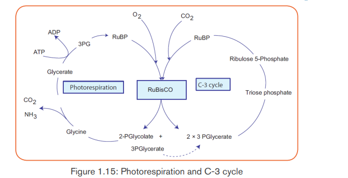
light (photo) and consumes O2 (respiration). However, unlike normal cellular
respiration, photorespiration generates no ATP, and unlike photosynthesis,
photorespiration generates no food. In fact, photorespiration decreasesphotosynthetic output by using material from the Calvin cycle.
Application activity 1.2
The diagram below illustrates photosynthesis process. Write each of the
following terms on the correct numbered line. Then answer the questions
that follow.
Carbon Dioxide Glucose Oxygen Water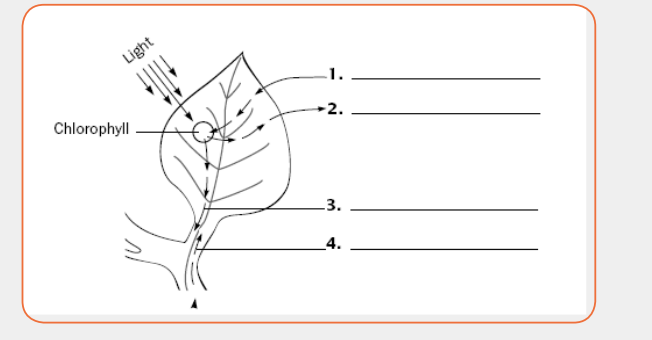 i) In photosynthesis, establish an equation of substrate with substances
produced
ii) What would happen if substance labeled in 1 and 4 are absent? Justify
your answer.iii) Explain how photosynthesis and respiration are interdependent?
1.3. Factors affecting the rate of photosynthesis
Activity 1.3
1. When beans are grown under banana trees, the farmers record poor
harvest. Explain why?
2. Make a research to find out how each of the following factors can
affect the rate of photosynthesis: Temperature – Light intensity –Concentration of CO2 – Amount of water
The photosynthesis rate varies with the species but also varies within individuals
for a same species; this varies under the influence of certain external factors
which are: the temperature, CO2 concentration in the atmosphere, light intensityand soil humidity.
a. Temperature
Photosynthesis is an enzyme-controlled process. At very low temperatures the
rate of photosynthesis is slow because the enzymes are inactive. As temperature
increases, the rate of photosynthesis increases because the enzymes become
more active. Rate of photosynthesis is optimum at (35-40) °C. Beyond 40°C
the rate of photosynthesis decreases and eventually stops since the enzymesbecome denatured.
b. CO2 concentration in the atmosphere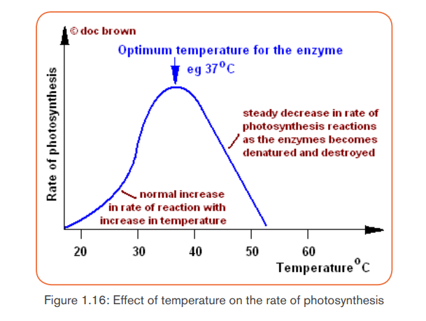
While the concentration of carbon (IV) oxide in the atmosphere is fairly constant
at 0.03%, an increase in carbon (IV) oxide concentration translates into an
increase in the rate of photosynthesis upto a certain point when the rate of
photosynthesis becomes constant. At this point, other factors such as lightintensity, water and temperature become limiting factors.
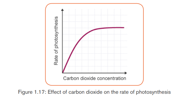
The photosynthetic rate is zero in place lacking CO2, it increases with the
increase concentration of CO2 in the atmosphere and reaches an optimumranging between 5 and 8%CO2 concentration.
c. Light intensity
The rate at of photosynthesis increases with an increase in light intensity up to
a certain level. Beyond the optimum light intensity, the rate of photosynthesis
becomes constant. To this effect, plants photosynthesize faster on bright and
sunny days than on dull cloudy days.
Light quality/wavelength also affects the rate of photosynthesis. Most plants
require red and blue wavelengths of light for photosynthesis. Light duration alsoaffects photosynthesis rate
The photosynthesis rate is low during night, it increases when the light intensity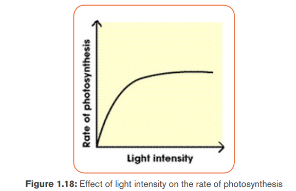
increases but the optimum varies according to the plants.
d. Availability of water for the plant
The photosynthesis rate is low when the soil is dry, it increases when the content
of water increases for the terrestrial plants, and for the aquatic plants it remains
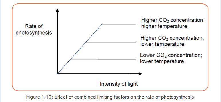
constant as long as they are fixed in water.Note: The limiting factors work together to influence the rate of photosynthesis
Application activity 1.3
Factors affecting rate of photosynthesis.
Materials Required:Elodea plant, glass rod, sodium bicarbonate.
Procedure:
Take a fresh, healthy twig of Elodea plant with one end intact and tie it
gently to a glass rod. Put the glass rod with plant in a boiling tube containing
water and add 1mg/mL sodium bicarbonate and keep it in moderate light
condition. Note the numbers of bubbles escaping from cut end per minute.
Again add same amount of sodium bicarbonate and note the number of
bubbles escaping from cut end per minute. Do you find number of bubbles
increasing? Repeat this step until bubbles escaping per minute do not
increase. Then take set up under high light intensity and note the numbersof bubbles.
1. What is the observation made when the mass of sodium bicarbonate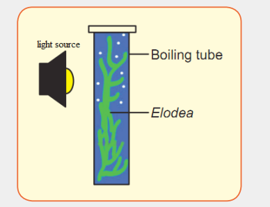
was increased?
2. What is your observation if the set up is under high light intensity?1.4. Importance of photosynthesis
Activity 1.4
The following diagram shows the link that exists between plant and animals.Observe the diagram and use it to answer the related questions.
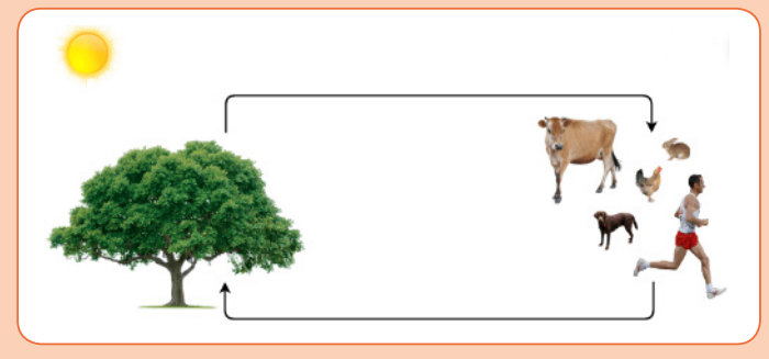 i) What do the animals receive from the plant and what do the plantsAutotrophic nutrition is a process by which living organisms (autotrophs:
receive from the animal on the diagram above?
ii) Discuss how the relation between plants and animals are
interdependent?
iii) Suggest the role of aquatic plants to aquatic life of animals.
photoautotrophs and chemoautotrophs) make their own food. The autotrophism
is very essential as it allows production of Oxygen and food for not only
themselves but also for heterotrophs. The roles of autotrophic nutrition include:
a. Independence of green plants from other living organisms
This importance relates to their capacity for synthesizing organic molecules from
glucose produced by CO2 and water, this completely make them independentsof the other living organisms to the nutrition point of view.
b. Energy storage
The autotrophs like green plants, by the process of photosynthesis synthesize
certain substances like the glucose, cellulose, starch… which are variablessources of energy.
b. Energy storage
The autotrophs like green plants, by the process of photosynthesis synthesize
certain substances like the glucose, cellulose, starch… which are variables
sources of energy.
c. Production of O2 for the living organisms’ respiration
The oxygen produced by the photosynthesis is necessary for the living organisms’
respiration. Thus without photosynthesis, no oxygen can be produced; withoutoxygen no respiration; without respiration no life on Earth.
d. Cleaning the atmosphere
Photoautotrophs absorb carbon dioxide from surrounding air, and release
Oxygen (produced by photosynthesis) in atmosphere.
e. Formation of Ozone layer
Ozone layer is a thick layer in the atmosphere which is formed Ozone
molecule (O3). Oxygen atoms which make ozone molecule are produced by
photosynthesis. Ozone layer protects the Earth from high solar radiations, and
this allows the existence of the life on the Earth.
Synthesis of the organic substances: food for the heterotrophs (animal and
mushrooms). The organic substances produced by photosynthesis are the food
for the heterotrophs which are unable to synthesize these substances by their
own means.
Application activity 1.4
(a) A well watered potted bean plant was destarched by putting it in the dark
for 36 hours. Three of its leaves were smeared with Vaseline as follows: leaf
I on both sides; leaf II on the lower surface only; leaf III on the upper surface
only. All the other leaves were left untreated. The plant was then placed in
sunlight for eight hours after which an iodine test for starch was carried out.The observation was as follows:
Leaf I-brown colour; Leaf II-slight blue-black stain; Leaf III-intense blueblack stain;
Leaf IV (untreated leaf)-very intense blue-black stain. Explainthese observations.
(b) A student carried out an investigation to show how light intensity affected
the rate of photosynthesis for a water plant, Elodea. The student used a test
containing water plant immersed in water at different light intensity. After 5
minutes of each experiment, the student counted the number of bubbles.The results are shown in the following table.
Plot a suitable graph for the result of experiment.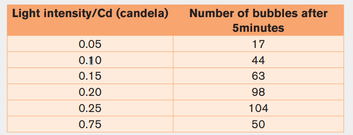
Explain the results recorded between 0.25 Cd and 0.75 Cd of light intensity
Skills Lab 1
Automobiles and machines must be supplied with gasoline or electricity
as a source of energy before they can move. Your muscles require energy
in the form of ATP to contract. Muscles can produce ATP by using oxygen
(aerobic respiration) or not using it (anaerobic respiration). Anaerobic
respiration in muscle cells produces lactic acid. When muscles do a lot
of work quickly, the buildup of lactic acid reduces their ability to contract
until exhaustion eventually sets in and contraction stops altogether. This iscalled muscle fatigue.
Materials: clothespin, timer
Procedure:- Hold a clothespin in the thumb and index finger of your dominant- Repeat this process for nine more 20-s periods, recording theresult
hand.
- Count the number of times you can open and close the clothespin in
a 20-s period while holding the other fingers of the hand straight out.
Make sure to squeeze quickly and completely to get the maximum
number of squeezes for each trial.
for each trial in a suitable table. Do not rest your fingers between
trials.
- Repeat the procedure for the nondominant hand.a) What happened to your strength as you progressed through eachEnd Unit Assessment 1
trial?
b) Describe how your hand and fingers felt during the end of your trials.
c) What factors might cause you to get more squeezes (to have less
fatigue)?
d) Were your results different for the dominant and then on dominant
hand? Explain why they would be different.
e) Your muscles would probably recover after 10 min of rest to operate
at the original squeeze rate. Explain why.f) Prepare a suitable graph of anaerobic respiration your muscle.
1. What are the products of the light dependent reactions of
photosynthesis?
a) ATP, RuBP and reduced NAD
b) ATP, oxygen and reduced NADP
c) GP, oxygen and reduced NAD
d) GP, reduced NADP and RuBP
2. Before the Krebs cycle can proceed,
pyruvic acid must be converted
intoa) Citric acid3. The net number of ATP made directly by glycolysis is
b) Glucose
c) Acetyl-CoA
d) Glucose
e) NADHa) 24. Cellular respiration is similar to photosynthesis in that they both
b) 4
c) 32
d) 38a) Produce ATP5. By accepting electrons and protons, the oxygen used in aerobic
b) Involve chemiosmosis
c) Make phosphoglyceraldehyde (PGAL)
d) All of the above
respiration turns intoa) CO26. The Krebs cycle occurs in the
b) H2O
c) C6H12O6
d) ATPa) Cytosold) Space between the inner and outer mitochondrial membrane
b) Outer mitochondrial membrane
c) Mitochondrial matrix
7. During each turn of the Krebs cycle,a) Two CO2 molecules are produced8. Most of the ATP synthesized in aerobic respiration is made
b) Two ATP molecules are consumed
c) Pyruvic acid combines with oxaloacetic acid
d) Glucose combines with a four-carbon molecule.a) During glycolysis9. Where does each stage of aerobic respiration occur in a eukaryotic
b) Through fermentation
c) In the cytosold) Through chemiosmosis
cell?
10. The diagram summarises how glucose can be used to produce ATP,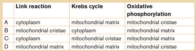
without the use of oxygen.
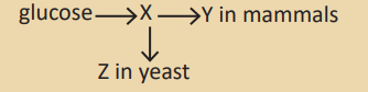
Which compounds are represented by the letters X, Y and Z ?
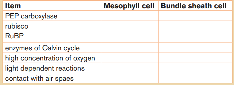
11. a. Copy and complete the table to show the differences between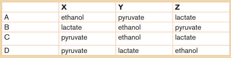
mesophyll and bundle sheath cells in C4 plants. Insert a tick (x) whenan item is present in the cell and a cross (√) when it is not.
 b. Explain what is meant by photorespiration.12. a. Explain what is meant by a limiting factor.b. List four factors that may be rate-limiting in photosynthesis.
c. At low light intensities, increasing the temperature has little effect
on the rate of photosynthesis. At high light intensities, increasing
the temperature increases the rate of photosynthesis. Explain theseobservations.
UNIT 2: HUMAN REPRODUCTION AND FAMILY PLANNING
Key Unit Competence:
Explain the role of human reproductive hormones,
stages of pregnancy and family planning methods.
Introductory activity 2
Observe the photo below and answer the questions that follow: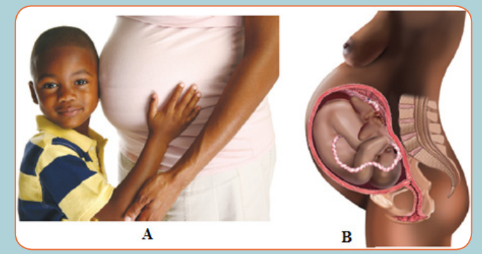 a) The woman in A is pregnant. What do you think about the origin of the
fetus in the womb of the pregnant woman?
b) Use the photo B to imagine the embryonic development in mother’s
uterus.
c) Use the photo in B and identify the observable parts of the female
reproductive system. Are you satisfy with that description? Justify your
response.
d) The child on the photo is a boy. Can you identify the parts of the male
reproductive system? What represent the gesture of that boy? Are
challenging!!!!e) What do you think about foetus position observed in mother’s uterusAnimals reproduce sexually, but some, on occasion, can reproduce asexually.
on the photo B?
f) The child boy on picture A is the first borne of her mother and the
fetus in the womb will be the second and last borne of this mother.Can you advocate for such a family planning? Give reasons
Sexually reproducing animals have gonads for the production of gametes,
and many have accessory organs for the storage and passage of gametes
into or out of the body. Animals have various means of ensuring fertilization of
gametes and protecting immature stages. Human reproduction is any form of
sexual reproduction resulting in human fertilization. It typically involves sexualintercourse between a man and a woman.
2.1. Human reproductive systems
The diagrams below represent male and female reproductive system.Observe and use them to answer questions that follow.
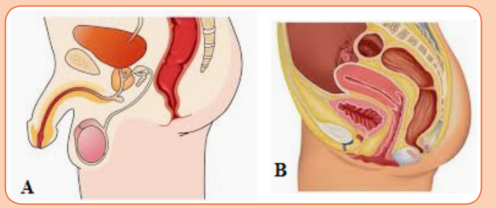 a) Identify the structures representing a male and female human
reproductive system.
b) Use the choice in (a) to locate and suggest the function of the following
male human reproductive organs on the diagram: testis, epididymis,
vas deferens, seminal vesicles and prostate gland.
c) Use the choice indicating the female human reproductive system
to locate and suggest the function of the following female human
reproductive organs on the diagram: urethra, vagina, uterus, ovariesand oviducts.
Human beings reproduce by sexual means where the male and female involve2.1.1. Structure of male reproductive system
in sexual intercourse, resulting in fertilization. During sexual intercourse, the
interaction between the male and female reproductive systems results in
fertilization of the woman’s ovum by the man’s sperm. The ovum and sperm are
specialized reproductive cells called gametes, generated by a process calledgametogenesis (i.e., spermatogenesis in males and oogenesis in females).
The main visible differences between boys and girls at birth are their reproductive
organs. The sex of a child is determined at the time of fertilization of the ovum by
the spermatozoon. The differences between a male and a female are geneticallydetermined by the chromosomes that each possesses in the nuclei of the cells.
The male gonads are paired testes, which are suspended within the sacs of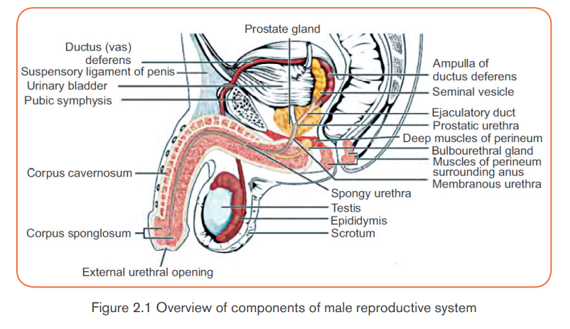
the scrotum. The testes begin their development inside the abdominal cavity,
but they descend into the scrotal sacs as development proceeds. If the testes
do not descend and the male does not receive hormone therapy or undergo
surgery to place the testes in the scrotum results in sterility (the inability to
produce offspring). Sterility occurs because undescended testes developing
in the body cavity are subject to higher body temperatures than those in the
scrotum. A cooler temperature is critical for the normal development of sperm.
Sperm produced in the seminiferous tubules of the testes mature within the
epididymides (sing., epididymis), which are tightly coiled tubules lying just
outside the testes. Maturation seems to be required for the sperm to swim tothe egg.
Once the sperm have matured, they are propelled into the vasa deferentia
(sing., vas deferens) by muscularcontractions. Sperm are stored in both the
epididymides and the vasa deferentia. When a male becomes sexually aroused,
sperm enter the ejaculatory ducts and then the urethra, part of which is locatedwithin the penis.
The penis is the male organ of sexual intercourse. The penis has a long shaft
and an enlarged tip called the glans penis. The glans penis is normally covered
by a layer of skin called the foreskin. Circumcision, the surgical removal of theforeskin, is often done soon after birth.
In addition to these organs, the male reproductive system consists of a series of
ducts and glands. Ducts include the vas deferens and ejaculatory ducts. They
transport sperm from the epididymis to the urethra in the penis.
Glands include the seminal vesicles, prostate gland, and bulbourethral glands
(also called Cowper’s glands). They secrete substances that become part ofsemen.
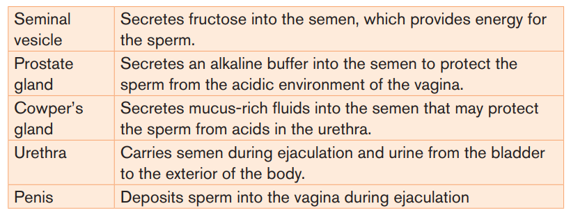
- Two seminal vesicles contribute about 60% of the volume of semen.
The fluid from the seminal vesicles is thick, yellowish, and alkaline. It
contains mucus, the sugar fructose (which provides most of the sperm’s
energy), a coagulating enzyme, ascorbic acid, and local regulators called
prostaglandins.
- The prostate gland secretes its products directly into the urethra through
several small ducts. This fluid is thin and milky; it contains anticoagulant
enzymes and citrate (a sperm nutrient).
- The bulbourethral glands are a pair of small glands along the urethra below
the prostate. Before ejaculation, they secrete clear mucus that neutralizes
any acidic urine remaining in the urethra. Bulbourethral fluid also carries
some sperm released before ejaculation, which is one reason for the highfailure rate of the withdrawal method of birth control (coitus interruptus).
Semen is the fluid that is ejaculated from the urethra. Semen contains secretions
from the glands as well as sperm. The secretions control pH and provide thesperm with nutrients for energy.
Table 2.1: Parts of the male reproductive system and their functions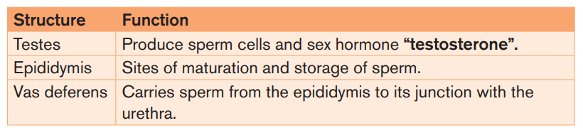
Note: Male infertility refers to a male’s inability to cause pregnancy in a fertile
female. In humans it accounts for 40-50% of infertility. It affects approximately
7% of all man. Male infertility may be due to:- Absence of sperms in the semen (Azoospermia).
- Low sperm count e.g. when ones ejaculate less than 1cm3 of semen.
- Abnormal sperm e.g. sperms with 2 tails, or without tail, or without
acrosomes,
- Auto-immunity e.g. antibodies attack one’s sperms
- Premature ejaculation: the man has orgasm before copulation
- Impotence i.e. inability to achieve or maintain an erection of the penis.2.1.2. Structure of female reproductive system
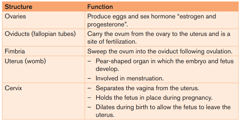
The female reproductive system is a collection of organs and other structures
located primarily in the pelvic region. Most of the structures are inside the body.
It includes the ovaries, the oviducts, the uterus, and the vagina. The female
reproductive system has several functions:- Producing eggs, which are female gametesDuring puberty, a girl develops into a sexually mature woman, capable of
- Secreting female sex hormones
- Receiving sperm during sexual intercourse
- Supporting the development of a fetus
- Delivering a baby during birthproducing eggs and reproducing.
The external genital organs of a female are known collectively as the vulva. The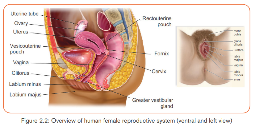
pubic hairs and two folds of skin called labia minora and labia majora are
on either side of the urethral and vaginal openings. Beneath the labia majora,
pea-sized greater vestibular glands (Bartholin glands) open on either side of thevagina. They keep the vulva moist and lubricated during intercourse.
At the juncture of the labia minora is the clitoris, which is homologous to the
penis in males. The clitoris has a shaft of erectile tissue and is capped by a peashaped glans. The many sensory receptors of the clitoris allow it to function
as a sexually sensitive organ. The clitoris has twice as many nerve endings as
the penis. Orgasm in the female is a release of neuromuscular tension in the
muscles of the genital area, vagina, and uterus.Table 2.2: Parts of the female reproductive system and their functions
Note: Female infertility is defined as the inability to conceive or carry a pregnancy
to term after 12 months of unprotected intercourse, or 6 months of unprotected
intercourse if the female is over 35 years old. In females, infertility may be due
to:- Failure to ovulate due to the lack of some hormones.Application activity 2.1
- Damage of the Fallopian tubes / oviducts, for example the tubes may be
completely blocked by nature or after an infection.
- Damage on the uterus; for example, the endometrium can be destroyed.
- Damage on the cervix, for example the cervix may be narrow or too wide or
may stop producing cervical mucus needed for the sperm to reach uterus.
- Antibodies against sperms, for example, the cervix, the uterus or the oviductof a woman can produce antibodies against her husband’s sperms.
The figure below represents the male reproductive system.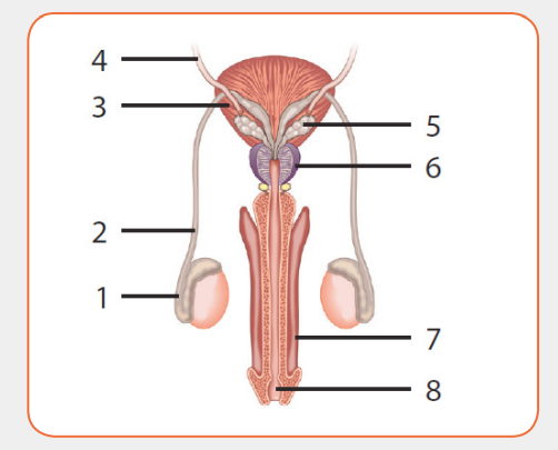
a) Justify why it is representing a male reproductive system.
b) Refer to the figure and identify the parts represented by the numbers
1 to 8.
c) How can you justify the function of organ 2, 5 and 7 according to thestructure of male reproductive system?
2.1.3. Gametogenesis
Activity 2.2
Gametes are haploid cells that are formed from diploid germ cells through
the process of gametogenesis. The significance of developing haploid
gametes lies in the fact that after fertilization, the developing zygote attains
the diploid status back. In this way, the developing embryo gets the single
copy of all the chromosomes from each parent.
Based on the chart diagrams of spermatogenesis and oogenesis shown
below.
i) Compute the number of chromosomes at each stage, assuming 2n= 46.
ii) Which diagram does it represent spermatogenesis and oogenesis?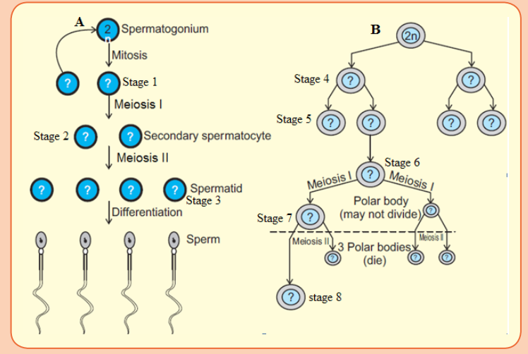
Explain why?
A) Spermatogenesis
The process of formation of haploid male gametes or spermatozoa from diploid
reproductive cells in males is called spermatogenesis. The complete process is
broadly divided into two parts, formation of spermatids and spermatogenesisor spermatoleosis.
Formation of Spermatids
The process of formation of spermatids is further divided into three stages as:
a) Multiplication phase: The primordial germ cells or sperm mother cells
differentiate from germinal epithelium of testis and increase in size with
prominent nuclei. These cells divide repeatedly by mitosis (i.e., equational
division) and produce a number of diploid daughter cells, known as
spermatogonia. Thus, in this stage, multiplication of germ cells takes place
mitotically.
b) Growth phase: In this phase, spermatogonia increase in size by
accumulating food reserves and are now called primary spermatocytes.
c) Maturation phase: The primary spermatocytes (which are diploid)
undergo first maturation division which is meiotic division (or
reductional division) to produce two haploid secondary spermatocytes.
These haploid secondary spermatocytes divide further by mitosis to give
rise to four haploid spermatids. This mitotic division is called secondmaturation division.
The spermatids so produced are non-motile rounded structures that
metamorphose into functional and motile spermatozoa through a process
known as spermiogenesis or spermatoleosis. The spermatozoa from testis
are incapable of fertilizing an ovum. They undergo several morphological,
physiological and biochemical changes as they move through the epididymis
to attain this structural and physiological maturity. The epididymis i) provides a
favorable environment to spermatozoa in acquiring fertilizing ability and ii) stores
them until they are ejaculated or move down to the vas deferens.
The morphological changes include structural remodeling of acrosome and
formation of disulfide linkages. The physiological and biochemical changes
include increase in net negative charge on spermatozoa, change in pattern of
motility, change in content of sialic acid, increase in specific activity and reflectionpower, resistance to pH and temperature and changes in metabolic patterns.
– Spermiogenesis
A series of changes in spermiogenesis that transform a non-motile spermatid
into motile, functional spermatozoa are listed below:- The nucleus shrinks and flattens by losing water. Only DNA is left in the
nucleus, making cells very light that aids to its motility.
- The two centrioles of a centrosome form proximal and distal centrioles
that lie at the posterior end of nucleus and give rise to axial filament of the
flagellum and acts as a basal granule respectively.- The mitochondria gather around axial filament and gradually unite toDuring all these steps, head of the developing sperm remains embedded in
form spiral sheath or nebenkern. It acts as power house of the sperm and
provides energy.
- The golgi bodies form the covering over nucleus called acrosome. During
acrosome formation, one or more vacuoles start enlarging with a small,
dense body called pro-acrosomal granule which further enlarges to
form acrosomal granule. The vacuole loses its liquid content and forms the
cap of spermatozoan. The remaining part of golgi apparatus is reduced
and discarded from sperm.
sertoli cells for nourishment. At the end, fully formed spermatozoan showsdistinct head, middle piece and tail region.

Structure of Spermatozoa
The sperms are microscopic and motile cell. Each sperm is composed of four
parts a head, a neck, a middle piece and a tail. A plasma membrane covers the
whole body of sperm.
i) Head is the enlarged end of the sperm, containing an elongated haploid
nucleus. The anterior of the nucleus is covered by a cap-like structure called
acrosome. The acrosome contains enzymes sperm or hyaluronidases,
which are used to contact and penetrate the ovum at the time offertilization.
ii) Neck is very short and is present between the head and middle piece. It
contains the proximal centriole towards the nucleus which plays a role in
the first cleavage of the zygote and the distal centriole which gives rise tothe axial filament of the sperm.
iii) Middle piece possesses numerous mitochondria which produce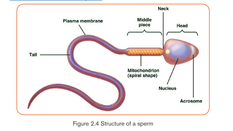
energy for the movement of the sperm. At the end of the middle
piece, there is a ring centriole (annulus) with unknown function.
iv) Tail is several times longer than the head. It consists of an axial filament
surrounded by a thin layer of cytoplasm. The tail provides motility tothe sperm, which is essential for fertilization.
b) Growth and differentiation phase: During this long phase, which may
last upto years, one cell in a follicle prepares for the formation of ovum. It
starts meiotic division but gets arrested at prophase-I stage and is called
primary oocyte. The remaining cells of the follicle lose the potential to
become primary oocyte and are known as the follicular cells or granulosa
cells. These follicular cells serve to protect and nourish the primary oocyte.
The complete follicle with a primary oocyte surrounded by a layer of
follicular cells is called the primary or the ovarian follicle.
c) Maturation phase: At puberty, only one of the primary oocytes resumes
division per menstrual cycle, alternately in each ovary. The tertiary follicle
matures into a Graafian follicle, within which the primary oocyte makes two
successive to form secondary oocyte. However, the secondary oocyte
again gets arrested at metaphase stage of meiosis-II and is released
from the ovary during ovulation. It waits in the oviduct for the sperm to
arrive. If fertilization occurs, sperm entry into the secondary oocyte marks
the resumption of meiosis. The 2nd maturation division (meiosis-II) again
divides the secondary oocyte into two unequal daughter cells: a large
ootid and a very small 2nd polar body. The ootid undergoes maturation into
a functional haploid ovum. A thin vitelline membrane develops outside theplasma membrane of the ovum that protects and nourishes the latter.
– Structure of Ovum
An ovum is a spherical, non-motile cell, in the secondary oocyte stage of
oogenesis. Human ovum is microlecithal with large amount of cytoplasm.
Cytoplasm is differentiated into outer, smaller and transparent exoplasm or egg
cortex and inner, larger and opaque endoplasm or ooplasm. Egg cortex is with
some cytoskeletal structures like microtubules and microfilaments, pigment
granules and cortical granules of mucopolysaccharides. Endoplasm is with cellorganelles,
informosomes, tRNAs, histones, enzymes etc.
The ovum is covered over by a thin, transparent vitelline membrane which is
further covered over by zona pellucida. There is a narrow space between these
two membranes known as perivitelline space. During discharge of ovum from
the Graafian follicle, several layers of follicular cells adhere to the outer surfaceof zona pellucida and are arranged radially forming corona radiata.
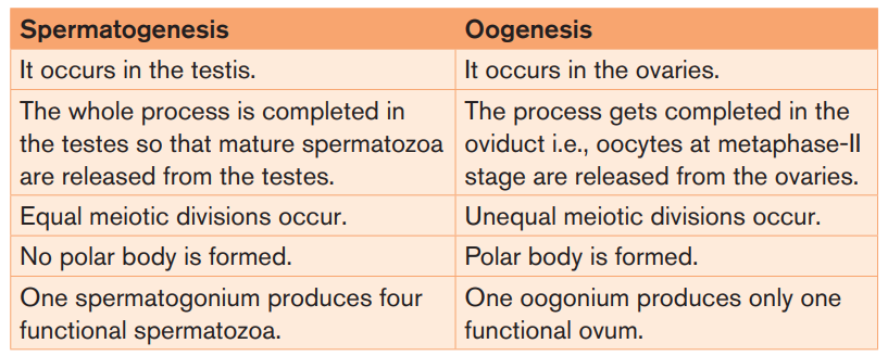
Table 2.3: Differences between spermatogenesis and oogenesis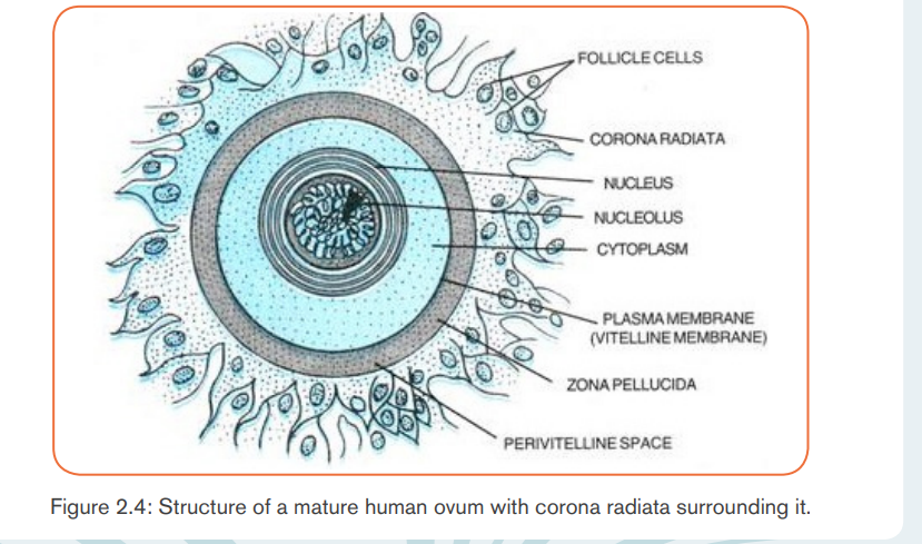
Application activity 2.2
1. Suppose that four hundred sperm mother cells have undergone a process
of spermatogenesis in a testis of human. How many chromosomes are
produced at the end of spermatogenesis? How many chromosomes
does each sperm have?
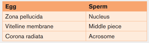
2. On the basis of your observations, use the drawn structure of a human
spermatozoan and an ovum and label their respective parts along with
the functions of each: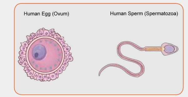
2.1.4. Cycle in humans
Activity 2.3
Human beings grow and develop from childhood to adulthood, during such
period of growth and development; there are changes in some parts of body
which may occur physiologically, physically and even psychologically. These
changes prepare individual adulthood to reproduce. Different researchers
indicated these changes to be coordinated by different types of hormones.1. Suggest the hormones involved during such period of changes in body
parts?
2. Discuss the significance of these hormones you have mentioned above
during such period of changes.
3. Describe the role of hormones involved during pregnancy and birth.4. Which day of the cycle will ovulation take place?
The menstrual cycle refers to the periodical changes in the reproductive behaviour
of a female which tend to occur in a sequence of events one after the other
in the periodical circle. At the onset of puberty, the cycle begins and repeats
after 28 days unless interrupted by pregnancy. The changes are stimulated
by the gonadotrophic hormone such as; follicle stimulating hormone (FSH)
and luteinizing hormone (LH). These hormones stimulate ovaries to secrete;
oestrogen (steroid) and progesterone hormones. These four hormones are
involved in menstrual cycle. Two of them including; FSH and LH are produced
by pituitary gland and the other two are released by ovaries respectively. The
most obvious sign of the cycle is the monthly discharge of blood a process
called menstruation. The first day of menstruation is regarded as the first dayof the cycle.
The three phases of the menstrual cycle are the follicular phase,ovulation and the luteal phase.
a. Follicular phase
Menstrual cycle usually begins when blood is first discharged from the uterus
during the first to fifth day (1-5 days). Following the reduction of progesterone,
the hypothalamus releases gonadotropin releasing hormone (GnRH) which
stimulates anterior pituitary gland to secrete follicle stimulating hormone (FSH).
FSH brings about the following effects:- Stimulates the development of a primary follicleCauses production of oestrogen by uterine cells. The oestrogen produced- Contributes to the shedding of uterine wall
promotes healing, repair and growth of uterine lining, inhibits further
secretion of FSH. Oestrogen levels keep on raising until day 13 where
they stimulate secretion of luteinizing hormone (LH) by anterior pituitarygland.
b. Ovulatory phase
Around the 14th day, the high levels of oestrogen causes release of luteinizing
hormone (LH).The release of LH brings about ovulation (release of mature egg
from the ovary). Immediately after and slightly before ovulation, a woman is fertile
and can conceive a baby if she has sexual intercourse or if sperm is present inher oviduct.
c. Luteal phase
After ovulation, the remains of ovarian follicle form corpus luteum also known
as Yellow body, which secrete large amounts of progesterone hormone
and smaller oestrogen. These two hormones; stimulate further development
of mammary glands, inhibit release of FSH and thickening wall of uterus in
anticipation of pregnancy. If oocyte (ovum) is not fertilized with in about 36
hours of being shed into oviduct, it dies and corpus luteum gets smaller. Thus
levels of progesterone and oestrogen keep on reducing until day 28 days i.e. 14
days after ovulation. Low levels of progesterone remove the inhibitory effect onFSH, causing its release thus menstruation and the cycle starts again.
The menstrual cycle is the regular natural changes that occurs in the female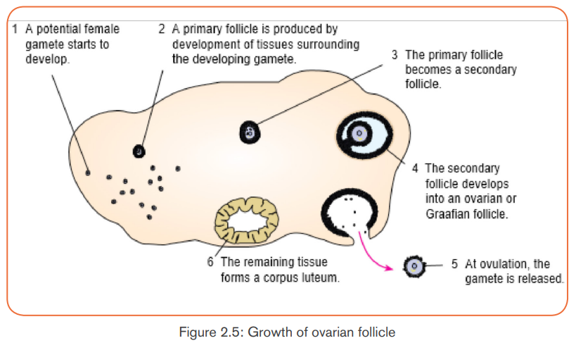
reproductive system (specifically the uterus and ovaries) that makes pregnancy
possible. The cycle is required for the production of oocytes, and for the
preparation of the uterus for pregnancy. The uterine events during menstrualcycle can also be divided into three phases:
Menstrual phase: when endometrium tissue is discharged and vaginal bleeding
occurs at the end of ovulatory cycle if pregnancy has not occurred. It is called
menstruation. It describes the shedding of endometrium when implantation
does not occur. When pregnancy does not occur, the level of progesterone
falls and this leads to the shedding of endometrium. Menstrual bleeding lasts
between 3 and 5 days. The first day of the period is the first day of the cycle.
Proliferative phase: It stimulates the thickening of endometrium of the uterus.
This thickness of endometrium is stimulated by estrogen from follicles before
ovulation which occurs when the ovarian follicles rupture and release the
secondary oocyte ovarian cells. This results to the development of ovary. It acts
like follicular phase.
Secretory phase: it occurs after ovulation for describes further thickening of
endometrium (endometrium tissue become more complex) in preparation for
implantation. This is stimulated by progesterone which is secreted by corpus
luteum and this occurs when corpus luteum is functioning. It acts like lactealphase.
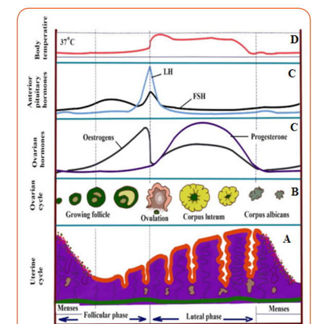
Figure 2.6: Menstrual cycle of human female
Application activity 2.3
1. The diagram below represents a section through a human ovary inovulation.
1. Use the diagram to locate the step at which ovulation take place. Explain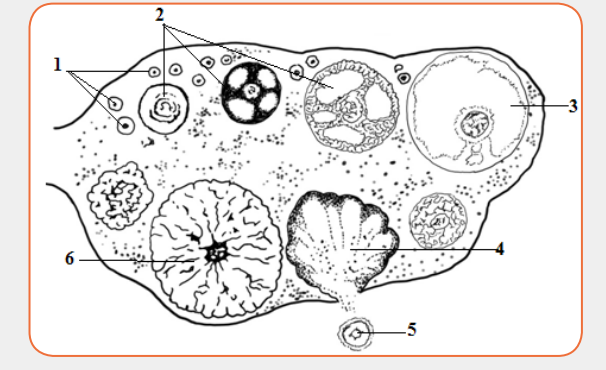
your answer.
2. State what will happen to this structure next if pregnancy has not
occurred.
3. State which hormone is needed to cause the changes seen in thediagram and indicated by the sequence (1), (2) and (3).
2.1.5. Fertilization and fatal developmentActivity 2.4
The following diagram represents different stages that happen before fetal
development. Use it to answer related questions.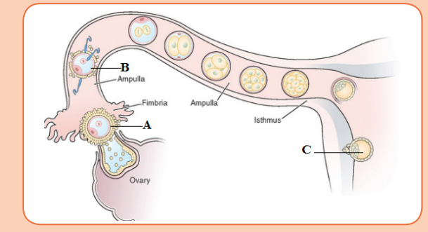 i) Suggest the name of the cell labeled A.a. Copulation
ii) Name the process which is happening on the cell labeled B. What
are the conditions required for the process to happen? Justify your
answer.
iii) Describe the process which is happening in C and what happensafter?
It is act of mating where sperms from male are transferred into the female tract.
Male mammals have an intromittent organ called penis which becomes erect
at a moment of mating for insertion into female’s vagina. The erection of penis is
brought by hydraulic action (penis becomes gorged with blood). This occurs as
a result of sexual arousal which brings about by ejaculation (release of sperm).
The semen’s are secreted from accessory glands into vas deferens and bladder
sphincter closes preventing urine from entering urethra. Sperms are expelled
from epididymis into vas deferens and out of the body by a series of muscle
contraction of penis. In a female, sexual arousal results in the swelling of clitoris
and stimulates the secretion of mucus which lubricates vagina during sexualintercourse.
b. Fertilization
Fertilization is the fusion of male and female nuclei to form zygote. Copulation
results in the ejection of spermatozoa into vagina. The spermatozoa swim in the
watery mucus of vagina and uterus up into the oviduct where the fertilization
takes place in the upper part of the oviduct. From the vagina or uterus
spermatozoa propel using energy from mitochondria. If ovulation has already
taken place, the egg and sperm meet in the upper part of oviduct and once they
come into contact, acrosome raptures and release lytic enzyme which dissolve
corona radiata of the egg and soften zona pellucida and vitelline membrane. The
following processes take place:
– Capacitation
This is a stage where by sperm undergoes essential changes while passing
through female genital track and this takes about 7 hours. These changes
include the removal of a layer of glycoprotein from outer surface of sperm, by
enzyme in uterus. Cholesterol also is removed to weaken the membrane.
– Acrosome reaction
This involves the releasing of enzyme found in acrosome such as hyaluronidases
and protease. These enzymes digest corona radiata (narrow path in the follicle
cells) and the zona pellucida (a protective glycoprotein surrounding the plasmamembrane of the egg).
Fusion
In this stage the head of sperm will fuse with the microvilli surrounding thesecondary oocyte and penetrate its cytoplasm.
– Cortical reaction
This stage involves the releasing of enzymes by lysosomes in cortical granules
(outer region of the secondary oocytes); the enzymes cause the zona pellucida
to thicken and harden forming a fertilization membrane. This cortical reaction
prevents the entry of other sperm inside ovum (polyspermy).
– Zygote formation
The secondary oocyte is stimulated to complete meiosis II, during this time of
stimulation the nucleus of sperm and secondary oocyte are called pro-nucleiand then the two nuclei fuse to form the zygote (2n).
2.1.6. Embryonic development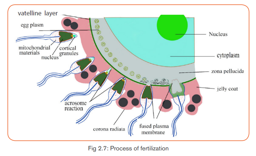
The zygote spends the next few days travelling down the oviduct (Fallopian
tube) by peristaltic contraction and by beatings of the cilia in wall of the oviduct
toward the uterus. As it travels, it divides by mitosis several times to form a ball
of cells called a morula. The cell divisions, which are called cleavage, increase
the number of cells but not their overall size. More cell divisions occur, and soon
a fluid-filled cavity forms inside the ball of cells. At this stage, the ball of cells iscalled a blastocyst.
The blastocyst reaches the uterus and becomes embedded in the endometriumat roughly the 5th – 10th day. Once in the uterus, the blastocyst burrows into theuterine wall a process called implantation. After implantation, the blastocyst
becomes embryo. It grows through multiplication and differentiation of its cells
forming tissues and organs. The heart and blood vessels are the first organsformed and embryo now called foetus.
During embryonic development, cells of the embryo migrate to form three distinct
cell layers: the ectoderm, mesoderm, and endoderm. Each layer will eventually
develop into certain types of tissues and cells in the body of vertebrates.- Ectoderm: forms tissues that cover the outer body; develops into cells
such as nerves skin, hair, and nails.
- Mesoderm: forms tissues that provide movement and support; develops
into cells such as muscles, bones, teeth, and blood.
- Endoderm: forms tissues involved in digestion and breathing; developinto organs such as lungs, liver, pancreas, and gall bladder.
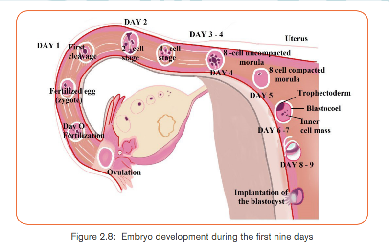
Application activity 2.4
The diagram below shows some of the events which take place in the ovaryand oviduct (Fallopian tube) around the time of fertilization.
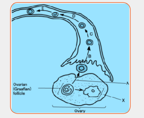
a) Name the following:
i) The process labeled A.b) On the diagram, use the letter F to label the region where fertilization
ii) The type of nuclear division taking place at D and E.
iii) The structure labeled X.
iv) One hormone produced by structure X.took place
2.1.7. Role of placenta in the development of an embryoActivity 2.5
The drawing below shows a developing human fetus inside the uterus.Observe the diagram and attempt the related questions.
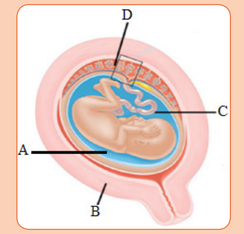 a) Suggest the name of the parts marked A to D.The placenta is a temporary organ in which nutrients and wastes are exchanged
b) Which part is involved in transport of substance from mother to fetus
on the diagram and why?
c) Suggest four substances which pass from the mother to the embryo.
d) Name one substance which passes from the embryo to the mother.e) What is the importance of the placenta?
between the mother and the embryo or foetus.
The foetal part of the placenta consists of the allantoids and chorion. The
chorion forms many large projections called chorionic villi which contain a
dense network of foetal capillaries which in turn are connected to two umbilical
arteries and umbilical vein in the umbilical cord. The umbilical arteries carry
blood from the foetus to the placenta, while the umbilical vein carries blood
in the opposite direction. Although maternal blood in the endometrium is in
close proximity with the foetal blood in the umbilical capillaries, they do not mixbecause they separated by membranes of the villi and capillary.
The placenta is an organ that develops in your uterus during pregnancy with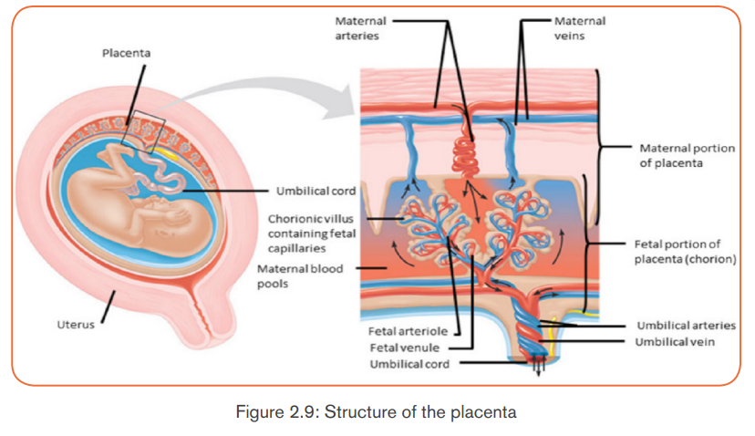
specifically the following functions:- It allows diffusion of nutrients such as water, glucose, amino acids, simpleNote: The action of HCG is similar to that of LH. HCG stimulates the corpus
proteins and mineral salts from maternal blood.
- It is a site of gaseous exchange: haemoglobin of the foetus has high affinity
to oxygen compared to adult haemoglobin.
- It offers passive natural immunity on the foetus. Certain maternal antibodies
can cross the placental barrier.
- It protects foetal circulation from the high pressure in the maternal
circulation
- Prevents mixing of maternal and foetal blood which would cause
agglutination (clotting) if the two blood types are incompatible.
- It produces and secretes hormones such as the HCG (human chorionicgonadotrophin), progesterone, oestrogen, and relaxin.
luteum to secrete progesterone and oestrogen throughout the first trimester.
HCG is produced in such large quantities that some of it is excreted in the urine
of a pregnant woman (positive test of pregnancy). Secretion of HCG declinesaround tenth week and the corpus luteum reduces.
The placenta does not give complete protection to the foetus. Certain pathogens,
toxins, and drugs can enter the foetal circulation and cause damage. Examplesare; HIV, rubella toxins, alcohol, nicotine and heroin.
Application activity 2.5
The diagram shows the structure of the placenta and parts of the fetal andmaternal circulatory systems.
a) Complete the table by listing the blood vessels that carry oxygenated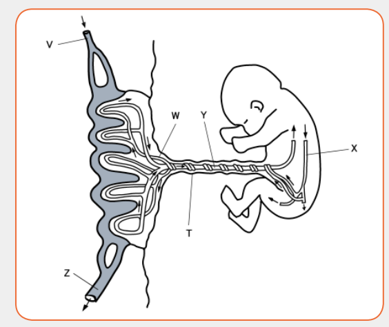
blood. Use the letters in the diagram to identify the blood vessels.
b) What happens on the structure T after birth?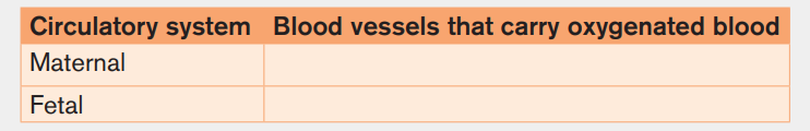
c) The placenta is adapted for the exchange of substances between the
maternal blood and the fetal blood. Describe the exchanges that occuracross the placenta to keep the fetus alive and well.
2.1.8. Physiological changes in females during pregnancy
and Parental care
Activity 2.6Observe the following images that show pregnant women.
Use your personal observation or conduct research from medical personnel,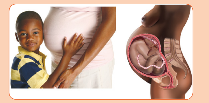
internet or library to answer the following questions:a) Suggest the physical changes that can be observed to the pregnantPregnancy refers to the development that take place between fertilization of the
women.
b) What physiological and behavioral changes that can happen when
women get pregnant.
c) It is necessary to practice a special parental care to pregnant women.Provide reasons that justify this statement.
ovum to birth of the foetus. When fertilized egg becomes implanted in uterine
wall, pregnancy starts. And a number of important events take place during
this period. The period from fertilization to birth is called gestation period. Inhuman it is about nine months.
A. Changes during pregnancy
A pregnant woman’s body undergoes various; physiological, physical and
behavioural changes.
a) Physiological changes during pregnancy- Respiration rate rises for increased maternal oxygen consumption which is
needed for demand of placenta, uterus and foetus.
- More blood vessels grow and pressure of expanding uterus on large veins
causes blood to slow in its return to the heart.
- Rise up and out of pelvic cavity this action displaces the stomach and
intestine.
- Blood volume increase greatly.- Placenta produces large amount of progesterone and oestrogen by 10 to12 week of pregnancy to control uterine activity.
- Increased requirement of calcium due to increase of parathyroid gland.b) Physical changes during pregnancy
- Experiences warm (hot flashes) caused by basal metabolic rate and
increased hormonal level.
- Stretching of abdomen wall and ligaments that support uterus.
- Kidney work extra hard to excrete waste products of both mother andfoetus.
- Breast may become large and more tender because of increased level ofc) Behavioural changes during pregnancy
oestrogen hormone progesterone thus breast gets even bigger to prepare
for breast feeding.
- Nipples may stick out more.
- By the end of third trimester, a yellow, watery, pre-milk may leak from
nipples.
- Changes in hair and nail growth and texture due to hormone changes.
- Leg cramp caused by fatigue from carrying pregnant weight.
- Feet and ankles may swell because of extra fluid in the body duringpregnancy.
- Physical discomfort such as urinary frequency can be frustrating.B. Delivery process
- Fear and anxiety lessen especially when foetal movements are felt.
- Self-introspection
- Nesting behaviour begins. Some woman exhibits mood swings andemotional liability.
By the end of pregnancy, near the time of birth, the amniotic sac raptures
(breaks) and amniotic fluid drains through birth canal and labour usually begins
which involves the contractions of muscular walls of the uterus.
Initiation of birth: Uterine contractions start when the foetal pituitary gland
secretes adrenocorticotrophic hormone (ACTH) which stimulates foetal adrenal
gland to secrete corticosteroids. These hormones pass into blood sinuses in
placenta to cause maternal cells to secrete prostaglandins (local hormone) and
cause uterine wall to contract. This contraction pushes the foetal head against
the cervix to stimulating stretcher receptor to send information to mother’s
brain and causes release of oxytocin hormone. The prostaglandin and oxytocin
hormone together result intense contraction of uterine walls called labour
which stimulates more release of oxytocin hormone and as positive feedbackmechanism.
The delivery process can be summarized into three main stages:- Dilation stage: During this stage, water sac filled with amniotic fluid forms
and precedes the head, widening soft tissue of birth canal, cervix, and
vagina for canal of constant diameter. The amnion raptures and amniotic
fluid drains through vagina.
- The expulsion stage: During this stage, cervix is fully dilated while
abdominal muscle bear down in supporting rhythmic contraction of uterus
shorten the uterine wall and baby is pushed into and through the birth
canal. The head and shoulder align themselves first.
- Placenta stage: This stage begins with complete expulsion of baby and
ends with expulsion of foetal membrane. The cord is clamped and cut
when delivery of baby is complete. This leads carbon dioxide enrichment
into baby’s blood which activates respiratory centre and baby begins to
breath with the first cry at the same time foetal circulation changes tobaby’s own systemic and pulmonary circulation.

Antenatal care is the care you get from health professionals during your
pregnancy. It is sometimes called pregnancy care or maternity care. Prenatal
care, also known as antenatal care, is a type of preventive healthcare. Its goal
is to provide regular check-ups that allow doctors or midwives to treat and
prevent potential health problems throughout the course of the pregnancy andto promote healthy lifestyles that benefit both mother and child.
a) Health needs of the pregnant mother
The pregnant mother needs to maintain good health status so that she has a
healthy baby. To remain healthy she needs:- To avoid contractions diseases such as malaria, STIs and HIV and AIDS
as these may harm the foetus.- To avoid smoking and drinking alcohol as these interfere with growth anddevelopment of the foetus, especially brain development.
- To eat an adequate balanced diet so that she maintains her good health
and is able to give birth to a healthy baby. Malnourished mothers usually
give birth to babies who are underweight. Such babies often have growth
and development problems because they do not eat well and tend to get
sick often.During the antenatal period, the promotion of the women healthy is the care
- To attend the ante-natal clinic once a moth so that her health and nutritional
needs and those of the foetus are monitored. In the ante-natal clinic the
mother has her weight monitored, blood pressure checked and urinechecked to establish the level of sugar.
and health of their babies before and after birth. Educating mothers about the
benefits of good nutrition, adequate rest, good hygiene, family planning and
exclusive breastfeeding, immunization and other disease prevention measures
aims to develop women’s knowledge of these issues so they can make better
decisions affecting their pregnancy outcome and never forget the difficultiessome women will face in being able to improve their lifestyles.
b) Nutritional needs of the pregnant mother
The pregnant mother needs additional nutritional requirements to meet the needs
of the growing foetus and those of her body. The pregnant mother therefore
needs to eat additional balanced diet to cater for these additional nutritional
requirements in her own body and for that of the growing foetus. The mother
needs the increased nutrients because of:- The increase in the rate at which her body burns energy. More carbohydrates
and fats are required. Adequate amounts of carbohydrates and fats are
required for the additional weight the mother puts on. The mother puts on
additional weight of about 10 to 12.5kg during pregnancy. More energy
giving foods are required to make up this additional weight.
- The increase in her blood volume of her additional weight. More iron is
required to form additional blood required by the body.
- The development of the placenta which requires nutrients to form and
making the amniotic fluid within which the foetus grows require more
nutrients.
- Increased muscles for both the mother and the growing foetus. The
mother’s body requires additional muscles, especially the breast and
uterine tissues. More proteins are required to develop these muscles.
- The need to store more fat. This fat is stored during the first four months
of pregnancy. From 5-9 months the stored nutrients are used by the fast
growing foetus. The mother starts to appear thinner.The breast milk contains antibodies that help your baby fight of viruses and- Preparation of breast-feeding. Nutrients are required to prepare milk to be
used to breast-feed the baby.
bacteria. Breastfeeding lowers your baby’s risk of having asthma or allergies.
Plus, babies who are breastfed exclusively for the first 6 months, without any
formula, have fewer ear infections, respiratory illnesses, and bouts of diarrhea.
In humans breastfeeding is associated with many other benefits:- It makes earlier a closer contact between the mother and her infantApplication activity 2.6
- The infant has a better control over its own milk intake, this prevents over
eating in late life
- Fats and irons from breast milk are better absorbed than those in cow’s
milk and milk is easily digested.
- Breast feeding provides important antibodies that help to prevent
respiratory infections and meningitis,
- Breastfeeding helps the mother’s reproduction organ return to a normal
state more rapidly
- Breast feeding promotes the secretion of LH (and prolactin) and this
makes a delay in follicle development and ovulation,
- The act of sucking on the breasts, promotes the development of the jaw,facial muscles and teeth (sucking from a bottle requires less effort).
1. Copy and complete the table to show, for each hormone, the precise
site of its secretion, and its effects on the ovary or on the endometriumof the uterus.
Alcohol consumption for pregnant women is generally more dangerous on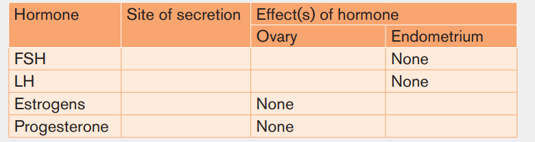
an embryo than a fetus. Suggest the reasons.
2.2. Family planning and contraceptive methods
Activity 2.7The photos below show various contraceptive methods.
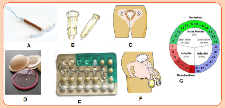 a) Use the photos to identify the letters that represent natural and artificialContraception is the prevention of conception that is preventing the fusion of
contraceptive methods. Justify your choice.
b) The most effective contraceptive method for young people is the useof condom. Provide the reasons for this statement.
the male gamete with the female gamete. Both natural and artificial methodsexist.
2.2.1 Natural contraceptive methods
Natural birth control methods include specific actions that people can do
naturally to help prevent an unintended pregnancy. Instead, these methods to
prevent pregnancy require that a man and woman not have sexual intercourse
during the time when an egg is available to be fertilized by a sperm.
The fertility awareness methods are based upon knowing when a woman
ovulates each month. In order to use a fertility awareness method, it is necessary
to watch for the signs and symptoms that indicate if ovulation has occurred oris about to occur.
i) Calendar rhythm method
The calendar rhythm method to avoid pregnancy relies upon calculating a
woman’s fertile period on the calendar. Based upon her 12 previous menstrual
cycles, a woman subtracts l8 days from her shortest menstrual cycle to determine
her first fertile day, and 11 days from her longest menstrual cycle to determine
her last fertile day. She can then calculate the total number of days during which
she may ovulate. If a woman’s menstrual cycles are quite irregular from month to
month, there will be a greater number of days during which she might become
pregnant.
The calendar method is only about 80% effective in preventing pregnancy and
when used alone, it is considered outdated and ineffective.
ii) Basal body temperature method
The basal body temperature (BBT) method is based upon the fact that a
woman’s temperature drops 12 to 24 hours before an egg is released from her
ovary and then increases again once the egg has been released. Unfortunately,
this temperature difference is not very large. It is less than 1-degree F (about ahalf degree C) when the body is at rest.
The basal body temperature method requires that a woman take her temperature
every morning before she gets out of bed. A special thermometer that is more
accurate and sensitive than a typical oral thermometer must be used, and the daily
temperature variations carefully noted. This must be done every month. Onlinecalculators are available to help a woman chart her basal body temperature.
To use the basal body temperature as a birth control method, a woman should
refrain from having sexual intercourse from the time her temperature drops untilat least 48 to72 hours after her temperature increases again.
iii) Mucus inspection method
The mucus inspection method depends on the presence or absence of a
particular type of cervical mucus that a woman produces in response to
estrogen. A woman will generate larger amounts of more watery mucus than
usual (like raw egg white) just before release of an egg from her ovary.This socalled egg-white cervical mucus stretches for up to an inch when pulled apart.
A woman can learn to recognize differences in the quantity and quality of her
cervical mucus by examining its appearance on her underwear, pads, and toilet
tissue; or she may gently remove a sample of mucus from the vaginal opening
using two fingers. She may choose to have intercourse between the time of her
last menstrual period and the time of change in the cervical mucus. During this
period, it is recommended that she have sexual intercourse only every other day
because the presence of seminal fluid makes it more difficult to determine the
nature of her cervical mucus. If the woman does not wish to become pregnant,
she should not have sexual intercourse at all for 3 to 4 days after she notices thechange in her cervical mucus.
iv) Withdrawal method
Withdrawal is a behavioral action where a man pulls his penis out of the vagina
before he ejaculates. The withdrawal method also relies on complete selfcontrol.
You must have an exact sense of timing to withdraw your penis in time.
Because this can be difficult for the man to complete successfully, the withdrawalmethod is only about 75%-80% effective in preventing pregnancy.
v) Abstinence
Abstinence from sexual activity means not having any sexual intercourse at all.
No sexual intercourse with a member of the opposite sex means that there is nochance that a man’s sperm can fertilize a woman’s egg.
vi) Lactation amenorrhea method
Lactation Amenorrhea method can postpone ovulation for up to 6 months after
giving birth. This natural birth control method works because the hormone
required to stimulate milk production prevents the release of the hormone that
triggers ovulation. This method is highly effective for the first six months after
childbirth. The mother has to breastfeed the baby at least every four hours
during the day and every six hours through the night. She also has to be aware
of her menstrual period. After six months fertility may return at any time.
Advantages of natural birth control- A woman does not need to take medication or use hormonal manipulation.Disadvantages of natural birth control include
- No procedures or fittings by a physician are required.- It can be difficult to estimate or know precisely when a woman is fertile,2.2.2. Artificial contraceptive methods
allowing increased chances for unplanned conception.
- Natural methods are not as effective as some forms of contraception.
- Ovulation test kits are used by some couples using natural methods of
contraception, and the cost of these kits is another potential disadvantage.
- Being unable to have intercourse at certain times of the month is a
disadvantage for some women.
Artificial contraception also known as birth control are medication used to
prevent pregnancy.
Oral Contraceptive pills: a chemical method of contraception. One version
uses a combination of progesterone and oestrogen that inhibits ovulation.Others are single hormones that require very careful management when taken.
Intrauterine device (IUD) the coil is placed inside the uterus an exact
understanding how this works is unclear. A possible explanation is that it
‘irritates’ the endometrium such that rejects implantation of embryos. The device
is made from plastic or copper and inserted by a doctor. Nevertheless, this
device is very effective.
Condom is another mechanical method of contraception that prevents the
sperm from reaching the egg. Composed of a thin barrier of latex this is placed
over the erect penis and captures semen on ejaculation. This is also a goodbarrier to prevent the transmission of sexual diseases.
Cap (diaphragm) is another barrier method again made from latex. The cap is
placed over the cervix to prevent the entry of sperm in semen. This technique
requires that the cap is put in position in advance of sexual intercourse and that
it is used in combination with a spermicidal cream. When used correctly this is
an effective contraceptive however this is not a barrier against the transmissionof sexual diseases.
Sterilization is a surgical and near permanent solution for contraception such
as: Vasectomy. In men this involves cutting the vas deferens and prevents sperm
entering the semen. In this state, man still ejaculates normally and releases
semen however this does not contain sperm. Tubal ligation involves the cutting
of fallopian tube so that eggs cannot reach the uterus. In women the surgery cutsor ties the oviducts thus preventing sperm from reaching the egg in fertilisation.
Advantages and disadvantages of birth control
Advantages of birth control/contraceptives- Gives great protection against unplanned pregnancy if one follows- Woman must begin using hormonal contraceptive in advance before they
instructions.
- Condoms to some extent protect against pregnancy and STDS.
- Combinations of pills reduce/prevent cysts in breasts and ovaries.
- Improved family wellbeing.- Improved maternal and infant health.
become effective.
- Some women experience several; headaches, breast tenderness, chest
pain, discharge from vagina, leg cramps and swelling or pain.
Disadvantages of birth control/contraceptives- Necessity of taking medication continually.Application activity 2.7
- High cost of medication.
- Hormonal contraceptive does not protect against STDS.
- Eggs may fail to mature in the ovary for a woman who uses hormonal
contraceptives.- Woman must remember to take them regularly.
1. Determining the fertile period
Count the number of days of your menstrual cycles and count the number of
days for 10 consecutive cycles. Choose the cycle with the highest number
of days and the cycle with the lowest number of days. Subtract 18 from the
lowest cycle and 11 from the highest cycle.
Example: Mary has 27 days as her shortest cycle and 36 as her longest
cycle. She has had her menstruation on 09/08/2019. What will be her fertile
period?2. The diagrams below represent different contraceptive methods.
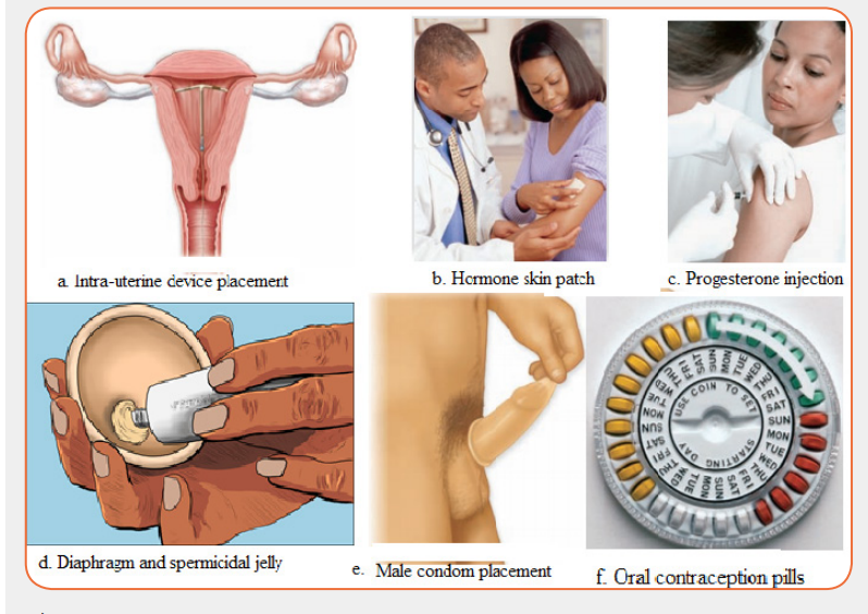 i) Use the diagrams to state contraceptive method that can prevent bothSkills Lab 2
STDs and pregnancy. Justify your answer.
ii) Suppose you are married, which contraceptive method do you preferto use and why?
Pregnancy test
The HCG Card Pregnancy Test is a rapid chromatographic immune assay for
the qualitative detection of human chorionic gonadotropin in urine to aid in
the early detection of pregnancy. The test utilizes a combination of antibodies
including a monoclonal HCG antibody to selectively detect elevated levels of
HCG.
The pregnancy test works by checking the urine for a hormone called human
chorionic gonadotropin (HCG). The woman body only makes this hormone if
she pregnant. HCG is released when a fertilized egg attaches to the lining of
the uterus when pregnancy begins. If pregnancy test is positive, it means that
woman is pregnant. If the pregnancy test is negative, it means that woman isnot pregnant.
Procedure:- Carefully read the instruction included in your test kit before collecting
your urine sample.
- Remove the plastic cap to expose the absorbent window.
- Use collected first morning urine one to two weeks after the first missed
period.
- Collect urine in a cup and then dip the indicator stick into the cup to
measure the HCG hormone level.
- Hold an indicator stick directly in the urine stream until it is soaked,
which should take about five seconds.
- Remove the HCG card pregnancy and make observation.- Take conclusion of the observation following the indicated interpretation.
End Unit Assessment 2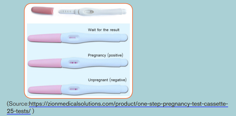
1. Which of the following do sperm NOT travel through?a) Ureter2. The placenta in humans is derived from the:
b) Urethra
c) Vas deferens
d) Epididymisa) Embryo only3. The graph below shows the level of reproductive hormones in the
b) Uterus only
c) Endometrium and embryo
d) None of the aboveblood of an un-named mammal during its reproductive cycle.
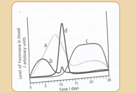 a) Name the hormones labelled (a) to (d)4. Answer the following questions:
b) Give the likely day of the cycle on which ovulation takes place and
give reasons for your answer.a) Define the term fertilizationb) The diagram below shows the structure of a human sperm.
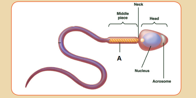 i) Explain the part played by the organelle labelled A in the process5. Which contraceptive methods can protect against sexually transmitted
leading to fertilization.
ii) The acrosome contains an enzyme that breaks down proteins.
Describe the function of this enzyme in the process leading to
fertilization.
diseases / infections?
6. The diagram shows the sequence of events in the development of a
mature ovarian (Graafian) follicle and corpus luteum
a) What is the main hormone produced by the ovary when stage B is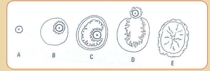
present?
b) Which two of stages A to E would you expect to find in the ovary of
a woman during the early stages of pregnancy?
c) Give the reason for your answer on b.
d) Some oral contraceptives contain only estrogens. Which of the
stages A to E would you expect to find in the ovary of a woman who
had been taking such an oral contraceptive for a prolonged period of
time?e) Give reasons for your answer on d.
UNIT 3: FERTILIZERS
Key Unit Competence:
Analyze the components of quality fertilizers and their benefits, effects ofmisuse and dangers associated with substandard fertilizers.
Introductory activity 3
Observe the pictures below showing some common fertilizers used inagriculture and attempt the following questions:
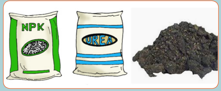 i) It is a must for a farmer to use fertilizers. Why?
ii) The fertilizers shown above are different. How are they different?
iii) Why a farmer does use different fertilizers (NPK, UREA, …) on only
one same plant?
iv) Can the fertilizers above become hazardous? When? What is
chemical hazard?
v) How the types of fertilizers mentioned above can have effect to the
environment or living things?
vi) Suggest the measures to avoid hazard while using fertilizers.3.1. Classification of fertilizers
Activity 3.1Observe the following fertilizers:
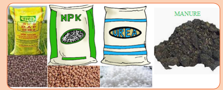 i) Read the labels of given fertilizers and propose the components ofA fertilizer is any material, organic or inorganic, that is used to supply nutrients
each.
ii) Categorize the above fertilizers according to their ways of
manufacturing?
iii) Suggest any other examples of fertilizers you have ever heard or used
at home.
iv) Use engine research or library textbook to describe the nutrients thatplants need in order to grow from fertilizers
to the soil.Fertilizer is a substance added to soil to improve plants’ growth and
yield.Fertilizers replace the chemical components that are taken from the soil
by growing plants. However, they are also designed to improve the growing
potential of soil, and fertilizers can create a better growing environment than
natural soil. They can also be tailored to suit the type of crop that is being grown.
Fertilizers are categorized into natural fertilizers (or organic fertilizers) andartificial fertilizers (or chemical fertilizers)
3.1.1. Natural Fertilizers
The name organic fertilizer refers to materials used as fertilizer that occur
regularly in nature, usually as a by-product or end product of a naturally occurring
process. They are made from remains of dead plants, wastes from animals or
they can be minerals. Examples include manures and minerals. Manure is anorganic material that is used to fertilize land.
Farmyard manure: animal manure that consists of feces.
Green manure: is a term used to describe specific plant or crop varieties that
are grown and turned into the soil to improve its overall quality.
Compost manure: is organic matter that has been decomposed and recycled
as a fertilizer and soil amendment. Minerals: Mineral mined powdered
limestone, rock phosphate and sodium nitrate, are inorganic compounds which
are energetically intensive to harvest and are approved for usage in organicagriculture in minimal amount.
3.1.2. Artificial Fertilizers
Artificial fertilizers are man-made chemical compounds that mimic the soil’s
natural minerals and elements to maximize plant growth. They usually contain
different ratios of nitrogen, phosphorus, potassium, calcium, magnesium andother elements.
Examples: Urea, N.P.K, ammonium dihydrogen phosphate, NH4 (H2PO4), etc.
Table 3.1: Differences between natural and artificial fertilizers
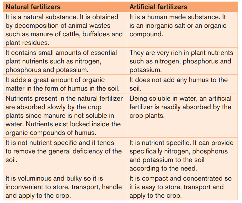
3.1.3. Components of a fertilizer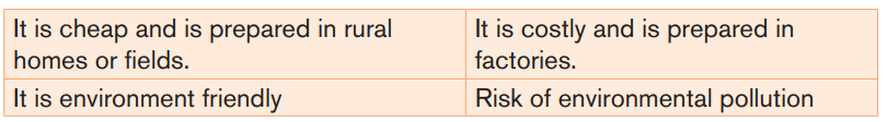
Typically, fertilizers are composed of nitrogen, phosphorus, and potassium
compounds. They also contain trace elements that improve the growth of
plants. The primary components in fertilizers are nutrients which are vital for
plant growth.
First it is important to understand that all industrial Fertilizers, by convention,
regardless of type and specific use, have something called a NPK ratio. The NPK
ratio will be prominently labeled on the package and indicates the percentage
of major (or primary) nutrients the fertilizer contains. Example: Urea is a fertilizer
with an NPK ratio of 46-00-00.
The nutrients of plants are classified into three types namely: major nutrients,secondary nutrients and micronutrients.
a. The major nutrients
The major nutrients for soil are nitrogen (N), phosphorus (P), and potassium
(K). These major nutrients usually are lacking or insufficient in the soil because
plants consume them in large amounts for their growth and survival.
The letter N represents the actual nitrogen content in the fertilizer by percentage
mass while P and K represent the amount of oxide in the form of phosphorus (V)oxide (P2O5) and potassium oxide (K2O) respectively.
Example:
- If a fertilizer is labeled 17-17-17, it means that the fertilizer contains17%
by mass N, 17% by mass P2O5 and 17% by mass K2O.
- If fertilizer is labeled 10-20-20, it means that the fertilizer contains 10% bymass N, 20% by mass P2O5 and 20% by mass K2O.
Table 3.2: Role of nutrients
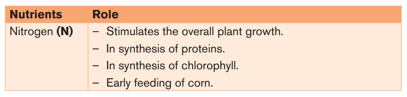
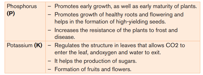
If a fourth number is included on the label of a fertilizer, it indicates the sulphur
content. That fertilizer is NPKS.
b. Secondary nutrients
The category of secondary nutrients, are calcium (Ca), magnesium (Mg), and
Sulphur (S). As, these nutrients are generally enough in the soil, so fertilization
is not always needed. Also, large amounts of Calcium are added when lime
is applied to acidic soils. In fact, Sulphur is usually found in sufficient amounts
from the slow decomposition of soil.
c. Micronutrients
Micronutrients are those elements essential for plant growth which are needed
but in only very small (micro) quantities. These elements are even called minor
elements or trace elements. The common micro nutrients are boron (B), copper
(Cu), iron (Fe), chlorine (Cl), manganese (Mn), molybdenum (Mo) and zinc (Zn).
In fact, recycling organic matter such as grass clippings and tree leaves is anexcellent way of providing micro nutrients to growing plants.
Table 3.3: Characteristics of some common artificial Fertilizers
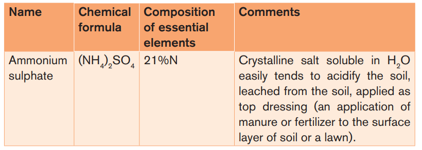
Depending on the nature of the essential elements that a fertilizer can supply to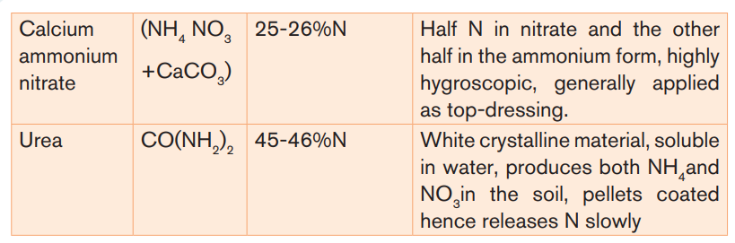
the soil, the fertilizers have been classified into the following groups:
– Nitrogenous Fertilizers: N-type Fertilizers
These Fertilizers supply only nitrogen as a major nutrient to the soil. Examples:
ammonium sulphate, urea, sodium nitrate (also called Chile saltpeter or Chile
nitre).
– Phosphorus Fertilizers: P-type Fertilizers
These fertilizers supply phosphorus as major nutrient to the soil. Examples:
Calcium dihydrogen phosphate, Ca (H2PO4)
2H2O, phosphate slag, Ca3
(PO4)
2CaSiO3
– Potassium Fertilizers: (K-type Fertilizers)
These Fertilizers supply only potassium as a major nutrient to the soil. Examples:potassium chloride, potassium sulphate.
– Mixed Fertilizers
Mixed fertilizers are those which can supply more than one essential element to
the soil.
Depending on the nature of the essential element supplied by the fertilizer,
mixed Fertilizers can be classified into the following groups:
- NP Fertilizers: These Fertilizers supply two essential elements,
nitrogen and phosphorus, to the plant. Examples: Ammonium dihydrogen
phosphate, (NH4)(H2PO4),(also called dihydrogen ammoniated
phosphate or ammophos). Calcium dihydrogen phosphate nitrate, Ca
(H2PO4)
22Ca(NO3)
2,(also called calcium superphosphate nitrate or
nitrophosphate).
- PK Fertilizers: It is a mixture of two compounds; containing phosphorus
and the other containing potassium. For example, a mixture of H2PO4)2H2O,
and K2SO4.
- KN fertilizer. Example: KNO3
- NPK Fertilizers: These are Fertilizers that contain %N, %P as P2O5
and %K as K2O.Example: A mixture of (NH4)
2SO4, (N-type fertilizer),(H2PO4)2H2O (P-type fertilizer), and K2SO4(K-type fertilizer).
3.1.4. Characteristics of a good fertilizer
A good fertilizer should have the following characteristics:- It should contain the required nutrients, in such a form that they can be
assimilated by the plants.
- It should be cheap.
- It should be soluble in water.
- It should be stable, so that it may be available for a long time for the
growing plant.
- It should not be injurious to the plants.
- It should be able to correct the acidity of the soil.Application activity 3.1
Andrew is a farmer in rural village. He always used to spray insecticides in
the farm in order to kill insects and use different fertilizers while growing
crops. In his casual work, Andrew uses to combine fertilizers. After getting
advice from the Sector Agronomist, Andrew is suspecting that he has not
used well the fertilizers, contributed to the pollution of atmosphere and has
contributed to water pollution.i) Which fertilizers does Andrew combine while growing his crops and
why?
ii) Referring to the mistakes done above by Andrew, suggest to Andrew
the advice to follow while selecting fertilizer to be used.iii) A NPK fertilizer is labeled 13-13-13. Interpret this labeling.
3.2. Use of organic and inorganic fertilizers\
Activity 3.2
A plot of land has been divided into two parts and in both Irish potatoes has
been cultivated by two cultivators.
One of them harvested 2000 kg of Irish potatoes of big size and the otherharvested 50kg of Irish potatoes of small size.
Given that on both plots of land, the following work has been done at the
same time- Cultivation,Organic fertilizers contain only plant- or animal-based materials that are either
- planting of the same seeds
- weeding (or hoeing)
- spraying with the same chemicals
- Harvesting
i) Suggest reason(s) which caused the difference in the harvest.ii) Provide advice to the cultivator who harvested 50 Kg.
a byproduct or end product of naturally occurring processes, such as manures,
leaves, and compost. Inorganic fertilizer, also referred to as synthetic fertilizer, ismanufactured artificially and contains minerals or synthetic chemicals.
3.2.1. Organic Fertilizers
The use of organic fertilizer may have many advantages but also it may have
some disadvantages
a. Advantages- The manures add organic matter (called humus) to the soil which
restores the soil texture for better retention of water and for aeration of
soil. For example, organic matter present in the manures increases the
water holding capacity in sandy soils and drainage in clay soil.
- The organic matter of manures provides food for the soil organisms
(decomposers such as bacteria, fungi, etc.) which help in making nutrientsavailable to plants.
- Nutrient release: slow and consistent at a natural rate that plants are able
to use. No anger of over concentration of any element, since microbes
must break down the material.
- Trace minerals: typically present in a broad range, providing more balanced
nutrition to the plant.
- They will not burn: safe for all plants with no danger of burning due to salt
concentration.
- Long lasting: does not leach out since the organic matter binds to the soil
particles where the roots have access to it.
- Fewer applications required: once a healthy soil condition is reached, it is
easier to maintain that level with less work- Controlled growth: does not over-stimulate to exceptional growth whichb. Disadvantagescan cause problems and more work.
- Many organic products produce inconsistent results.3.2.2. Inorganic Fertilizers
- The level of nutrients present in organic fertilizer is often low.- The time of their preparation is too long.
The use of inorganic fertilizers may have many advantages but also it may havesome disadvantages.
a. Advantages- Chemical fertilizers are made with synthetic ingredients designed tob. Disadvantages
stimulate plant growth.
- Commercial chemical fertilizers have the advantage of predictability and
reliability
- Formulations are blended with accuracy and you can buy different blends
for different types of plants; commercial formulated fertilizers allow you
to know exactly which nutrients you’re giving your plants, rather thanguessing at the composition of organic formulas.
- They can burn plants
- They require a specific timetable of application and watering because of
fast release of nutrients- On groundwater, artificial fertilizers have the following disadvantages:
– Increased nitrate levels increase the risks of blue baby syndrome, a rare
form of anaemia which affects babies below 6 months of age. The cause
is the oxidation by nitrite ions of Fe2+ in haemoglobin to Fe3+. The oxidized
hemoglobin cannot bind oxygen, and the baby turns blue from lack of
oxygen. Conditions in the digestive tracks of young children are more
favorable to the bacteria which reduce nitrates to nitrites than those in
adults.
– Another hazard of chemical fertilizers is that carcinogenic nitrosoamines
(yellow oil substance) may be formed in the human digestive track by the
conversion of nitrate into nitrite. The nitrite produced in the stomach it
combines with HCl to produce nitrous acid. Nitrous acid can react with any
secondary amine in foods to form nitrosoamines and the reaction of nitritewith amino acids.
- Repeated use or excess use of the same fertilizer producing acidic ionsOther causes of acid soils include:
(NH4
+). Example of such a fertilizer is (NH4)
2SO4.
- Repeated use or excess use of the same fertilizer producing basic ions.
Example of such a fertilizer is CaCO3.
- Warm temperatures and high rain fall: Cations such as Ca++, Mg++, K+
which are essential to living organisms, are leached (dissolved) from the
soil profile, leaving behind more stable materials rich in Fe and Al oxides.This natural weathering process makes soils acid.
- Man-made processes also contribute significantly to soil acidity. For
example, Sulphur dioxide (SO2) and nitrogen oxides (NOx) released
primarily by industrial activities react with water to form acid rain, which
acidifies soils, particularly forest soils with.
- Organic acids from plants during decomposition;- CO2 from root respiration and microbial respiration.
Application activity 3.2
Jane is a farmer in rural village. He always used to spray insecticides in
the farm in order to kill insects and use different fertilizers while growing
crops. In his casual work, Jane uses to combine fertilizers and varies artificial
fertilizers according to plant crop she wants to grow. After getting advice
from the Sector Agronomist, Jane is now using the fertilizers appropriately.i) Provide the advantages of using combined fertilizers while growing
crops.ii) Why do the farmers use specific fertilizer on specific plant crop?
3.3. Dangers of the use of the substandard fertilizers
Activity 3.3
Agriculture practice contributes to pollution of atmosphere and waterpollution. Observe the photos below and answer related questions.
i) What is the situation of living things’ life on the picture above?
ii) In which way agriculture practice can lead to the consequences
observed on the photos?
iii) Suggest the advice to prevent the consequences observe on thephotos.
One of the problems with chemical fertilizers is they seep through the soil into
the groundwater and other water sources, leading to contamination. Now, NPK
in small quantities is non-toxic, but a lot can kill the balance of nature in various
ways. Nitrogen is especially tricky.
Sub-standard fertilizer means any fertilizer which does not conform to therequired NPK ratio.
Example: A fertilizer may be labeled 16-00-00, while the real NPK ratio is for
example 25-00-00, 10-00-05, etc
Using these Fertilizers can lead to:
- Soil pollution (basic soil or acidic soil) due to accumulation of ions which
are acidic or basic- Poor growth of plantsEffects of acid soil
- Poor harvest
- Eutrophication
- Fertilizer burn: leaf scorch resulting from over-fertilization, usually referring
to excess nitrogen salts. Fertilizer burn is the result of desiccation of plant
tissues due to osmotic stress, creating a state of hypertonicity.
Major effects of extremes in pH levels include gaps in nutrient availability andthe presence of high concentrations of minerals that are harmful to plants.
In very alkaline soil, certain micronutrients such as zinc and copper become
chemically unavailable to plants. In very acidic soil, macronutrients such as
calcium, magnesium and phosphorous are not absorbed while others reach
toxic levels,
Acid soil, particularly in the subsurface, will also restrict root access to water
and nutrients.
In addition to affecting how nutrients are dispensed to growing plants, pH levels
also influence microorganism activity that contributes to the decomposition
of organic materials. A neutral pH is ideal for microbial action that produces
chemical changes in soil, making nitrogen, sulfur and phosphorus more available.
A pH that is either too high or too low may also interfere with the effectiveness
of pesticides by changing their basic composition or weakening their ability to
kill unwanted insects.
Plant growth and most soil processes, including nutrient availability and microbialactivity, are favored by a soil pH range of 5.5 – 8.
Example: The optimal pH range for most plants is between 5.5 and 7.0 as it isshown in the table below.
For soils the pH should be maintained at above 5.5 in the topsoil and 4.8 in the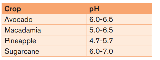
subsurface.
Eutrophication: the undesirable overgrowth of vegetation caused by high
concentration of plants nutrients (Nitrogen and Phosphorous) in bodies of water
(lakes, rivers, etc).
As consequence, water plants (e.g: water hyacinth: amarebe) grow more
vigorously and this prevents the sun light from reaching the water and stops
photosynthesis of aquatic plants which provide oxygen in the water to animals
needed then animals die, deposits of organic matter on the bottom of the lake
build up.
When lake water is enriched with nutrients (e.g.: nitrates and phosphates), algal
flourish, and produce an algae bloom, a green scum with an unpleasant smell.
When algal die they are decomposed by aerobic bacteria. When the oxygencontent falls too low to support aerobic bacteria, anaerobic bacteria take over.
They convert the dead matter into unpleasant-smelling decay products and
debris which falls to the bottom. Gradually, a layer of dead plant material builds
up on the bottom of the lake. The lowering of the oxygen concentration leads tothe death of aquatic animals (fish, crabs, etc).
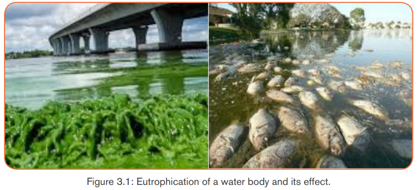
In order to reduce the effects of substandard fertilizers different measure can
be taken:- Standardization of the fertilizer before use.
- Production of fertilizers in Rwanda, as this will help us to choose good
minerals (where necessary) in producing fertilizers.- Use of chemical fertilizers with coated pellets so that nutrients are releasedApplication activity 3.3slowly.
- Regular watering.
Fertilizers application normally results in increased yield with diminishing
returns until maximum yield is reached. A cultivator James is advised to
use NPK 17-17-17 in growing Irish potatoes. When James reached the
Agrotech, he missed NPK 17-17-17 and bought NPK 16-00-00 and he
used in excess to fit with the fertilizer he wanted.i) Were fertilizers used by James the same? How are they different?
ii) Show how using NPK 16-00-00 will be dangerous than using NPK
17-17-17.iii) What can be done to avoid or minimize those dangers?
Skills Lab 3
Making rich organic fertilizer
The best manure for gardens is properly composted manure. It’s often
called black gold, especially when it contains cow manure. Well rotted
farmyard manure is rich and full of slow releasing natural plant nutrients.
Procedure:
- Select an area in a farm that is protected from strong wind and sun,
for instance, under the shade of a tree.
- Mark the area you intend to locate the compost (the minimum area is
1.25m x 1.25m).
- Dig a shallow trench, same size as the compost heap 20cm deep.
Cover the sides of the trench with water or a mixture of water and
cow dung to prevent moisture and nutrients from leaking from the
compost heap. The shallow trench will become the foundation of
the compost heap. The trench also helps to hold moisture especiallyduring the dry season.
Foundation layer
- Put the dry plants material such as small tree branches, maize stalks
or sorghum stalks. Cut the plant material into small pieces. Spread
the dry material evenly over the bottom of the trench to make a layer
of 15-25cm. Sprinkle with water using a watering can or basin to
ensure all material is moist but not wet.
- Layer 1: put dry plant material such as grass, dry leaves mixed with
top soil, manure and ashes. The layer should be about 20-25cm
thick. Mix the material with soil, manure and ashes and sprinkle water
to make it moist.
- Layer 2: Make another layer of moist (green) material which is fresh or
wilted such as weeds or grass cuttings, stems and vegetable leaves,
tree branch leaves, damaged fruits, or vegetables or even kitchen
waste. Do not sprinkle water in this layer. But you can spread it to
remain even or flat.
- Layer 3: is composed of animal manure collected from fresh or
dried cow dung, chicken waste, donkey manure and sheep or goat
droppings. The animal manure can be mixed with soil, old compost
and some ashes to make a layer that is 5 -10 cm thick. Make a watery
mixture and spread it over as a thin layer about 1-2cm thick.
Covering layer: protect the heap from the sun or animals or anything
that might interrupt with the mixture. The cover should be sealed with
only the ventilation stick.
- Turning the compost: open up the compost heap mixing all the layers
while sprinkling water to make it moist but not wet after three weeks.
- Decomposition progress checking: using the ventilation or temperature
stick, you can keep on checking the decomposition process of your
compost every week by pulling out the stick. If it has a white substance
on it and has a bad smell, it means the decomposition is not going on
well. You can turn the compost further and sprinkle some more water
to make it moist.
- Ready compost: A mature compost heap is about the half the size
of the original heap. Check to ensure the compost has a dark brown
colour or black soil, which has a nice smell. All the original materialshould not be seen if the decomposition process went on well.
End Unit Assessment 3
I. Multiple choice (Choose the best answer)
1. If nitrogen is the main element of fertilizers then fertilizers are classified
asa) Structural fertilizers2. Increased ratio of chemical nutrients in ecosystem is classified as
b) Non-structural fertilizers
c) Nitrogen fertilizers
d) Respiratory fertilizersa) Triplication3. Greenhouse gas which can be emitted from storage of nitrogen-based
b) Eutrophication
c) Crystallization
d) Distillation
Fertilizers isa) Nitrous oxide4. Organic Fertilizers can be derived from
b) Nitric oxide
c) Oxygen
d) Hydroxidea) Animal materialsII. Open-ended questions
b) Carbon materials
c) Plant materials
d) Both (a) and (c)
1. Ammonia itself can be used as a fertilizer but has some disadvantages.
Explain the disadvantages of using ammonia as a fertilizer.
2. Give any two advantages of the use ofa) Natural Fertilizers3. Give any two causes of acid soils
b) Artificial Fertilizers
4. Discuss the advantages and disadvantages of the use of organic and
inorganic Fertilizers.
5. Identify the effects of misusing Fertilizers and the dangers of substandard
fertilizers.
REFERENCES
1. Barbara, A., Jean, B, Niamh, G., Douglas W. (2009). Biology, CK-12
Foundation, California, USA.
2. Benjamin Cummings. 8th Ed. San Francisco, US. Pp. 142-148.
3. Biggs.Hagins.Kapicka.Lundgren.Rillero. Tallman.Zike (2005) The Dynamics
of life, McGraw-Hill, USA
4. Campbell, N.A., Reece, J.B., Urry, A.L., Cain, L.M, Wasserman, A.S.,
Minorksy, V.P and Jackson, B.R. (2008). Biology. 8th edition. Pearson
international, San Francisco, USA.
5. Cram, D. and de Kretser, D. 2002 Genetic Diagnosis: the future, in C. Jonge
and C. Barratt (eds) Assisted
6. Fullick, A. (2006). GCSE Biology. AQA Science Exclusively endorsed and
approved by AQA. Nelson Thornes Ltd. Cheltenham
7. Fullick, A., Mwinshekhe, H., Maddu, M., and Nyabua, S. (2011). A level
Biology. Malaysia Ashton Editorial and Juice Creative, Ashton, Malasia.
8. Kent, M. (2000). Advanced biology. Anew mainstream text for the new
specifications. Oxford university press, New York, USA.
9. Margaret Kabiru and Anne Njenga. (2010) Health, nutrition and care,
Nairobi, Kenya.
10. Reproductive Technology: Accomplishments and New Horizons, Cambridge
University Press, Cambridge.
11. https://healthyeating.sfgate.com/cellular-respiration-making-bread-12156.
html
12. https://www.britannica.com/science/human-reproductive-system
13. https://www.nhs.uk/conditions/pregnancy-and-baby/antenatal-midwifecare-pregnant/
14. https: //www.google.rw/
15. s e a r c h ? e i = R Ya y X a y E M s G 8 9 Q O C 4 I 2 Q B A & q = P u b l i c +
health+services&oq=Public+health+services&gs_l=psy-ab.3.0i67j0i7i30l9.15205.15428.23076.0.2.0.1381.2544.7-2......0...